Note: Descriptions are shown in the official language in which they were submitted.
CA 02905010 2015-09-10
WO 2014/143205
PCT/US2013/069702
HUMAN ANTIBODIES THAT BIND HUMAN TNF-ALPHA
AND METHODS OF PREPARING THE SAME
RELATED APPLICATION
[001] This application claims priority to U.S. Provisional Patent
Application
No. 61/777,883, filed March 12, 2013, which is incorporated by reference into
the
present application in its entirety and for all purposes.
SEQUENCE LISTING
[002] This application is accompanied by a sequence listing in a computer
readable form that accurately reproduces the sequences described herein.
Field of the Invention
[003] This disclosure relates to antibodies that specifically bind to human
TNF-
alpha. More particular, Methylglyoxal (MG0)-modified recombinant TNF-alpha
antibodies are disclosed. Methods for reducing MGO-modified TNF-alpha
antibodies are
also provided.
Background
[004] Tumor necrosis factor alpha ("TNF-alpha") is a cytokine produced by
many cell types such as monocytes and macrophages. See e.g., Old, L. Science
230:630-
632 (1985). TNF-alpha plays an important role in many biological processes and
has
been implicated in the pathophysiology of a variety of other human diseases
and
disorders, including sepsis, infections, autoimmune diseases, transplant
rejection and
graft-versus-host disease. See e.g., Vasilli, P., Annu. Rev. Immunol. 10:411-
452 (1992);
and Tracey, K. J. and Cerami, A. Annu. Rev. Med. 45:491-503 (1994).
[005] In an effort to treat/prevent these diseases, various therapeutic
strategies
have been designed to inhibit or counteract TNF-alpha activities. U.S. Patent
No.
6,090,382 disclosed human antibodies (e.g., recombinant human antibodies) that
specifically bind to human TNF-alpha with high affmity and slow dissociation
kinetics.
Nucleic acids, vectors and host cells for expressing the recombinant human TNF-
alpha
antibodies were also disclosed. One example of such recombinant TNF-alpha
antibodies
is called Adalimumab, which is marketed under the trade name Humira . The
entire
1
CA 02905010 2015-09-10
WO 2014/143205
PCT/US2013/069702
contents of U.S. Patent No. 6,090,382 is hereby incorporated by reference into
the
present disclosure.
[006] Recombinant biotherapeutics are typically produced by live cells and
are
inherently more complex as compared to traditional small molecule drug.
Various post-
translational modifications have been reported as major contributors to
heterogeneity in
recombinant monoclonal antibodies (References 1-4). Some of these
modifications, for
example, glycosylation and sialic acid incorporation, may occur during
fermentation
(References 5-7). Some other modifications, such as oxidation and disulfide
bond
scrambling, may occur during production, purification and storage.
[007] One example of such modifications is the so-called acidic species
(charge
variants). Acidic species are observed when recombinant monoclonal antibodies
are
analyzed by weak-cation exchange chromatography (WCX) (Figure 1). One major
contributing factor is the removal of the C-terminal lysine of the heavy chain
by cell-
derived carboxypeptidease, reducing the overall positive charge (Reference 8).
These
variants are commonly referred to as Lys0, Lysl and Lys2 species,
respectively.
[008] C-terminal amidation (Reference 9) is another enzymatic process during
fermentation. Another type of variant is caused by spontaneous non-enzymatic
transformations, which include the formation of pyruglutamate (Pyro-Glu) from
an N-
terminal glutamine (Gin) that remove the positive charge of the free N-
terminus
(Reference 10), and the deamidation of asparagine (Asn) to aspartic (Asp) or
isoaspartic
acid (isoAsp or isoD) that introduces negatively charged carboxylic acids
(References 11
and 12).
[009] Some modifications may shift the retention time of antibody on weak
cation exchange chromatography even though they do not alter the formal
charges of the
antibody molecule. These modifications may exert their effects through
perturbation of
local charge and conformation. For instance, incomplete glycosylation
(Reference 13) or
the presence of free sulfhydryl (References 14-16) may shift the retention
time of
antibody on weak cation exchange chromatography. It is worth noting that some
modifications are imparted by metabolites, such as glycation by glucose,
methionine
oxidation by reactive oxygen species (ROS), cysteinylation by cysteine
(Reference 17),
and 5-homocysteinylation and N-homocysteinylation by homocysteine (References
2, 18-
2
CA 02905010 2015-09-10
WO 2014/143205
PCT/US2013/069702
23). Although the mechanisms of many modifications have been reported, these
mechanisms cannot fully explained the observed heterogeneity of recombinant
monoclonal antibodies on weak cation exchange chromatography.
Summary
[010] This disclosure advances the art by identifying novel species of
modified
recombinant antibodies that may negatively impact the functionalities of such
antibodies.
The disclosure also provides methods for reducing the amount of such species
without
substantially compromising the overall yield of the antibody production.
[011] In one embodiment, two acidic species of the Adalimumab antibody are
disclosed which exist when the antibody are expressed in Chinese hamster ovary
(CHO)
cells cultured in chemically defined media (CDM). Detailed analyses have
revealed that
several arginine residues in Adalimumab are modified by methylglyoxal (MGO),
which is
further confirmed by the treatment of native antibody with authentic MOO. The
reaction
between MOO and arginine result in formation of hydroxylimide and/or
hydroimidazolone. The resulting hydroxylimide and hydroimidazolone adducts
increase
the molecular weight of the antibody by 54 and 72 Dalions, respectively.
[012] In another embodiment, these modifications cause the antibody to
elute
earlier in the weak cation exchange chromatogram as compared to the elution
time of
unmodified forms. Consequently, the extent to which an antibody was modified
at
multiple sites corresponds to the degree of shift in acidity and the elution
time. The
modification of Adalimumab antibody by MOO is the first reported modification
of a
recombinant monoclonal antibody by MOO.
[013] In another embodiment, a composition is disclosed which contains a
binding protein capable of binding TNF-alpha. In one aspect, the binding
protein may
contain at least one methylglyoxal (MOO)-susceptible amino acid, and at least
a portion
of the binding protein may contain one or more MOO-modified amino acids.
[014] In another embodiment, a composition is disclosed which contains a
binding protein capable of binding TNF-alpha. In one aspect, the binding
protein may
contain at least one methylglyoxal (MOO)-susceptible amino acid and the
composition
may be prepared by substantially removing molecules of the binding protein
that contain
at least one MOO-modified amino acid. The term "substantially" may mean at
least 50%.
3
CA 02905010 2015-09-10
WO 2014/143205
PCT/US2013/069702
In another aspect, the term "substantially" may mean at least 60%, 70%, 80%,
90%, or
even 100% removal of the molecules that contain at least one MGO-modified
amino acid.
[015] For purpose of this disclosure, the term "methylglyoxal (MG0)-
susceptible" refers to groups or residues (e.g., arginine) that may react with
MOO under
appropriate cell culture conditions. List of MOO-susceptible arginines in
Adalimumab is
shown in Table 1. Examples of MOO-susceptible peptides in Adalimumab are shown
in
Table 2.
[016] The term "at least a portion of the binding protein" means that
although
all molecules of the binding protein in the composition are capable of binding
TNF-alpha,
at least two populations of these molecules exist in the composition, wherein
one
population contain one or more amino acids that have been modified by MOO,
while the
other population does not contain amino acids that have been modified by MOO.
In
another aspect, all molecules of the binding protein may contain one or more
amino acids
that have been modified by MOO.
[017] In one aspect, the portion of the binding protein that contains at
least one
MOO-modified amino acid is less than 15%, 14%, 13%, 12%, 11%, 10%, 9%, 8%, 7%,
6%, 5%, 4%, 3%, 2% or 1% of the total amount of the binding protein.
[018] In another embodiment, the binding protein is a human antibody or an
antigen-binding portion thereof, wherein the binding protein dissociates from
human
TNF-alpha with a Kid of 1x10⁻⁸M or less and a Koff rate constant of lx 10-3 s-
1 or less,
both as determined by surface plasmon resonance. In one aspect, the binding
protein
neutralizes human TNF-alpha cytotoxicity in a standard in vitro L929 assay
with an ICso
of lx10-7 M or less, described in Example 4 of U.S. Patent No. 6,090,382. In
another
aspect, the binding protein is the D2E7 antibody as described in U.S. Patent
No.
6,090,382.
[019] In another embodiment, cell culture parameters may affect the extent of
modifications by methylglyoxal (MOO). MOO is a highly reactive metabolite that
may be
generated from glucose, lipids or other metabolic pathways. In one aspect,
cell culture
conditions may be modified to decrease the production of MOO thereby reducing
modification of the recombinant antibodies by MOO. Taken together, the
disclosed
findings highlight the impact of cell culture conditions on the critical
quality attributes of
4
CA 02905010 2015-09-10
WO 2014/143205
PCT/US2013/069702
recombinantly produced antibodies. These findings provide additional
parameters for
improving manufacturing processes and may prove useful for the quality by
design (QbD)
approach.
[020] In another embodiment, methods are disclosed for purifying a target
protein product from both process and/or product related impurities.
Specifically, method
for purifying a composition containing a target protein is disclosed. In one
aspect,
methods are provided for reducing product related charge variants (i.e. acidic
and basic
species). In another aspect, the method involves contacting the process
mixture with an
ion (anion or cation) exchange adsorbent in an aqueous salt solution under
loading
conditions that permit both the target and non-target proteins to bind to the
adsorbent
and allowing the excess target molecule to pass through the column and
subsequently
recovering the bound target protein with a wash at the same aqueous salt
solution used in
the equilibration (i.e. pre-loading) condition.
[021] In another embodiment, a method for purifying a composition containing
a target protein is disclosed which may include at least the following steps:
(a) loading the
composition to a cation exchange adsorbent using a loading buffer, wherein the
pH of the
loading buffer is lower than the pI of the target protein; (b) washing the
cation exchange
adsorbent with a washing buffer, wherein the pH of the washing buffer is lower
than the
pI of the target protein; (c) eluting the cation exchange adsorbent with an
elution buffer,
said elution buffer being capable of reducing the binding between the target
protein and
the cation exchange adsorbent; and (d) collecting the eluate, wherein the
percentage of
the target protein is higher in the eluate than the percentage of the target
protein in the
composition. In one aspect, the washer buffer and the loading buffer are the
same. In
another aspect, the conductivity of the elution buffer is higher than the
conductivity of the
washer buffer. In another aspect, the pH of the elution buffer may be between
5.5 and
9.0, between 6 and 8, or between 6.5 and 8. The conductivity of the elution
buffer may
be raised by increasing the salt concentration of the elution buffer. The salt
concentration
of the elution buffer may be between 20 mM NaC1 and 200 mM NaC1, between 40 mM
NaC1 and 160 mM NaC1, or between 60 mM NaC1 and 120 mM NaCl.
[022] In another embodiment, a method for purifying a composition containing
a target protein is disclosed which may include at least the following steps:
(a) loading the
composition to an anion exchange adsorbent using a loading buffer, wherein the
pH of the
CA 02905010 2015-09-10
WO 2014/143205
PCT/US2013/069702
loading buffer is lower than the isoelectric point (pI) of the target protein;
(b) allowing
the majority of the target protein to pass through without binding to the
anion exchange
adsorbent; (c) collecting the pass-through loading buffer containing said
unbound target
protein; (d) washing the anion exchange adsorbent with a washing buffer; (e)
allowing the
target protein bound to the anion exchange adsorbent to disassociate from the
anion
exchange adsorbent; (f) collecting the washing buffer containing said
disassociated target
protein. In another aspect, the method may further include a step (g) of
pooling the
collections from steps (c) and (f) to obtain a purified composition containing
the target
protein. The percentage of the target protein is higher in the pooled
collections than the
percentage of the target protein in the original composition.
[023] In one aspect, the loading buffer may contain an anionic agent and a
cationic agent, wherein the conductivity and pH of the loading buffer is
adjusted by
increasing or decreasing the concentration of a cationic agent and maintaining
a constant
concentration of an anionic agent in the loading buffer. In another aspect,
the anionic
agent is selected from the group consisting of acetate, citrate, chloride
anion, sulphate,
phosphate and combinations thereof. In another aspect, the cationic agent is
selected
from the group consisting of sodium, Tris, tromethalmine, ammonium cation,
arginine,
and combinations thereof.
[024] In one embodiment, the target protein is a human antibody or an antigen-
binding portion thereof that is substantially free from MOO modification. In
one aspect,
the target protein dissociates from human TNF-alpha with a Kid of 1 x 10⁻⁸M or
less and
a Koff rate constant of 1 x 10⁻³ s-1 or less, both as determined by surface
plasmon
resonance. In another aspect, the target protein neutralizes human TNF-alpha
cytotoxicity
in a standard in vitro L929 assay with an IC50 of 1 x 10 M or less, described
in Example
4 of U.S. Patent No. 6,090,382. In another aspect, the target protein is the
D2E7
antibody as described in U.S. Patent No. 6,090,382.
Brief Description of the Drawings
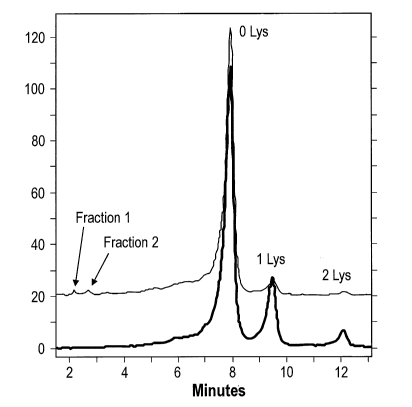
[025] Figure 1 shows a typical WCX chromatogram of adalimumab after
protein A purification.
[026] Figure 2 shows deconvoluted mass spectra of the light chain and heavy
chains in fractions 1 and 2.
6
CA 02905010 2015-09-10
WO 2014/143205
PCT/US2013/069702
[027] Figure 3 shows representative MS/MS mass spectra of peptides
containing Arg residues modified by MOO.
[028] Figure 4 shows chemical modification of arginine by MOO.
[029] Figure 5 shows modification of a purified 0 lysine fraction by MOO over
a 5-hour time course.
[030] Figure 6 shows the mass spectra of peaks a and b from Figure 5.
[031] Figure 7 shows comparison of peptide MS/MS data between acidic
fraction 1 from cell culture and acidic fraction 1 from methylglyoxal
incubation.
[032] Figure 8 shows the crystal structure of the adalimumab Fab subunit in
complex with TNF-alpha, indicating that modification by MOO may cause
conformational change which may impede adalimumab's ability to bind TNF-alpha.
[033] Figure 9 shows Surface Plasmon Resonance (SPR) data for 0 Lys
Fraction (Top ¨ 0 Lys) and for the MOO enriched fraction (Bottom - Peak 1).
[034] Figure 10 shows comparison of acidic region affected by methylglyoxal
before and after two-step chromatographic separation, wherein the top trace is
an
expanded view of the acidic region in which the two distinctive MOO peaks are
denoted,
and the lower trace shows a complete clearance of this acidic region and the
MOO
variants.
[035] Figure 11 shows the CEX chromatogram when reversible binding mode
was performed using Adalimumab with a Tris-acetate buffer system.
[036] Figure 12 shows the removal of acidic species by Poros XS resin with
NaCl/Tris-acetate solution.
Detailed Description
[037] The instant disclosure identifies novel species of methylglyoxal
(MOO)-
modified recombinant antibodies which may have negative impact on the
structure and
function of the antibodies. The disclosure also provides methods for reducing
the
percentage of such variant species without substantially compromising the
yield of
antibody production. More specifically, this disclosure describes
methylglyoxal (MOO)-
7
CA 02905010 2015-09-10
WO 2014/143205
PCT/US2013/069702
modified forms of Adalimumab in cell culture when Adalimumab is expressed in
CHO
cells using chemically defined media (CDM).
[038] In one embodiment, modification of the side chain of certain
arginines
(e.g., R30 in CDR1 of Adalimumab) by MOO may result in the formation of a five-
member ring originating at the guanidinium terminal of the side chain which
may further
penetrate into the TNF-alpha structure. These MOO modifications may impede
Adalimumab's ability to bind TNF-alpha due to steric constraints.
[039] In one embodiment, control of acidic species heterogeneity may be
attained by purifying a protein of interest from a mixture comprising the
protein with an
anion exchange (AEX) adsorbent material and an aqueous salt solution under
loading
conditions that permit both the protein of interest and non-target proteins to
bind to the
AEX adsorbent, wherein the bound protein of interest is subsequently recovered
with a
wash buffer comprising the same aqueous salt solution used in the
equilibration (i.e.
loading) buffer. In one aspect, the aqueous salt solution used as both the
loading and
wash buffer has a pH that is greater than the isoelectric point (pI) of the
protein of
interest.
[040] In another embodiment, the disclosed purification method may include
adjusting the conductivity and/or pH of the aqueous salt solution. In one
aspect, the
adjustments may include decreasing the conductivity of the aqueous salt
solution. In
another aspect, the adjustment to achieve the desired control over acidic
species
heterogeneity may involve an increase in the load conductivity of the
solution. In another
aspect, the adjustment may increase the pH of the aqueous salt solution. In
another
aspect, the adjustment to achieve the desired control over acidic species
heterogeneity
may involve a decrease in the pH of the aqueous salt solution. Such increases
and/or
decreases in the conductivity and/or pH may be of a magnitude of 1%, 5%, 10%,
15%,
20%, 25%, 30%, 35%, 40%, 45%, 50%, 55%, 60%, 65%, 70%, 75%, 80%, 85%, 90%,
95%, or 100%, and ranges within one or more of the preceding, of the
conductivity
and/or pH of the aqueous salt solution.
[041] In another embodiment, the conductivity and pH of the aqueous salt
solution is adjusted by increasing or decreasing the concentration of a
cationic agent and
maintaining a constant concentration of an anionic agent in the aqueous salt
solution. In
8
CA 02905010 2015-09-10
WO 2014/143205
PCT/US2013/069702
one aspect, the anionic agent is maintained at a concentration of between
about 0.05 mM
and 100 mM, or between about 0.1 mM and 90 mM, or between about 0.5 mM and 80
mM, or between about 1 mM and 70 mM, or between about 1.5 mM and 60 mM, or
between about 2 mM and 50 mM, or between about 2.5 mM and 40 mM, or between
about 3 mM and 30 mM, or between about 3.5 mM and 25 mM, or between about 4 mM
and 20 mM, or between about 4.5 mM and 15 mM, or between about 4.5 mM and 10
mM, or between about 5 mM and 7 mM. In another aspect, the anionic agent is
maintained at a concentration of about 5 mM. In another aspect, the anionic
agent is
maintained at a concentration of about 10 mM. In another aspect, the anionic
agent is
maintained at a concentration of about 18.5 mM.
[042] In another embodiment, the concentration of the cationic agent in the
aqueous salt solution is increased or decreased to achieve a pH of between
about 5 and
12, or between about 5.5 and 11.5, or between about 6 and 11, or between about
6.5 and
10.5, or between about 7 and 10, or between about 7.5 and 9.5, or between
about 8 and
9, or between about 8.5 and 9. In certain embodiments, the concentration of
cationic
agent is increased or decreased in the aqueous salt solution to achieve a pH
of 8.8. In
certain embodiments, the concentration of cationic agent in the aqueous salt
solution is
increased or decreased to achieve a pH of 9.
[043] In another embodiment, the protein load of the protein mixture is
adjusted to a protein load of between about 50 g/L and 500 g/L, or between
about 100
g/L and 450 g/L, or between about 120 g/L and 400 g/L, or between about 125
g/L and
350 g/L, or between about 130 g/L and 300 g/L or between about 135 g/L and 250
g/L,
or between about 140 g/L and 200 g/L, or between about 145 g/L and 200 g/L, or
between about 150 g/L and 200 g/L, or between about 155 g/L and 200 g/L, or
between
about 160 g/L and 200 g/L. In certain embodiments, the protein load of the
protein or
antibody mixture is adjusted to a protein load of about 100 g/L. In certain
embodiments,
the protein load of the protein or antibody mixture is adjusted to a protein
load of about
20 g/L. In certain embodiments, the protein load of the protein or antibody
mixture is
adjusted to a protein load of about 105 g/L. In certain embodiments, the
protein load of
the protein or antibody mixture is adjusted to a protein load of about 140
g/L. In certain
embodiments, the protein load of the protein or antibody mixture is adjusted
to a protein
9
CA 02905010 2015-09-10
WO 2014/143205
PCT/US2013/069702
load of about 260 g/L. In certain embodiments, the protein load of the protein
or antibody
mixture is adjusted to a protein load of about 300 g/L.
[044] In another embodiment, the concentration of cationic agent in the
aqueous salt solution is increased or decreased in an amount effective to
reduce the
amount of acidic species heterogeneity in a protein or antibody sample by
about 1%,
1.2%, 1.5%, 2%, 2.2%, 2.5%, 3%, 3.2%, 3.5%, 4%, 4.2%, 4.5%, 5%, 10%, 15%, 20%,
25%, 30%, 35%, 40%, 45%, 50%, 55%, 60%, 65%, 70%, 75%, 80%, 85%, 90%, 95%,
or 100%, and ranges within one or more of the preceding, when the aqueous salt
solution
is used as a load and wash buffer to purify the protein of interest (for
example, an
antibody) from the sample containing the protein.
[045] In another embodiment, the anionic agent is acetate, citrate,
chloride
anion, sulphate, phosphate or combinations thereof. In certain embodiments,
the cationic
agent is sodium, Tris, tromethalmine, ammonium cation, arginine, or
combinations
thereof.
[046] By way of example but not limitation, as detailed in this disclosure,
up to
60% of the acidic species in an antibody preparation was removed when the
antibody was
purified using chromatography comprising an anion exchange adsorbent material,
a
protein load of 150 g/L, and a load/wash buffer containing 5 mM
Acetate/Arginine at pH
8.8.
[047] In another embodiment of the instant disclosure, control of acidic
species
heterogeneity can be attained by purifying a protein of interest from a
mixture comprising
the protein with a cation exchange (CEX) adsorbent material and an aqueous
salt solution
under loading conditions that permit both the protein of interest and non-
target proteins
to bind to the CEX adsorbent, washing off the acidic species, charged
variants, molecular
variants and impurities using the same buffer conditions as the loading
buffer, and eluting
the bound protein target from the CEX adsorbent with a buffer having a higher
conductivity than the loading buffer. In certain embodiments, the aqueous salt
solution
used as both the loading and wash buffer has a pH that is lower than the
isoelectric point
(pI) of the protein of interest.
[048] In another embodiment, the purification method may include adjusting
the conductivity and/or pH of the aqueous solution. In certain embodiments,
such
CA 02905010 2015-09-10
WO 2014/143205
PCT/US2013/069702
adjustments will be to decrease the conductivity, while in other embodiments
the
necessary adjustment to achieve the desired control over acidic species
heterogeneity will
involve an increase in the load conductivity. In certain embodiments, such
adjustments
will also be to increase the pH of the aqueous salt solution, while in other
embodiments
the necessary adjustment to achieve the desired control over acidic species
heterogeneity
will involve a decrease in the pH of the aqueous salt solution. Such increases
and/or
decreases in the conductivity and/or pH can be of a magnitude of 1%, 5%, 10%,
15%,
20%, 25%, 30%, 35%, 40%, 45%, 50%, 55%, 60%, 65%, 70%, 75%, 80%, 85%, 90%,
95%, or100%, and ranges within one or more of the preceding, of the
conductivity and/or
pH of the aqueous salt solution.
[049] In certain embodiments, the conductivity and pH of the aqueous salt
solution is adjusted by increasing or decreasing the concentration of a
anionic agent and
maintaining a constant concentration of a cationic agent in the aqueous salt
solution. In
certain embodiments, the cationic agent is maintained at a concentration of
between about
0.5 mM and 500 mM, or between about 1 mM and 450 mM, or between about 5 mM and
400 mM, or between about 10 mM and 350 mM, or between about 15 mM and 300 mM,
or between about 20 mM and 250 mM, or between about 25 mM and 200 mM, or
between about 30 mM and 150 mM, or between about 35 mM and 100 mM, or between
about 40 mM and 50 mM. In certain embodiments, the anionic agent is maintained
at a
concentration of about 15 mM, or about 20 mM, or about 25 mM, or about 30 mM,
or
about 35 mM, or about 40 mM, or about 45 mM, or about 50 mM, or about 60 mM,
or
about 65 mM, or about 75 mM, or about 90 mM, or about 115 mM, or about 120 mM,
or about 125 mM, or about 135 mM, or about 140 mM, or about 145 mM, or about
150
mM, or about 175 mM, or about 250 mM, or about 275 mM, or about 300 mM, or
about
350 mM, or about 375 mM, or about 400 mM.
[050] In certain embodiments, the concentration of the anionic agent in
aqueous salt solution is increased or decreased to achieve a pH of between
about 2 and
12, or between about 2.5 and 11.5, or between about 3 and 11, or between about
3.5 and
10.5, or between about 4 and 10, or between about 4.5 and 9.5, or between
about 5 and
9, or between about 5.5 and 8.5, or between about 6 and 8, or between about
6.5 and
7.5. In certain embodiments, the concentration of anionic agent is increased
or decreased
in the aqueous salt solution to achieve a pH of 5, or 5.5, or 6, or 6.5, or
6.8, or 7.5.
11
CA 02905010 2015-09-10
WO 2014/143205
PCT/US2013/069702
[051] In certain embodiments, the protein load of the protein mixture is
adjusted to a protein load of between about 50 and 500 g/L, or between about
100 and
450 g/L, or between about 120 and 400 g/L, or between about 125 and 350 g/L,
or
between about 130 and 300 g/L or between about 135 and 250 g/L, or between
about
140 and 200 g/L, or between about 145 and 150 g/L. In certain embodiments, the
protein
load of the protein or antibody mixture is adjusted to a protein load of about
40 g/L.
[052] In certain embodiments, the concentration of anionic agent in the
aqueous salt solution is increased or decreased in an amount effective to
reduce the
amount of acidic species heterogeneity in a protein or antibody sample by
about 1%,
1.2%, 1.5%, 2%, 2.2%, 2.5%, 3%, 3.2%, 3.5%, 4%, 4.2%, 4.5%, 5%, 10%, 15%, 20%,
25%, 30%, 35%, 40%, 45%, 50%, 55%, 60%, 65%, 70%, 75%, 80%, 85%, 90%, 95%,
or 100%, and ranges within one or more of the preceding, when the aqueous salt
solution
is used as a load and wash buffer to purify the protein of interest (for
example, an
antibody) from the sample containing the protein.
[053] In certain embodiments, the cationic agent is sodium, Tris,
tromethalmine, ammonium cation, arginine, or combinations thereof. In certain
embodiments, the anionic agent is acetate, citrate, chloride anion, sulphate,
phosphate or
combinations thereof.
[054] By way of example but not limitation, as detailed in this disclosure,
the
presence of acidic species in an antibody preparation was reduced by 6.5% from
starting
material after purification using a cation exchange adsorbent material, and a
load and
wash buffer comprising 140 mM Tris at pH 7.5.
[055] Unless otherwise defmed herein, scientific and technical terms used
herein have the meanings that are commonly understood by those of ordinary
skill in the
art. In the event of any latent ambiguity, definitions provided herein take
precedent over
any dictionary or extrinsic defmition. Unless otherwise required by context,
singular terms
shall include pluralities and plural terms shall include the singular. The use
of "or" means
"and/or" unless stated otherwise. The use of the term "including", as well as
other forms,
such as "includes" and "included", is not limiting.
[056] Generally, nomenclatures used in connection with cell and tissue
culture,
molecular biology, immunology, microbiology, genetics and protein and nucleic
acid
12
CA 02905010 2015-09-10
WO 2014/143205
PCT/US2013/069702
chemistry and hybridization described herein are those well known and commonly
used in
the art. The methods and techniques provided herein are generally performed
according
to conventional methods well known in the art and as described in various
general and
more specific references that are cited and discussed throughout the present
specification
unless otherwise indicated. Enzymatic reactions and purification techniques
are performed
according to manufacturer's specifications, as commonly accomplished in the
art or as
described herein. The nomenclatures used in connection with, and the
laboratory
procedures and techniques of, analytical chemistry, synthetic organic
chemistry, and
medicinal and pharmaceutical chemistry described herein are those well known
and
commonly used in the art. Standard techniques are used for chemical syntheses,
chemical
analyses, pharmaceutical preparation, formulation, and delivery, and treatment
of patients.
10571 That the disclosure may be more readily understood, select terms are
defined below.
10581 The term "antibody" refers to an immunoglobulin (Ig) molecule, which is
generally comprised of four polypeptide chains, two heavy (H) chains and two
light (L)
chains, or a functional fragment, mutant, variant, or derivative thereof, that
retains the
epitope binding features of an Ig molecule. Such fragment, mutant, variant, or
derivative
antibody formats are known in the art. In an embodiment of a full-length
antibody, each
heavy chain is comprised of a heavy chain variable region (VH) and a heavy
chain
constant region (CH). The heavy chain variable region (domain) is also
designated as
VDH in this disclosure. The CH is comprised of three domains, CH1, CH2 and
CH3.
Each light chain is comprised of a light chain variable region (VL) and a
light chain
constant region (CL). The CL is comprised of a single CL domain. The light
chain
variable region (domain) is also designated as VDL in this disclosure. The VH
and VL
can be further subdivided into regions of hypervariability, termed
complementarity
determining regions (CDRs), interspersed with regions that are more conserved,
termed
framework regions (FRs). Generally, each VH and VL is composed of three CDRs
and
four FRs, arranged from amino-terminus to carboxy-terminus in the following
order:
FR1, CDR1, FR2, CDR2, FR3, CDR3, and FR4. Immunoglobulin molecules can be of
any type (e.g., IgG, IgE, IgM, IgD, IgA and IgY), class (e.g., IgGl, IgG2,
IgG3, IgG4,
IgAl and IgA2), or subclass.
13
CA 02905010 2015-09-10
WO 2014/143205
PCT/US2013/069702
[059] The term "antigen-binding portion" of an antibody (or "antibody
portion"), as used herein, refers to one or more fragments of an antibody that
retain the
ability to specifically bind to an antigen (e.g., hTNF-alpha). It has been
shown that the
antigen-binding function of an antibody can be performed by fragments of a
full-length
antibody. Examples of binding fragments encompassed within the term "antigen-
binding
portion" of an antibody include (i) a Fab fragment, a monovalent fragment
consisting of
the VL, VH, CL and CH I domains; (ii) a F(ab')2 fragment, a bivalent fragment
comprising two Fab fragments linked by a disulfide bridge at the hinge region;
(iii) a Fd
fragment consisting of the VH and CH1 domains; (iv) a Fv fragment consisting
of the VL
and VH domains of a single arm of an antibody, (v) a dAb fragment (Ward et
al., (1989)
Nature 341:544-546), which consists of a VH domain; and (vi) an isolated
complementarity determining region (CDR). Furthermore, although the two
domains of
the Fv fragment, VL and VH, are coded for by separate genes, they can be
joined, using
recombinant methods, by a synthetic linker that enables them to be made as a
single
protein chain in which the VL and VH regions pair to form monovalent molecules
(known as single chain Fv (scFv); see e.g., Bird et al. (1988) Science 242:423-
426: and
Huston et al. (1988) Proc. Natl. Acad. Sci. USA 85:5879-5883). Such single
chain
antibodies are also intended to be encompassed within the term "antigen-
binding portion"
of an antibody. Other forms of single chain antibodies, such as diabodies are
also
encompassed. Diabodies are bivalent, bispecific antibodies in which VH and VL
domains
are expressed on a single polypeptide chain, but using a linker that is too
short to allow
for pairing between the two domains on the same chain, thereby forcing the
domains to
pair with complementary domains of another chain and creating two antigen
binding sites
(see e.g., Holliger, P., et al. (1993) Proc. Natl. Acad. Sci. USA 90:6444-
6448; Poljak, R.
J., et al. (1994) Structure 2:1121-1123).
[060] The term "human antibody", as used herein, is intended to include
antibodies having variable and constant regions derived from human germline
immunoglobulin sequences. The human antibodies of the invention may include
amino
acid residues not encoded by human germline immunoglobulin sequences (e.g.,
mutations
introduced by random or site-specific mutagenesis in vitro or by somatic
mutation in
vivo), for example in the CDRs and in particular CDR3.
14
CA 02905010 2015-09-10
WO 2014/143205
PCT/US2013/069702
[061] The term "recombinant human antibody", as used herein, is intended to
include all human antibodies that are prepared, expressed, created or isolated
by
recombinant means, such as antibodies expressed using a recombinant expression
vector
transfected into a host cell, antibodies isolated from a recombinant,
combinatorial human
antibody library, antibodies isolated from an animal (e.g., a mouse) that is
transgenic for
human immunoglobulin genes (see e.g., Taylor, L. D., et al. (1992) Nucl. Acids
Res.
20:6287-6295) or antibodies prepared, expressed, created or isolated by any
other means
that involves splicing of human immunoglobulin gene sequences to other DNA
sequences.
Such recombinant human antibodies have variable and constant regions derived
from
human germline immunoglobulin sequences. In certain embodiments, however, such
recombinant human antibodies are subjected to in vitro mutagenesis (or, when
an animal
transgenic for human Ig sequences is used, in vivo somatic mutagenesis) and
thus the
amino acid sequences of the VH and VL regions of the recombinant antibodies
are
sequences that, while derived from and related to human germline VH and VL
sequences,
may not naturally exist within the human antibody germline repertoire in vivo.
[062] The term "surface plasmon resonance", as used herein, refers to an
optical phenomenon that allows for the analysis of real-time biospecific
interactions by
detection of alterations in protein concentrations within a biosensor matrix,
for example
using the BIAcore system (Pharmacia Biosensor AB, Uppsala, Sweden and
Piscataway,
N.J.). For further descriptions, see Example 1 and Jonsson, U., et al. (1993)
Ann. Biol.
Clin. 51:19-26; Jonsson, U., et al. (1991) Biotechniques 11:620-627; Johnsson,
B., et al.
(1995) J. Mol. Recognit. 8:125-131; and Johnnson, B., et al. (1991) Anal.
Biochem.
198:268-277.
[063] The term "biological activity" refers to any one or more biological
properties of a molecule (whether present naturally as found in vivo, or
provided or
enabled by recombinant means). Biological properties include, but are not
limited to,
binding a receptor or receptor ligand, inducing cell proliferation, inhibiting
cell growth,
inducing other cytokines, inducing apoptosis, and enzymatic activity.
[064] The term "neutralizing" refers to counteracting the biological
activity of
an antigen/ligand when a binding protein specifically binds to the
antigen/ligand. In an
embodiment, the neutralizing binding protein binds to an antigen/ligand (e.g.,
a cytokine)
CA 02905010 2015-09-10
WO 2014/143205
PCT/US2013/069702
and reduces its biologically activity by at least about 20%, 40%, 60%, 80%,
85% or
more.
[065] "Specificity" refers to the ability of a binding protein to
selectively bind
an antigen/ligand.
[066] "Affinity" is the strength of the interaction between a binding
protein and
an antigen/ligand, and is determined by the sequence of the binding domain(s)
of the
binding protein as well as by the nature of the antigen/ligand, such as its
size, shape,
and/or charge. Binding proteins may be selected for affmities that provide
desired
therapeutic end-points while minimizing negative side-effects. Affinity may be
measured
using methods known to one skilled in the art (US 20090311253).
[067] The term "potency" refers to the ability of a binding protein to
achieve a
desired effect, and is a measurement of its therapeutic efficacy. Potency may
be assessed
using methods known to one skilled in the art (US 20090311253).
[068] The term "cross-reactivity" refers to the ability of a binding
protein to
bind a target other than that against which it was raised. Generally, a
binding protein will
bind its target tissue(s)/antigen(s) with an appropriately high affinity, but
will display an
appropriately low affinity for non-target normal tissues. Individual binding
proteins are
generally selected to meet two criteria. (1) Tissue staining appropriate for
the known
expression of the antibody target. (2) Similar staining pattern between human
and tox
species (mouse and cynomolgus monkey) tissues from the same organ. These and
other
methods of assessing cross-reactivity are known to one skilled in the art (US
20090311253).
[069] The term "biological function" refers the specific in vitro or in
vivo
actions of a binding protein. Binding proteins may target several classes of
antigens/ligands and achieve desired therapeutic outcomes through multiple
mechanisms
of action. Binding proteins may target soluble proteins, cell surface
antigens, as well as
extracellular protein deposits. Binding proteins may agonize, antagonize, or
neutralize the
activity of their targets. Binding proteins may assist in the clearance of the
targets to
which they bind, or may result in cytotoxicity when bound to cells. Portions
of two or
more antibodies may be incorporated into a multivalent format to achieve
distinct
16
CA 02905010 2015-09-10
WO 2014/143205
PCT/US2013/069702
functions in a single binding protein molecule. The in vitro assays and in
vivo models
used to assess biological function are known to one skilled in the art (US
20090311253).
[070] The term "solubility" refers to the ability of a protein to remain
dispersed
within an aqueous solution. The solubility of a protein in an aqueous
formulation depends
upon the proper distribution of hydrophobic and hydrophilic amino acid
residues, and
therefore, solubility can correlate with the production of correctly folded
proteins. A
person skilled in the art will be able to detect an increase or decrease in
solubility of a
binding protein using routine HPLC techniques and methods known to one skilled
in the
art (US 20090311253).
[071] Binding proteins may be produced using a variety of host cells or may be
produced in vitro, and the relative yield per effort determines the
"production efficiency."
Factors influencing production efficiency include, but are not limited to,
host cell type
(prokaryotic or eukaryotic), choice of expression vector, choice of nucleotide
sequence,
and methods employed. The materials and methods used in binding protein
production, as
well as the measurement of production efficiency, are known to one skilled in
the art (US
20090311253).
[072] The term "conjugate" refers to a binding protein, such as an
antibody,
that is chemically linked to a second chemical moiety, such as a therapeutic
or cytotoxic
agent. The term "agent" includes a chemical compound, a mixture of chemical
compounds, a biological macromolecule, or an extract made from biological
materials. In
an embodiment, the therapeutic or cytotoxic agents include, but are not
limited to,
pertussis toxin, taxol, cytochalasin B, gramicidin D, ethidium bromide,
emetine,
mitomycin, etoposide, tenoposide, vincristine, vinblastine, colchicin,
doxorubicin,
daunorubicin, dihydroxy anthracin dione, mitoxantrone, mithramycin,
actinomycin D, 1-
dehydrotestosterone, glucocorticoids, procaine, tetracaine, lidocaine,
propranolol, and
puromycin and analogs or homologs thereof. When employed in the context of an
immunoassay, the conjugate antibody may be a detectably labeled antibody used
as the
detection antibody.
[073] The term "vector" refers to a nucleic acid molecule capable of
transporting another nucleic acid to which it has been linked. One type of
vector is a
"plasmid", which refers to a circular double stranded DNA loop into which
additional
17
CA 02905010 2015-09-10
WO 2014/143205
PCT/US2013/069702
DNA segments may be ligated. Another type of vector is a viral vector, wherein
additional DNA segments may be ligated into the viral genome. Other vectors
include
RNA vectors. Certain vectors are capable of autonomous replication in a host
cell into
which they are introduced (e.g., bacterial vectors having a bacterial origin
of replication
and episomal mammalian vectors). Other vectors (e.g., non-episomal mammalian
vectors)
can be integrated into the genome of a host cell upon introduction into the
host cell, and
thereby are replicated along with the host genome. Certain vectors are capable
of
directing the expression of genes to which they are operatively linked. Such
vectors are
referred to herein as "recombinant expression vectors" (or simply, "expression
vectors").
In general, expression vectors of utility in recombinant DNA techniques are
often in the
form of plasmids. In the present specification, "plasmid" and "vector" may be
used
interchangeably as the plasmid is the most commonly used form of vector.
However,
other forms of expression vectors are also included, such as viral vectors
(e.g., replication
defective retroviruses, adenoviruses and adeno-associated viruses), which
serve
equivalent functions. A group of pHybE vectors (US Patent Application Serial
No.
61/021,282) were used for parental binding protein cloning.
[074] The terms "recombinant host cell" or "host cell" refer to a cell into
which
exogenous DNA has been introduced. Such terms refer not only to the particular
subject
cell, but to the progeny of such a cell. Because certain modifications may
occur in
succeeding generations due to either mutation or environmental influences,
such progeny
may not, in fact, be identical to the parent cell, but are still included
within the scope of
the term "host cell" as used herein. In an embodiment, host cells include
prokaryotic and
eukaryotic cells. In an embodiment, eukaryotic cells include protist, fungal,
plant and
animal cells. In another embodiment, host cells include but are not limited to
the
prokaryotic cell line E.Coli; mammalian cell lines CHO, HEK293, COS, NSO, 5P2
and
PER.C6; the insect cell line Sf9; and the fungal cell Saccharomyces
cerevisiae.
[075] The term "transfection" encompasses a variety of techniques commonly
used for the introduction of exogenous nucleic acid (e.g., DNA) into a host
cell, e.g.,
electroporation, calcium-phosphate precipitation, DEAE-dextran transfection
and the
like.
[076] The term "cytokine" refers to a protein released by one cell
population
that acts on another cell population as an intercellular mediator. The term
"cytokine"
18
CA 02905010 2015-09-10
WO 2014/143205
PCT/US2013/069702
includes proteins from natural sources or from recombinant cell culture and
biologically
active equivalents of the native sequence cytokines.
[077] The term "biological sample" means a quantity of a substance from a
living thing or formerly living thing. Such substances include, but are not
limited to,
blood, (e.g., whole blood), plasma, serum, urine, amniotic fluid, synovial
fluid, endothelial
cells, leukocytes, monocytes, other cells, organs, tissues, bone marrow, lymph
nodes and
spleen.
[078] The term "component" refers to an element of a composition. In relation
to a diagnostic kit, for example, a component may be a capture antibody, a
detection or
conjugate antibody, a control, a calibrator, a series of calibrators, a
sensitivity panel, a
container, a buffer, a diluent, a salt, an enzyme, a co-factor for an enzyme,
a detection
reagent, a pretreatment reagent/solution, a substrate (e.g., as a solution), a
stop solution,
and the like that can be included in a kit for assay of a test sample. Thus, a
"component"
can include a polypeptide or other analyte as above, that is immobilized on a
solid
support, such as by binding to an anti-analyte (e.g., anti-polypeptide)
antibody. Some
components can be in solution or lyophilized for reconstitution for use in an
assay.
[079] "Control" refers to a composition known to not analyte ("negative
control") or to contain analyte ("positive control"). A positive control can
comprise a
known concentration of analyte. "Control," "positive control," and
"calibrator" may be
used interchangeably herein to refer to a composition comprising a known
concentration
of analyte. A "positive control" can be used to establish assay performance
characteristics
and is a useful indicator of the integrity of reagents (e.g., analytes).
[080] The term "Fc region" defines the C-terminal region of an
immunoglobulin heavy chain, which may be generated by papain digestion of an
intact
antibody. The Fc region may be a native sequence Fc region or a variant Fc
region. The
Fc region of an immunoglobulin generally comprises two constant domains, a CH2
domain and a CH3 domain, and optionally comprises a CH4 domain. Replacements
of
amino acid residues in the Fc portion to alter antibody effector function are
known in the
art (e.g., US Patent Nos. 5,648,260 and 5,624,821). The Fc region mediates
several
important effector functions, e.g., cytokine induction, antibody dependent
cell mediated
cytotoxicity (ADCC), phagocytosis, complement dependent cytotoxicity (CDC),
and half-
19
CA 02905010 2015-09-10
WO 2014/143205
PCT/US2013/069702
life/ clearance rate of antibody and antigen-antibody complexes. In some cases
these
effector functions are desirable for a therapeutic immunoglobulin but in other
cases might
be unnecessary or even deleterious, depending on the therapeutic objectives.
[081] The terms "Kabat numbering", "Kabat definitions" and "Kabat labeling"
are used interchangeably herein. These terms, which are recognized in the art,
refer to a
system of numbering amino acid residues which are more variable (i.e.,
hypervariable)
than other amino acid residues in the heavy and light chain variable regions
of an
antibody, or an antigen binding portion thereof (Kabat et al. (1971) Ann. NY
Acad. Sci.
190:382-391 and, Kabat et al. (1991) Sequences of Proteins of Immunological
Interest,
Fifth Edition, U.S. Department of Health and Human Services, NIH Publication
No. 91-
3242). For the heavy chain variable region, the hypervariable region ranges
from amino
acid positions 31 to 35 for CDR1, amino acid positions 50 to 65 for CDR2, and
amino
acid positions 95 to 102 for CDR3. For the light chain variable region, the
hypervariable
region ranges from amino acid positions 24 to 34 for CDR1, amino acid
positions 50 to
56 for CDR2, and amino acid positions 89 to 97 for CDR3.
[082] The term "CDR" means a complementarity determining region within an
immunoglobulin variable region sequence. There are three CDRs in each of the
variable
regions of the heavy chain and the light chain, which are designated CDR1,
CDR2 and
CDR3, for each of the heavy and light chain variable regions. The term "CDR
set" refers
to a group of three CDRs that occur in a single variable region capable of
binding the
antigen. The exact boundaries of these CDRs have been defined differently
according to
different systems. The system described by Kabat (Kabat et al. (1987) and
(1991)) not
only provides an unambiguous residue numbering system applicable to any
variable region
of an antibody, but also provides precise residue boundaries defining the
three CDRs.
These CDRs may be referred to as Kabat CDRs. Chothia and coworkers (Chothia
and
Lesk (1987) J. Mol. Biol. 196:901-917; Chothia et al. (1989) Nature 342:877-
883) found
that certain sub- portions within Kabat CDRs adopt nearly identical peptide
backbone
conformations, despite having great diversity at the level of amino acid
sequence. These
sub-portions were designated as Li, L2 and L3 or H1, H2 and H3 where the "L"
and the
"H" designates the light chain and the heavy chain regions, respectively.
These regions
may be referred to as Chothia CDRs, which have boundaries that overlap with
Kabat
CDRs. Other boundaries defining CDRs overlapping with the Kabat CDRs have been
CA 02905010 2015-09-10
WO 2014/143205
PCT/US2013/069702
described by Padlan (1995) FASEB J. 9:133-139 and MacCallum (1996) J. Mol.
Biol.
262(5):732-45). Still other CDR boundary definitions may not strictly follow
one of the
herein systems, but will nonetheless overlap with the Kabat CDRs, although
they may be
shortened or lengthened in light of prediction or experimental findings that
particular
residues or groups of residues or even entire CDRs do not significantly impact
antigen
binding. The methods used herein may utilize CDRs defined according to any of
these
systems, although certain embodiments use Kabat or Chothia defined CDRs.
[083] The term "epitope" means a region of an antigen that is bound by a
binding protein, e.g., a polypeptide and/or other determinant capable of
specific binding
to an immunoglobulin or T-cell receptor. In certain embodiments, epitope
determinants
include chemically active surface groupings of molecules such as amino acids,
sugar side
chains, phosphoryl, or sulfonyl, and, in certain embodiments, may have
specific three
dimensional structural characteristics, and/or specific charge
characteristics. In an
embodiment, an epitope comprises the amino acid residues of a region of an
antigen (or
fragment thereof) known to bind to the complementary site on the specific
binding
partner. An antigenic fragment can contain more than one epitope. In certain
embodiments, a binding protein specifically binds an antigen when it
recognizes its target
antigen in a complex mixture of proteins and/or macromolecules. Binding
proteins "bind
to the same epitope" if the antibodies cross-compete (one prevents the binding
or
modulating effect of the other). In addition, structural definitions of
epitopes
(overlapping, similar, identical) are informative; and functional definitions
encompass
structural (binding) and functional (modulation, competition) parameters.
Different
regions of proteins may perform different functions. For example specific
regions of a
cytokine interact with its cytokine receptor to bring about receptor
activation whereas
other regions of the protein may be required for stabilizing the cytokine. To
abrogate the
negative effects of cytokine signaling, the cytokine may be targeted with a
binding protein
that binds specifically to the receptor interacting region(s), thereby
preventing the binding
of its receptor. Alternatively, a binding protein may target the regions
responsible for
cytokine stabilization, thereby designating the protein for degradation. The
methods of
visualizing and modeling epitope recognition are known to one skilled in the
art (US
20090311253).
21
CA 02905010 2015-09-10
WO 2014/143205
PCT/US2013/069702
[084] "Pharmacokinetics" refers to the process by which a drug is absorbed,
distributed, metabolized, and excreted by an organism. To generate a
multivalent binding
protein molecule with a desired pharmacokinetic profile, parent binding
proteins with
similarly desired pharmacokinetic profiles are selected. The PK profiles of
the selected
parental binding proteins can be easily determined in rodents using methods
known to one
skilled in the art (US 20090311253).
[085] "Bioavailability" refers to the amount of active drug that reaches
its
target following administration. Bioavailability is function of several of the
previously
described properties, including stability, solubility, immunogenicity and
pharmacokinetics,
and can be assessed using methods known to one skilled in the art (US
20090311253).
[086] The term "Kon" means the on rate constant for association of a binding
protein (e.g., an antibody) to the antigen to form the, antibody/antigen
complex. The term
"Kon" also means "association rate constant", or "ka", as is used
interchangeably herein.
This value indicating the binding rate of a binding protein to its target
antigen or the rate
of complex formation between a binding protein, e.g., an antibody, and antigen
also is
shown by the equation below:
Antibody ("Ab") + Antigen ("Ag")Ab-Ag
[087] The term "Koff" means the off rate constant for dissociation, or
"dissociation rate constant", of a binding protein (e.g., an antibody) from
the,
antibody/antigen complex as is known in the art. This value indicates the
dissociation rate
of a binding protein, e.g., an antibody, from its target antigen or separation
of Ab-Ag
complex over time into free antibody and antigen as shown by the equation
below:
Ab + AgAb-Ag
[088] The terms "Kd" and "equilibrium dissociation constant" means the value
obtained in a titration measurement at equilibrium, or by dividing the
dissociation rate
constant (Koff) by the association rate constant (Kon). The association rate
constant, the
dissociation rate constant and the equilibrium dissociation constant, are used
to represent
the binding affmity of a binding protein (e.g., an antibody) to an antigen.
Methods for
determining association and dissociation rate constants are well known in the
art. Using
fluorescence based techniques offers high sensitivity and the ability to
examine samples in
22
CA 02905010 2015-09-10
WO 2014/143205
PCT/US2013/069702
physiological buffers at equilibrium. Other experimental approaches and
instruments such
as a BIAcore0 (biomolecular interaction analysis) assay, can be used (e.g.,
instrument
available from BIAcore International AB, a GE Healthcare company, Uppsala,
Sweden).
Additionally, a KinExA0 (Kinetic Exclusion Assay) assay, available from
Sapidyne
Instruments (Boise, Idaho), can also be used.
[089] The term "variant" means a polypeptide that differs from a given
polypeptide in amino acid sequence or in post-translational modification. The
difference
in amino acid sequence may be caused by the addition (e.g., insertion),
deletion, or
conservative substitution of amino acids, but that retains the biological
activity of the
given polypeptide (e.g., a variant TNF-alpha antibody can compete with anti-
TNF-alpha
antibody for binding to TNF-alpha). A conservative substitution of an amino
acid, i.e.,
replacing an amino acid with a different amino acid of similar properties
(e.g.,
hydrophilicity and degree and distribution of charged regions) is recognized
in the art as
typically involving a minor change. These minor changes can be identified, in
part, by
considering the hydropathic index of amino acids, as understood in the art
(see, e.g., Kyte
et al. (1982) J. Mol. Biol. 157: 105-132). The hydropathic index of an amino
acid is
based on a consideration of its hydrophobicity and charge. It is known in the
art that
amino acids of similar hydropathic indexes in a protein can be substituted and
the protein
still retains protein function. In one aspect, amino acids having hydropathic
indexes of 2
are substituted. The hydrophilicity of amino acids also can be used to reveal
substitutions
that would result in proteins retaining biological function. A consideration
of the
hydrophilicity of amino acids in the context of a peptide permits calculation
of the
greatest local average hydrophilicity of that peptide, a useful measure that
has been
reported to correlate well with antigenicity and immunogenicity (see, e.g., US
Patent No.
4,554,101). Substitution of amino acids having similar hydrophilicity values
can result in
peptides retaining biological activity, for example immunogenicity, as is
understood in the
art. In one aspect, substitutions are performed with amino acids having
hydrophilicity
values within 2 of each other. Both the hydrophobicity index and the
hydrophilicity
value of amino acids are influenced by the particular side chain of that amino
acid.
Consistent with that observation, amino acid substitutions that are compatible
with
biological function are understood to depend on the relative similarity of the
amino acids,
and particularly the side chains of those amino acids, as revealed by the
hydrophobicity,
23
CA 02905010 2015-09-10
WO 2014/143205
PCT/US2013/069702
hydrophilicity, charge, size, and other properties. The term "variant" also
includes
polypeptide or fragment thereof that has been differentially processed, such
as by
proteolysis, phosphorylation, or other post-translational modification, yet
retains its
biological activity or antigen reactivity, e.g., the ability to bind to TNF-
alpha. The term
"variant" encompasses fragments of a variant unless otherwise defmed. A
variant may be
99%, 98%, 97%, 96%, 95%, 94%, 93%, 92%, 91%, 90%, 89%, 88%, 87%, 86%,85%,
84%, 83%, 82%, 81%, 80%, 79%, 78%, 77%, 76%, or 75% identical to the wild-type
sequence.
[090] The difference in post-translational modification may be effected by
addition of one or more chemical groups to the amino acids of the modified
molecule, or
removal of one or more such groups from the molecule. Examples of modification
may
include but are not limited to, phosphorylation, glysosylation, or MOO
modification.
[091] It will be readily apparent to those skilled in the art that other
suitable
modifications and adaptations of the methods described herein are obvious and
may be
made using suitable equivalents without departing from the scope of the
embodiments
disclosed herein. Having now described certain embodiments in detail, the same
will be
more clearly understood by reference to the following examples, which are
included for
purposes of illustration only and are not intended to be limiting.
EXAMPLES
Example 1: Identification of Different Forms of MGO-mAb
[092] In a traditional process for making Adalimumab, antibody expression
typically takes place by using Hydrolysate and Phytone as raw materials. When
adalimumab was expressed with CHO cells using chemically defmed media (CDM),
the
percentage of acidic species as defined by the weak cation exchange
chromatography
method increased as compared to the percentage of acidic species generated by
the
traditional production process. Specifically, two distinct early eluting
chromatographic
peaks were observed as shown in Figure 1. The peaks labeled as Lys 0, Lys 1
and Lys 2
are antibody without C-terminal Lys, with one C-terminal Lys and with two C-
terminal
Lys on the heavy chains, respectively. The top trace is from adalimumab
produced using
chemically defined media (CDM) and the bottom trace is from adalimumab
produced
using yeastolate. Two peaks were observed in antibody expressed in cell
culture using
24
CA 02905010 2015-09-10
WO 2014/143205
PCT/US2013/069702
CDM and are denoted by Fractions 1 and 2, respectively. These peaks are unique
to
adalimumab production with CDM. The peaks were subsequently isolated using
weak
cation exchange fractionation.
[093] Analysis of the isolated peaks by reduced LC/MS revealed mass spectra
of the expected values for the adalimumab heavy chain and light chain but with
additional
peak corresponding to mass increases of +54 Da and +72 Da with additional
lower
intensity peaks which are likely due to additional modifications at multiple
sites of the
respective chains (Figure 2). As shown in Fig. 2 left panel, three major peaks
corresponding to the theoretical molecular weight of the light chain at 23408
Da plus
masses of 23462 and 23480 were observed. The two peaks that shift from the
theoretical
molecular weight diverge from the expected mass by increases of 54 and 72
daltons,
respectively. As shown in Fig. 2 Right Panel, three peaks corresponding to the
theoretical molecular weight of the heavy chain at 50637 Da plus an additional
ladder of
masses corresponding to 54 and 72 Da increases were observed. Peaks with these
molecular weight increases were observed for both the light chain and heavy
chain from
fractions 1 and 2 but were noticeably absent from the Lys-0 controls (bottom
spectra of
Fig. 2).
[094] The peaks were subsequently analyzed by peptide mapping with
LC/MS/MS detection. Modifications that resulted in the molecular weight
increases of
both 54 Da and 72 Da were localized to a particular Arg for this peptide and
has resulted
in a tryptic mis-cleavage (Fig. 3). This observation supports the hypothesis
of
hydroxylimidine conversion to a hydroimadazolone after loss of water. The
results
suggest that the modifications are localized to miscleaved tryptic peptides
where the
adduction is on the arginine side chain.
[095] Based on these observations, it is likely that the adduction of the
antibody was due to methylglyoxal (MOO) accumulation in cell cultures grown in
the
presence of chemically defined media (CDM). The reaction scheme for
methylglyoxal
modification of arginine residues is shown in Fig. 4. The initial adduction of
MOO with
an arginine side chain results in the formation of a hydroxylimidine with an
observed mass
increase of + 72Da. Following a dehydration to a hydroimadazolone, the
resulting
product has a + 54Da mass increase.
CA 02905010 2015-09-10
WO 2014/143205
PCT/US2013/069702
[096] In order to confirm that an accumulation of methylglyoxal is the cause
of
the +54 Da and +72 Da mass increases associated with the early eluting acidic
peaks,
antibody was incubated with synthetic methylglyoxal and analyzed over a time
course.
WCX-10 fractionation was used to isolate zero lysine species, which is the
adalimumab
antibody with only the dominant main peak of the weak cation exchange
chromatogram
present. The 0 Lys species was incubated in the presence of 2.7 mM MOO over
the
course of five hours at 37 C.
[097] As shown in Fig. 5, over the time course, nearly all of the 0 Lys was
converted to the two distinct acidic peaks found in the initial material
analyzed from the
CDM expressions. The lysine 0 after incubation under the same condition
without
exposure to MOO is also shown as a control. Peaks a and b from the sample
treated with
MOO for 120 minutes were subsequently collected and analyzed by LC/MS to
assess the
level of chemical modifications which have resulted.
[098] Subsequent analysis of 0 Lys material incubated with MOO showed the
previously observed ladder of +54 Da and +72 Da mass heterogeneity as a
prevalent
pattern in the mass spectra of both the adalimumab light chain and heavy chain
(Fig. 6).
More specifically, peaks a and b from the 0 Lys recombinant antibody species
treated
with MOO were fractionated and analyzed by reduced LC/MS. The top pane shows
the
corresponding light chain mass spectra of the two peaks and the bottom pane
depicts the
heavy chain for the fractionated peaks. Mass heterogeneity of the chains
corresponding
to +54 Da and +72 Da were observed for both fractions. The resulting
modifications are
in agreement with the observations found in the cell culture acidic peaks
supporting the
previous data that the modification is due to methylglyoxal. Thus,
fractionation of the
acidic-shifted 0 Lys material followed by LC/MS/MS tryptic mapping confirmed
that
MOO modification of arginine residues was the cause of the observed
adductions.
[099] In addition, acid species from both cell culture and the MOO spike were
compared to each other by LC/MS/MS. The resulting MS/MS spectra showed
fragmentation profiles that were highly comparable for mis-cleavages at
arginine residues
with the MOO adduction characteristic + 54 Da and + 72 Da mass increases
(Figure 7).
The data provide a confirmation that the acidic peaks resulting from the use
of chemically
defined media are due to modifications of the expressed adalimumab recombinant
antibody by methylglyoxal which has accumulated in the cell culture
bioreactor.
26
CA 02905010 2015-09-10
WO 2014/143205
PCT/US2013/069702
Moreover, the modification of the arginine may influence the fragmentation of
the peptide
backbone. The strong similarities between the two mass spectra further support
the
notion that the arginine has undergone a modification which may result in
destabilization
of the peptide backbone.
Example 2 Functional Liabilities Associated with Methylglyoxal
Modifications to Adalimumab Antibodies
[0100] Methylglyoxal modifications of arginine residues lead to miscleavages
due to the steric constraints imparted by the adducted MOO to the active site
of trypsin.
In order to better quantitate and determine all susceptible arginine residues
in the
adalimumab primary structure, an endoprotease Lys-C digestion was performed
where
arginine residues were no longer recognized as target substrates in the
peptide mapping
protocol. All Lys-C peptides were evaluated using the Sequest algorithm
against the
FASTA sequence for adalimumab. Several sites were identified as potential
susceptible
sites but one site of particular susceptibility was identified at R30 of the
light chain. The
sequences of the light chain and heavy chain of the Adalimumab D2E7 are
designated as
SEQ ID No. 1 and SEQ ID No. 2, respectively. A list of all potential
susceptible arginine
residues is shown in Table 1. Different sites may have different level of
susceptibility to
MOO modification. Not all sites have to be modified by MOO in a single
molecule. Table
2 lists peptide fragments on Adalimumab that are susceptible to modification
by
methylglyoxal.
Table 1 Potential Sites of MOO modification in Adalimumab
Ab Chain Type Adalimumab Light Chain Adalimumab Heavy Chain
(SEQ ID No. 1) (SEQ ID No. 2)
Arginine Sites Arginine 30 Arginine 16
Arginine 93 Arginine 259
Arginine 108 Arginine 359
Arginine 420
27
CA 02905010 2015-09-10
WO 2014/143205 PCT/US2013/069702
Table 2. List of peptides susceptible to modification by methylglyoxal
Activation RT MS
Sequence Modifications Charge miz [Da] MH+ [OA
Type [min] Order
EPQVYTL PPS r0 ELTK HCD R11 (MGO (R) 72) 2 97 2.998 8 1944.99
27.71 MS2
EPQVYTL PPS r0 ELTK CID R11 (MGO (R) 72) 3 64 9.001 4 1944.99
27.72 MS2
EPQVYTL PPS r0 ELTK CID R11 (MGO) 3 64 2.998 8 1929 6.82
27.81 MS2
EPQVYTL PPS r0 ELTK HCD R11 (MGO) 3 64 2.998 8 1929 6.82
27.82 MS2
EPQVYTL PPS r0 ELTK CID R11 (MGO) 2 96 3.9942 1 92 6.981
27.88 MS2
EPQVYTL PPS r0 ELTK HCD R11 (MGO) 2 96 3.9942 1 92 6.981
27.89 MS2
EVQLVESGGGLVQPGrSLR CID R16 (MGO (R) 72) 2 10 27.05 5 2051
3.03 32 MS2
EVQLVESGGGLVQPGrSLR HCD R16 (MGO (R) 72) 2 10 27.05 5 2 05
3.103 32.01 MS2
EVQLVESGGGLVQPGrSLR CID R16 l3) 3 67 9.035 3 2 03 5.091
32.11 MS2
EVQLVESGGGLVQPGrSLR CID R16 l3) 2 1018.05 2 03 5.02 32.13
MS2
EVQLVESGGGLVQPGrSLR HCD R16 l3) 2 1018.05 2 03 5.02 32.15
MS2
R18 l3),
DI QMTQSPSSLS ASVGDrVTITcR HCD C23 (Carboxymet hyl) 3
888.7587 2 66 4.261 35.6 MS2
R18 l3),
DI QMTQSPSSLS ASVGDrVTITcR HCD C23 (Carboxymet hyl) 3
888.7583 2 66 4.26 36.63 MS2
YNrAPYTFGQGTK CID R3(M30 (R) 7 2) 2 78 7.8835 1 57 4.76
17.61 MS2
YNrAPYTFGQGTK HCD R3(M30 (R) 7 2) 2 78 7.8835 1 57 4.76
17.62 MS2
YNrAPYTFGQGTK CID R3(M30 (R) 7 2) 3 52 5.591 1 1 57
4.759 17.63 MS2
YNrAPYTFGQGTK HCD R3(M30 (R) 7 2) 3 52 5.591 1 1 57
4.759 17.64 MS2
YNrAPYTFGQGTKVEIK CID R3(M30 (R) 7 2) 2 10 22.461 204916
46.16 MS2
R3(M30 (R) 72),
SLrL&AASGFTFDDYAMHWVR CID C6(Carboxymethyl) 3 888.462 2 66a
204 49.36 MS2
R3(M30 (R) 72),
SLrL&AASGFTFDDYAMHWVR HCD C6(Carboxymethyl) 3 888.4C62 2 66a
204 49.38 MS2
YNrAPYTFGQGTK CID R3(M30) 2 778.8782 1 55 6.749
17.49 MS2
YNrAPYTFGQGTK HCD R3(M30) 2 778.8782 1 55 6.749
17.5 MS2
YNrAPYTFGQGTK CID R3(M30) 3 51 9.587 8 1 55 6.749
17.56 MS2
YNrAPYTFGQGTK HCD R3(M30) 3 51 9.587 8 1 55 6.749
17.57 MS2
R4(M3 0) ,
SFNrGEc HCD C7(Carboxymethyl) 2 '162.8614 9 24.7
1% 5.29 MS2
ASQGMLAVVYQQKPGK CID R6(M30 (R) 7 2) 3 72 7.3791 2180.123
32.15 MS2
ASQGMLAVVYQQKPGK HCD R6(M30 (R) 7 2) 3 72 7.3791 2180.123
32.16 MS2
28
CA 02905010 2015-09-10
WO 2014/143205 PCT/US2013/069702
ASQ GI rNY LAWY QQKP GK CID R6(M3 0 (R) 7 2) 2 10 90.56 6 218
0.125 32.2 MS2
ASQ GI rNY LAWY QQKP GK H CD R6(M3 0 (R) 7 2) 2 10 90.56 6
218 0.125 32.21 MS2
ASQ GI rNY LAWY QQKP GK CID R6(3 O) 3 72 1.375 6 2162 112
31.52 MS2
ASQ GI rNY LAWY QQKP GK H CD R6(3 O) 3 72 1.375 6 2162 112
31.53 MS2
ASQ GI rNY LAWY QQKP GK CID R6(v30) 2 10 81.561 2162 115
31.55 MS2
ASQ GI rNY LAWY QQKP GK H CD R6(3 O) 2 10 81.561 2162 115
31.56 MS2
R7(M3 0 (R) 72),
DTL MI SrTPEVTcVVVDV SH ED PEV K CID C13 (Carboxymet hyl) 3
10 10.15 5 3 02 8.451 44.42 MS2
R7(M3 0 (R) 72),
DTL MI SrTPEVTcVVVDV SH ED PEV K H CD C13 (Carboxymet hyl) 3
10 10.15 5 3 02 8.451 44.43 MS2
R7(3 O),
DTL MI SrTPEVTcVVVDV SH ED PEV K CID C13 (Carboxymet hyl) 3
10 04.15 2 3 01 0.442 44.14 MS2
R7(3 O),
DTL MI SrTPEVTcV VVDV SH ED PEV K H CD C13 (Carboxymet hyl) 3
10 04.15 2 3 01 0.442 44.15 MS2
[0101] The crystal structure of the adalimumab Fab unit in complex with its
cognate binding partner TNF-alpha shows that R30 is intimately involved in the
contact
surface between CDR1 and the antigen surface (Fig. 8). The figure shows the
side chain
of arginine 30 (indicated by arrow) protruding into the TNF-alpha structure
(indicated by
arrow). A modification of this side chain by MOO would result in the formation
of a
five-member ring originating at the guanidinium terminal of the side chain and
further
penetrating into the TNF-alpha structure. The MOO modification is therefore
likely to
impede adalimumab's ability to bind TNF-alpha due to steric constraints.
[0102] In order to further elucidate any functional liabilities associated
with
adalimumab and chemical modifications due to an accumulation of MOO in a cell
culture
expression using chemically defmed media, an enriched MOO-modified fraction
was
isolated using weak cation exchange chromatography. A control fraction of a
pure 0 Lys
fraction was also obtained. The two fraction were analyzed by surface plasmon
resonance to calculate the association and dissociation rates of TNF-alpha to
the
immobilized antibody. A three-fold reduction was observed for the MOO modified
adalimumab as compared to the 0 Lys control (Fig. 9). Thus, it appears that
the
methylglyoxal modification of Arginine 30 (R30) of the light chain does impart
a
functional liability to the affected population of adalimumab drug substance.
These data
support the hypothesis that a chemical modification on the side chain of
Arginine 30 of
the light chain induces steric interference with the CDR1 and the TNF-alpha
binding
29
CA 02905010 2015-09-10
WO 2014/143205
PCT/US2013/069702
surface which may lead to a significant drop in adalimumab potency. It is
therefore
desirable to reduce the amount of this modified form of antibody in adalimumab
drug
substance or drug product.
Example 3 Removal of Methylglyoxal-modified Adalimumab Using an AEX
and/or CEX Strategy
[0103] A chromatographic strategy was employed to remove the early eluting
acidic region on the WCX-10 chromatogram. After the removal process is
performed,
adalimumab drug substance devoid of this region was generated. As disclosed
herein,
expression of adalimumab in chemically defined media may cause an increase of
species
eluting in this acidic region as a result of the accumulating MOO adducting to
the
positively charged guanidinium groups of the affected arginine residues. The
disclosed
chromatographic strategy helps clear this functional liability of the
adalimumab
preparation. The resulting adalimumab BDS is free of or substantially free of
the negative
impact from the methylglyoxal modification and has normal binding to its
target, TNF-
alpha.
[0104] The decision whether to use cationic exchange chromatography (CEX),
anionic exchange chromatography (AEX), or both, to purify a protein is
primarily based
on the overall charge of the protein. Therefore, it is within the scope of
this invention to
employ an anionic exchange step prior to the use of a cationic exchange step,
or a
cationic exchange step prior to the use of an anionic exchange step.
Furthermore, it is
within the scope of this invention to employ only a cationic exchange step,
only an
anionic exchange step, or any serial combination of the two.
[0105] In performing the separation, the initial protein mixture can be
contacted
with the ion exchange material by using any of a variety of techniques, e.g.,
using a batch
purification technique or a chromatographic technique.
[0106] For example, ion exchange chromatography is used as a purification
technique to separate the MOO-modified forms from the non-MOO-modified forms.
Ion
exchange chromatography separates molecules based on differences between the
overall
charge of the molecules. In the case of an antibody, the antibody has a charge
opposite to
that of the functional group attached to the ion exchange material, e.g.,
resin, in order to
bind. For example, antibodies, which generally have an overall positive charge
in a buffer
CA 02905010 2015-09-10
WO 2014/143205
PCT/US2013/069702
having a pH below its pI, will bind well to cation exchange material, which
contain
negatively charged functional groups.
[0107] In ion exchange chromatography, charged patches on the surface of the
solute are attracted by opposite charges attached to a chromatography matrix,
provided
the ionic strength of the surrounding buffer is low. Elution is generally
achieved by
increasing the ionic strength (i.e., conductivity) of the buffer to compete
with the solute
for the charged sites of the ion exchange matrix. Changing the pH and thereby
altering
the charge of the solute is another way to achieve elution of the solute. The
change in
conductivity and/or pH may be gradual (gradient elution) or stepwise.
Example 3.1 Removal of Methylglyoxal-modified Adalimumab Using AEX
[0108] A process is described here for purifying a target protein product from
both process and product related impurities. Specifically, a method is
provided for
reducing product related charge variants (i.e. acidic and basic species). The
method
involves contacting the process mixture with an anion exchange (AEX) adsorbent
in an
aqueous salt solution under loading conditions that permit both the target and
non-target
proteins to bind to the AEX adsorbent and allowing the excess target molecule
to pass
through the column and subsequently recovering the bound target protein with a
wash at
the same aqueous salt solution used in the equilibration (i.e. pre-loading)
condition.
[0109] Source Material-The antibody used in this study was derived from cell
culture conditions employing both chemically defined media (CDM) and
hydrolysate
media. The antibody was captured from the clarified harvest through affmity
chromatography (Protein-A, GE MabSuRe) where the eluate is in a buffer system
of
about 20 mM acetic acid at a pH of about 4.2.
[0110] Induced pH Gradient Anion Exchange Chromatography-POROS
50PI (Applied Biosystems) resin was packed in 1.0 cm x 10.0 cm (OrrmiFit)
column. The
column was equilibrated in a two-component buffer containing acetate as the
anion and
either tromethalmine (Tris) or arginine as the cation. In these experiments,
the anion (i.e.
acetate) concentration was held constant and the cation (Tris/Arginine) was
added to
achieve the desired pH. Induced pH gradients were initially performed, without
protein,
by equilibrating the column with an Acetate/Tris or Acetate/Arginine buffer at
pH 9.0
followed by a step change of the equivalent buffer at pH 7Ø Induced pH
gradients
31
CA 02905010 2015-09-10
WO 2014/143205 PCT/US2013/069702
without protein were run at controlled acetate concentrations of 5 mM, 10 mM,
20 mM,
and 30 mM.
[0111] The POROS 50P1 column was then loaded with 20 g/L of D2E7 in 5mM
Acetate/Tris (or Arginine) pH 9.0, followed by a 10 column volume (CV)
isocratic wash,
and then an induced pH gradient elution with a step change in the running
buffer to 5 mM
Acetate/Tris (or Arginine) pH 7Ø The column was then regenerated (5 CVs of
100 mM
acetate + 1 M NaC1), cleaned in place (3 CVs 1M NaOH, 60 min hold), and stored
(5
CVs 20% ethanol). During elution, the column effluent was fractionated into
0.5 x CV
and analyzed for UV280, WCX-10, and SEC (described below). The D2E7 AEX-load
was prepared by diluting the source material described above with Milli-Q
water to 5 mM
acetate and titrating with arginine to the desired pH.
[0112] Flow-Through Anion Exchange Chromatography-Using the induced
pH gradient results, an operational pH was selected to operate the POROS 50P1
column
in flow-through mode. The pH was selected (e.g. pH 8.8) to optimize the
resolution
between the acidic species and Lysine variants. The first run was performed by
loading
150 g/L in a 5 mM Acetate/Arginine pH 8.8 buffer system, followed with a 20 CV
isocratic wash. A FTW fraction was collected from 50-150 mAU and analyzed for
UV280, WCX-10, and SEC. The results from this run are shown in Table 3. This
run was
able to reduce acidic species by 60% and remove almost all detectable high
molecular
weight species (i.e. aggregates) with about 68% recovery.
Table 3 Acidic species and aggregates reduction by AEX
SEC
=
AEX Poros 50P1, 150 git FT MAC.Ii0W P, iFiliiiiiiiii ' ii=
mM Acetate/Arginine pH 8.8 m,,,,u=,,on ''''''''''''''.i,,i,:.:.
:.:.i,i,i,''''''''''''.= .:.ii
' *Tlitkiiiit4m5ipirliii HMW Mono LMW
AEX Load (t=0) 17 O5 ninAgt 1.704 97.947 0.348 ..
AEX Load (t=10 days, 4 C) M49-3WMPLY46 1.975 .. 97.831 ... 0.194
AEX FTVV (t=0) Ii111111111738,51111111111np411, 0.019
99.889 0.092 .11
AEX FTVV (1=10 days, 4 C) miktlfiSm utTIT 0.04 99.853 0.107
**:::::::: **:::::-
.......................................... ..............i'i
:i'......................................i:,
[0113] The data presented here demonstrates a method for the fine purification
of D2E7 from both product related (i.e. charge variants and molecular weight
variants)
impurities by loading the process stream to an anion exchange adsorbent under
aqueous
32
CA 02905010 2015-09-10
WO 2014/143205
PCT/US2013/069702
salt conditions (i.e. low conductivity and high pH) that permit both the
target and non-
target proteins to bind to the AEX adsorbent and allowing the excess target
molecule to
pass through the column and subsequently recovering the bound target protein
with a
wash at the same aqueous salt solution used in the equilibration (i.e. pre-
loading)
condition.
Example 3.2 Removal of Methylglyoxal-modified Adalimumab Using CEX
[0114] This Example describes a process for purifying a target protein product
from both process and product related impurities by using a cation exchange
(CEX)
technique. Specifically, a reversible binding method is disclosed for reducing
product
related charge variants (i.e. acidic species) of the target molecule. By way
of example, the
method may involve some or all of the following steps.
[0115] In one step, the process mixture is caused to be in contact with a
cation
exchange (CEX) adsorbent in an controlled aqueous buffer solution with pH and
conductivity under loading conditions that permit both the target and non-
target proteins
to bind to the CEX adsorbent. The pH of the loading buffer is below the pI of
the
antibody molecule.
[0116] In another step, the charged variants, molecular variants and
impurities
are washed off using the same buffer conditions as the loading buffer. The
product may
then be eluted with a buffer having higher conductivity than that of the
loading buffer.
[0117] In this Example, three antibody molecules were used. Adalimumab
antibody was obtained from concentrated fractogel eluate in AY04 manufacturing
process
and CDM 300 L scale up run Protein A eluate. They were buffer exchanged into
29 mM
Tris-acetate buffer pH 7.5 as CEX loading material.
[0118] Poros XS, (Applied Biosystems) strong CEX resin, CM Hyper D (Pall),
weak CEX resin, Nuvia S (Bio-Rad) strong resin and GigaCap S 650 (Tosoh
Biosciences) strong resin were packed in 1.0 cm x 10.0 cm (OmniFit) columns.
The
column was equilibrated in a buffer system with appropriate pH and
conductivity. The
column load was prepared in the equilibration buffer and loaded on the column
at 40 g
protein/L resin followed by washing with the equilibration buffer for 20 CV.
The antibody
product was eluted out with 150 mM sodium chloride and 30 mM Tris-acetate
buffer
33
CA 02905010 2015-09-10
WO 2014/143205 PCT/US2013/069702
solution. 1M of NaC1 was used for column regeneration and 1M of NaOH solution
was
used for column cleaning.
[0119] Four buffer/salt systems, sodium chloride/Tris-acetate, Tris-acetate,
Ammonium sulfate/Tris-acetate and arginine/Tris-acetate at different pH and
conductivity
were evaluated. The buffer conditions are listed in Table 4.
Table 4. Buffer conditions
Resin Buffer pH Conductivity
Tris-acetate 7.5, 6.5, 3 conductivity for each
5.5 pH
Poros XS 7.5 6.5 3 conductivity for each
Sodium chloride''
(strong) 5.5 pH
Ammonium 3 conductivity for each
sulfate . pH
Tris-acetate 7.5 3 conductivity
CM Hyper D Sodium chloride ' ' 7 5 6 8 3 conductivity for
each
(weak) 6.0 pH
Ammonium
sulfate 7.5 3 conductivity
Tris-acetate 3 conductivity
Nuvia S (strong) Sodium chloride 7.5 3 conductivity
Ammonium
3 conductivity
sulfate
GigaCap S 650 Tris-acetate 7.5 3 conductivity
[0120] A reversible binding mode was performed using Adalimumab with Tris-
acetate buffer system. The loading utilized buffer at pH 7.5 and Tris
concentration at 145
mM with 40 g protein /L resin. The column wash was fractionated. The wash
fractions
and elute pool were analyzed by UV280, WCX-10 and SEC assays. The chromatogram
is
shown in Fig. 11.
Example 4 Charge variants reduction in Adalimumab by Poros XS resin
[0121] In this Example, different resins and buffer conditions were evaluated.
The starting material contained 14% total AR and 3% AR1. Experiments were
performed
34
CA 02905010 2015-09-10
WO 2014/143205 PCT/US2013/069702
by varying resins and buffer conditions for acidic species removal. The
results are
described in the following sections.
[0122] Experiments were performed on Poros XS resin using NaC1 to vary the
conductivity with a fixed 29 mM Tris-acetate buffer for pH control. Three pH
levels were
tested, pH 7.5, 6.8 and 6Ø Each pH was studied at conductivities wherein the
amount of
NaC1 was varied. As shown in Fig. 12, acidic species can be removed by 3% with
90%
yield. For further reduction in acidic species, the yields achieved vary under
different
buffer conditions. At pH 7.5 and 45 mM NaC1, the amount of acidic species was
reduced
by 6.8%, with 75% yield of Adalimumab. AR1 was significantly reduced to about
zero
percent, with a yield of 72% of Adalimumab, and to less than 0.5% with over
80% yield
of Adalimumab, as shown in Table 5. The column wash was fractionated and
specified as
Fraction 1 to Fraction 5 by the order of adjacent to the eluate. The AR1, AR2,
Lys sum
versus yield was calculated based on the results of each fraction.
Table 5. AR1 removal versus yield by CEX
Wash fractions `YoAR1 `YoAR2 %Lys Yield
SUM %
Load 2.9 12.1 84.3 n/a
Eluate 0 7.8 92.2 72
Eluate + Fraction 1 0.3 8.8 91.0 79
Eluate + Fraction 1+ Fraction 2 0.6 9.6 89.8 83
Eluate + Fraction 1+ Fraction 2 + Fraction
1.6 10 88.4 88
3
Eluate + Fraction 1+ Fraction 2 + Fraction
2.2 1 0.9 8 6.8 92
3 +_Fraction 4
Eluate + Fraction 1+ Fraction 2 + Fraction
2.9 11 8 6.1 93
3 +_Fraction 4 + Fraction 5
[0123] In summary, methods for the purification of Adalimumab from product
related impurities (i.e. charge variants and molecular weight variants) are
disclosed.
More particularly, the process stream may be loaded to a cation exchange
adsorbent
under appropriate aqueous conditions, wherein the pH and conductivity of the
loading
and wash buffer is below the pI of the target protein that permit both the
target protein
and impurities to bind to the CEX adsorbent. The acidic species and other
impurities may
then be washed off by using wash buffer which is the same as the loading
buffer. Lastly,
the bound target protein may be recovered by using a high conductivity aqueous
solution.
CA 02905010 2015-09-10
WO 2014/143205
PCT/US2013/069702
[0124] The present disclosure is not to be limited in scope by the specific
embodiments described herein. Indeed, various modifications of the invention
in addition
to those described herein will become apparent to those skilled in the art
from the
foregoing description and the accompanying figures. Such modifications are
intended to
fall within the scope of this disclosure and the claims.
36
CA 02905010 2015-09-10
WO 2014/143205
PCT/US2013/069702
References
[0125] The contents of all cited references (including literature references,
patents, patent applications, and websites) that may be cited throughout this
application
or listed below are hereby expressly incorporated by reference in their
entirety for any
purpose into the present disclosure. The disclosure may employ, unless
otherwise
indicated, conventional techniques of immunology, molecular biology and cell
biology,
which are well known in the art.
[0126] The present disclosure also incorporates by reference in their entirety
techniques well known in the field of molecular biology and drug delivery.
These
techniques include, but are not limited to, techniques described in the
following
publications:
1. Awdeh, Z.L., A.R. Williamson, and B.A. Askonas, One cell-one
immunoglobulin.
Origin of limited heterogeneity of myeloma proteins. Biochem J, 1970. 116(2):
p. 241-8.
2. Liu, H., et al., Heterogeneity of monoclonal antibodies. Journal of
Pharmaceutical
Sciences, 2008. 97(7): p. 2426-2447.
3. Vlasak, J. and R. Ionescu, Heterogeneity of Monoclonal Antibodies Revealed
by
Charge-Sensitive Methods. Current Pharmaceutical Biotechnology, 2008. 9(6): p.
468-
481.
4. Manning, M., et al., Stability of Protein Pharmaceuticals: An Update.
Pharmaceutical
Research, 2010. 27(4): p. 544-575.
5. Mizuochi, T., et al., Structural and numerical variations of the
carbohydrate moiety of
immunoglobulin G. J Immunol, 1982. 129(5): p. 2016-20.
6. Parekh, R.B., et al., Association of rheumatoid arthritis and primary osteo
arthritis with
changes in the glycosylation pattern of total serum IgG. Nature, 1985.
316(6027): p. 452-
7.
7. Jefferis, R., Glycosylation of Recombinant Antibody Therapeutics.
Biotechnology
Progress, 2005. 21(1): p. 11-16.
37
CA 02905010 2015-09-10
WO 2014/143205
PCT/US2013/069702
8. Reed J, H., Processing of C-terminal lysine and arginine residues of
proteins isolated
from mammalian cell culture. Journal of Chromatography A, 1995. 705(1): p. 129-
134.
9. Johnson, K.A., et al., Cation exchange HPLC and mass spectrometry reveal C-
terminal amidation of an IgG1 heavy chain. Analytical Biochemistry, 2007.
360(1): p. 75-
83.
10. Moorhouse, K.G., et al., Validation of an HPLC method for the analysis of
the
charge heterogeneity of the recombinant monoclonal antibody IDEC-C2B8 after
papain
digestion. Journal of Pharmaceutical and Biomedical Analysis, 1997. 16(4): p.
593-603.
11. Harris, R.J., et al., Identification of multiple sources of charge
heterogeneity in a
recombinant antibody. Journal of Chromatography B: Biomedical Sciences and
Applications, 2001. 752(2): p. 233-245.
12. Huang, L., et al., In Vivo Deamidation Characterization of Monoclonal
Antibody by
LC/MS/MS. Analytical Chemistry, 2005. 77(5): p. 1432-1439.
13. Gaza-Bulseco, G., et al., Characterization of the glycosylation state of a
recombinant
monoclonal antibody using weak cation exchange chromatography and mass
spectrometry. J Chromatogr B Analyt Technol Biomed Life Sci, 2008. 862(1-2):
p. 155-
60. Epub 2007 Dec 8.
14. Zhang, W. and M.J. Czupryn, Free Sulfhydryl in Recombinant Monoclonal
Antibodies. Biotechnology Progress, 2002. 18(3): p. 509-513.
15. Chumsae, C., G. Gaza-Bulseco, and H. Liu, Identification and localization
of
unpaired cysteine residues in monoclonal antibodies by fluorescence labeling
and mass
spectrometry. Anal Chem, 2009. 81(15): p. 6449-57.
16. Xiang, T., C. Chumsae, and H. Liu, Localization and Quantitation of Free
Sulfhydryl
in Recombinant Monoclonal Antibodies by Differential Labeling with 12C and 13C
Iodoacetic Acid and LCY'MS Analysis. Analytical Chemistry, 2009. 81(19): p.
8101-
8108.
17. Ren, D., et al., Reversed-phase liquid chromatography-mass spectrometry of
site-
specific chemical modifications in intact immunoglobulin molecules and their
fragments.
Journal of Chromatography A, 2008. 1179(2): p. 198-204.
38
CA 02905010 2015-09-10
WO 2014/143205
PCT/US2013/069702
18. Jakubowski, H., Protein N-homocysteinylation: implications for
atherosclerosis.
Biomedicine & Pharmacotherapy, 2001. 55(8): p. 443-447.
19. Chumsae, C., et al., Comparison of methionine oxidation in thermal
stability and
chemically stressed samples of a fully human monoclonal antibody. Journal of
Chromatography B, 2007. 850(1-2): p. 285-294.
20. Zhang, B., et al., Unveiling a Glycation Hot Spot in a Recombinant
Humanized
Monoclonal Antibody. Analytical Chemistry, 2008. 80(7): p. 2379-2390.
21. Quan, C., et al., A study in glycation of a therapeutic recombinant
humanized
monoclonal antibody: Where it is, how it got there, and how it affects charge-
based
behavior. Analytical Biochemistry, 2008. 373(2): p. 179-191.
22. Cordoba, A.J., et al., Non-enzymatic hinge region fragmentation of
antibodies in
solution. Journal of Chromatography B, 2005. 818(2): p. 115-121.
23. Liu, H., G. Gaza-Bulseco, and E. Lundell, Assessment of antibody
fragmentation by
reversed-phase liquid chromatography and mass spectrometry. J Chromatogr B
Analyt
Technol Biomed Life Sci, 2008. 876(1): p. 13-23. Epub 2008 Oct 15.
24. U.S. Patent No. 6,090,382.
Equivalents
[0127] The disclosure may be embodied in other specific forms without
departing from the spirit or essential characteristics thereof. The foregoing
embodiments
are therefore to be considered in all respects illustrative rather than
limiting of the
disclosure. Scope of the disclosure is thus indicated by the appended claims
rather than by
the foregoing description, and all changes that come within the meaning and
range of
equivalency of the claims are therefore intended to be embraced herein.
39