Note: Descriptions are shown in the official language in which they were submitted.
=
CA2905086
1
DEVICE FOR ABLATING BODY TISSUE
[0001] [DELETED]
[0002] [DELETED]
FIELD OF THE INVENTION
[0003] In this invention we describe a device and a method for
creating ablation zones in human
tissue. More specifically, this invention pertains to the treatment of atrial
fibrillation of the heart by
using ultrasound energy.
BACKGROUND OF THE INVENTION
[0004] The condition of atrial fibrillation is characterized by the
abnormal (usually very rapid)
beating of left atrium of the heart which is out of synch with the normal
synchronous movement
("normal sinus rhythm") of the heart muscle. In normal sinus rhythm, the
electrical impulses originate in
the sino-atrial node ("SA node") which resides in the right atrium. The
abnormal beating of the atrial
heart muscle is known as fibrillation and is caused by electrical impulses
originating instead in the
pulmonary veins ("PV") [Haissaguerre, M. et al., Spontaneous Initiation of
Atrial Fibrillation by Ectopic
Beats Originating in the Pulmonary Veins, New England J Med., Vol. 339:659-
666].
[0005] There are pharmacological treatments for this condition with
varying degree of success. In
addition, there are surgical interventions aimed at removing the aberrant
electrical pathways from PV to
the left atrium ("LA") such as the Cox-Maze III Procedure [J. L. Cox et al.,
The development of the
Maze procedure for the treatment of atrial fibrillation, Seminars in Thoracic
& Cardiovascular Surgery,
2000; 12: 2-14; J. L. Cox et al., Electrophysiologic basis, surgical
development, and clinical results of
the maze procedure for atrial flutter and atrial fibrillation, Advances in
Cardiac Surgery, 1995; 6: 1 -67;
and J. L. Cox et al., Modification of the maze procedure for atrial flutter
and atrial fibrillation. II,
Surgical technique of the maze III procedure, Journal of Thoracic &
Cardiovascular
CA 2905086 2019-06-17
CA 02905086 2015-09-21
WO 2007/134258 PCT/US2007/068818
2
Surgery, 1995;2110:485-95]. This procedure is shown to be 99% effective [J. L.
Cox, N. Ad, T.
Palazzo, et al. Current status of the Maze procedure for the treatment of
atrial fibrillation, Seminars in
Thoracic & Cardiovascular Surgery, 2000; 12: 15-19] but requires special
surgical skills and is time
consuming.
[0006] There has been considerable effort to copy the Cox-Maze procedure
for a less invasive
percutaneous catheter-based approach. Less invasive treatments have been
developed which involve
use of some form of energy to ablate (or kill) the tissue surrounding the
aberrant focal point where the
abnormal signals originate in PV. The most common methodology is the use of
radio-frequency
("RF") electrical energy to heat the muscle tissue and thereby ablate it. The
aberrant electrical
impulses are then prevented from traveling from PV to the atrium (achieving
conduction block within
the heart tissue) and thus avoiding the fibrillation of the atrial muscle.
Other energy sources, such as
microwave, laser, and ultrasound have been utilized to achieve the conduction
block. In addition,
techniques such as cryoablation, administration of ethanol, and the like have
also been used.
[0007] There has been considerable effort in developing the catheter based
systems for the
treatment of AF using radiofrequency (RF) energy. One such method is described
in US Patent
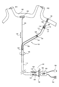
6,064,902 to Haissaguerre et al. In this approach, a catheter is made of
distal and proximal electrodes
at the tip. The catheter can be bent in a J shape and positioned inside a
pulmonary vein. The tissue of
the inner wall of the PV is ablated in an attempt to kill the source of the
aberrant heart activity. Other
RF based catheters are described in US Patents 6,814,733 to Schwartz et al.,
6,996,908 to Maguire et
al., 6,955,173 to Lesh; and 6,949,097 to Stewart et al.
[ODOR] Another source used in ablation is microwave energy. One such device
is described by
Dr. Mark Levinson [(Endocardial Microwave Ablation: A New Surgical Approach
for Atrial
Fibrillation; The Heart Surgery Forum, 2006] and Maessen et al. [Beating heart
surgical treatment of
atrial fibrillation with microwave ablation. Ann Thorac Surg 74: 1160-8,
20021. This intraoperative
device consists of a probe with a malleable antenna which has the ability to
ablate the atrial tissue.
Other microwave based catheters are described in US Patents 4,641,649 to
Walinslcy; 5,246,438 to
Langberg; 5,405,346 to Grundy, et al.; and 5,314,466 to Stem, et al.
[0009] Another catheter based method utilizes the cryogenic technique where
the tissue of the
atrium is frozen below a temperature of -60 degrees C. This results in killing
of the tissue in the
vicinity of the PV thereby eliminating the pathway for the aberrant signals
causing the AF [A. M.
Gillinov, E. H. Blackstone and P. M. McCarthy, Atrial fibrillation: current
surgical options and their
assessment, Annals of Thoracic Surgery 2002;74:2210-7]. Cryo-based techniques
have been a part of
the partial Maze procedures [Sueda T., Nagata H., Orihashi K., et al.,
Efficacy of a simple left atrial
procedure for chronic atrial fibrillation in mitral valve operations, Ann
Thorac Surg 1997;63:1070-
1075; and Sueda T., Nagata H., Shikata H., et al.; Simple left atrial
procedure for chronic atrial
fibrillation associated with mitral valve disease, Ann Thorac Surg
1996;62:1796-1800]. More
CA 02905086 2015-09-21
WO 2007/134258
PCT/1JS2007/068818
3
recently, Dr. Cox and his group [Nathan H., Eliakim M., The junction between
the left atrium and the
pulmonary veins, An anatomic study of human hearts, Circulation 1966;34:412-
422, and Cox J.L.,
Schuessler R.B., Boineau J.P., The development of the Maze procedure for the
treatment of atrial
fibrillation, Semin Thorac Cardiovasc Surg 2000;12:2-141 have used cryoprobes
(cryo-Maze) to
duplicate the essentials of the Cox-Maze III procedure. Other cryo-based
devices are described in US
Patents 6,929,639 and 6,666,858 to Lafmtaine and 6,161,543 to Cox et al.
[00010] More recent approaches for the AF treatment involve the use of
ultrasound energy. The
target tissue of the region surrounding the pulmonary vein is heated with
ultrasound energy emitted by
one or more ultrasound transducers. One such approach is described by Lesh et
al. in US Patent
6,502,576. Here the catheter distal tip portion is equipped with a balloon
which contains an ultrasound
element The balloon serves as an anchoring means to secure the tip of the
catheter in the pulmonary
vein. The balloon portion of the catheter is positioned in the selected
pulmonary vein and the balloon
is inflated with a fluid which is transparent to ultrasound energy. The
transducer emits the ultrasound
energy which travels to the target tissue in or near the pulmonary vein and
ablates it. The intended
therapy is to destroy the electrical conduction path around a pulmonary vein
and thereby restore the
normal sinus rhythm. The therapy involves the creation of a multiplicity of
lesions around individual
pulmonary veins as required. The inventors describe various configurations for
the energy emitter and
the anchoring mechanisms.
[00011] Yet another catheter device using ultrasound energy is described by
Gentry et al.
[Integrated Catheter for 3-D Intracardiac Echocardiography and Ultrasound
Ablation, IEEE
Transactions on Ultrasonics, Ferroelectrics, and Frequency Control. Vol. 51,
No. 7, pp 799-8071. Here
the catheter tip is made of an array of ultrasound elements in a grid pattern
for the purpose of creating
a three dimensional image of the target tissue. An ablating ultrasound
transducer is provided which is
in the shape of a ring which encircles the imaging grid. The ablating
transducer emits a ring of
ultrasound energy at 10 MHz frequency. In a separate publication [Medical
Device Link, Medical
Device and Diagnostic Industry, February 2006], in the description of the
device, the authors assert
that the pulmonary veins can be imaged and "a doctor would be able to
electrically isolate the
pulmonary veins by putting a linear lesion around them" (emphasis by
inventors). It is unclear from
this statement whether the ablation ring is placed around one single target
vein, or around a plurality
of veins. In the described configuration of the catheter tip, it can be easily
seen that the described ring
ultrasound energy source can only emit the ultrasound beam of a size to ablate
only one pulmonary
vein at a time.
[00012] Other devices based on ultrasound energy to create circumferential
lesions are described
in US Patent Nos. 6,997,925; 6,966,908; 6,964,660; 6,954,977; 6,953,460;
6,652,515; 6,547,788; and
6,514,249 to Maguire et al.; 6,955,173; 6,052,576; 6,305,378; 6,164,283; and
6,012,457 to Lesh;
6,872,205; 6,416,511; 6,254,599; 6,245,064; and 6,024,740; to Lesh et al.;
6,383,151; 6,117,101; and
CA 02905086 2015-09-21
WO 2007/134258 PCT/US2007/068818
4
WO 99/02096 to Diederich etal.; 6,635,054 to Fjield et al.; 6,780,183 to
Jimenez et al.; 6,605,084 to
Acker et at; 5,295,484 to Marcus et al.; and WO 2005/117734 to Wong etal..
[00013] In all above approaches, the inventions involve the ablation of tissue
inside a pulmonary
vein or at the location of the ostium. The anchoring mechanisms engage the
inside lumen of the target
pulmonary vein. In all these approaches, the anchor is placed inside one vein,
and the ablation is done
one vein at a time.
SUMMARY OF THE INVENTION
[00014] One aspect of the invention provides a cardiac ablation system
including an ablation
catheter having an anchor adapted to support the ablation catheter within an
atrium of a heart and an
ultrasound emitter disposed radially outward from a rotation axis and from the
anchor, and a control
mechanism adapted to rotate the ultrasound emitter about the rotation axis and
to provide ablation
energy to the ultrasound emitter to ablate heart tissue. Some embodiments also
include an ultrasound
emitter support extending radially outward from the rotation axis and
supporting the ultrasound
emitter, which may be the a distal portion of the ablation catheter or may be
a separate element.
[00015] In some embodiments, the emitter is disposed to emit ultrasound energy
through a distal
end of the support, and in other embodiments the emitter is disposed to emit
ultrasound energy
radially outward from a side of the support. In some embodiments, the emitter
is disposed at an angle
greater than zero with respect to the outer surface of the support.
[00016] In some embodiments, the emitter includes an ultrasound transducer and
an ultrasound
reflective surface disposed to reflect ultrasound energy from the transducer.
The transducer may be
disposed to direct ultrasound energy proximally toward the reflective surface.
[00017] In some embodiments, the control mechanism is adapted to bend the
emitter support at a
desired angle from the rotation axis. This angle may be formed at a first
location along the emitter
support, with the control mechanism being further adapted to bend the emitter
support at a second
location along the emitter support.
[00018] In some embodiments, the ultrasound emitter support includes or serves
as an electrode in
electrical communication with the control mechanism and the anchor includes or
serves as an
electrode in electrical communication with the control mechanism.
[00019] The control mechanism may be adapted to move the anchor within a left
atrium. The
anchor may extend substantially along the rotation axis, with the ablation
catheter being adapted to
rotate with respect to the anchor. Alternatively, the anchor may extend along
an axis other than the
rotation axis. In embodiments in which the system further includes a delivery
sheath adapted to
contain the ablation catheter, either the delivery sheath or the ablation
catheter may have a port
through which the anchor extends. Some embodiments also include a second
anchor supporting the
ablation catheter.
CA 02905086 2015-09-21
WO 2007/134258
PCT/US2007/068818
[00020] In some embodiments, the emitter is distally and proximally
translatable with respect to
the anchor. In some embodiments, the emitter is supported by a transducer
support extending radially
outward from the rotation axis and is distally and proximally translatable
with respect to the anchor.
[00021] The anchor may be adapted to contact a heart tissue surface, such as
the interior wall of
the atrium or an interior surface of a pulmonary vein. Some embodiments have a
delivery sheath
surrounding the ablation catheter, and the anchor is expandable to contact a
support catheter
surrounding the ablation catheter.
[00022] In embodiments in which the ultrasound emitter includes an ultrasound
transducer, the
system may also include a fluid source and a fluid flow path adjacent to the
transducer. The system
may also have a fluid exit port adjacent to the transducer and extending from
the fluid flow path to the
exterior of the ablation catheter. In embodiments in which the ultrasound
emitter is disposed
proximal to a distal end of the ablation catheter, the ablation catheter may
also have a fluid chamber in
communication with the fluid source, disposed between the ultrasound emitter
and the distal end of
the catheter, and in fluid communication with the distal end of the catheter.
The fluid chamber may
have a plurality of fluid exit channels formed in the distal end of the
catheter.
[00023] Some embodiments also have a distance sensor adapted to sense distance
between the
ultrasound emitter and a tissue surface. The ultrasound emitter and the
distance sensor may both be
an ultrasound transducer. Some embodiments may also have an ablation depth
sensor. The
ultrasound emitter and ablation depth sensor may both be an ultrasound
transducer.
[00024] Another aspect of the invention provides a cardiac ablation system
including an ablation
catheter having an ultrasound emitter and an ultrasound emitter support
extending radially outward
from a rotation axis and supporting the ultrasound emitter, and a control
mechanism adapted to rotate
the ultrasound emitter about the rotation axis and to provide ablation energy
to the ultrasound emitter
to ablate heart tissue and adapted to bend the emitter support at a desired
angle from rotation axis. In
some embodiments, the desired angle is formed at a first location along the
emitter support, the
control mechanism being further adapted to bend the emitter support at a
second location along the
emitter support.
[00025] In some embodiments, the ultrasound emitter includes an ultrasound
transducer, with the
system further comprising a fluid source and a fluid flow path adjacent to the
transducer. The system
may also include a fluid exit port adjacent to the transducer and extending
from the fluid flow path to
the exterior of the ablation catheter.
[000261 Some embodiments also have a distance sensor adapted to sense distance
between the
ultrasound emitter and a tissue surface. The ultrasound emitter and the
distance sensor may both be
an ultrasound transducer. Some embodiments may also have an ablation depth
sensor. The
ultrasound emitter and ablation depth sensor may both be an ultrasound
transducer.
CA 02905086 2015-09-21
WO 201)7/134258 PCT/US2007/068818
6
[00027] Yet another aspect of the invention provides a cardiac ablation method
including the
following steps: inserting a treatment catheter into an atrium of a heart, the
treatment catheter
including an ultrasound emitter; positioning the ultrasound emitter to face
heart tissue within the left
atrium outside of a pulmonary vein; emitting ultrasound energy from the
ultrasound emitter while
rotating the ultrasound emitter about a rotation axis; and ablating heart
tissue with the ultrasound
energy to form a lesion outside of a pulmonary vein. In some embodiments, the
positioning step
includes the step of bending an ultrasound emitter support. In some
embodiments, the positioning
step includes the step of moving the ultrasound emitter parallel to the
rotation axis. In some
embodiments, the positioning step includes the step of anchoring the treatment
catheter, such as
against the heart wall or by placing an anchor against an atrial wall outside
of a pulmonary vein or
within a pulmonary vein. The anchoring step may also involve placing a
plurality of anchors within a
plurality of pulmonary veins and/or expanding an anchor within a support
catheter.
[00028] In some embodiments, the rotating step includes the step of rotating
the treatment catheter
about the anchor. The rotation may include the step of rotating the ultrasound
emitter less than 3600
around the rotation axis or rotating the ultrasound emitter at least 3600
around the rotation axis.
[00029] In some embodiments, the ablating step includes the step of forming a
lesion encircling at
least two pulmonary vein ostia. The method may also include forming a second
lesion around two
other pulmonary vein ostia, possibly forming a third lesion extending from the
first lesion to the
second lesion, and possibly forming a fourth lesion extending from the first,
second or third lesion
substantially to a mitral valve annulus.
[00030] In some embodiments, the emitting step includes the step of
transmitting ultrasound
energy distally from a distal end of the treatment catheter and/or radially
from the treatment catheter.
In some embodiments, the emitting step includes the step of transmitting
ultrasound energy from an
ultrasound transducer (possibly in a proximal direction) and reflecting the
ultrasound energy from a
reflector. These embodiments may also include the step of rotating the
reflector.
[00031] Some embodiments include the step of passing fluid through the
ablation catheter and
through an exit port adjacent the ultrasound emitter. The fluid may pass into
a fluid chamber disposed
between the ultrasound emitter and the heart tissue.
[00032] Some embodiments include the step of sensing distance between the
ultrasound emitter
and a tissue surface, such as by using the ultrasound emitter to sense
distance between the emitter and
the tissue surface. The distance sensing step may include the step of sensing
distance between the
ultrasound emitter and the tissue surface over an intended ablation path prior
to the ablating step and
may include the step of repositioning the ultrasound emitter as a result of
sensed distance determined
in the sensing step.
[00033] Some embodiments include the step of sensing depth of ablation in the
heart tissue, such
as by using the ultrasound emitter to sense depth of ablation in the heart
tissue. The speed of rotation
CA2905086
7
of the ultrasound emitter and/or the power delivered to the ultrasound emitter
may be based on sensed
depth of ablation.
[00034] Some embodiments include the step of sensing thickness of the heart
tissue. The speed of
rotation of the ultrasound emitter and/or the power delivered to the
ultrasound emitter may be based on
sensed tissue thickness. In some embodiments, the ablating step includes the
step of forming a
substantially elliptical lesion segment in the heart tissue.
[00035] Still another aspect of the invention provides a cardiac ablation
method including the
following steps: inserting a treatment apparatus into an atrium of a heart,
the treatment apparatus having
an ultrasound emitter and an ultrasound emitter support; positioning the
ultrasound emitter to face heart
tissue within the left atrium outside of a pulmonary vein; emitting ultrasound
energy from the ultrasound
emitter while changing a bend angle in the ultrasound emitter support; and
ablating heart tissue with the
ultrasound energy to form a lesion outside of a pulmonary vein. In some
embodiments, the positioning
step includes the step of bending an ultrasound emitter support. In some
embodiments, the positioning
step includes the step of anchoring the treatment catheter.
[00036] Some embodiments add the step of rotating the ultrasound emitter
about a rotation axis
during the emitting step. In some embodiments, the ablating step includes the
step of forming a
substantially linear lesion and/or a substantially elliptical lesion segment
in the heart tissue.
[0036A] Various aspects of the disclosure relate to a cardiac ablation
system comprising: an ablation
catheter comprising an anchor adapted to support the ablation catheter within
a heart and an ultrasound
emitter disposed radially outward from a rotation axis and from the anchor,
and a control mechanism
adapted to rotate the ultrasound emitter about the rotation axis and to
provide ablation energy to the
ultrasound emitter to ablate heart tissue.
[0036B] Various aspects of the disclosure relate to a cardiac ablation
system comprising: an ablation
catheter comprising an ultrasound emitter and an ultrasound emitter support
extending radially outward
from a rotation axis and supporting the ultrasound emitter, and a control
mechanism adapted to rotate the
ultrasound emitter about the rotation axis and to provide ablation energy to
the ultrasound emitter to
ablate heart tissue and adapted to bend the emitter support at a desired angle
from rotation axis.
[0036C] Various aspects of the claimed invention also relate to a cardiac
ablation system
comprising: a single ultrasound transducer for positioning adjacent a target
tissue; and a
processor configured with instructions to: ablate the target tissue with a
beam of energy from the
single ultrasound transducer when positioned adjacent the target tissue in
order to form a
Date Re9ue/Date Received 2020-06-12
CA2905086
7a
lesion in the target tissue, wherein the lesion is formed without contact
between the single
ultrasound transducer and the target tissue; sense a distance between the
single ultrasound
transducer and the target tissue with the single ultrasound transducer; move a
distal portion of an
elongate flexible shaft and the single ultrasound transducer coupled thereto
in order to form a
continuous lesion in the target tissue, wherein the ablation is performed
while the single
ultrasound transducer and the distal portion of the elongate flexible shaft
are moving; and control
the movement of the single ultrasound transducer or control the beam of energy
based on the
sensed distance between the single ultrasound transducer and the target
tissue.
[0036D] Various aspects of the claimed invention also relate to a cardiac
ablation system
comprising: an elongate flexible shaft having a proximal portion and a distal
portion; and an
ultrasound transducer disposed within a housing adjacent the distal portion of
the elongate
flexible shaft, wherein the ultrasound transducer is configured to emit a beam
of ultrasound
energy, wherein the beam of ultrasound energy is configured to ablate target
tissue without
contact between the ultrasound transducer and the target tissue, and wherein
the beam of
ultrasound energy forms a continuous lesion in the target tissue while the
distal portion of the
elongate flexible shaft is moving.
[0036E] Various aspects of the claimed invention relate to a cardiac
ablation system comprising:
an elongate flexible shaft having a proximal portion and a distal portion; and
an ultrasound
transducer adjacent the distal portion of the elongate flexible shaft, wherein
the ultrasound
transducer comprises a flat disc having a front face configured to emit a beam
of ultrasound
energy, wherein the beam of ultrasound energy is configured to ablate target
tissue without
contact between the ultrasound transducer and the target tissue, and wherein
the beam of
ultrasound energy forms a continuous lesion in the target tissue while the
distal portion of the
elongate flexible shaft is moving.
BRIEF DESCRIPTION OF THE DRAWINGS
[00037] The novel features of the invention are set forth with
particularity in the claims that follow.
A better understanding of the features and advantages of the present invention
will be obtained by
reference to the following detailed description that sets forth illustrative
embodiments, in which the
principles of the invention are utilized, and the accompanying drawings of
which:
[00038] Figure 1 shows the device including a catheter in one embodiment of
the invention.
Date Recue/Date Received 2020-06-12
CA2905086
7b
[00039] Figure 2 shows the construction of the shaft of the catheter in one
embodiment of the
invention.
[00040] Figures 3A-C show bending of a distal portion of the catheter of
Figure 1.
[00041] Figure 3D shows bending of the distal end of the catheter of Figure
1 and an anchor
mechanism.
[00042] Figure 4 shows the distal tip assembly of the catheter of Figure 1.
[00043] Figure 5 is a view of the device in a second embodiment.
[00044] Figure 6 shows the distal tip assembly of the catheter of Figure 5.
[00045] Figure 7 is a view of the device in a third embodiment.
[00046] Figure 8 shows the distal tip assembly of the catheter of Figure 7.
[00047] Figure 9 is a view of the device in a fourth embodiment.
[00048] Figure 10 shows the distal tip assembly of the catheter of Figure
9.
CA 2905086 2019-06-17
CA 02905086 2015-09-21
WO 2007/134258 PCT/US2007/068818
8
[00049] Figure 11 shows an ablation zone encircling four pulmonary veins and
the device in one
embodiment of the invention.
[00050] Figure 12 shows two ablation zones each around two pulmonary veins.
[00051] Figure 13 shows an ablation zone around three pulmonary veins.
[00052] Figures 14 to 17 show various mechanisms for the anchoring a portion
of the catheter.
[00053] Figure 18 shows yet another embodiment of the invention as positioned
in the left atrium
of the heart.
[00054] Figure 19 shows the use of the device of Figure 18 in the atrium of
the heart.
[00055] Figure 20 shows the distal end of the device of Figure 18 beyond the
guiding sheath.
[00056] Figure 21A shows the details of the transducer housing at the distal
tip of the catheter.
[00057] Figure 21B shows the transducer mounting header with fluid flow
channels.
[00058] Figure 21C shows an alternative design for the fluid pocket
containment component.
[00059] Figure 22 is a view of the construction of the therapy catheter.
[00060] Figure 23 shows a view of the construction of the outer catheter.
[00061] Figure 24 is a view of the characteristics of the ultrasound beam as
it exits from the
trancrhicer
[00062] Figure 25 shows formation of the shape of an ablation lesion.
[00063] Figures 26 A-D show the development of the ablation lesion as function
of time.
[00064] Figures 27 A-D show the interaction of the ultrasound beam with the
tissue at various
distanucs Twin the ultrasound transducer.
[00065] Figures 28 A-B are views of the interaction of the ultrasound beam
with the tissue when
the tissue is presented to the beam at an angle.
[00066] Figure 29 shows the effect of the movement of heart muscle during
ablation.
[00067] Figure 30 shows the transmission and retlections of ultrasound beam
from the target
tissue.
[00068] Figure 31 shows position of the catheter set in the left atrium in a
condition when it may
not be desirable to create an ablation zone.
[00069] Figure 32 shows a catheter set designed to address the right pulmonary
veins.
[00070] Figure 33 shows a lesion set according to one embodiment of this
invention.
[00071] Figure 34 shows the creation of an ablation zone near the left
pulmonary veins.
[00072] Figures 35A-C show the formation of a line lesion from the left
pulmonary veins to the
right pulmonary veins.
[00073] Figure 36 shows a vertical line of ablation ending at the mitral valve
annulus.
[00074] Figure 37 shows the use of the device of Figure 31 in creating the
ablation zone in the
right pulmonary veins.
9
[00075] Figures 38 A-J show a variety of candidate lesion sets in the left
atrium.
DETAILED DESCRIPTION OF THE INVENTION
[00076] The invention described herein includes a device and methods for
creating ablation zones
in tissue. The device of the invention includes an elongated member (e.g., a
catheter) and an anchor
mechanism. The elongate member includes a distal tip assembly for directing
energy to a tissue.
Uses of the invention include but are not limited to providing a conduction
block for treatment of
atrial fibrillation in a subject, for example, in a patient.
[00077] One aspect of a first embodiment of the invention is shown in Figure
1. As shown, the
device 100 includes an elongate member that can be a catheter 110. In other
implementations, the
elongate member is a cannula, tube or other elongate structure having one or
more lumens. The
catheter 110 can be made of a flexible multi-lumen tube. As shown, the
catheter 110 can include a
distal tip assembly 112 positioned at a distal portion of the catheter 110.
The tip assembly 112 can
house an energy delivery structure, for example, an ultrasound transducer
subassembly 114 (described
in more detail in reference to Figure 4).
[00078] Although the ablation device described herein includes a distal tip
assembly having an
ultrasound transducer as a source of ablation energy, it is envisioned than
any of a number of energy
sources can be used with various implementations of the invention. Suitable
sources of ablation
energy include but are not limited to, radio frequency (RF) energy,
microwaves, photonic energy, and
thermal energy. It is envisioned that ablation could alternatively be achieved
using cooled fluids (e.g.,
cryogenic fluid). Additionally, although use of a single ultrasound transducer
is described herein as
an exemplary energy delivery structure, it is envisioned that a plurality of
energy delivery structures,
including the alternative energy delivery structures described herein, can be
included in the distal
portion of the elongate member. In one implementation the elongate member is a
catheter wherein the
distal portion of the catheter includes multiple energy delivery structures,
for example, multiple
ultrasound transducers. Such a catheter distal portion can be deployable as a
loop or other shape or
arrangement to provide positioning of one or more of the energy delivery
structures for a desired
energy delivery.
[00079] The elongate member of the device can include a bending mechanism for
bending a distal
portion of the elongate member (e.g., a catheter) at various locations (an
example of such bending is
shown in Figures 3A-D). The bending mechanism can include but is not limited
to lengths of wires,
ribbons, cables, lines, fibers, filament or any other tensional member. In one
implementation the
bending mechanism includes one or more pull wires, for example, a distal pull
wire and a proximal
pull wire. A variety of attachment elements for connecting the bending
mechanism and the elongate
member are envisioned. As shown in Figure 1, in one implementation where the
elongate member is
a catheter 110, the distal pull wire 116 and the transducer subassembly 114
are secured to the tip
Date Recue/Date Received 2020-11-13
CA 02905086 2015-09-21
WO 2007/134258
PCT/US2007/068818
assembly 112 by means of a distal adhesive band 118. Other means of attaching
the distal pull wire
116 to a portion of the tip assembly 112 include but are not limited to
attachment using: adhesive,
welding, pins and/or screws or the likes. Pull wire 116 can be contained in a
lumen (not shown) of
the catheter 110 and can terminate at a slider 120 in a proximal housing 122.
The proximal housing
122 can include various actuating mechanisms to effect various features of the
catheter, as described
below. In one implementation, the slider 120 can move in a slot 124 which
pulls or pushes the wire
116. Since the distal end of the wire 116 is secured to the tip 112, the
result is that the catheter tip 112
can be bent and unbent as desired at a distal bend location 126 by moving the
slider 120. Distal bend
location 126 can be positioned on the distal tip assembly 112 as needed to
achieve the desired bending
of the catheter 110.
[00080] A second analogous bending mechanism can be provided in the catheter
which is more
proximally positioned with respect to the distal tip assembly. As shown in
Figure 1, a proximal pull
wire 128 can reside in a lumen (not shown) of the catheter 110 and the wire
128 distal end can be
secured in the catheter 110 by a proximal adhesive band 130. This proximal
pull wire 128 can
terminate in a second slider 132 at the proximal housing 122. The slider 132
can move in a second
slot 134 which allows the distal tip assembly 112 to be bent at a proximal
bend location 136.
[00081] The elongate member can further include an anchor mechanism by which
the distal
portion of the elongate member can be held in a relatively predictable
position relative to a tissue, for
example, inside a chamber such as the left atrium of the heart As shown in
Figure 1, in one
implementation an anchor mechanism 140 includes a pre-shaped wire loop 138. In
a specific
implementation, the wire loop 138 is made of a shapeable wire, for example,
made from a shape-
memory material such as Nitinol (nickel ¨ titanium alloy). Alternatively, the
anchor mechanism can
include a loop made from any of a number of materials such as metal, plastic
and/or fiber or
combinations thereof. Although a loop is described, it is envisioned that any
of a number of shapes,
curved and/or angular, two-dimensional and/or three-dimensional can provide
the anchoring required.
The anchor 140 can reside in a lumen (not shown) of the catheter 110, and can
exit from the catheter
110 through a notch 142 near the distal end of the catheter 110 (see Figure
1). The proximal end of
the anchor mechanism 140 can terminate in a third slider 148 at the proximal
housing 122. The third
slider 148 can move in a third slot 150 at the proximal housing 122, thereby
producing a
corresponding anchor mechanism movement 144 of the anchor mechanism 140.
[000821 In one implementation, when the slider 148 is in a proximal position,
the wire loop 138
can be maintained in a substantially linear shape inside the lumen of the
catheter 110 (not shown). In
use, as third slider 148 is advanced distally in the slot 150, a distal tip of
the wire loop 138 exits the
notch 142 (not shown). As the slider 148 is further advanced, the wire loop
138 can take on the shape
of a pre-formed loop as it is unrestricted by the confines of a lumen (see
Figure 3D). As shown in
Figure 1, the wire loop 138 of the anchor 140 can be advanced further until it
makes a firm contact
CA 02905086 2015-09-21
WO 2007/134258 PCT/US2007/068818
11
with the tissue such as the ceiling wall 146 of the left atrium of the heart.
One function of the wire
loop 138 is to provide a fiim contact and/or stabilization between the anchor
mechanism 140 and the
tissue, and thereby between a region of the catheter 110 and the tissue (see
Figure 1). An additional
function of the anchor mechanism is to provide an axis around which all or a
portion of the catheter
shaft can be rotated. Such rotation of the catheter is illustrated in Figure
1, as arrow 152. As shown
in Figure 1, in one implementation a rotation mechanism 154, for example, a
wheel, is provided at the
proximal housing 122 by which all or a portion of the catheter 110 shaft can
be rotated around the axis
defined by the anchor mechanism 140. As can be easily envisioned, through
rotational movement
about such an axis, the most distal portion of the tip assembly 112 can be
swept in a desired path in
relation to target tissue. In one implementation, the path of the tip assembly
212 can be a
substantially circular path 262 inside a tissue chamber such as the left
atrium of the heart (see Figure
11).
1000831 A transducer subassembly can be secured in the distal tip assembly of
the catheter. As
shown in Figure 1, in one implementation a transducer subassembly 114 is
secured by the distal
adhesive band 118. The transducer subassembly is described in more detail
herein for various
embodiments of the invention. In one implementation, the transducer
subassembly 114 includes a
tenipciatule ineasuling device such as a then-Ms-tot or a thermocouple (not
shown). The transducer
can be energized by the wires which, along with the temperature sensor wires,
can be contained in a
lumen of the catheter (not shown). As shown in Figure 1, such wires can
terminate in a connector, for
example, a transducer connector 156 at the proximal housing 122. The connector
156 can be attached
to and detached from a power generator and/or controller (not shown). It is
envisioned that such a
power generator and/or controller can energize the transducer, display
temperature readings and
perform any of a number of functions relating to such transducers as well
understood in the art. For
example, monitoring A-mode signal and the like (e.g., B-mode). In use, as the
transducer is
energized, it can emit an ultrasound beam 158 towards the tissue 146. As the
energy is transferred
from the ultrasound beam into the tissue, the targeted tissue portion can be
heated sufficiently to
achieve ablation. Thus, as shown in Figure 1, an ablation zone 160 can be
created in the tissue.
1000841 During the energizing of the transducer, the transducer may become
heated. It is
envisioned that the transducer can be maintained within a safe operating
temperature range by cooling
the transducer. In one implementation cooling of the transducer can be
accomplished by contacting
the transducer subassembly with a fluid, for example, saline. In some
implementations the transducer
can be cooled using a fluid having a lower temperature relative to the
temperature of the transducer.
In one implementation a fluid for cooling the transducer is flushed past the
transducer subassembly
from a lumen in the catheter (see e.g., Figure 2). Accordingly, as shown in
Figure 1, the proximal end
of a lumen of the catheter 110 can be connected to a fluid port 162, for
example, a luer fitting, in the
proximal housing 122. As further shown in Figure 1, in one implementation
fluid used for cooling the
transducer can exit the catheter tip 112 through a one or more apertures 164.
The apertures can be a
CA 02905086 2015-09-21
WO 2007/134258
PCT/US2007/068818
12
grating, screen, holes, weeping structure or any of a number of suitable
apertures. In one
implementation apertures 164 are drip holes.
[00085] Referring to Figure 2, in one implementation where the elongate member
of the device is
a catheter, the shaft of the catheter 110 includes a multi-lumen tubing 170
having one or more lumens
176, which is encased in a braid 166 of suitable metallic or non-metallic
filaments and is encased in a
smooth jacket 168 made of conventional biocompatible material. Lumens 176 can
accommodate any
of a number of features of the invention including but not limited to, pull
wires, fluids, gases, and
electrical connections.
[00086] In Figures 3A-C, an exemplary series of drawings illustrate bending
of the catheter distal
portion in more detail. In the implementation shown, the distal pull wire 116
is secured at a distal
portion of the tip assembly 112 by means of the distal adhesive band 118. In
use, as the distal pull
wire 116 is pulled by moving the first slider 120 (see Figure 1), the catheter
distal portion is bent at
location 126 in the direction 172, thereby moving from position X to position
Y, as shown in Figure
3B. Next, the proximal pull wire 128, which is secured in the catheter lumen
at a position by
proximal adhesive band 130, is pulled by moving the second slider 132 (see
Figure 1). This results in
the catheter 110 distal portion bending at location 136 and moving in the
direction 174 to position Z,
away awl the longitudinal axis of the catheter, as shown Figure 3C.
[00087] It is envisioned that the pull wire attachment points, and
correspondingly the bend
locations in the device can be configured, in any of a number of ways, not
limited to the examples
described herein. For example, it is envisioned that a single pull wire or
other bend inducing
mechanism can be used. Alternatively, the use of three in MUM such mechanism
is envisioned. With
respect to attachment points for bend inducing mechanism, it is envisioned
that any location along the
distal tip assembly as well as the catheter distal portion are suitable
optional attachment points. With
respect to the number and location of bend locations in the device, it is
envisioned that a spectrum of
suitable bend locations can be provided. For example, while one and two bends
arc illustrated herein,
it is envisioned that three or more bends can be used to achieve a desired
catheter configuration and/or
application of energy using the device.
[00088] The anchor mechanism 140 of the device can be deployed in a separate
or simultaneous
step from bending the device, as shown in Figure 3D. The anchor mechanism 140,
which can be
configured to reside in a lumen (not shown) of the catheter 110, is advanced
out of the catheter 110
and through the anchor notch 142 by moving the third slider 148 (see Figure
1). In the
implementation shown in Figure 3D, as the anchor mechanism 140 exits the notch
142 a distal portion
of the mechanism 140 takes on the pre-formed shape of a loop 138. This loop
138 is advanced further
in axial direction 144 until it firmly engages tissue, for example in the
inside wall of a tissue chamber
such as the left atrium of the heart. The anchor mechanism provides a
rotational axis for the distal tip
assembly. The transducer subassembly 114 can be intentionally displaced away
from this axis so that
CA 02905086 2015-09-21
WO 2007/134258
PCT/US2007/068818
13
when the catheter shaft is rotated (see arrow 152) around the axis provided by
the anchor mechanism
140, the transducer can traverse a substantially circular loop inside the
tissue chamber. The result of
this motion is to create a substantially circular ablation zone inside the
tissue chamber (described in
more detail in Figure 11). It is envisioned that an arc-shaped or other curved
ablation zone could
alternatively be created with the device.
[00089] The design of the distal tip subassembly can include a variety of
configurations providing
alternative means of delivering energy to tissue. A first embodiment of the
distal tip subassembly
1112 is shown in Figure 4. As illustrated, the tip assembly 1112 can include a
closed end tube casing
1142 which is transparent to ultrasound waves. It can further contain a
transducer subassembly 1114
including an ultrasound transducer 1120. The transducer 1120 can be made of a
piezoelectric material
such as PZT (lead zirconate titanate) or PVDF (polyvinylidine difluoride) and
the like. The
transducer 1120 can be configured as a disc and the faces of the disc can be
coated with a thin layer of
a metal such as gold. In one implementation the disc is a circular flat disc.
Other suitable transducer
coating metals include but are not limited to stainless steel, nickel-cadmium,
silver or a metal alloy.
As shown in Figure 4, in one implementation the transducer 1120 can be
connected to electrical
attachments 1130 and 1132 at two opposite faces. These connections can be made
of insulated wires
1134 which can bc, for example, a twisted pair or a coaxial cable so as to
minimize electromagnetic
interference. When a voltage is applied across the transducer, ultrasonic
sound beam 1158 is emitted.
The frequency of the ultrasound beam is in the range of about Ito 50
megaHertz.
[00090] As shown in Figure 4, a temperature sensor 1136 can be coupled with
the transducer
1120, for example, attached to the back face of the transducer 1120. The
temperature sensor can be
comprised of a thermocouple or a thermistor or any other suitable means. As
shown in Figure 4, the
sensor 1136 can include wires 1138 which carry the temperature information to
the catheter proximal
end. The wires 1134 and 1138 together can form a wire bundle 1140 extending to
the catheter
proximal end.
[00091] As further shown in Figure 4, the transducer 1120 can be attached to a
backing 1126 by
means of an adhesive ring 1122 or other attachment, which creates a void or
pocket 1124 between the
transducer 1120 and the backing 1126. The pocket 1124 can include a material
which efficiently
reflects sound waves generated by the transducer 1120. The material of the
pocket 1124 can be air Or
any other suitable material such as metal or plastic which reflects the sound
waves. Advantageously,
the sound waves thus can be directed to exit from the front face of the
transducer, resulting in a
minimum amount of sound energy lost out through the transducer back face into
the backing. The
backing can be made of a thermally conductive material such as metal or
plastic for aiding in the
dissipation of heat which is created when the transducer is energized.
[00092] As illustrated in Figure 4, the wire bundle 1140 can be fed through a
passageway or hole
1128 in the backing 1126 and can be housed in a lumen of the catheter 1110.
The wire bundle can
CA 02905086 2015-09-21
WO 2007/134258 PCT/US2007/068818
14
terminate in the connector 156 at the proximal housing 122 (see Figure 1). As
shown in Figure 4, the
proximal end of the backing 1126 can be secured to the casing 1142 by means of
the distal adhesive
band 1118. This creates a void or chamber 1146 between the distal end of the
casing 1142 and the
distal adhesive band 1118. The chamber 1146 is configured to be filled with a
thermally conductive
fluid such as saline so that the transducer 1120 can be cooled while
energized. The distal adhesive
band 1118 can include a passageway 1148 which is used in connecting the
chamber 1146 to a fluid
carrying lumen. The passageway 1148 can be in fluid communication with the
fluid port 162 at the
proximal housing 122 through one of the lumens (not shown) of the catheter
1110 (see Figures 1 and
4). As shown in Figure 4, the chamber 1146 can include one or more apertures
1164, for example,
drip holes distributed circumferentially at the chamber 1146 distal portion.
In use, prior to insertion
of the device into the body, the chamber can be filled with a fluid such as
saline. This can be
accomplished using a suitable fluid supply device such as a syringe connected
to the fluid port (not
shown). The fluid from the syringe can flow through the passageway of the
distal adhesive band, into
the chamber while expelling the air out from the chamber through the
apertures. During the use of the
device in the body, a constant drip of saline can be maintained, if necessary,
to cool the transducer.
[00093] Still referring to Figure 4, a distal pull wire 1116 can be secured
to the distal tip
subassembly 1112 by the distal adhesive band 1118. The distal pull wire 1116
can reside in one of
the lumens 1176 of the catheter 1110 and can be connected to the slider 120 in
the proximal housing
122 (see Figure 1 and Figure 4). As described above in reference to Figure 3A,
the distal pull wire
1116 can be utilized in bending the distal portion of the catheter 1110. As
shown in Figure 4, the
distal lip subassembly 1112 can be securely attached to the catheter tubing
1170 of thc catheter 1110
by the proximal adhesive band 1144. As further shown in Figure 4, lumens 1176
of the catheter
tubing 1170 can be utilized for passage of various elements of the tip
subassembly 1112 and any of
their related features, in addition to instruments, gases, fluids, or other
substances.
[00094] A second embodiment of the invention including an alternative distal
tip assembly
arrangement is shown in Figure 5. Here the transducer subassembly 1214 is
mounted in the distal tip
assembly 1212 such that the ultrasound transducer 1220 face is substantially
parallel to the
longitudinal axis of the catheter 1210 (that is to say the longitudinal axis
of the catheter 1210 before
bending the distal tip assembly 1212 or catheter 1210). In this configuration,
the sound beam 1258
exits from a lateral surface of the tip assembly 1212. The construction of the
catheter in this
configuration can be essentially same as that described herein for the first
embodiment (see Figures 1-
4).
[00095] As shown in Figure 5, the distal tip assembly 1212 and catheter
1210 bend points, distal
bend location 1272 and proximal bend location 1274 respectively, can be
arranged and configured
such that the ultrasound beam 1258 is presented to the tissue 146 in a
substantially right angle from
the catheter 1210 longitudinal axis. In this manner an ablation zone 1260 is
produced laterally
CA 02905086 2015-09-21
WO 2007/134258
PCT/US2007/068818
through the tip assembly 1212. Figure 6 shows details of the distal tip
assembly 1212 for this
embodiment. As illustrated, the tip assembly 1212 can be assembled in a tube
1242 which is
substantially transparent to the ultrasound waves 1258. The transducer
subassembly 1214 can include
a transducer 1220 which has electrical connections 1230 and 1232 on opposite
flat faces. As
discussed herein, the transducer 1220 can include a temperature sensor 1236
on, for example, a back
side which has wire connections. The transducer wires and the temperature
sensor wires together
form a bundle 1240 which resides in a lumen 1276 of the catheter tubing 1270.
[00096] Still referring to Figure 6, the distal end of the tube housing 1242
can be sealed. As
shown in Figure 6, in one implementation the distal end is sealed with a
thermally conductive
adhesive 1250. The back side of the transducer subassembly 1214 can be secured
to an adhesive ring
1222 that is connected to a backing 1226. Thus, a void or pocket 1224 is
created between the
transducer 1220 and the backing 1226. As shown in Figure 6, the backing 1226
can be secured to the
inner wall of the tube 1242, for example, by the distal adhesive band 1218.
There can be a
passageway 1248 in the adhesive band 1218 to allow the flow of a fluid such as
saline to be
introduced into the chamber 1246. The passageway 1248 can be in fluid
communication with the
fluid port 162 at the proximal housing 122 of the catheter 1210 (see Figures 1
and 6). As discussed
herein the chamber 1246 can include a number of apertures 1264, for example,
drip holes distributed
circumferentially at the chamber 1246 distal end. As further described herein,
prior to insertion of the
device into the body, the chamber 1246 can be filled with a fluid such as
saline. In addition, during
the use of the device in the body, a constant drip of saline can be
maintained, as required to cool the
transducer 1220.
[00097] Again referring to Figure 6, a distal pull wire 1216 can be secured to
the distal tip
subassembly 1212 by the distal adhesive band 1218. The distal pull wire 1216
can reside in one of
the lumens 1276 of the catheter 1210 and can be connected to the slider 120 in
the proximal housing
122 (see Figure 1 and Figure 6). As described above in reference to Figure 3A,
the distal pull wire
1216 can be utilized in bending the distal portion of the catheter 1210. As
shown in Figure 6, the
distal tip subassembly 1212 can be securely attached to the catheter tubing
1270 of the catheter 1210
by the proximal adhesive band 1244. As further shown in Figure 6, lumens 1276
of the catheter
tubing 1270 can be utilized for passage of various elements of the tip
subassembly 1212 and any of
their related features, in addition to instruments, gases, fluids, or other
substances.
[00098] A third embodiment of the invention including an alternative distal
tip assembly
arrangement is shown in Figure 7. Various details, features and uses of this
embodiment include
those as described herein regarding other embodiments. In this embodiment an
alternative transducer
subassembly is provided as shown in detail in Figure 8. As shown in Figure 8,
the ultrasound
transducer 1320 can be mounted on an angled backing 1326. The angle of the
backing can range
between substantially 0-90 . In one implementation the angle is substantially
10-80o. In another
CA 02905086 2015-09-21
WO 2067/134258
PCT/US2007/068818
16
implementation the angle is substantially 30-600. In another implementation
the angle is substantially
40-50 . In a further embodiment the angle is substantially 45 . The transducer
can include a shape.
In one implementation the transducer is in the shape of an elliptical disc. In
another implementation
the transducer has a rectangular shape. As shown in Figures 7 and 8, in one
implementation the
transducer 1320 can emit energy in the form of an ultrasound beam 1358 at an
angle to the
longitudinal axis of the catheter 1310. As shown in Figure 7, the ultrasound
beam 1358 can be
directed to the tissue 146 by appropriately bending the distal tip assembly
1312 using, for example,
pull wires as described herein. The ultrasound energy beam 1358 can create an
ablation zone 1360 in
the tissue 146. Cooling of the transducer in this implementation can be
achieved as described herein.
[00099] As shown in Figure 8 the angled backing 1326 can be secured in the
distal tip assembly
1312 by the distal adhesive band 1318. It is envisioned that other means of
securing the backing to
the distal tip assembly can include but are not limited to attachment using:
adhesive, welding, pins
and/or screws or the likes. Still referring to Figure 8, a distal pull wire
1316 can be secured to the
distal tip subassembly 1312 by the distal adhesive band 1318. The distal pull
wire 1316 can reside in
one of the lumens 1376 of the catheter 1310 and can be connected to the slider
120 in the proximal
housing 122 (see Figure 1 and Figure 8). As described above in reference to
Figure 3A, the distal pull
wire 1316 can be utilized in bending the distal portion of the catheter 1310.
As shown in Figure 8, the
distal tip subassembly 1312 can be securely attached to the catheter tubing
1370 of the catheter 1310
by the proximal adhesive band 1344. As further shown in Figure 8, lumens 1376
of the catheter
tubing 1370 can be utilized for passage of various elements of the tip
subassembly 1312 and any of
their related features, in addition to instruments, gases, fluids, or other
substances.
[000100] A fourth embodiment of the invention including an alternative distal
tip assembly
arrangement is shown in Figure 9, and the details of the tip assembly are
shown in Figure 10. Various
details, features and uses of this embodiment include those as described
herein regarding other
embodiments_ In this embodiment an alternative transducer subassembly is
provided as shown in
detail Figure 10. As shown in Figure 10, in this implementation, the
ultrasound transducer 1420 is
mounted at a distal portion of the distal tip assembly 1412. Further, the
transducer 1420 is directed
substantially toward the proximal direction. As illustrated, in this
orientation the transducer 1420 can
emit an ultrasound wave 1457 substantially parallel to the longitudinal axis
of the distal tip assembly
1412.
[000101] As shown in Figure 10, proximal to the transducer 1420 an angled
reflector device can be
mounted. For example, the reflector device can be a cylindrical reflector 1452
with having a face cut
at an angle to the distal tip assembly 1412 longitudinal axis. The reflector
1452 can be arranged to
reflect the ultrasound energy wave 1457 produced by the transducer 1420 as an
outgoing ultrasound
wave 1458 which exits the tubing 1442 and travels to the intended ablation
site 1460 in the tissue 146.
It is envisioned that the reflector can alternatively include a non-planar
face, for example, a curved,
CA 02905086 2015-09-21
WO 2007/134258
PCT/US2007/068818
17
convex or concave surface. The angle of the reflector can range between
substantially 0-900. In one
implementation the angle is substantially 10-80 . In another implementation
the angle is substantially
30-60 . In another implementation the angle is substantially 40-50 . In a
further embodiment the
angle is substantially 450.
[000102] The reflector 1452 can be secured to the tubing 1442 by means of the
distal adhesive band
1418 which can also secure the distal pull wire 1416. The adhesive band 1418
can include a
passageway 1448 for the flow of a cooling fluid as describe herein. The
transducer subassembly 1414
can be secured at the distal portion of the tip assembly 1412 by means of
thermally conductive
adhesive 1450 which, together with the adhesive band 1418 forms a chamber
1446. The chamber
1446 can include one or more apertures 1464. As shown in Figure 10, in one
implementation the
apertures 1464 are drip holes distributed circumferentially about the distal
portion of the distal tip
assembly 1412.
[0001031 In use, a cooling fluid can be flowed from the passageway 1448 in the
distal adhesive
band, past the reflector 1452 and exit by way of the apertures 1464. This
fluid flow can serve to cool
the transducer 1420 and keep it within nominal operating temperatures. It is
envisioned that cooling
of the transducer can be controlled to provide nominal transducer operation.
As shown in Figure 10,
the transducer 1420 can include a temperature sensor 1436, for example,
attached to the back side of
the transducer. The temperature sensor 1436 can include associated lead wires,
which along with the
wires for the transducer can form a bundle 1440 which is subsequently
contained in a lumen 1476 of
the catheter tube 1470. Similarly, the fluid passageway 1448 can be in fluid
communication with a
lumen 1476 of the catheter tubing 1470. As further shown in Figure 10, the
distal pull wire 1416 can
also be contained in a lumen 1476 of the catheter tubing 1470. As shown in
Figure 10, in one
implementation tubing 1442 is bonded to the catheter tubing 1470 by means of
proximal adhesive
band 1444.
[000104] Still referring to Figure 10, a distal pull wire 1416 can be secured
to the distal tip
subassembly 1412 by the distal adhesive band 1418. The distal pull wire 1416
can reside in one of
the lumens 1476 of the catheter 1410 and can be connected to the slider 120 in
the proximal housing
122 (see Figure 1 and Figure 10). As described above in reference to Figure
3A, the distal pull wire
1416 can be utilized in bending the distal portion of the catheter 1410. As
shown in Figure 10, the
distal tip subassembly 1412 can be securely attached to the catheter tubing
1470 of the catheter 1410
by the proximal adhesive band 1444. As further shown in Figure 10, lumens 1476
of the catheter
tubing 1470 can be utilized for passage of various elements of the tip
subassembly 1412 and any of
their related features, in addition to instruments, gases, fluids, or other
substances.
[000105] The anchoring mechanism of the device can be configured in any of a
number ways in
addition to the mechanism as illustrated, for example in Figures 3 and 14
wherein a wire loop is
included. One function of the anchor mechanism is to provide a firm axis of
rotation to the catheter as
CA 02905086 2015-09-21
WO 2007/134258
PCT/US2007/068818
18
it is rotated so that the ultrasound beam can be directed to provide a partial
or complete zone of
ablation. Another function of the anchor mechanism in some implementations is
to provide
stabilization of the catheter when manipulating the catheter distal portion.
As shown in Figure 14 the
anchor mechanism 140 can include a wire loop 138 that can be firmly pressed
against the ceiling wall
of a heart chamber.
[000106] As shown in Figure 15, in another implementation anchor mechanism 370
including an
expandable member, for example, an inflatable balloon is provided. The
anchoring member can be in
the shape of a disc 372 that is inflatable, for example, an inflatable
balloon. The shaft of the anchor
mechanism 370 in this case can be made of a suitable tubing 374 for inflating
and deflating the disc
372. The disc can be constructed such that when in a deflated profile, the
disc can move through an
assigned lumen in the catheter (not shown). In use, the device is placed in a
heart chamber as
described herein. The implementation of the anchor member 374 illustrated in
Figure 15 can be
advanced beyond the notch 342, and after deployment the disc 372 can be
inflated. The inflated disc
can be firmly pressed against the ceiling wall of the heart chamber (not
shown). The shaft 374 of the
anchor mechanism 370 in this implementation provides an axis of catheter
rotation 352 around which
the distal tip assembly can be rotated to sweep the ultrasound energy beam to
create a zone of
ablation. Anchor mechanism 370 shown in Figure 15 can be withdrawn into the
catheter by deflating
the disc and pulling the anchor mechanism 370 proximally into the lumen
through the notch 342, for
example, by actuating a slider mechanism provided at the proximal housing of
the catheter.
[000107] Although the disc 372 of this anchor mechanism 370 implementation is
described as a
balloon (see Figure 15), it is envisioned that any type of expandable member
could be used. Suitable
expandable members can include but are not limited to a cage, stent, or other
self-expanding device
that can be deployed and collapsed as required. Such structures are well known
in the art.
1000108] Another implementation of an anchor mechanism is illustrated in
Figure 16. In this
implementation, the distal portion of the anchor mechanism 470 includes one or
more barb members
472 or similar tissue engaging hooks. As the anchor mechanism 470 is deployed
by advancing the
mechanism 470 distally beyond the catheter notch 442, the barb members 472
deploy to an open
configuration. Upon further advancement of the anchor mechanism, the barb
members can engage
firmly in the tissue, for example the ceiling wall of the heart chamber (not
shown). Again, as shown
in Figure 16, the shaft 474 of the anchor mechanism 470 provides an axis of
rotation 452 for the
catheter 410 when the catheter 410 is used for creating a zone of ablation.
The barb members 472 can
collapse as the anchor mechanism 470 is withdrawn into a lumen of the catheter
by way of the notch
442, for example, by actuating a slider mechanism at the proximal housing of
the catheter.
1000109] In general, in another aspect, an ablation device including a
catheter having a distal tip
assembly as described herein, but without a need for physical anchoring to the
ceiling wall of the
heart chamber is provided. As shown in Figure 17, in one implementation, the
anchor mechanism 570
CA 02905086 2015-09-21
WO 2007/134258
PCT/US2007/068818
19
of the ablation device includes a double wall tubing 580 having an annulus 582
between an inner wall
584 and an outer wall 586. Anchor mechanism 570 is an elongate structure
spanning from a distal
portion of the ablation catheter (see Figure 17) to substantially the proximal
portion of the device (not
shown). The distal portion of the anchor mechanism 570 includes an expandable
member, for
example, an inflatable balloon 588 (see Figure 17) which can communicate with
a connector, for
example, a luer fitting at the proximal end of the anchor mechanism 570 (not
shown). Although a
balloon is described as an exemplary expandable member, it is envisioned that
other expandable
members including but not limited to a cage or stent can be used. The inner
lumen 590 of the anchor
mechanism 570 provides a passageway for the ablation catheter 510 such that
the catheter is free to
move axially 554 and radially 552 within. As shown in Figure 17, during use,
the anchor mechanism
570 can be positioned inside the guide catheter 522 and advanced distally
until a distal portion of the
anchor mechanism 570 extends beyond the guide catheter 522 while the balloon
588 remains inside
the guide catheter 522 substantially proximal to the guide catheter 522 end.
In another
implementation at least a part of the expandable member of the anchor
mechanism remains inside the
guide catheter, while another part of the expandable member extends distally
beyond the guide
catheter end (not shown). In yet another implementation the distal portion of
the anchor mechanism
remains substantially proximal to the distal end of the guide catheter (not
shown).
[000110] To effect anchoring, the balloon can be inflated with a suitable
fluid (e.g., saline or CO2)
sufficiently such that a distal portion of the anchor mechanism is held firmly
in the guide catheter.
The ablation catheter 510 can then be advanced distally (see arrow 554 in
Figure 17) through the inner
lumen 590 of the anchor 570. As shown in Figure 17, when the balloon 588 is
inflated, the distal
portion of the catheter 510 exiting from the anchor mechanism 570 is free to
rotate in a manner 552
about a longitudinal axis, yet is held firmly in the guide catheter 522. As
required, the catheter distal
portion can be shaped by bending as described herein to a desired position
(e.g., see Figures 3A-C).
When anchored at the end of the guide catheter, the distal portion of the
ablation catheter can be
caused to follow a fixed rotational path without being susceptible to wavering
or wandering as the
catheter is rotated or otherwise guided in the heart chamber to create a zone
of ablation.
1000111] The creation of a zone of ablation is facilitated by moving the
distal portion of the
catheter sufficiently away from the longitudinal axis of the catheter followed
by rotation around an
axis of rotation provided by an anchor mechanism. The location and orientation
of the distal tip
assembly, and the resulting direction of the ultrasound energy beam, is
determined by the bending of
the catheter distal portion at one, two or more locations along the catheter.
In one implementation an
ultrasound beam is presented to the tissue at a substantially orthogonal angle
to achieve efficient
ablation of the tissue. The direction of the sound beam can be adjusted by
manipulating the bending
of the catheter distal portion. This can be achieved by presenting the beam to
the tissue in a duty
cycle manner where the beam is energized for a pre-determined period followed
by a quiet period.
During this quiet period, a portion of the sound beam is reflected by the
tissue, and the intensity of the
CA 02905086 2015-09-21
WO 2007/134258 PCT/US2007/068818
reflection is measured by the same transducer being used in a receive mode. An
operator or a control
system can manipulate the angle of the ultrasound energy beam to maximize the
intensity of the
reflected sound beam. This ensures that the beam is substantially orthogonal
to the tissue. As the
beam is swept along the tissue, the distal tip assembly angle can be
continuously manipulated such
that the beam is presented to the tissue in a substantially orthogonal manner
at all times. This can be
achieved by a microprocessor controlled system (not shown) which utilizes the
information provided
by the reflected signal and then manipulates the tip bending through the pull
wires connected to
appropriate stepping motors. The motor mechanism can be contained in a
separate module connected
to the generator by means of an electrical cable (not shown). The proximal
housing of the ablation
catheter can be arranged to engage with the motor module making appropriate
connections between
the slider mechanisms and the corresponding motors (not shown). The resulting
zone of ablation
would then achieve maximum ablation, and the irregular anatomy, if any, of the
heart chamber would
be effectively addressed.
[000112] It is envisioned that a zone of ablation produced using the device
described herein can be
lesion in tissue having a shape including but not limited to a ring,
elliptical, linear, and curvilinear as
created by a combination of bending and/or rotating motions of the device.
[000113] In general, in another aspect, methods of using the embodiments
described herein, for
example, in treating atrial fibrillation, are provided. By way of example, a
use of the device of the
first embodiment is illustrated in Figure 11. One method of using the device
can include the
following steps:
[000114] 1. A guide catheter sheath 222 is positioned across the atrial septum
224 of a heart in a
conventional way. One such technique is described by Gill (J.S. Gill, How to
perform pulmonary
vein isolation, Europace 2004 6(2):83-91). The opening of the guide catheter
222 is directed towards
the ceiling 226 of the heart chamber.
[000115] 2. Ablation catheter 210 is advanced through the guide catheter 222
and beyond the
guide catheter 222 open end towards the tissue area in the middle of the
pulmonary veins (PV) such
that the distal tip assembly 212 points generally towards a part of the tissue
surrounded by the PV.
[000116] 3. Anchor mechanism 240 is deployed from within the catheter 210 and
wire loop 238 is
securely positioned against the tissue of the ceiling 226 of the heart chamber
thereby providing an
axis of rotation for the catheter 210.
[000117] 4. Tip assembly 212 of the catheter 210 is moved away from the wire
loop 238 by using
the bending mechanism described herein and as shown Figures 3A-C. In general,
the distal pull wire
116 is pulled by moving the first slider 120 (see Figure 1), the catheter
distal portion is bent at
location 126 in the direction 172, thereby moving from position X to position
Y, as shown in Figure
3B. Next, the proximal pull wire 128, which is secured in the catheter lumen
at a position by
proximal adhesive band 130, is pulled by moving the second slider 132 (see
Figure 1). This results in
CA 02905086 2015-09-21
WO 2007/134258 P C T/U
S20071068818
21
the catheter 110 distal portion bending at location 136 and moving in the
direction 174 to position Z,
away from the longitudinal axis of the catheter, as shown Figure 3C. In this
way a portion or all of
the tip assembly 212 can be positioned outside an area circumscribing the PV.
More specifically, it is
envisioned that the tip assembly 212 can be positioned suitably, in terms of
distance and incident
angle (e.g., orthogonal), to ablate tissue outside of an area defined by the
PV.
[000118] 5. The tip assembly 212 is oriented towards the tissue 226, and the
device is energized
by a generator (not shown) to provide a beam 258 of emitted ultrasound energy
which impinges on
the tissue 226. This energy beam 258 creates an ablation zone 260 in the
tissue 226.
[000119] 6. The treatment of the tissue is continued until a complete ablation
of transmural
thickness is achieved.
[000120] 7. Catheter 210 is progressively rotated in a manner 252 about an
axis as indicated in
Figure 11, such that the tip assembly 212 and the sound beam 258 traverses in
a substantially circular
path in the heart chamber (indicated as dashed lines 262 in Figure 11). The
treatment of tissue along a
tissue path is continued until a complete ablation of transmural thickness is
achieved along the entire
path to create a partial or a complete zone of ablation 262 around all the
targeted pulmonary veins,
thereby achieving a conduction block.
[000121] 8. The anchor mechanism 740 is retracted into a lumen through the
notch 242 by
actuating the appropriate slider mechanism at the proximal housing (not
shown).
[000122] 9. Distal tip assembly 212 is returned to a relaxed position by
releasing the pull tension
on the respective pull wires (not shown) thereby readying the catheter 210 for
retraction into the guide
catheter 222.
[000123] 10. The ablation catheter 212 and the guide catheter 222 are removed
from the body.
[000124] The method outlined above provides for a zone of ablation, having a
shape as described
herein, around four pulmonary veins. However, as shown in Figure 12, in
another method of using
the device a conduction block can be achieved by providing two zones of
ablation, for example,
ablation rings 264 and 266, each around two PV. Alternatively, an ablation
ring 268 can be placed
around three PV as shown in Figure 13. It is envisioned that any combination
of ablation zones
including but not limited to rings could be placed around one, two, three, or
four pulmonary veins to
achieve a complete conduction block.
[000125] In another implementation a method of using the device described
herein can include the
following steps:
[000126] 1. A guide catheter sheath 222 is positioned across the atrial septum
224 of a heart in a
conventional way. The opening of the guide catheter 222 is directed towards
the ceiling 226 of the
heart chamber.
CA 02905086 2015-09-21
WO 2007/134258 PCT/US2007/068818
22
[0001271 2. Ablation catheter 210 is advanced through the guide catheter 222
and beyond the
guide catheter 222 open end towards the tissue area in the middle of the
pulmonary veins (PV) such
that the distal tip assembly 212 points generally towards a part of the tissue
surrounded by the PV.
[000128] 3. Tip assembly 212 of the catheter 210 is moved away from the wire
loop 238 by using
the bending mechanism described herein and as shown Figures 3A-C. In general,
the distal pull wire
116 is pulled by moving the first slider 120 (see Figure 1), the catheter
distal portion is bent at
location 126 in the direction 172, thereby moving from position X to position
Y, as shown in Figure
3B. Next, the proximal pull wire 128, which is secured in the catheter lumen
at a position by
proximal adhesive band 130, is pulled by moving the second slider 132 (see
Figure 1). This results in
the catheter 110 distal portion bending at location 136 and moving in the
direction 174 to position Z,
away from the longitudinal axis of the catheter, as shown Figure 3C. In this
way a portion or all of
the tip assembly 212 can be positioned outside an area circumscribing the PV.
More specifically, it is
envisioned that the tip assembly 212 can be positioned suitably, in terms of
distance and incident
angle (e.g., orthogonal), to ablate tissue outside of an area defined by the
PV.
[000129] 4. Anchor mechanism 240 is deployed from within the catheter 210 and
wire loop 238 is
securely positioned against the tissue of the ceiling 226 of the heart chamber
thereby providing an
axis of rotation for the catheter 210.
[000130] 5. The device is energized by a generator (not shown) to provide a
beam 258 of emitted
ultrasound energy which impinges on the tissue 226. This energy beam 258
creates an ablation zone
260 in the tissue 226.
[000131] 6. The treatment of the. tissue is continued until a complete
ablation of transmural
thickness is achieved.
[000132] 7. Catheter 210 is progressively rotated in a manner 252 about an
axis as indicated in
Figure 11, such that the tip assembly 212 and the sound beam 258 traverses in
a substantially circular
path in the heart chamber (indicated as dashed lines 262 in Figure 11) The
treatment of tissue along a =
tissue path is continued until a partial or a complete zone of ablation of
transmural thickness is
achieved along the entire path to create complete ablation, for example,
shaped as a ring 262 around
all the targeted pulmonary veins, thereby achieving a conduction block.
1000133] 8. The anchor mechanism 240 is retracted into a lumen through the
notch 242 by
actuating the appropriate slider mechanism at the proximal housing (not
shown).
[000134] 9. Distal tip assembly 212 is returned to a relaxed position by
releasing the pull tension
on the respective pull wires (not shown) thereby readying the catheter 210 for
retraction into the guide
catheter 222.
[000135] 10. The ablation catheter 212 and the guide catheter 222 are removed
from the body.
CA 02905086 2015-09-21
WO 2007/134258 PCT/US2007/068818
23
[000136] In a further implementation, wherein the anchor mechanism of the
device is the
mechanism as shown in Figure 17 and as described herein, a method of using the
device can include
the following steps:
[000137] 1. Referring to generally to Figure 11 (disregarding the anchor
mechanism 240 depicted
therein), a guide catheter sheath 222 is positioned across the atrial septum
224 of a heart in a
conventional way. The opening of the guide catheter 222 is directed towards
the ceiling 226 of the
heart chamber.
[000138] 2. Referring now to Figure 17, anchor mechanism 570 is advanced
through the guide
catheter 522 and beyond the guide catheter 522 open end towards the tissue
area in the middle of the
pulmonary veins (PV) (not shown) such that the anchor mechanism 522 points
generally towards a
part of the tissue surrounded by the PV.
[000139] 3. Referring still to Figure 17, the balloon 588 of the anchor
mechanism 570 is inflated
with a fluid such that a distal portion of the anchor mechanism 570 is held
firmly in the guide catheter
522.
[000140] 4. The ablation catheter 510 is advanced through the inner lumen 590
of the anchor
mechanism 570 and into the heart chamber.
[000141] 5. Referring generally again to Figure 11 (disregarding the anchor
mechanism 240
depicted therein), the tip assembly 212 of the catheter 210 is bent into a
shape using the bending
mechanism described herein and as shown Figures 3A-C. Thus, a portion or all
of the tip assembly
212 is positioned outside of an area circumscribing the PV.
[000142] 6. Tim device is energized by a generator (not shown) to provide a
beam 258 of emitted
ultrasound energy which impinges on the tissue 226. This energy beam 258
creates an ablation zone
260 in the tissue 226.
[000143] 7. The treatment of the tissue is continued until a complete ablation
of transmural
thickness is achieved.
[000144] 8. Referring again to Figure 17, catheter 510 is progressively
rotated about an axis in a
manner 552 such that the tip assembly and the sound beam traverses in a
substantially circular path in
the heart chamber (indicated as dashed lines 262 in Figure 11). The treatment
of tissue along a tissue
path is continued until a partial or a complete ablation of transmural
thickness is achieved along the
entire path. Thus, a complete ablation ring 262 is made around all the
targeted pulmonary veins,
thereby achieving a conduction block.
[000145] 9. The catheter 512 is returned to a relaxed position by releasing
the pull tension on the
respective pull wires (not shown) and the catheter 510 is retracted through
the anchor mechanism.
[000146] 10. The balloon 588 of the anchor mechanism 570 is deflated and the
anchor mechanism
570 is retracted through the guide catheter 522 and the guide catheter 522 is
removed from the body.
CA 02905086 2015-09-21
WO 2007/134258
PCT/US2007/068818
24
[000147] In another implementation, the methods described herein can be used
to treat the left atrial
appendage of the heart. In this case, the method can include use of the
ablation device as described
herein to produce a conduction block circumscribing the atrial appendage. It
is envisioned that the
afrial appendage can be treated alone or in conjunction with treatment of the
PV using the ablation
device of the invention.
[000148] Referring to the embodiment of Figure 18, the system consists of a
catheter set 100, two
positioning wires 2128 and 2130, and a guide sheath 2118. The catheter set 100
is composed of two
catheters, a therapy catheter 2110 which is slideably contained in an outer
catheter 2112. Catheter
2110 consists of a housing 2114 which contains the ultrasound transducer 2116.
A more detailed
description of the housing 2114 is presented later in this specification.
Catheter 2110 is contained in
the outer catheter 2112. The catheter 2112 is further contained in the
transseptal guiding tube 2118.
Catheter 2112 has three independent movements available. First, the catheter
2112 can move axially
in the guide tube 2118 as depicted by 2120. The distal tip of the catheter
2112 is equipped to be bent
in a manner 2122. Finally, the catheter 2112 can be rotated in the guide
sheath 2118 in a manner
2124. Catheter 2112 contains a lumen 2126 which houses the locating wire
springs 2128 and 2130.
Wires 2128 and 2130 are independently movable in the lumen 2126 of catheter
2112.
[000149] The elements of the catheter systems are positioned in the left
atrium (LA) of the heart.
The wires 2128 and 2130 are positioned in the left pulmonary veins (LPV). The
therapy catheter
2110, outer catheter 2112, and the distal portion of the guide sheath 2118 are
positioned in the
chamber of the left atrium. Other structures of the heart shown in Figure 18
are the mitral valve
opening (MV), left atrial appendage (LAA), and right pulmonary veins (RPV).
[000150] At the proximal end, the various catheter elements are connected to a
variety of controls
in a connector console 2132. After placement in the septum of the heart, the
guide sheath 2118 is
locked in position by means of the lever 2134. The locating wires 2128 and
2130 have markers 129
and 131 respectively at their proximal ends. The locating wires 2128 and 2130
are designed to be
guided by hand by the surgeon, and after the intended positioning, are locked
in by means of the lever
mechanisms 2136 and 2138 at the position of the markers 129 and 131. The
linear movement 2120 of
the outer catheter 2112 is achieved by moving the slider 2140 which moves
linearly in slot 2142.
Once the desired position of the catheter 2112 is achieved, the slider 2140
can be locked in position.
The rotational movement 2124 of the outer catheter 2112 is achieved by the
gear mechanism 2144
and 2146. Gear 2144 is attached to the proximal end of the outer catheter
2112. Gear 2144 is driven
by the pinion 2146 which is attached to a motor (not shown). The bending
mechanism 2122 of the
distal tip of the catheter 2112 is achieved by means of the pull wire 2148
which terminates in a slider
mechanism 2150 which is lockable once the desired position of the bending of
the catheter 2112 is
achieved. All the motions described here can be achieved by hand or by using
appropriate motors,
linkages, and actuators in the console 2132.
CA 02905086 2015-09-21
WO 2007/134258
PCT/US2007/068818
[000151] Similar to the outer catheter 2112, the catheter 2110 also is
provided with three
independent movements. First, the catheter 2110 can be moved axially in the
catheter 2112 as shown
by movement 2152. This movement 2152 is controlled at the proximal end by
means of the slider
2158 which is lockable once the desired position of the therapy catheter 2110
is achieved in the outer
catheter 2112. Second, the distal portion of the catheter 2110 can be bent in
the manner 2124 by
means of a pull wire (not shown) connected to the slider mechanism 2160 at the
proximal end console
2132. Again, the slider 2160 is lockable in position once the desired position
of the bend of the tip of
the catheter 2110 is achieved. Finally, the catheter 2110 can be rotated in
the outer catheter 2112 in a
manner shown as 2156. This motion is effected by the gear mechanism 2162 and
2164 in the console
2132. Gear 2162 is attached to the proximal end of the catheter 2110, and it
is driven by the pinion
2164 which is connected to a motor (not shown). The catheters 2110 and 2112
contain the
corresponding orientation marks 2166 and 2168 provided on the shafts thereof.
The console also
consists of a connector 2170 which electrically connects to a power generator
and controller (not
shown). The connector 2170 also provides electrical connections to the
positioning wires 2128 and
2130 by means of being connected to the locking levers 2136 and 2138 in the
console 2132. As
described later, the connector 2170 provides electrical connections to the
ultrasound transducer 2116,
a temperature sensor at the housing 2114, and the positioning wires 2128 and
2130.
[000152] Figure 36 shows the positions of the catheter elements in the left
atrium. The locating
wires 2128 and 2130 are positioned in the two pulmonary veins (LPV1 and LPV2).
As shown in the
figure, the housing 2114 at the tip of the catheter 2110 points towards the
wall tissue 2174 of the
atrium. As described in detail later, the ultrasound element 2116 in the
housing 2114 emits an
ultrasound beam to establish an ablation window 2172. Now, as the outer
catheter 2112 is rotated
inside the guide sheath 2118 in the manner 2124 and around the locating wires
2128 and 2130, the
ultrasound beam 2172 sweeps a generally circular path 2176 creating a section
of a conical shell. The
purpose of the two positioning wires 2128 and 2130 is to assure that the
rotation of the housing 2114
will occur in a path outside the pulmonary vein LPV1 and LPV2. The objective
of the invention is to
find at least one such curve where the sweep path 2176 of the ultrasound beam
2172 intersects with
the atrial wall tissue 2172 in a contiguous locus.
[000153] Figure 20 shows the catheter apparatus. The therapy catheter 2110 and
the outer catheter
2112 form a conjoined set 100 which can be freely moved axially in the guide
sheath 2118. The very
tip section 186 of the sheath 2118 has a snug fit over the outer catheter 2112
so as to provide a firm
grip on the catheter 2112 while it is performing its rotation 2124. Catheter
2112 can also be moved
axially inside the guide sheath 2118 in a manner 2120. In addition, the tip of
the catheter 2112 can be
bent about a pivot point 182 in a manner 2122. Catheter 2112 has a separate
lumen 2126 which
houses the locating wires 2128 and 2130. These wires exit at the notch 127 and
can be advanced or
retracted in a manner 178 and 180. The wires 2128 and 2130 are constructed
from a material such as
CA 02905086 2015-09-21
WO 2007/134258
PCT/US2007/068818
26
nitinol so as to take the shape of conical springs 194 and 196 respectively
when in free space. The
ends of the positioning wires can also be shaped in a suitable configuration
other than the conical
shapes described herein. The tips 190 and 192 of the wires 2128 and 2130 are
made of a soft spring
coil so as not to cause any injury to the tissue of the heart where the tips
might be in contact and move
against. The wires 2128 and 2130 can be advanced in the atrial chamber with
the intention of being
positioned in the two pulmonary veins. The wires 2128 and 2130, when residing
completely inside
the lumen 2126 of the catheter 2112, are held in a generally straight shape
conforming to confines of
the lumen 2126 (ref. Figure 23). As they are advanced outwards, and as they
exit the notch 127, they
take on the predetermined shape of conical springs 194 and 196. The rotation
2124 of the catheter
2112 is essentially around the wires 2128 and 2130 with lumen 2126 serving as
the axis of said
rotation.
[000154] As described earlier, the therapy catheter 2110 similarly has three
degrees of motion. It
can move axially in the outer catheter 2112 in a manner 2152. Catheter 2110
can be bent in a manner
2154 around a pivot point 184. Finally, the catheter 2110 can be rotated in
the manner 2156. The tip
end 188 of the outer catheter 2112 has a snug fit over the catheter 2110 to
provide a firm support
during the rotation 2156 of the catheter 2110. Otherwise, the catheter 2110 is
freely movable inside
the outer catheter 2112 in a manner 2152.
[000155] The tip of the catheter 2110 has a housing 2114 which contains an
ultrasound transducer
2116. Figure 21A shows the details of the housing 2114. The transducer 2116,
which is of a generally
circular shaped disc fabricated from a suitable piezoelectric material, is
bonded to the end of a
cylindrical hacking 198 by means of an adhesive ring 200. The attachment of
the transducer 2116 to
the backing 198 is such that there is an air pocket 202 between the back
surface of the transducer 2116
and the backing 198. This air pocket 202 is useful in the sense that when the
transducer 2116 is
energized by the application of electrical energy, the emitted ultrasound beam
is reflected by the air
pocket 202 and directed outwards from the transducer 2116. The air pocket 202
can be replaced by
any other suitable material such that a substantial portion of the ultrasound
beam is directed outwards
from the transducer 2116. Backing 198 can be made of a metal or a plastic, as
shown in more detail
in Figure 21B, such that it provides a heat sink for the transducer 2116. The
cylindrical backing 198
has a series of grooves 204 disposed longitudinally along the outside
cylindrical wall. The purpose of
the grooved backing is to provide for the flow of a cooling fluid 2224
substantially along the outer
surface of backing 198 and past the face of the transducer 2116. The resulting
fluid flow lines are
depicted as 206 in Figure 21A. In an actual clinical situation, saline or any
other physiologically
compatible fluid can be used as the cooling fluid 2224 at any safe temperature
preferably below the
body temperature of 37 Celsius.
[000156] The transducer 2116 has an electrical contact 208 on the front
surface of the transducer
using a suitably insulated wire 214. The electrical contact 208 can be made by
standard bonding
CA 02905086 2015-09-21
WO 2007/134258
PCT/US2007/068818
27
techniques such as soldering or wire bonding. The contact 208 is preferably
placed closer to the edge
of the transducer 2116 so as not to disturb the ultrasound beam 2226 emitted
by the transducer 2116
upon being electrically energized. The front face of the transducer 2116 is
covered with another
material known as the matching layer 228. The purpose of the matching layer
228 is to increase the
efficiency of coupling of the ultrasound wave 2226 into the surrounding fluid
2224. Generally, as the
ultrasound energy moves from the transducer 2116 into the fluid 2224, the
acoustic impedances are
different in the two media, resulting in a reflection of some of the
ultrasound energy back into the
transducer 2116. A matching layer 228 provides a path of intermediate
impedance so that the sound
reflection is minimized, and the output sound from the transducer 2116 into
the fluid 2224 is
maximized. The thickness of the matching layer 228 is maintained at one
quarter of the wavelength
of the sound wave in the matching layer material. There are a number of
material candidates,
generally from a family of plastics, which can serve as the matching layer.
One such material is
parylene which can be easily placed on the transducer face by a vapor
deposition technique. In
addition one can deposit a multitude of matching layers, generally two or
three, on the face of the
transducer to achieve maximum energy transmission from the transducer 2116
into the fluid 2224.
Conversely, same reflection principle is used on the backside of the
transducer 2116. Here the air
pocket 202 is provided. Ultrasound energy sees a large impedance mismatch, so
a majority of energy
is reflected back into the transducer 2116 and emitted from its front face.
Thus by using a
combination of the air pocket 202 on the back and matching layer(s) 228 on the
front, the efficiency
of the transducer 2116 is greatly enhanced. Alternatively, the air pocket 202
could be replaced with a
backing block material that minimizes reflections from the behind the
transducer 2116. While this
backing block can reduce the amount of energy transmitted from the front of
transducer 2116, it
removes reverberations and other artifacts when transducer 2116 is operating
as an ultrasound
receiver. The backing block material is designed to maximize the efficiency of
transducer 2116 while
providing adequate suppression of imaging artifacts.
[000157] The back side of the transducer 2116 also has an electrical
connection 2210 to a suitably
insulated wire 216. Again, the bonding can be done in any of the conventional
manner such as a
solder joint or wire bonding. Wires 214 and 216 together form a pair 218 which
can be a twisted pair
or miniature coaxial cable. On the backside of the transducer 2116, there is
temperature sensor 2212.
Its purpose is to monitor the temperature of the transducer 2116 during its
use. The sensor can be a
thermocouple or a thermistor of appropriate size so as to cover a small
portion of the transducer
surface. Two wires 220 provide the electrical connection to the temperature
sensor 2212. The wire
pairs 218 and 220 form a bundle 2222. The flow of the cooling fluid is
achieved through a lumen
2242 which is terminated in a fluid port 254 at the proximal end (ref. Figure
18).
10001581 The transducer¨backing subassembly is encased in a tubular jacket
230. The material of
the jacket can be metal or plastic. The tubular jacket protrudes distally
beyond the transducer 2116 to
form a fluid chamber or pocket 236. This pocket 236 provides for a column of
fluid 2224 which is in
CA 02905086 2015-09-21
WO 2007/134258
PCT/US2007/068818
28
a physical and thermal contact with the transducer 2116. This invention
provides for the fluid column
2224 for two distinct objectives. First, the column 2224 provides for the
thermal cooling of the
ultrasound transducer 2116. This column 2224 is at a lower temperature than
the transducer face and
therefore aids in cooling the transducer 2116. The temperature of the fluid
2224 can be easily
controlled by providing the cooling fluid at a suitable temperature. The
temperature of the transducer
is constantly monitored by the temperature sensor 2212 disposed on the back of
the transducer 2116.
Secondly, the fluid column provides for a separation medium between the
ultrasound transducer 2116
and the blood surrounding the housing 2114 during the use of the device in a
clinical setting.
10001591 Still referring to Figure 21A, the tubular jacket 230 is shown at its
distal end in a "castle
head" configuration with slots 239. The purpose of the slots 239 is to provide
for exit ports for the
flowing fluid 2224. The slots 239 are desirable for the situation when the
front tip of the catheter is in
contact with the tissue or other structures during the use of the device, to
maintain the important flow
of the cooling fluid. The fluid flow lines 206 flow along the grooves 204,
bathe the transducer 2116,
form the fluid column 236 arid exit through the slots 239 at the castle head
2238. The maintenance of
the fluid flow through the tubular jacket 230 can be achieved in a number of
different ways. One
additional such way is shown in Figure 21C where the tubular jacket 230
consists of an enclosed
chamber with small holes 2240 on the cylindrical surface closer to the distal
end. These holes 2240
provide for the exit path for the flowing fluid.
[000160] It is important to maintain the transducer functioning at a lower
temperature so as to
operate at a safe temperature for the patient, and to preserve consistent
performance of the
piezoelectric material, which can be damaged by exposure to excessive heat.
[000161] Another important function of the housing design of this invention is
to provide a barrier
between the face of the transducer 2116 and the blood residing in the atrium
of the heart. If the fluid
flow is not incorporated, and the transducer face is directly in contact with
blood, the blood will
coagulate on the surface of the transducer 2116. The coagulation will be
further aggravated if the
transducer gets hotter during its operation. The coagulated blood will provide
a barrier to
transmission of the ultrasound energy in an unpredictable way depending on the
coverage of the
transducer face by the coagulated blood. Additionally, there is serious risk
of forming a blood clot at
the interface of the transducer 2116 and the surrounding blood. The incidence
of any blood clot is
undesirable in any situation in the heart chamber. The flow of the cooling
fluid, as described in this
invention, keeps the blood from getting in contact with the transducer face,
thus avoiding the
formation of blood clots. We have determined that a flow rate of approximately
1 ml per minute is
sufficient to maintain the fluid column 236 and keep the separation between
the blood and the face of
the transducer.
[000162] Figure 21A shows the mounting of the transducer 2116 at an angle of
90 degrees to the
axis of the catheter housing 2114. However, the transducer 2116 can also be
mounted at any other
CA 02905086 2015-09-21
WO 2007/134258
PCT/US2007/068818
29
angle. The exit path of the beam will be at 90 degrees to the face of the
transducer. The remaining
details of the catheter and the presentation of the ultrasound beam to the
tissue will vary accordingly
in order to achieve the intended effect of tissue ablation.
[000163] The transducer disc 2116, as shown in Figure 21A, has a flat front
surface. This front
surface of the transducer can be either concave or convex to achieve an effect
of a lens.
[000164] The tubular jacket 230 of the above description is attached to a
catheter tubing 234 by
means of adhesive 232. A pull wire 248 also is secured in the adhesive 232.
The pull wire 248 is
contained in a lumen 244. This pull wire 248 is utilized in bending the tip of
the catheter 2110 in a
manner 2154 (ref. Figure 18). Another lumen 2242 provides the path for the
fluid flow. The wire
bundle 2222 is contained in a yet separate lumen 246 in the catheter tube 234.
[000165] Referring to Figure 22 showing the cut-away section, the catheter
tubing 234 constitutes
of a multilumen inner tubing 235 covered with a braid 250 and a jacket 2252.
The multilumen tubing
235 has three lumens. The lumen 2242 is terminated in a fluid port 254 (ref.
Figure 18) at the
proximal end of the catheter 2110. This allows the cooling fluid to be passed
through the length the
catheter and exit at the 'castle head' 2238 of housing 2114. The lumen 246
contains the wire bundle
2222, and the lumen 244 contains the pull wire 248. The tubing 2240 is encased
in a braid 250 in a
conventional way. The material of the braid can be round or flat metal wires,
plastic filaments, or
Kevlar. It is understood that the braid can be replaced with a spring like
wrapping or a wrapping of
foil. Finally, the braid 250 is covered in a smooth jacket 2252. The material
of the jacket is generally
plastic, and can be placed using conventional extrusion techniques. The braid
250 and the jacket 2252
ingether pmvide the tortional control of the catheter tubing 234. The
tortional control is required to
achieve the rotation 2156 (ref. Figure 18) of the therapy catheter 2110.
[000166] Next, the construction of the outer catheter 2112 is shown in a cut-
away section in Figure
23. The catheter tubing 256 consists of a multilumen tubing 257 which is
encased in a braid 2268 and
a jacket 270. The multilumen tubing 256 has three lumens, one lumen 2258
contains a pull wire 2260
which is terminated at the tip in an adhesive band 2262. This pull wire is
utilized in bending the outer
catheter tubing in the manner 2122 (ref. Figure 18). Another lumen 2126 is
provided for the
positioning wires 2128 and 2130. The multilumen tubing 256 is encased in a
braid 2268 in a
conventional way. The material of the braid can be round or flat metal wires,
plastic filaments, or
Kevlar. It is understood that the braid can be replaced with a spring like
wrapping or a wrapping of
foil. Finally, the braid 2268 is covered in a smooth jacket 270. The material
of the jacket is generally
plastic, and can be placed using conventional extrusion techniques. The braid
2268 and the jacket 270
together provide the tortional control of the outer catheter tubing 2112. The
tortional control is
required to achieve the rotation 2124 (ref. Figure 18) of the outer catheter
2112.
[0001671 When energized with an electrical pulse or pulse train, the
transducer emits a sound wave
with properties determined by the characteristics of the transducer 2116, the
matching layer 228, the
CA 02905086 2015-09-21
WO 2007/134258
PCT/US2007/068818
backing 202, the electrical pulse, and the tissue in front of the transducer.
These elements determine
the frequency, bandwidth and amplitude of the sound wave propagated into the
tissue. Typically, the
frequencies of the emitted sound are in the low megahertz range. For the
intended use in this
invention, for tissue imaging and ablation near the transducer, the useful
frequencies range from 5 to
25 megahertz.
10001681 During one of the actual uses of the device of this invention, it
will be placed in the
atrium of the heart. Referring to Figure 24, the transducer 2116 is maintained
separated from the
surrounding blood 284 by a fluid column 236. When the transducer 2116 is
energized with an
appropriate electrical pulse, it emits a beam 272 of ultrasound energy. A
typical beam pattern is
shown for the ultrasound wave as it is emitted by the transducer 2116. This
beam pattern illustrates
the outline of the ultrasound beam by mapping where the sound pressure falls
by 6dB relative to the
midline of the beam. The sound beam 272 travels in the direction 274 away from
the transducer 2116
in a generally collimated manner up to a distance of L and then diverges
thereafter. The diameter at
the origin of the ultrasound beam 272 corresponds to the diameter D of the
transducer disc 2116. If
the device relies on the natural focusing of a flat disc transducer, the
ultrasound beam 272 converges
slightly up to a depth of L, beyond which the beam diverges. The minimum
beamwidth D' occurs at
the distance L. It is well known that the distance L is determined by the
diameter of the transducer
disc D and the operating frequency. These relationships are well summarized by
Bushberg et al [The
Essential Physics of Medical Imaging, 2nd edition, Bushberg, Seibert,
Leidholdt and Boone,
Lippincott Williams & Wilkins, 2002; p. 491]. In this invention, a relatively
large L is desired, since
it establishes the size of the ablation window 2172. A variety of disc
diameters and operating
frequencies can be used. In general, D is selected as large as possible for a
given device diameter, so
that L is maximized. A higher operating frequency will also increase the
distance L. However since
ultrasound is attenuated in tissue as a function of increasing frequency, the
required depth of the
lesions determines the useable maximum frequency. Given the constraints of
device size and
ultrasound attenuation, this invention uses, for example, an operating
frequency of 12 MHz and a disc
diameter of 2.5 mm, resulting in a depth L @1 12 mm and a minimum beamwidth D'
of 1.6 mm.
10001691 The natural focusing of a flat disc transducer provides adequate beam
forming for typical
uses of this invention. Adding an acoustic lens in front of transducer 2116
provides additional
flexibility in adjusting the beam pattern. For example, an acoustic lens could
create a beam that is
snore uniformly collimated, such that the minimum beamwidth D' approaches the
diameter of the disc
D. This will provide a more uniform energy density in the ablation window
2172, and therefore more
uniform lesions as the tissue depth varies within the window. A lens could
also be used to move the
position of the minimum beamwidth D', for those applications that may need
either shallower or
deeper lesion. This lens could be fabricated from plastic or other material
with the appropriate
acoustic properties, and bonded to the face of transducer 2166. Alternatively,
the circular
piezoelectric disc could be fabricated with a front face that is curved
instead of flat A slight concave
CA 02905086 2015-09-21
WO 2007/134258
PCT/US2007/068818
31
shape, for example, would move the focal point (i.e. smallest D') in towards
the transducer, while a
slight convex shape would move the focus outwards.
[000170] The interaction of the ultrasound beam with the tissue is shown in
Figure 25. The tissue
276 is presented to the ultrasound beam 272 within the collimated length L.
The front surface 280 of
the tissue 276 is at a distance d (282) away from the face of the castle head
2238. As the ultrasound
beam 272 travels through the tissue 276, its energy is absorbed by the tissue
276 and converted to
thermal energy. This thermal energy heats the tissue to temperatures higher
than the surrounding
tissue. The result is a heated zone 278 which has a typical shape of an
elongated tear drop. The
diameter DI of the zone 278 is smaller than the beam diameter D at the tissue
surface 280. This is
due to the thermal cooling provided by the surrounding fluid (cooling fluid
286 or blood 284) which is
flowing past the tissue surface 280. As the ultrasound beam travels deeper
into the tissue, the thermal
cooling is provided by the surrounding tissue, which is not as efficient as
that on the surface. The
result is that the ablation zone 278 has a larger diameter D2 than DI as
determined by the heat transfer
characteristics of the surrounding tissue as well as the continued input of
the ultrasound energy from
the beam 272. During this ultrasound¨tissue interaction, the ultrasound energy
is being absorbed by
the tissue, and less of it is available to travel further into the tissue.
Thus a correspondingly smaller
diameter heated zone is developed in the tissue, and the overall result is the
formation of the heated
ablation zone 278 which is in the shape of an elongated tear duct limited to a
depth 288 into the tissue.
[000171] The interaction of ultrasound energy with the live tissue is well
studied and understood.
One such description is presented in the article by Gail ter Haar "Acoustic
Surgery, Physics Today,
December 2001". In the 7one 97R where the tissue is heated, the tissue cells
are rendered dead due to
heat. The temperatures of the tissue typically are above 55 Celsius in the
heated zone 278 and the
tissue is said to be ablated. Hence, the zone 278 can be depicted as the
ablation zone.
[000172] Referring to Figure 25, it is important to present the tissue 276 to
the ultrasound beam
272 such that the tissue is within the collimated length L to achieve
effective ablation. As the beam
272 is presented to the tissue for an extended period of time, the ablation
zone 278 extends into the
tissue, but not indefinitely. There is a natural limit of the depth of the
ablation zone 278 as
determined by the factors such as the attenuation of the ultrasound energy,
heat transfer provided by
the healthy surrounding tissue, and the divergence of the beam beyond the
collimated length L. This
effect is beneficial in the sense that there is a natural safety limit to the
penetration of the ultrasound
energy such that the ablation zone 278 stops growing as a steady state is
reached between the input of
ultrasound energy and its conversion in to thermal energy which is dissipated
by the surrounding
tissue.
10001731 The ablation zone in the tissue is formed by the conversion of the
ultrasound energy to
thermal energy in the tissue. The formation of the ablation zone is dependent
on time as shown in
Figures 26 A-D, which show the formation of the lesion at times tl, t2, t3 and
t4, respectively. As the
CA 02905086 2015-09-21
WO 2007/134258 PCT/US2007/068818
32
sound beam 272 initially impinges on the front surface 280 of the tissue 276
at time ti, heat is created
which begins to form the lesion 278 (Figure 26A). As time passes on to t2, and
t3 (Figs. 26B and 26C,
the ablation zone 278 continues to grow in diameter and depth. This time
sequence from ti to 13 takes
as little as 3 to 5 seconds, depending on the ultrasound energy density. As
the incidence of the
ultrasound beam is continued beyond time 13, the ablation lesion 278 grows
slightly in diameter and
length, and then stops growing due to the steady state achieved in the energy
transfer from its
ultrasound form to the thermal form. The example shown in of Figure 26D shows
the lesion after an
exposure t4 of approximately 30 seconds to the ultrasound beam 272. Thus the
lesion reaches a
natural limit in size and does not grow indefinitely.
[000174] The ultrasound energy density determines the speed at which the
ablation occurs. The
acoustic power delivered by the transducer divided by the cross sectional area
of the beamwidth
determines the energy density per unit time. In this invention, effective
acoustic power ranges from
0.3 watt to >10 watts, and the corresponding energy densities range from 3
watts/cm2 to >100
watts/cm2. These energy densities are developed in the ablation zone. As the
beam diverges beyond
the ablation zone, the energy density falls such that ablation will not occur,
regardless of the time
exposure.
[000175] One aspect of this invention is to provide a device which will
produce an ablation zone
across the entire thickness of the wall of the atrial tissue in order to
completely block the conduction
of abnormal electrical impulses. This is termed as a transmural lesion. The
transmural lesion 279, as
shown in Figure 26C, is formed when the entire thickness of the tissue 276 is
in the ablation window
2172, and sufficient time is allowed for the lesion to develop.
[000176] The dependence of the formation of the ablation zone 278 on the gap
distance 282
between the catheter tip and the tissue surface is shown in Figures 27A-D. For
a uniformly
collimated beam, as the gap distance 282 increases, the depth 288 of the
ablation zone 278 remains
constant. Even for cases where the beam is not uniformly collimated, as in the
case of this invention
where the beam convergences slightly over distance L, the depth 288 of the
ablation zone 278 varies
little as long as the tissue resides in an approximately collimated zone L.
This distance L where the
ultrasound beam 272 is approximately collimated, and where an ablation zone is
effectively created, is
termed as the ablation window 2172. Thereafter the depth 288 decreases
dramatically mainly due to
the divergence of the ultrasound beam 272.
[000177] In practice, the amount of beam convergence can be varied to
partially compensate for
tissue attenuation, thereby creating more uniform energy densities within the
ablation window. This
compensation helps reduce the variations in depth 288 of the ablation zone 278
for tissues falling in
the ablation window 2172.
[000178] There is another important factor contributing to uniform ablation
depths 288 within the
ablation window 2172 independent of the gap distance 282. The sound beam
travels through the
CA 02905086 2015-09-21
WO 2007/134258
PCT/US2007/068818
33
cooling fluid and blood in the gap 282 with very little attenuation. Therefore
almost the entire
acoustic energy is available and presented to the tissue 276 beginning at the
front surface of the tissue
280.
[000179] For the practical use of the device of this invention, the discussion
of some of the
important parameters is presented. Above, we discussed the gap distance 282.
The gap distance 282
is the distance between the distal end of the castle head 2238 and the front
surface 280 of the tissue
276. Now we discuss the angle of incidence as shown in Figures 28A and 28B.
The tissue 276 is
presented to the ultrasound beam 272 such that its front face 280 is at an
angles 01 and 02 to the beam
272 at a gap distance 282. The resulting ablation 278 is formed in the tissue
in the line of the
direction 274 of the beam travel. The formation of the zone 278 is somewhat
independent of the
angle of incidence 0. Again, as long as the tissue 278 is presented to the
ultrasound beam 272 within
the ablation window 2172, the resulting ablation zone 278 profiles will be
generally similar in shape,
size, and depth and somewhat independent of the incidence angle O.
[000180] In the actual clinical setting, the wall of the atrial tissue is
moving within some physical
distances. In order to achieve a contiguous transmural lesion in the moving
wall of the atrium, the
entire movement must be within the ablation window 2172. As shown in Figure
29, the atrial wall
tissue 976 is moving over a distance of R within the ablation window 2172. So
long as the movement
R is within the ablation window 2172, an effective transmural lesion 278 will
be created. Therefore it
is important to position the castle head 2238 close enough to the endocardial
surface of the atrial wall
to ensure a transmural lesion in a moving wall.
[000181] One aspect of this invention is to present the ultrasound beam to the
atrial tissue and
move it across the tissue such that a contiguous ablation zone (lesion) is
created in the tissue wall.
Referring to Figure 19, the zone 2172 depicts the cylindrical region in front
of the transducer 2116
where the atrial wall tissue 2174 is effectively ablated. As the catheter 2112
is rotated in the manner
2124, the zone 2172 sweeps in a circle creating a section 2176 of a cone. The
catheter housing 2114
can also be moved inside the atrium in geometry other than a circle by
utilizing the various other
movements available for the catheters 2110 and 2112. Thus the sweeping
ultrasound beam will form
a complex pattern 2176 inside the atrium. The atrial wall tissue 2174
intersects this pattern 2176
forming a somewhat complex shaped lesion of ablated tissue. The important
requirement for effective
therapy is to create a contiguous transmural lesion which will serve as a
conduction block in stopping
the aberrant electrical pathways in the atrium which cause the fibrillation of
atrial tissue.
[000182] Referring to Figure 18, the ultrasound transducer 2116 is connected
to an electrical
generator (not shown) by means of the connector 2170 which contains the wires
214 and 216
connected to the two faces of the transducer 2116. When energized by the
generator (not shown), the
transducer 2116 emits ultrasound energy at a frequency in the range of 1 to 20
megaHertz (MHz). A
practical range of frequency is 5 to 15 MHz. It is well understood in physics
of ultrasound, as the
CA 02905086 2015-09-21
WO 2007/134258
PCT/US2007/068818
34
frequency increases, the depth of penetration of ultrasound energy in to the
tissue is reduced resulting
in an ablation zone 276 (ref. Figure 25) of shallower depth 288. The energy of
the ultrasound beam
272 is determined by the excitation voltage applied to the transducer. The
generator provides the
appropriate frequency and voltage to the transducer to create the desired
sound beam 272. For the
purpose of the description of this invention, we are using a frequency in the
range of 5 to 15 MHz,
and a voltage in the range of 10 to 100 volts peak-to-peak. In addition, a
variable duty cycle can be
used to control the average power delivered to the transducer. The duty cycle
ranges from 0% to
100%, with a repetition frequency of approximately 40 kHz, faster than the
time constant of thermal
conduction in the tissue. This results in an ablation zone 278 which is
created within 2 to 5 seconds,
and is of depth 288 of approximately 5 millimeters (mm), and of a maximum
diameter of
approximately 2.5 mm in correspondence to the diameter of the transducer 2116.
It is understood that
the ultrasound transducer of different diameters and frequencies can be used
and different voltages
and duty cycles can be applied to get various outputs of ultrasound power
resulting in different sized
ablation zones 278.
[0001831 A contiguous transmural lesion is intended as the ultrasound beam 272
is swept across the
atrial wall. Therefore, it would be desirable to know if a contiguous
transmural lesion is indeed being
created as the ultrasound beam is moved across the moving atrial wall. This is
achieved by using the
same ultrasound transducer 2116 in a diagnostic mode as described below.
[000184] The effectiveness of the creation of a transmural lesion 279 is in
knowing and ensuring
that the atrial wall tissue 2174 is being presented to the ultrasound beam
with the pattern 2176 for
effective ablation (ref. Figute 19). This is achieved by using the same
ultrasound transducer 2116 for
the purpose of tissue detection. On the one hand, in order to achieve ablation
(i.e. killing of the live
tissue cells), the ultrasound beam of sufficient energy is delivered to the
tissue in a substantially
continuous manner such that the energy input exceeds the thermal relaxation
provided by the cooling
due to the surrounding tissue This mode of energizing the ultrasound
transducer 2116 is termed as the
ablation mode. On the other hand, the tissue detection is done by utilizing a
pulse of ultrasound of
short duration which is generally not sufficient for heating of the tissue.
Ultrasound has been
traditionally used for diagnostic purposes for a number of years. Typical uses
are fetal ultrasound
imaging, intravascular ultrasound imaging, and the like. For the purpose of
this invention, we use the
ultrasound to detect the gap (namely, the distance of the tissue surface from
the castle head), the
thickness of the tissue targeted for ablation, and the characteristics of the
ablated tissue. This mode of
energizing the transducer 2116 is termed as the diagnostic mode. One objective
of this invention is to
utilize the diagnostic mode in guiding the therapy provided by the ablation of
the tissue.
[000185] This invention uses a simple ultrasound imaging technique, referred
to in the art as A
Mode, or Amplitude Mode imaging. A short electrical pulse or train of pulses
excites the ultrasound
transducer creating a short duration ultrasound pulse wave that propagates
into the blood and tissue.
CA 02905086 2015-09-21
WO 2007/134258 PCT/US2007/068818
As the ultrasound pulse travels through the tissue, some of the acoustic
energy is backscattered to the
transducer, which converts the returning acoustic signal into an electrical
voltage. The amplitude of
the voltage is sensed in a receiver (not shown), as a function of the time
elapsed from the initial
transmitted pulse. Since ultrasound travels through blood and soft tissue at a
known and
approximately constant speed, the receiver can determine the distance from
which the returning
signals originate. The amplitude of the returning signals depends on the
acoustic properties of the
tissue. Homogeneous tissue backscatters the sound as the pulse wave propagates
through it. Different
tissues create differing amounts of backscatter, so the returning ultrasound
signal has different
amplitudes depending on the type of tissue. As the pulse travels passes from
one tissue to another, a
reflection occurs, the amplitude of which is determined by the acoustic
impedance difference of the
two tissues.
[000186] Referring to Figure 30, the transducer 2116 sends a pulse 290 of
ultrasound towards the
tissue 276. A portion of the beam is reflected and backscattered as 292 from
the front surface 280 of
the tissue 276. This reflected beam 292 is detected by the transducer 2116 a
short time later and
converted to an electrical signal which is sent to the electrical receiver
(not shown). The reflected
beam 292 is delayed by the amount of time it takes for the sound to travel
from the transducer 2116 to
the front boundary 280 of the tissue 276 and back to the transducer 2116 now
serving as an ultrasound
detector. This travel time represents a delay in receiving the electrical
signal from the transducer
2116. Based on the speed of sound in the intervening media (saline fluid 286
and blood 284), the gap
distance d (282) cm be determined. As the sound beam travels further into the
tissue 276, a portion
994 of it is reflected from the back surface and travels towards the
transducer. Again, the transducer
converts this sound energy into electrical signals and the generator converts
this information into the
thickness t (300) of the tissue 276 at the point of the incidence of the
ultrasound pulse 290. As the
catheter housing 2114 is traversed in a manner 301 across the tissue 276, the
ultrasound transducer
continuously detects the gap distance d (282) and the tissue thickness t
(300). This information is
used in delivering continuous ablation of the tissue 276 during therapy as
discussed below.
[000187] The returning echo from tissue boundaries has the same time duration
as the transmitted
pulse. The returning backscattered signal from the bulk of the tissue has a
time duration equal to the
path length of the pulse through the tissue. The returning signal from tissue
276 then is a composite
of two short relatively high amplitude pulses returning from the front wall
280 and back wall 298,
along with the backscatter from within the tissue. The amplitude of the
backscatter from the tissue
will change as the pulse traverses the ablated tissue and the normal tissue.
Therefore, by measuring
the relative amplitudes of the returning signal, the receiver can determine
the depth of the front wall,
the depth of the lesion, residual tissue depth that is not yet ablated, and
the depth of the back wall.
[000188] The receiver compares the time delay of the first echo from the face
of tissue 280 to a
time threshold corresponding to the ablation window length 2172. If the time
delay is less than the
CA 02905086 2015-09-21
WO 2007/134258 PCT/US2007/06881
8
36
threshold, this indicates that the front face of the tissue 280 lies within
the window length 2172. The
receiver can indicate this by a display means, for example lighting a 'green'
display. If the receiver
detects the echo arriving later than the time threshold, then a 'red' display
can be lit indicating that the
gap 282 is too large, and a lesion may not be created in the tissue.
[000189] The use of the above information in an actual clinical setting is
depicted in Figure 31.
The catheter 100 of catheters 2110 and 2112 is introduced into the atrial
chamber through the guide
sheath 2118. The positioning wires 2128 and 2130 are advanced in to the two
left pulmonary veins
LPV I and LPV2. In the diagnostic mode, as the outer catheter 2112 is rotated
in a manner 2124, the
housing 2114 at the tip of the therapy catheter 2110 rotates in the atrial
chamber. When the catheter is
in position A near the LPV1, the ablation window 2172 intersects with the
tissue wall 302. This
indicates a condition that the ablation of the tissue in its entire thickness
can be achieved and is
indicated by a `green' light. As the housing 2114 continues to sweep the
atrial chamber, it reaches
position B near the LPV2. Here the ablation window 2172 does not intersect the
tissue wall 304.
This indicates a condition that the tissue is either too far, or the
ultrasound beam is pointed towards a
structure such as a PV, or the atrial appendage, or the mitral valve opening.
In this case, transmural
ablation will not be achieved and a `red' light will be indicated.
[000190] It is the objective of the user physician to establish a contiguous
beam path 2176 (ref.
Figure 19) indicated by the `green' light continuously lit during the movement
along the entire
intended lesion path. A check for this continuous green light, before
energizing the ultrasound
transducer, will insure that the proposed path will result in a contiguous
ablation zone in the atrial
wall. The situation shown in Figure 31 does not yield a contiguous beam path,
therefore the physician
would move the catheters 2110 and/or 2112 and sweep another circle of the
housing 2114 in
diagnostic mode to arrive at a situation such as that shown in Figure 19. Once
such contiguous path
2176 is established in the diagnostic mode, the physician can proceed with the
ablation of the said
path using the ablation mode.
[000191] As an added safety feature, the system can regularly, on a time-
shared basis, convert from
ablation mode briefly to diagnostic mode. In this way, the correct gap can be
checked even during the
ablation. If the red light goes on, the system will automatically exit the
ablation mode, until a correct
gap (i.e. green light) is again detected. Then the ablation mode will be
automatically resumed. This
diagnostic sampling can occur at a relatively fast sampling frequency. In the
current invention, it
occurs at about 40 kHz, corresponding to the duty cycle repetition rate for
the diagnostic power
generator. Conversely, if the 'green' light remains lit throughout the
movement along entire ablation
path, then a contiguous lesion has been created. This measure off goodness can
result in an additional
display (flashing `green' light, for example) to inform the physician that he
has created a complete
contiguous lesion.
CA 02905086 2015-09-21
WO 2007/134258
PCT/US2007/068818
37
[000192] Furthermore, since the wall thickness and the lesion depth can also
be checked in the
diagnostic mode on a time-shared basis during the ablation, the system can
dynamically control the
lesion depth by varying the sweep rate along the intended ablation path,
and/or changing the power
provided fain the generator. In this way the lesion is even more likely to be
transmural contiguously
all along the lesion path. In addition, the system can minimize the
possibility of creating a lesion
beyond the atrial wall. If the system detects the lesion extending beyond the
outer wall, the generator
will be turned off. Alternatively, the system can be configured such that the
generator is turned off
when the depth of the lesion reaches or exceeds a preset depth.
[000193] The above description of the design and construction of the catheter
set 100 is aimed at
creating the ablation zone for the left pulmonary veins. A different catheter
set is used for the right
pulmonary veins, essentially of the same functioning principles but of a
different geometry
appropriate for the anatomical location of the right pulmonary veins in the
left atrium of the heart.
This catheter set 400 is shown in Figure 32. The outer catheter 412 has a
preset shape of a
'shepherd's hook' so as to point towards the right pulmonary veins when placed
in the atrial chamber.
The catheter 412 can move in the axial direction in the guide sheath 418 in a
manner 420. The
therapy catheter 2410 moves inside the outer catheter 412 in the axial
direction in a manner 2452. In
ddition, catheter 412 can rotate in a manner 424. A lumen 426 (not shown) in
the catheter 2410 is
used to house the positioning wires 428 and 430 which exit from the said lumen
at the notch 427. The
catheter 2410 can also be rotated in the catheter 412 in a manner 456. The
distal tip portion of the
catheter 2410 can be bent by means of a pull wire (not shown) in the manner
454. The distal tip of the
catheter 2410 is compoce.r1 of a 'castle head' housing 414 which contains the
ultrasound transducer
416. The transducer has an ablation window 2472 similar to the ablation window
2172 (ref. Figure
19) of catheter 2110. The additional construction of the elements of the
catheter 2410 are identical to
those of the catheter 2110 as described earlier in this specification. In
addition, the catheter set 400
engages with the console 2112 in a similar manner as the catheter set 100.
1000194] Under the current state of knowledge, certain ablation lines are
drawn in the atrium
around the pulmonary veins in an attempt to block the conduction of aberrant
electrical signals. This
set of ablation lines is called a lesion set In this invention, ills proposed
to have a lesion set as
shown in Figure 33. One ablation ring 306 encircles the two left PV's and
another ablation ring 308
encircles the right PV's. An ablation line 3310 is drawn joining the ablation
rings 306 and 308.
Finally, another ablation line 312 is drawn intersecting the ablation line
3310 and down to the annulus
of the mitral valve (MV).
[000195] Next, a method for the use of the device of this invention in a
clinical setting is presented
as follows:
CA 02905086 2015-09-21
WO 2007/134258 PCT/US2007/068818
38
[000196] 1. Referring to Figure 18, position the guide sheath 2118 across the
atrial septum S using
the conventional femoral vein approach. One technique for this procedure is
described by Gill (J.S.
Gill, How to perform pulmonary vein isolation, Europace 2004 6(2):83-91).
[000197] 2. Pre-load the positioning wires 2128 and 2130 in the lumen 2126 of
the outer catheter
2112 such that the distal tips of the wires are entirely inside the lumen
2126.
[000198] 3. Advance the catheter set 100 through the guide sheath 2118 into
the atrial chamber.
[000199] 4. Advance one of the positioning wire 2128 through the opening notch
127 of the outer
catheter 2112. The conical spring like shape 194 of the wire will now deploy.
Under conventional
fluoroscopic guidance, position the wire in the pulmonary vein LPV1. The wire
can be rotated gently
to help it find and navigate the ostium and the opening of the pulmonary vein.
Advance the wire
slightly beyond the marker 129 at the proximal end to ensure its position
inside the LPV I then lock it
in position using the lever 2136.
[000200] 5. Advance the second positioning wire 2130, and guide its conical
spring 196 into to
second vein LPV2 in a similar manner., positioning it beyond the marker 131 at
its proximal end and
lock in position using the lever 2138.
[000201] 6. Referring to Figure 34, move the outer catheter 2112 and the inner
catheter 2110 to the
most proximal position in the atrial chamber I king the transducer 2116 in a
diagnostic mode, rotate
the outer catheter 2112 (either manually or using the motor drive of console
2132) in the chamber.
The generator/receiver will sense for the position of the atrial wall tissue
and indicate appropriately
with a green or a red light.
[000202] 7. If the red light indication exists in R portion of the rotation,
use the linear or bending
motions of the catheters 2112 and/or 2110 to achieve a complete green circle.
At this point, a
contiguous beam path 2176 has been established. In the diagnostic mode, the
navigation through a
circle is quite rapid and can be completed in several seconds. Since the
circular movement can not
continue in one direction only, reverse the direction of rotation after a
rotation of 160 degrees plus an
overlap of about 10 to 15 degrees. If the physician chooses for the motor
drive to achieve this
function, the drive unit is programmed to automatically reverse the direction
after a complete circle
plus an overlap.
[000203] 8. Energize the transducer in the ablation mode and start the rotary
motion of the catheter
tip housing 2114 using the motor drive in the console 2132. This movement is
much slower, and will
typically take several minutes to complete. Confirm that the green light stays
green through the entire
movement
[000204] 9. If the red light persists over a portion of the circle, proceed
with the ablation in the
green zone, and later cover the red zone ablation in the following manner:
CA 02905086 2015-09-21
WO 2007/134258
PCT/IJS2007/068818
39
a. The physician can use the other linear and bending movements of the
catheters
to establish a path in a set of other planes which would yield a green path
covering the region
where the original red arc appeared.
b. The computer in the generator/receiver can memorize this complex green
path,
and upon activation, can establish an ablation zone in the tissue which is
contiguous with the
original green zone.
[000205] 10. The ablation around the two left pulmonary veins LPV1 and LPV2 is
now complete
as shown as curve 306 in Figure 34.
[000206] 11. Next, the ablation lines 3310 and 312 of Figure 33 are created
using a method as
shown in Figures 35A, 35B, 35C, and Figure 36.
[000207] 12. Starting at the position of the tip housing 2114 of the catheter
2110 at the end point of
the just completed ablation ring 306 (Figure 34), orient the tip 2114
posteriorly in the atrium using the
orientation markers 2166 and 2168 (ref. Figure 18) on the proximal ends of the
catheters 2110 and
2112.
[000208] 13. Advance the catheter 2112 distally towards the LPV1 a few
millimeters to establish
the starting point 324 of the ablation line 3310.
[0002091 14. Using the diagnostic mode, move the catheter 7117 towards the
right pulmonary
veins in a manner 314 by pulling it into the guide sheath 2118. At the same
time, bend the tip of the
catheter 2112 in a manner 316. If necessary, move the therapy catheter 2110
inside the outer catheter
2112 in a manner 318, and bend the tip of the therapy catheter 2110 in a
manner 320. All these
movements are carried out to establish the locus of the ablation window 2172
in the 'green' reginn
Generally, this locus will be achieved by a combination of various movements
of the catheters 2110
and 2112 and can be carried out by the computer in the generator/receiver. The
finishing point 326 of
this 'green' line is intended to be past the ostium of one of the right
pulmonary veins. Once this
horizontal green line 3310 is established, the computer can memorize the
actual motions required
therefor.
[000210] 15. Follow through with the formation ablation line 3310 (Figure 33)
by moving the tip
2114 in the ablation mode all the while maintaining the 'green' light. The
successive positions of the
ablation window 2172 and the resulting ablation line is shown in the top view
of the atrium in Figures
35B and 35C.
[000211] 16. When the catheter tip is at its most proximal position, an
ablation zone around the
right pulmonary veins can be created as follows:
a. In diagnostic mode, rotate the catheter 2112 in a manner 2124 to establish
a
'green' curve around the right pulmonary veins. Other available motions of the
catheter set
100 can be utilized to establish a 'green' curve.
CA 02905086 2015-09-21
WO 2007/134258
PCT/US2007/068818
b. Once the 'green' curve is established, using the ablation mode, create the
ablation zone 308.
[000212] 17. Now referring to Figure 36, move the tip 2114 of the catheter
2110 to an
approximately middle position of the ablation line 3310, and a few millimeters
clockwise (i.e. above
the line 3310) to establish the starting position 328 for the vertical
ablation line 312, as shown in
Figure 33.
[000213] 18. Using the catheter in the diagnostic mode, rotate the catheter
2112 counterclockwise
in the manner 2124, and ensure a 'green' path is established. The end point
330 of this line 312 is at
the mitral valve annulus which can be detected by the transducer by virtue of
the movements of the
leaflet of the valve itself. If required, additional movements of the
catheters can be used as
appropriate to determine the locus of the 'green' line. Once this 'green' line
is established, enable the
computer to memorize the required movements.
[000214] 19. Using the transducer in the ablation mode, form an ablation line
312 from the
horizontal line 2110 down to the annulus of the mitral valve (MV).
[000215] 20. Withdraw the positioning wires into the lumen of the catheter
2112 and withdraw the
catheter set 100 from the body of the patient through the guide sheath 2118
while leaving the said
guide sheath 2118 in position across the septum.
[000216] 21. The ablation zone encircling the right pulmonary veins is made
using a different
catheter set specifically designed for that anatomy of the region of the
atrium.
[000217] 22. Referring to Figure 37, advance the outer catheter 412 distally
until its curved surface
498 is in contact with the inside left wall of the atrium.
[000218] 23. Place the positioning wires 428 and 430 in the lumen 426 (not
shown) of the catheter
using the technique described earlier.
[000219] 24. Position the wires 428 and 430 into the right pulmonary veins
using the technique
described collier.
[000220] 25. Advance the therapy catheter 2410 to its most distal position.
Using the diagnostic
mode, rotate the tip housing 414 of the catheter 2410 in the manner 456. Look
for the presence of the
'green' circle.
[000221] 26. If the 'green' circle is not established, move the catheter 2410
a few millimeters
proximal in the manner 2452 and repeat step 25. Repeat this step 26 until a
'green' circle is
established.
[000222] 27. Now energize the transducer in ablation mode, and create the
lesion 308 (Figure 33).
[000223] 28. If the 'red' light appears, follow the procedure in step 9 above.
[000224] 29. The formation of the right PV ablation zone 308 is now complete.
[000225] 30. Retract the positioning wires 428 and 430 from the atrium by
withdrawing them
through the lumen of the catheter 412.
CA 02905086 2015-09-21
WO 2007/134258 PCT/US2007/068818
41
[000226] 31. Remove the catheter set 400 from the atrium through the guide
sheath 2118.
[000227] 32. Remove the guide sheath 2118 from the heart and follow the
conventional closure
technique for the femoral vein.
[000228] The procedure above describes the formation of one lesion set. As the
catheter sets 100
and 400 are provided with multiple degrees of motions, the physician can
create a variety of other
lesion sets to achieve a conduction block. Figure 38 shows some of the lesion
sets which can be
created with the device of the present invention. The possible lesion sets are
not limited to those
presented here, and it is important to recognize that the device of this
invention allows the physician
to create any other lesion set in the atrium of the heart.
[000229] In a conventional catheter-based ablation procedures, the physician
check the presence or
absence of the conduction block by mapping of the atrial tissue. The technique
involves checking the
electrical conduction between the pulmonary veins and the other parts of the
atrial wall on the
endocardial side. The wires 428 and 430 are already positioned inside the
pulmonary veins and can
be easily used as electrodes for the sensing and mapping purposes. The
electrical connections to the
positioning wires 428 and 430 are provided at the console 2132.
[000230] This specification for the present invention discusses an ultrasound
transducer as a single
element in the shape of a disc mounted at the end of a cylindrical catheter.
This invention is not
intended to be limited to the use of a single element circular disc. A
rectangular or oval shaped
transducer can be mounted on the cylindrical side of the catheter tip.
Appropriate fluid flow
mechanism can be provided to cool the said transducer and to provide for the
separation of the
surrounding blood from the surface of the transducer In addition, the
transducer configuration is not
intended to be limited to that of a disc. The transducer can be in the form of
an array of multiple
transducers. The transducer can also be fabricated as a set of concentric
circles (known in the art as
an annular array), for example, instead of the single element disc described
in this invention. One
skilled in the art will appreciate the wide possibility of possible shapes,
sizes, and configurations
which can be used for the transducer in this invention.
10002311 This specification of the present invention discusses the use of a
console 2132 that allows
simple control of the catheter sets 100 and 400. This invention is not
intended to be limited to the use
of this console. The catheter sets, with appropriate modifications, can also
be controlled and
manipulated by other means, for example mechanical robotic or magnetic
controllers with remote user
interfaces that manage all motions, with or without haptic feedback.
[000232] In some embodiments, the tip of the treatment catheter and the anchor
can both be made
of metal and can communicate electrically with the control system so that they
can serve as mapping
electrodes for determining the electrical characteristics of the heart tissue.
[000233] The description above of the device of this invention has been
limited to the treatment of
atrial fibrillation in the left atrium of the heart. However, the device, with
appropriate modifications,
CA 02905086 2015-09-21
WO 2007/134258
PCT/US2007/068818
42
can be used in other parts of the body. For example, if it is determined that
the right atrium is also
involved in the condition of atrial fibrillation, appropriate lesion set can
be created in the wall of the
right atrium as well. Another example is the use of another version of the
device in the ventricular
space for the treatment of ventricular arrhythmia. The transducer creates an
ultrasound beam which is
capable of creating transmural lesions in the myocardial tissue, and this beam
can be moved around in
the chambers of the heart to create intended lesions in the wall of the heart.
10002341 While preferred embodiments of the present invention have been shown
and described
herein, it will be obvious to those skilled in the art that such embodiments
are provided by way of
example only. Numerous variations, changes, and substitutions will now occur
to those skilled in the
art without departing from the invention. It should be understood that various
alternatives to the
embodiments of the invention described herein may be employed in practicing
the invention.