Note: Descriptions are shown in the official language in which they were submitted.
MONOLITHIC MEDICAL DEVICES, METHODS OF MAKING
AND USING THE SAME
BACKGROUND
[001] The invention generally relates to medical devices.
[002] Various types of intravascular stents have been used in recent years. An
intravascular stent generally refers to a device used for the support of
living tissue during
the healing phase, including the support of internal structures. Intravascular
stents, or
stents, placed intraluminally, as by use of a catheter device, have been
demonstrated to be
highly efficacious in initially restoring patency to sites of vascular
occlusion.
Intravascular stents, or stents, may be of the balloon-expandable type, such
as those of
U.S. Pat. Nos. 4,733,665; 5,102,417; or 5,195,984, which are distributed by
Johnson &
Johnson Interventional Systems, of Warren, N.J., as the PalmazTM and the
Palmaz-
SchatzTM balloon-expandable stents or balloon expandable stents of other
manufacturers,
as are known in the art. Other types of intravascular stents are known as self-
expanding
stents, such as Nitinol coil stents or self-expanding stents made of stainless
steel wire
formed into a zigzag tubular configuration.
[003] Prior art stents have some functional limitations due to their current
design. For
example, the prior art stent can collapse when it is bent around a sharp
angle. What is
needed is an improved stent that is more flexible and can be implanted in
tightly bent
vessels.
SUMMARY OF THE INVENTION
10041 Provided herein are systems, methods and compositions for monolithic
medical
devices and methods of making and using the same. The medical devices and
methods of
making and using the devices address the device flexibility and flow diversion
shortcomings of prior art devices.
1004.11 In one aspect, the present invention comprises a monolithic medical
device and a
method of making monolithic medical devices.
[004.2] The inventive monolithic devices may be intravascular stents, stent-
grafts, grafts,
heart valves, venous valves, filters, occlusion devices, catheters, sheaths,
osteal implants,
implantable contraceptives, implantable antitumor pellets or rods, shunts and
patches,
- 1-
CA 2905515 2019-10-17
pacemakers, needles, temporary fixation rods, medical wires or medical tubes
for any
type of medical device, or other implantable medical devices, as will also be
hereinafter
described. Monolithic devices are favored due to the fact that the tedious and
often
questionable joining/assembly of the two components as historically achieved
could
possibly be circumvented, in-turn potentially improving quality and
performance while
reducing overall costs.
1004.31 In another aspect, the monolithic device can be used to prevent plaque
from
embolizing downstream during a stent placement. A monolithically constructed
covered
stent ensures a secure bond between the scaffolding members and the mesh
patterned
member webbed between the scaffolding members about the entire length and
circumference of the device.
1004.41 In yet another aspect, the monolithic device may comprise a low
profile stent that
promotes thrombosis of an aneurysm by diverting blood flow through the parent
vessel.
The monolithic device may comprise an ultra-dense stent cell pattern including
a plurality
of structural members that diverts the majority of blood flow without
restricting blood
flow completely, thus providing the opportunity for the aneurysm to shrink
over time.
The monolithic device includes an expanded state and a contracted state for
delivery.
1004.51 In yet another aspect, the monolithic device may alternatively be used
as an
embolic protection stent cover or in any other application where a low
profile, high
.. density pattern is desirable. Alternatively, the monolithic device may be
used a liner for a
catheter tip, scaffold/indenter for drug-eluting balloons, and vascular
stenting, including;
vulnerable plaque containment (carotid, coronary), flow diversion, adjunct to
coiling
(neurological), and vascular perforation.
10051 The methods, systems, and apparatuses are set forth in part in the
description
which follows, and in part will be obvious from the description, or can be
learned by
practice of the methods, apparatuses, and systems. The advantages of the
methods,
apparatuses, and systems will be realized and attained by means of the
elements and
- 2a-
CA 2905515 2019-10-17
combinations described herein. It is to be understood that both the foregoing
general
description and the following detailed description are exemplary and
explanatory only
and are not restrictive of the methods, apparatuses, and systems, as claimed.
BRIEF DESCRIPTION OF THE DRAWINGS
10061 In the accompanying figures, like elements are identified by like
reference
numerals among the several preferred embodiments of the present invention.
[007] FIG. 1 is a diagram of one embodiment of a method to make a monolithic
medical
device.
10081 FIG. 2 is a diagram of one embodiment of a method to make a monolithic
medical
device.
[009] FIG. 3A is a perspective view of one embodiment of a monolithic medical
device.
10101 FIG. 3B is an enlarged view of a section of the device of FIG. 3A,
showing the
scaffolding members and the mesh patterned members.
[011] FIG. 4A is a perspective view of one embodiment of a monolithic medical
device.
[012] FIG. 4B is an enlarged view of a section of FIG. 4A showing the scaffold
members and the mesh patterned members.
[013] FIG. 5A is a perspective view of one embodiment of the monolithic
medical
device.
[014] FIG. 5B is an enlarged view of a section of FIG. 5A showing the scaffold
members and the mesh patterned members.
10151 FIGS. 6A-6B, are enlarged photographs of the photoresist and the exposed
metal
from the metal tube 600 is shown after steps 105 through 120 and steps 205
through 220
from FIGS. 1-2, at 100X magnification.
10161 FIG. 6C shows an embodiment of the device after steps 125 through 130
and
steps 225 through 230 from FIGS. 1-2, displaying the scaffolding members and
mesh
surface that can be later patterned by laser machining or chemically
machining.
[017] FIG. 7 is a perspective view of one embodiment of a monolithic device
preserving
flow in a blood vessel while diverting flow from an aneurysm.
10181 FIG. 8A is a perspective view of one embodiment the monolithic device.
- 2b-
CA 2905515 2019-10-17
[019] FIG. 8B is an enlarged view of a photograph of the distal end of one
embodiment
of the monolithic device, in the expanded configuration at 50X magnification.
[020] FIG. 8C is an enlarged view of a photograph of the embodiment of the
monolithic
device of FIG. 8B, in the unexpanded configuration at 100X magnification.
[021] FIG. 8D is an enlarged view of a photograph of the distal end of the
embodiment
of the monolithic device of FIG. 8B, in the unexpanded configuration at 100X
magnification.
[022] FIG. 9 is an enlarged view of a photograph of one embodiment of the
monolithic
device in a bent configuration.
[023] FIG. 10 is an enlarged side view of a photograph of one embodiment of
the
monolithic device crimped around a guidewire.
10241 FIG. 11A is a side view of an enlarged photograph of the distal end of
an
alternative embodiment of the monolithic device.
[025] FIG. 11B is an exploded version of portion 11B from FIG. 11A of the side
view of
the distal end of an alternative embodiment of the monolithic device.
10261 FIG. 11C is a side view of an enlarged photograph of the distal end of
the
alternative embodiment of the monolithic device of FIG. 11A.
DETAILED DESCRIPTION OF THE INVENTION
10271 The foregoing and other features and advantages of the invention are
apparent
from the following detailed description of exemplary embodiments, read in
conjunction
with the accompanying drawings. The detailed description and drawings are
merely
illustrative of the invention rather than limiting, the scope of the invention
being defined
by the appended claims and equivalents thereof.
[028] In one aspect, the present invention comprises a monolithic medical
device and a
method of making monolithic medical devices.
[029] Generally speaking, the monolithic device may comprise a covered stent
300, as
shown in FIG. 3A. In one embodiment, the monolithic device can be used to
prevent
plaque from embolizing downstream during a stent placement. The covered (or
webbed)
stent 300 comprises of a plurality of scaffolding members 310 and a mesh
patterned
members 320 webbed between the scaffolding members 310. The mesh patterned
- 3 -
CA 2905515 2018-10-31
member 320 webbed between the scaffolding members 310 surround a lumen 302 and
may generally expand from a contracted state to an expanded state. The
scaffold
members 310 may generally for polygonal shapes, including, but not limited to,
squares,
rectangles, trapezoids, pentagons, diamond-shapes, hexagons, octagons,
circles, ellipses,
and the like. The mesh patterned member 320 may general includes a plurality
of
openings 330 traversing the thickness of the mesh patterned member 320. The
mesh
patterned members 320 includes a surface on which a pattern of openings 330 is
formed.
The covered stent 300 can be monolithically constructed out of one starting
work-piece
tube using subtractive processing. The covered stent made monolithically is
favored due
the fact that the tedious and often questionable joining/assembly of the two
components
as historically achieved could possibly be circumvented, in-turn potentially
improving
quality and performance while reducing overall costs. The monolithically
constructed
covered stent ensures a secure bond between the scaffolding members 310 and
the mesh
patterned member 320 webbed between the scaffolding members 310 about the
entire
length and circumference of the device.
10301 FIG. 1 highlights the process flow steps 100 of how the monolithic
covered stent
may be made according to one embodiment. A start tube is prepared 105, and
then
photoresist is applied to the start tube 110. The start tube may be a wrought
metal,
polymer, composite, or ceramic tube, or may be vacuum deposited metal or
polymer tube.
The start tube may be fabricated by a deposition procedure. Alternatively, the
monolithic
device may be produced from drawn metal or polymer tubing, or wrought tubing,
provided that fatigue life is adequate. Radiopaque markers could be added as
an
interdispersed deposited layer or a ternary alloy deposition (e.g., NiTiTa or
NiTiNb) if
vacuum deposition is used. Different metal layers may be used to foi __ in the
monolithic
device. The positioning of the layers can be optimized for mechanical, or
other reasons.
Furthermore, ternary additions to binary Nitinol can be used to strengthen or
otherwise
alter the material properties, allowing for lower profile devices, enhanced
fatigue
resistance, etc. These ternary additions can also double as radiopacity
enhancers. The
stent pattern is then exposed 115 and the stent pattern's exposed photoresist
is developed
120. Methods for UV exposure of the pattern (stent or mesh) can include using
contact
- 4 -
CA 2905515 2018-10-31
mask methods, non-contact methods (e.g., DLP pattern projection), or UV laser
writing.
Then the stent pattern is chemically machined 125, and the photoresist is
removed 130.
Photoresist is then reapplied 135, and the mesh pattern is exposed 140. The
exposed
photoresist for the mesh pattern members is developed 142, and the mesh
pattern
members are chemically machined 144. The final step is to remove the
photoresist 146.
Photo-chemical machining enables the tiered levels of tube wall material from
which the
stent scaffold and fine mesh patterned members can be made. Steps 105 through
130
shown in Figure 1 detail how the larger scaffolding patterns of a stent may be
chemically
machined to achieve a partial through-wall pattern. It is preferred that the
photoresist be
coated electro-phoretically due to the nature of the coating process that
results in uniform
and even conformal coatings over complex 3D work-piece geometries. Steps 135
through
146 highlight methods for machining the fine mesh pattern(s) within the cells
of the
larger stent struts either by using photo-chemical or laser machining.
Alternatively, the
mesh pattern members may include grooved features along with the openings on
at least
one surface of the monolithic device. In other embodiments, the pattern may be
a
plurality of microgrooves imparted onto the luminal and/or abluminal surface
of the
monolithic device. The plurality of microgrooves may be formed either as a
post-
deposition process step, such as by etching, or during deposition, such as by
depositing
the stent-forming material onto a mandrel which has a microtopography on the
surface
thereof which causes the metal to deposit with the microgroove pattern as part
of the
deposited material.
[031] An alternative process 200 is shown in FIG. 2, which comprises the
preparation
of the start tube 205, and applying a photoresist to the start tube 210. Then,
the stent
pattern is exposed 215 and the photoresist is developed for the stent pattern
220. The
stent pattern is then chemically machined 225, and the photoresist is removed
230. The
last step is to laser machine the mesh pattern 240.
[032] The processes 100 and 200 previously mentioned include the use of
electrophoretically depositable (ED) photoresist, and photochemical machining
of a 3D
work-piece geometry to make the monolithic medical device. The use of ED
photoresist
allows for pattern designs that encompass different circumferential planes,
which is
- 5 -
CA 2905515 2018-10-31
necessary for the monolithic covered stent to resolve the stent and mesh
patterns.
Through the methods 100 and 200 disclosed above, a vast assoitnient of stent
and mesh
patterns may be formed which enable optimal designs.
10331 Although it is preferable that the photoresist be applied to the work-
piece tube (or
other geometry) electrophoretically using either an anionic or cationic
electrophoretic
depositable photoresist, the photoresist may be applied using other techniques
including
but not limited to lamination, spraying, dipping, or Chemical Vapor Deposition
(CVD).
Although chemical machining has been initially disclosed as the method for
through-
resist machining, other selective methods including but not limited to
reactive ion etching
(RIE), dry etching, electrochemical machining, or photo-activated chemical
machining
may be used. RIE may utilize Cl or F (or mixtures thereof) based chemistries
or others
compatible with etching SS, PtCr, Nitinol, SS, CoCr alloys (to include MP35N
and L-
605). Dry etching may use inert gases such as Ar, Kr, Xe, and the like.
[034] As shown in FIG. 3B, the device 300 includes a plurality of scaffold
members
310 and mesh patterned members 320 webbed between the scaffolding members 310.
The scaffolding members 310 may include a raised surface feature that includes
a
thickness T above the surface of the mesh patterned members 320. The mesh
patterned
members 320 may form generally polygonal shapes with the scaffolding members
310
forming the borders thereabout. A plurality of openings 330 may be patterned
in a first
row 332, a second row 334, and/or a third row 336 in the mesh patterned
members 320.
The scaffolding members 310 may intersect at points 312 to form larger hinge
regions
312 to allow for the expansion of the scaffolding members 310. In one
embodiment, the
mesh patterned members 320 have a length or a width between at least 0.1 to
50.0
microns in length or width, alternatively between at least 10.0 to 100.0
microns in length
or width, or alternatively between at least 1.0 to 1000.0 microns in length or
width. The
length and/or width of the mesh patterned members 320 may be selected
according to the
type of pattern and openings employed with the mesh patterned members.
[035] An alternative embodiment of the monolithic medical device 400 is shown
in
FIGS. 4A-4B. The monolithic medical device 400 comprises a plurality of
scaffold
members 410 interconnected by a plurality of mesh patterned members 420. The
mesh
- 6 -
CA 2905515 2018-10-31
patterned members 420 may include a plurality of openings 430 throughout the
surface of
the mesh patterned members 420, and exterior borders 422 around the perimeters
of the
mesh patterned members 420, as shown in FIG. 4B. The plurality of openings 430
may
generally form a diamond shaped pattern 432. The generally diamond shaped
pattern 432
may generally include between at least 4 to 16 openings 430 in a mesh
patterned member
420. Generally, the scaffold members 410 include a thickness T that is raised
from the
surface of the mesh patterned members 420, and the scaffold members 410
intersect at
hinge regions 412.
[036] An alternative embodiment of the monolithic medical device 500 is shown
in
FIGS. 5A-5B. The monolithic medical device 500 may comprise a plurality of
scaffold
members 510 interconnected by a plurality of mesh patterned members 520. The
mesh
patterned members 520 may include a plurality of openings 530 in the corner
features of
the mesh patterned members 520, and a plurality of L-shaped openings 532
traversing the
width and length of the mesh patterned members 520. In one embodiment, each
corner
opening 530 includes at least 2 to 5 L-shaped openings 532 of progressively
larger L-
shapes. As shown in FIG. 5B, corner openings 530 adjacent to scaffold members
510
may be a different size. Generally, the scaffold members 510 include a
thickness T that is
raised from the surface of the mesh patterned members 520, and the scaffold
members
510 intersect at hinge regions 512.
[037] As shown in FIGS. 6A-6B, the photoresist and the exposed metal from the
metal
tube 600 are shown after steps 105 through 120 and steps 205 through 220. The
exposed
photoresist 610 defines the location of the scaffold members and the exposed
metal 620 is
shown for locations of the mesh pattern members. FIG. 6C shows the result of
steps 125
through 130 and steps 225 through 230, displaying the scaffold members 630 and
mesh
pattern surface 640 that can be later patterned by laser machining or chemical
machining.
[038] The monolithic device may be used with any type of cell, which cell has
a cellular
membrane. Most distinct cell types arise from a single totipotent cell that
differentiates
into hundreds of different cell types during the course of development.
Multicellular
organisms are composed of cells that fall into two fundamental types: germ
cells and
somatic cells. During development, somatic cells will become more specialized
and form
- 7 -
CA 2905515 2018-10-31
the three primary germ layers: ectoderm, mesodeini, and endoderm. After
formation of
the three germ layers, cells will continue to specialize until they reach a
terminally
differentiated state that is much more resistant to changes in cell type than
its progenitors.
The ectoderm differentiates to form the nervous system (spine, peripheral
nerves and
brain), tooth enamel and the epidermis (the outer part of integument). It also
forms the
lining of mouth, anus, nostrils, sweat glands, hair and nails. The endoderm
forms the
gastrointestinal tract cells, the respiratory tract cells, the endocrine
glands and organ cells,
the auditory system cells, and the urinary system cells. The mesoderm forms
mesenchyme (connective tissue), mesothelium, non-epithelial blood cells and
coelomocytes. Mesothelium lines coeloms; forms the muscles, septa (cross-wise
partitions) and mesenteries (length-wise partitions); and forms part of the
gonads (the rest
being the gametes).
10391 The inventive monolithic devices may be intravascular stents, stent-
grafts, grafts,
heart valves, venous valves, filters, occlusion devices, catheters, sheaths,
osteal implants,
implantable contraceptives, implantable antitumor pellets or rods, shunts and
patches,
pacemakers, needles, temporary fixation rods, medical wires or medical tubes
for any
type of medical device, or other implantable medical devices, as will also be
hereinafter
described. A pacemaker (or artificial pacemaker, so as not to be confused with
the heart's
natural pacemaker) is a medical device that uses electrical impulses,
delivered by
electrodes contacting the heart muscles, to regulate the beating of the heart.
The
electrodes may be covered by tubing or other material that includes a surface
that may
require endothelialization and grooves thereon. Earrings and other piercings
may benefit
from the topographical features, as well as any other implant, whether the
implant is an
organic, inorganic, mechanical, electrical, or biological device.
1040] In some embodiments, such as those discussed above in relation to FIGS.
1-6C
the monolithic device is formed from a metal, a polymer, a composite, or a
ceramic
material. In some embodiments, materials to make the inventive stents are
chosen for
their biocompatibility, mechanical properties, i.e., tensile strength, yield
strength, and
their ease of deposition include the following: elemental titanium, vanadium,
aluminum,
nickel, tantalum, zirconium, chromium, silver, gold, silicon, magnesium,
niobium,
- 8 -
CA 2905515 2018-10-31
scandium, platinum, cobalt, palladium, manganese, molybdenum and alloys
thereof, such
as zirconium-titanium-tantalum alloys, nitinol, and stainless steel.
10411 In another aspect, the present invention may comprise a monolithic
medical
device and a method of using the monolithic medical device.
10421 Generally speaking, the monolithic device 700 may comprise a low profile
stent
that promotes thrombosis of an aneurysm 12 by diverting blood flow through the
parent
vessel 10, as shown in FIG. 7. As shown in FIG. 8A, the monolithic device 700
comprises an ultra-dense stent cell pattern 710 including a plurality of
structural members
720 that diverts the majority of blood flow without restricting blood flow
completely,
thus providing the opportunity for the aneurysm to shrink over time. The
monolithic
device includes an expanded state and a contracted state for delivery. The
monolithic
device may include an end ring 730 on the proximal and/or distal ends. This
monolithic
device may alternatively be used as an embolic protection stent cover or in
any other
application where a low profile, high density pattern is desirable.
Alternatively, the
monolithic device may be used a liner for a catheter tip, scaffold/indenter
for drug-eluting
balloons, and vascular stenting, including; vulnerable plaque containment
(carotid,
coronary), flow diversion, adjunct to coiling (neurological), and vascular
perforation.
[043] As shown in FIG. 8B, the expanded monolithic device 700 includes the end
ring
730 on either the proximal or distal end or both ends of the device 700. The
ultra-dense
cell pattern 710 includes a first Z-pattern 740 of the structural members 720
and a second
Z-pattern 742 of the structural members 720. The structural members 720 of the
first and
second Z-patterns 740, 742 form a plurality of peaks 744 and a plurality of
troughs 746
along the longitudinal axis 702 of the monolithic device 700. The first and
second Z-
patterns 740, 742 are interconnected by a plurality of curved interconnecting
members
750 that connect a peak 744 of the first Z pattern 740 with a trough 746 of
the second Z
pattern 742. Preferably, the curved interconnecting members 750 do not
connect
adjacent peaks 744 of the first Z pattern to adjacent troughs 746 of the
second Z pattern.
In one embodiment, the curved interconnecting members 750 connect a peak 744
of the
first Z pattern with a trough 746 of the second Z pattern that is displaced
along the
longitudinal axis and at least one trough below the peak 744 along the
vertical axis 704 of
- 9 -
CA 2905515 2018-10-31
the monolithic device 700. In other embodiments, the curved interconnecting
members
750 may connect a peak 744 of the first Z pattern 740 with a trough 746 of the
second Z
pattern 742 that is at least two troughs below the peak 744 along the vertical
axis 704 of
the monolithic device. This connection of the peak 744 of the first Z pattern
740 with a
nonadjacent trough 746 of the second Z pattern 742 by the curved
interconnecting
member 750 forms the curved portion of the curved interconnecting member 750.
As
shown in FIGS. 8B-8C, the second Z pattern 742 is connected with a second set
of
curved interconnecting members 752 at the peak 744 that is angled at an
opposite angle
or non-parallel angle from the first set of the curved interconnecting members
750. The
opposite or non-parallel angle may be between about 10-100 degrees,
alternatively,
between about 20-90 degrees, alternatively, between about 30-80 degrees. The
tight first
and second Z patterns 740, 742 allow the monolithic device to maintain
adequate radial
force despite its small size. The interior cell structure 710 could be
modified to optimize
performance. In some embodiments, the monolithic device 700 may include a
radiopaque
layer, as more fully discussed below.
[044] As shown in FIGS. 8B-8D, the end ring 730 includes an end Z pattern 732
comprising a plurality of peaks 734 and a plurality of troughs 736. In one
embodiment, a
peak 734 of the end ring 730 connects to every other trough 746 of the first Z
pattern 740,
such that the peak 734 of each end Z pattern 730 does not connect to adjacent
troughs 746
of the first Z pattern 740. This connection forms a larger end Z pattern 732.
In one
embodiment, the peak 734 of the end Z pattern 732 connects to every third
trough 746 of
the first Z pattern 740, while in other embodiments the peak 734 may connect
to every
fourth trough 746 of the first Z pattern 740. The modified end rings of the
stent geometry
can prevent cell migration as well as be used for marker placement.
Alternatively, the end
rings could be modified or eliminated completely from the monolithic device.
[045] As shown in FIG. 9, the monolithic device 700 may be bent along its
longitudinal
axis to conform to the shape or curvature of a blood vessel. After being
deployment and
bending along its longitudinal axis, the monolithic device 700 is retrievable.
The spacing
between the curved interconnecting members 750 and 752 is maintained between
about
least 0.1 and 20 microns, and the spacing between the peaks 744 and the
troughs 746 of
- 10 -
CA 2905515 2018-10-31
the first and second Z patterns 740, 742 is maintained between about at least
0.1 and 20
microns to permit blood flow therebetween. The monolithic device 700 is able
to bend,
while the wall thickness of the monolithic device 700 is between about 0.1-
100.0
microns.
[046] As shown in FIG. 10, the monolithic device 700 may be crimped around a
guide
wire 790. The crimping may collapse the first Z pattern 740, the end ring 730,
and the
curved interconnecting members 750 to a diameter between about 0.2 and 2.0 mm.
After
the monolithic device 700 is uncrimped, the monolithic device 700 may expand
to a
diameter between about 2.0 and 7.0 mm while maintaining adequate radial force
and wall
apposition. In one embodiment, the wall thickness of the monolithic device 700
is less
than about 75 microns.
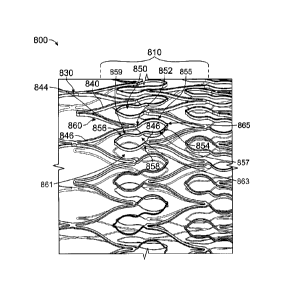
1047] An alternative embodiment of the monolithic device is shown in FIGS. 11A-
11C.
The monolithic device 800 comprises a dense cell pattern 810 and may include
circumferential ring members comprising a first Z pattern 840, a second Z
pattern 842,
and a plurality of looped or generally S-shaped interconnecting members 850
connecting
the first Z pattern 840 and the second Z pattern 842. The proximal and/or
distal end of
the monolithic device 800 may include an end ring 830 in an end Z pattern 832
that is
connected to the first Z pattern 840. The first and second Z patterns 840, 842
include a
plurality of interconnected peaks 844 and troughs 846. As shown in FIG. 11B,
the peak
844 of the first Z pattern 840 is connected to the first end 852 of the looped
or S-shaped
interconnecting member 850, whereby the first end 852 of the looped or S-
shaped
interconnected member 850 forms a generally first loop or first generally
elliptical
section 854 facing the proximal end of the monolithic device 800, while the
first loop or
first generally elliptical section 854 connects to a second loop or second
generally
elliptical section 856 that faces in the opposite direction of the first loop
or first generally
elliptical section 854 and towards the distal end of the monolithic device.
The second
loop or second generally elliptical section 856 ends at the second end 858
that is
connected to the trough 846 of the second Z pattern 842. In one embodiment,
the first
loop or first generally elliptical section 854 fits within the peak 844 of the
second Z
pattern 844, and the second loop or second generally elliptical section 856
fits within the
- 11 -
CA 2905515 2018-10-31
trough 846 of the first Z pattern 840. As shown in FIG. 11C, the end ring 830
includes an
end Z pattern 832, which includes a plurality of interconnected peaks 834 and
troughs
836. The peak 834 of the end Z pattern 832 connects with the trough 846 of the
first Z
pattern 840, and in one embodiment, the peak 834 of the end Z pattern 832
connects with
every other trough 846 of the first Z pattern 840, or every third trough 846
of the first Z
pattern 840. Optionally, the end Z pattern 832 may include additional peaks
834b and
troughs 836b, whereby the peaks 834b are to the troughs 836, as to further
extend the
distal end. A radiopaque layer 860 of Tantalum may be between two layers of
metal for
the monolithic device 800. The Tantalum is the white layer 860 that appears as
a stripe
along the side walls of the stent, as shown in FIG. 11B. Alternatively,
radiopaque layer
860 may comprise another biocompatible radiopaque material.
[047.1] In some embodiments as described above and shown in further detail in
FIGS.
11A-11C, the first generally elliptical section 854 has a major axis generally
parallel to a
longitudinal axis of the intravascular stent device. The first generally
elliptical section
further comprises a first portion 855 connected to a peak of a first
circumferential ring
member at a first end of the major axis and to a second portion 857 at a
second end of the
major axis. The second portion 857 is further coupled to the second generally
elliptical
section 856 proximate to the first end of the major axis. Additionally, the
second
generally elliptical section 856 has a second major axis generally parallel to
a
longitudinal axis of the intravascular stent device and circumferentially off-
set from the
major axis. The second generally elliptical section 856 further comprises a
third portion
859 coupled to the first generally elliptical section 854 proximate a second
end of the
second major axis and further coupled to a fourth portion 861 at a first end
of the second
major axis. The fourth portion 861 is further connected to a peak of the
second
circumferential ring member at the second end of the second major axis.
[047.2] In additional embodiments as described above and shown in further
detail in
FIGS. 11A-11C, the intravascular stent device further comprises a curvilinear
member
863 connecting the second portion 857 of the first generally elliptical
section 854 to the
third portion 859 of the second generally elliptical section 856. The
curvilinear member
.. 863 is oriented generally along a longitudinal axis of the intravascular
stent device.
- 12 -
CA 2905515 2018-10-31
[047.3] In yet additional embodiments as described above and shown in further
detail in
FIGS. 11A-11C, the intravascular stent device further comprises hinge regions
865 at the
junctions of the portions of the generally elliptical sections. For example, a
hinge region
865 interconnects the first portion 855 and the second portion 857 of the
first generally
elliptical section 854 at the second end of the major axis of the first
generally elliptical
section 854 and a second hinge region 865 interconnects the third portion 859
and the
fourth portion 861 of the second generally elliptical section 856 at the first
end of the
major axis connect of the second generally elliptical section 856.
[048] The monolithic device 800 may have a wall thickness between about 0.1-
100.0
microns. The monolithic device 800 may be crimped around a guide wire. The
crimping
may collapse the first Z-pattern, the end ring, and the looped interconnecting
members to
a diameter between about 0.2 and 2.0 mm. After the monolithic device 800 is
uncrimped,
the monolithic device 800 may expand to a diameter between about 2.0 and 7.0
mm while
maintaining adequate radial force and wall apposition. In one embodiment, the
wall
thickness of the monolithic device 800 is less than about 75 microns.
10491 In some embodiments, such as those discussed above in relation to FIGS.
7-11C,
the monolithic device is formed from a material that is a metal, a polymer, a
composite,
or a ceramic material. In some embodiments, materials to make the inventive
stents are
chosen for their biocompatibility, mechanical properties, i.e., tensile
strength, yield
strength, and their ease of deposition include the following: elemental
titanium,
vanadium, aluminum, nickel, tantalum, zirconium, chromium, silver, gold,
silicon,
magnesium, niobium, scandium, platinum, cobalt, palladium, manganese,
molybdenum
and alloys thereof, such as zirconium-titanium-tantalum alloys, nitinol, and
stainless
steel.
10501 In one embodiment, a coating of deposited metal film or polymer is about
0.1 ¨
100.0 microns in a tube form, which is laser cut using ultra short pulsed
femtosecond
laser to minimize heat affected zones and recast. The final monolithic device
may be heat
treated to optimize spring back effects. The stent's one piece construction
allows many
advantages over many currently available braided stent designs, such as a
lower profile,
self-expanding, and ease of manufacturing. Alternatively, the monolithic
device may be
- 13 -
CA 2905515 2018-10-31
produced from drawn metal or polymer tubing, wrought tubing, provided that
fatigue life
is adequate. Radiopaque markers could be added as an interdispersed deposited
layer if
vacuum deposition is used. Different metal layers may be used to form the
monolithic
device.
10511 In some embodiments, the method further comprises the step of patterning
at least
one surface of the monolithic device. In some embodiments, the patterning
comprises
laser patterning to impart at least one feature on the at least one surface of
the monolithic
device. In some embodiments, the pattern is a series of grooves on at least
one surface of
the monolithic device, preferably the surface that will comprise the inner
diameter of the
finished stent. In other embodiments, the pattern may be a plurality of
microgrooves
imparted onto the luminal and/or abluminal surface of the monolithic device.
The
plurality of microgrooves may be formed either as a post-deposition process
step, such as
by etching, or during deposition, such as by depositing the stent-forming
material onto a
mandrel which has a microtopography on the surface thereof which causes the
metal to
deposit with the microgroove pattern as part of the deposited material.
10521 The inventive monolithic devices may be intravascular stents, stent-
grafts, grafts,
heart valves, venous valves, filters, occlusion devices, catheters, sheaths,
osteal implants,
implantable contraceptives, implantable antitumor pellets or rods, shunts and
patches,
pacemakers, needles, temporary fixation rods, medical wires or medical tubes
for any
type of medical device, or other implantable medical devices, as will also be
hereinafter
described. A pacemaker (or artificial pacemaker, so as not to be confused with
the heart's
natural pacemaker) is a medical device that uses electrical impulses,
delivered by
electrodes contacting the heart muscles, to regulate the beating of the heart.
The
electrodes may be covered by tubing or other material that includes a surface
that may
require endothelialization and grooves thereon. Earrings and other piercings
may benefit
from the topographical features, as well as any other implant, whether the
implant is an
organic, inorganic, mechanical, electrical, or biological device.
[053] The monolithic device may be used with any type of cell, which cell has
a cellular
membrane. Most distinct cell types arise from a single totipotent cell that
differentiates
into hundreds of different cell types during the course of development.
Multicellular
- 14 -
CA 2905515 2018-10-31
=
organisms are composed of cells that fall into two fundamental types: germ
cells and
somatic cells. During development, somatic cells will become more specialized
and form
the three primary germ layers: ectoderm, mesoderm, and endoderm. After
formation of
the three germ layers, cells will continue to specialize until they reach a
terminally
differentiated state that is much more resistant to changes in cell type than
its progenitors.
The ectoderm differentiates to form the nervous system (spine, peripheral
nerves and
brain), tooth enamel and the epidermis (the outer part of integument). It also
forms the
lining of mouth, anus, nostrils, sweat glands, hair and nails. The endoderm
forms the
gastrointestinal tract cells, the respiratory tract cells, the endocrine
glands and organ cells,
the auditory system cells, and the urinary system cells. The mesoderm forms
mesenchyme (connective tissue), mesothelium, non-epithelial blood cells and
coelomocytes. Mesothelium lines coeloms; forms the muscles, septa (cross-wise
partitions) and mesenteries (length-wise partitions); and forms part of the
gonads (the rest
being the gametes).
10541 In one embodiment, the apparatus comprises: an ultra-dense stent cell
pattern
including a plurality of structural members that diverts the majority of blood
flow without
restricting blood flow completely.
10551 While the invention has been described in connection with various
embodiments,
it will be understood that the invention is capable of further modifications.
This
application is intended to cover any variations, uses or adaptations of the
invention
following, in general, the principles of the invention, and including such
departures from
the present disclosure as, within the known and customary practice within the
art to
which the invention pertains.
- 15 -
CA 2905515 2018-10-31