Note: Descriptions are shown in the official language in which they were submitted.
WO 2014/140926
PCT/IB2014/001235
SYSTEMS, METHODS, AND COMPUTER-READABLE MEDIA FOR
IDENTIFYING WHEN A SUBJECT IS LIKELY TO BE AFFECTED BY A
MEDICAL CONDITION
Technical Field.
[0001] The present disclosure relates to the field of image analysis. For
example, systems,
methods, and computer-readable media are disclosed for identifying when a
subject is likely to be
affected by a medical condition using image analysis.
Background
[0002] There are thousands of known rare diseases that collectively affect
more than 8% of
the world's population, Rare diseases are often chronic, progressive,
degenerative, and life threatening.
Children affected by rare diseases often suffer from many associated medical
complications and need
critical and timely medical intervention.
[0003] Many rare diseases are genetic in origin, inborn, and exhibit
symptomatic
malformations. Symptomatic malformations are often the first sign of a rare
disease. A
dysmorphic evaluation performed by a qualified specialist often plays a key
factor in
recognizing a disease. But due to the rarity of many diseases, the scarcity of
dysmorphology
experts, and the complexity of a clinical diagnosis, is it often not possible
to provide proper
and comprehensive dysmorphology training to a large number of physicians.
[0004] The diagnosis of rare diseases is often very difficult, particularly
for physicians
that lack the relevant awareness, knowledge, and experience. Most children
that do reach a
diagnosis are typically diagnosed later in life when physical symptoms,
developmental delay,
intellectual disability, and other medical complications are observed by their
families or
treating physician. This can result in an unmanaged and untreated disease that
can cause a
child's condition to deteriorate,
[0005] Early identification of diseases is often critical. Accordingly,
systems and
methods are needed that can efficiently and noninvasively determine whether a
person is likely
to be affected by a medical condition.
1
23592617.1
CA 2905637 2019-03-04
CA 02905637 2015-09-11
WO 2014/140926 PCT/1132014/001235
SUMMARY
[0006] Embodiments consistent with the present disclosure provide systems,
methods, and computer-readable media for identifying when a subject is likely
to be affected
by a medical condition using image analysis,
[0007] In one disclosed embodiment, a system for identifying when a subject is
likely to be affected by a medical condition is disclosed. The system includes
at least one
processor that is configured to receive information reflective of an external
soft tissue image
of the subject, perform a first evaluation of the external soft tissue image
information using at
least one of an anchored cells analysis, a shifting patches analysis and a
relative
measurements analysis, generate first evaluation result information based, at
least in part, on
the first evaluation, perform a second evaluation of the external soil tissue
image information
using at least one of the anchored cells analysis, the shifting patches
analysis, and the relative
measurements analysis, generate second evaluation result intbrmation based, at
least in part,
on the second evaluation, and predict a likelihood that the subject is
affected by the medical
condition based, at least in part, on the first evaluation result information
and the second
evaluation result information.
[0008] in another disclosed embodiment, a system for identifying when a
subject is
likely to be affected by a medical condition is disclosed. The system includes
at least one
processor that is configured to receive information reflective of an external
soft tissue image
of the subject, divide the external soft tissue image information into a
plurality of regions,
generate an analysis of each of the plurality of regions, aggregate the
analyses of the plurality
of regions, and determine a likelihood that the subject is affected by the
medical condition
based on the aggregated analyses.
[0009] In another disclosed embodiment, a system for identifying when a
subject is
likely to be affected by a medical condition is disclosed. The system includes
at least one
processor that is configured to receive information reflective of an external
soft tissue image
of the subject, use image information analysis to compare the external soft
tissue image
information with a plurality of external soft tissue images of other subjects
in a database,
determine, based on the image information analysis, dysmorphic features
included in the
external soft tissue image information, access descriptors associated with the
dysmorphic
features, and output at least some of the descriptors.
[0010] In another disclosed embodiment, a system for identifying when a
subject is
likely to be affected by a medical condition is disclosed. The system includes
at least one
2
CA 02905637 2015-09-11
WO 2014/140926 PCT/1112014/001235
processor that is configured to receive information reflective of an external
soft tissue image
of the subject, analyze the external soft tissue image information, identify
one or more
external soft tissue attributes in the external soft tissue image information
based, at least in
part, on the analysis, access at least one database of external soft tissue
attributes associated
with a plurality of medical conditions, compare the one or more identified
external soft tissue
attributes with the external soft tissue attributes of the at least one
database, and output
information about at least one medical condition likely possessed by the
subject based on the
corn pan i son.
[0011] In another disclosed embodiment, a system for identifying when a
subject is
likely to be affected by a medical condition is disclosed. The system includes
at least one
processor that is configured to receive first information reflective of a
first external soft tissue
image of the subject recorded at a first time, analyze the first image
information, receive
second information reflective of a second external soft tissue image of the
subject recorded at
a second time, analyze the second image information, compare the analysis or
the first image
information with the analysis of the second image information, and predict a
likelihood that
the subject is affected by the medical condition based, at least in part, on
the comparison.
[0012] Additional aspects related to the disclosed embodiments will be set
forth in
part in the description which follows, and in part will be understood from the
description, or
may be learned by practice of the disclosed embodiments.
[0013] It is to be understood that both the foregoing general description and
the
following detailed description are exemplary and explanatory only and are not
restrictive of
the claims.
BRIEF DESCRIPTION OF THE DRAWINGS
[0014] The accompanying drawings, which are incorporated in and constitute a
part
of this disclosure, illustrate various disclosed embodiments, In the drawings:
[0015] Figure 1 illustrates art example system for identifying when 8 subject
is
likely to he affected by a medical condition that may be used for implementing
the disclosed
embodiments.
[0016] Figure 2 illustrates example operations that a processor of a medical
condition analysis system may be configured to perform to predict a likelihood
that a subject
is affected by a medical condition using two evaluations, in accordance with
some of the
disclosed embodiments.
3
CA 02905637 2015-09-11
WO 2014/140926 PCT/1B2014/001235
[0017] Figure 3 illustrates example operations that a processor of a medical
condition analysis system may be configured to perform to predict a likelihood
that a subject
is affected by a medical condition using an image division, in accordance with
some of the
disclosed embodiments.
[0018] Figure 4 illustrates example operations that a processor of a medical
condition analysis system may be configured to perform to predict a likelihood
that a subject
is affected by a medical condition using information of at least one relative
of the subject, in
accordance with some of the disclosed embodiments.
[0019] Figure 5 illustrates example operations that a processor of a medical
condition analysis system may be configured to perform to output at least some
descriptors
associated with dysmorphic features, in accordance with some of the disclosed
embodiments.
[00201 Figure 6 illustrates example operations that a processor of a medical
condition analysis system may be configured to perform to predict a likelihood
that a subject
is affected by a medical condition using at least one hundred defined
locations on an image,
in accordance with some of the disclosed embodiments,
[0021] Figure 7 illustrates example operations that a processor of a medical
condition analysis system may be configured to perform to superimpose
indicates of at least
one dysmorphology on an image, in accordance with some of the disclosed
embodiments.
[0022] Figure 8 illustrates example operations that a processor of a medical
condition analysis system may be. configured to perform to identify
information about
dysmorphie features in a selected region, in accordance with some of the
disclosed
embodiments.
[0023] Figure 9 illustrates example operations that a processor of a medical
condition analysis system may be configured to perform to output information
about at least
one medical condition likely possessed by a subject, in accordance with some
of the disclosed
embodiments.
[0024] Figure 10 illustrates example operations that a processor of a medical
condition analysis system may be configured to perform to predict a likelihood
that a subject
is affected by a medical condition based on analyses at two different times,
in accordance
with some of the disclosed embodiments.
[0025] Figure 11 illustrates example operations that a processor of a medical
condition analysis system may be configured to perform to determine a
previously
unrecognized medical condition likely possessed by two subjects, in accordance
with some of
the disclosed embodiments,
4
CA 02905637 2015-09-11
WO 2014/140926 PCT/IB2014/001235
[0026] Figure 12 illustrates example operations that a processor of a medical
condition analysis system may be configured to perform to mediate
communications between
a health service provider and a healthcare professional, in accordance with
some of the
disclosed embodiments.
100271 Figure 13 illustrates example operations that a processor of a medical
condition analysis system may be configured to perform to alert a healthcare
provider when
an image of a subject meets a threshold of being likely to be affected by a
medical condition,
in accordance with some of the disclosed embodiments.
[0028] Figure 14 illustrates example operations that a processor of a medical
condition analysis system may be configured to perform to predict whether a
subject has a
medical condition, in accordance with some of the disclosed embodiments.
[0029] Figure 15 illustrates example operations that a processor of a medical
condition analysis system may be configured to perform to generate a list of
tests to be
performed, in accordance with sonic of the disclosed embodiments.
[0030] Figure 16 illustrates example operations that a processor of a medical
condition analysis system may be configured to perform to predict whether a
dysmorphic
feature is indicative of a medical condition based on a severity score, in
accordance with
some of the disclosed embodiments.
[0031] Figure 17 illustrates example operations that a processor of a medical
condition analysis system may be configured to perform to predict whether a
subject is likely
to be affected by a medical condition by discounting at least one dysmorphic
feature, in
accordance with some of the disclosed embodiments.
[0032] Figure 18 illustrates exemplary depictions of an image processing
pipeline
in accordance with some of the disclosed embodiments.
[0033] Figures 19-22 illustrate exemplary depictions of image segmentation in
accordance with some of the disclosed embodiments.
[0034] Figures 23A-23C illustrate exemplary depictions of an anchored cell
analysis in accordance with sonic of the disclosed embodiments.
[0035] Figure 24 illustrates exemplary depictions of a shifting patch analysis
in
accordance with some of the disclosed embodiments.
[0036] Figure 25 illustrates exemplary depictions of a relative measurements
analysis in accordance with some of the disclosed embodiments.
[0037] Figure 26 illustrates exemplary depictions of a plurality of analyses
in
accordance with some of the disclosed embodiments.
CA 02905637 2015-09-11
WO 2014/140926 PCT/1B2014/001235
[0038] Figure 27 illustrates exemplary depictions of an ear analysis in
accordance
with some of the disclosed embodiments.
[0039] Figure 28 illustrates exemplary depictions of an undiagnosed patient
analysis in accordance with some of the disclosed embodiments.
DETAILED DESCRIPTION
[0040] Reference will now be made in detail to the example embodiments, which
are illustrated in the accompanying drawings. Wherever possible, the same
reference
numbers will be used throughout the drawings to refer to the same or like
parts.
[0041] Figure 1 is a diagram illustrating an example system 100 for
identifying
when a subject is likely to be affected by a medical condition, consistent
with the disclosed
embodiments. A subject may include, among other things, any person or type of
person, such
as a male or female person and a child or adult. A child may include, for
example, a neonate,
an infant, a toddler, a preschooler, a school age child, or an adolescent. For
example, a male
or female person from birth to 1 month old may be referred to as a neonate,
from I month to
1 year old may be referred to as an infant, from 1 year to 3 years old may be
referred to as a
toddler, from 3 years to 6 years old may be referred to as a preschooler, from
6 years to 12.
years old may be referred to as a school age child, and from 12 years to 18
years old may be
referred to as an adolescent, An adult may include, for example, a male or
female person
from 18 years old and onwards. These age ranges, however, are exemplary only.
For
example, a 19 year old person may be referred to as an adolescent in certain
contexts.
[0042] A medical condition may include, among other things, any medical
disease.
A subject possessing a medical condition may include, for example, at least
one of possessing
a genetic syndrome and being a carrier of a genetic syndrome. A medical
condition may also
include, among other things, any association of clinically recognizable,
features, signs,
symptoms, phenomena, or other characteristics that often occur together, such
that the
presence of one feature, sign, symptom, phenomena, or other characteristic may
imply,
indicate, or alert to the possible presence of the others. A medical condition
may also include
one or more abnormal findings in physical growth and development over time
(e.g., growth
deficiencies and craniofaciai deformations that develop over time). For
example, a medical
condition may be one or more of the medical conditions disclosed in "Gorlin's
syndromes of
the head and neck," 2010. Oxford University Press, to RCM. H.ennekam et al,
"The Bedside
Dysmorphologist," 2008, Oxtbrd University Press, to William Reardon, and
"Smith's
0
CA 2,905,637
Blakes Ref 12665/00001
Recognizable Patterns of Human Malformation," 2005, WB Saunders, to Kenneth
Lyons Jones.
[0043] In some embodiments, a medical condition includes one or more
conditions that may
cause a person to exhibit one or more dysmorphic features. A dysmorphic
feature may include, for
example, any feature that affects the appearance of a subject. A dysmorphic
feature may, for example,
reflect an external soft tissue dysmorphology. For example, a medical
condition may cause a child's
skull to form in an irregular manner, which may cause the child's facial
appearance to also be irregular
in a manner that may be described by one or more dysmorphic features. For
example, a dysmorphic
feature may be one or more of the dysmorphic features disclosed in "Elements
of morphology:
Introduction," 2009, Am J Med Genet Part A 149A:2-5, to Allanson et al.,
"Elements morphology:
Standard of terminology for the head and face," 2009, Am J Med Genet Part A
149A:6-28, to Allanson
et al., "Elements of morphology: Standard terminology for the lips, mouth, and
oral region," 2009, Am
J Med Genet Part A 149A:77-92, to Carey et al., "Elements of morphology:
Standard Terminology for
the periorbital region," 2009, Am J Med Genet Part A 149A:29-39, to Hall et
al., "Elements of
morphology: Standard terminology for the Nose and philtrum," 2009, Am J Med
Genet Part A
149A:61-76, to Hennekam et al., "Elements of morphology: Standard terminology
for the ear," 2009,
Am .1 Med Genet Part A 149A:40-60, to Hunter et al., and "Elements of
morphology: Standard
terminology for the hands and feet," 2009, Am J Med Genet. Part A 149A:93-127,
to Biesecker et at.
[0044] System 100 may include, among other things, at least one processor 110,
at least one
memory device 120, at least one input device 130, at least one camera 140, and
at least one output
device 150. Processor 110 may include any electrical circuit that may he
configured to perform an
operation on at least one input variable, including, for example one or more
integrated circuits,
microchips, microcontrollers, and microprocessors, which may be all or part of
a central processing
unit (CPU), a digital signal processor (DSP), a field programmable gate array
(FPGA), a graphical
processing unit (OPU), or any other circuit known to those skilled in the art
that may be suitable for
executing instructions or performing logic operations. Multiple functions may
be accomplished using
a single processor or multiple related and/or unrelated functions may be
divided among multiple
processors. Processor 110 may be configured to access memory device 120, which
may include, for
example, persistent memory, ROM, EEPROM, EAROM, flash memory devices, magnetic
disks,
magneto optical disks, CD-ROM, DVD-ROM, Blu-ray, and the like. Memory device
7
23648050.1
CA 2905637 2019-08-28
CA 02905637 2015-09-11
WO 2014/140926 PCT/1B2014/001235
120 may contain instructions (i.e., software or firmware) or other data,
Processor 110 may
receive instructions and data stored memory device 120. Thus, in some
embodiments,
processor 110 may execute the software or firmware to perform functions by
operating on
input data and generating output. However, processor 110 may also receive or
access data
stored remotely over a network (not depicted in Figure I), For example, device
100 may
include a communication device (not depicted in Figure 1) that enables
processor 110 to
receive or access data stored remotely on a server or user device over a
network. Moreover,
processor 110 may also be, for example, dedicated hardware or an application
specific
integrated circuit (ASIC) that performs processes by operating on input data
and generating
output. Processor 110 may be any combination of dedicated hardware, one or
more ASICs,
one or more general purpose processors, one or more DSPs, one or more GPUs, or
one or
more other processors capable of processing digital information. For example,
in some
embodiments, processor 110 may comprise multiple processors that may provide
parallel
processing capabilities,
[0045] Figure 2 illustrates an exemplary process 200 that at least one
processor may
be configured to perform. For example, as discussed above, processor 110 may
be
configured to perform process 200 by executing software or firmware stored in
memory
device 120, or may be configured to perform process 200 using dedicated
hardware or one or
more ASICs.
[0046] Processor 110 may be configured to receive information reflective of an
external soft tissue image of the subject (step 210). For example, processor
110 may receive
information reflective of an external soft tissue image of a subject captured
by camera 140.
Camera 140 may include, among other things, one or more image sensors, such as
a CCD
image sensor, a CMOS image sensor, a camera, a light sensor, an IR sensor, an
ultrasonic
sensor, a proximity sensor, a shortwave infrared (SWIR) image sensor, a
reflectivity sensor,
or any other image sensor that is capable of capturing an external soft tissue
image. An
image sensor may be configured to capture any quantity of image data, such as
single pixel
data, one-dimensional line data, two-dimensional data, or three-dimensional
data. Camera
140 may be a fixed camera, mobile camera, or any other image capturing device
or
equipment, which may, for example, be further incorporated into a computer, a
tablet, a
phone, glasses, or any other device.
[0047] An external soft tissue image may include, among other things, an.
image of a
subject or any portion of a subject. In some embodiments, the external soft
tissue image may
include an image of at least one of a face of the subject, a cranium of the
subject, a hand of
CA 02905637 2015-09-11
WO 2014/140926 PCT/1132014/001235
the subject, and a foot of the subject. However, the external soft tissue
image may also
include other portions of the subject, such as, such as a hairline, forehead,
ocular region,
eyebrow, nose, eye, mid-face region, philtrum region, mouth, ear, mandibular
region, chin,
cheek, neck, chest, mid-body, back, torso, hips, genitalia, limbs, joints,
hands, and fingers. in
some embodiments, the external soft tissue image is a cranio-ilicial image
that includes at
least one of a frontal view, a lateral view, an angled view, a top view, and a
back view. As
used herein, a crania-facial image is an image that includes at least a
portion of a cranium or
face of the subject. A frontal view may include an image of the front of the
face of the
subject. A lateral view may include an image taken at or approximately at a 45
degree angle
(to the left and/or right, side of the face) from the vertical midline of the
head of the subject.
A top view may include an image of the top of the head of the subject. A back
view may
include an image of the back of the head of the subject. As described in more
detail below, in
some embodiments the external soft tissue image is associated with a
dysinorphology.
[0048] The information reflective of an external soft tissue image received by
processor 110 may include the external soft tissue image itself or any data
derived from the
external soft tissue image (e.g., a separate processor at camera 140 may
derive data from the
external soft tissue image and transmit the derived data to processor 110),
For example, if the
external soft tissue image is an analog image (although the external soft
tissue image may be
captured as a digital image), information. reflective of an external soft
tissue image may
include a digitally converted version of the external soft tissue image. The
information
reflective of an external soft tissue image may be, for example, a vector
image or a raster
image. The information reflective of an external soft tissue image may also be
non-image
data, such as a set of parameters derived from the external soft tissue image,
which may
include, thr example, one or more intensities of the image, one or more
locations of edges in
the image, and one or more textures in the image.
[0049] In some embodiments, processor 110 may preprocess the external soft
tissue
image information as it receives the information and/or after it receives the
information.
Figure 18 depicts one example of a preprocessing routine that may be performed
by
processor 110. As depicted in Figure 18, as part of a preprocessing routine,
processor 110
may be configured to detect a face region of the external soft tissue image
information, detect
a number of points in the face region, and align the face region, One example
of a face
detection routine is shown in Figures 19-22. For example, as graphically
depicted in Figure
20, processor 110 may be configured to detect a face region by first placing a
plurality of
patches (i.e., sub-regions of the image information), which optionally may
overlap one
9
CA 02905637 2015-09-11
WO 2014/140926 PCT/IB2014/001235
another, over the image information. For each patch, a descriptor vector may
be computed.
A descriptor vector may include, for example, data derived from at least one
of a scale-
invariant feature transform (SIFT), a histogram of oriented gradients (HOG), a
self-similarity
descriptor, a histogram of Local Binary Patterns, and any other determinable
feature known
in the image analysis and computer vision fields.
[0050] During a training phase for the face detection routine, one or more
regions of
a set of training images may be manually outlined For
example, processor 110 may
determine an outline of a head and one or more regions within a face or side
of a face (e.g.,
an outline of eyes, nose, mouth, ears, etc.). Processor 110 may determine a
center of mass of
the outline of the head (e.g., a point where the weighted relative position of
the points on the
outline sums to zero). Processor 110 may further determine a descriptor vector
for each patch
of each training image and store the descriptor vectors in a. database. The
database may be
stored by memory device 120, or may be stored on, for example, a remote server
that is
accessible to processor 110 over a network. Moreover, information regarding
the location of
the center of mass of the head shape relative to the center of each patch
associated with a
descriptor vector may also be stored in the database. Figure 19 depicts an
example of an
outlined head shape with information regarding the location of the center of
mass of the head
shape relative to the center of a plurality of patches.
[0051] Further regarding the face detection routine, processor 110 may be
configured to compare the descriptor vector of each patch in the image
information of the
subject to descriptor vectors associated with patches of training images to
determine a set of
the most similar descriptor vectors (e.g., the 25 most similar descriptor
vectors). To perform
the comparison, processor 110 may be configured to retrieve the descriptor
vector of each
patch in the image information from memory device 120 and/or from a remote
source over a
network (e.g., a server or user device), and may be configured to retrieve the
descriptor
vectors associated with patches of training images from MCMOry device 120
and/or from a
remote source over a network (e.g., a server or user device). Processor 110
may be
configured to calculate one or more of a Euclidean distance, a Chebyshev
distance, a chi-
square distance, and a Mahalanobis distance between the descriptor vector of
each patch in
=
the image information of the subject and the descriptor vectors associated
with patches of
training images. The set of the most similar descriptor vectors may include,
for example,
those descriptor vectors that are associated with a shortest Euclidean
distance, Chebyshev
distance, chi-square distance, or Mahalanobis distance. The patches associated
with the set of
the most similar descriptor vectors may be retrieved from the database. Each
patch retrieved
CA 02905637 2015-09-11
WO 2014/140926 PCT/1B2014/001235
from the database may provide one vote for a location of the center of mass of
the image
information of the subject. A vote may be determined by adding a relative
location of the
center of mass in the, training image associated with a given patch to the
location of the patch
in the image information used to retrieve the patch from the database. A vote,
as used in this
context, refers to one estimate for a location of the center of mass of the
image information of
the subject. All votes from all patches of the image information may be
integrated and the
location with the most support may be selected as the center of mass of the
head. Figure 20
graphically depicts an example of the described voting operations.
[0052] In some embodiments, for each patch in the image information of the
subject, patches in the set of retrieved patches that point to the selected
center of mass
location and are within a threshold distance from the selected center of mass
may he
discarded. The rest. are assigned a score that is proportional to two
elements: the similarity
between the retrieving patch descriptor vector and the retrieved patch
descriptor vector, and a
distance between the selected center of mass and the center of mass implied by
.the retrieved
patch. For each retrieving patch (i.e., a patch in the image information of
the subject), the
scores of the retrieved patches may be accumulated, A threshold filter may be
applied to the
accumulated score of each of the retrieving patches to obtain an initial rough
estimate of a
foreground region of the head shape in the image information. Processor 110
may be
configured to apply one or more morphological operations on the initial rough
estimate to
produce a closed shape. As graphically depicted, for example, in Figure 21,
the contour of
the closed shape may serve as a first segmentation hypothesis. In some
embodiments,
processor I 10 may also be configured to apply a mean shin segmentation
algorithm and/or a
GrabCut segmentation algorithm to the image information using the first
segmentation
hypothesis as a starting position for the computation.
[0053] in some embodiments, processor 110 may use the contour described above
as the detected head or face region. However, in some embodiments, the
determination is
further refined. For example, in some embodiments a contour of each training
image may be
represented by a vector containing one or more for the following: (i) the (x,
y) coordinates of
a number of points (e.g., 50) sampled along the contour at equal distance,
wherein the first
point is taken, for example, to be the top most point along the contour; (ii)
the location of the
first point as well as the differences in (x, y) coordinates between every
point and the next
point (e.g., a number of pairs of (dx, dy) summing up to zero); (iii) the
distance of each such
contour point from the center of the mass of the contour's captured area and
the angle of the
ray from the center of mass to a contour point; and (iv) the distances from
the center of mass
ii
CA 02905637 2015-09-11
WO 2014/140926 PCT/1B2014/001235
to the points on the contour. To refine the estimated contour of the image
information,
processor 110 may employ a Principal Components Analysis to compute the
Principal
Components of the training vectors. The estimated contour may be refined by
projecting it
onto the space of the Principal Components.
[00541 Processor 110 may identify regions in which one or more of the
previously
determined contours are consistent. For example, densely sampled rays may be
projected
from the center of mass in all directions, As graphically depicted, for
example, in Figure 22,
each ray may intersect the various contours. Processor 110 may be configured
to compute
the mean and standard deviation of the obtained intersection locations for
each ray. If the
standard deviation is below a threshold (which may mean that the intersections
are nearby
and the contours are consistent along the ray), the mean point may be used as
a high
confidence location for a contour. High confidence locations from a plurality
of rays may be
grouped into high confidence segments, which may produce a contour that has
missing parts
and multiple sub-segments. The missing segments may be reconstructed by
examining the
head shapes in the training images, selecting the head shape that is most
consistent with the
high confidence segments, and copying the values from the selected head shape
into the
missing segments.
100551 To detect a number of points in the face region of the image
information,
processor 110 may be configured to perform a similar voting teehnique to that
described
above. For example, while the contour described above is with respect to a
head shape or
face shape, the same operations may be performed for any other definable
contour in the
image information of the subject. Processor 110 may be configured, for
example, to select
points on one or more contours of the image information (e.g., points
surrounding the face,
eyes, eyebrows, nose, mouth, and/or chin of the subject). For example,
processor 110 may he
configured to select a number of evenly spaced points along a given contour.
[0056] To align the face region, processor 110 may be configured to perform
one or
more of a translation, rotation, and scaling operation such that the resulting
face is maximally
aligned with an average face model. In some embodiments, regions of interest
may be
determined based on their known association to corresponding facial regions
determined
based on the detected points. The regions of interest may then be aligned to
the average face
model.
[0057] Processor 110 may be configured to perform a first evaluation of the
external
soft tissue image information using at least one of an anchored cell analysis,
a shifting
patches analysis, and a relative measurements analysis (step 220): To perform
the anchored
12
CA 02905637 2015-09-11
WO 2014/140926 PCTAB2014/001235
cell analysis, processor 110 may be configured to overlay a grid with a
plurality or cells on
the external sort tissue information, calculate descriptors for each of the
plurality of cells,
aggregate the descriptors to produce a vector, and compare the vector to
previously produced
vectors from external soft tissue images or other individuals previously
diagnosed with the
medical condition.
[005N Overlaying the grid of cells may include a number of different
complementary and/or alternative options. For example, as depicted in Figure
23A,
processor 110 may be configured to overlay a fixed grid with a plurality of
cells on the
external soft tissue image information, A fixed grid of cells may include, for
example, a
Plurality of adjacent squares, rectangles, or triangles that are overlaid on a
region (e.g., a face
region) or the external soil tissue image information. As another example, as
depicted in
Figure 23B, processor 110 may be configured to overlay a small grid of cells,
which may be
smaller in size than the cells of the fixed grid discussed above, on a
particular defined region,
such as at least one of a forehead region, a periorbital region, a nasal
region, a mid-face
region, an ear region, and an oral region of the external soil tissue image
information;
processor 110 may be configured to discount at least one other region of the
external soft
tissue image information (e.g., a. region from which minimal, or no, relevant
information can
be obtained, such as a hair region, may not be overlaid with a small grid of
cells). As another
example, as depicted in Figure 23C, processor 110 may also be configured to
overlay a
plurality of triangle cells generated by connecting points detected on the
external soft tissue
image information. For example, processor 110 may determine a plurality of
feature points
on the image information and connect the feature points to form triangular
regions.
[0059] To calculate descriptors for each of the plurality of cells, processor
110 may
be configured, for example, to analyze at least one of an intensity, a
texture, and an edge
associated with the external soft tissue image information. In some
embodiments, the
descriptor for each cell is a vector that includes, for example, data derived
from at least one
of a SIFT. HOG, a self-similarity descriptor, a histogram of Local Binary
Patterns, and any
other type or feature used in image analysis and computer vision.
[0060] To aggregate the descriptors to produce a vector, processor 110 may be
configured, for example, to create a vector that includes one or more of the
calculated
descriptors. For example, each of the descriptors associated with a cell in a
fixed grid may be
aggregated into a vector, each of the descriptors associated with a set of
triangle cells may be
=
aggregated into a vector, or each of the descriptors associated with a small
grid of cells may
he aggregated into a vector. Additionally or alternatively, if mere than one
set of cells is
13
CA 02905637 2015-09-11
WO 2014/140926 PCT/IB2014/001235
created (e.g., both a fixed grid and a set of triangle cells are formed, both
a fixed grid and a
small grid of cells are formed, both a set of triangle cells and a fixed grid
are formed, or a
fixed grid, set of triangle cells, and small grid are all formed), a single
vector may be created
that includes the descriptors of multiple sets of cells. The vector that
includes the aggregated
descriptors may be referred to as, for example, a combined appearance vector.
[00611 To compare the vector to previously produced vectors from external soft
tissue images of other individuals previously diagnosed with the medical
condition, processor
110 may be configured, for example, to access a database. The database may
include, for
example, vectors produced using an anchored cell analysis of external soft
tissue images of
other individuals. The database may be annotated by one or more of patient ID,
age, age
group, gender, ethnic group, race group, dysinorphic feature, phenotypic
feature,
anthropornetrie measurements (including without limitation, height, weight,
head
circumference, facial height, skull height, upper facial height, lower facial
height, head
length, head width, facial width, mandibular width, anterior fontanelle size,
posterior
fontanelle size, inner canthal distance, outer candial distance,
interpupillary distarux,
interorbital distance, palpebral fissure length, palpebral fissure height,
obliquity of the
palpebral fissure, orbital protrusion, corneal dimensions, ear length, ear
width, ear protrusion,
ear position, ear rotation, nasal height, length of the columella, nasal
protrusion, nasal width,
philtrum len2th, philtrum width, mouth width, ONC angle, maxillomandibular
differential,
and mandible width), relative ratios and proportions between bodily and facial
landmarks,
known diagnosis,, suspected diagnosis, mutations and/or genetic variants,
source of image,
informed consent, pose, illumination, quality of image, expression type, and
association with
cohort (e.g., part of a control group of individuals known not to be affected
by a medical
condition or part of a group of individuals known to be affected by a medical
condition). The
database may also be annotated by, for example, linking data regarding
individuals in the
database that are family members (e.g., siblings, parents, children, cousins,
etc.) and/or
indicating the relationship of other family members in the database that are
affected by a
medical condition or dysmorphic ftature to an individual (e.g., sister of
grandmother from the
mother's side). In some embodiments, the previously produced vectors used in
the
comparison are associated with one or more annotations that are in common. For
example,
the previously produced vectors may be associated with an annotation
indicating that they
were derived from images associated with individuals of the same age, gender,
and ethnicity
as the subject. Additionally or alternatively, previously produced vectors
used in the
comparison may be associated with a suspected dysmorphie feature and/or a
suspected
14
CA 02905637 2015-09-11
WO 2014/140926 PCT/1B2014/001235
medical condition of the subject, That is, for example, the previously
produced vectors may
be associated with one or more of individuals affected by a dysmorphic
feature, individuals in
a control group for the dysmorphic feature, individuals affected by a medical
condition, and
individuals in a control group for the medical condition.
[0062] Data associated with a set of the most similar previously produced
vectors
(e.g., the 25 most similar previously produced vectors) may be determined. For
example,
processor 110 may be configured to calculate one or more of a Euclidean
distance, a
Chebyshev distance, a chi-square distance, and a Mahalanobis distance between
the
combined appearance vector and the previously produced vectors in the database
to
determine a set of the most similar previously produced vectors (e.g., the 25
previously
produced vectors associated with the 25 shortest computed distances may be
selected).
[0063] The set of the most similar previously produced vectors may be analyzed
(at,
for example, a server associated with the database or by processor 110) to
determine how
many of the previously produced vectors are associated with a positive example
of a
particular dysmorphic feature (that is, a previously produced vector
associated with an
individual known to have the dysmorphic feature) and how many of the
previously produced
vectors are associated with a negative example of a particular dysmorphic
feature (that is, a
previously produced vector associated with an individual known to not have the
dysmorphic
feature). Based on the number of positive examples and the number of negative
examples, a
probability score may be determined for the dysmorphic feature. A probability
score, as used
herein, may be an actual probability or some value that is reflective of a
probability. For
example, a probability score may provide some indication of the likelihood
that a subject has
dysmorphic feature. For example, if only positive examples are included in the
set of most
similar previously presented vectors, a very high or maximum probability score
may be
determined (e.g., a probability score of 100). If only negative examples are
included, then a
very low probability score may he determined (e.g., a probability score of I).
If a mixture of
positive and negative examples is included, then the probability score may
reflect the number
of positive examples and the number of negative examples. The probability
score may or
may not be directly proportional to the number of positive and negative
examples. For
example, in some embodiments, if a threshold number of positive examples are
obtained,
then the same very high or maximum probability score may he determined
regardless of the
number of positive examples or negative examples. Moreover, a probability
score is not
necessarily a positive value. In some embodiments, the probability score may
be a negative
score. Moreover, all of the probability scoresi do not necessarily add up to
100%. In some
CA 02905637 2015-09-11
WO 2014/140926 PCT/1B2014/001235
embodiments, a probability score may be any real valued score that is
approximately
monotonic with respect to the underlying probability of a certain medical
condition or
dysmorphology.
[0064] In some embodiments, a probability score for the dysmorphic feature may
also, or alternatively, be calculated based on a degree of similarity of the
combined
appearance vector to a given one of the previously produced vectors. For
example, if an
equal number of positive and negative examples are retrieved, but the combined
appearance
vector is more similar to the previously produced vectors associated with
positive examples
than the previously produced vectors associated with negative examples, then
the probability
score may be relatively high. In contrast, if an equal number of positive and
negative
examples are retrieved, but the combined appearance vector is more similar to
the previously
produced vectors associated with negative examples than the previously
produced vectors
associated with positive examples, then the probability score may be
relatively low.
[0065] In some embodiments, processor 110 may calculate more than one
probability score for a given dysmorphic feature. For example, one probability
score may be
determined by treating all positive and negative examples equally, and another
probability
score may be determined that considers the similarity of the combined
appearance vector to
the positive and negative samples. Moreover, probability scores for a
plurality of different
dysmorphic features may be calculated in the same or substantially the same
way.
[0066] While the above anchored cell analysis description refers to dysmorphic
features, the same process may also, or alternatively, be performed to
determine probability
scores for one or more medical conditions. For example, rather than
determining the
association of previously presented vectors to dysmorphic features, processor
110 may
determine which medical conditions are associated with the previously
presented vectors.
[0067] To perform the shifting patches analysis, processor 110 may be
configured to
overlay a plurality of densely spaced or overlapping patches on the external
soft tissue image
information, calculate a descriptor vector for each of the plurality of
patches, and compare
each descriptor vector to previously produced vectors from a similar region in
external soft
tissue images of other individuals previously determined to be affected by the
medical
condition.
[0068] As depicted in Figure 24, to overlay patches on the external soft
tissue
image information, processor 110 may be configured, for example, to overlay
multiple
densely spaced or overlapping patches, which optionally may be of varying
sizes, onto a
region of the image inthrmation (e.g., a face reeion). For example, a square
patch of a first
16
CA 02905637 2015-09-11
WO 2014/140926 PCT/1B2014/001235
size may he overlaid on the region of the image information at every possible
position (e.g.,
the square patch may be shifted by one pixel in every direction until the
patch is overlaid in
every possible position) or at a subset of the possible positions (e.g,, the
square patch may be
shifted by ten pixels in every direction in every direction until the patch is
overlaid over the
entire region of the image. information). In some embodiments, a square patch
of one or more
different sizes may also he overlaid on the region of the image information.
[00691 To calculate a descriptor vector for each of the plurality of patches,
processor
110 may be configured to compute, for example, data derived from at least one
of a scale-
invariant feature transform (SIFT), a histogram of oriented gradients (HOC), a
self-similarity
descriptor, a histogram of Local Binary Patterns, and any other type of
feature used in image
analysis and computer vision.
[0070] To compare each descriptor vector to previously produced vectors from a
similar region in external soft tissue images of other individuals previously
determined to be
affected by the medical condition, processor 110 may be configured to access a
database. For
example, the same database discussed above with respect to the anchored cell
analysis, or a
similar database, may include previously produced vectors for patches of
images of
individuals previously determined to he affected by the medical conditionõks
described
above, the database may annotate the previously produced vectors with a
variety of data such
as, for example, one or more of patient ID, age, age group, gender, ethnic
group, race group,
dysmorphic feature, phenotypic feature, anthropometric measurements (including
without
limitation, height, weight, head circumference, facial height, skull height,
upper facial height,
lower facial height, head length, head width, facial width, mandibular width,
anterior
fontanelle size, posterior fontanelle size, inner canthal distance, outer
canthal distance,
interpupillary distance, interorbital distance, palpebrai fissure length,
palpebral fissure height,
obliquity of the palpebral fissure, orbital protrusion, corneal dimensions,
ear length, ear
width, ear protrusion, ear position, ear rotation, nasal height, length of the
columella, nasal
protrusion, nasal width, philtrum length, obiltrum width, mouth width, ONC
angle,
imixillomandibular differential, and mandible width), relative ratios and
proportions between
bodily and facial landmarks, known diagnosis, suspected diagnosis, mutations
and/or genetic
variants, source of image, informed consent, pose, illumination, quality of
image, expression
type, and association with cohort (e.g., part of a control group of
individuals known not to be
affected by a medical condition or part of a group of individuals known to be
affected by a
medical condition). The database may also be annotated by, for example,
linking data
regarding individuals in the database that are family members (e,g., siblings,
parents,
17
CA 02905637 2015-09-11
WO 2014/140926 PCT/1B2014/001235
children, cousins, etc.) and/or indicating the relationship of other family
members in the
= database that are affected by a medical condition or dysmorphic feature
to an individual (e.g.,
sister of grandmother from the mother's side).
[0071] Processor 110 may compare one or more of the descriptor vectors
associated
with the patches of the image infbrmation with the previously produced vectors
in the
database. The comparison may occur at a server associated with the database,
or may occur
directly by processor 110 using, for example, information retrieved from the
database. The
previously produced vectors used in the comparison may be from a similar
region as the
descriptor vector in external soft tissue images of other individuals
previously determined to
be affected by one or more medical conditions and of other individuals in a
control group. A
similar region may include, for example, a patch in image information in the
database that is
the same, or substantially the same, distance from a center of mass of a face
in the same or
substantially the same direction. A similar region may also include, for
example, a patch in
image information in the database that is associated with a same organ or type
of region as a
patch associated with a respective descriptor vector associated with the image
int-Orin:Am of
= the subject. For example, if the descriptor vector associated with a
particular patch of the
image information of the subject is within an nose region, the descriptor
vector may be
compared to one or more descriptor vectors in the database that are also
associated with a
nose region. In some embodiments, only patches in the database that point to a
center of
mass location that is not relatively far away from the center of mass of the
face region of the
image information and/or a center of mass of a particular organ or type of
region are
considered.
[0072] In sonic embodiments, the descriptor vector may be compared only to
previously produced vectors that are associated with one or more annotations
that are in
common. For example, the previously produced vectors used in the comparison
may be
associated with an annotation indicating that they were derived from images
associated with
individuals of the same age, gender, and weight. Additionally or
alternatively, the previously
produced vectors used in the comparison may be associated with a suspected
dysmorphie
feature and/or a suspected medical condition. That is, for example, the
previously produced
vectors may be associated with one or more of individuals affected by a
dysmorphie feature,
individuals in a control group for the dysmorphie feature, individuals
affected by a medical
condition, and individuals in a control group for the medical condition.
[0073] Data associated with a set of the most similar previously produced
vectors in
the database (e.g., the 25 most similar previously produced vectors) may be
determined. For
18
CA 02905637 2015-09-11
WO 2014/140926 PCT/IB2014/001235
example, processor 110 may be configured to calculate one or more of a
Euclidean distance,
a Chebyshey distance, a chi-square distance, and a Mahalanobis distance
between each
descriptor vector and the previously produced vectors in the database to
determine a set of the
most similar previously produced vectors (e.g., the 25 previously produced
vectors associated
with the 25 shortest computed distances may he selected). Data associated with
the set of the
most similar previously produced vectors for each descriptor vector may be
determined. The
data may include, for example, one or more dysmorphic features and/or one or
more medical
conditions associated with the set of the most similar previously produced
vectors.
[0074] The set of the most similar previously produced vectors may be analyzed
(at,
for example, a server associated with the database Or by processor 110) to
determine how
many of the previously produced vectors are associated with a positive example
of a
particular dysmorphic feature (that is, a previously produced vector
associated with an
individual known to have the dysmorphic feature) and how many of the
previously produced
vectors are associated with a negative example of a particular dysmorphic
feature (that is, a
previously produced vector associated with an individual known to net have the
dysmorphic
feature). Based on the number of positive examples and the number of negative
examples, a
probability score may be determined for the dysmorphic feature. For example,
if only
positive examples are retrieved, a very high or maximum probability score may
be
determined (e.g., a probability score of 100). If, for example, only negative
examples are
retrieved, a very low probability score may be determined (e.g., a probability
score of 1). If a
mixture. of positive and negative examples is determined then the probability
score may
reflect the number of positive examples and the number of negative examples.
However, the
probability score may or may not be directly proportional to the number of
positive and
negative examples. For example, if a threshold number of positive examples are
obtained,
then in some embodiments the same very high or maximum probability score as if
only
positive examples were found may be determined.
[0075] In some embodiments, a probability score for the dysmorphic feature may
also, or alternatively, be calculated based on a degree of similarity of the
descriptor vector to
a given one of the previously produced vectors. For example, if an equal
number of positive
and negative examples are retrieved, but the descriptor vector is more similar
to the
previously produced vectors associated with positive examples than the
previously produced
vectors associated with negative examples, then the probability score may be
relatively high.
In contrast, if an equal number of positive and negative examples are
retrieved, but the
descriptor vector is more similar to the. previously produced vectors
associated with negative
19
CA 02905637 2015-09-11
WO 2014/140926 PCT/1B2014/001235
examples than the previously produced vectors associated with positive
examples, then the
probability score may be relatively low. Thus, more than one probability score
may be
calculated for a given dysmorphic feature. Moreover, probability scores for a
plurality of
different dysmorphic features may be calculated in the same or substantially
the same way.
[0076] in some embodiments, a probability score for a dysmorphic feature may
also
depend on a degree to which a center of mass associated with the patch
associated with the
descriptor vector corresponds to a center of mass associated with a patch
associated with a
particular previously produced vector. For example, a center of mass of a face
of the subject
may be a first distance. and direction from the patch associated with the
descriptor vector, and
a center of mass of a face in a previously presented image may be a second
distance and
direction from a patch associated with the particular previously produced
vector. The data
associated with the particular previously produced vector (e.g., whether it
comes from a
positive or negative example of a dysmorphic feature) may have more or less
significance on
the probability score based on the degree to which the two distances and
directions
correspond,
[0077] While the above shifting patch analysis description refers to
dysmorphic
features, the same process may also, or alternatively, be performed to
determine probability
scores for one or more medical conditions. For example, rather than
determining the
association of previously presented vectors to dysmorphic features, processor
110 may
determine which medical conditions are associated with the previously
presented vectors.
[0078] To perform the relative measurements analysis, processor 110 may be
configured to calculate a plurality of relative measurements between a
plurality of locations
within the external soft tissue image information, aggregate the plurality of
measurements to
produce a vector for the plurality of measurements, and compare the vector to
previously
produced vectors from external soft tissue images of other individuals
previously determined
to be affected by the medical condition.
[0079] To calculate a plurality of relative measurements between a plurality
of
locations within the external soft tissue image information, processor 110 may
be configured
to detect a plurality of feature points in the external soft tissue image
information. For
example, as depicted in Figure 25, a plurality of points in a face region of
the image
inthrmation may he detected, including, for example, one or more points
surrounding eye
regions, eyebrow regions, a nose region, a mouth region, and a chin region of
the image
information. These feature points may be detected using, for example, the
operations
described above.
CA 2,905,637
Blakes Ref: 12665/00001
[0080] Using the feature points, a plurality of relative measurements may be
calculated. The
plurality of relative measurements may include, for example, one or more
distances between feature
points, angles formed by sets of feature points, sizes of areas formed by sets
of feature points, shapes"
defined by sets of feature points, ratios established by' sets of distances,
angles, and sizes, and any
other relative measurement that may be performed using the detected feature
points. Other relative
measurements may include, for example, any of the measurements disclosed in
"Handbook of normal
physical measurements," 2nd edition, 2009, Oxford University Press, to Hall et
al.
[0081] To aggregate the plurality of measurements to produce a vector for the
plurality of
measurements, processor 110 may be configured create a vector that includes
one or more of the
calculated relative measurements. For example, each of the relative
measurements may be aggregated
into a single vector or each of the relative measurements .of a certain type
(e.g., relative measurements
relating to distance measurements) may be aggregated into a vector.
[0082] To compare the vector to previously produced vectors from external soft
tissue images
of other individuals previously determined to be affected by the medical
condition, processor 110 may
be configured to access a database. For example, the same database discussed
above with respect to
the anchored cell analysis and the shifting patch analysis, or a similar
database, may include previously
produced vectors for relative measurements of individuals previously
determined to be affected by the
medical condition. As described above, the database may annotate the
previously produced vectors
with a variety of data such as, for example, one or more of patient ID, age,
age group, gender, ethnic,
group, race group, dysmorphic feature, phenotypic feature, anthropometric
measurements (including
without limitation, height, weight, head circumference, facial height, skull
height, upper facial height,
lower facial height, head length, head width, facial width, mandibular width,
anterior fontanelle size,
posterior fontanelle size, inner canthal distance, outer canthal distance,
interpupillary distance,
interorbital distance, palpebral fissure length, palpebral fissure height,
obliquity of the palpebral
fissure, orbital protrusion, corneal dimensions, ear length, ear width, ear
protrusion, ear position, ear
rotation, nasal height, length of the columella, nasal protrusion, nasal
width, philtrum length, philtrum
width, mouth width, ONC angle, maxillomandibular differential, and mandible
width), relative ratios
and proportions between bodily and facial landmarks, known diagnosis,
suspected diagnosis, mutations
and/or genetic variants, source of image, informed consent, pose,
illumination, quality of
21
23648050.1
CA 2905637 2019-08-28
CA 02905637 2015-09-11
WO 2014/140926 PCT/IB2014/001235
image, expression type, and association with cohort (e.g., part of a control
group of
individuals known not to be affected by a medical condition or part of a group
of individuals
known to be affected by a medical condition). The database may also be
annotated by, for
example, linking data regarding individuals in the database that are family
members (e.g.,
siblings, parents, children, cousins, etc.) and/or indicating the relationship
of other family
members in the database that are affected by a medical condition or dysmorphic
feature to an
individual (e.g., sister of grandmother from the mother's side).
[00831 Processor 110 may compare an aggregated vector of relative measurements
with the previously produced vectors in the database. The comparison may occur
at a server
associated with the database, or may occur directly by processor 110 using,
for example,
information retrieved from the database,
[0084] In some embodiments, the previously produced vectors used in the
comparison may be associated with one or more annotations that are in common.
For
example, the previously produced vectors used in the comparison may be
associated with an
annotation indicating that they were derived from images associated with
individuals of the
same age, gender, and weight. Additionally or alternatively, the previously
produced vectors
used in the comparison may be associated with a suspected dysmorphic feature
and/or a
suspected medical condition. That is, for example, the previously produced
vectors used in
the comparison may he associated with one or more of individuals affected by a
dystnorphic
feature, individuals in a control group for the dysmorphic feature,
individuals affected by a
medical condition, and individuals in a control group for the medical
condition,
[0085] Data associated with a set of the most similar previously produced
vectors in
the database (e.g., the 25 most similar previously produced vectors) may be
determined for at
least one aggregated vector of relative measurements. For example, processor
110 may be
configured to calculate one or more of a Euclidean distance, a Chebyshcv
distance, a chi-
square distance, and a Mahalanobis distance between the aggregated vector of
relative
measurements and the previously produced vectors in the database to determine
a set of the
most similar previously produced vectors (e.g., the 25 previously produced
vectors associated
with the 25 shortest computed distances may be selected). The data associated
with the set of
the most similar previously produced vectors may include, for example, one or
more
dysmorphic features and/or one or more medical conditions associated with the
set of the
most similar previously produced vectors. For example, for each aggregated
vector of
relative measurements, one or more dysmorphic features associated with a
predefined number
of the most similar previously produced vectors may be determined.
Additionally or
22
CA 02905637 2015-09-11
WO 2014/140926 PCT/1B2014/001235
alternatively, for each aggregated vector of relative measurements, one or
more medical
conditions associated with a predefined number of the most similar previously
produced
vectors may be determined.
[0086] The set of the most similar previously produced vectors may be analyzed
(at,
for example, a server associated with the database or by processor 110) to
determine how
many of the previously produced vectors are associated with a positive example
of a
particular dysmorphic feature (that is, a previously produced vector
associated with an
individual known to have the dysmorphic feature) and how many of the
previously produced
vectors are associated with a negative example of a particular dysmorphic
feature (that is, a
previously produced vector associated with en individual known to not have the
dysmorphic
feature). Based on the number of positive examples and the number of negative
examples, a
probability score may be determined for the dysmorphic feature. For example,
if only
positive examples are retrieved, a very high or maximum probability score may
be
determined (e.g., a probability score of 100). If, for example, only negative
examples are
retrieved, a very low probability score may be determined (e.g., a probability
score of 1). If a
mixture of positive and negative examples is determined then the probability
score may
reflect the number of positive examples and the number of negative examples.
However, the
probability score may or may not be directly proportional to the number of
positive and
negative examples. For example, if a threshold number of positive examples are
obtained,
then in some embodiments the same very high or maximum probability score as if
only
positive examples were found may be determined.
[00871 In some embodiments, a probability score for the dysmorphic feature may
also, or alternatively, be calculated based on a degree of similarity of the
aggregated vector of
relative measurements to a given one of the previously produced vectors. For
example, if an
equal number of positive and negative examples are retrieved, but the
aggregated vector of
relative measurements is more similar to the previously produced vectors
associated with
positive examples than the previously produced vectors associated with
negative examples,
then the probability score may he relatively high. In contrast, if an equal
number of positive
and negative examples are retrieved, but the aggregated vector of relative
measurements is
more similar to the previously produced vectors associated with negative
examples than the
previously produced vectors associated with positive examples, then the
probability score
may be relatively low. Thus, more than one probability score may be calculated
for a Riven
dysmorphic feature. Moreover, probability scores for a plurality of different
dysmorphie
features may be calculated in the same or substantially the same way.
23
CA 02905637 2015-09-11
WO 2014/140926 PCT/1B2014/001235
[0088] In sonic embodiments, the comparison to previously produced vectors may
not be a direct comparison. For example, the previously produced vectors may
be analyzed
to determine percentiles regarding various relative measurements in a
population. Processor
110 may determine where various relative measurements in the aggregated vector
of relative
measurements fall in a particular population. Processor 110 may, for example,
determine the
percentile of a particular dysinorphie feature of the subject in a population
(e.g., the length of
a facial feature is compared to a population). The population may be a general
population or
may be some subset defined by, for example, an aspect of the subject (e.g.,
the subject's age,
gender, ethnicity, etc.) Based on the percentile, processor 110 may determine
whether the
subject is likely to exhibit a dysmorphic feature and determine a probability
score of the
dysmorphic feature. Processor 110 may also, or alternatively, be configured to
determine a
severity score associated with a dysmorphic feature. For example, if processor
110
determines that the subject is likely to exhibit a dysmorphic feature, a
determination may be
made as to a severity score based on the. determined percentile associated
with the subject.
[0089] As another example of an indirect comparison, in some embodiments, one
or
more dysmorphic features may be defined directly by one or more relative
measurements.
For example, an analysis of the previously produced vector may demonstrate
that a triangular
face dysmorphic feature or an up-slanting eye dysmorphic feature may be
defined by one or
more angles or ranges of angles defined by a set of feature points. Thus,
processor 110 may,
for example, compare an aggregated vector of relative measurements to a
defined dysmorphic
feature. A probability score may be determined based on whether or not the
aggregated
vector of relative measurements satisfies the. defined dysmorphic feature
and/or a degree to
which the aggregated vector of relative measurements satisfies the defined
dysmorphic
feature. Processor 110 may also, or alternatively, be configured to determine
a severity score
associated with a dysmorphic feature. For example, if processor 110 determines
that the
subject is likely to exhibit a dysmorphic feature, a determination may be made
as to a severity
score based on the degree to which the aggregated vector of relative
measurements satisfies
the defined dysmorphic feature. The probability score andfor the severity
score determined
using the relative measurements may be determined based on a normalization
procedure. For
example, in some embodiments, the lengths of mouth-related measurements may be
normalized based on the width of the face. The normalized measurements may
then he
analyzed using the defined dysmorphic features.
[0090] White the above relative measurement analysis description refers to
dysmorphic features, the same process may also, or alternatively, be performed
to determine
24
CA 02905637 2015-09-11
WO 2014/140926 PCT/IB2014/001235
probability scores for one or more medical conditions. For example, rather
than determining
the association of previously produced vectors to dysmorphic features,
processor 110 may
determine which medical conditions are associated with the previously produced
vectors.
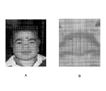
[0091] Figure 26 depicts an example of each of the three techniques that may
be
used for relative measurements analysis. For example, the geometric position
of the feature
points may define a square face dysmorphic feature. The location on a philtrum
length
distribution curve may suggest that the subject has a short philtrum. The
aggregated vector of
relative measurements may be most similar to two positive examples of a short
nose and one
negative example of a short nose.
[00921 Processor 110 may be configured to generate first evaluation result
information based, at least in 1)art, on the first evaluation (step 230),
Processor 110 may
utilize the data derived from the first evaluation to generate the first
evaluation result. For
example, as discussed above, one or more probability scores for one or more
dysmorphic
features and/or one or more medical conditions may be determined in the first
evaluation. If
a plurality of probability scores for any particular dysmorphic feature and/or
any particular
medical condition are determined in the first evaluation, then the probability
scores for the
particular dysmorphie feature and/or the particular medical condition may be
combined. As
one example, an average of the probability scores may be determined. As
another example,
the plurality of probability scores for a particular dysmorphic feature and/or
a particular
medical condition may be input into a classifier that is calibrated to output
another probability
score attributed to the particular dysmorphic feature and/or the particular
medical condition.
For example, the classifier may he trained with positive and negative examples
of a medical
condition in order to determine a single probability score and/or severity
score of a particular
dysmorphic feature and/or a particular medical condition based on a received
set of
probability scores. In some embodiments, the classifier may also, or
alternatively, be
configured to receive a set of severity scores in order to determine a
probability score for the
medical condition.
1:0093] Processor 110 may he configured to perform a second evaluation of the
external soft tissue image information using at least one of the anchored cell
analysis, the
shifting patches analysis, and the relative measurements analysis (step 240).
For example, if
the first evaluation includes an anchored cell analysis, then a shifting
patches analysis or
relative measurements analysis may be performed as the second evaluation. If
the first
= evaluation includes a shifting patches analysis, then an anchored cell
analysis or relative
measurements analysis may be performed. If the first evaluation includes a
relative
CA 02905637 2015-09-11
WO 2014/140926 PCT/IB2014/001235
measurements analysis, then an anchored cell analysis or shifting patches
analysis may be
performed as the second evaluation, The anchored cell analysis, shifting
patches analysis,
and relative measurements analysis may be performed in the same or
substantially the same
manner as described above with respect to step 220.
[0094] - In some embodiments, the first evaluation and second evaluation may
be of
the same general type (e.g., both may be an anchored cell analysis, both may
be a shilling
patches analysis, or both may be relative measurements analysis). In such
embodiments, one
of the evaluations may result in, for example, one or more probability scores
associated with
one or more dysmorphic features, whereas another one of the evaluations may
result in, for
example, one or more probability scores associated with one or more medical
conditions,
Likewise, even if the general types of analysis are different, one of the
evaluations may result
in, for example, one or more probability scores associated with one or more
dysmorphic
features, whereas another one of the evaluations may result in, for example,
one or more
probability scores associated with one or more medical conditions,
[0095] Processor 110 may be configured to generate second evaluation result
information based, at least in part, on the second evaluation (step 250).
Processor 110 may
utilize the data derived from the second evaluation to generate the second
evaluation result.
For example, one or more probability scores and/or severity scores for one or
more
dysmorphic features and/or one or more medical conditions may be determined in
the second
evaluation that are combined using one or more classifiers to generate a
simile probability
score associated with one or more particular dysmorphic features and/or
medical conditions.
[0096] Processor 110 may be configured to predict a likelihood that the
subject is
affected by the medical condition based, at least in pad, on the first
evaluation result
information and the second evaluation result information (step 260). For
example, if the first
evaluation result information includes one or more probability scores
associated with one or
more dysmorphic features, and the second evaluation result information
includes one or more
probability scores associated with one or more dysmorphic features, processor
110 may be
configured to analyze the information to determine the likelihood.
Alternatively, for
example, if the first evaluation result information includes one or more
probability scores
associated with one or more medical conditions, and the second evaluation
result information
includes one or more probability scores associated with one or more medical
conditions,
processor 110 may be configured to analyze the information to determine the
likelihood.
Alternatively, for example, if the first evaluation result information
includes one or more
probability scores associated with one or more medical conditions, and the
second evaluation
26
CA 02905637 2015-09-11
WO 2014/140926 PCT/IB2014/001235
result information includes one or more probability scores associated with one
or more
dysmorphie features, processor 110 may be configured to analyze the
information to
determine the likelihood. Likewise, if, for example, the first evaluation
result infOrmation
includes one or more probability scores associated with one or more dysmorphic
features, and
the second evaluation result information includes one or more probability
scores associated
with one or more medical conditions, processor 110 may be configured to
analyze the
information to determine the likelihood.
[0097] If both evaluations return a set of probability scores for a set of
dysmorphic
features, then the set of probability scores for the set of dysmorphic
features may be input
into a trained classifier that is calibrated to output a probability score
attributed to a particular
medical condition. For example, the classifier may be trained with positive
and negative
examples of the medical condition in order to determine a probability score of
the particular
medical condition.
[0098] If both evaluations return a set of probability scores for a set of
medical
conditions, then the set of probability scores for a particular medical
condition may be input
into a trained classifier that is calibrated to output another probability
score attributed to the
particular medical condition. For example, the classifier may he trained with
positive and
negative examples in order to determine a probability score of the particular
medical
condition, in this way, a more accurate medical condition probability score
may be
determined than any one of the individual evaluations that produced a medical
condition
likelihood.
[0099] If one of the evaluations returns a set of probability scores for a set
of
dysmorphic features and another one of the evaluations results a set of
probability scores frir
a medical condition, then the set of probability scores for the set of
dysmorphic features from
one evaluation and the set of probability scores for a particular medical
condition from the
other evaluation, may be input into a trained classifier that is calibrated to
output another
probability score attributed to a particular medical condition. For example,
the classifier may
be trained with positive. and negative examples of a medical condition in
order to determine a
probability score of the particular medical condition.
[001001 In some embodiments an initial determination of the likelihood that
the
subject is affected by the medical condition may be revised. For example, a
likelihood of a
plurality of medical conditions may initially be determined, Then, a revised
likelihood of one
or more of the medical conditions may be determined based on the other
likelihoods. For
example, if two medical conditions are related such that they typically occur
together, a low
27
CA 02905637 2015-09-11
WO 2014/140926 PC171B2014/001235
probability score for one medical condition may decrease an otherwise high
probability score
for the other medical condition. Similarly, if two medical conditions are
related such that
they typically do not occur together, a high probability score for both
medical conditions may
cause both probability scores to decrease. Moreover, for example, if a set of
medical
conditions are initially determined to have a high probability score, but are
known to
commonly he misdiagnosed for another medical condition, then the probability
score of each
medical condition in the set may be decreased and the probability score of the
other medical
condition may be increased.
[00101] Processor 110 may be configured to consider other data as well when
determining the likelihood of the medical condition. For example, more than
two evaluations
may he performed in substantially the same manner as the first and second
evaluations
described above. Processor 110 may be configured to analyze the additional
evaluations
using the techniques described above. Moreover, features associated with the
subject may be
determined from other sources. For example, a physician may provide (e.g.,
dictate or type)
one or more known features (e.g., dysinorphic features, biographical
information,
demographic information, etc.) of the subject that are used, for example, to
limit the images
in the database that the subject is compared to (e.g., the subject may only be
compared
against other individuals sharing one or more of the features provided by the
physician).
[00102] Figure 3 illustrates an exemplary process 300 that processor 110 may
be
configured to perform. For example, as discussed above, processor 110 may be
configured to
perform process 300 by executing software or firmware stored in memory device
120, or may
be configured to perform process $00 using dedicated hardware or one or more
AS1Cs.
[00103] Processor 110 may be configured to receive inthrmation reflective of
an
external soft tissue image of the subject (step 310). Processor 110 may be
configured, for
example, to perform step 310 in the same manner as step 210, discussed above.
(00104] Processor 110 may be configured to divide the external soft tissue
image
information into a plurality of regions (step 320), For example, processor 110
may be
configured to process at least one of a forehead region of the external soft
tissue image
information, a periorbital region of the external soft tissue ima.e.,e
information, a nasal region
of the external soft tissue image information, a mid-face region of the
external soft tissue
image information, an ear region of the external soft tissue image
information, and an oral
region of the external soft tissue image information, and discount at least
one other region of
the external soft tissue image information. Particular regions in the external
soft tissue image
information may be defined in accordance with, for example, any oithe
techniques described
28
CA 02905637 2015-09-11
WO 2014/140926 PCT/IB2014/001235
above. For example, as depicted in Figure 23B, a small grid may be applied to
a nasal region
of the external soft tissue image information. Areas surrounding the nasal
region may be
discounted by, for example, not having a small grid applied to them.
[00105] in addition, while regions associated with a face of the subject are
discussed,
other regions may also he processed. For example, the external soft tissue
image information
may also, or alternatively, include a side view of the subject that includes
an ear region.
Figure 27 depicts an example of an ear region that is further divided into a
plurality of
regions.
[00106] Processor 110 may be configured to generate an analysis of each of the
plurality of regions (step 330). For example, within each region, at least one
of an anchored
cell analysis and a shifting patch analysis may be performed. The anchored
cell analysis and
shifting patch analysis may be performed in the manner described above with
respect to step
220. As described above, processor 110 may be configured to calculate a
descriptor for each
of the plurality of regions. The descriptor may include, for example, at least
one of a
combined appearance vector if an anchored cell analysis is performed and a
descriptor vector
if a shifting patch analysis is perfomied.
[00107] As described above, processor 110 may be configured to compare the
plurality of regions with data derived from images of individuals known to be
affected by the
medical condition. For example, as described above, processor 110 may be
configured to
compare the. descriptor to previously produced descriptors from additional
external soft tissue
images of other individuals previously determined to be affected by the
medical condition,
Based on the comparison, for each region, one or more probability scores
associated with one
or more dysmorphic features and/or one or more medical conditions may be
determined.
[00108] Processor 110 may further be configured to aggregate the analyses of
the
plurality of regions (step 340). For example, an analysis of a nasal region
may result in a first
set of probability scores regarding dysmorphic features and/or medical
conditions associated
with the nasal region and an analysis of an ear region may result in a second
set of probability
scores regarding dysmorphic features and/or medical conditions associated with
the ear
region. The probability scores may be produced using, for example, the
techniques described
above. In some embodiments, some of the probability scores may be aggregated
by
combining the probability scores. For example, a particular dysmorphic feature
and/or
medical condition may be associated with both the nasal region and the ear
region. A
probability score for the particular dysmorphic feature and/or medical
condition determined
from the nasal region and another probability score for the particular
dysmorphic feature
29
CA 02905637 2015-09-11
WO 2014/140926 PCT/IB2014/001235
and/or medical condition determined from the ear region may be input into a
classifier trained
using, for example, positive and negative examples of the particular
dystuorphic feature
and/or medical condition, to generate a third probability score reflective of
a probability score
associated with the particular dysmorphie feature and/or medical condition. As
a result of the
aggregation, a single probability score, for example, may be determined for a
set of
dysmorphic features and/or medical conditions.
[00109] Processor 110 may further be configured to determine a likelihood that
the
subject is affected by the medical condition based on the aggregated analyses
(step 350). For
example, in the same or substantially the same manner as described above with
respect to
step 260, one or more classifiers may be trained to receive a set of scores
for a plurality of
dysmorphic features and/or medical conditions and output a score
representative of the
likelihood that the subject is affected by the medical condition.
[00110] in some embodiments, the medical condition may be a known medical
condition. However, some medical conditions can have unknown genetic causes.
Processor
110 may be configured to identify an underlying genetic variation likely to
cause a medical
condition. For example, a database, such as the database discussed above, may
include a
plurality of external soft tissue images of individuals associated with a
medical condition
caused by an unknown genetic variation. The database may also include a
plurality of
genetic variation information of individuals having a medical condition caused
by an
unknown genetic. variation. Processor 110 may be further configured to
determine whether a
common dysmorphology exists at the location of at least some of the plurality
of external soft
tissue images. For example, processor 110 may be configured to analyze the
plurality of
external soft tissue images in the manner described above to determine one or
more
dysmorphic features. A common dysmorphology may exist if, for example, a
dysmorphie
feature exists at the same or substantially the same location in at least two
images.
[00111] Processor 110 may be further configured to analyze the plurality of
genetic
variation information to identify at least one common genetic variation. A
common genetic
variation may include, for example, a determination that a gene associated
with one image
matches a gene associated with another image. Processor 110 may be further
configured to
compare the location of the plurality of external soft tissue images with the
common genetic
variation. For example, a determination may be made whether the genetic
variation is known
to affect a body part containing the common dysmorphology location in the
images.
Processor 110 may he further configured to associate, in the database, at
least one common
CA 02905637 2015-09-11
WO 2014/140926 PCT/1B2014/001235
location in the plurality of external soft tissue images and at least one
common genetic
variation in the plurality of genetic variation information.
[00112] Figure 4 illustrates an exemplary process 400 that at least one
processor may
be configured to perform. For example, as discussed above, processor 110 may
be
configured to perform process 400 by executing software or firmware stored in
memory
device 120, or may be configured to perform process 400 using dedicated
hardware or one or
more A.S I Cs.
[00113] Processor 110 may be configured to receive information reflective of
an
external soft tissue image of the subject (step 410). Step 410 may include
substantially the
same operations as step 2.10, discussed above.
[00114] Processor 110 may be further configured to analyze the external soft
tissue
image information of the subject to generate medical-condition related
information (step
420). For example, medical-condition related information may be generated
using the same
or substantially the same operations described above with respect to steps 220-
260.
Optionally, however, only one analysis may be performed (e.g., the analysis in
steps 220-
230) rather than the two (or more) analyses described in steps 220-250,
Medical-condition
related information may include, for example, one or more scores for one or
more
dysmorphie features and/or one or more medical conditions.
[00115] Processor 110 may be further configured to analyze external soft
tissue
image information of at least one relative of the subject known not to be
affected by the
medical condition to generate additional medical-condition related information
(step 430).
For example, processor 110 may be configured to determine one or more scores
associated
with one or more dysmorphie features associated with the relative. Processor
110 may be
configured to identify, for example, dysmorphic features having a high score
that are usually
indicative of a medical condition that the relative is known not to be
affected by. In some
embodiments, external soft tissue image information of a plurality of
relatives is analyzed and
dysmorphic features are identified having a high score for all of the
relatives, or a number of
relatives greater than a threshold, that are usually indicative of a. medical
condition that the
relatives are known not to be affected by.
[001161 Additionally, or alternatively, processor 110 may be configured to
determine
one or more scores associated with one or more medical conditions associated
with the
relative. For example, processor 110 may initially utilize a classifier used
for the general
population to determine a score associated with a medical condition that the
relative is known
not to have. However, despite the relative not having the medical condition,
processor 110
31
CA 02905637 2015-09-11
WO 2014/140926 PCT/IB2014/001235
may determine a high score for the medical condition based on the analysis of
the image
information associated with the relative.
[00117] Processor 110 may be further configured to predict a likelihood that
the
subject is affected by a medical condition by discounting features in the
medical-condition
information that are common to the additional medical-condition related
information (step
440). For example, as described above, processor 110 may determine one or more
scores of
one or more dysmorphic features from the image information of the subject.
Based on the
identified dysmorphic features of one or more relatives (e.g., the dysmorphic
features of the
relative that exhibit a high score usually indicative of a medical condition
despite the relative
being known not to be affected by the medical condition), processor 110 may be
configured
to modify or construct a classifier associated with a particular medical
condition. For
example, if a high score for a particular dysmorphic feature is usually used
to increase the
probability of a medical condition, but the relative has a high score for the
particular
dysmorphie feature and is known not to have the medical condition, the
classifier may be
modified or constructed such that the dysmorphic feature is ignored, is used
to reduce the
likelihood of the medical condition, or is used less heavily than for the
general population in
the determination of the likelihood of the medical conditionõks another
example, if
processor 110 determines a high score for a medical condition for a relative
of the subject
despite the relative being known not to be affected by the medical condition,
processor 110
may reduce any score determined for the medical condition for the subject. As
another
example, one or more images of the relative may be used to train the
classifier. For example,
one or more images of one or more relatives known not to he affected by a
medical condition
may be used a negative examples when training a classifier. As another
example, only
dysmorphic features of the subject that are different than one or more
dysmorphic features of
one or more relatives known not to be affected by a medical condition may he
used in the
likelihood determination.
[00118] Figure 5 illustrates an exemplary process 500 that at least one
processor may
be configured to perform. For example, as discussed above, processor 110 may
be
configured to perform process 500 by executing software or firmware stored in
memory
device 120, or may he configured to perform process 500 using dedicated
hardware or one or
more ASICs,
[00119] Processor 110 may be configured to receive information reflective of
an
external soft tissue image. of the subject (step 510), Processor 110 may be
configured, for
example, to perform step 510 in the same manner as step 210, discussed above.
32
CA 02905637 2015-09-11
WO 2014/140926 PCT/IB2014/001235
[00120] Processor 110 may be configured to use image information analysis to
compare the external soft tissue image information with a plurality of
external soft tissue
images of other subjects in a database (step 520). For example, processor 110
may be
configured to use image information analysis, wherein the image information
analysis
includes at least one of anchored cells analysis, shifting patches analysis,
and relative
measurements analysis. The anchored cell analysis, shifting patches analysis,
and relative
measurements analysis may be performed in the same or substantially the same
manner as
described above, As described above (for example, hi step 220), processor 110
may analyze
the external soli tissue image information based on a plurality of objective
criteria, including
at least one of age, gender, and ethnicity. For example, the only external
soft tissue images in
the database of other subjects of the same age, gender, and ethnicity as the
subject may be
utilized in the analysis.
[00121] Processor 110 may be further configured to determine, based on the
image
information analysis, dysmorphic features included in the external soft tissue
image
information (step 530), For example, as described above, one or more of an
anchored cells
analysis, shitting patches analysis, and relative measurements analysis may be
used to assign
a probability score to each dysmorphic feature in a set of dysmorphie features
being
analyzed.
[00122] Processor 110 may be further configured to access descriptors
associated
with the dysmorphic features (step 540). Tn some embodiments, the accessed
descriptors
include a list of words associated with dysmorphic features and that are
potential indicators of
at least one medical condition. For example, the accessed descriptors may
include terms that
are compatible with a plurality of databases for searching medical conditions.
The
descriptors associated with the dysmorphic features may be obtained, fbr
example, from a
variety of sources including, for example, the international Statistical
Classification of
Diseases and Related health Problems (e.g., .1.C1J-9 or ICD-10), the Human
Phenotype
Ontology (I-11>0), and various other sources of descriptions for dysmorphic
features, such as
medical books, published journal papers, and computerized datasets. Processor
110 may be
configured to link the descriptors associated with the dysmorphic features
obtained from
different sources (e.g., a descriptor for a particular dysmorphic feature used
in 1CD-10 may
be linked to a descriptor for the particular dysmorphic feature used in HF0).
Each descriptor
for a dysmorphic feature may include, for example, a textural description and
a list of
synonyms. in some embodiments, HPOsbased descriptors may be used as a
reference list and
all other lists from other sources may be mapped to it. If there is a term
dysmorphic feature
33
CA 02905637 2015-09-11
WO 2014/140926 PCT/1B2014/001235
that is missing from IIPO, it may be given a unique HPO-like numeric
identifier and added to
the reference list. in some embodiments, processor 110 may determine when HP0
is updated
and, based on a determined that HPO has updated, update the reference list.
Moreover, in
some embodiments, the accessed descriptors are a list of words that includes
at least one
description of a general appearance of a medical condition.
[00123] As one example, six high scoring dysmorphic features may be determined
for image information of a subject, The descriptors for the dysmorphie
features may include,
for example, "Vermillion, Lower Lip, Thick", "Columelia, tligh Insettion",
"Hairline, High
Anterior or Forehead, Tall", "Palpebral Fissure, Upslanted", "Eyebrow, Thick
or
Hypertrichosis of the Eyebrow or Bushy Eyebrow", "and "Philtrum, Tented."
Thus, each of
the descriptors may include, for example, a name of the dysmorphie feature
(e.g., "Eyebrow,
Thick") and possible alternatives to the dysmorphie feature (e.g.,
"Hypertrichosis of the
Eyebrow").
[00124] Processor 110 may be further configured to output at least some of the
descriptors (step 550). For example, processor 110 may be configured to output
at least some
of the descriptors to output device 150. Output device 150 may be, for
example, a display,
In some embodiments, as depicted in Figure 1, output device 150 may be part of
system 100.
However, in other embodiments, output device 150 may be located remotely and
processor
110 may be configured to send data to a device that includes output device 150
or is in
communication with output device 150, A display may include, for example, one
or more of
a television set, computer monitor, head-mounted display, broadcast reference
monitor, a
liquid crystal display (LCD) screen, a light-emitting diode (LED) based
display, an LED-
backlit LCD display, a cathode ray tube (CRT) display, an electrolumineseent
(ELI)) display,
an electronic paper/ink display, a plasma display panel, an organic light-
emitting diode
(01,ED) display, thin-film transistor display ('ITT), High-Performance
Addressing- display
(HPA), a surface-conduction electron-emitter display, a quantum dot display,
an
interferometrie modulator display, a swept-volume display, a carbon nanotube
display, a
variforcal mirror display, an emissive volume display, a laser display, a
holographic display,
a light field display, a projector and surface upon which images are
projected, a printer
configured to generate a printout of data, or any other electronic device for
outputting visual
information,
[0012.5] Output device ISO may also be an audio device configured to output
audio
representative of, for example, at least some of the descriptors. The audio
device may
include, for example, a sound card and one or more speakers. Processor 110 may
be
34
CA 02905637 2015-09-11
WO 2014/140926 PCT/1B2014/001235
configured, for example, to convert at least some of the descriptors into
audio using a text-to-
speech program.
[00126] In seine embodiments, the descriptors may be presented in a list. in
some
embodiments, an image may be displayed near a descriptor that is indicative of
the general
location of the dysinorphic feature associated with the. descriptor. For
example, an image of
an eye may be displayed next to a descriptor of "Eyebrow, Thick".
[00127] in some embodiments, a descriptor may be displayed at a location at,
or close
to, a dystnorphic feature to which it is associated. For example, the image
information of the
subject .may be presented on the display. A descriptor (e.g., "Eyebrow,
Thick") may be
displayed on top of a region of the image information associated with a
dysinorphic feature
associated with the descriptor (e.g., "Eyebrow, Thick" may be displayed on top
of an eye or
eyebrow region of the image information),
[00128] Figure 6 illustrates an exemplary process 600 that at least one
processor may
be configured to perform. For example, as discussed above, processor 110 may
be
configured to perform process 600 by executing software or firmware stored in
memory
device 120, or may be confuzured to perform process 600 using dedicated
hardware or one or
more ASICs.
[00129] Processor 110 may be configured to receive information reflective of
an
external soft tissue image of the subject (step 610), Processor 110 may be
configured, for
example, to perform step 610 in the some manner as step 210 discussed above,
[00130] Processor 110 may be configured to define at least one hundred
locations in
the received external soft tissue image information, wherein information about
the at least
one hundred locations constitutes subject information (step 620). Processor
110 may be
configured to define the at least one hundred locations by determining at
least one hundred
feature points in the manner described above for determining feature points.
[00131] Processor HO may also be configured to receive first information
defining at
least one hundred locations in at least an external soft tissue image of at
least a first
individual known to be affected by the medical condition (step 630) and to
receive second
information defining at least one hundred locations in at least an external
soft tissue image of
at least a second individual known to be affected by the medical condition
(step 640). The
subject information, the first information, and the second information may
include, for
example, vector data, ratio data, distance data, angular data, area data, and
shape data
associated with a relative measurements analysis calculated between at least
some of the at
least one hundred locations.
CA 02905637 2015-09-11
WO 2014/140926 PCT/IB2014/001235
[00132] Processor 110 may be configured to determine a likelihood that the
subject is
affected by a medical condition by comparing the subject information with the
first
information and the second information (step 650). For example, processor 110
may be
configured to determine a likelihood that the subject is affected by a medical
condition by
comparing the subject information with the first information and the second
information
using the relative measurements analysis described above.
[00133] In some embodiments, processor 110 may initially define a first number
of
feature points (e.g., one hundred feature points). The first number of feature
points may
permit processor 110 to determine the likelihood at a first speed. However, if
the likelihood
determination is inconclusive result (e.g,, the likelihood is above a first
threshold but below a
second threshold), the above process may be repeated with a second number of
feature points
greater than the first number of feature points (e.g,, one thousand feature
points) that requires
more time, but may be more accurate.
[00134] Figure 7 illustrates an exemplary process 700 that at least one
processor may
be configured to perform. For example, as discussed above, processor 110 may
be
configured to perform process 700 by executing software or firmware stored in
memory
device 120, or may be configured to perform process 700 using dedicated
hardware or one or
more ASICs,
[00135] Processor 110 may be configured to receive information reflective of
an
external soft tissue image of the subject (step 710). Processor 110 may be
configured, for
example, to perform step 710 in the same mariner as step 210 discussed above.
[00136] Processor 110 may be configured to analyze the external soft tissue
image
information to identify locations likely to be associated with at least one
dysmorphology
corresponding to a medical condition (step 720). For example, processor 110
may be
configured to identify one or more dysmorphic features having high probability
scores in the
manner described above.
[00137] Processor 110 may be configured to superimpose indicators of the at
least
one dysmorphology on the external soft tissue image information (step 730).
For example,
processor 110 may be configured to output to a display the external soft
tissue image
information along with a superimposed indication of at least one
dysmorphology. For
example, points detected in the image information may be superimposed on the
image
information. As another example, regions in the image information associated
with high
probability dysmorphie features may be highlighted. As another example, a heat
map may be
superimposed on the image information such that, at locations in the external
soil tissue
36
CA 02905637 2015-09-11
WO 2014/140926 PCT/1B2014/001235
image information associated with a dysmorphic feature having a low score, a
first
translucent color may be used, whereas at locations in the external soft
tissue image
information associated with a dysmorphic feature having a high score, a second
translucent
color, different than the first translucent color, may he used, The locations
may be chosen,
for- example, based on cells or patches used to determine the presence of the
dysmorphic
feature. In some embodiments, processor 110 may be configured to blur the heat
map to
produce a more appealing heat map.
[00138] Figure 8 illustrates an exemplary process 800 that at least one
processor may
be configured to perform. For example, as discussed above, processor 110 may
be
configured to perform process 800 by executing software or firmware stored in
memory
device 120, or may be configured to perform process 800 using dedicated
hardware or one or
more ASiCs.
[00139] Processor 110 may be configured to receive information reflective of
an
external soft tissue image of the subject (step 810). Processor 110 may be
configured, for
example, to perform step 810 in the same manner as step 210 discussed above.
[00140] Processor 110 may be configured to display the external soft tissue
image
information (step 820). For example, processor 110 may be configured to output
the external
soft tissue image information to a display that is configured in the manner
described above.
[00141] Processor 110 may be configured to enable a user to select a region of
the
external soft tissue image information (step 830), For example, processor 110
may be
configured to enable a user to select a region of the external soft tissue
image information
presented on the display. In some embodiments, the region selected by the user
may be
expanded after processor 110 detects the selection.
[00142] Processor 110 may be configured to identify, to the user, information
about
dysmorphic features in the selected region (step 840), For example,
information about the
dysmorphic features may be displayed in a list or may be superimposed onto the
external soft
tissue image information. The list of dysmorphic features may be presented,
for example, in
descending or ascending order of probability score.
[00143] Figure 9 illustrates an exemplary process 900 that at least one
processor may
be configured to perform. For example, as discussed above, processor 110 may
be
configured to perform process 900 by executing software or firmware stored in
memory
device 120, or may be configured to perform process 900 using dedicated
hardware or One or
more ASICs.
37
CA 02905637 2015-09-11
WO 2014/140926 PCT/IB2014/001235
[001441 Processor 110 may be configured to receive information reflective of
an
external soft tissue image of the subject (step 910). Processor 110 may be
configured, for
example, to perform step 910 in the same manner as step 210 discussed above.
[001451 Processor 110 may be configured to analyze the external soft tissue
image
information (step 920). For example, processor 110 may analyze the external
soft tissue
image information using the same or substantially the same operations
described above with
respect to steps 220-260. Optionally, however, only one analysis may be
performed (e.g., the
analysis in steps 220-230) rather than the two (or more analyses described in
steps 220-250.
For example, as described above, processor 110 may be configured to perform at
least one of
at least one of anchored cells analysis, a shifting patches analysis, and a
relative
measurements analysis,
[00146] Processor 110 may be configured to identify one or more external soft
tissue
attributes in the external soft tissue image information based, at least in
part, on the analysis
(step 930). The one or more external soft tissue attributes may include, for
example, one or
more dysmorphic features. For example, as described above, processor 110 may
be
configured to identify potential external soft tissue attributes in the
external soft tissue image
information and to assign a confidence level to the potential external soft
tissue attributes
reflective of a likelihood that the potential external soft tissue attributes
appear in the
In some embodiments, processor 110 may be configured to identify which
external soft tissue
attributes are indicators of medical conditions by taking into account a
weighting of each
external soft tissue attribute as an indicator of each medical condition. For
example,
processor 110 may identify all dysmorphic features having a high probability
score or a
probability score above a predetermined threshold. In some embodiments, the
weighting of
each external soft tissue attribute includes at least one of a severity of
each external soft tissue
attribute, a commonality of each external soft tissue attribute in a general
population, and a
relevance of each external soft tissue attribute to a medical condition.
[001471 Processor 110 may be configured to access at least one database of
external
soft tissue attributes associated with a plurality of medical conditions (step
940). For
example, processor 110 may be configured to access a database containing data
regarding one
or more dysmorphic features andlor one or more medical conditions in the same
manner as
described above in, for example, step 220,
[00148] Processor 110 may be configured to compare the one or more identified
external soft tissue attributes with the external soft tissue attributes of
the at least one
database (step 950), For example, processor 110 may be configured to compare
the one or
38
CA 02905637 2015-09-11
WO 2014/140926 PCT/IB2014/001235
more identified external soft tissue attributes with the external soft tissue
attributes of the at
least one database in the same manner as described above in, for example, step
220, The
comparison may generate one or more probability scores associated with one or
more
dysmorphic features.
[00149] Processor 110 may be configured to output information about st least
one
medical condition likely possessed by the subject based on the comparison
(step 960). For
example, as described above, processor 110 may be configured to input the one
or more
probability scores of the one or more dysmorphie features into a classifier to
generate a
probability score for a medical condition. In some embodiments, processor 110
is configured
to determine additional information about the at least one medical condition
likely possessed
by the subject based directly on the analysis; and output the information
about at least one
medical condition likely possessed by the subject based on the comparison and
the additional
information. For example, as described in step 260, processor 110 may
determine a
likelihood of a medical condition based on both an =initial medical condition
likelihood and a
set of dysmorphic feature probability scores.
[001501 Figure 10 illustrates an exemplary process 1000 that at least one
processor
may be configured to perform. For example, as discussed above, processor 110
may be
configured to perform process 1000 by executing software or -firmware stored
in memory
device 120, or may be configured to perform process 1000 using dedicated
hardware or one
or more ASICs,
[00151] Processor 110 may be configured to receive first information
reflective of a
first external soft tissue image of the subject recorded at a first time (step
1010). Processor
110 may be configured, for example, to perform step 1010 in the same manner as
step 210,
discussed above.
1001521 Processor 110 may be configured to analyze the first image information
(step
1020). Processor 110 may be configured, for example, to analyze the first
image information
in the same manner as described above With respect to steps 220-260.
Optionally, however,
only one analysis may be performed (e.g., only the analysis in steps 220-230)
rather than the
two (or more) analyses described in steps 220-250. For example; in some
embodiments the
analysis includes at least one of anchored cells analysis, shifting patches
analysis and relative
measurements analysis. Moreover, as described above, in some embodiments the
analysis
includes a comparison of the first soft tissue image information to an
external soft tissue
image of at least one individual having at least one of substantially the same
age, ethnicity,
and gender as the subject.
39
CA 02905637 2015-09-11
WO 2014/140926 PCT/IB2014/001235
[00153] Processor 110 may be configured to receive second information
reflective of
a second external soft tissue image of the subject recorded at a second time
(step 1034
Processor 110 may be configured, for example, to perform step 1010 in the same
manner as
step 210 discussed above. The second time may be, for example, a predetermined
amount of
time after the first. time or a non-scheduled time after the first time, in
some embodiments,
processor 110 may be configured to send an alert that second information
reflective of a
second external soft tissue image of the subject should be recorded. For
example, if second
information reflective of a second external soft tissue image of the subject
has not been
received within a predetermined amount of time, an alert may be sent to the
subject's
physician. As another example, if the analysis of the first information
provided an indication
that there is a low probability that a patient has a medical condition, an
alert may be sent if
second information reflective of a second external soft tissue image of the
subject has not
been received within a predetermined amount of time.
[00154] Processor 110 may be configured to analyze the second image
information
(step 1040). Processor 110 may be configured, for example, to analyze the
second image
information in the same manner as described above with respect to steps 220-
260.
Optionally, however, only one analysis may be performed (en., the analysis in
steps 220-
230) rather than the two (or more) analyses described in steps 220-250. For
example, in
some embodiments the analysis includes at least one of anchored cells
analysis, shilling
patches analysis and relative measurements analysis. Moreover, as described
above, in some
embodiments the analysis includes a comparison of the second soft tissue image
information
to an external soft tissue image of at least one individual having at least
one of substantially
the same age, ethnicity, and gender as the subject.
[00155] In some embodiments, processor 110 is configured to apply the same
technique to analyze the first external soft tissue image information and to
analyze the second
external soft tissue image information. For example, the technique applied at
the first time
may be recorded and reused the next time a subject is imaged. Alternatively,
in some
embodiments, if the analysis of the first image information did not indicate a
likelihood of
any medical condition, the analysis may be changed at the second time when the
second
external soft tissue image information is analyzed. As another alternative,
if, for example, the
analysis of the first image information indicated that there was a likelihood
of a medical
condition associated with a particular body part (e.g., an ear), the analysis
of the second
image information may be focused on the particular body part. In some
embodiments,
processor 110 may be configured to alert a part,' associated with capturing
the second
CA 02905637 2015-09-11
WO 2014/140926 PCT/IB2014/001235
external soft tissue image to capture one or more images of the particular
body part. The
second external soft tissue image thus may include one or more images of the
particular body
part.
[00156] Processor 110 may be configured to compare the analysis of the first
image
information with the analysis of the second image information (step 1050). For
example, the
first image information and the second image information may include
probability scores for
a number of dysmorphic features. Processor 110 may be configured to determine
changes in
probability scores of the dysmorphic features over time. The first image
information and
second image information may also include, for example, probability scores for
a number of
medical conditions. Processor 110 may be configured to determine changes in
probability
scores of the medical conditions over time.
[00157] As another example, the first image information and the second image
information may also include one or more severity scores of one or more
dysmorphic
features. For example, the first image information and the second image
information may
include one or more severity scores based on one or more distances between
feature points,
angles formed by feature points, ratios between distances, ratios between
angles, and the like.
Thus, in some embodiments, processor 110 may be configured to monitor progress
of the
medical condition to determine a change in severity of an attribute over time.
[00158] Processor 110 may be configured to predict a likelihood that the
subject is
affected by the medical condition based, at least in part, on the comparison
(step 1060). For
example, probability scores and severities of one or more dysmorphic features
at the first
time, probability scores and severities of one or more dysmorphic features at
the second time,
and/or changes in probability scores and severities of one or more dysmorphic
features from
the first time to the second time may be input into a classifier trained on,
for example,
positive and negative examples of a medical condition. IL for example, the
severity or
probability scores associated with a set of dysmorphic features increases from
the first time to
the second time, and the dysmorphic features are associated with a medical
condition, a
relatively high likelihood for the medical condition may be determined.
[001591 However, not all changes in severity will necessarily result in a high
likelihood for the medical condition. For example, as a child ages, some the
size of various
dysmorphic features may he expected to change. Thus, in some embodiments, the
change in
severity of a dysmerphie feature may be compared to known changes that occur
from the
subject's age at the first time to the subject's age at the second time
(optionally, known
changes that occur from the subject's age at the first time to the subject's
age at the second
41
CA 02905637 2015-09-11
WO 2014/140926 PCT/1B2014/001235
time may be examined in the context of at least one of the subject's gender,
ethnicity, and any
other category describing the subject). The known changes that occur .from the
subject's age
at the first time to the subject's am at the second time may be determined,
for example, by
analyzing images in a database and determining norms for a given patient
population. Thus,
in some embodiments, if the change in severity of a dysmorphic feature
deviates from an
expected change, then a high likelihood that the subject is affected by the
medical condition
may be predicted. If the change in severity of a dysrnorphic feature does not
deviate from an
expected change, then a low likelihood that the subject is affected by the
medical condition
may be predicted.
1001601 In some embodiments, processor 110 may increase a probability score
determined at the second time if an increase in probability score from the
first time to the
second time is determined, in some embodiments, processor 110 may decrease a
probability
score determined at the second time if a decrease in probability score from
the first time to
the second time is determined.
[00161] in, some embodiments, a plurality of additional sets of information
may be
received reflecting a plurality of additional external soft tissue images
recorded at a plurality
of additional times. Processor 110 may be configured to analyze the plurality
of additional
images, compare the analysis of the first soft tissue image information, the
second soft tissue
image information, and the additional sets of soft tissue image information,
and predict the
likelihood that the subject is affected by the medical condition based on the
comparison of
the analysis of the first soft tissue image information, the second soft
tissue image
information, and the additional sets of soft tissue image information
[00162] Figure 11 illustrates an exemplary process 1100 that at least one
processor
may be configured to perform. For example, as discussed above, processor 110
may be
configured to perform process 11.00 by executing software or firmware stored
in memory
device 120, or may be configured to perform process 1100 using dedicated
hardware or one
or more ASICs.
[001631 Processor 110 may be configured to receive first information
reflective fan
external soil tissue image of a first subject suspected of having an
unrecognized medical
condition (step 1110), Processor 110 may be configured, for example, to
perform step 1110
in the same manner as step 210 discussed above,
[001641 Processor 110 may be configured to receive second information
reflective of
an external soft tissue image of a second subject suspected of having an
unrecognized
42
CA 02905637 2015-09-11
WO 2014/140926 PCT/M2014/001235
medical condition (step 1120). Processor 110 may be configured, for example,
to perform
step 1120 in the same manner as step 210 discussed above.
[00165j Processor 110 may be configured to compare the first image information
and
the second image information (stop 1130). For example, a relative measurements
analysis
may be used to generate a vector of relative measurements for the first image
information and
the second image information. In some embodiments, the first image information
is
associated with a new subject and the second image information is associated
with a
previously presented individual. Thus, for example, as depicted in Figure 28,
a vector of
relative measurements associated with the first image information may be
compered against a
set of vectors of relative measurements in a database (including the vector of
relative
measurements associated with the second image information). As another
example,
processor 110 may be configured to receive first image information from a
first healthcare
provider and receive the second image information from a second healthcare
provider.
Processor 110 may enable the first healthcare provider to access image
information provided
by the second healthcare provider but deny access to text data (e.g., a
patient name) provided
by the second healthcare provider, and vice versa,
[00166] Processor 110 may be configured to determine, based on the comparison,
that the first subject and the second subject are likely to possess the same
previously
unrecognized medical condition (step 1140). For example, processor 110 may be
configured
to determine that the first subject and the second subject are likely to
possess the same
previously unrecognized medical condition if the first image information and
the second
image information have a high degree of similarity to each other and a high
degree of
dissimilarity to other images in the database. The similarity may be
determined, for example,
based on a comparison of the vectors of relative measurements (e.g., as
graphically depicted
in the bottom right of Figure 28, a distance may be determined from a vector
of relative
measurements associated with the first image information to a vector of
relative
measurements associated with the second image information). Processor 110 may
enable the
first healthcare provider to communicate with the second healthcare provider
if it is
determined that the first subject and the second subject are likely to possess
the same
previously unrecognized medical condition.
[001671 Figure 12 illustrates an exemplary process 1200 that at least one
processor
may be configured to perform. For example, as discussed above, processor 110
may be
configured to perform process 1200 by executing soil-ware or firmware stored
in memory
43
CA 02905637 2015-09-11
WO 2014/140926 PCT/1132014/001235
device 120, or may be configured to perform process 1200 using dedicated
hardware or one
= or more AS1Cs,
[001681 Processor 110 may be configured to use a computer-based external soft
tissue image analyzer to determine a likelihood that a specific individual is
affected by a
medical condition for which at least one health service provider offers
products or services
relating to the medical condition, wherein a user of the computer-based
external soft tissue.
image analyzer is a healthcare professional (step 1210). For example,
processor 110 may be
configured to determine a likelihood that a specific individual is affected by
a medical
condition using the same operations as described above with respect to steps
210-260.
Processor 110 may be configured to access a database that includes products or
services
offered by one or more health service providers and data associated the
products or services
with one or more related medical conditions. Thus, after determining a
likelihood of a
medical condition, processor 110 may determine whether at least one health
service provider
offers products or services relating to the medical condition.
[001691 Processor 110 may be configured = to identify information about the
healthcare professional (step 1220). For example, processor 110 may be
configured to
identify information including one or more of healthcare professional contact
information,
education, expertise, training, experience with the medical condition, and the
like.
[001701 Processor 110 may be configured to mediate communication between the
at
least one health service provider and the healthcare professional based on the
likelihood that
the specific individual is affected by the medical condition (step 1230). For
example,
mediating may include alerting the healthcare professional of the existence of
at least one of
information regarding clinical trials, registries, diagnostics, and second
opinions. An alert
may be sent, for example, using a text message to a telephone number of the
healthcare
professional, using an email message to an email address of the healthcare
professional, or
using a telephone call to a telephone number of the healthcare professional.
The alert may
provide the healthcare professional with an option to contact (e.g., using
text message, email,
or telephone communication) the health service provider. In some embodiments,
processor
110 may be configured to mediate if the likelihood is above a threshold. In
some
embodiments, processor 110 may be configured to mediate differently depending
on the
likelihood. For example, if the likelihood of the medical condition is
moderate, then only the
healthcare professional may receive a communication. If the likelihood of the
medical
condition is high, then both the health service provider and the healthcare
professional may
receive a communication.
44
CA 02905637 2015-09-11
WO 2014/140926 PCT/1B2014/001235
[00171] Figure 13 illustrates an exemplary process 1300 that at least one
processor
may be configured to perform. For example, as discussed above, processor 110
may he
configured to perform process 1300 by executing software or firmware stored in
memory
device 120, or may be configured to perform process 1300 using dedicated
hardware or one
or more AS1Cs.
[00172] Processor 110 may be configured to maintain a database of external
soft
tissue images of individuals having a medical condition (step 1310). For
example, processor
110 may be configured to maintain a database in accordance with any of the
databases
discussed above.
[00173] Processor 110 may be configured to receive from a healthcare provider
an
image feed of external soft tissue images of subjects from the healthcare
provider (step 1320).
For example, processor 110 may be configured receive each image in the image
feed in
accordance with the operations described above with respect to step 210,
Moreover,
processor 110 may be configured to receive one or more dysmorphie feature
annotations
associated with each received image. The dysmorphic feature annotations may be
generated
by, for example the healthcare provider or by any of the other techniques
described herein.
Moreover, processor 110 may be configured to receive one or more medical
conditions
associated with each received image. The medical condition may be generated by
the
healthcare provider or by any of the other techniques described herein.
[00174] Processor 110 may be configured to use a computer-based external soft
tissue image analyzer to compare images in the feed with images in the
database (step 1330).
For example, processor 110 may be configured to compare each image in the feed
with
images in the database in accordance with the operations described above with
respect to
steps 220-250. Optionally, however, only one analysis may be performed (e.g.,
the analysis
in steps 220..230) rather than the two (or more) analyses described in steps
220-250. In some
embodiments, the annotated dysmorphie features and medical conditions may be
used to limit
the analysis. For example, in some embodiments, only a likelihood of the
annotated
dysmorphic features may be determined, Likewise, in some embodiments, only a
likelihood
fete medical conditions received with each image may be determined,
[00175] Processor 110 may be configured to, based on the comparison, determine
when an image of a subject in the feed meets a threshold of being likely to be
affected by a
medical condition (step 1340). For example, processor 110 may he configured to
determine
when an image of a subiect in the feed meets a threshold of being likely to be
affected by a
medical condition in accordance with the operations described above with
respect to step 260.
CA 02905637 2015-09-11
WO 2014/140926 PCT/182014/001235
[00176] Processor 110 may be configured to alert the healthcare provider when
the
image of the subject meets the threshold (step 1350). For example, processor
110 may be
configured to send an alert to the healthcare provider using, for example, a
text message, a
telephone call, an email, and the like, As another example, processor 110 may
be configured
to present an alert on a display of the device that is used by the healtheare
provider to capture,
the image. The alert may include, for example, a patient name or other patient
identifier and
data regarding the medical condition that triggered the alert. The data
regarding the medical
condition may include, for example, the name of the medical condition,
dysmorphic features
associated with the medical condition, suggested treatments for the medical
condition,
suggested additional tests for the medical condition, etc.
[00177] Processor 110 may be configured to add the image to the database of
external
soft tissue images based on whether the subject actually possesses the medical
condition (step
1360). For example, processor 110 may be configured to receive confirmation
from the
healthcare provider that the subject possesses the medical condition. The
confirmation may
be based on, for example, the additional tests included in the alert.
[00178] In some embodiments, the images of the confirmed subjects and/or data
derived from the confirmed subjects may be linked with the medical condition,
For example,
the database may be configured to annotate the confirmed subject data with the
medical
condition. The confirmed subject data may then be used to train one or more
classifiers for
the medical condition as a positive example of the medical condition.
Likewise, processor
110 may also be configured to receive an indication from the healthcare
provider that the
subject dues not possess the medical condition. The negative subject data may
then be. used
to train one or more classifiers for the medical condition as a negative
example (i.e,, a
control) or a false positive example.
[00179] Figure 14 illustrates an exemplary process 1400 that at least one
processor
may he configured to perform. For example, as discussed above, processor 110
may be
configured to perform process 1400 by executing software or firmware stored in
memory
device 120, or may be configured to perform process 1000 using dedicated
hardware or one
or more ASICs.
[00180] Processor 110 may be configured to associate, in a database, a
plurality of
external soft tissue images of individuals having a medical condition (step
1410). For
example, processor 110 may be configured to associate a plurality of external
soft tissue
images of individuals having a medical condition in a database in accordance
with the
operations described above.
46
CA 02905637 2015-09-11
WO 2014/140926 PCT/IB2014/001235
[00181) Processor 110 may be configured to analyze the plurality of external
soft
tissue images to identify at least one predictor location associated with a
dysmorphic feature
predictive of the medical condition (step 1420). For example, processor 110
may be
configured to detect one or more dysmorphic features using the operations
described above.
Processor 110 may be configured to determine whether a number of the external
soft tissue
images that are associated with individuals having a medical condition are
also associated
with one or more of the same dysmorphic features at, for example, a same or
similar location.
[00182] Processor 110 may be configured to receive an external soft tissue
image eta
subject, undiagnosed with the medical condition (step 1430). For example,
processor 110
may be configured to receive an external soft tissue image of a subject in
accordance with the
same operations described with respect to step 210.
[00133] Processor 110 may be configured to analyze the subject's external soft
tissue
image to identify the predictor location (step 1440). For example, processor
110 may be
configured to identify a region in the subject's external soft tissue image
information
corresponding to or containing the dysmorphic feature identified in step 1420.
[00184] Processor 110 may be configured to compare the predictor location of
the
plurality of external soft tissue images with the predictor location in the
subject's external
soft tissue image (step 1450) and to determine whether a common dysmorphology
exists at
the predictor location of at least some of the plurality of external soft
tissue images and the
subject's external soft tissue image (step 1460). For example, processor 110
may be
configured to determine whether the region of the subject's external soft
tissue image
containing the predictor location is similar to one or more of the regions
containing the
predictor location in the plurality of external soft tissue images. For
example, processor 110
may be configured to determine that one or more of the identified dysmorphic
features are
contained in the region of the subject's external soft tissue image containing
the predictor
location.
[00185] Processor 110 may be configured to predict, based on the
determination,
whether the subject has the medical condition (step 1470). For example, if one
or more of the
identified dysmorphie features are detected at the predicted location in the
subject's external
soft tissue image, then processor 110 may predict that the subject has the
medical condition.
Similarly, processor 110 may be configured to predict that the subject has the
medical
condition if a sufficient number of dysinorphic features are detected in a
sufficient number of
predictor locations.
47
CA 02905637 2015-09-11
WO 2014/140926 PCT/IB2014/001235
[00186] Figure 15 illustrates an exeinplary process 1500 that at least one
processor
may be configured to perform. For example, as discussed above, processor 110
may be
configured to perform process 1500 by executing software or firmware stored in
memory
device 120, or may be configured to perform process 1500 using dedicated
hardware or one
or more AS1Cs.
[00187] Processor 110 may be configured to receive information reflective of
an
external soft tissue image of the subject (step 1510). Processor 110 may be
configured, for
example, to perform step 1510 in the same manner as step 210, discussed above,
[00188] Processor 110 may be configured to analyze the external soft tissue
image
information for a. dysmorphology (step 1520). For example, processor 110 may
he
configured to analyze the external soft tissue image information using the
same or
substantially the same operations described above with respect to steps 220-
250. Optionally,
however, only one analysis may be performed (e.g., the analysis in steps 220-
230) rather than
the two (or more) analyses described in steps 220-250.
[001891 Processor 110 may be configured to determine a plurality of potential
medical conditions associated with the dysmorphology (step 1530). For example,
processor
11.0 may be configured to determine a plurality of potential medical
conditions using the
same operations described above with respect to step 260,
[00190] Processor 110 may be configured to generate a list of tests to be
performed in
order to determine whether the individual possesses at least one of the
plurality of potential
medical conditions (step 1540). For example, processor 110 may be configured
to determine
all tests having diagnostic value for a potential medical condition. in some
embodiments,
processor 110 may be configured to generate a list of tests based on at least
one of the price
of the test, the accuracy of the test, and the compatibility of the test with
the plurality of
potential medical condition.
[00191] In some embodiments, processor 110 may receive information reflective
of
previous tests that were selected in response to a generated list. Based on
the received
information, processor 110 may be configured to favor the selected tests, For
example, if one
test is more expensive than another test, processor 110 may be initially
configured to output
the cheaper test first. However, if processor 110 receives information
indicating that the
more expensive test is more widely used, then the more expensive test may be
included first
for subsequent generated lists.
[00192] Figure 16 illustrates an exemplary process 1600 that at least one
processor
may be configured to perform. For example, as discussed above, processor 110
may be
48
CA 02905637 2015-09-11
WO 2014/140926 PCT/1B2014/001235
configured to perform process 1600 by executing software or firmware stored in
memory
device 120, or may be configured to perform process 1600 using dedicated
hardware or one
or more ASICs.
1001931 Processor 110 may be configured to receive information reflective of
an
external soft tissue image of the subject (step 1610). Processor 110 may be
configured, for
example, to perform step 1610 in the same manner as step 210, discussed above.
[00194] Processor 110 may be configured to analyze. the external soil tissue
image
information (step 1620). For example, processor 110 may be configured to
analyze the
external soft tissue image information using the same or substantially the
same operations
described above with respect to steps 220-250. Optionally, however, only one
analysis may
be performed (esg., the analysis in steps 220-230) rather than the two (or
more analyses
described in steps 220-250.
[00195] Processor 110 may be configured to determine, based on the analysis, a
probability that a dysmorphic feature exists (step 1630). For example,
processor 110 may be
configured to determine a probability score of a dysmorphic feature using
operations
described above.
[00196] Processor 110 may be configured to assign a severity score to the
dysmorphic feature (step 1640). For example, processor 110 may be configured
to assign a
severity score to a dysmorphic feature based on a relative measurements
analysis. For
example, the severity of a long philtrutn dysmorphic feature may be measured
by the ratio of
the length defined by feature points associated with the philtrum to lengths
defined by feature
points associated with one or more of the nose, mouth, and height of a face of
the subject. in
some embodiments, the severity score is determined after a probability score
is determined.
For example, in some embodiments, if the probability score for a dysmorphic
feature is above
a threshold, the severity score may then be determined. The severity score may
be
determined as a function of a probability score for the dysmorphic feature
(e.g., a higher
probability score may receive a higher severity score) or by using a second
classifier trained,
for example, on data from individuals known to have various predetermined
severities of the
dysmorphic feature.
[00197] Processor 110 may be configured to predict whether the dysmorphic
feature
is indicative of a medical condition based on the severity score (step 1650).
For example, a
classifier may be trained on positive and negative examples of the medical
condition to
receive probability scores and severity scores associated with a set of
dysmorphic features
and to output a probability score fbr the medical condition. Processor 110
may, for example,
49
CA 02905637 2015-09-11
WO 2014/140926 PCT/182014/001235
use the trained classifier to determine whether the severity score of the
dysmorphic feature is
indicative of a medical condition.
[00198] Figure 17 illustrates an exemplary process 1700 that at least one
processor
may be configured to perform. For example, as discussed above, processor 110
may be
configured to perform process 1700 by executing software or firmware stored in
memory
device 120, or may be configured to ,perform process 1700 using dedicated
hardware or one
or more ASICs,
[00199] Processor 110 may be configured to receive information reflective of
an
external soft tissue image of the subject (step 1710). Processor 110 may be
configured, for
example, to perform step 1710 in the same mariner as step 210, discussed
above.
[00200] Processor 110 may be configured to analyze the external soft tissue
image
information and identify a first dysmorphic attribute and a second dysmorphic
attribute (step
1720). For example, processor 110 may be configured to analyze the external
soft tissue
image information and identify at least two dysmorphic features in the
external soft tissue
image information using the same or substantially the same operations
described above with
respect to steps 220-250. Optionally, however, only one analysis may be
performed (e.g., the
analysis in steps 220-230) rather than the two (or more) analyses described in
steps 220-250.
[00201] Processor 110 may be configured to determine that the first dysmorphic
attribute is less likely to be a predictor of the medial condition than the
second dysmorphic
attribute (step 1730). For example, processor 110 may be configured to
determine that the
first dysmorphic attribute is less likely to be a predictor of the medical
condition than the
second dysmorphic attribute based on information that the first dysmorphic
attribute typically
does not coincide with the second dysmorphie attribute. As another example,
processor I 10
may be configured to determine that the first dysmorphic attribute is less
likely to be a.
predictor of the medical condition than the second dysmorphic attribute based
on information
that the first dysmorphic attribute is common amongst family members of the
subject. As
another example, processor 110 may be configured to determine that the first
dysmorphic
attribute is less likely to be a predictor of the medical condition than the
second dysmorphic
attribute based on information that the first dysmorphic attribute is common
amongst
members of the individual's ethnicity. As another example, processor 110 may
be configured
to determine that the first dysmorphic attribute is less likely to be a
predictor of the medical
condition than the second dysmorphic attribute based on information that the
first dysmorphic
attribute is common amongst members of the individual's gender.
CA 02905637 2015-09-11
WO 2014/140926 PCT/IB2014/001235
[00202j Processor 110 may be configured to predict whether the subject is
likely to
be affected by a medical condition, wherein, during the prediction, the first
dysmorphic
attribute is discounted (step 1740). For example, processor 110 may be
configured to predict
whether the subject is likely to be affected by a medical condition using
substantially the
same operations described above with respect to step 260. However, the
classifier used to
make the prediction may, for example, be configured to ignore the first
dysmorphic attribute
or provide it with a reduced weight.
[00203] Certain features which, for clarity, are described in this
specification in the
context of separate embodiments, may also be provided in combination in a
single
embodiment. Conversely, various features which, for brevity, arc described in
the context of
a single embodiment, may also be provided in multiple embodiments separately
or in any
suitable sub-combination. Moreover, although features may be described above
as acting in
certain combinations and even initially claimed as such, one or more features
from a claimed
combination can in some cases be excised from the combination, and the claimed
combination may be directed to a subcombination or variation of a
subcombination.
[00204-] Particular embodiments have been described, Other embodiments are
within
the scope of the following claims.
51