Note: Descriptions are shown in the official language in which they were submitted.
CA 02905718 2015-09-11
WO 2014/149545 PCT/US2014/019463
1
METHOD AND APPARATUS FOR FLUID STERILIZATION
FOR PATIENT SUPPORT SURFACES
FIELD
[0001] The disclosure relates in general to patient supports and, more
particularly, to a
method and apparatus for fluid sterilization for patient supports surfaces.
BACKGROUND
[0002] Patient support surfaces are known. These support surfaces are
inflated at least
in part using air or other fluid. Such fluids may include impurities and/or
organisms such as
mold spores, fungi, bacteria and viruses. It is undesirable to place patients
in close proximity to
such impurities and/or organisms as they may cause infection and/or illness,
aggravate allergies
and create undesirable odors.
[0003] Accordingly, it is desirable to provide an apparatus for
sterilizing fluid for use in
patient support surfaces that overcomes one or more of the aforementioned
drawbacks or other
limitations of the prior art.
BRIEF DESCRIPTION OF THE DRAWINGS
[0004] The above-mentioned and other features and advantages of this
disclosure, and
the manner of attaining them, will become more apparent and the disclosure
itself will be better
understood by reference to the following description of embodiments taken in
conjunction with
the accompanying drawings, wherein:
[0005] FIGS. 1(a)-(c) illustrate conceptual views of various orientations
of a UV chamber
according to the present disclosure relative to a fluid inlet, a blower
device, and a patient support;
[0006] FIG. 2 is a conceptual view of a UV chamber according to the
present disclosure
mounted within a valve assembly;
[0007] FIG. 3 is a perspective view of a UV chamber according to the
present disclosure;
[0008] FIG. 4 is a block diagram of the electronics of the UV chamber of
FIG. 3;
[0009] FIG. 5 is a flowchart of a control loop implemented by the
electronics of FIG. 4;
and
[0010] FIG. 6 is a schematic diagram of the electronics of FIG. 4.
SUBSTITUTE SHEET (RULE 26)
CA 02905718 2015-09-11
WO 2014/149545 PCT/US2014/019463
-2-
[0011] Corresponding reference characters indicate corresponding parts
throughout the
several views. The exemplifications set out herein illustrate exemplary
embodiments of the
disclosure and such exemplifications are not to be construed as limiting the
scope of the
disclosure in any manner.
DETAILED DESCRIPTION OF THE DRAWINGS
[0012] The embodiments disclosed herein are not intended to be exhaustive
or to limit
the disclosure to the precise forms disclosed in the following detailed
description. Rather, the
embodiments are chosen and described so that others skilled in the art may
utilize their
teachings.
[0013] In an exemplary embodiment of the present disclosure an apparatus
for treating
fluid provided to a patient support is provided. The apparatus comprises a
housing defining an
interior space; an inlet in flow communication with a source of untreated
fluid and the interior
space; an outlet in flow communication with the patient support and the
interior space; and a
light source directed into the interior space such that light emitted by the
light source bombards
fluid flowing through the interior space with light within a wavelength range
that destroys
organisms within the fluid to thereby provide treated fluid to the patient
support. In one
example, the apparatus further comprises an opening into the interior space
and
a cover configured to cover the opening, the light source being mounted to the
cover. In another
example, the light source is a UVC LED and the wavelength range is between 255
nm and 285
nm. In a variation thereof, the UVC LED emits light at a wavelength of
approximately 280 nm.
In another example, the apparatus further comprises a microcontroller
configured to receive a
flow sensor signal indicating whether fluid is flowing through the interior
space, the
microcontroller outputting a control signal that causes deactivation of the
light source when the
flow sensor signal indicates that fluid is not flowing through the interior
space. In a variation
thereof, the apparatus further comprises an opening into the interior space, a
cover configured to
cover the opening, the light source being mounted to the cover, and a sensor
mounted to the
cover, the sensor being configured to provide to enable logic a first signal
when the cover covers
the opening to the interior space and a second signal when the cover does not
cover the opening,
the enable logic causing activation of the light source upon receipt of both
the control signal
from the microprocessor and the first signal from the sensor. In a further
variation, the sensor
CA 02905718 2015-09-11
WO 2014/149545 PCT/US2014/019463
-3-
comprises a Hall Effect sensor mounted to the cover and a magnet mounted to
the housing at a
location proximate to the Hall Effect sensor when the cover is attached to the
housing to cover
the opening to the interior space. In yet another example, the housing is
mounted within a valve
assembly configured to route fluid from the fluid source to the patient
support.
[0014] In another embodiment of the present disclosure, a patient support
system is
provided, comprising: a patient support having an air inlet for receiving air
to at least partially
inflate the patient support; and a UV chamber having an inlet for receiving
unsterilized air from
an air source into an interior space of a housing, a light source that emits
light toward the
unsterilized air in the interior space at a wavelength that sterilizes the
air, and an outlet in flow
communication with the patient support inlet to provide sterilized air to the
patient support. In
one example, the patient support is one of a wheel chair cushion, a mattress,
an overlay pad, a
chair cushion, a chair pad, and a dialysis chair. In another example, the
housing comprises an
opening into the interior space, the light source being positioned to emit
light through the
opening. In a variation thereof, the UV chamber further comprises a removable
cover that
covers the opening when attached to the housing, the light source being
mounted to the cover. In
yet a further variation, the cover further comprises a printed circuit board
attached thereto having
a first side with electronics mounted thereon and a second side with the light
source mounted
thereon. In still a further variation, the electronics include a
microprocessor, enable logic
connected to the microprocessor, a driver circuit connected between the enable
logic and the
light source, and a Hall Effect sensor connected to the enable logic. In a
further variation, the
microprocessor responds to an input that indicates air is flowing through the
UV chamber by
providing a control signal to the enable logic, the enable logic being
configured to activate the
driver circuit, which thereby activates the light source, upon receipt of both
the control signal
from the microprocessor and an input signal from Hall Effect sensor indicating
that the cover is
attached to the housing and covering the opening into the interior space. In
another example, the
light source comprises a plurality of UVC LEDs. In another variation, the Hall
Effect sensor is
mounted to the printed circuit board such that when the cover covers the
opening, the Hall Effect
sensor is in close proximity to a magnet mounted to the housing.
[0015] In still another embodiment of the present disclosure, a method of
sterilizing fluid
for use in a patient support, comprising: passing unsterilized fluid from a
fluid source through an
CA 02905718 2015-09-11
WO 2014/149545 PCT/US2014/019463
-4-
inlet into an interior space of a chamber; emitting ultraviolet light toward
the fluid in the interior
space at a wavelength of between about 255 nm and about 285 nm to thereby
sterilize the fluid;
and passing the sterilized fluid through an outlet of the chamber to an inlet
of a patient support to
thereby at least partially inflate the patient support with sterilized fluid.
In one example, the
interior space is defined by a housing of the chamber, the housing having an
opening, and the
ultraviolet light is emitted toward the fluid through the opening. In a
variation thereof, the
method further comprises sensing whether a cover covers the housing opening,
and
discontinuing the emission of ultraviolet light upon sensing that the cover
does not cover the
opening. In another example, the emitting step comprises activating at least
one UVC LED. In
yet another example, the method further comprises sensing whether fluid is
flowing through the
interior space, and discontinuing the emission of ultraviolet light upon
sensing that fluid is not
flowing through the interior space.
[0016] As used herein, the term "support surface" refers to all support
surfaces and
immersion surfaces which support humans or animals and which are inflated at
least in part,
including, but not limited to wheel chair cushions, mattresses (including
standard, bariatric,
pediatric and neonatal), overlay pads, chair cushions, chair pads, and
dialysis chairs. Additional
details about such support surfaces are provided in US Patent Application S/N
61/713,856, filed
on October 15, 2012, titled PATIENT SUPPORT APPARATUS AND METHOD, the
disclosure
of which is expressly incorporated herein by reference.
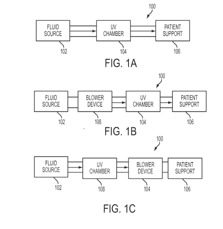
[0017] Referring now to the drawings, in its most basic form a system 100
according to
the present disclosure generally includes a fluid source 102, a UV chamber 104
and a patient
support 106 as depicted in FIG. 1(a). Fluid source 102 functions as a supply
of air or other fluid
in an untreated, unsterilized state. The fluid is then passed through UV
chamber 104 where it is
sterilized using ultra-violet technology as is further described below. The
sterilized fluid is then
passed to patient support 106 to inflate (or maintain a semi-inflated state
of) patient support 106.
[0018] FIG. 1(b) is another generalized embodiment of system 100 wherein
a blower
device 108 such as a fan, compressor, or other device is used to force air
from fluid source 102
through UV chamber 104 and into patient support 106. In FIG. 1(b), blower
device 108 is
situated upstream of UV chamber 104. In yet another embodiment as shown in
FIG. 1(c),
blower device 108 is situated downstream of UV chamber 104. In either case,
blower device 104
CA 02905718 2015-09-11
WO 2014/149545 PCT/US2014/019463
-5-
104 forces unsterilized air through UV chamber 104 for sterilization prior to
being provided to
patient support 106.
[0019] In any of the above-mentioned embodiments, UV chamber 104 may be
positioned
within an existing valve assembly of the fluid supply circuit for the patient
support 106 and
coupled to the valve assembly electronics in the manner described below. More
specifically,
referring to FIG. 2, valve assembly 200 generally includes a housing 202, an
inlet 204 and an
outlet 206. Inlet 204 is in fluid communication with fluid source 102 (FIGS.
1(a), (c)) or blower
device 108 (FIG. 1(b)) and outlet 206 is in fluid communication with patient
support 106 (FIGS.
1(a)-(b)) or blower device 104 (FIG. 1(c)). Valve assembly 200 further
includes control and
power circuitry 208, which includes, among other things, a flow sensor 210 and
a connector 212.
[0020] UV chamber 104 generally includes a housing 214, an inlet 216, an
outlet 218 and
a cover 220. Inlet 216 is in fluid communication with valve assembly inlet 204
via conduit 222.
Outlet 218 is in fluid communication with valve assembly outlet 206 via
conduit 224. Cover 220
includes a printed circuit board and electronics as described below, which
receive power from
and are in communication with valve control and power circuitry 208.
Specifically, a cable 226
is connected to cover 220 and a connector 228, which mates with connector 212
of valve control
and power circuitry 208.
[0021] UV chamber 104 is shown in more detail in FIG. 3. As shown,
housing 214
defines an interior space 300 which is in flow communication with inlet 216
and outlet 218.
Housing 214 also includes an opening 302. When attached to housing 214, cover
220 entirely
covers opening 302. Cover 220 includes a printed circuit board ("PCB") 304,
which is mounted
to an interior side 306 of cover 220 using standoffs or other conventional
mounting techniques.
In one embodiment, PCB 304 is a standard, double sided FR4, 0.062 inch copper
clad PCB.
Electronics 308 are mounted to the top side of PCB 304 (i.e., facing interior
side 306 of cover
220) and connected to cable 226 and connector 228 as is further described
below. PCB 304 also
includes at least one UV light source 310 (three shown), which is mounted to
the bottom side of
PCB 304. In one embodiment of the disclosure, light source 310 is a UVC LED as
is further
described below. More or fewer than three light sources 310 may be used. When
cover 220 is
attached to housing 214 (using screws or other conventional fastening
techniques), the bottom
CA 02905718 2015-09-11
WO 2014/149545 PCT/US2014/019463
-6-
side of PCB 304 and more particularly light sources 310 are directed toward
interior space 300.
As should be apparent from the foregoing, light sources 310 may thereby
sterilize the fluid
flowing through UV chamber 104.
[0022] In one embodiment of the present disclosure, light sources 310 are
ultraviolet C
wavelength LEDs, which are LEDs that emit ultraviolet light in the sub range
of wavelengths
from 100 nm to 290 nm. Light sources 310 may be LEDs, tubes, bulbs or a
combination thereof.
In one embodiment, light sources 310 emit ultraviolet light having wavelengths
of between 255
nm and 285 nm, or more specifically at a wavelength of about 280 nm. Light
sources 310
emitting this wavelength have demonstrated the result of providing
sterilization of almost any
surface or media. In the present disclosure, light sources 310 bombard the
potentially germ-
laden fluid flowing through UV chamber 104 with ultraviolet C light and
viruses, bacteria and
mold spores are destroyed. All of the fluid (e.g., air) provided to patient
support 106 will be
purified or processed in this manner. By processing the fluid provided to
patient support 106
with light sources 310, UV chamber 104 reduces the spread of airborne germs
and other
contaminants, kills odors, and provides a filterless, quiet, energy efficient
fluid processing
system.
[0023] It should be understood that the fluid provided to patient support
106 may be
purified or processed by light sources 310 at any point upstream of patient
support 106, and the
depicted embodiments are only examples. In any such embodiment, however, all
or substantially
all of the fluid passed to patient support 10 should be passed through UV
chamber 104 such that
all or substantially all of the fluid passed through UV chamber 104 is passed
under light sources
310.
[0024] It should be further understood that housing 214, while shown as a
substantially
rectangular enclosure in FIG. 3, may be formed in any of a variety of shapes
which permit the
flow of fluid between inlet 216 and outlet 218. For example, housing 214 may
be formed such
that the passage between inlet 216 and outlet 218 is reduced in any dimension
shown, or is
curved or cylindrical, or formed as a conduit between inlet 216 and outlet 218
in any appropriate
shape. Moreover, UV chamber 104 may be disposed inside patient support 106 or
outside
patient support 106 as depicted in the figures described above.
CA 02905718 2015-09-11
WO 2014/149545 PCT/US2014/019463
-7-
[0025] Referring now to FIG. 4, electronics 308 of PCB 304 generally
include a
microcontroller 400, enable logic 402, a Hall Effect interlock 404, a light
source driver / current
limit circuit 406 and light sources 310. In one embodiment, microcontroller
400 is the
PIC18F13K22-I/55 device manufactured by Microchip. Microcontroller 400 is
connected to
connector 228, which provides power, ground and control signals from valve
control / power
circuitry 208 to electronics 308. Microcontroller 400 provides control signals
to enable logic
402 to control light sources 310 in the manner described below. Enable logic
402 provides
control signals to circuit 406 based on the control signals from
microcontroller 400 and an input
signal from Hall Effect interlock 404. As such, depending upon the signals
provided to enable
logic 402, circuit 406 either activates or deactivates light sources 310 and
limits the current
provided to light sources 310 to approximately 20 mA.
[0026] Referring back to FIG. 3, Hall Effect interlock 404 includes a
Hall Effect sensor
312 mounted to PCB 304 and a magnet 314 mounted to housing 214. When cover 220
is
attached to housing 214 and covering opening 302, sensor 312 is in close
proximity to magnet
314, and therefore provides a signal to enable logic 402 indicating that UV
chamber 104 is
closed. When cover 220 is removed from housing 214 or no longer covers opening
302, sensor
312 provides a different signal to enable logic 402 indicating that UV chamber
104 is opened. It
should be understood that while Hall Effect sensor 312 is described herein,
any of a variety of
different sensing technologies may be used to provide signals to enable logic
402 indicating
whether UV chamber 104 is opened or closed. For example, a switch could be
attached to cover
220 such that it is mechanically toggled when cover 220 is removed from
opening 302.
Alternatively, cover 220 could include an optical sensor, an IR sensor, or
other suitable sensor
that switches from one state to another when cover 220 is removed from opening
302.
[0027] As is described below, enable logic 402 uses the signals from Hall
Effect
interlock 404 to ensure that light sources 310 are deactivated whenever cover
220 is removed
from opening 302. Additionally, enable logic 402 deactivates light sources 310
when fluid is not
not flowing through UV chamber 104 as indicated by flow sensor 210 (FIG. 2). A
flowchart
depicting a control loop 500 implemented by electronics 308 for controlling
the activation of
light sources 310 is provided in FIG. 5. Unless activated by control loop 500,
light sources 310
are by default deactivated. At block 502, it is determined whether fluid is
being provided to
CA 02905718 2015-09-11
WO 2014/149545 PCT/US2014/019463
-8-
patient support 106 (i.e., whether fluid is flowing through UV chamber 104).
In the
implementation of FIG. 2, this determination is made based on the signal
provided by flow
sensor 210. If fluid is flowing through conduit 222, flow sensor 210 provides
a signal to
circuitry 208 indicating such flow. Circuitry 208 in turn provides a signal
through connectors
212 and 228 and cable 226 to electronics 308 on PCB 304. Specifically, this
signal is provided
to microcontroller 400, which provides a control signal to enable logic 402
indicating that fluid is
flowing through UV chamber 104. If fluid is not flowing, flow sensor 210
provides a different
signal to circuitry 208, which causes microprocessor 400 to provide a
different control signal to
enable logic 402. If the determination at block 502 is that fluid is not
flowing, then control loop
500 continues to monitor the status of fluid flow and light sources 310 remain
deactivated.
[0028] If, on the other hand, it is determined at block 502 that fluid is
flowing through
UV chamber 104, then the determination is made at block 504 whether housing
214 is closed.
More specifically, Hall Effect interlock 404 provides one input to enable
logic 402 if cover 220
is covering opening 302 and another input to enable logic 402 if cover 220 is
not covering
opening 302. This input causes enable logic 402 to provide a control signal to
light source driver
/ current limit circuit 406 to enable light sources 310 (block 506) if cover
220 is in place (i.e.,
housing 214 is closed) and to disable light sources 310 (block 508) if cover
220 is not in place
(i.e., housing 214 is opened). It should be noted that interlock 404 operates
independently of
microprocessor 400, and therefore functions to prevent activation of light
sources 310 even in the
event that microprocessor 400 malfunctions. As such, control loop 500 enables
light sources 310
only if fluid is flowing through UV chamber 104 and housing 214 is closed.
Light sources 310
are disabled if either fluid is not flowing through housing 214 or housing 214
is opened.
[0029] FIG. 6 provides a schematic diagram of one implementation of the
high level
diagram of electronics 308 of FIG. 4 and control loop 500 of FIG. 5. As shown,
in addition to
the components depicted in FIG. 4, electronics 308 also includes a power
indicator 600 which is
mounted to housing 214 and activated when power is supplied to electronics
308, three light
source activated indicators 602, a Hall Effect active indicator 604, and
various passive
components. The light source indicators 602 are also mounted to housing 214
and are activated
when light sources 310 are activated, thereby providing a visual indication
external to UV
chamber 104 that light sources 310 are activated. Similarly, indicator 604 is
mounted to housing
CA 02905718 2015-09-11
WO 2014/149545 PCT/US2014/019463
-9-
housing 214 and activated when Hall Effect interlock 404 is activated (i.e.,
when cover 220
covers opening 302), thereby providing a visual indication external to UV
chamber that interlock
404 is activated.
[0030] Depending upon how microcontroller 400 is programmed, each of
light sources
310 may be activated individually and in a sequential order, or all of light
sources 310 may be
activated simultaneously. If one of the light sources 310 is sensed as being
defective,
microcontroller 400 may skip activation of that light source 310 if sequential
activation is
implemented.
[0031] While this disclosure includes particular examples, it is to be
understood that the
disclosure is not so limited. Numerous modifications, changes, variations,
substitutions, and
equivalents will occur to those skilled in the art without departing from the
spirit and scope of
the present disclosure upon a study of the drawings, the specification, and
the following claims.