Note: Descriptions are shown in the official language in which they were submitted.
CA 02906022 2015-09-11
WO 2014/152199
PCT/US2014/027062
ANTIBODY DRUG CONJUGATE (ADC) PURIFICATION
RELATED APPLICATIONS
This application claims the benefit of U.S. Provisional Application No.
61/792,834,
filed on March 15, 2013. The contents of the aforementioned priority document
are hereby
incorporated by reference in its entirety.
BACKGROUND OF THE INVENTION
Antibody drug conjugates (ADC) are an emerging class of potent anti-cancer
agents,
which have recently demonstrated remarkable clinical benefit. ADCs are
comprised of a
cytotoxic agent attached to an antibody via a stable linker. Putatively, by a
series of events,
including antigen binding at the cell surface, endocytosis, trafficking to the
lysosome, ADC
degradation, release of payload, interruption of cellular processing (e.g.
mitosis) and
apoptosis, ADCs may destroy cancer cells possessing an over-expression of cell-
surface
proteins. ADCs combine the antigen-driven targeting properties of monoclonal
antibodies
with the potent anti-tumor effects of cytoxic agents. For example, in 2011
ADCETRIS (an
anti-CD30 antibody-MMAE ADC) gained regulatory approval for the treatment of
refractory
Hodgkin lymphoma and systemic anaplastic lymphoma.
Studies have demonstrated deleterious effects of a high drug loaded ADCs (Liu
et al.
(2010) Analy. Chem. 82:5219). These deleterious effects of higher levels of
conjugation
include increased propensity towards aggregate formation (King et al. (2002) J
Med. Chem.
45:4336; Hollander et al. (2008) Bioconjugate Chem 19:358; Burke et al. (2009)
Bioconjugate Chem 20:1242; and Zhao et al. (2011) J Med. Chem. 54:3606).
Controlling the drug load of an ADC has been attempted using various methods,
including: (i) limiting the molar excess of drug-linker intermediate or linker
reagent relative
to antibody, (ii) limiting the conjugation reaction time or temperature, and
(iii) partial or
limiting reductive conditions for cysteine thiol modification. While reduction
methods that
limit the number of attachment sites on the antibody have been used to achieve
ADCs with
fewer drugs per antibody (Alley et al. (2004) Proc Amer Assoc Cancer Res
45:Abst 627),
there remains a need for methods and compositions that can provide optimal
drug loaded
species.
1
CA 02906022 2015-09-11
WO 2014/152199
PCT/US2014/027062
SUMMARY OF INVENTION
The invention provides effective methods for separating antibody drug
conjugates
(ADCs) having different drug loads, as well as compositions obtained using
such methods.
The invention also provides compositions where higher drug load species of
ADCs are
removed.
The invention features, in one embodiment, a method of obtaining a composition
comprising Antibody Drug Conjugates (ADCs), said method comprising contacting
an ADC
mixture comprising a drug loaded species of 4 or less and a drug loaded
species of 6 or more
with a hydrophobic resin, wherein the amount of hydrophobic resin contacted
with the ADC
mixture is sufficient to allow binding of the drug loaded species of 6 or more
to the resin but
does not allow significant binding of the drug loaded species of 4 or less;
and removing the
hydrophobic resin from the ADC mixture, such that the composition comprising
ADCs is
obtained, wherein the composition comprises less than 15% of the drug loaded
species of 6 or
more, and wherein the ADC comprises an antibody conjugated to an auristatin.
In one
embodiment, the composition comprises less than 10% of the drug loaded species
of 6 or
more. In another embodiment, the composition comprises 5% or less of the drug
loaded
species of 6 or more.
In one embodiment, the method of the invention includes the use of a
hydrophobic
resin weight which is 3 to 12 times the weight of the drug loaded species of 6
or more in the
ADC mixture. In one embodiment, the hydrophobic resin weight is 4 to 8 times
the weight of
the drug loaded species of 6 or more in the ADC mixture. In a further
embodiment, the
hydrophobic resin weight is 5 to 10 times the weight of the drug loaded
species of 6 or more
in the ADC mixture.
In yet another embodiment, the hydrophobic resin weight is 6 to 12 times the
weight
of the drug loaded species of 6 or more in the ADC mixture, and wherein the
ADC mixture
comprises between 0 to 1 N NaC1, or an equivalent ionic strength thereof. In a
further
embodiment, the hydrophobic resin weight is 3 to 6 times the weight of the
drug loaded
species of 6 or more in the ADC mixture, and wherein the ADC mixture comprises
between 1
to 2N NaC1, or an equivalent ionic strength thereof. In yet another
embodiment, the
hydrophobic resin weight is 3 to 7 times the weight of the drug loaded species
of 6 or more in
the ADC mixture, and wherein the auristatin is monomethylauristatin E (MMAE).
In a
2
CA 02906022 2015-09-11
WO 2014/152199
PCT/US2014/027062
further embodiment, the hydrophobic resin weight is 5 to 10 times the weight
of the drug
loaded species of 6 or more in the ADC mixture, and wherein the auristatin is
monomethylauristatin F (MMAF). In a further embodiment, the hydrophobic resin
weight is
3 to 7 times the weight of the drug loaded species of 6 or more in the ADC
mixture, and
wherein the auristatin is monomethylauristatin E (MMAE).
The invention further provides a method of producing a composition comprising
ADCs with an average Drug-to-Antibody Ratio (DAR) of 4.5 or less and
comprising less
than 15% undesired ADCs, said method comprising contacting an ADC mixture with
a
hydrophobic resin, wherein the amount of hydrophobic resin contacted with the
ADC mixture
is sufficient to allow binding of the undesired ADCs; and removing the
hydrophobic resin
from the ADC mixture, such that the composition with an average DAR of 4.5 or
less and
comprising less than 15% undesired ADCs is produced, wherein the ADC comprises
an
antibody conjugated to an auristatin. In one embodiment, the composition with
an average
DAR of 4 or less comprises less than 10% undesired ADCs. In one embodiment,
the
undesired ADCs are 6 and 8 drug loaded species. In a further embodiment, the
undesired
ADCs are an 8 drug loaded species.
In one embodiment, the amount of hydrophobic resin added to the ADC mixture is
a
resin weight which is 3 to 12 times the weight of the undesired ADCs in the
ADC mixture.
In a further embodiment, the amount of hydrophobic resin added to the ADC
mixture
is a resin weight which is 4 to 8 times the weight of the drug loaded species
of 6 or more in
the ADC mixture. In yet another embodiment, the amount of hydrophobic resin
added to the
ADC mixture is a resin weight which is 5 to 7 times the weight of the drug
loaded species of
6 or more in the ADC mixture. In a further embodiment, the hydrophobic resin
weight is 6 to
12 times the weight of the drug loaded species of 6 or more in the ADC mixture
and wherein
the ADC mixture comprises between 0 to 1 N NaC1, or an equivalent ionic
strength thereof.
In one embodiment, the hydrophobic resin weight is 3 to 6 times the weight of
the drug
loaded species of 6 or more in the ADC mixture and the ADC mixture comprises
between 1
to 2 N NaC1, or an equivalent ionic strength thereof. In another embodiment,
the
hydrophobic resin weight is 3 to 7 times the weight of the drug loaded species
of 6 or more in
the ADC mixture, and wherein the auristatin is monomethylauristatin E (MMAE).
In still
another embodiment, the hydrophobic resin weight is 5 to 10 times the weight
of the drug
3
CA 02906022 2015-09-11
WO 2014/152199
PCT/US2014/027062
loaded species of 6 or more in the ADC mixture, and wherein the auristatin is
monomethylauristatin F (MMAF). In yet a further embodiment, the hydrophobic
resin
weight is 3 to 7 times the weight of the drug loaded species of 6 or more in
the ADC mixture,
and wherein the auristatin is monomethylauristatin E (MMAE). In one
embodiment, the
hydrophobic resin weight is 5 to 10 times the weight of the drug loaded
species of 6 or more
in the ADC mixture, and wherein the auristatin is monomethylauristatin F
(MMAF).
In one embodiment, the method of the invention is used to obtain a composition
comprising ADCs with an average DAR of 4.5 or less. In one embodiment, the
method of
the invention is used to obtain a composition comprising ADCs with an average
DAR of 4 or
less. In one embodiment, the method of the invention is used to obtain a
composition
comprising ADCs with an average DAR of 3.5 or less. In one embodiment, the
method of
the invention is used to obtain a composition comprising ADCs with an average
DAR of 3 or
less. In one embodiment, the composition has an average DAR of 2.5 or less.
In one embodiment, the method of the invention features adding a hydrophobic
resin
to an ADC mixture to form a resin mixture, wherein the resin mixture has an
ionic strength
which is equal to or higher than the ADC mixture.
In a further embodiment of the invention, the ADC mixture was obtained
following an
ultrafiltration /diafiltration process.
In one embodiment of the invention, the hydrophobic resin used in the methods
of the
invention is a butyl hydrophobic resin.
In one embodiment of the invention, the method of the invention is a batch
process or,
alternatively, a circulation process or a flow through process.
In one embodiment, the invention features a composition obtained using the
methods
described herein.
The invention further features a composition comprising ADCs, wherein 70% of
the
ADCs have a drug loaded species of 4 or less, wherein the ADC comprises an
anti-EGFR
antibody (e.g., antibody 1) and an auristatin (e.g., MMAE or MMAF). In one
embodiment of
the invention, the composition comprises 75% ADCs present having a drug loaded
species of
4 or less. In another embodiment of the invention, the composition comprises
80% ADCs
present having a drug loaded species of 4 or less. In one embodiment of the
invention, the
composition comprises 85% ADCs present having a drug loaded species of 4 or
less. In a
4
CA 02906022 2015-09-11
WO 2014/152199
PCT/US2014/027062
further embodiment of the invention, the composition comprises 90% ADCs having
a drug
loaded species of 4 or less. In one embodiment, the composition of the
invention comprises
95% ADCs present having a drug loaded species of 4 or less. In another
embodiment, the
composition of the invention comprises ADCs wherein 70% or more of the ADCs
have a
drug loaded species of 4 to 1, 3 to 1, or, alternatively, 2 to 1.
The invention further features a composition comprising ADCs with an average
DAR
of 4.5 or less, wherein the ADC comprises an anti-EGFR antibody (e.g.,
antibody 1) and an
auristatin (e.g., MMAE or MMAF). In one embodiment of the invention, the
composition
comprises ADCs having an average DAR of 4 or less. In another embodiment of
the
invention, the composition comprises ADCs having an average DAR of 4 or less.
In yet
another embodiment of the invention, the composition comprises ADCs having an
average
DAR of 3.5 or less. In one embodiment of the invention, the composition
comprises ADCs
having an average DAR of 3 or less. In yet another embodiment of the
invention, the
composition comprises ADCs having an average DAR of 2.5 or less. In another
embodiment,
the composition of the invention comprises anti-EGFR ADCs (e.g., antibody 1
conjugated to
MMAE or MMAF) with an average DAR of 4.5 to 0.001, 4 to 0.001, 3.5 to 0.001, 3
to 0.001,
or, alternatively, 2.5 to 0.001.
In one embodiment, the methods and compositions of the invention include an
ADC
comprising an anti-Epidermal Growth Factor Receptor (EGFR) antibody. In one
embodiment of the invention, the anti-EGFR antibody comprises a light chain
variable region
comprising a Complementarity Determining Region 1 (CDR1), CDR2, and CDR3
domain
comprising the amino acid sequence as set forth in SEQ ID NO: 7, SEQ ID NO: 8,
and SEQ
ID NO: 9, respectively, and comprises a heavy chain variable region comprising
a CDR1,
CDR2, and CDR3 domain comprising the amino acid sequence as set forth in SEQ
ID NO: 2,
SEQ ID NO: 3, and SEQ ID NO: 4. In another embodiment of the invention, the
anti-EGFR
antibody comprises a light chain variable region comprising the amino acid
sequence set
forth in SEQ ID NO: 6 and a heavy chain variable region comprising the amino
acid
sequence set forth in SEQ ID NO: 1.
In another embodiment of the invention, the anti-EGFR ADC comprises CDRs
(i.e.,
light chain CDR1, CDR2, and CDR3) described in the light chain variable region
set forth in
5
CA 02906022 2015-09-11
WO 2014/152199
PCT/US2014/027062
the amino acid sequence of SEQ ID NO: 6, and CDRs (i.e., heavy chain CDR1,
CDR2, and
CDR3) described in the amino acid sequence of SEQ ID NO: 1.
In a further embodiment, the methods and compositions of the invention feature
an
auristatin which is either monomethylauristatin E (MMAE) or
monomethylauristatin F
(MMAF).
In one embodiment, the MMAE is conjugated to the antibody via a valine-
citrulline
(vc) linker (vc-MMAE). In another embodiment, the MMAF is conjugated to the
antibody
via a maleimidocaproyl linker (mc-MMAF).
In one embodiment, the composition of the invention is a pharmaceutical
composition.
Also included in the invention are methods of treating cancer in a subject
comprising
administering a composition described herein to the subject such that cancer
is treated. In
one embodiment, the cancer is selected from the group consisting of squamous
tumors
(including, squamous tumors of the lung, head and neck, cervical, etc.),
glioblastoma, glioma,
non-small cell lung cancer, lung cancer, colon cancer, head and neck cancer,
breast cancer,
squamous cell tumors, anal cancer, skin cancer, and vulvar cancer.
In one embodiment, the compositions of the invention are used to treat
glioblastoma
multiforme.
In one embodiment, the compositions of the invention are used to treat a solid
tumor
having overexpression of EGFR. In one embodiment, the compositions of the
invention are
used to treat a subject having an advanced solid tumor likely to overexpress
EGFR.
In one embodiment, the compositions of the invention are administered
intravenously.
BRIEF DESCRIPTION OF THE DRAWINGS
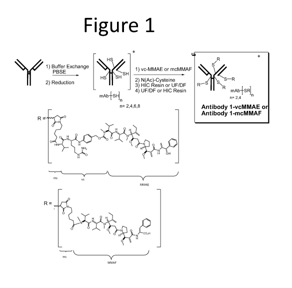
Figure 1 provides an overview of the processes described in the examples,
including
reduction of Antibody 1, conjugation of Antibody 1 to vcMMAE, and purification
of the
ADC using batch purification with HIC resin.
Figure 2 graphically depicts HIC HPLC analysis of an Antibody 1 ADC solution
before and
after HIC resin batch purification.
Figure 3 provides an overview of the process described in Example 6 for the
purification of
ADC mixtures with average DARs of 2.7, 4, and 5.5.
6
CA 02906022 2015-09-11
WO 2014/152199
PCT/US2014/027062
DETAILED DESCRIPTION
I. Definitions
In order that the present invention may be more readily understood, certain
terms are
first defined. In addition, it should be noted that whenever a value or range
of values of a
parameter are recited, it is intended that values and ranges intermediate to
the recited values
are also intended to be part of this invention.
The term "antibody-drug-conjugate" or "ADC" refers to a binding protein, such
as an
antibody or antigen binding fragment thereof, chemically linked to one or more
chemical
drug(s) (also referred to herein as agent(s)) that may optionally be
therapeutic or cytotoxic
agents. In a preferred embodiment, an ADC includes an antibody, a cytotoxic or
therapeutic
drug, and a linker that enables attachment or conjugation of the drug to the
antibody. An
ADC typically has anywhere from 1 to 8 drugs conjugated to the antibody,
including drug
loaded species of 2, 4, 6, or 8. Non-limiting examples of drugs that may be
included in the
ADCs are mitotic inhibitors, antitumor antibiotics, immunomodulating agents,
vectors for
gene therapy, alkylating agents, antiangiogenic agents, antimetabolites, boron-
containing
agents, chemoprotective agents, hormones, antihormone agents, corticosteroids,
photoactive
therapeutic agents, oligonucleotides, radionuclide agents, topoisomerase
inhibitors, tyrosine
kinase inhibitors, and radio sensitizers.
The terms "anti-Epidermal Growth Factor antibody drug conjugate," "anti-EGFR
antibody drug conjugate," or "anti-EGFR ADC", used interchangeably herein,
refer to an
ADC comprising an antibody that specifically binds to EGFR, whereby the
antibody is
conjugated to one or more chemical agent(s). In one embodiment, the anti-EGFR
antibody
drug conjugate is Antibody 1 conjugated to an auristatin, e.g., MMAE or MMAF.
Amino
acid sequences corresponding to the light and heavy chains of Antibody 1 are
provided in
SEQ ID NOs: 1-10.
The term "auristatin", as used herein, refers to a family of antimitotic
agents.
Auristatin derivatives are also included within the definition of the term
"auristatin".
Examples of auristatins include, but are not limited to, auristatin E (AE),
monomethylauristatin E (MMAE), monomethylauristatin F (MMAF), and synthetic
analogs
of dolastatin.
7
CA 02906022 2015-09-11
WO 2014/152199
PCT/US2014/027062
The term "drug-to-antibody ratio" or "DAR" refers to the number of drugs,
e.g.,
auristatin, attached to the antibody of the ADC. The DAR of an ADC can range
from 1 to 8,
although higher loads, e.g., 10, are also possible depending on the number of
linkage site on
an antibody. The term DAR may be used in reference to the number of drugs
loaded onto an
individual antibody, or, alternatively, may be used in reference to the
average or mean DAR
of a group of ADCs.
The term "undesired ADC species", as used herein, refers to any drug loaded
species
which is to be separated from an ADC species having a different drug load. In
one
embodiment, the term undesired ADC species may refer to drug loaded species of
6 or more,
i.e.., ADCs with a DAR of 6 or more, including DAR6, DAR7, DAR8, and DAR
greater than
8 (i.e., drug loaded species of 6, 7, 8, or greater than 8). In a separate
embodiment, the term
undesired ADC species may refer to drug loaded species of 8 or more, i.e.,
ADCs with a
DAR of 8 or more, including DAR8, and DAR greater than 8 (i.e., drug loaded
species of 8,
or greater than 8).
The term "ADC mixture", as used herein, refers to a composition containing a
heterogeneous DAR distribution of ADCs. In one embodiment, an ADC mixture
contains
ADCs having a distribution of DARs of 1 to 8, e.g., 2, 4, 6, and 8 (i.e., drug
loaded species of
2, 4, 6, and 8). Notably, degradation products may result such that DARs of 1,
3, 5, and 7
may also be included in the mixture. Further, ADCs within the mixture may also
have DARs
greater than 8. The ADC mixture results from interchain disulfide reduction
followed by
conjugation. In one embodiment, the ADC mixture comprises both ADCs with a DAR
of 4 or
less (i.e., a drug loaded species of 4 or less) and ADCs with a DAR of 6 or
more (i.e., a drug
loaded species of 6 or more).
As used herein, the term "hydrophobic resin" or "hydrophobic interaction
resin"
refers to a medium consisting of hydrophobic ligands used for purposes of
purifying a
mixture of molecules, wherein the presence of hydrophobic surface moieties on
the
molecules within the mixture facilitates an interaction with the medium such
that interacting
molecules are at least transiently bound to the medium. In one embodiment, the
hydrophobic resin is a resin comprising alkyl moieties, e.g., a C4-C8 alkyl
hydrophobic resin,
which is a resin comprising a four to eight straight or branched chain carbon
membered
alkane radical group such as butyl, pentyl, hexyl, heptyl, or octyl group
coupled to a solid
8
CA 02906022 2015-09-11
WO 2014/152199
PCT/US2014/027062
support (e.g., agarose, silica, etc.). Examples of hydrophobic alkyl resins
include a
hydrophobic butyl resin or a hydrophobic hexyl resin. In one embodiment, the
hydrophobic
resin is a resin comprising aryl moieties, e.g., a hydrophobic phenyl resin.
In one
embodiment, the hydrophobic resin comprises an alkenyl moiety. In one
embodiment, the
hydrophobic resin comprises an ether moiety. In yet another embodiment, the
hydrophobic
resin comprises a phenyl moiety. The hydrophobic moieties (e.g., alkyl, aryl,
etc.) may be
linked to an inert substance (e.g., silica, agarose, and/or other
polysaccharide polymers). In
one embodiment, the resin is a methacrylate resin.
The term "ionic strength" broadly refers to a measure of the concentration of
ions in a
solution, i.e., the conductivity of a solution. Exemplary salts that may be
used to modulate
the ionic strength of a solution include, but are not limited to sodium
bromide, sodium
chloride, sodium citrate, sodium iodide, sodium phosphate, sodium sulfate,
potassium
bromide, potassium chloride, potassium citrate, potassium iodide, potassium
phosphate,
potassium sulfate, cesium chloride, lithium chloride, or other salts of
ammonia (e.g., NH4C1,
(NH4)2SO4), carbonates (NaHCO3), citric acid (NaH2(C3H50(C00)3),
Na2H(C3H50(C00)3),
Na3H(C3H50(C00)3)), phosphoric acid (e.g., KH2PO4, K2HPO4, K3PO4), nitrates
(KNO3),
or any mixture of these components. Those skilled in the art appreciate that
both the anion
and the cation can be varied as is known to the person skilled in the art, as
long as sufficient
ionic strength is provided without precipitation or other undesired side-
effects.
The term "anti-EGFR antibody" is meant to refer to an antibody that
specifically
binds to EGFR. An antibody "which binds" an antigen of interest, i.e., EGFR,
is one capable
of binding that antigen with sufficient affinity such that the antibody is
useful in targeting a
cell expressing the antigen. Antibody 1 is an example of an anti-EGFR
antibody.
The term "antibody" broadly refers to an immunoglobulin (Ig) molecule,
generally
comprised of four polypeptide chains, two heavy (H) chains and two light (L)
chains, or any
functional fragment, mutant, variant, or derivative thereof, that retains the
essential target
binding features of an Ig molecule.
In a full-length antibody, each heavy chain is comprised of a heavy chain
variable
region (abbreviated herein as HCVR or VH) and a heavy chain constant region.
The heavy
chain constant region is comprised of three domains, CH1, CH2 and CH3. Each
light chain is
comprised of a light chain variable region (abbreviated herein as LCVR or VL)
and a light
9
CA 02906022 2015-09-11
WO 2014/152199
PCT/US2014/027062
chain constant region. The light chain constant region is comprised of one
domain, CL. The
VH and VL regions can be further subdivided into regions of hypervariability,
termed
complementarity determining regions (CDR), interspersed with regions that are
more
conserved, termed framework regions (FR). Each VH and VL is composed of three
CDRs
and four FRs, arranged from amino-terminus to carboxy-terminus in the
following order:
FR1, CDR1, FR2, CDR2, FR3, CDR3, FR4. Immunoglobulin molecules can be of any
type
(e.g., IgG, IgE, IgM, IgD, IgA and IgY) and class (e.g., IgG 1, IgG2, IgG 3,
IgG4, IgAl and
IgA2) or subclass.
The term "antigen-binding portion" of an antibody (or simply "antibody
portion"), as
used herein, refers to one or more fragments of an antibody that retain the
ability to
specifically bind to an antigen (e.g., hIL-13). It has been shown that the
antigen-binding
function of an antibody can be performed by fragments of a full-length
antibody. Such
antibody embodiments may also be bispecific, dual specific, or multi-specific
formats;
specifically binding to two or more different antigens. Examples of binding
fragments
encompassed within the term "antigen-binding portion" of an antibody include
(i) a Fab
fragment, a monovalent fragment consisting of the VL, VH, CL and CH1 domains;
(ii) a
F(abt)2 fragment, a bivalent fragment comprising two Fab fragments linked by a
disulfide
bridge at the hinge region; (iii) a Fd fragment consisting of the VH and CH1
domains; (iv) a
Fv fragment consisting of the VL and VH domains of a single arm of an
antibody, (v) a dAb
fragment (Ward et al., (1989) Nature 341:544-546, Winter et al., PCT
publication WO
90/05144 Al herein incorporated by reference), which comprises a single
variable domain;
and (vi) an isolated complementarity determining region (CDR). Furthermore,
although the
two domains of the Fv fragment, VL and VH, are coded for by separate genes,
they can be
joined, using recombinant methods, by a synthetic linker that enables them to
be made as a
single protein chain in which the VL and VH regions pair to form monovalent
molecules
(known as single chain Fv (scFv); see e.g., Bird et al. (1988) Science 242:423-
426; and
Huston et al. (1988) Proc. Natl. Acad. Sci. USA 85:5879-5883). Such single
chain antibodies
are also intended to be encompassed within the term "antigen-binding portion"
of an
antibody. Other forms of single chain antibodies, such as diabodies are also
encompassed.
Diabodies are bivalent, bispecific antibodies in which VH and VL domains are
expressed on
a single polypeptide chain, but using a linker that is too short to allow for
pairing between the
CA 02906022 2015-09-11
WO 2014/152199
PCT/US2014/027062
two domains on the same chain, thereby forcing the domains to pair with
complementary
domains of another chain and creating two antigen binding sites (see e.g.,
Holliger, P., et al.
(1993) Proc. Natl. Acad. Sci. USA 90:6444-6448; Poljak, R.J., et al. (1994)
Structure 2:1121-
1123). Such antibody binding portions are known in the art (Kontermann and
Dubel eds.,
Antibody Engineering (2001) Springer-Verlag. New York. 790 pp. (ISBN 3-540-
41354-5).
An "isolated antibody", as used herein, is intended to refer to an antibody
that is
substantially free of other antibodies having different antigenic
specificities (e.g., an isolated
antibody that specifically binds EGFR is substantially free of antibodies that
specifically bind
antigens other than EGFR). An isolated antibody that specifically binds EGFR
may,
however, have cross-reactivity to other antigens, such as EGFR molecules from
other species.
Moreover, an isolated antibody may be substantially free of other cellular
material and/or
chemicals.
The term "humanized antibody" refers to antibodies which comprise heavy and
light
chain variable region sequences from a non-human species (e.g., a mouse) but
in which at
least a portion of the VH and/or VL sequence has been altered to be more
"human-like", i.e.,
more similar to human germline variable sequences. In a particular embodiment,
the term
"humanized antibody" refers to an antibody or antibody variant, derivative or
fragment,
which specifically binds to an antigen of interest, and comprises a framework
(FR) region
having substantially the amino acid sequence of a human antibody, and
comprises CDRs
having substantially the amino acid sequence of a non-human antibody. As used
herein, the
term "substantially" in the context of a CDR refers to a CDR having an amino
acid sequence
at least 80%, preferably at least 85%, at least 90%, at least 95%, at least
98% or at least 99%
identical to the amino acid sequence of a non-human antibody CDR. In one
embodiment,
one type of humanized antibody is a CDR-grafted antibody, in which human CDR
sequences
are introduced into non-human VH and VL sequences to replace the corresponding
nonhuman CDR sequences.
As used herein, the term "CDR" refers to the complementarity determining
region
within antibody variable sequences. There are three CDRs in each of the
variable regions of
the heavy chain and the light chain, which are designated CDR1, CDR2 and CDR3,
for each
of the variable regions. The term "CDR set" as used herein refers to a group
of three CDRs
that occur in a single variable region capable of binding the antigen. The
exact boundaries of
11
CA 02906022 2015-09-11
WO 2014/152199
PCT/US2014/027062
these CDRs have been defined differently according to different systems. The
system
described by Kabat (Kabat et al., Sequences of Proteins of Immunological
Interest (National
Institutes of Health, Bethesda, Md. (1987) and (1991)) not only provides an
unambiguous
residue numbering system applicable to any variable region of an antibody, but
also provides
precise residue boundaries defining the three CDRs. These CDRs may be referred
to as Kabat
CDRs. Chothia and coworkers (Chothia &Lesk, J. Mol. Biol. 196:901-917 (1987)
and
Chothia et al., Nature 342:877-883 (1989)) found that certain sub- portions
within Kabat
CDRs adopt nearly identical peptide backbone conformations, despite having
great diversity
at the level of amino acid sequence. These sub-portions were designated as Li,
L2 and L3 or
H1, H2 and H3 where the "L" and the "H" designates the light chain and the
heavy chains
regions, respectively. These regions may be referred to as Chothia CDRs, which
have
boundaries that overlap with Kabat CDRs. Other boundaries defining CDRs
overlapping with
the Kabat CDRs have been described by Padlan (FASEB J. 9:133-139 (1995)) and
MacCallum (J Mol Biol 262(5):732-45 (1996)). Still other CDR boundary
definitions may
not strictly follow one of the above systems, but will nonetheless overlap
with the Kabat
CDRs, although they may be shortened or lengthened in light of prediction or
experimental
findings that particular residues or groups of residues or even entire CDRs do
not
significantly impact antigen binding. The methods used herein may utilize CDRs
defined
according to any of these systems, although preferred embodiments use Kabat or
Chothia
defined CDRs.
The term "disorder" refers to any condition that would benefit from treatment
with the
formulations of the invention, e.g. a disorder requiring treatment with the
anti-EGFR
antibody in the formulation. This includes chronic and acute disorders or
diseases including
those pathological conditions that predispose the subject to the disorder in
question.
The term "cancer" is meant to refer to or describe the physiological condition
in
mammals that is typically characterized by unregulated cell growth. Examples
of cancer
include, but are not limited to, carcinoma, lymphoma, blastoma, sarcoma, and
leukemia or
lymphoid malignancies. More particular examples of such cancers include
glioblastoma, non-
small cell lung cancer, lung cancer, colon cancer, head and neck cancer,
breast cancer,
squamous cell tumors, anal cancer, skin cancer, and vulvar cancer. In one
embodiment, the
compositions of the invention are administered to a patient having a tumor(s)
containing
12
CA 02906022 2015-09-11
WO 2014/152199
PCT/US2014/027062
amplifications of the EGFR gene, whereby the tumor expresses the truncated
version of the
EGFR de2-7. In one embodiment, the formulation of the invention comprising ADC-
1 may
be administered to a subject for the treatment of colorectal cancer, head and
neck cancer
(including, but not limited to, hypopharyngeal cancer, oropharyngeal cancer,
esophageal
cancer, laryngeal cancer, and oral cavity cancer), non-small cell lung cancer,
pancreatic
cancer, gastric cancer, and breast cancer. More particular examples of such
cancers include
squamous tumors (including, squamous tumors of the lung, head and neck,
cervical, etc.),
glioblastoma, glioma, non-small cell lung cancer, lung cancer, colon cancer,
head and neck
cancer, breast cancer, squamous cell tumors, anal cancer, skin cancer, and
vulvar cancer. In
one embodiment of the invention, the composition is used to treat a subject
having a solid
tumor, e.g., a solid tumor likely to over-express the Epidermal Growth Factor
Receptor
(EGFR), or glioblastoma multiforme.
The term "administering" as used herein is meant to refer to the delivery of a
substance (e.g., an anti-EGFR antibody drug conjugate) to achieve a
therapeutic objective
(e.g., the treatment of an EGFR- associated disorder). Modes of administration
may be
parenteral, enteral and topical. Parenteral administration is usually by
injection, and includes,
without limitation, intravenous, intramuscular, intraarterial, intrathecal,
intracapsular,
intraorbital, intracardiac, intradermal, intraperitoneal, transtracheal,
subcutaneous,
subcuticular, intraarticular, subcapsular, sub arachnoid, intraspinal and
intrasternal injection
and infusion.
The term "therapeutically effective amount" or "effective amount" of an
antibody as
used herein refers to an amount effective in the prevention or treatment or
alleviation of a
symptom of a disorder for the treatment of which the antibody is effective.
The term "treatment" refers to both therapeutic treatment and prophylactic or
preventative measures. Those patients in need of treatment include those
already with the
disorder as well as those in which the disorder is to be prevented.
Various aspects of the invention are described in further detail in the
following
subsections.
13
CA 02906022 2015-09-11
WO 2014/152199
PCT/US2014/027062
II. Methods for Purifying Antibody Drug Conjugates and Compositions
Thereof
The invention provides a method for purifying antibody drug conjugates (ADCs),
and
provides an effective means for removing undesired species of ADC, e.g., drug
loaded
species of 6 or more, from a mixture of ADCs. While the methods of the
invention may be
used to separate any drug loaded species, in a preferred embodiment, the
methods described
herein are used to separate high drug loaded ADCs from ADCs having optimal
drug to
antibody ratios (DARs), e.g. a DAR of 4 or less. In certain embodiments, the
methods of the
invention may provide numerous advantages over traditional column
chromatography,
including improved recovery, as fractionation and subsequent pooling of
fractions may not be
necessary. It should be understood that the methods and compositions described
throughout
may be used to purify anti-EGFR antibody-auristatin ADCs, particularly, in a
certain
embodiment, anti-EGFR ADCs comprising Antibody 1 either coupled via a
maleimidocaproyl linker to MMAF (mc-MMAF) or coupled via a maleimidocaproyl
valine-
citrulline linker to MMAE (vc-MMAE).
The method of the invention generally includes adding a hydrophobic resin to
an
ADC mixture such that undesired ADCs, i.e., higher drug loaded ADCs, bind the
resin and
can be selectively removed from the mixture. In certain embodiments,
separation of the
ADCs may be achieved by contacting an ADC mixture (e.g., a mixture comprising
a drug
loaded species of ADC of 4 or less and a drug loaded species of ADC of 6 or
more) with a
hydrophobic resin, wherein the amount of resin is sufficient to allow binding
of the drug
loaded species which is being removed from the ADC mixture. The resin and ADC
mixture
are mixed together, such that the ADC species being removed (e.g., a drug
loaded species of
6 or more) binds to the resin and can be separated from the other ADC species
in the ADC
mixture. The amount of resin used in the method is based on a weight ratio
between the
species to be removed and the resin, where the amount of resin used does not
allow for
significant binding of the drug loaded species that is desired. Thus, the
invention provides
methods for reducing the average DAR of an ADC mixture from, for example, 5.5
to less
than 4. Further, the purification methods described herein may be used to
isolate ADCs
having any desired range of drug loaded species, e.g., a drug loaded species
of 4 or less, a
drug loaded species of 3 or less, a drug loaded species of 2 or less, a drug
loaded species of 1
or less.
14
CA 02906022 2015-09-11
WO 2014/152199
PCT/US2014/027062
The invention provides a purification method whereby a certain species of
molecule(s) binds to a surface based on hydrophobic interactions between the
species and a
hydrophobic resin. In one embodiment, method of the invention refers to a
purification
process that relies upon the intermixing of a hydrophobic resin and a mixture
of ADCs,
wherein the amount of resin added to the mixture determines which species
(e.g., ADCs with
a DAR of 6 or more) will bind.
Following production and purification of an antibody from an expression system
(e.g.,
a mammalian expression system), the antibody is reduced and coupled to a drug
through a
conjugation reaction. The resulting ADC mixture often contains ADCs having a
range of
DARs, e.g., 1 to 8. In one embodiment, the ADC mixture comprises a drug loaded
species of
4 or less and a drug loaded species of 6 or more. According to the methods of
the invention,
the ADC mixture may be purified using a process, such as, but not limited to,
a batch process,
such that ADCs having a drug loaded species of 4 or less are selected and
separated from
ADCs having a higher drug load (e.g., ADCs having a drug loaded species of 6
or more).
Notably, the purification methods described herein may be used to isolate ADCs
having any
desired range of DAR, e.g., a DAR of 4 or less, a DAR of 3 or less, a DAR of 2
or less.
Thus, in one embodiment, the method of the invention comprises contacting an
ADC
mixture comprising a drug loaded species of 4 or less and a drug loaded
species of 6 or more
with a hydrophobic resin to form a resin mixture, wherein the amount of
hydrophobic resin
contacted with the ADC mixture is sufficient to allow binding of the drug
loaded species of 6
or more to the resin but does not allow significant binding of the drug load
species of 4 or
less; and removing the hydrophobic resin from the ADC mixture, such that the
composition
comprising ADCs is obtained, wherein the composition comprises less than 15%
of the drug
loaded species of 6 or more, and wherein the ADC comprises an antibody
conjugated to an
auristatin. In a separate embodiment, the method of the invention comprises
contacting an
ADC mixture comprising a drug loaded species of 4 or less and a drug loaded
species of 6 or
more with a hydrophobic resin to form a resin mixture, wherein the amount of
hydrophobic
resin contacted with the ADC mixture is sufficient to allow binding of the
drug loaded
species of 6 or more to the resin but does not allow significant binding of
the drug load
species of 4 or less; and removing the hydrophobic resin from the ADC mixture,
such that the
composition comprising ADCs is obtained, wherein the composition comprises
less than 15%
CA 02906022 2015-09-11
WO 2014/152199
PCT/US2014/027062
of the drug loaded species of 6 or more, and wherein the ADC comprises an
antibody
conjugated to an auristatin, wherein the hydrophobic resin weight is 3 to 12
times the weight
of the drug loaded species of 6 or more in the ADC mixture.
The method of the invention provides an effective method of separating low and
high
DAR ADCs. In one embodiment, the method may be performed using a batch
purification
method. The batch purification process generally includes adding the ADC
mixture to the
hydrophobic resin in a vessel, mixing, and subsequently separating the resin
from the
supernatant. For example, in the context of batch purification, a hydrophobic
resin may be
prepared in or equilibrated to the desired equilibration buffer. A slurry of
the hydrophobic
resin may thus be obtained. The ADC mixture may then be contacted with the
slurry to
adsorb the specific species of ADC(s) to be separated by the hydrophobic
resin. The solution
comprising the desired ADCs that do not bind to the hydrophobic resin material
may then be
separated from the slurry, e.g., by filtration or by allowing the slurry to
settle and removing
the supernatant. The resulting slurry can be subjected to one or more washing
steps. In order
to elute bound ADCs, the salt concentration can be decreased. In one
embodiment, the
process used in the invention includes no more than 50 g of hydrophobic resin.
Thus, in one embodiment of the invention, a batch method may be used to
contact an
ADC mixture comprising a drug loaded species of 4 or less and a drug loaded
species of 6 or
more with a hydrophobic resin to form a resin mixture, wherein the amount of
hydrophobic
resin contacted with the ADC mixture is sufficient to allow binding of the
drug loaded
species of 6 or more to the resin but does not allow significant binding of
the drug load
species of 4 or less; and removing the hydrophobic resin from the ADC mixture,
such that the
composition comprising ADCs is obtained, wherein the composition comprises
less than 15%
of the drug loaded species of 6 or more, and wherein the ADC comprises an
antibody
conjugated to an auristatin. In a separate embodiment, a batch method is used
to contact an
ADC mixture comprising a drug loaded species of 4 or less and a drug loaded
species of 6 or
more with a hydrophobic resin to form a resin mixture, wherein the amount of
hydrophobic
resin contacted with the ADC mixture is sufficient to allow binding of the
drug loaded
species of 6 or more to the resin but does not allow significant binding of
the drug load
species of 4 or less; and removing the hydrophobic resin from the ADC mixture,
such that the
composition comprising ADCs is obtained, wherein the composition comprises
less than 15%
16
CA 02906022 2015-09-11
WO 2014/152199
PCT/US2014/027062
of the drug loaded species of 6 or more, and wherein the ADC comprises an
antibody
conjugated to an auristatin, wherein the hydrophobic resin weight is 3 to 12
times the weight
of the drug loaded species of 6 or more in the ADC mixture.
Alternatively, in a separate embodiment, the invention may be performed using
a
circulation process, whereby the resin is packed in a container and the ADC
mixture is passed
over the hydrophobic resin bed until the specific species of ADC(s) to be
separated have been
removed. The supernatant (containing the desired ADC species) is then pumped
from the
container and the resin bed may be subjected to washing steps.
A circulation process may be used to contact an ADC mixture comprising a drug
loaded species of 4 or less and a drug loaded species of 6 or more with a
hydrophobic resin to
form a resin mixture, wherein the amount of hydrophobic resin contacted with
the ADC
mixture is sufficient to allow binding of the drug loaded species of 6 or more
to the resin but
does not allow significant binding of the drug load species of 4 or less; and
removing the
hydrophobic resin from the ADC mixture, such that the composition comprising
ADCs is
obtained, wherein the composition comprises less than 15% of the drug loaded
species of 6 or
more, and wherein the ADC comprises an antibody conjugated to an auristatin.
In a separate
embodiment, a circulation process is used to contact an ADC mixture comprising
a drug
loaded species of 4 or less and a drug loaded species of 6 or more with a
hydrophobic resin to
form a resin mixture, wherein the amount of hydrophobic resin contacted with
the ADC
mixture is sufficient to allow binding of the drug loaded species of 6 or more
to the resin but
does not allow significant binding of the drug load species of 4 or less; and
removing the
hydrophobic resin from the ADC mixture, such that the composition comprising
ADCs is
obtained, wherein the composition comprises less than 15% of the drug loaded
species of 6 or
more, and wherein the ADC comprises an antibody conjugated to an auristatin,
wherein the
hydrophobic resin weight is 3 to 12 times the weight of the drug loaded
species of 6 or more
in the ADC mixture.
Alternatively, in a separate embodiment of the invention, the purification
method may
be performed using a flow through process, whereby resin is packed in a
container, e.g., a
column, and the ADC mixture is passed over the packed resin such that the
desired ADC
species does not substantially bind to the resin and flows through the resin,
and the undesired
ADC species is bound to the resin. A flow through process may be performed in
a single
17
CA 02906022 2015-09-11
WO 2014/152199
PCT/US2014/027062
pass mode (where the ADC species of interest are obtained as a result of a
single pass
through the resin of the container) or in a multi-pass mode (where the ADC
species of interest
are obtained as a result of multiple passes through the resin of the
container). The flow
through process is performed such that the weight of resin selected binds to
the undesired
ADC population, and the desired ADCs (e.g., DAR 2-4) flow over the resin and
are collected
in the flow through after one or multiple passes.
In one embodiment of the invention, a flow through process may be used to
contact an
ADC mixture comprising a drug loaded species of 4 or less and a drug loaded
species of 6 or
more with a hydrophobic resin, wherein the amount of hydrophobic resin
contacted with the
ADC mixture is sufficient to allow binding of the drug loaded species of 6 or
more to the
resin but does not allow significant binding of the drug load species of 4 or
less, where the
drug load species of 4 or less passes over the resin and is subsequently
collected after one or
multiple passes, such that the composition comprising the desired ADCs (e.g.
DAR 2-4) is
obtained, wherein the composition comprises less than 15% of the drug loaded
species of 6 or
more, and wherein the ADC comprises an antibody conjugated to an auristatin.
In a separate
embodiment, a flow through process is used to contact an ADC mixture
comprising a drug
loaded species of 4 or less and a drug loaded species of 6 or more with a
hydrophobic resin
by passing the ADC mixture over the resin, wherein the amount of hydrophobic
resin
contacted with the ADC mixture is sufficient to allow binding of the drug
loaded species of 6
or more to the resin but does not allow significant binding of the drug load
species of 4 or
less, where the drug load species of 4 or less passes over the resin and is
subsequently
collected, such that the composition comprising ADCs is obtained, wherein the
composition
comprises less than 15% of the drug loaded species of 6 or more, and wherein
the ADC
comprises an antibody conjugated to an auristatin, wherein the amount of
hydrophobic resin
weight is 3 to 12 times the weight of the drug loaded species of 6 or more in
the ADC
mixture.
In one embodiment of the invention, the resin is washed with a one or more
washes
following the flow through process in order to further recover ADCs having the
desired DAR
range (found in the wash filtrate). For example, a plurality of washes having
decreasing
conductivity may be used to further recover ADCs having the DAR of interest.
The elution
material obtained from the washing of the resin may be subsequently combined
with the
18
CA 02906022 2015-09-11
WO 2014/152199
PCT/US2014/027062
filtrate resulting from the flow through process for improved recovery of ADCs
having the
DAR of interest.
The purification methods of the invention are based on the use of a
hydrophobic resin
to separate high vs. low drug loaded species of ADC. Hydrophobic resin
comprises
hydrophobic groups which interact with the hydrophobic properties of the ADCs.
Hydrophobic groups on the ADC interact with hydrophobic groups within the
hydrophobic
resin. The more hydrophobic a protein is the stronger it will interact with
the hydrophobic
resin.
Hydrophobic resin normally comprises a base matrix (e.g., cross-linked agarose
or
synthetic copolymer material) to which hydrophobic ligands (e.g., alkyl or
aryl groups) are
coupled. Many hydrophobic resins are available commercially. Examples include,
but are
not limited to, Phenyl Sepharosen4 6 Fast Flow with low or high substitution
(Pharmacia
LKB Biotechnology, AB, Sweden); Phenyl Sepharosen4 High Performance (Pharmacia
LKB
Biotechnology, AB, Sweden); Octyl SepharoseTm High Performance (Pharmacia LKB
Biotechnology, AB, Sweden); FractogelTm EMD Propyl or Fractogeff EMD Phenyl
columns (E. Merck, Germany); Macro-Prep Th4 Methyl or Macro-Prep. t-Butyl
Supports
(Bio-Rad, California); WP HI-Propyl (C3)Tm (J. T. Baker, New Jersey); and
Toyopearff
ether, hexyl, phenyl or butyl (TosoHaas, PA). In one embodiment, the
hydrophobic resin is a
butyl hydrophobic resin. In another embodiment, the hydrophobic resin is a
phenyl
hydrophobic resin. In another embodiment, the hydrophobic resin is a hexyl
hydrophobic
resin, an octyl hydrophobic resin, or a decyl hydrophobic resin. In one
embodiment, the
hydrophobic resin is a methacrylic polymer having n-butyl ligands (e.g.
TOYOPEARL
Butyl-600M).
The methods of the invention are based, at least in part, on the discovery
that a
hydrophobic resin may be used in certain amounts to selectively bind to ADCs
having certain
DARs. The binding between the resin and ADCs having a given DAR is dependent
upon the
weight of the resin relative to the weight of the ADCs which are to be removed
from the
ADC mixture. By varying the amount of resin load (calculated based on the dry
weight)
contacted to the ADC mixture relative to the specific drug load species weight
in the ADC
mixture, the resin will selectively bind ADCs having, for example, a DAR of 8
or more,
ADCs having a DAR of 6-8, ADCs having a DAR of 5-8, etc. Thus, the selectivity
of the
19
CA 02906022 2015-09-11
WO 2014/152199
PCT/US2014/027062
hydrophobic resin is dependent upon the weight ratio of the resin and the
weight of the ADC
species to be removed by the resin. In one embodiment, the hydrophobic resin
weight
contacted with the ADC mixture is 3 to 12 times the weight of the drug loaded
species of 6 or
more in the ADC mixture. In one embodiment, the hydrophobic resin weight
contacted with
the ADC mixture is 4 to 8 times the weight of the drug loaded species of 6 or
more in the
ADC mixture. In one embodiment, the hydrophobic resin weight contacted with
the ADC
mixture is 5 to 10 times the weight of the drug loaded species of 6 or more in
the ADC
mixture. In another embodiment, the hydrophobic resin weight contacted with
the ADC
mixture is 5 to 7 times the weight of the drug loaded species of 6 or more in
the ADC
mixture. In another embodiment, the hydrophobic resin weight contacted with
the ADC
mixture is 5 to 6 times the weight of the drug loaded species of 6 or more in
the ADC
mixture. For example, as described in Table 5 in the examples below, about 5-
10 weights of
resin (dry) were employed to reduce the 6 and 8 loaded drug species (3.2 mg
resin / 0.54 mg
6/8 load species = approx. 6), resulting in an enriched composition (enriched
for ADCs
having DARs of less than 6). In another example, as described in Table 7
below, a resin
weight of approximately 8 to 12 times that of the 6 and 8 drug load species
was proven to be
effective for reducing those species from the ADC mixture. In a further
example, as described
in Table 7 below, a resin weight of approximately 4 times that of the 6 and 8
drug load
species was proven to be effective for significantly reducing those species
from the ADC
mixture.
The selectivity of the resin for ADCs may be impacted by the ionic strength of
the
resin mixture in combination with the ratios identified herein as providing
appropriate load
resin:ADC weight ratios that result in selective binding of ADCs having a
certain desired
DAR distribution, e.g., a DAR distribution of 6-8. Generally, by decreasing
the ionic
strength of the resin mixture, the hydrophobic resin will be less adsorbent,
whereas an
increase in the ionic strength of the resin mixture will provide a more
adsorbent resin.
Adsorption of ADCs to hydrophobic resin is favored by high salt
concentrations, but the
actual concentrations may vary over a wide range depending on the nature of
the ADC and
the particular hydrophobic resin chosen. In general, Na, K or NH4 sulfates
effectively
promote ligand-protein interaction in hydrophobic resin. Salts may be
formulated that
influence the strength of the interaction as given by the following
relationship:
CA 02906022 2015-09-11
WO 2014/152199
PCT/US2014/027062
(NH4)2SO4>Na2SO4>NaC1>NH4C1>NaBr>NaSCN. In general, salt concentrations of
between about 0.75 and about 2 M ammonium sulfate or between about 1 and 4 M
NaC1 are
useful. In one embodiment, the resin mixture has an ionic strength of 0-2 N
NaCl. The ionic
strength of the ADC mixture may be adjusted prior to, concurrently with, or
following the
addition of the hydrophobic resin.
In one embodiment, the method of the invention uses a hydrophobic resin weight
which is 6 to 12 times the weight of the drug loaded species of 6 or more in
the ADC mixture
where the ADC mixture has an ionic strength of 0 to 1 N NaC1, or an equivalent
ionic
strength thereof. In a separate embodiment, the separation method of the
invention is carried
out using a hydrophobic resin weight which is 3 to 6 times the weight of the
drug loaded
species of 6 or more in the ADC mixture, and where the ADC mixture comprises
between 1
to 2 N NaC1, or an equivalent ionic strength thereof. The method may also be
carried out
using a hydrophobic resin weight which is 3 to 7 times the weight of the drug
loaded species
of 6 or more in the ADC mixture, and wherein the auristatin is
monomethylauristatin E
(MMAE). An additional method for separating a drug loaded species of 6 or more
includes
contact of an ADC mixture with a hydrophobic resin weight that is 5 to 10
times the weight
of the drug loaded species of 6 or more, wherein the auristatin is
monomethylauristatin F
(MMAF).
Additional purification or processing steps may be performed prior to or
following the
methods described herein. For example, in one embodiment, the ADC mixture is
obtained
following an ultrafiltration /diafiltration process. In another embodiment,
the purified
composition of ADCs is subjected to ultrafiltration / diafiltration.
In one embodiment, the method of the invention includes contacting an ADC
mixture
with a hydrophobic resin, wherein the amount of hydrophobic resin contacted
with the ADC
mixture is sufficient to allow binding of the drug loaded species of 6 or more
to the resin but
does not allow significant binding of the drug loaded species of 4 or less,
and removing the
hydrophobic resin from the ADC mixture. The hydrophobic resin binds the higher
drug
loaded species, e.g., drug loaded species of 6 or more, while the lower drug
loaded species,
e.g., the drug loaded species of 4 or less, largely remains in the
supernatant. The amount of
hydrophobic resin which is contacted with the ADC mixture and does not allow
significant
binding of the drug loaded species of 4 or less is an amount of resin which,
in one
21
CA 02906022 2015-09-11
WO 2014/152199
PCT/US2014/027062
embodiment, binds 35% or less drug loaded species of 4 or less. In certain
embodiments,
significant binding of the drug loaded species of 4 or less is defined as 30%
or less, 25% or
less, 20% or less, 15 % or less, 10% or less, or 5% or less. In other
embodiments, significant
binding of the drug loaded species is defined as 30% to 1%, 25% to 1%, 20% to
1%, 15% to
1%, 10% to 1%, or 5% to 1%.
In one embodiment, the methods of the invention may be used to obtain
compositions
having low levels of an undesired ADC species, e.g., a drug loaded species of
6 or more. In
one embodiment, the composition of the invention has 15% or less of the drug
loaded species
of 6 or more. In one embodiment, the composition of the invention has 14% or
less of the
drug loaded species of 6 or more. In one embodiment, the composition of the
invention has
13% or less of the drug loaded species of 6 or more. In one embodiment, the
composition of
the invention has 12% or less of the drug loaded species of 6 or more. In one
embodiment,
the composition of the invention has 11% or less of the drug loaded species of
6 or more. In
one embodiment, the composition of the invention has 10% or less of the drug
loaded species
of 6 or more. In one embodiment, the composition of the invention has 9% or
less of the
drug loaded species of 6 or more. In one embodiment, the composition of the
invention has
8% or less of the drug loaded species of 6 or more. In one embodiment, the
composition of
the invention has 7% or less of the drug loaded species of 6 or more. In one
embodiment, the
composition of the invention has 6% or less of the drug loaded species of 6 or
more. In one
embodiment, the composition of the invention has 5% or less of the drug loaded
species of 6
or more. In one embodiment, the composition of the invention has 4% or less of
the drug
loaded species of 6 or more. In further embodiments, the composition has 15%
to 1% of the
drug loaded species of 6 or more, 10% to 1% of the drug loaded species of 6 or
more, 5% to
1% of the drug loaded species of 6 or more, 10% to 0.5% of the drug loaded
species of 6 or
more, or 5% to 0.5% of the drug loaded species of 6 or more.
In one embodiment, the methods of the invention may be used to produce a
composition comprising ADCs with an average DAR of 4. Such a composition may
be
obtained by contacting an ADC mixture with an amount of hydrophobic resin in
an species
absorption process to form a resin mixture, wherein the ADC mixture comprises
drug loaded
species of 4 or less and drug loaded species of 6 or more, and wherein the
amount of
hydrophobic resin is 5 to 10 times the weight of the drug loaded species of 6
or more in the
22
CA 02906022 2015-09-11
WO 2014/152199
PCT/US2014/027062
ADC mixture, and obtaining a supernatant from the resin mixture, such that the
composition
comprising ADCs with an average DAR of 4 or less is produced. In one
embodiment, the
composition of the invention comprises ADCs with an average DAR of 3.5 or
less. In one
embodiment of the invention, the composition comprises ADCs with an average
DAR of 3
or less. In one embodiment, the composition of the invention comprises ADCs
with an
average DAR of 2-4. In one embodiment of the invention, the composition
comprises ADCs
with an average DAR of 2.4 ¨ 3.6. In one embodiment, the composition comprises
ADCs
and has an average DAR of 4 or less, or, alternatively, an average DAR of 3.5
or less, an
average DAR of 3 or less, or an average DAR of 2.5 or less.
In one embodiment, the methods of the invention may be used to produce a
composition comprising ADCs with an average Drug-to-Antibody Ratio (DAR) of 4
or less
and comprising less than 15% undesired ADCs. The method includes contacting an
ADC
mixture with a hydrophobic resin, wherein the amount of hydrophobic resin
contacted with
the ADC mixture is sufficient to allow binding of the undesired ADCs; and
removing the
hydrophobic resin from the ADC mixture, such that the composition with a mean
DAR of 4
or less and comprising less than 15% undesired ADCs is produced. In one
embodiment, the
undesired ADCs are 6 and 8 drug loaded species. In one embodiment, the amount
of
hydrophobic resin added to the ADC mixture is a resin weight which is 5 to 10
times the
weight of the undesired ADCs in the ADC mixture. In another embodiment, the
amount of
hydrophobic resin added to the ADC mixture is a resin weight which is 5 to 7
times the
weight of the undesired ADCs in the ADC mixture. In one embodiment, the amount
of
hydrophobic resin added to the ADC mixture is a resin weight which is 3 to 12
times the
weight of the undesired ADCs in the ADC mixture.
The DAR of an ADC may be measured according to common methods known in the
art, including, but not limited to UV/VIS spectroscopic analysis of the ADC
and analytical
HIC and HPLC, e.g., HPLC-MS.
III. Antibody Drug Conjugates
The compositions and methods described herein are based, at least in part, on
antibody drug conjugates (ADCs) comprising anti-EGFR antibodies, or antigen-
binding
portions thereof, that specifically bind to EGFR conjugated to auristatin.
23
CA 02906022 2015-09-11
WO 2014/152199
PCT/US2014/027062
In particular, the present invention pertains to methods and compositions
comprising
an anti-EGFR antibody drug conjugate comprising an antibody or an antigen-
binding portion
thereof, that recognizes an EGFR epitope which is found in tumorigenic,
hyperproliferative
or abnormal cells, wherein the epitope is not detectable in normal or wild-
type cells.
Preferably, the antibody or antigen-binding portion thereof, does not bind to
or recognize
normal or wild-type cells containing normal or wild-type EGFR epitope in the
absence of
overexpression and in the presence of normal EGFR post-translational
modification.
Anti-EGFR antibodies suitable for use in accordance with the present
compositions
and methods are typically monoclonal and can include, for example, chimeric
(e.g., having a
human constant region and mouse variable region), humanized, or human
antibodies; single
chain antibodies; or the like. The immunoglobulin molecules can be of any type
(e.g., IgG,
IgE, IgM, IgD, IgA and IgY), class (e.g., IgG 1, IgG2, IgG3, IgG4, IgAl and
IgA2) or
subclass of immunoglobulin molecule. For example, the anti-EGFR antibody used
in the
anti-EGFR antibody drug conjugate of the invention may be Antibody 1. The
sequences and
characteristics of antibody 1 are described below (see also WO 2011/041319 and
US20110076232 (see, e.g., antibody sequence of Figure 55), incorporated by
reference in its
entirety herein). Antibody 1 targets the over-expressed form of the epidermal
growth factor
receptor (EGFR) present in 50% of all cancers of epithelial origin.
In a particular embodiment of the present invention, the anti-EGFR antibody
used in
the anti-EGFR antibody drug conjugate of the invention recognizes amplified
wild-type
EGFR and the de2-7 EGFR. The anti-EGFR antibody of the invention demonstrates
useful
specificity, in that it recognizes de2-7 EGFR and amplified EGFR, but does not
recognize
normal, wild-type EGFR or the unique junctional peptide which is
characteristic of de2-7
EGFR. Sequences for Antibody 1 are provided below.
As described above, Antibody 1 is a humanized anti-EGFR antibody. The heavy
chain variable (VH) and constant (CH) regions of Antibody 1 are shown below as
SEQ ID
NOS: 1 and 5, respectively. The VH region CDR1, CDR2, and CDR3 (SEQ ID NOS: 2,
3,
and 4, respectively) are indicated by underlining.
24
CA 02906022 2015-09-11
WO 2014/152199
PCT/US2014/027062
Heavy Chain Variable Region amino acid sequence (SEQ ID NO: 1) (CDRs are
underlined):
QVQLQESGPGLVKPSQTLSLTCTVSGYSISSDFAWNVVIRQPPGKGLEWMGYISYSGNTR
CDR1 (SEQ ID NO: 2) CDR2 (SEQ ID NO: 3)
YQPSLKSRITISRDTSKNQFFLKLNSVTAADTATYYCVTAGRGFPYWGQGTLVTVSS
CDR3 (SEQ ID NO: 4)
Heavy Chain Constant Region amino acid sequence (SEQ ID NO: 5):
ASTKGPSVFPLAPSSKSTSGGTAALGCLVKDYFPEPVTVSWNSGALTSGVHTFPAVL
QSSGLYSLSSVVTVPSSSLGTQTYICNVNHKPSNTKVDKKVEPKSCDKTHTCPPCPAP
ELLGGPSVFLFPPKPKDTLMISRTPEVTCVVVDVSHEDPEVKFNWYVDGVEVHNAKT
KPREEQYNSTYRVVSVLTVLHQDWLNGKEYKCKVSNKALPAPIEKTISKAKGQPREP
QVYTLPPSRDELTKNQVSLTCLVKGFYPSDIAVEWESNGQPENNYKTTPPVLDSDGS
FFLYSKLTVDKSRWQQGNVFSCSVMHEALHNHYTQKSLSLSPGK
The light chain variable (VL) and constant (CL) regions of Antibody 1 are
shown
below as SEQ ID NOS: 6 and 10, respectively. The VL region CDR1, CDR2, and
CDR3
(SEQ ID NOS: 7, 8, and 9, respectively) are indicated by underlining.
Light Chain Variable Region amino acid sequence (SEQ ID NO: 6) (CDRs are
underlined)::
DIQMTQSPSSMSVSVGDRVTITCHSSQDINSNIGWLQQKPGKSFKGLIYHGTNLDDGVPS
CDR1 (SEQ ID NO: 7)
CDR2 (SEQ ID NO: 8)
RFSGSGSGTDYTLTISSLQPEDFATYYCVQYAQFPWTFGGGTKLEIKR
CDR3 (SEQ ID NO: 9)
Light Chain Constant Region amino acid sequence (SEQ ID NO: 10):
TVAAPSVFIFPPSDEQLKSGTASVVCLLNNFYPREAKVQWKVDNALQSGNSQESVTE
QDSKDSTYSLSSTLTLSKADYEKHKVYACEVTHQGLSSPVTKSFNRGEC
CA 02906022 2015-09-11
WO 2014/152199
PCT/US2014/027062
Thus, in one embodiment, the anti-EGFR antibody (used in the ADCs described
herein) comprises a light chain variable region comprising a Complementarity
Determining
Region 1 (CDR1), CDR2, and CDR3 domain comprising the amino acid sequence as
set forth
in SEQ ID NO: 7, SEQ ID NO: 8, and SEQ ID NO: 9, respectively, and comprises a
heavy
chain variable region comprising a CDR1, CDR2, and CDR3 domain comprising the
amino
acid sequence as set forth in SEQ ID NO: 2, SEQ ID NO: 3, and SEQ ID NO: 4.
In one embodiment, the invention provides a formulation comprising an ADC
comprising an anti-EGFR antibody (conjugated to an auristatin) having a light
chain variable
region comprising CDRs as described in the amino acid sequence of SEQ ID NO:
6, a heavy
chain variable region comprising CDRs as described in the amino acid sequence
of SEQ ID
NO: 1.
In one embodiment, the anti-EGFR antibody (used in the ADCs described herein)
comprises a light chain variable region comprising the amino acid sequence set
forth in SEQ
ID NO: 6 and a heavy chain variable region comprising the amino acid sequence
set forth in
SEQ ID NO: 1.
In a preferred embodiment, the ADC used in the methods and compositions of the
invention comprises an anti-EGFR antibody, e.g., Antibody 1, and an
auristatin. In one
embodiment, the auristatin is monomethylauristatin E (MMAE), e.g., vc-MMAE. In
one
embodiment, the auristatin is or monomethylauristatin F (MMAF), .e.g, mc-MMAF.
Alternatively, other auristatin-based ADCs may be made in accordance with the
methods of the invention, Examples of antibodies that may be used in making
auristatin-
ADCs include chimeric antibodies, human antibodies, and humanized antibodies.
Antibodies, including anti-EGFR antibodies, that may be used make ADCs,
including
anti-EGFR antibody drug conjugates, can be generated by any suitable method
known in the
art. For example, monoclonal antibodies can be prepared using a wide variety
of techniques
including, e.g., the use of hybridoma, recombinant, and phage display
technologies, or a
combination thereof. Hybridoma techniques are generally discussed in, for
example, Harlow
et al., Antibodies: A Laboratory Manual, (Cold Spring Harbor Laboratory Press,
2nd ed.,
1988); and Hammerling, et al., In Monoclonal Antibodies and T-Cell Hybridomas,
pp. 563-
681 (Elsevier, N.Y., 1981). Examples of phage display methods that can be used
to make the
anti-CD70 antibodies include, e.g., those disclosed in Brinkman et al., 1995,
J Immunol
26
CA 02906022 2015-09-11
WO 2014/152199
PCT/US2014/027062
Methods 182:41-50; Ames et al., 1995, J Immunol Methods 184:177-186;
Kettleborough et
al., 1994, Eur J Immunol 24:952-958; Persic et al., 1997, Gene 187:9-18;
Burton et al., 1994,
Advances in Immunology 57:191-280; PCT Application No. PCT/GB91/01 134; PCT
Publications WO 90/02809; WO 91/10737; WO 92/01047; WO 92/18619; WO 93/11236;
WO 95/15982; WO 95/20401; and U.S. Pat. Nos. 5,698,426; 5,223,409; 5,403,484;
5,580,717; 5,427,908; 5,750,753; 5,821,047; 5,571,698; 5,427,908; 5,516,637;
5,780,225;
5,658,727; 5,733,743 and 5,969,108 (the disclosures of which are incorporated
by reference
herein).
Mammalian host cells for expressing the recombinant antibodies of the
invention
include Chinese Hamster Ovary (CHO cells) (including dhfr-CHO cells, described
in Urlaub
and Chasin (1980) Proc. Natl. Acad. Sci. USA 77:4216-4220, used with a DHFR
selectable
marker, e.g., as described in Kaufman and Sharp (1982) J. Mol. Biol. 159:601-
621) and
DG44 or DUXB11 cells (Urlaub et al. (1986) Som. Cell Molec. Genet. 12:555;
Haynes et al.
(1983) Nuc. Acid. Res. 11:687-706; Lau et al. (1984) Mol. Cell. Biol. 4:1469-
1475), NSO
myeloma cells, monkey kidney line (e.g., CVI and COS, such as a COS 7 cell),
5P2 cells,
human embryonic kidney (HEK) cells, such as a HEK-293 cell, Chinese hamster
fibroblast
(e.g., R1610), human cervical carcinoma (e.g., HELA), murine fibroblast (e.g.,
BALBc/3T3),
murine myeloma (P3x63-Ag3.653; NSO; 5P2/0), hamster kidney line (e.g., HAK),
murine L
cell (e.g., L-929), human lymphocyte (e.g., RAJI), human kidney (e.g., 293 and
293T). Host
cell lines are typically commercially available (e.g., from BD Biosciences,
Lexington, Ky.;
Promega, Madison, Wis.; Life Technologies, Gaithersburg, Md.) or from the
American Type
Culture Collection (ATCC, Manassas, Va.).
When recombinant expression vectors encoding the antibody are introduced into
mammalian host cells, the antibodies are produced by culturing the host cells
for a period of
time sufficient to allow for expression of the antibodies in the host cells or
secretion of the
antibodies into the culture medium in which the host cells are grown.
Antibodies can be
recovered from the culture medium using standard protein purification methods.
In an exemplary system for recombinant expression of antibodies, a recombinant
expression vector encoding both the antibody heavy chain and the antibody
light chain is
introduced into dhfr-CHO cells by calcium phosphate-mediated transfection.
Within the
recombinant expression vector, the antibody heavy and light chain cDNAs are
each
27
CA 02906022 2015-09-11
WO 2014/152199
PCT/US2014/027062
operatively linked to CMV enhancer/AdMLP promoter regulatory elements to drive
high
levels of transcription of the cDNAs. The recombinant expression vector also
carries cDNA
encoding DHFR, which allows for selection of CHO cells that have been
transfected with the
vector using methotrexate selection/amplification. The selected transformant
host cells are
cultured to allow for expression of the antibody heavy and light chains and
intact antibody is
recovered from the culture medium. Standard molecular biology techniques are
used to
prepare the recombinant expression vector, transfect the host cells, select
for transformants,
culture the host cells and recover the antibody from the culture medium. Still
further, the
invention provides a method of synthesizing an antibody by culturing a host
cell of the
invention in a suitable culture medium until the antibody is synthesized. The
method can
further comprise isolating the antibody from the culture medium.
In a preferred embodiment, the anti-EGFR antibody, or an antigen-binding
portion
thereof, is conjugated to an auristatin (one or more). Auristatins have been
shown to interfere
with microtubule dynamics, GTP hydrolysis, and/or nuclear and cellular
division and have
anticancer and/or antifungal activity. Auristatins represent a group of
dolastatin analogs that
have generally been shown to possess anticancer activity by interfering with
microtubule
dynamics and GTP hydrolysis, thereby inhibiting cellular division. For
example, Auristatin E
(U.S. Patent No. 5,635,483, incorporated by reference herein) is a synthetic
analogue of the
marine natural product dolastatin 10, a compound that inhibits tubulin
polymerization by
binding to the same site on tubulin as the anticancer drug vincristine (G. R.
Pettit, Prog.
Chem. Org. Nat. Prod, 70: 1-79 (1997)). Dolastatin 10, auristatin PE, and
auristatin E are
linear peptides having four amino acids, three of which are unique to the
dolastatin class of
compounds. Exemplary embodiments of the auristatin subclass of mitotic
inhibitors include,
but are not limited to, monomethyl auristatin D (MMAD or auristatin D
derivative),
monomethyl auristatin E (MMAE or auristatin E derivative), monomethyl
auristatin F
(MMAF or an MMAF derivative), auristatin F phenylenediamine (AFP), auristatin
EB
(AEB), auristatin EFP (AEFP), and 5-benzoylvaleric acid-AE ester (AEVB). The
synthesis
and structure of auristatin derivatives are described in U.S. Patent
Application Publication
Nos. 2003-0083263, 2005-0238649 and 2005-0009751; International Patent
Publication No.
WO 04/010957, International Patent Publication No. WO 02/088172, and U.S. Pat.
Nos.
6,323,315; 6,239,104; 6,034,065; 5,780,588; 5,665,860; 5,663,149; 5,635,483;
5,599,902;
28
CA 02906022 2015-09-11
WO 2014/152199
PCT/US2014/027062
5,554,725; 5,530,097; 5,521,284; 5,504,191; 5,410,024; 5,138,036; 5,076,973;
4,986,988;
4,978,744; 4,879,278; 4,816,444; and 4,486,414, each of which is incorporated
by reference
herein.
In one embodiment, the anti-EGFR antibody of the invention is conjugated to at
least
one MMAF (monomethylauristatin F). Monomethyl auristatin F (MMAF) inhibits
cell
division by blocking the polymerization of tubulin. It has a charged C-
terminal phenylalanine
residue that attenuates its cytotoxic activity compared to its uncharged
counterpart MMAE.
Because of its super toxicity, it cannot be used as a drug itself, but can be
linked to a
monoclonal antibody (mAb) that directs it to the cancer cells. In one
embodiment, the linker
to the anti-EGFR antibody is stable in extracellular fluid, but is cleaved by
cathepsin once the
conjugate has entered a tumor cell, thus activating the anti-mitotic
mechanism. In one
embodiment, Antibody 1 is conjugated to MMAF using a noncleavable
maleimidocaproyl
(mc) linkage. The structure of MMAF is provided in Figure 1.
In one embodiment, the anti-EGFR antibody of the invention is conjugated to at
least
one MMAE (mono-methyl auristatin E). Monomethyl auristatin E (MMAE, vedotin)
inhibits
cell division by blocking the polymerisation of tubulin. Because of its super
toxicity, it also
cannot be used as a drug itself. In recent cancer therapy developments, it is
linked to a
monoclonal antibody (mAb) that recognizes a specific marker expression in
cancer cells and
directs MMAE to the cancer cells. In one embodiment, the linker linking MMAE
to the anti-
EGFR antibody is stable in extracellular fluid (i.e., the medium or
environment that is
external to cells), but is cleaved by cathepsin once the ADC has bound to the
specific cancer
cell antigen and entered the cancer cell, thus releasing the toxic MMAE and
activating the
potent anti-mitotic mechanism. The structure of MMAE is provided in Figure 1.
Techniques for conjugating therapeutic agents to proteins, and in particular
to
antibodies, are well-known. (See, e.g., Amon et al., "Monoclonal Antibodies
For
Immunotargeting Of Drugs In Cancer Therapy," in Monoclonal Antibodies And
Cancer
Therapy (Reisfeld et al. eds., Alan R. Liss, Inc., 1985); Hellstrom et al.,
"Antibodies For
Drug Delivery," in Controlled Drug Delivery (Robinson et al. eds., Marcel
Dekker, Inc., 2nd
ed. 1987); Thorpe, "Antibody Carriers Of Cytotoxic Agents In Cancer Therapy: A
Review,"
in Monoclonal Antibodies '84: Biological And Clinical Applications (Pinchera
et al. eds.,
1985); "Analysis, Results, and Future Prospective of the Therapeutic Use of
Radiolabeled
29
CA 02906022 2015-09-11
WO 2014/152199
PCT/US2014/027062
Antibody In Cancer Therapy," in Monoclonal Antibodies For Cancer Detection And
Therapy
(Baldwin et al. eds., Academic Press, 1985); and Thorpe et al., 1982, Immunol.
Rev. 62:119-
58. See also, e.g., PCT publication WO 89/12624.)
In one embodiment, the anti-EGFR-ADC comprises a linker region between the
cytotoxic drug and the antibody. For example, such linker, spacer and/or
stretcher
compounds include, but are not limited to, the following: amino benzoic acid
spacers (see, for
example and without limitation, U.S. Patent Nos. 7,091,186 and 7,553,816, each
of which is
hereby incorporated by reference in its entirety); maleimidocaproyl; p-
aminobenzylcarbamoyl (PAB); lysosomal enzyme-cleavable linkers (see, for
example and
without limitation, U.S. Patent No. 6,214,345, hereby incorporated by
reference in its
entirety); maleimidocaproyl-polyethylene 20 glycol (MC(PEG)6-0H); N-methyl-
valine
citrulline; N-succinimidyl 4-(N-maleimidomethyl) cyclohexane-l-carboxylate
(SMCC) (see,
for example and without limitation, Yoshitake et al. (1979) Eur. J. Biochem.,
101, 395-399,
hereby incorporated by reference in its entirety); N- succinimidyl 4-(2-
pyridyldithio)
butanoate (SPDB) (see, for example and without limitation, U.S. Patent No.
4,563,304,
hereby incorporated by reference 25 in its entirety); N-Succinimidyl 4-(2-
pyridylthio)pentanoate (SPP); valine-citrulline (vc); and other linker,
spacer, and/or stretcher
compounds (see, for example and without limitation, U.S. Patent Nos.
7,090,843, 7,223,837,
and 7,659,241, and U.S. Patent Publication Nos. 2004/0018194, 2004/0121940,
2006/0116422, 2007/0258987, 2008/0213289, 2008/0241128, 2008/0311136,
2008/0317747,
and 2009/0010945, each of which is hereby incorporated by reference in its
entirety).
Generally speaking, techniques for attaching and/or conjugating the agents set
forth above, as
well as other agents, to specific binding members of the present invention,
particularly
antibodies and fragments thereof, are known in the art. See, for example and
without
limitation, Amon et al., "Monoclonal Antibodies For Immunotargeting Of Drugs
In Cancer
Therapy", in Monoclonal Antibodies And Cancer Therapy, Reisfeld et al. (eds.),
pp. 243-56
(Alan R. Liss, Inc. 1985); Hellstrom et al., "Antibodies For Drug Delivery",
in Controlled
Drug Delivery (2nd Ed.), Robinson et al. (eds.), pp. 623-53 (Marcel Dekker,
Inc. 1987);
Thorpe, "Antibody Carriers Of Cytotoxic Agents In Cancer Therapy: A Review",
In
Monoclonal Antibodies '84: Biological And Clinical Applications, Pinchera et
al. (eds.), pp.
475-506 (1985); "Analysis, Results, And Future Prospective Of The Therapeutic
Use Of
CA 02906022 2015-09-11
WO 2014/152199
PCT/US2014/027062
Radiolabeled Antibody In Cancer Therapy", in Monoclonal Antibodies For Cancer
Detection
And Therapy, Baldwin et al. (eds.), pp. 303-16 (Academic Press 1985), and
Thorpe et al.,
"The Preparation And Cytotoxic Properties Of Antibody-Toxin Conjugates",
Immunol. Rev.,
62:119-58 (1982), each of which is hereby incorporated by reference in its
entirety.
A number of different reactions are available for covalent attachment of drugs
to
antibodies. This is often accomplished by reaction of the amino acid residues
of the antibody
molecule, including the amine groups of lysine, the free carboxylic acid
groups of glutamic
and aspartic acid, the sulfhydryl groups of cysteine and the various moieties
of the aromatic
amino acids. One of the most commonly used non-specific methods of covalent
attachment is
the carbodiimide reaction to link a carboxy (or amino) group of a compound to
amino (or
carboxy) groups of the antibody. Additionally, bifunctional agents such as
dialdehydes or
imidoesters have been used to link the amino group of a compound to amino
groups of the
antibody molecule. Also available for attachment of drugs to antibodies is the
Schiff base
reaction. This method involves the periodate oxidation of a drug that contains
glycol or
hydroxy groups, thus forming an aldehyde which is then reacted with the
antibody molecule.
Attachment occurs via formation of a Schiff base with amino groups of the
antibody
molecule. Isothiocyanates can also be used as coupling agents for covalently
attaching drugs
to antibodies. Other techniques are known to the skilled artisan and within
the scope of the
present invention. Non-limiting examples of such techniques are described in,
e.g., U.S. Pat.
Nos. 5,665,358; 5,643,573; and 5,556,623, which are incorporated by reference
in their
entireties herein.
In certain embodiments, an intermediate, which is the precursor of the linker,
is
reacted with the drug under appropriate conditions. In certain embodiments,
reactive groups
are used on the drug and/or the intermediate. The product of the reaction
between the drug
and the intermediate, or the derivatized drug, is subsequently reacted with
the anti-EGFR
antibody under appropriate conditions.
Other examples of conjugation methods are described in US Patent No. 7,837,980
(Seattle Genetics), Carter and Senter (2008) Cancer J, 14(3):154, as well as
U.S. Published
Application Nos. 2004-0157782 Al and 2005-0238649 and International Patent
Application
No, PCT/U504/038392.
31
CA 02906022 2015-09-11
WO 2014/152199
PCT/US2014/027062
In one embodiment of the invention, the anti-EGFR antibody, or antigen-binding
portion thereof, is conjugated to an auristatin which is MMAF. In one
embodiment, the anti-
EGFR ADC is Antibody 1-mc-MMAF. Antibody 1-mc-MMAF comprises antibody 1
(described above and in SEQ ID NOs: 1 to 10) covalently linked to one or more
molecules of
monomethyl auristatin F (MMAF) (see Figure 1 for structure). To generate
Antibody 1-mc-
MMAF, the interchain disulfide bonds of antibody 1 are reduced to sulfhydryl
groups.
MMAF is then coupled to the antibody via these sulfhydryl groups. Antibody 1-
mc-MMAF is
generated using a noncleavable linker, i.e., a noncleavable maleimidocaproyl
(mc) linkage, as
shown in Figure 1.
In one embodiment of the invention, the anti-EGFR antibody, or antigen-binding
portion thereof, is conjugated to an auristatin which is MMAE. In one
embodiment, the anti-
EGFR ADC comprises Antibody 1-vc-MMAE. Antibody 1-vc-MMAE comprises Antibody
1 (described above and in SEQ ID NOs: 1 to 10) covalently linked to one or
more molecules
of monomethyl auristatin E (MMAE) (see Figure 1 for structure). To generate
Antibody 1-vc-
MMAE, the interchain disulfide bonds of Antibody 1 are reduced to sulfhydryl
groups.
vcMMAE is then coupled to the antibody via these sulfhydryl groups. Antibody 1-
vc-MMAE
is generated using a valine citrulline linker (vc), as shown in Figure 1, thus
forming Antibody
1-vc-MMAE
IV. Composition Uses
In accordance with the present methods, a composition comprising anti-EGFR
ADCs
having a desired average DAR is administered to a subject having (or at risk
of having) a
disorder requiring treatment with the anti-EGFR antibody. The formulation
comprising the
anti-EGFR ADC may be administered either alone or in combination with other
compositions
in the prevention or treatment of the disorder requiring treatment with the
anti-EGFR
antibody.
As used herein, the term "a disorder in which EGFR activity is detrimental" is
intended to include diseases and other disorders in which the presence of EGFR
in a subject
suffering from the disorder has been shown to be or is suspected of being
either responsible
for the pathophysiology of the disorder or a factor that contributes to a
worsening of the
disorder. Accordingly, a disorder in which EGFR activity is detrimental is a
disorder in which
32
CA 02906022 2015-09-11
WO 2014/152199
PCT/US2014/027062
inhibition of EGFR activity is expected to alleviate the symptoms and/or
progression of the
disorder. Such disorders may be evidenced, for example, by an increase in the
activity of
EGFR or an increase in the amount of EGFR present in a biological sample from
a subject
suffering from the disorder (e.g., an increase in the concentration of EGFR in
a tissue sample,
in serum, plasma, synovial fluid, etc. of the subject), which can be detected,
for example,
using an anti-EGFR antibody.
Thus, the ADC compositions of the invention having an average DAR of, for
example, 2-4, may be used to treat cancer. Examples of cancer that may be
treated include,
but are not limited to, glioblastoma, non-small cell lung cancer, squamous non-
small cell lung
cancer (NSCLC), lung cancer, colon cancer, head and neck cancer, breast
cancer, squamous
cell tumors, anal cancer, skin cancer, and vulvar cancer. ADC compositions
having a desired
average DAR may also be used to treat a subject having a solid tumor likely to
over-express
the Epidermal Growth Factor Receptor (EGFR) or glioblastoma multiforme.
In one embodiment, the purified ADC compositions of the invention having an
average DAR of, for example, 2-4, are used to treat colorectal cancer, head
and neck cancer
(including, but not limited to, hypopharyngeal cancer, oropharyngeal cancer,
esophageal
cancer, laryngeal cancer, and oral cavity cancer), non-small cell lung cancer,
squamous non-
small cell lung cancer (NSCLC), pancreatic cancer, gastric cancer, solid
tumors, a solid
tumor likely to over-express the Epidermal Growth Factor Receptor (EGFR),
glioblastoma
multiforme, and breast cancer. More particular examples of such cancers
include squamous
tumors (including, squamous tumors of the lung, head and neck, cervical,
etc.), glioblastoma,
glioma, lung cancer, colon cancer, head and neck cancer, breast cancer,
squamous cell
tumors, anal cancer, skin cancer, and vulvar cancer.
The unique specificity of the compositions comprising anti-EGFR ADCs provides
diagnostic and therapeutic uses to identify, characterize, target and treat,
reduce or eliminate a
number of tumorigenic cell types and tumor types, for example, but not limited
to,
glioblastoma, non-small cell lung cancer, lung cancer, colon cancer, head and
neck cancer,
breast cancer, squamous cell tumors, anal cancer, skin cancer, a solid tumor
likely to over-
express the Epidermal Growth Factor Receptor (EGFR), glioblastoma multiforme,
and vulvar
cancer, without the problems associated with normal tissue uptake that may be
seen with
previously known EGFR antibodies. Thus, cells overexpressing EGFR (e.g. by
amplification
33
CA 02906022 2015-09-11
WO 2014/152199
PCT/US2014/027062
or expression of a mutant or variant EGFR), and in particular embodiments,
those
demonstrating aberrant post-translational modification may be recognized,
isolated,
characterized, targeted and treated or eliminated utilizing the antibody(ies)
or fragments
thereof of the present invention. The compositions of the invention may be
used to treat
EGFR positive tumors. Methods for detecting expression of EGFR in a tumor are
known in
the art, e.g., the EGFR pharmDxTM Kit (Dako). In contrast, an "EGFR negative
tumor" is
defined as a tumor having an absence of EGFR membrane staining above
background in a
tumor sample as determined by immunohistochemical techniques.
In one aspect of the invention, there is provided a method for treating a
subject
comprising administering a therapeutically effective amount of an anti-EGFR
ADC in any of
the compositions as described herein, wherein the subject has a disorder
requiring treatment
with the anti-EGFR antibody in the composition (e.g. a tumor, a cancerous
condition, a
precancerous condition, and any condition related to or resulting from
hyperproliferative cell
growth).
A composition comprising anti-EGFR ADCs can thus specifically categorize the
nature of EGFR tumors or tumorigenic cells, by staining or otherwise
recognizing those
tumors or cells wherein EGFR overexpression, particularly amplification and/or
EGFR
mutation, particularly de2-7EGFR, is present.
Therefore, in a further aspect of the invention, there is provided a method of
treatment
of a tumor, a cancerous condition, a precancerous condition, and any condition
related to or
resulting from hyperproliferative cell growth comprising administration of a
composition of
the invention comprising an anti-EGFR ADC comprising Antibody 1.
Various delivery systems are known and can be used to administer the anti-EGFR
ADC composition of the invention. Methods of introduction include but are not
limited to
intradermal, intramuscular, intraperitoneal, intravenous, subcutaneous,
intranasal, epidural,
and oral routes. The ADCs can be administered, for example by infusion or
bolus injection,
by absorption through epithelial or mucocutaneous linings (e.g., oral mucosa,
rectal and
intestinal mucosa, and the like) and can be administered together with other
biologically
active agents such as chemotherapeutic agents. Administration can be systemic
or local. In
one embodiment, the formulation of the invention is delivered to a subject
intravenously. In
another embodiment, the formulation of the invention is delivered to a subject
34
CA 02906022 2015-09-11
WO 2014/152199
PCT/US2014/027062
subcutaneously. In one embodiment, the subject administers the formulation to
himself/herself (self-administration).
The amount of the ADC that is effective in the treatment or prevention of a
disorder
requiring treatment with the anti-EGFR antibody in the formulation, e.g. a
cancer, can be
determined by standard clinical techniques. In addition, in vitro assays may
optionally be
employed to help identify optimal dosage ranges. The precise dose to be
employed in the
formulation will also depend on the route of administration, and the stage of
immunological
disorder or EGFR-expressing cancer, and should be decided according to the
judgment of the
practitioner and each patient's circumstances. In one embodiment, a
therapeutically effective
amount of the formulation is administered. The term "therapeutically effective
amount" or
"effective amount" of an antibody as used herein refers to an amount effective
in the
prevention or treatment or alleviation of a symptom of a disorder for the
treatment of which
the antibody is effective. An example of a therapeutically effective amount of
the
formulation is an amount sufficient to inhibit detrimental EGFR activity or
treat a disorder in
which EGFR activity is detrimental.
A dose of an anti-EGFR ADC can be administered, for example, daily, once per
week
(weekly), twice per week, thrice per week, four times per week, five times per
week,
biweekly, every three weeks, monthly, every four weeks, two weeks on / one
week off or
otherwise as needed.
Thus, pharmaceutical compositions according to the present invention, and for
use in
accordance with the present invention, may comprise, in addition to the active
ingredient
(ADC), a pharmaceutically acceptable excipient, carrier, buffer, stabilizer or
other materials
well known to those skilled in the art. Such materials should be non-toxic and
should not
interfere with the efficacy of the active ingredient. The precise nature of
the carrier or other
material may depend on the route of administration, which may be oral, or by
injection, e.g.
intravenous. In one embodiment, the pharmaceutical composition comprises an
ADC (e.g.,
an anti-EGFR antibody such as Antibody 1 conjugated to a MMAE or MMAF), and a
pharmaceutically acceptable carrier. As used herein, "pharmaceutically
acceptable carrier"
includes any and all solvents, dispersion media, coatings, antibacterial and
antifungal agents,
isotonic and absorption delaying agents, and the like that are physiologically
compatible.
Examples of pharmaceutically acceptable carriers include one or more of water,
saline,
CA 02906022 2015-09-11
WO 2014/152199
PCT/US2014/027062
phosphate buffered saline, dextrose, glycerol, ethanol and the like, as well
as combinations
thereof. In many cases, it will be preferable to include isotonic agents, for
example, sugars,
polyalcohols such as mannitol, sorbitol, or sodium chloride in the
composition.
In certain embodiments, the anti-EGFR ADC can be co-administered to a subject
with
one or more additional therapeutic agents to treat cancer. The term "co-
administered" means
the administration of two or more different pharmaceutical agents or
treatments (e.g.,
radiation treatment) that are administered to a subject by combination in the
same
pharmaceutical composition or separate pharmaceutical compositions. Thus co-
administration involves administration at the same time of a single
pharmaceutical
composition comprising two or more pharmaceutical agents or administration of
two or more
different compositions to the same subject at the same or different times.
Anti-EGFR ADCs can be co-administered with a therapeutically effective amount
of
one or more agents to treat a cancer, where examples of the agents include,
such as radiation,
alkylating agents, angiogenesis inhibitors, antibodies, antimetabolites,
antimitotics,
antiproliferatives, antivirals, aurora kinase inhibitors, apoptosis promoters
(for example,
Bc1-xL, Bcl-w and Bfl-1) inhibitors, activators of death receptor pathway, Bcr-
Abl kinase
inhibitors, BiTE (Bi-Specific T cell Engager) antibodies, antibody drug
conjugates, biologic
response modifiers, cyclin-dependent kinase inhibitors, cell cycle inhibitors,
cyclooxygenase-
2 inhibitors, DVDs (dual variable domain antibodies), leukemia viral oncogene
homolog
(ErbB2) receptor inhibitors, growth factor inhibitors, heat shock protein
(HSP)-90 inhibitors,
histone deacetylase (HDAC) inhibitors, hormonal therapies, immunologicals,
inhibitors of
inhibitors of apoptosis proteins (IAPs), intercalating antibiotics, kinase
inhibitors, kinesin
inhibitors, Jak2 inhibitors, mammalian target of rapamycin inhibitors,
microRNA's, mitogen-
activated extracellular signal-regulated kinase inhibitors, multivalent
binding proteins, non-
steroidal anti-inflammatory drugs (NSAIDs), poly ADP (adenosine diphosphate)-
ribose
polymerase (PARP) inhibitors, platinum chemotherapeutics, polo-like kinase
(Plk) inhibitors,
phosphoinositide-3 kinase (bromodomain) inhibitors, proteosome inhibitors,
purine analogs,
pyrimidine analogs, receptor tyrosine kinase inhibitors, etinoids/deltoids
plant alkaloids,
small inhibitory ribonucleic acids (siRNAs), topoisomerase inhibitors,
temozolomide,
ubiquitin ligase inhibitors, and the like, and in combination with one or more
of these agents.
36
CA 02906022 2015-09-11
WO 2014/152199
PCT/US2014/027062
Anti-EGFR ADCs can be co-administered with a therapeutically effective amount
of
one or more agents to treat a cancer, including BiTE antibodies, which are bi-
specific
antibodies that direct T-cells to attack cancer cells by simultaneously
binding the two cells.
The T-cell then attacks the target cancer cell. Examples of BiTE antibodies
include
adecatumumab (Micromet MT201), blinatumomab (Micromet MT103) and the like.
Without
being limited by theory, one of the mechanisms by which T-cells elicit
apoptosis of the target
cancer cell is by exocytosis of cytolytic granule components, which include
perforin and
granzyme B. In this regard, Bc1-2 has been shown to attenuate the induction of
apoptosis by
both perforin and granzyme B. These data suggest that inhibition of Bc1-2
could enhance the
cytotoxic effects elicited by T-cells when targeted to cancer cells (V.R.
Sutton, D.L. Vaux
and J.A. Trapani, J. of Immunology 1997, 158 (12), 5783).
Anti-EGFR ADCs can be co-administered with a therapeutically effective amount
of
one or more agents to treat a cancer, including siRNA. SiRNAs are molecules
having
endogenous RNA bases or chemically modified nucleotides. The modifications do
not
abolish cellular activity, but rather impart increased stability and/or
increased cellular
potency. Examples of chemical modifications include phosphorothioate groups,
2'-
deoxynucleotide, 2'-OCH3-containing ribonucleotides, 2'-F-ribonucleotides, 2'-
methoxyethyl
ribonucleotides, combinations thereof and the like. The siRNA can have varying
lengths
(e.g., 10-200 bps) and structures (e.g., hairpins, single/double strands,
bulges, nicks/gaps,
mismatches) and are processed in cells to provide active gene silencing. A
double-stranded
siRNA (dsRNA) can have the same number of nucleotides on each strand (blunt
ends) or
asymmetric ends (overhangs). The overhang of 1-2 nucleotides can be present on
the sense
and/or the antisense strand, as well as present on the 5'- and/ or the 3'-ends
of a given strand.
Anti-EGFR ADCs can be co-administered with a therapeutically effective amount
of
one or more agents to treat a cancer, including DVDs and other multivalent
binding proteins.
Multivalent binding proteins are binding proteins comprising two or more
antigen binding
sites. Multivalent binding proteins are engineered to have the three or more
antigen binding
sites and are generally not naturally occurring antibodies. The term
"multispecific binding
protein" means a binding protein capable of binding two or more related or
unrelated targets.
Dual variable domain (DVD) binding proteins are tetravalent or multivalent
binding proteins
binding proteins comprising two or more antigen binding sites. Such DVDs may
be
37
CA 02906022 2015-09-11
WO 2014/152199
PCT/US2014/027062
monospecific (i.e., capable of binding one antigen) or multispecific (i.e.,
capable of binding
two or more antigens). DVD binding proteins comprising two heavy chain DVD
polypeptides and two light chain DVD polypeptides are referred to as DVD Ig's.
Each half of
a DVD Ig comprises a heavy chain DVD polypeptide, a light chain DVD
polypeptide, and
two antigen binding sites. Each binding site comprises a heavy chain variable
domain and a
light chain variable domain with a total of 6 CDRs involved in antigen binding
per antigen
binding site. Multispecific DVDs include DVD binding proteins that bind DLL4
and VEGF,
or C-met and EGFR or ErbB3 and EGFR.
Anti-EGFR ADCs can be co-administered with a therapeutically effective amount
of
one or more agents to treat a cancer, including alkylating agents. Alkylating
agents include
altretamine, AMD-473, AP-5280, apaziquone, bendamustine, brostallicin,
busulfan,
carboquone, carmustine (BCNU), chlorambucil, CLORETAZINE (laromustine, VNP
40101M), cyclophosphamide, decarbazine, estramustine, fotemustine,
glufosfamide,
ifosfamide, KW-2170, lomustine (CCNU), mafosfamide, melphalan, mitobronitol,
mitolactol,
nimustine, nitrogen mustard N-oxide, ranimustine, temozolomide, thiotepa,
TREANDA
(bendamustine), treosulfan, rofosfamide and the like.
Anti-EGFR ADCs can be co-administered with a therapeutically effective amount
of
one or more agents to treat a cancer, including angiogenesis inhibitors.
Angiogenesis
inhibitors include endothelial-specific receptor tyrosine kinase (Tie-2)
inhibitors, epidermal
growth factor receptor (EGFR) inhibitors, insulin growth factor-2 receptor
(IGFR-2)
inhibitors, matrix metalloproteinase-2 (MMP-2) inhibitors, matrix
metalloproteinase-9
(MMP-9) inhibitors, platelet-derived growth factor receptor (PDGFR)
inhibitors,
thrombospondin analogs, vascular endothelial growth factor receptor tyrosine
kinase
(VEGFR) inhibitors and the like.
Anti-EGFR ADCs can be co-administered with a therapeutically effective amount
of
one or more agents to treat a cancer, including antimetabolites.
Antimetabolites include
ALIMTA (pemetrexed disodium, LY231514, MTA), 5-azacitidine, XELODA
(capecitabine), carmofur, LEUSTAT (cladribine), clofarabine, cytarabine,
cytarabine
ocfosfate, cytosine arabinoside, decitabine, deferoxamine, doxifluridine,
eflornithine, EICAR
(5-ethyny1-1-13 -D-ribofuranosylimidazole-4-carboxamide), enocitabine,
ethnylcytidine,
38
CA 02906022 2015-09-11
WO 2014/152199
PCT/US2014/027062
fludarabine, 5-fluorouracil alone or in combination with leucovorin, GEMZAR
(gemcitabine), hydroxyurea, ALKERAN (melphalan), mercaptopurine, 6-
mercaptopurine
riboside, methotrexate, mycophenolic acid, nelarabine, nolatrexed, ocfosfate,
pelitrexol,
pentostatin, raltitrexed, Ribavirin, triapine, trimetrexate, S-1, tiazofurin,
tegafur, TS-1,
vidarabine, UFT and the like.
Anti-EGFR ADCs can be co-administered with a therapeutically effective amount
of
one or more agents to treat a cancer, including antivirals. Antivirals include
ritonavir,
hydroxychloroquine and the like.
Anti-EGFR ADCs can be co-administered with a therapeutically effective amount
of
one or more agents to treat a cancer, including aurora kinase inhibitors.
Aurora kinase
inhibitors include ABT-348, AZD-1152, MLN-8054, VX-680, Aurora A-specific
kinase
inhibitors, Aurora B-specific kinase inhibitors and pan-Aurora kinase
inhibitors and the like.
Anti-EGFR ADCs can be co-administered with a therapeutically effective amount
of
one or more agents to treat a cancer, including Bc1-2 protein inhibitors. Bc1-
2 protein
inhibitors include AT-101 ((-)gossypol), GENASENSE (G3139 or oblimersen (Bc1-
2-
targeting antisense oligonucleotide)), IPI- 194, IPI-565, N-(4-(4-((4'-
chloro(1,1'-bipheny1)-2-
yl)methyl)piperazin-1-y1)benzoy1)-4-(((1R)-3-(dimethylamino)-1-
((phenylsulfanyl)methyl)propyl)amino)-3-nitrobenzenesulfonamide) (ABT-737), N-
(4-(4-((2-
(4-chloropheny1)-5,5-dimethy1-1-cyclohex-1-en-1-y1)methyl)piperazin-1-
y1)benzoy1)-4-
(((1R)-3-(morpholin-4-y1)-1-((phenylsulfanyl)methyl)propyl)amino)-3-
((trifluoromethyl)sulfonyl)benzenesulfonamide (ABT-263), GX-070 (obatoclax),
ABT-199,
and the like.
Anti-EGFR ADCs can be co-administered with a therapeutically effective amount
of
one or more agents to treat a cancer, including Bcr-Abl kinase inhibitors,
such as
DASATINIB (BMS-354825), GLEEVEC (imatinib) and the like.
Anti-EGFR ADCs can be co-administered with a therapeutically effective amount
of
one or more agents to treat a cancer, including CDK inhibitors. CDK inhibitors
include
AZD-5438, BMI-1040, BMS-032, BMS-387, CVT-2584, flavopyridol, GPC-286199,
MCS-5A, PD0332991, PHA-690509, seliciclib (CYC-202, R-roscovitine), ZK-304709
and
the like.
39
CA 02906022 2015-09-11
WO 2014/152199
PCT/US2014/027062
Anti-EGFR ADCs can be co-administered with a therapeutically effective amount
of
one or more agents to treat a cancer, including COX-2 inhibitors. COX-2
inhibitors include
ABT-963, ARCOXIA (etoricoxib), BEXTRA (valdecoxib), BMS347070, CELEBREX
(celecoxib), COX-189 (lumiracoxib), CT-3, DERAMAXX (deracoxib), JTE-522, 4-
methyl-
2-(3,4-dimethylpheny1)-1-(4-sulfamoylpheny1-1H-pyrrole), MK-663 (etoricoxib),
NS-398,
parecoxib, RS-57067, SC-58125, SD-8381, SVT-2016, S-2474, T-614, VIOXX
(rofecoxib)
and the like.
Anti-EGFR ADCs can be co-administered with a therapeutically effective amount
of
one or more agents to treat a cancer, including other EGFR inhibitors. EGFR
inhibitors
include EGFR antibodies, ABX-EGF, anti-EGFR immunoliposomes, EGF-vaccine, EMD-
7200, ERBITUX (cetuximab), HR3, IgA antibodies, IRESSA (gefitinib), TARCEVA
(erlotinib or OSI-774), TP-38, EGFR fusion protein, TYKERB (lapatinib) and
the like.
Anti-EGFR ADCs can be co-administered with a therapeutically effective amount
of
one or more agents to treat a cancer, including HER2 inhibitors. ErbB2
receptor inhibitors
include CP-724-714, CI-1033 (canertinib), HERCEPTIN (trastuzumab), TYKERB
(lapatinib), OMNITARG (2C4, petuzumab), TAK-165, GW-572016 (ionafarnib),
GW-282974, EKB-569, PI-166, dHER2 (HER2 vaccine), APC-8024 (HER-2 vaccine),
anti-HER/2neu bispecific antibody, B7.her2IgG3, AS HER2 trifunctional
bispecfic
antibodies, mAB AR-209, mAB 2B-1 and the like.
Anti-EGFR ADCs can be co-administered with a therapeutically effective amount
of
one or more agents to treat a cancer, including histone deacetylase
inhibitors, such as
depsipeptide, LAQ-824, MS-275, trapoxin, suberoylanilide hydroxamic acid
(SAHA), TSA,
valproic acid and the like.
Anti-EGFR ADCs can be co-administered with a therapeutically effective amount
of
one or more agents to treat a cancer, including HSP-90 inhibitors include 17-
AAG-nab, 17-
AAG, CNF-101, CNF-1010, CNF-2024, 17-DMAG, geldanamycin, IPI-504, KOS-953,
MYCOGRAB (human recombinant antibody to HSP-90), NCS-683664, PU24FC1, PU-3,
radicicol, SNX-2112, STA-9090 VER49009 and the like.
CA 02906022 2015-09-11
WO 2014/152199
PCT/US2014/027062
Anti-EGFR ADCs can be co-administered with a therapeutically effective amount
of
one or more agents to treat a cancer, including inhibitors of inhibitors of
apoptosis proteins,
such as HGS1029, GDC-0145, GDC-0152, LCL-161, LBW-242 and the like.
Anti-EGFR ADCs can be co-administered with a therapeutically effective amount
of
one or more agents to treat a cancer, including other ADCs, such as anti-CD22-
MC-MMAF,
anti-CD22-MC-MMAE, anti-CD22-MCC-DM1, CR-011-vcMMAE, PSMA-ADC, MEDI-
547, SGN-19Am SGN-35, SGN-75 and the like.
Anti-EGFR ADCs can be co-administered with a therapeutically effective amount
of
one or more agents to treat a cancer, including activators of death receptor
pathway, such as
TRAIL, antibodies or other agents that target TRAIL or death receptors (e.g.,
DR4 and DRS)
such as Apomab, conatumumab, ETR2-ST01, GDC0145, (lexatumumab), HGS-1029, LBY-
135, PRO-1762 and trastuzumab.
Anti-EGFR ADCs can be co-administered with a therapeutically effective amount
of
one or more agents to treat a cancer, including kinesin inhibitors, such as
Eg5 inhibitors such
as AZD4877, ARRY-520; CENPE inhibitors such as GSK923295A and the like.
Anti-EGFR ADCs can be co-administered with a therapeutically effective amount
of
one or more agents to treat a cancer, including JAK-2 inhibitors, such as CEP-
701
(lesaurtinib), XL019 and INCB018424 and the like.
Anti-EGFR ADCs can be co-administered with a therapeutically effective amount
of
one or more agents to treat a cancer, including MEK inhibitors, such as ARRY-
142886,
ARRY-438162 PD-325901, PD-98059 and the like.
Anti-EGFR ADCs (or formulations comprising anti-EGFR ADCs) can be co-
administered with a therapeutically effective amount of one or more agents to
treat a cancer,
including mTOR inhibitors, such as AP-23573, CCI-779, everolimus, RAD-001,
rapamycin,
temsirolimus, ATP-competitive TORC1/TORC2 inhibitors, including PI-103, PP242,
PP30,
Torin 1 and the like.
Anti-EGFR ADCs can be co-administered with a therapeutically effective amount
of
one or more agents to treat a cancer, including non-steroidal anti-
inflammatory drugs
(NSAIDs), such as AMIGESIC (salsalate), DOLOBID (diflunisal), MOTRIN
41
CA 02906022 2015-09-11
WO 2014/152199
PCT/US2014/027062
(ibuprofen), ORUDIS (ketoprofen), RELAFEN (nabumetone), FELDENE
(piroxicam),
ibuprofen cream, ALEVE (naproxen) and NAPROSYN (naproxen), VOLTAREN
(diclofenac), INDOCIN (indomethacin), CLINORIL (sulindac), TOLECTIN
(tolmetin),
LODINE (etodolac), TORADOL (ketorolac), DAYPRO (oxaprozin) and the like.
Anti-EGFR ADCs can be co-administered with a therapeutically effective amount
of
one or more agents to treat a cancer, including PDGFR inhibitors, such as C-
451, CP-673,
CP-868596 and the like.
Anti-EGFR ADCs can be co-administered with a therapeutically effective amount
of
one or more agents to treat a cancer, including platinum chemotherapeutics,
such as cisplatin,
ELOXATIN (oxaliplatin) eptaplatin, lobaplatin, nedaplatin, PARAPLATIN
(carboplatin),
satraplatin, picoplatin and the like.
Anti-EGFR ADCs can be co-administered with a therapeutically effective amount
of
one or more agents to treat a cancer, including polo-like kinase inhibitors,
e.g., BI-2536 and
the like.
Anti-EGFR ADCs can be co-administered with a therapeutically effective amount
of
one or more agents to treat a cancer, including phosphoinositide-3 kinase (P13
K) inhibitors,
such as wortmannin, LY294002, XL-147, CAL-120, ONC-21, AEZS-127, ETP-45658, PX-
866, GDC-0941, BGT226, BEZ235, XL765 and the like.
Anti-EGFR ADCs can be co-administered with a therapeutically effective amount
of
one or more agents to treat a cancer, including thrombospondin analogs, such
as ABT-510
(thrombospondin mimetic), ABT-567, ABT-898 (thrombospondin-1 mimetic peptide),
TSP-
1 and the like.
Anti-EGFR ADCs can be co-administered with a therapeutically effective amount
of
one or more agents to treat a cancer, including VEGFR inhibitors, such as
AVASTIN
(bevacizumab), ABT-869, AEE-788, ANGIOZYMETm (a ribozyme that inhibits
angiogenesis
(Ribozyme Pharmaceuticals (Boulder, CO.) and Chiron, (Emeryville, CA)),
axitinib (AG-
13736), AZD-2171, CP-547,632, IM-862, MACUGEN (pegaptamib), NEXAVAR
(sorafenib, BAY43-9006), pazopanib (GW-786034), vatalanib (PTK-787, ZK-
222584),
SUTENT (sunitinib, SU-11248), VEGF trap, ZACTIMATm (vandetanib, ZD-6474),
GA101,
42
CA 02906022 2015-09-11
WO 2014/152199
PCT/US2014/027062
ofatumumab, ABT-806 (mAb-806), ErbB3 specific antibodies, BSG2 specific
antibodies,
DLL4 specific antibodies and C-met specific antibodies, and the like.
Anti-EGFR ADCs can be co-administered with a therapeutically effective amount
of
one or more agents to treat a cancer, including antibiotics, such as
intercalating antibiotics
aclarubicin, actinomycin D, amrubicin, annamycin, adriamycin, BLENOXANE
(bleomycin), daunorubicin, CAELYX or MYOCET (liposomal doxorubicin),
elsamitrucin,
epirbucin, glarbuicin, ZAVEDOS (idarubicin), mitomycin C, nemorubicin,
neocarzinostatin,
peplomycin, pirarubicin, rebeccamycin, stimalamer, streptozocin, VALSTAR
(valrubicin),
zinostatin and the like.
Anti-EGFR ADCs can be co-administered with a therapeutically effective amount
of
one or more agents to treat a cancer, including topoisomerase inhibitors, such
as aclarubicin,
9-aminocamptothecin, amonafide, amsacrine, becatecarin, belotecan, BN-80915,
CAMPTOSAR (irinotecan hydrochloride), camptothecin, CARDIOXANE
(dexrazoxine),
diflomotecan, edotecarin, ELLENCE or PHARMORUBICIN (epirubicin), etoposide,
exatecan, 10-hydroxycamptothecin, gimatecan, lurtotecan, mitoxantrone,
orathecin,
pirarbucin, pixantrone, rubitecan, sobuzoxane, SN-38, tafluposide, topotecan
and the like.
Anti-EGFR ADCs can be co-administered with a therapeutically effective amount
of
one or more agents to treat a cancer, including therapeutic antibodies, such
as AVASTIN
(bevacizumab), CD40-specific antibodies, chTNT-1/B, denosumab, ERBITUX
(cetuximab),
HUMAX-CD4 (zanolimumab), IGF1R-specific antibodies, lintuzumab, PANOREX
(edrecolomab), RENCAREX (WX G250), RITUXAN (rituximab), ticilimumab,
trastuzimab, CD20 antibodies types I and II and the like.
Anti-EGFR ADCs can be co-administered with a therapeutically effective amount
of
one or more agents to treat a cancer, including hormonal therapies, such as
ARIIVIIDEX
(anastrozole), AROMASIN (exemestane), arzoxifene, CASODEX (bicalutamide),
CETROTIDE (cetrorelix), degarelix, deslorelin, DESOPAN (trilostane),
dexamethasone,
DROGENIL (flutamide), EVISTA (raloxifene), AFEMATm (fadrozole), FARES TON
(toremifene), FASLODEX (fulvestrant), FEMARA (letrozole), formestane,
glucocorticoids, HECTOROL (doxercalciferol), RENAGEL (sevelamer carbonate),
lasofoxifene, leuprolide acetate, MEGACE (megesterol), MIFEPREX
(mifepristone),
43
CA 02906022 2015-09-11
WO 2014/152199
PCT/US2014/027062
NILANDRONTM (nilutamide), NOLVADEX (tamoxifen citrate), PLENAXISTM
(abarelix),
prednisone, PROPECIA (finasteride), rilostane, SUPREFACT (buserelin),
TRELSTAR
(luteinizing hormone releasing hormone (LHRH)), VANTAS (Histrelin implant),
VETORYL (trilostane or modrastane), ZOLADEX (fosrelin, goserelin) and the
like.
Anti-EGFR ADCs can be co-administered with a therapeutically effective amount
of
one or more agents to treat a cancer, including deltoids and retinoids, such
as seocalcitol
(EB1089, CB1093), lexacalcitrol (KH1060), fenretinide, PANRETIN
(aliretinoin),
ATRAGEN (liposomal tretinoin), TARGRETIN (bexarotene), LGD-1550 and the
like.
Anti-EGFR ADCs can be co-administered with a therapeutically effective amount
of
one or more agents to treat a cancer, including PARP inhibitors, such as ABT-
888 (veliparib),
olaparib, KU-59436, AZD-2281, AG-014699, BSI-201, BGP-15, INO-1001, ONO-2231
and
the like.
Anti-EGFR ADCs can be co-administered with a therapeutically effective amount
of
one or more agents to treat a cancer, including plant alkaloids, such as, but
are not limited to,
vincristine, vinblastine, vindesine, vinorelbine and the like.
Anti-EGFR ADCs can be co-administered with a therapeutically effective amount
of
one or more agents to treat a cancer, including proteasome inhibitors, such as
VELCADE
(bortezomib), MG132, NPI-0052, PR-171 and the like.
Anti-EGFR ADCs can be co-administered with a therapeutically effective amount
of
one or more agents to treat a cancer, including immunologicals. Examples of
immunologicals include interferons and other immune-enhancing agents.
Interferons include
interferon alpha, interferon alpha-2a, interferon alpha-2b, interferon beta,
interferon gamma-
la, ACTIMMUNE (interferon gamma-lb) or interferon gamma-n1, combinations
thereof
and the like. Other agents include ALFAFERONE ,(IFN-cc), BAM-002 (oxidized
glutathione), BEROMUN (tasonermin), BEXXAR (tositumomab), CAMPATH
(alemtuzumab), CTLA4 (cytotoxic lymphocyte antigen 4), decarbazine,
denileukin,
epratuzumab, GRANOCYTE (lenograstim), lentinan, leukocyte alpha interferon,
imiquimod, MDX-010 (anti-CTLA-4), melanoma vaccine, mitumomab, molgramostim,
MYLOTARGTm (gemtuzumab ozogamicin), NEUPOGEN (filgrastim), OncoVAC-CL,
OVAREX (oregovomab), pemtumomab (Y-muHMFG1), PROVENGE (sipuleucel-T),
44
CA 02906022 2015-09-11
WO 2014/152199
PCT/US2014/027062
sargaramostim, sizofilan, teceleukin, THERACYS (Bacillus Calmette-Guerin),
ubenimex,
VIRULIZIN (immunotherapeutic, Lorus Pharmaceuticals), Z-100 (Specific
Substance of
Maruyama (SSM)), WF-10 (Tetrachlorodecaoxide (TCDO)), PROLEUKIN
(aldesleukin),
ZADAXIN (thymalfasin), ZENAPAX (daclizumab), ZEVALIN (90Y-Ibritumomab
tiuxetan) and the like.
Anti-EGFR ADCs can be co-administered with a therapeutically effective amount
of
one or more agents to treat a cancer, including biological response modifiers,
such as agents
that modify defense mechanisms of living organisms or biological responses,
such as
survival, growth or differentiation of tissue cells to direct them to have
anti-tumor activity
and include krestin, lentinan, sizofiran, picibanil PF-3512676 (CpG-8954),
ubenimex and the
like.
Anti-EGFR ADCs can be co-administered with a therapeutically effective amount
of
one or more agents to treat a cancer, including pyrimidine analogs, such as
cytarabine (ara C
or Arabinoside C), cytosine arabinoside, doxifluridine, FLUDARA
(fludarabine), 5-FU (5-
fluorouracil), floxuridine, GEMZAR (gemcitabine), TOMUDEX (ratitrexed),
TROXATYLTm (triacetyluridine troxacitabine) and the like.
Anti-EGFR ADCs can be co-administered with a therapeutically effective amount
of
one or more agents to treat a cancer, including purine analogs, such as LANVIS
(thioguanine) and PURI-NETHOL (mercaptopurine).
Anti-EGFR ADCs can be co-administered with a therapeutically effective amount
of
one or more agents to treat a cancer, including antimitotic agents, such as
batabulin,
epothilone D (KOS-862), N-(2-((4-hydroxyphenyl)amino)pyridin-3-y1)-4-
methoxybenzenesulfonamide, ixabepilone (BMS 247550), paclitaxel, TAXOTERE
(docetaxel), PNU100940 (109881), patupilone, XRP-9881 (larotaxel), vinflunine,
ZK-EPO
(synthetic epothilone) and the like.
Anti-EGFR ADCs can be co-administered with a therapeutically effective amount
of
one or more agents to treat a cancer, including ubiquitin ligase inhibitors,
such as MDM2
inhibitors, such as nutlins, NEDD8 inhibitors such as MLN4924 and the like.
CA 02906022 2015-09-11
WO 2014/152199
PCT/US2014/027062
Compounds of this invention can also be used as radiosensitizers that enhance
the
efficacy of radiotherapy. Examples of radiotherapy include external beam
radiotherapy,
teletherapy, brachytherapy and sealed, unsealed source radiotherapy and the
like.
Anti-EGFR ADCs can be co-administered with a therapeutically effective amount
of
one or more agents to treat a cancer, including chemotherapeutic agents such
as
ABRAXANETM (ABI-007), ABT-100 (farnesyl transferase inhibitor), ADVEXIN
(Ad5CMV-p53 vaccine), ALTOCOR or MEVACOR (lovastatin), AMPLIGEN (poly
I:poly C12U, a synthetic RNA), APTOSYN (exisulind), AREDIA (pamidronic
acid),
arglabin, L-asparaginase, atamestane (1-methy1-3,17-dione-androsta-1,4-diene),
AVAGE
(tazarotene), AVE-8062 (combreastatin derivative) BEC2 (mitumomab), cachectin
or
cachexin (tumor necrosis factor), canvaxin (vaccine), CEAVAC (cancer
vaccine),
CELEUK (celmoleukin), CEPLENE (histamine dihydrochloride), CERVARIX (human
papillomavirus vaccine), CHOP (C: CYTOXAN (cyclophosphamide); H:
ADRIAMYCIN (hydroxydoxorubicin); 0: Vincristine (ONCOVIN ); P: prednisone),
CYPATTm (cyproterone acetate), combrestatin A4P, DAB(389)EGF (catalytic and
translocation domains of diphtheria toxin fused via a His-Ala linker to human
epidermal
growth factor) or TransMID-107RTm (diphtheria toxins), dacarbazine,
dactinomycin, 5,6-
dimethylxanthenone-4-acetic acid (DMXAA), eniluracil, EVJZONTM (squalamine
lactate),
DIMERICINE (T4N5 liposome lotion), discodermolide, DX-8951f (exatecan
mesylate),
enzastaurin, EP0906 (epithilone B), GARDASIL (quadrivalent human
papillomavirus
(Types 6, 11, 16, 18) recombinant vaccine), GASTRIMMUNE , GENASENSE , GMK
(ganglioside conjugate vaccine), GVAX (prostate cancer vaccine),
halofuginone, histerelin,
hydroxycarbamide, ibandronic acid, IGN-101, IL-13-PE38, IL-13-PE38QQR
(cintredekin
besudotox), IL-13-pseudomonas exotoxin, interferon-a, interferon-y, JUNOVANTM
or
MEPACTTm (mifamurtide), lonafarnib, 5,10-methylenetetrahydrofolate,
miltefosine
(hexadecylphosphocholine), NEOVASTAT (AE-941), NEUTREXIN (trimetrexate
glucuronate), NIPENT (pentostatin), ONCONASE (a ribonuclease enzyme),
ONCOPHAGE (melanoma vaccine treatment), ONCOVAX (IL-2 Vaccine),
ORATHECINTm (rubitecan), OSIDEM (antibody-based cell drug), OVAREX MAb
(murine monoclonal antibody), paclitaxel, PANDIMEXTm (aglycone saponins from
ginseng
comprising 20(S)protopanaxadiol (aPPD) and 20(S)protopanaxatriol (aPPT)),
panitumumab,
46
CA 02906022 2015-09-11
WO 2014/152199
PCT/US2014/027062
PANVAC -VF (investigational cancer vaccine), pegaspargase, PEG Interferon A,
phenoxodiol, procarbazine, rebimastat, REMOVAB (catumaxomab), REVLIMID
(lenalidomide), RSR13 (efaproxiral), SOMATULINE LA (lanreotide), SORIATANE
(acitretin), staurosporine (Streptomyces staurospores), talabostat (PT100),
TARGRETIN
(bexarotene), TAXOPREXIN (DHA-paclitaxel), TELCYTA (canfosfamide, TLK286),
temilifene, TEMODAR (temozolomide), tesmilifene, thalidomide, THERATOPE (STn-
KLH), thymitaq (2-amino-3,4-dihydro-6-methy1-4-oxo-5-(4-
pyridylthio)quinazoline
dihydrochloride), TNFERADETm (adenovector: DNA carrier containing the gene for
tumor
necrosis factor-a), TRACLEER or ZAVESCA (bosentan), tretinoin (Retin-A),
tetrandrine,
TRISENOX (arsenic trioxide), VIRULIZIN , ukrain (derivative of alkaloids from
the
greater celandine plant), vitaxin (anti-alphavbeta3 antibody), XCYTRIN
(motexafin
gadolinium), XINLAYTM (atrasentan), XYOTAXTm (paclitaxel poliglumex), YONDELIS
(trabectedin), ZD-6126, ZINECARD (dexrazoxane), ZOMETA (zolendronic acid),
zorubicin and the like.
In one embodiment, the formulation comprising the anti-EGFR-ADC is
intravenously
administered to a subject having glioblastoma in combination with radiation
and/or
TEMODAR (temozolomide).
Further, in one embodiment, the composition of the invention can be provided
as a
pharmaceutical kit comprising (a) a container containing an anti-EGFR ADC in
lyophilized
form and (b) a second container containing a pharmaceutically acceptable
diluent (e.g., sterile
water) for injection. The pharmaceutically acceptable diluent can be used for
reconstitution or
dilution of the lyophilized ADC. Optionally associated with such container(s)
can be a notice
in the form prescribed by a governmental agency regulating the manufacture,
use or sale of
pharmaceuticals or biological products, which notice reflects approval by the
agency of
manufacture, use or sale for human administration.
The invention is further described in the following examples, which are in not
intended to limit the scope of the invention.
47
CA 02906022 2015-09-11
WO 2014/152199
PCT/US2014/027062
EXAMPLES
Example 1: Conjugation of Auristatin vc-MMAE to Anti-EGFR Antibody 1
The following example describes the conjugation of an antibody to an
auristatin to
form an antibody drug conjugate (ADC), specifically the conjugation of MMAE to
anti-
EGFR Antibody 1. Generation of the Antibody 1 ADC with reduced drug loads of
vc-
MMAE molecules per antibody, involved a partial reduction of the mAb followed
by reaction
with Val-Cit-MMAE (vcMMAE) to complete the conjugation, as described in detail
below.
Disulfide Reduction of Antibody
Reduction of Antibody 1 was achieved using TCEP (tricarboxyethyl phosphine).
Recombinant monoclonal Antibody 1 was produced by a transfected Chinese
hamster ovary
(CHO) cell line and purified at Abbott Bioresearch Center (Worcester, MA).
Following
antibody purification, the antibody solution (148 mg/mL, 6 mL) was charged
into a 50 mL
polypropylene centrifuge tube. The antibody solution was then diluted to a
total volume of
41 mL by adding PBSE Buffer (360 mL; 125 mM K2HPO4, 150 mM NaCl; 6.3 mM EDTA,
pH 7.7). Protein content was 21.6 mg/ml as determined by A280. 19 ml of
antibody solution
was charged into a reactor for a total of 410.6 mg. The antibody solution was
warmed to 37
C. Antibody 1 (20 mg/mL) was then partially reduced by the addition of TCEP
(Sigma
Aldrich Fine Chemical (St. Louis, MO)) to the antibody solution. Specifically,
9.67 mM
TCEP solution (0.592 mL, 2.05 equiv) was added to the antibody solution (molar
equivalents
of TCEP :mAb was 2.05). Following the addition of TCEP, the antibody solution
was
incubated at 37 C for 1 hour. The reduction reaction was then chilled to 20 C.
This process
resulted in the reduction of the disulfide bonds of Antibody 1.
Conjugation of MMAE and Antibody]
The following describes the process by which MMAE was conjugated to exemplary
Antibody 1 following reduction of the antibody.
To conjugate the thiols of Antibody 1, Val-Cit-MMAE (vc-MMAE). Val-Cit (para-
aminobenzylcarbamate-monomethylauristatin E; Sigma Aldrich Fine Chemical (St.
Louis,
MO)) was added to the antibody solution to a final vc-MMAE:reduced Cysteine
(Cys) molar
48
CA 02906022 2015-09-11
WO 2014/152199
PCT/US2014/027062
ratio of 1.15. The conjugation reaction was carried out in the presence of 10%
v/v of DMSO
(dimethylsulfoxide; Sigma Aldrich Fine Chemical (St. Louis, MO)), and allowed
to proceed
at 20 C for 45 minutes.
After the conjugation reaction, excess free N(acety1)-Cysteine (Sigma Aldrich
Fine
Chemical (St. Louis, MO) (2.3 equivalents vs. vcMMAE charge) was added to
quench
unreacted vc-MMAE to produce N(acety1)-Cys-vc-MMAE. The N(aceyt1)-Cys
quenching
reaction was allowed to proceed at 20 C for approximately 30 minutes. The
quenched
reaction mixture (also referred to as the crude solution) was then purified,
as described below
in Example 2.
Alternatively, the following protocol was also used for a smaller scale
reaction.
conjugation was performed by charging 10mM vcMMAE DMSO solution (1.32 mL, 4.72
equiv.). Charge DMSO (0.86 mL). The reaction mixtures was then stirred at
ambient
temperature for 1 hour. Excess drug linker was quenched by the addition of 50
mM N-
(acetyl) Cysteine (0.53 mL). The mixture was then stirred for about 15
minutes. The reaction
mixture was then stored in the refrigerator.
Analytical Analysis of Reaction Mixture
Analytical analysis of the reaction mixture was performed. Analysis of the
supernatant samples was accomplished by hydrophobic interaction
chromatography¨high-
performance liquid chromatography (HIC-HPLC) using an TSKgel Butyl-NPR column
(4.6
mm ID x 3.5 cm, 2.5 um;Tosoh Bioscience LLC, Japan)).
UV analysis of the protein content showed 386.4 mg of protein. HIC trace
analysis
showed that the average Drug to Antibody Ratio (DAR) for Antibody 1-vcMMAE was
3.85,
as described below in Table 1. The average DAR was determined by summing up
the 2, 4, 6
and 8 ADC product of multiplying PA% (PA% is the peak area percent as
determined by the
area measured under the peak at A280) by requisite drug load and dividing by
100, e.g., [(6.3
PA% x 0) + (24.8 PA% x 2) + (34.8 x 4) + (20.9 x 6) + (8.8 x 8)] / 100 = 3.85.
49
CA 02906022 2015-09-11
WO 2014/152199
PCT/US2014/027062
Table 1: HIC Trace Analysis of Reaction Mixture
HIC
DAR PA% Drug Load Equivalent
0 6.34 0
2 24.84 49.680
4 34.84 139.360
6 20.92 125.520
8 8.79 70.320
Average DAR 3.8488
As described in Table 1, the conjugation reaction resulted in a mixture of
species of ADCs
having a range of drug loads, i.e., DARs of 2 to 8. A small percentage of ADCs
had no drug
load as described in Table 1.
Example 2: Batch Purification of Antibody Drug Conjugate (ADC) Using a
Hydrophobic Resin
The following example describes batch purification of an ADC (Antibody 1-vc-
MMAE), where the resulting purified composition had an average DAR of 2.8. The
following purification process selectively removed the higher loaded ADCs,
i.e., the six and
eight drug-loaded species, resulting in a purified distribution comprising
lower ordered drug
load species, i.e., DARs of 2-4. The purification process utilized small
amounts of a
hydrophobic resin that could be titrated in to the crude antibody solution (or
mixture) in
order to selectively remove ADCs of varying degree of conjugation.
The purification process provides a practical, scalable process to selectively
modulate
the distribution of both Auristatin E and Auristatin F conjugates resulting
from partial inter-
chain disulfide reduction and subsequent alkylation with vc-MMAE or mc-MMAF.
The
purification method described below has been demonstrated on both a milligram
to multi-
gram scale in either batch mode or in circulation mode affording the purified
distribution in
86% yield. An overview of the antibody reduction, conjugation, and
purification process is
described in Figure 1.
CA 02906022 2015-09-11
WO 2014/152199
PCT/US2014/027062
Materials and Methods
The buffers described herein were prepared as follows:
Buffer A (50 mM K2HPO4 buffer pH 7 Buffer / 2M NaC1) was prepared by charging
K2HPO4 (0.87 g) (K2HPO4; Fisher Scientific) and NaC1 (11.7 g) (NaCl; EMD)
diluting with
WIFI to approximately 90 mL. The resulting solution was treated with 1.0 N HC1
to a final
pH of 7.0 and further diluted to a total volume of 100 mL.
Buffer A' (50 mM K2HPO4 / 4M NaC1) was prepared by charging NaC1 (2.92 g) into
a flask followed by charging Mobile Phase A to achieve a final volume of 25
mL.
Buffer B (50 mM K2HPO4 buffer pH 7 Buffer) was prepared by charging K2HPO4
(0.87 g) and NaC1 (11.7 g) diluting with WIFI (water-intended for injection;
Gibco) to
approximately 90 mL. The resulting solution was treated with 1.0 N HC1 (1.0 N
HC1; JT
Baker), to a final pH of 7.0 and further diluted to a total volume of 100 mL.
Pre-treated Butyl-HIC (Bu-HIC) resin was prepared by briefly mixing the bulk
container of ToyoPearl Butyl - 600M Resin slurry (ToyoPearl Bu-HIC Resin
(600M); Tosoh
Bioscience), pouring out (1 gram) into a coarse polypropylene filter. The
slurry was filtered
and rinsed with Buffer A (3 x 2 mL). The wet cake was dried by passing
filtered nitrogen
through the wet cake for 10 minutes or until no more droplets were observed on
the bottom of
the coarse funnel. The dry weight basis was calculated by subtracting the
amount of water
present on the wet cake. The amount of moisture was measured by Karl Fisher
analysis
(typically contains 55% water).
Titration Screening Study to Determine Conditions for ADC DAR 2-4
A solid phase titration study was performed to determine the conditions for
removing
ADCs having a DAR of 6-8. Analysis of the supernatant samples was accomplished
by
hydrophobic interaction chromatography¨high-performance liquid chromatography
(HIC-
HPLC) using an TSKgel Butyl-NPR column (4.6 mm ID x 3.5 cm, 2.5 um; Tosoh
Bioscience
LLC, Japan)). The method consisted of a linear gradient from 100% buffer A [25
mM sodium
phosphate, 1.5 M (NH4)2504, pH 7.0] to 100% buffer B [75% v/v 25 mmol/L sodium
phosphate (pH 7.0), 25% v/v isopropanol] in 12 minutes. The flow rate was set
at 0.8
mL/min, inject 30 uL, the temperature was set at 30 C, and detection was
followed at 280
nm.
51
CA 02906022 2015-09-11
WO 2014/152199
PCT/US2014/027062
Sample Preparation for HIC-HPLC Analysis
The sample was prepared by filtering off the reaction mixture slurry through a
5 um
syringe filter. The filtrate was diluted 5-fold with Buffer A (30
uL/injection).
Eight conditions (Assays-Bu-0, Bu-1, Bu-2, Bu-4, Bu-8, Bu-16, Bu-32, and scale
up)
were tested with varying amounts of resin in each. Assay-Bu-0 was performed by
charging
100 uL of crude Antibody 1-vcMMAE reaction solution (Example 1) (18 mg/mL)
into a vial.
Buffer A' (100 uL) was added, followed by the addition of 100 uL of Buffer B.
The solution
was then shaken on the lowest setting (orbital mixer). A sample of the
supernatant was taken
at 20 minutes. Supernatant was sampled and measured according to HIC-HPLC.
Specifically, sample preparation was conducted by removing 30 uL of the
supernatant,
diluting it with 120 uL of Buffer A, and measuring the contents by HIC-HPLC.
See Table 5
for a summary of distribution.
Assays-Bu-1 to Bu-32 were all variants of Assay-Bu-0, which did not contain
any
hydrophobic interaction resin and was the control. Assay-Bu-1 was the same as
Assay-Bu-0
except 0.8 mg of preconditioned n-Bu HIC 600M resin was added to the solution
prior to
adding Buffer B. Assay-Bu-2 was the same as Assay-Bu-0 except 1.6 mg of
preconditioned
n-Bu HIC 600M resin was added prior to adding Buffer B. Assay-Bu-4 was the
same as
Assay-Bu-0 except 3.2 mg of preconditioned n-Bu HIC 600M resin was added prior
to
adding Buffer B. Assay-Bu-8 was the same as Assay-Bu-0 except 6.4 mg of
preconditioned
n-Bu HIC 600M resin was added prior to adding Buffer B. Assay-Bu-16 was the
same as
Assay-Bu-0 except 12.8 mg of preconditioned n-Bu HIC 600M resin was added
prior to
adding Buffer B. Assay-Bu-32 was the same as Assay-Bu-0 except 25.6 mg of
preconditioned n-Bu HIC 600M resin was added prior to adding Buffer B. For
each of these
experiments, the final NaC1 concentration was 1.3 M NaCl. The results from the
eight
conditions are summarized in Tables 2-5 below.
Table 2 provides a summary of the distribution of various ADC species (e.g.,
antibody
alone / unconjugated (% mAb), an ADC having a DAR of 2 (%2 Load), an ADC
having a
DAR of 4 (% 4 Load), etc.). The load to protein ratio described in Table 2
represents the dry
weight of the resin vs. the calculated antibody protein weight. ADC weight is
calculated by
total protein content as measured by UV absorption at 280 nm multiplied by
peak area% of
52
CA 02906022 2015-09-11
WO 2014/152199
PCT/US2014/027062
the drug loaded species. As described in Table 2, the resin load:protein ratio
impacted the %
ADC having certain DARs. For example, a resin load vs. total protein of 1.8
(or 5.9 weights
of resin versus the 6-8 load; see row "Assay-Bu-4" of Table 2) resulted in 94%
purity of the 2
or 4 Drug-loaded Species (47% each) and no detectable level of ADCs having a
DAR of 6 or
8, as determined by HIC-HPLC. Table 3 describes the amount of Bu-HIC resin
that was
added for each experiment described in Table 2, while Table 4 describes the
calculation of
net weight of drug loaded species present in the screen.
Table 2. Summary of Distribution as Measured by HIC-HPLC
Experiment Resin Resin % mAb %2 % 4 % 6 % 8
Average
Load vs load vs. Load Load Load Load DAR
Total 6-8
Protein
Assay-Bu-O* 0 0 3.4 30.1 36.6 21.8 8.1
4.02
Assay-Bu-1* 0.44 1.5 4.4 31 40.3 22 2.2 3.73
Assay-Bu-2* 0.9 3.0 4.4 37 47.3 4.3 0 2.89
Assay-Bu-4* 1.8 5.9 5.9 47 47 0 0 2.82
Assay-Bu-8* 3.5 11.7 10.4 74.2 15.5 0 0 2.10
Assay-Bu-16 7.1 23.7 59.3 40.7 0 0 0 0.81
Assay-Bu-32 14.2 47.5 100 0 0 0 0 0
Assay (Scale- 2 6.7 8.7 40 47 1.5 0 2.82
up)
* As measured at > 24 hours of residence time with addition of 1 volume% of
IPA
20
53
CA 02906022 2015-09-11
WO 2014/152199
PCT/US2014/027062
Table 3: Charge Amounts of Bu-HIC Resin
Experiment Weight of Wet Weight* Bu- Dry Weight Dry Dry
weight
Total Protein Resin Charge Bu-resin Weight vs. 6-8
in mgs in (mg) Charge (mg) Resin /
vial** Weight of
Total
Protein
Assay-Bu-0 1.8 0 0 0 0
Assay-Bu-1 1.8 1.8 0.8 0.44 1.5
Assay-Bu-2 1.8 3.6 1.6 0.9 3.0
Assay-Bu-4 1.8 7.2 3.2 1.8 6.0
Assay-Bu-8 1.8 14.4 6.4 3.5 11.7
Assay-Bu-16 1.8 28.8 12.8 7.1 23.7
Assay-Bu-32 1.8 57.6 25.6 14.2 47.5
*(water content of resin = 55%); ** Based on 280 nm protein concentration and
volume delivered per
vial
Table 4: Calculation of Net Weight of Drug Loaded Species Present in Screen
Drug Load PA%* of Drug Calculated Net
Species Load Species Weight of Drug
Load Species**
(mg) in vial
0-Load (mAb) 2.8 0.05
2-Load 29.8 0.54
4-Load 37.3 0.67
6-Load 22.3 0.40
8-Load 7.8 0.14
6/8 Load 30.1 0.54
4/6/8 Load 67.4 1.21
2/4/6/8 Load 97.2 1.75
0/2/4/6/8 100 1.8
* Odd Species were not included; ** 1.8 mg total protein / vial
54
CA 02906022 2015-09-11
WO 2014/152199
PCT/US2014/027062
The above analytical HIC results demonstrated sufficient selectivity to reduce
the
presence of certain drug loaded species (e.g., ADCs having a DAR of 6-8). The
data from
Tables 2 and 5 suggest that, in the presence of an end ionic strength of - 1.3
M NaC1
concentration, or an equivalent ionic strength thereof, reduction of the 8-
loaded species can
largely be achieved by utilizing a 0.5 (specifically 0.44% wt) wt resin
charge; the reduction
of the 6/8 loaded species can be achieved through the addition of about two
weights of
hydrophobic resin to crude conjugation reaction mixture. The 4-8 drug loaded
species can
largely be removed by using - 3.5 weights of resin (versus total protein (see
"Assay-Bu-8"
row of Table 5) and the 2-8 loaded species can be removed by utilizing - 7
weights of resin
versus total protein (see "Assay-Bu-16" row of Table 5) . These data also
suggest that the 4
weight load or 2 weight loading (versus total protein) on a dry weight basis
was selective at
removing the high drug loads, with minimal attrition of the 2 or 4 drug loaded
species.
The weight ratio loading of resin to species to be reduced was calculated and
summarized in Table 5.
Summaries of the analytical HIC study results are provided in Table 5.
Table 5: Summary of Drug Loaded Species* vs. Resin Charge
Experiment Net weight Weights of Weights of Weights Weights
Weights
of resin in Resin (dry Resin 6/8 of Resin of Resin of
resin /
mg (dry Weight) Load (0.54 4/6/8 Load 2/4/6/8
Total
Weight) vs. 8 Load mg/vial) (1.21 Load
Protein
(0.14 mg/vial) (1.75
(1.8
mg/vial) mg/vial)
mg/vial)
Assay-Bu-0 0 0 0 0 0 0
Assay-Bu-1 0.8 5.7 1.5 0.7 0.5 0.44
Assay-Bu-2 1.6 11.5 3.0 1.3 0.9 0.9
Assay-Bu-4 3.2 22.9 5.9 2.6 1.8 1.8
Assay-Bu-8 6.4 45.7 11.7 5.3 3.7 3.5
Assay-Bu-16 12.8 91.4 23.7 10.6 7.3 7.1
Assay-Bu-32 25.6 183 47.5 21.2 14.7 14.2
CA 02906022 2015-09-11
WO 2014/152199
PCT/US2014/027062
* See Table 3 for calculation of drug load species
** Underlined values denote approximate amount of resin load where the amount
of 6/8 species was
less than the 2% (as shown in Table 2)
Summary
In sum, the reaction mixture obtained from Example 1 was diluted with Buffer
A' (4N
NaC1, 0.05 M pH 7 K2HPO4 phosphate buffer, 1 mL/mL conjugation reaction
mixture). The
diluted reaction mixture was treated with the calculated amount of pre-treated
Bu-HIC resin,
filtered through a coarse polypropylene filter, and further diluted with
Buffer B (0.05 M pH 7
K2HPO4 phosphate buffer, 1 mL/ mL conjugation reaction mixture). The
calculated amount
of resin, as shown in Table 5, depends on the drug load species that is to be
removed from the
crude mixture. For example, a resin weight of approximately 5 to 10 times that
of the 6 and 8
drug load species was proven to be effective for removing these species from
the crude
mixture. The resin / diluted reaction mixture was stirred for the appropriate
time, and
monitored by analytical hydrophobic interaction chromatography for reduction
of the
specified drug conjugate products.
Example 3: Scale Up of Batch Hydrophobic Interaction ADC Purification
The optimal conditions for removing high DAR ADCs that were identified from
the
titration screen were scaled up for larger scale purification.
In order to first reduce the antibody, a solution containing Antibody 1 (151
mg/mL,
50 mL, 7.52g) was added to a 500 mL flask. The solution was diluted to a total
volume of
395 mL by the addition of a solution prepared by mixing a pH 6, 15 mM
Histidine buffer (30
mL) and PBSE Buffer (360 mL; 125 mM K2HPO4, 150 mM NaCl; 6.3 mM EDTA, pH 7.7).
The resulting antibody solution was warmed to 37 C. 10.98 mM TCEP solution
(12.1 mL,
2.05 equiv) was then added to the solution, which was stirred for 30 minutes.
The antibody
solution was then cooled to ambient temperature over 20 minutes.
Once reduced, Antibody 1 was conjugated to vcMMAE by adding 10mM vcMMAE
DMSO solution (28.8 mL, 4.72 equiv.) to the antibody solution. DMSO (21.2 mL)
was added
next, whereupon the solution was stirred at ambient temperature for 45
minutes. Excess drug
linker was quenched by the addition of 50 mM N-Acetyl cysteine (9.7 mL). The
solution was
stirred for about 15 minutes. UV protein concentration was determined to be
7.4 g of protein
56
CA 02906022 2015-09-11
WO 2014/152199
PCT/US2014/027062
following the conjugation of Antibody 1 to vcMMAE, and HIC analysis showed a
DAR of
4.1 (about a 25 PA% (or 1.85 g) combined 6-8 drug load species).
The crude reaction mixture was then diluted with an equal volume of 4N NaC1 /
0.05
M pH 7 K2HPO4 buffer. 30.7 g pre-washed Bu HIC wet resin (KF = 56 wt% water;
net 17.2
g resin on dry weight basis; 2.3 weights resin vs total protein; 9.3 weights
resin vs 6-8 loaded
species) was then added, followed by the addition of 50 mM KH2PO4 pH7 Buffer
(460 mL).
The antibody resin solution was gently stirred for 3 hours at room
temperature. Alternatively,
the solution was stored for 12 hours in the refrigerator, and subsequently
stirred for an
additional 2.5 hours.
Resin was then filtered off through a coarse polypropylene filter. Clear
filtrate was
poured into a new container. Weight of 1469 g (density = 1.06 g/mL) was
determined.
Analytical analysis of purified ADC solution showed that the solution had a UV
protein
content of 3.7 mg/ml or 5.07 g of total protein (a 67% overall yield). The HIC
trace analysis
showed a DAR of 2.8.
A graph showing an overlay of HIC-HPLC of the antibody solutions before and
after
purification is provided in Figure 2. The two late eluting peaks in Figure 2
represent ADCs
having a DAR of 6-8 (retention time: 8.6 minutes and 9.6 minutes
respectively). These peaks
are missing following purification, demonstrating that the 0, 2, and 4 DAR ADC
species are
not affected and that this purification process is selective in that it
removes only the high
(e.g., 6 ¨ 8) DAR ADC species.
UF/DF (Concentration and Final Buffer Exchange)
Following purification, the purified ADC solution was subjected to
ultrafiltration /
diafiltration (UF/DF) and final buffer exchange. The filtrate was added to the
UF/DF
reservoir, concentrating the solution to ¨ 50 mg/mL and removing 1400 g on a
Pall
Centramate Omega 30K LV1 part 0S030C12P1 serial number 31061058R at a
transmembrane pressure of ¨ 25 psi and a peristaltic pump speed of 80 ¨ 100
mL/minute
(approximately 1 hour to concentrate). After concentrating to ¨ 50 mg/mL, 10
DV of a 15
mM pH 6.0 Histidine buffer was run. The UF/DF system was drained and
subsequently
flushed with 15 mM pH 6.0 Histidine buffer (2 x 20 mL). Concentration was
measured at
(127 g solution) 40.1 mg/mL. Diluted with 15 mM Histidine pH6.0 buffer to a
concentration
57
CA 02906022 2015-09-11
WO 2014/152199
PCT/US2014/027062
of 35.1 mg/mL (141 g BDS). The BDS was filtered through 0.45 micron syringe
filter,
followed by a 0.2 micron sterile filtration. Filtrate was charged into 12
vials at 100 mg each
and Falcon tubes (3 x 35 mL).
The above UF/DF process may be used prior to or following the batch
purification
process.
In a separate experiment, 2 grams of pre-treated Toyopearl Butyl-600M HIC
resin
(Tosoh Bioscience, Japan)/ 1 gram of mAb and further dilution with 0.05 M pH 7
K2HPO4
phosphate buffer and stirred at ambient for 6 hours. The slurry was filtered
through a coarse
polypropylene filter and washed with Buffer A (2 wet cake bed volumes). The
resulting
filtrate and rinses were combined and buffer-exchanged into 0.015 M pH 6
histidine buffer
by diafiltration to afford the purified Antibody 1-Val-Cit-MMAE referred to as
Antibody 1-
vcMMAEp. In this example, the overall yield was 67% of Antibody 1-vcMMAEp.
Based on
the amount of 0-4 drug loaded species in the crude reaction mixture, the
purification yield
was 87%.
Example 4: Preparation of Anti-EGFR Antibody 1/ mc-MMAF ADC
The following example describes the preparation of Anti-EGFR Antibody 1 mc-
MMAF ADC.
Reduction of Anti-EGFR Antibody]
Following antibody purification, the antibody solution (151 mg/mL, 86 mL) was
charged into a 1 L flask. The antibody solution was then diluted to a total
volume of 729 mL
by adding PBSE Buffer (600 mL; 125 mM K2HPO4, 150 mM NaCl; 6.3 mM EDTA, pH
7.7)
and 15 mM Histidine buffer (43 mL, pH 6). Protein content was 20.0 mg/ml as
determined
by UV spectroscopy (A280). The solution containing Antibody 1 was heated to 37
C. A 9.67
mM TCEP solution (0.592 mL, 2.05 equiv) was then added to the solution of
Antibody 1
under stirring for 30 minutes. The reaction was subsequently cooled to ambient
temperature
over 20 minutes.
58
CA 02906022 2015-09-11
WO 2014/152199
PCT/US2014/027062
Conjugation of mcMMAF to Anti-EGFR Antibody]
Anti-EGFR Antibody 1 was subsequently conjugated to maleimidocaproyl-MMAF
(Antibody 1-mcMMAF). Charge 10 mM mcMMAF/DMS0 (38 mL, 4.72 equivalents).
Charge DMSO (18.6 mL). Stir for 1 hour at ambient temperature. Excess mc-MMAF
was
quenched by the addition of 100 mM N-Acetyl cysteine (7.6 mL) and stirring for
15 minutes.
The quenched reaction was placed in the refrigerator.
Analysis of the Crude Antibody 1-mcMMAF Reaction Mixture
The reaction mixture was analyzed to determine the protein concentration. UV
spectroscopy (A280) showed a protein concentration of 17.7 mg/mL. Analysis of
the resulting
HIC trace revealed a DAR of 3.93 (Table 6). The average DAR was determined by
summation of the 2, 4, 6 and 8 ADC product of multiplying PA% by requisite
drug load) and
dividing by 100, e.g., [(5.72 PA% x 0) + (27.27 PA% x 2) + (41.08 x 4) +
(16.79 x 6) + (9.13
x 8)] / 100 = 3.93.
Table 6: HIC Trace Analysis of Antibody 1-mc-MMAF Reaction Mixture
DAR PA% Drug Load
0 5.72 0
2 27.27 49.680
4 41.08 139.360
6 16.79 125.520
8 9.13 70.320
Average DAR* 3.93
* Summation of the Drug Load Equivalents / 100
As described in Table 6, the conjugation reaction resulted in a mixture of
species of ADCs
having a range of drug loads, i.e., DARs of 2 to 8. A small percentage of ADCs
had no drug
load as described in Table 6.
UF/DF (Concentration and Final Buffer Exchange)
The purified ADC solution was subjected to ultrafiltration /diafiltration
(UF/DF) and
final buffer exchange. Tangential flow filtration was performed on a Millipore
Biomax
Pellicon 3 88 cm2 membrane. The sample was concentrated to 100 mg/mL at 20 psi
(TMP)
59
CA 02906022 2015-09-11
WO 2014/152199
PCT/US2014/027062
and 40 mL/min crossflow. The protein was subsequently diluted to a
concentration of 60
mg/mL with 15 mM Histidine buffer (pH 6) by performing 10 DVs at approximately
20 psi
(TMP) at a rate of 40 mL/min. The resulting solution was filtered through a
0.45 lam
Millipak 20 filter (Millipore) . The protein concentration was determined to
be 59.7 mg/mL
via UV spectroscopy (A280). A 58.6 mL sample of the UF/DF purified bulk mc-
MMAF
Antibody 1 solution was subsequently diluted with 15 mM Histidine buffer (pH
6.0) to a final
volume of 100 mL. The concentration as determined by UV spectroscopy was 35.7
mg/mL.
The protein solution was then filtered through a 0.2 lam Millipak 20 filter
(Millipore) into a
sterile 125 ml PETG bottle. The purified mc-MMAF Antibody 1 solution was
frozen and
stored in a -80 C cryofreezer.
Example 5: Batch Purification of mc-MMAF ADC Using Hydrophobic Resin
The purified Antibody 1-mc-MMAF from Example 4 was subjected to a resin
treatment purification screen. The screen was performed by varying the total
resin charge
(0.5, 1, 2, and 3 wts; the purified Antibody 1-mc-MMAF varied from 9.5 mg/mL
to 34
mg/mL), the NaC1 concentration (0 N, 0.65 N, 1.3 N), and the residence time
(0.5 hours, 4
hours, and 20 hours). Table 7 provides a summary of the distribution of the
various ADC
species as a function of the resin charge, NaC1 concentration, and residence
time. The DAR
values were determined by analysis of the HIC trace as described above. The
calculated yield
was determined by UV spectroscopy and are summarized in Table 8.
Table 7: Summary of Distribution as Measured by HIC-HPLC
0
t,..)
o
,-,
.6.
,-,
Resin Resin Residenc Salt
un
n.)
1-,
Load Loading e Time Concentration
vs. vs. 6-8 (Hours) (Molarity of
Calculated
Total drug NaCH 0 2 4 6 8
Yield
Description Protein Load
Load Load Load Load Load DAR (280 nm)
TO ON NaCI 0 0 0 0 5 28.1 43.2 17 6.7
3.85 N/A
TO 0.6N NaCI 0 0 0 0.65 5.1 29.1 42.6 16.6
6.6 3.81 N/A
TO 1.3N NaCI 0 0 0 1.3 5.1 29.7 42.3 16.5
6.5 3.8 N/A P
2
M30 ON NaCI 0.5 2.1 0.5 0
0.5 wt Resin 5.2 30 43.5 16.2 5 3.71
97% o'
2
cs)
_. M30 ON NaCI 1 4.2 0.5 0
1 wt Resin 5.5 31.3 44.4 15.4 3.4
3.6 94% `,:'-µ
,
M30 ON NaCI 2 8.4 0.5
0 .
2 wt Resin 6.1 35.2 47.8 10.5 0.4
3.28 85% ,
M30 ON NaCI 3 3 12.6 0.5 0
WI Resin 6.7 38.2 48.9
6.2 0 3.09 78%
M30 0.6N NaCI 0.5 2.1 0.5 0.65
0.5 wt Resin 5.4 31.2 44.7 15.8 2.9
3.59 90%
M30 0.6N NaCI 1 4.2 0.5 0.65
1 wt Resin 5.9 34.5 47.8 11.2 0.6
3.32 86%
M30 0.6N NaCI 2 8.4 0.5 0.65
IV
2 wt Resin 6.7 38.7 50.1 4.5 0
3.05 74% n
1-3
M30 0.6N 3 12.6 0.5 0.65
cp
NaCI
t=.)
3 wt Resin 8 44.4 46.5 1.1 0 2.81
63% o
1-,
.6.
M30 1.3N NaCI 0.5 2.1 0.5 1.3
'a
n.)
0.5 wt Resin 5.5 33.2 46 13.8 1.4
3.44 93%
o
CA
M30 1.3N NaCI 1 4.2 0.5 1.3 6.2 35.8 49 9
0 3.22 81% N
ME1 17417134v.1
0
n.)
1 wt Resin
0
1-,
M30 1.3N 2 8.4 0.5 1.3
.6.
1-,
NaCI
2 wt Resin 7.7 42 48.1 2.2
0 2.9 68% 4
1-,
o
o
M30 1.3N 3 12.6 0.5 1.3
NaCI
3 wt Resin 9.4 52.4 38.2
0 0 2.58 55%
H4 ON NaCI 0.5 2.1 4 0
0.5 wt Resin 5.3 30 44.2
16.2 4.3 3.68 1%
H4 ON NaCI 1 4.2 4 0
1 wt Resin 5.6 31.3 46.7
14.6 1.8 3.51 90%
H4 ON NaCI 2 8.4 4 0
2 wt Resin 6.4 36.6 48.9
8.1 0 3.17 84% P
H4 ON NaCI 3 12.6 4 0
''
3 wt Resin 7.2 40.3 48.4 4
0 2.98 72% c,9
2'
cs)
r.,
N.) H4 0.6N NaCI 0.5 2.1 4 0.65
0.5 wt Resin 5.6 32 45.7
14.8 1.9 3.51 92% r.,
.
"
,
H4 0.6N NaCI 1 4.2 4 0.65
,T,
1 wt Resin 6.1 34.6 48.5
10.8 0 3.28 86 /0
,
H4 0.6N NaCI 2 8.4 4 0.65
2 wt Resin 7.3 40.8 49 2.8
0 2.94 70%
H4 0.6N NaCI 3 12.6 4 0.65
3 wt Resin 8.8 49.4 41.8
0 0 2.66 59%
H4 1.3N NaCI 0.5 2.1 4 1.3
0.5 wt Resin 5.6 33.3 46.7
13.6 0.8 3.41 88%
H4 1.3N NaCI 1 4.2 4 1.3
Iv
6.2 36 49.3 8.4 0 3.2 80%
1 wt Resin
n
1-3
H4 1.3N NaCI 2 8.4 4 1.3
2 wt Resin 7.8 44.7 47.1
0.5 0 2.81 64% cpw
o
H4 1.3N NaCI 3 12.6 4 1.3
3wtResin
10.6 56.1 33.4 0 0 2.46 48% .6.
'a
n.)
H20 ON NaCI 0=5 2.1 20 0 5.1 29.4 44.6
16.4 4.4 3.71 100% --.1
2
ME1 17417134v.1
0
N
0.5 wt Resin
0
1-,
H20 ON NaCI 1 4.2 20 0
.6.
1 wt Resin 5.4 31.4 47.1
14.3 1.8 3.51 95% 1-
vi
w
H20 ON NaCI 2 8.4 20 0
1-
vD
2w1 Resin 6.1 34.7 50 9.1
0 3.24 85% vD
H20 ON NaCI 3 12.6 20 0
3 wt Resin 7.3 40.5 48.4
3.8 0 2.97 70%
H20 0.6N NaCI 0.5 2.1 20 0.65
0.5 wt Resin 5.5 28.9 47.2
16.3 2.1 3.61 91%
H20 0.6N NaCI 1 4.2 20 0.65
1 wt Resin 5.9 33.9 49.7
10.5 0 3.3 83%
H20 0.6N NaCI 2 8.4 20 0.65
2 wt Resin 7.2 39.7 50.6
2.5 0 2.97 71% P
.
H20 0.6N NaCI 3 12.6 20 0.65
3 wt Resin 9.2 49 41.8 0
0 2.65 58% .
cs)
r.,
co H20 1.3N NaCI 0.5 2.1 20
1.3 rõ
0.5 wt Resin 7.1 45.9 37.8
9.1 0 2.98 90% o
,
,
H20 1.3N NaCI 1 4.2 20 1.3
1 wt Resin 6.2 35 50 8.8
0 3.23 77% '
,
,
H20 1.3N NaCI 2 8.4 20 1.3
2 wt Resin 7.8 43.8 48.4
0 0 2.81 67%
H20 1.3N NaCI 3 12.6 20 1.3
3 wt Resin 10.8 56.9 32.3 0 0
2.43 47%
Bold font in Table 7 indicates conditions that removed 6-loaded species < 3
PA% (peak area%); the 8-loaded species were undetectable under 1-d
n
these experimental conditions. "TO" refers to a residence time of 0 minutes
(i.e. control experiment with no resin); "M30" refers to a residence
cp
time of 0.5 hours; "H4" refers to a residence time of 4 hours; "H20" refers to
a residence time of 20 hours. t..)
o
,-,
.6.
O-
t..)
-4
o
N
ME1 17417134v.1
CA 02906022 2015-09-11
WO 2014/152199
PCT/US2014/027062
Table 8: UV Antibody 1-mc-MMAF Concentration Results as a Function of the
Resin
Treatment Purification Screen
0.5 Hours Residence 0.5 Hours Residence Protein Recovery of
0 Protein Protein
Time Time N NaC1
Recovery of 0.6 Recovery of
N NaC1
1.3 N NaC1
Resin Loading Resin Loading (Dry
(Dry Weight Basis) Weight Basis) vs 6-8
vs. Protein Load Load
0 [0] 100% 99% 98%
0.5 [2.1] 97% 90% 93%
1 [4.2] 94% 86% 81%
2 [8.4] 85% 74% 68%
3 [12.6] 78% 63% 55%
4 Hours Residence 4 Hours Residence
Time Time
Resin Loading Resin Loading (Dry
(Dry Weight Basis) Weight Basis) vs 6-8
vs. Protein Load Load
0 0 100% 99% 98%
0.5 2.1 101% 92% 88%
1 4.2 90% 86% 80%
2 8.4 84% 70% 64%
3 12.6 72% 59% 48%
20 Hours Hour 20 Hours Hour
Residence Time Residence Time
Resin Loading Resin Loading (Dry
(Dry Weight Basis) Weight Basis) vs 6-8
vs. Protein Load Load
0 0 100% 99% 98%
0.5 2.1 100% 91% 90%
1 4.2 95% 83% 77%
2 8.4 85% 71% 67%
3 12.6 70% 58% 47%
In sum, the resin titration screens describes in Tables 7 and 8 were performed
to
determine the impact of the resin load, NaC1 concentration, and residence time
on the
purification process (DAR, protein concentration) obtained from the UF/DF
purified
Antibody 1-mc-MMAF ADC from Example 4.. The calculated amount of resin as
shown in
Table 7, depends on the drug load species to be removed from the crude
distribution. A
series of reaction conditions using Antibody 1-mcMMAF from Example 4 were
tested as
described below (referred to in Tables 7 and 8). Buffer A contains the
following: 4.35g
K2HPO4; 58.5g NaCl; 495 mL water (WFI); pH adjusted to 7.0 with 5 mL 1N HC1.
64
CA 02906022 2015-09-11
WO 2014/152199
PCT/US2014/027062
Reaction 1: 0 N NaC1; No Resin:
1) Charge Antibody 1-mcMMAF (1.00 mL, 35 mg) into a 4 mL vial
2) Charge 0 mg Bu-HIC Resin
3) Shake
4) Pull supernatant sample at 0.5, 4, and 20 hours (remove 50 [t.L through
syringe
filter, dilute 20 [t.L of this supernatant sample with 2N NaC1 / 0.05 M K2HPO4
pH 7
buffer (Buffer A) to afford a 1/50x).
5) Assay for UV protein concentration and perform HIC analysis.
Reaction 2: 0 N NaCI; 0.5 wt Resin:
1) Charge Antibody 1-mcMMAF (1.00 mL, 35 mg) into a 4 mL vial
2) Charge 17.5 mg Bu-HIC Resin
3) Shake
4) Pull supernatant sample at 0.5, 4, and 20 hours (remove 50 [t.L through
syringe
filter, dilute 20 [t.L of this supernatant sample with 2N NaC1 / 0.05 M K2HPO4
pH 7
buffer (Buffer A) to afford a 1/50x).
5) Assay for UV protein concentration and perform HIC analysis.
Reaction 3: 0 N NaC1; 1 wt Resin:
1) Charge Antibody 1-mcMMAF (1.00 mL, 35 mg) into a 4 mL vial
2) Charge 350 mg Bu-HIC Resin
3) Shake
4) Pull supernatant sample at 0.5, 4, and 20 hours (remove 50 [t.L through
syringe
filter, dilute 20 [t.L of this supernatant sample with 2N NaC1 / 0.05 M K2HPO4
pH 7
buffer (Buffer A) to afford a 1/50x).
5) Assay for UV protein concentration and perform HIC analysis.
Reaction 4: 0 N NaC1; 2 wt Resin:
1) Charge Antibody 1-mcMMAF (1.00 mL, 35 mg) into a 4 mL vial
2) Charge 70 mg Bu-HIC Resin
3) Shake
CA 02906022 2015-09-11
WO 2014/152199
PCT/US2014/027062
4) Pull supernatant sample at 0.5, 4, and 20 hours (remove 50 [t.L through
syringe
filter, dilute 20 [t.L of this supernatant sample with 2N NaC1 / 0.05 M K2HPO4
pH 7
buffer (Buffer A) to afford a 1/50x).
5) Assay for UV protein concentration and perform HIC analysis.
Reaction 5: 0 N NaC1; 3 wt Resin:
1) Charge Antibody 1-mcMMAF (1.00 mL, 35 mg) into a 4 mL vial
2) Charge 105 mg Bu-HIC Resin
3) Shake
4) Pull supernatant sample at 0.5, 4, and 20 hours (remove 50 [t.L through
syringe
filter, dilute 20 [t.L of this supernatant sample with 2N NaC1 / 0.05 M K2HPO4
pH 7
buffer (Buffer A) to afford a 1/50x).
5) Assay for UV protein concentration and perform HIC analysis.
Reaction 6: 0.65 N NaCI; No Resin:
1) Charge Antibody 1-mcMMAF (1.00 mL, 35 mg) into a 4 mL vial
2) Charge 0.195 mL of a 4 N NaC1 / 0.05 M K2HPO4 pH 7 buffer (Buffer A').
3) Charge 0 mg Bu-HIC Resin
4) Shake
5) Pull supernatant sample at 0.5, 4, and 20 hours (remove 50 [t.L through
syringe
filter, dilute 20 [t.L of this supernatant sample with 2N NaC1 / 0.05 M K2HPO4
pH 7
buffer (Buffer A) to afford a 1/50X).
6) Assay for UV protein concentration and perform HIC analysis.
Reaction 7: 0.65 N NaC1; 0.5 wt Resin:
1) Charge Antibody 1-mcMMAF (1.00 mL, 35 mg) into a 4 mL vial
2) Charge 0.195 mL of a 4 N NaC1 / 0.05 M K2HPO4 pH 7 buffer (Buffer A').
3) Charge 17.5 mg Bu-HIC Resin
4) Shake
66
CA 02906022 2015-09-11
WO 2014/152199
PCT/US2014/027062
5) Pull supernatant sample at 0.5, 4, and 20 hours (remove 50 [t.L through
syringe
filter, dilute 20 [t.L of this supernatant sample with 2N NaC1 / 0.05 M K2HPO4
pH 7
buffer (Buffer A) to afford a 1/50X).
6) Assay for UV protein concentration and perform HIC analysis.
Reaction 8: 0.65 N NaCI; 1 wt Resin:
1) Charge Antibody 1-mcMMAF (1.00 mL, 35 mg) into a 4 mL vial
2) Charge 0.195 mL of a 4 N NaC1 / 0.05 M K2HPO4 pH 7 buffer (Buffer A').
3) Charge 35 mg Bu-HIC Resin
4) Shake
5) Pull supernatant sample at 0.5, 4, and 20 hours (remove 50 [t.L through
syringe
filter, dilute 20 [t.L of this supernatant sample with 2N NaC1 / 0.05 M K2HPO4
pH 7
buffer (Buffer A) to afford a 1/50X).
6) Assay for UV protein concentration and perform HIC analysis.
Reaction 9: 0.65 N NaC1; 2 wt Resin:
1) Charge Antibody 1-mcMMAF (1.00 mL, 35 mg) into a 4 mL vial
2) Charge 0.195 mL of a 4 N NaC1 / 0.05 M K2HPO4 pH 7 buffer (Buffer A').
3) Charge 70 mg Bu-HIC Resin
4) Shake
5) Pull supernatant sample at 0.5, 4, and 20 hours (remove 50 [t.L through
syringe
filter, dilute 20 [t.L of this supernatant sample with 2N NaC1 / 0.05 M K2HPO4
pH 7
buffer (Buffer A) to afford a 1/50X).
6) Assay for UV protein concentration and perform HIC analysis.
Reaction 10: 0.65 N NaC1; 3 wt Resin:
1) Charge Antibody 1-mcMMAF (1.00 mL, 35 mg) into a 4 mL vial
2) Charge 0.195 mL of a 4 N NaC1 / 0.05 M K2HPO4 pH 7 buffer (Buffer A').
3) Charge 105 mg Bu-HIC Resin
4) Shake
67
CA 02906022 2015-09-11
WO 2014/152199
PCT/US2014/027062
5) Pull supernatant sample at 0.5, 4, and 20 hours (remove 50 [t.L through
syringe
filter, dilute 20 [t.L of this supernatant sample with 2N NaC1 / 0.05 M K2HPO4
pH 7
buffer (Buffer A) to afford a 1/50X).
6) Assay for UV protein concentration and perform HIC analysis.
Reaction 11: 1.3 N NaCI; No Resin:
1) Charge Antibody 1-mcMMAF (1.00 mL, 35 mg) into a 4 mL vial
2) Charge 0.48 mL of a 4 N NaC1/0.05 M K2HPO4 pH 7 Buffer (Buffer A').
3) Charge 0 mg Bu-HIC Resin
4) Shake
5) Pull supernatant sample at 0.5, 4, and 20 hours (remove 50 [t.L through
syringe
filter, dilute 20 [t.L of this supernatant sample with 2N NaC1 / 0.05 M K2HPO4
pH 7
buffer (Buffer A) to afford a 1/50X).
6) Assay for UV protein concentration and perform HIC analysis.
Reaction 12: 1.3N NaC1; 0.5 wt Resin:
1) Charge Antibody 1-mcMMAF (1.00 mL, 35 mg) into a 4 mL vial
2) Charge 0.48 mL of a 4 N NaC1/0.05 M K2HPO4 pH 7 Buffer (Buffer A').
3) Charge 17.5 mg Bu-HIC Resin
4) Shake
5) Pull supernatant sample at 0.5, 4, and 20 hours (remove 50 [t.L through
syringe
filter, dilute 20 [t.L of this supernatant sample with 2N NaC1 / 0.05 M K2HPO4
pH 7
buffer (Buffer A) to afford a 1/50X).
6) Assay for UV protein concentration and perform HIC analysis.
Reaction 13: 1.3N NaC1; 1 wt Resin:
1) Charge Antibody 1-mcMMAF (1.00 mL, 35 mg) into a 4 mL vial
2) Charge 0.48 mL of a 4 N NaC1/0.05 M K2HPO4 pH 7 Buffer (Buffer A').
3) Charge 35 mg Bu-HIC Resin
4) Shake
68
CA 02906022 2015-09-11
WO 2014/152199
PCT/US2014/027062
5) Pull supernatant sample at 0.5, 4, and 20 hours (remove 50 [t.L through
syringe
filter, dilute 20 [t.L of this supernatant sample with 2N NaC1 / 0.05 M K2HPO4
pH 7
buffer (Buffer A) to afford a 1/50X).
6) Assay for UV protein concentration and perform HIC analysis.
Reaction 14: 1.3N NaC1; 2 wt Resin:
1) Charge Antibody 1-mcMMAF (1.00 mL, 35 mg) into a 4 mL vial
2) Charge 0.48 mL of a 4 N NaC1/0.05 M K2HPO4 pH 7 Buffer (Buffer A').
3) Charge 70 mg Bu-HIC Resin
4) Shake
5) Pull supernatant sample at 0.5, 4, and 20 hours (remove 50 [t.L through
syringe
filter, dilute 20 [t.L of this supernatant sample with 2N NaC1 / 0.05 M K2HPO4
pH 7
buffer (Buffer A) to afford a 1/50X).
6) Assay for UV protein concentration and perform HIC analysis.
Reaction 15: 1.3N NaC1; 3 wt Resin:
1) Charge Antibody 1-mcMMAF (1.00 mL, 35 mg) into a 4 mL vial
2) Charge 0.48 mL of a 4 N NaC1/0.05 M K2HPO4 pH 7 Buffer (Buffer A').
3) Charge 105 mg Bu-HIC Resin
4) Shake
5) Pull supernatant sample at 0.5, 4, and 20 hours (remove 50 [t.L through
syringe
filter, dilute 20 [t.L of this supernatant sample with 2N NaC1 / 0.05 M K2HPO4
pH 7
buffer (Buffer A) to afford a 1/50X).
6) Assay for UV protein concentration and perform HIC analysis.
Resin Preparation and Calculations:
Karl Fischer (KF) titration of resins:
Butyl = 72 wt% water (28% potency). Note all charges of resin are based on a
dry
weight basis.
A sample calculation of dry weight resin charge: 35 mg resin charge/0.28 dry
weight
potency= 125 mg of wet resin charge.
69
CA 02906022 2015-09-11
WO 2014/152199
PCT/US2014/027062
Generally, the potency of resin was calculated by 100% - w/w% of water from
Karl Fisher
analysis.
Example 6: Purification of ADC Mixtures Having Average DARs of 2.7, 4 and 5.5
The following example describes batch purification of several different ADC
mixtures
comprising either Antibody 1-vc-MMAE or Antibody 1-mc-MMAF having either an
average
DAR of 2.7 or a more heavily loaded average DAR of 5.5. Additionally, an ADC
mixture
comprising Antibody 1-vcMMAE with an average DAR of 4 is also described. More
specifically, a screen was performed to determine the impact of the resin
weight, NaC1
concentration, on the purification process (DAR, protein concentration) of two
differentially
loaded ADCs (antibody 1-vc-MMAE and antibody 1-mc-MMAF) with varying amounts
of 6
and 8 loaded species as inputs in the purification process. A series of
reaction conditions
were tested as described below.
The five crude ADC mixtures (1-5) that were used in Example 6 were prepared as
described below. Four crude ADC mixtures (1) Antibody 1-vcMMAE DAR 2.7 (avg),
(2)
Antibody 1-mcMMAF DAR 2.7 (avg), (3) Antibody 1-vcMMAE DAR 5.5 (avg), and (4)
Antibody 1-mcMMAF DAR 5.5 (avg) were prepared in accordance with the methods
described in Example 1 and Figure 1 where the reduction of antibody 1 was
achieved using
TCEP (1.3 or 2.65 molar equivalents) and conjugation was achieved using and of
either mc-
MMAF or vcMMAE (3 or 6 equivalents) resulting in the preparation of antibody-1-
vcMMAE DAR 2.7 (avg) and 5.5 (avg) ADC mixtures and antibody-l-mcMMAF DAR 2.7
(avg) and 5.5 (avg) ADC mixtures. Additionally, a fifth crude ADC mixture (5)
Antibody 1-
vcMMAE DAR 4 (avg) prepared in accordance of Example 1.
The screening procedure was performed according to the following protocol.
First, a
respective amount of wet Bu-HIC Resin (representatively prepared as described
in Example 2
in the Materials and Method Section) was weighed into a 4 mL vial. The amount
of resin
was based on a few calculations. First, the amount of dry resin needed was
based on the mass
amount of 6 and 8 loaded species. The mass amount was calculated based on the
crude ADC
starting material solution as follows:
70
CA 02906022 2015-09-11
WO 2014/152199
PCT/US2014/027062
Mass amount of species to remove:
E(6 load + 8 load), mg = 20 mg of crude ADC (1-5) x (E(6 load pa% + 8 load
pa%))/100
For example for Vial 13:
E(6 load + 8 load), mg = 20 mg of mAb x (10.7/100) = 2.14 mg E(6 load + 8
load)
Next, the total mass amount of 6 load and 8 load was multiplied by the target
weight of resin.
For example for Vial 13:
5 weights of resin = 5 x 2.14 mg E(6 load + 8 load) = 10.7 mg dry resin
7.5 weights of resin = 7.5 x 2.14 mg E(6 load + 8 load) = 16.1 mg dry resin
10 weights of resin = 10 x 2.14 mg E(6 load + 8 load) = 21.4 mg dry resin
Lastly, the resin was corrected for water, sodium chloride, and K2HPO4
content. The
inorganic salt correction was made because the resin was previously isolated
by filtration and
washed with 1.95M NaC1/0.05M K2HPO4 solution. ( The Bu-HIC resin was filtered
and
washed multiple times with 1.95M NaC1/0.05M K2HPO4 solution. The moisture
content of
the resin was determined by KF (Karl Fisher moisture titration) analysis at
59.0 w/w%. The
w/w % concentration of the wash components (10.6 w/w% NaC1, 0.8w/w % K2HPO4,
88.6%
Water) was then used to estimate the masses of NaC1 and K2HPa4in the wet resin
(51.8g wet
resin, 30.6g water, 3.6g NaC1, and 0.3g K2HPO4) which were then subtracted to
calculate the
dry resin amount (17.3g).)
For example for Vial 13 at 5 weights of dry resin:
10.7 mg dry resin / (0.334 mg dry resin/ mg wet resin) = 32.0 mg wet resin
Following the addition of the Bu-HIC resin, the crude ADC mixtures (1-5) (1.1
¨ 1.2
mL, 20 mg) was charged into a 4 mL vial. The volume of crude ADC solution was
adjusted
to target 20mg of total protein.
Next, a range of sodium chloride solutions was prepared. 0.55 ¨ 0.6 mL,
(approximately 1/2 the volume of ADC solution), of the respective molarity of
sodium
chloride solution/50mM K2PO4/pH 7 was charged into various vials. The initial
concentration of NaC1 solutions were 0 M, 1.95 M, 3.9 M, and 5.85 Mat constant
concentration of 50mM K2HPO4 at pH 7. After addition to the ADC solutions, the
NaC1
concentration was reduced to 0, 0.65, 1.3 and 1.95 M NaC1 on account of the
dilution. The
71
CA 02906022 2015-09-11
WO 2014/152199 PCT/US2014/027062
vial contents (ADC of DAR 2.5 or 5.5 + salt solution at various
concentrations) were then
shaken overnight (approximately 20 hrs)
Following stirring overnight (>20 hours), a supernatant sample was taken
(remove 0.6
mL through syringe filter, dilute 75 [t.L of this supernatant sample with 924
[1.1_, 1.95N NaC1/
0.05 M K2HPO4 pH 7 buffer). Subsequent HPLC-HIC analysis was performed to
determine
the PA% of each loaded species, as well as protein recovery. The results of
the study are
shown below in Tables 9 and 10.
15 Table 9: Summary of Distribution as Measured by HIC-HPLC of vc-MMAE1
(.7)
co
z *
'45 =
co
*
....= ==
. r
00
CD
CO i + >
0. CO -I 0
O (.0 ''' * * * * =
CO i
==? zzr co co co co co
00 2 12. 12. 12. 12. 12. a.) a)
TO + ul
> -0 -0 -0 -0 -0 >
0 >
0
.> -I
kr) e
O -0 co co co co co .
zzi =.47. co 0 0 0 0 0 & 0.,
_. _. _. _. _.
co 0 a cc
4., 0 eN .zr g.o oo 0 c
.-
LA a.)
c a)
- t.) CD
C cc t)
o 0.
(..) *
4.,
ra
L.)
13 10.7 0 5 16.4 51.6 30.9 1.0 0.0 2.35
106
14 10.7 0 7.5 16.9 53.5 29.2 0.4 0.0 2.28
102
10.7 0 10 17.8 55.7 26.2 0.2 0.0 2.20 97
16 10.7 0.65 5 16.9 52.5 29.5 1.0 0.0 2.32 104
17 10.7 0.65 7.5 18.0 55.0 26.8 0.2 0.0 2.20
97
18 10.7 0.65 10 18.8 57.8 22.6 0.3 0.4 2.13 93
1 Odd species were not included
72
CA 02906022 2015-09-11
WO 2014/152199 PCT/US2014/027062
(.7)
co
Z *
45 73
*>.
+, :7
co aJ
.
ea c + >
o. ea _1 0
, to * * * * 73
:7 i) iri ea ea ea ea ea E
o. o. o. o. o. aJ a)v) >
T co a + > -cs -cs -cs -cs -cs 0 8
*>, _.
(.0 e
O -0 co co co co co u
1;1 .47. Ca 0 0 0 0 0 & a)
J J J J J
Ca
0 < cc
+, o (NI .zr to co GI c
.-
aJ
c aJ v)
- u aJ
c cc Ia.
o a.
(..) *
+,
co
I,,
19 10.7 1.3 5 17.5 52.9 27.9 0.6 1.0 2.30 100
20 10.7 1.3 7.5 20.2 55.2 23.3 0.6 0.7 2.13 86
21 10.7 1.3 10 20.0 57.9 21.2 0.4 0.5 2.08 88
22 10.7 1.95 5 25.2 52.2 18.6 1.5 2.5 2.08 69
23 10.7 1.95 7.5 19.2 55.1 24.8 0.3 0.6 2.17
92
24 10.7 1.95 10 21.9 58.2 18.8 0.5 0.5 2.00 77
37 60.2 0 5 1.4 26.8 70.5 1.3 0.0 3.41 39
38 60.2 0 7.5 2.9 52.2 41.2 1.2 2.4 2.94 21
39 60.2 0 10 6.1 89.0 4.9 0.0 0.0 2.00 10
40 60.2 0.65 5 2.0 30.7 65.8 0.8 0.7 3.30 31
41 60.2 0.65 7.5 6.9 87.7 5.4 0.0 0.0 1.98 9
42 60.2 0.65 10 21.8 78.2 0.0 0.0 0.0 1.47 3
43 60.2 1.3 5 7.5 89.7 1.0 1.8 0.0 2.60 21
44 60.2 1.3 7.5 44.3 55.7 0.0 0.0 0.0 1.09 1
45 60.2 1.3 10 100.0 0.0 0.0 0.0 0.0 0.00 0
46 60.2 1.95 5 1.9 28.3 68.4 0.7 0.7 3.37 20
47 60.2 1.95 7.5 100.0 0.0 0.0 0.0 0.0 0.00
0
48 60.2 1.95 10 0.00 0.00 0.00 0.00 0.00 0.00 0
49 25.9 0 5 7.0 44.4 47.0 1.6 0.0 2.91 88
50 25.9 0 7.5 8.1 50.6 41.3 0.0 0.0 2.68 75
51 25.9 0 10 9.3 58.2 32.5 0.0 0.0 2.49 66
52 25.9 0.65 5 7.5 46.2 45.9 0.3 0.0 2.81 82
53 25.9 0.65 7.5 8.9 53.8 37.3 0.0 0.0 2.59
69
73
CA 02906022 2015-09-11
WO 2014/152199 PCT/US2014/027062
(.7)
co
Z *
45 =
co
* >.
.+, :7
oo a)co c + >
o. co _1
o to * * * * * = 0
co E
n's iri co co co co co
oo 2 o. o. o. o. o. aJ" a)>
Ta + v)
e 0 0
.> (_.
4) 0 -0 co co co co co .
1;1 .47. Ca 0 0 0 0 0 & a)
J J J J J
Ca
0 < cc
+.. o (NI .zr to oo 0 c
.-
o. c .- aJ
c aJ v)
_ u aJ
c cc Ia.
o a.
(..) *
+..
co
(r)
54 25.9 0.65 10 11.4 66.6 22.0 0.0 0.0 2.24 55
55 25.9 1.3 5 8.1 47.0 44.3 0.5 0.0 2.76 76
56 25.9 1.3 7.5 9.9 54.5 35.6 0.0 0.0 2.53 63
57 25.9 1.3 10 14.3 68.8 16.9 0.0 0.0 2.07 43
58 25.9 1.95 5 8.3 46.9 43.9 0.8 0.0 2.77
74
59 25.9 1.95 7.5 10.2 55.1 34.7 0.0 0.0 2.50
58
60 25.9 1.95 10 14.2 72.1 13.7 0.0 0.0 2.02 40
* Protein recovery was calculated using UV by HPLC at 280 nm.
Table 10: Summary of Distribution as Measured by HIC-HPLC of mc-MMAF2
(.7)
co
z *
"5 =
co
* >== aJ
...... ns
co E E E E E "
ca c + c c c c c >
o. ca _1 o o o o o 0
o to co co co co co
To- E
'as iri
co 2 a a a a a aJ" a)Ta +e v)
> * * * * * >
0 >
o
. _1
>
to
O -0 co co co co co & .
1;1 .47. a)
Ca0 < cc
+.. -cs -cs -cs -cs -cs
o o o o o .-
CJ
C C.)
- (.7C.)
c cc o (NI .zr to oo II
o a.
(..) *
+..
co
(r)
1 9.4 0 5 17.8 37.2 31.3 6.7 0.9 2.64 97
2 9.4 0 7.5 17.8 37.8 31.6 6.0 0.6 2.61 97
3 9.4 0 10 18.1 38.3 31.4 5.4 0.2 2.55 95
74
CA 02906022 2015-09-11
WO 2014/152199
PCT/US2014/027062
(.7)
co
Z *
45 =
co
* >.
+, :7
oo
. E E E E E C.)"
CO i + C C C C C >
0. CO -I 0 0 0 0 0 0
1
:Ts i) 0 -7:1 00 00 00 00 00 =
CO >.00 a a a a a C.)"
C.)
V) > >
TO + > * * * * * 0 0
.> -I
(.0 e
O -0 co co co co co & õ,
1;1 .47. CO 0. 0. 0. 0. 0. a.)
CO-
0=a -
CC cc
+.. a -a -a -a
o o o o o .-
C.)
C C.)
- (.7C.)
c cc o (NI .zr to oo II
o a.
(..) *
+..
co
I,,
4 9.4 0.65 5 18.1 39.2 32.4 5.1 0.2 2.55 97
9.4 0.65 7.5 18.5 39.8 32.2 3.6 0.4 2.49 95
6 9.4 0.65 10 18.6 40.0 32.2 3.0 0.5 2.47 95
7 9.4 1.3 5 19.4 42.3 33.9 4.5 0.3 2.50 95
8 9.4 1.3 7.5 19.7 42.3 30.3 2.6 0.3 2.37 90
9 9.4 1.3 10 20.5 45.3 28.9 1.8 0.2 2.31 86
9.4 1.95 5 19.0 40.8 31.6 3.8 0.2 2.45 95
11 9.4 1.95 7.5 19.9 42.7 30.8 1.9 0.1 2.33 89
12 9.4 1.95 10 21.2 45.2 28.1 1.2 0.1 2.21 82
25 56.8 0 5 1.7 9.6 36.3 29.3 14.1 4.93 84
26 56.8 0 7.5 0.8 10.8 41.8 27.4 9.5 4.69 72
27 56.8 0 10 0.8 11.9 46.1 26.9 4.4 4.45 61
28 56.8 0.65 5 0.9 12.0 45.4 25.3 4.7 4.43
67
29 56.8 0.65 7.5 1.0 16.5 59.4 12.6 0.5 3.89
45
30 56.8 0.65 10 1.6 20.1 64.1 5.3 0.0 3.59
35
31 56.8 1.3 5 1.0 15.4 54.5 16.7 2.2 4.08 51
32 56.8 1.3 7.5 1.6 22.8 64.7 2.9 0.0 3.48 30
33 56.8 1.3 10 3.2 36.2 51.8 0.0 0.0 3.01 18
34 56.8 1.95 5 1.5 18.9 57.0 9.5 3.6 3.88 38
35 56.8 1.95 7.5 3.0 33.1 57.2 0.0 0.0 3.11
18
36 56.8 1.95 10 7.9 58.1 22.6 0.0 0.0 2.29
7
* Protein recovery was calculated using UV by HPLC at 280 nm. 2 Odd species
were not included.
CA 02906022 2015-09-11
WO 2014/152199
PCT/US2014/027062
In sum, the resin titration screen described in Example 6, Tables 9 and 10 was
performed to determine the impact of the resin load and NaC1 concentration on
the
purification process (DAR, protein concentration) obtained from the crude
Antibody-1-
mcMMAF and Antibody-l-vcMMAE ADC mixtures (1) - (5) possessing a DAR (avg)
range
of 2.7 to 5.5. The calculated amount of resin as shown in Tables 9 and 10,
depends on the
drug load species to be removed from the crude distribution. For example, a
resin weight of
approximately 5 to 10 times that of the 6 and 8 drug load species was proven
to be effective
for removing these species from the crude reaction mixture.
15 Example 7: Batch and Flow Through Purification of Antibody 1-vc-MMAE
Batch Purification Method
Generation of a crude distribution of an ADC, i.e., Antibody 1-vc-MMAE, was
performed in accordance with the methods described in Example 1.
The reaction mixture was then treated with Bu-HIC resin that was previously
washed
with 50 mM potassium phosphate, 2 M NaC1, and buffer at a pH of 6.8. The resin
/ reaction
mixture was subsequently stirred, and monitored by analytical hydrophobic
interaction
chromatography for removal of drug conjugate products (according to the
methods described
in the previous examples). Results of this additional experiment describing
batch purification
of Antibody 1-vc-MMAE are described in Table 11. Retention time referred to in
Table 11 is
the time for which the compound takes to elute off of the analytical HPLC
analysis.
76
CA 02906022 2015-09-11
WO 2014/152199
PCT/US2014/027062
Table 11: Peak Area % Results (PA%) of the Reaction Mixture Before (Labeled
"Reaction"
Below) and After Purification (Labeled "Purified" Below)
HIC pa% results @ 280nm
Retention time
(min) DAR Reaction Purified
6.4 0 5.15 6.54
7.2 1 0.36 1.46
8.4 2 27.26 33.37
10.2 4 40.74 50.54
11.3 5 6.03 5.86
11.8 6 11.45 1.67
12.2 7 1.67 0.55
12.4 7 2.00
12.7 8 5.32
DAR(avg) =
3.9 3.1
E4/E2 = 1.5 1.5
1 (>E6), pa% 280nm = 20.4 2.2
As described in Table 11, batch purification of Antibody-l-vc-MMAE resulted in
a
lower average DAR relative to the initial reaction mixture.
Flow Through Purification Method
Alternatively, the purification of Antibody 1-vc-MMAE may be performed using a
flow through purification mode.
Flow through purification was generally performed according to the following
method: A two liter batch of Tosoh Bioscience Butyl 600 M resin was made.
Resin was
filtered into a 2 L sintered funnel (note the funnel had been previously
washed with IPA and
dried). Filtered resin was washed with 2 x 2 L of 50 mM potassium phosphate, 2
M NaC1
phosphate buffer at pH 6.8. The resin potency was determined to be 27% by Karl
Fisher
moisture analysis (analysis showed the presence of 73 w/w% water; note in this
example the
modest amount of inorganic residue (NaC1 and K2HPO4) was not used in
calculating the
potency of the resin).
77
CA 02906022 2015-09-11
WO 2014/152199
PCT/US2014/027062
Reduction / conjugation methods described in Example 1 were used, starting
with
134.9 g of Antibody 1. One change relative to the protocol described in
Example 1 was that
2.15 equiv TCEP was used (which resulted in a slightly higher average DAR).
The process
resulted in 6-8 load species that were 33.8 pa%. Thus 45.6 g of 6-8 load
species was present
in the crude reaction mixture.
Using a 5x loading of hydrophobic resin, 228.5 g dry weight hydrophobic resin
was
required (i.e. 45.6 g of 6-8 load species multiplied by 5 = 228.5 g of potency
adjusted resin).
At a resin potency of 27% (note: potency of resin is calculated by 100% - w/w%
of water
from Karl Fisher analysis), 856 g wet weight resin equivalent to 228.5 grams
of potency
adjusted resin (i.e. calculation: 228.5 grams of potency adjusted resin / (27
grams dry-weight
resin/100 grams wet resin)) = 856 g wet resin required) was loaded into a
stainless steel
column that was 4 inches x 7 inches. The crude reaction mixture was then
pumped from a
sterile 20 L carboy over the resin bed through a pressure sensor and a
peristaltic pump using
size 35 Pharmed tubing and into a second 20 L sterile carboy at 185 ml/min.
The collected
filtrate was then pumped across the resin bed again, collecting the desired
ADC mixture
containing the lower DAR species in the final filtrate. The flow through
process used was a
double pass process.
The resin bed was then washed numerous times to remove residual unbound lower
DAR species while leaving the high DAR species (drug loads 6-8) bound to the
resin.
Specifically, first, the resin bed was washed with 1200 mL 1 N NaC1 (95 mS)
prepared by
diluting 600 ml of the 50 mM potassium phosphate, 2 M NaC1 to 1200m1 with WFI.
The
resin bed was then washed with 1200 ml 0.75 N NaC1 (71 mS) prepared by
diluting 450 ml of
the 50 mM potassium phosphate, 2 M NaC1 to 1200 mL with WFI. A third wash was
performed using 1200 mL 0.5 N NaC1 (50 mS) prepared by diluting 300 ml of the
50 mM
potassium phosphate, 2 M NaC1 with 900 ml WFI. A fourth wash was performed
using 1200
mL 0.25 N NaC1 (26 mS) prepared by diluting 150 ml of the 50 mM potassium
phosphate, 2
M NaC1 to 1200 ml with WFI. The filtrate from resin washes was largely
collected and
combined with the final filtrate from the above flow through process,
affording the bulk (i.e.,
final filtrate + washes).
Notably, the washing of the resin bed is optional, as purified ADCs having a
DAR of
2-4 were obtained in the final filtrate from the initial multi-pass procedure
described above.
78
CA 02906022 2015-09-11
WO 2014/152199
PCT/US2014/027062
The first wash provided about 10% recovery from the wash, while the subsequent
washes
resulted in about 1-2% recovery.
The bulk was concentrated by tangential flow filtration (TFF) to approximately
1200
g of concentrated ADC solution and then exchanged with 10 diavolumes of 15 mM
Histidine
buffer at pH 6 to yield the desired DAR 0 ¨ 4 species of Antibody 1-vc-MMAE at
a final
protein concentration of 35 mg/mL (isolated 81 grams, 66% yield, 91% recovery
vs DAR 0-
4). Thus, the flow through purification method was successful at separating
DAR species,
i.e., high load species of 6-8, away from the lower DAR species (described in
more detail
below in Table 12).
A comparison of the batch purification mode to the flow through purification
mode
(Table 12) was performed. In both cases, the loading of resin vs. protein was
5 weights of
resin vs. the mass of the high load DAR 6-8 species of Antibody 1-vc-MMAE.
Although
there was a slight variance of the relative amount of the individual species
(due to the slightly
higher equivalents of TCEP used in the flow through experiment example), the
effectiveness
of removing the higher DAR species of both methods was comparable. The
material referred
to in Table 12 in the "Flow Through Method" column includes the combined
filtrate from the
flow through method and the material from the washes (referred to collectively
as "bulk").
Table 12. Comparison of Batch vs Flow-Through Methods
HIC PA% results @ 280 nm
Purified
Retention time, Purified (Flow Through
(min) DAR (Batch) Method)
4.3 0 10.7 7.9
5.9 2 46.2 41.6
7.5 4 41.7 47.0
8.8 6 1.4 3.5
9.9 8 Not detected Not detected
DAR= 2.7 2.9
In conclusion, both the batch and flow through purification methods were used
to
enrich for ADCs having DARs of 2-4. Both purification methods relied on the
ratio of
protein (ADC) weight (coupled with fraction of high drug load species) to the
load of resin
79
CA 02906022 2015-09-11
WO 2014/152199
PCT/US2014/027062
used, where a hydrophobic resin weight which is 5 to 6 times the weight of the
drug loaded
species of 6-8 (6 or more) in the ADC mixture resulted in substantially
reduced levels of
ADCs having a DAR of 6-8. As described in Table 12, both processes resulted in
compositions comprising at least 95% ADCs having a DAR of 4 or less or
compositions
comprising ADCs with less than 4% of the drug loaded species or 6 or more.
It is understood that the examples and embodiments described herein are for
illustrative purposes only and that various modifications or changes in light
thereof will be
suggested to persons skilled in the art and are to be included within the
spirit and purview of
the appended claims. All publications, patents, and patent applications cited
herein are
hereby incorporated by reference in their entirety for all purposes.