Note: Descriptions are shown in the official language in which they were submitted.
-1-
HCV ANTIGEN-ANTIBODY COMBINATION ASSAY AND METHODS AND
COMPOSITIONS FOR USE THEREIN
RELATED APPLICATIONS
[0001] The present application is filed as a PCT application claiming the
benefit
of priority of U.S. Provisional Patent Application No. 61/785,124, which was
filed March
14, 2013, and U.S. Provisional Patent Application No. 61/788,136, which was
filed
March 15, 2013.
FIELD OF THE INVENTION
[0002] The present invention generally relates to immunoassays for
detection
and diagnosis of HCV infection. More particularly, the present invention
relates to
combination immunoassays, reagents and kits for simultaneous detection of HCV
antigens and anti-HCV antibodies in a test sample.
BACKGROUND OF THE INVENTION
[0003] According to WHO statistics, as many as 170 million people worldwide
are
infected by hepatitis C virus (HCV), a viral infection of the liver. 75 to 85%
of persons
infected with HCV progress to chronic infection, approximately 20% of these
cases
develop complications of chronic hepatitis C, including cirrhosis of the liver
or
hepatocellular carcinoma after 20 years of infection. The current recommended
treatment for HCV infections is a combination of interferon and ribavirin
drugs, however
the treatment is not effective in all cases and the liver transplantation is
indicated in
hepatitis C-related end-stage liver disease. At present, there is no vaccine
available to
prevent HCV infection, therefore all precautions to avoid infection must be
taken.
[0004] Thus, patient care, as well as the prevention of transmission of
Hepatitis C
Virus (HCV) by blood and blood products or by close personal contact requires
extreme
vigilance using sensitive detection assays. This creates a need for specific
methods for
screening and identifying carriers of HCV and HCV-contaminated blood or blood
products. Serological determination of HCV exposure relies on the detection of
HCV
Date Recue/Date Received 2020-06-18
CA 02906421 2015-09-14
WO 2014/158272 PCT/US2013/077504
-2-
present in human blood plasma or sera. This can be accomplished by detection
of
distinct structural and non-structural proteins encoded by the virus or
alternatively by
detection of antibodies to HCV.
[0005] After exposure to the HCV pathogen, there is initially no evidence
of viral
presence, i.e. no detectable viral RNA or serology markers. This is referred
to as the
"window period" (WP). Generally, after 10 days following exposure to HCV,
viral RNA
can be detected while anti-HCV antibodies become detectable approximately 70
days
later (Busch M P and Dodd fl Y, Transfusion 40(10): 1157-1160, 2000).
Prevention of
HCV infection spread it is ever more important to have reliable blood-
screening tests
that are designed to narrow the detection window.
[0006] There are numerous methods for the detection of HCV infection based
on
serological screening of the blood for detecting the presence of HCV core
antigen or
antibodies against HCV polypeptides in patient serum or plasma. It has been
noted that
the assays directed at detection of HCV core antigen assay detects HCV
infection
between 40 to 50 days earlier than the HCV screening based on antibody
screening
assays. HCV core protein is a structural protein of HCV comprising the first
191 amino
acids of the polyprotein and that forms the internal viral coat encapsidating
the genomic
RNA. Two different types of serologic assays have been developed which permit
detection of HCV core antigens in serum. One assay format detects HCV core
antigens
in subjects prior to seroconversion and is utilized in screening blood donors,
while the
other assay format detects core antigens only in hepatitis C patients,
regardless of their
HCV antibody status, and is utilized in clinical laboratories to diagnose
exposure to HCV
or to monitor antiviral therapy.
[0007] Typically however, the HCV core antigen blood screening assays only
detect core antigen at pre-seroconversion or early post-seroconversion phase.
Furthermore, HCV core antigen assays are unable to detect core antigen when
the
antigen forms immune-complexes with anti-core antibodies in the late
seroconversion
phase. This creates a need for a serological assay that can detect HCV core
antigen in
the pre-seroconversion phase as well as anti-HCV antibodies in the
seroconversion
phase, thus narrowing the WP significantly.
CA 02906421 2015-09-14
WO 2014/158272 PCT/US2013/077504
-3-
[0008] The utility of such combination HCV screening assays is significant
as
such assays will be a significant improvement over the current serology blood
screening
method with respect to narrowing the WP. However, one of the challenges to the
successful antigen antibody combined assay is to select appropriate antigens
and
antibodies for performing such assays. The present invention addresses this
need.
BRIEF SUMMARY OF THE INVENTION
[0009] The present invention generally relates to combination immunoassays,
reagents and kits for simultaneous detection of HCV antigens and anti-HCV
antibodies
in a test sample More particularly, the present invention describes an
immunoassay for
the combined detection of HCV antigen and HCV antibody in a test sample
comprising:
a) simultaneously providing the following reagents:
a solid phase capable of binding to biotin
biotinylated anti-HCV antibody for the capture of an HCV
antigen present the test sample;
iii. a biotinylated HCV antigen for the capture of anti-HCV
antibody in the test sample; and
iv. a detectably labeled HCV antigen for binding to anti-HCV
antibody captured by the biotinylated HCV antigen of (iii); and
b) incubating the reagents of step (a) under conditions to produce a
reaction mixture that
(i) the biotinylated anti-HCV antibody of (a)(ii) binds to the solid
phase through the biotin and specifically binds to HCV antigen present in the
test
sample to produce an anti-HCV antibody-HCV antigen complex captured on the
solid
phase;
(ii) the biotinylated antigen of (a)(iii) binds to the solid phase
through the biotin and specifically binds to anti-HCV antibodies present in
the test
sample to produce an HCV antigen-anti-HCV antibody complex captured on the
solid
phase and the detectably labeled HCV antigen of (a)(iv) specifically binds to
the anti-
CA 02906421 2015-09-14
WO 2014/158272 PCT/US2013/077504
-4-
HCV antibody in the an HCV antigen-anti-HCV antibody complex captured on the
solid
phase;
C) isolating solid phase that comprises attached captured antibody, and
captured HCV antigen from unreacted test sample and reagents
d. contacting the isolated solid phase with a detectably labeled
conjugate antibody that binds to the HCV antigen captured in the an anti-HCV
antibody-
HCV antigen complex of (b)(ii); and
e. detecting the signal generated from the detectable label moieties
upon triggering of the signal, wherein presence of the signal indicates
presence of HCV
in the test sample.
[0010] In an exemplary embodiment, the immunoassay may further comprise:
(a) providing
(v) a second biotinylated HCV antigen for the capture of anti-HCV
antibody in the test sample wherein the second HCV antigen is distinct from
the HCV
antigen in step (aiii); and
(vi). a detectably labeled HCV antigen for binding to anti-HCV antibody
captured by the biotinylated HCV antigen of (v); and
(b) (iii) the biotinylated antigen of (a)(v) binds to the solid phase through
the biotin and specifically binds to anti-HCV antibodies present in the test
sample to
produce an HCV antigen-anti-HCV antibody complex captured on the solid phase
and
the detectably labeled HCV antigen of (a)(vi) specifically binds to the anti-
HCV antibody
in the an HCV antigen-anti-HCV antibody complex captured on the solid phase.
[0011] Such an immunoassay may also detect a third or a plurality of
additional
HCV antigens by:
(a) providing
(vii) a third (or plurality of additional) biotinylated HCV antigen
for the
capture of anti-HCV antibody in the test sample wherein the third HCV antigen
is distinct
from the HCV antigen in step 1(a)(iii) or step 2(a)(v); and
CA 02906421 2015-09-14
WO 2014/158272 PCT/US2013/077504
-5-
(viii) a detectably labeled HCV antigen for binding to anti-HCV antibody
captured by the biotinylated HCV antigen of (vii); and
(b) (iv) the
biotinylated antigen of (a)(vii) binds to the solid phase
through the biotin and specifically binds to anti-HCV antibodies present in
the test
sample to produce an HCV antigen-anti-HCV antibody complex captured on the
solid
phase and the detectably labeled HCV antigen of (a)(viii) specifically binds
to the anti-
HCV antibody in the an HCV antigen-anti-HCV antibody complex captured on the
solid
phase.
[0012]
Another aspect of the invention describes an immunoassay for the
simultaneous detection of both HCV antigens and HCV antibodies in a test
sample,
wherein the combination assay comprises:
a. a
first capture antigen comprising a peptide sequence of a first HCV
protein;
a first detection antigen comprising a peptide sequence of a first
HCV protein and further comprising a detectable label
c. a second capture antigen comprising an antigenic sequence from a
second HCV protein
d. a second detection antigen comprising an antigenic sequence from
a second HCV protein and further comprising a detectable label
e. a third capture antigen comprising an antigenic sequence from a
third HCV protein
f. a third detection antigen comprising an antigenic sequence from a
third HCV protein and further comprising a detectable label
g. a first capture antibody
h. a conjugate antibody comprising a detectable label
wherein the capture antibody and the conjugate antibody specifically bind
a fourth HCV protein from the test sample, and the combination assay is
performed by:
CA 02906421 2015-09-14
WO 2014/158272 PCT/US2013/077504
-6-
(i)
contacting the test sample with the capture antigen, the detection
antigen, the capture antibody and the conjugate antibody under conditions to
allow:
a) formation of a sandwich complex between the first capture antigen and
the detection antigen and first anti-HCV antibody present in the test sample;
b) formation of a sandwich complex between the second capture antigen
and the second detection antigen and an anti-HCV antibody against the second
HCV
protein present in the test sample;
c) formation of a sandwich complex between the third capture antigen and
the third detection antigen and an anti-HCV antibody against the third HCV
protein
present in the test sample; and
d) formation of a complex between the capture antibody, the conjugate
antibody and an HCV antigen present in the sample; and
(ii) measuring a signal generated from the detectable labels as a result of
formation of the complexes, thereby simultaneously detecting HCV antigens and
HCV
antibodies present in the sample.
[0013] In any
of the immunoassay summarized above, the first, second, third and
fourth HCV proteins are independently selected from the group consisting of
core
antigen, El, E2, NS2, NS3, NS4 and NS5 or distinct and independent portions of
any
one of core antigen, El, E2, NS2, NS3, NS4 and NS5.
[0014] In
certain embodiments, two or more of the first, second, third and fourth
HCV proteins are independently selected from different portions of the same
protein
selected from the group consisting of core antigen, El, E2, NS2, NS3, NS4 and
NS5.
[0015] In
specific preferred embodiments, in the immunoassays of the invention
the first HCV protein is core antigen designed for the detection of anti-Core
antibodies
present in a test sample. More specifically, the capture antigen for capturing
anti-Core
antibodies is a core peptide that comprises a deletion of amino acids 34 and
48 and
amino acids 115-121. In some embodiments, the detection antigen for detection
anti-
Core antibodies also is a core peptide that comprises a deletion of amino
acids 34 and
48 and amino acids 115-121. In
particular embodiments, the combination
CA 02906421 2015-09-14
WO 2014/158272 PCT/US2013/077504
-7-
immunoassay is designed for the detection of both core antigens and anti-core
antibodies in the test sample. Such a detection is facilitated by use of the
core deletion
antigens summarized above as capture and detection antigens. Hence,
in the
immunoassays outlines above, both the first antigen and the fourth protein
each are
Core related proteins, namely, the first antigen is supplied in the test assay
and the
fourth protein is present in the test sample as a result of presence of HCV in
the
sample.
[0016] In
particular embodiments that employ capture of antigens from the test
sample, the immunoassays may employ a plurality of antibodies wherein each of
the
plurality of antibodies is directed to distinct epitope of the same HCV
antigen (e.g., an
antibody directed to the lipid binding region of Core and an antibody directed
to the DNA
binding region of Core as two separate capture antibodies for capturing Core
antigen).
[0017] The
immunoassays of the invention further comprise providing a second
pair of capture antibody and conjugate antibody, wherein the second
capture/conjugate
antibody pair specifically bind to the same HCV protein as the first
capture/conjugate
antibody pair of the immunoassay summarized above or specifically bind a
different
HCV protein.
[0018] In
particular embodiments, the capture antigens and the capture antibody
are attached to a solid support.
[0019] In
other embodiments, the first capture antigen is a biotinylated core
peptide, and the first detection antigen is an acridinylated core peptide
wherein each the
biotinylated and detection antigen is a core peptide comprising a deletion of
amino acids
34 and 48 and amino acids 115-121.
[0020]
Additional specific embodiment comprise the second capture antigen is a
biotinylated NS3 recombinant antigen and the second detection antigen is an
acridinylated NS3 recombinant antigen. In other specific embodiments the third
capture
antigen is biotinylated NS4 peptide and the third detection antigen is an
acridinylated
NS4 peptide.
CA 02906421 2015-09-14
WO 2014/158272 PCT/US2013/077504
-8-
[0021] In particular embodiments, the capture antibody is biotinylated 011-
7
monoclonal antibody.
[0022] In other embodiments, the detection antibody conjugate comprises
antibodies selected from the group consisting of 011-9 and 011-14 or
combinations
thereof.
[0023] Also described herein is an immunoassay for detection of multiple
HCV
components from a test sample comprising:
a. providing a biotin-binding solid phase
b. contacting the solid phase with a mixture that comprises:
biotinylated first capture antigen, biotinylated second capture
antigen, biotinylated third capture antigen and biotinylated antibody specific
for a fourth
HCV antigen; and
ii detectably labeled first, second, and third detection
antigens
[0024] under conditions and time sufficient for
(1) immune complexes to form between antibodies in the test sample
that are independently immunoreactive with and captured by the first, second
and third
biotinylated antigens, respectively and HCV proteins in the sample that are
immunoreactive with the biotinylated antibody, and
(2) immune complexes to form between the capture antibodies and the
respective first, second and third detectably labeled antigens;
c. isolating solid phase that comprises attached detectably
labeled
captured antibodies, and captured fourth HCV antigen from unreacted test
sample and
reagents
d. contacting the isolated solid phase with a detectably labeled
conjugate antibody that binds to the captured fourth HCV antigen; and
e. detecting the signal generated from the detectably labeled
moieties
upon triggering of the signal, wherein presence of the signal indicates
presence of HCV
in the test sample.
CA 02906421 2015-09-14
WO 2014/158272 PCT/US2013/077504
-9-
[0025] Again in such an assay, the first, second, third, and fourth HCV
protein is
independently selected from the group consisting of group consisting of core
antigen,
El, E2, NS2, NS3, NS4 and NS5. More specifically, in one particular
embodiment, the
HCV core antigen comprising a deletion of amino acids 34 and 48 and amino
acids 115-
121; the second antigen is NS3 antigen; the third antigen is NS4 antigen; and
the
biotinylated antibody is directed against HCV core antigen. In specific
embodiments,
the anti-Core monoclonal antibody is an antibody specific for the lipid
binding domain of
HCV core. Alternatively, or additionally, the NS3 antigen is a recombinant HCV
NS3
antigen comprising a NS3 helicase sequence that comprises each of domains I,
II and
III of the helicase, wherein the antigen has increased immunoreactivity
against HCV
antibodies from serum as compared to C33 antigen.
[0026] Also contemplated herein is an immunoassay for detection of multiple
HCV antibodies from a test sample comprising:
a. providing a biotin-binding solid phase
b. contacting the solid phase with a mixture that comprises:
biotinylated first capture antigen, biotinylated second capture
antigen, biotinylated third capture antigen; and
ii detectably labeled first, second, and third detection
antigens
under conditions and time sufficient for
(1) immune complexes to form between antibodies in the test sample
that are independently immunoreactive with and captured by the first, second
and third
biotinylated antigens, respectively, and
(2) immune complexes to form between the capture antibodies and the
respective first, second and third detectably labeled antigens;
c. isolating solid phase that comprises attached detectably
labeled
captured antibodies, from unreacted test sample and reagents; and
d. detecting the signal generated from the detectably labeled
moieties
upon triggering of the signal, wherein presence of the signal indicates
presence of HCV
CA 02906421 2015-09-14
WO 2014/158272 PCT/US2013/077504
-10-
in the test sample. Again, in such an immunoassay the first, second, and third
HCV
protein is independently selected from the group consisting of group
consisting of core
antigen, El, E2, NS2, NS3, NS4 and NS5. In one particular exemplary assay, the
first
antigen is HCV core antigen; the second antigen is NS3 antigen; and the third
antigen is
NS4 antigen. More particularly, the capture core antigen may be, but need not
be an
antigen comprises a deletion of amino acids 34 and 48 and amino acids 115-121.
The
NS3 antigen also may be any NS3 antigen derived from NS3. In certain
embodiments,
the NS3 antigen is a recombinant HCV NS3 antigen comprising a NS3 helicase
sequence that comprises each of domains I, II and III of the helicase, wherein
the
antigen has increased immunoreactivity against HCV antibodies from serum as
compared to C33 antigen.
[0027] The invention further comprises kits for the simultaneous detection
of HCV
antigens and antibodies in a sample comprising:
a first pair of capture antigen and detection antigen for detecting a first
anti-HCV antibody against a first HCV protein, wherein the detection antigen
is
detectably labeled
a second pair of capture antigen and detection antigen for detecting a
second anti-HCV antibody against a second HCV protein; wherein the detection
antigen
is detectably labeled
[0028] a third pair of capture antigen and detection antigen for detecting
a third
anti-HCV antibody against a third HCV protein, wherein the detection antigen
is
detectably labeled
a first pair of capture antibody and conjugate antibody for detecting a
fourth HCV protein, wherein the conjugate antibody is detectably labeled.
[0029] In the kits, the first, second, third and fourth HCV proteins are
independently selected from the group consisting of core antigen, El, E2, NS2,
NS3,
NS4 and NS5 or distinct and independent portions of any one of core antigen,
El, E2,
NS2, NS3, NS4 and NS5. Specifically, the kits are designed to detect two or
more of
the first, second, third and fourth HCV proteins which are independently
selected from
CA 02906421 2015-09-14
WO 2014/158272 PCT/US2013/077504
-11-
different portions of the same protein selected from the group consisting of
core antigen,
E1, E2, NS2, NS3, NS4 and NS5. In preferred kits, the first HCV protein is
core
antigen, preferably, it is core peptide that comprises a deletion of amino
acids 34 and 48
and amino acids 115-121. The kit comprising an anti-Core antibody detection
antigen
wherein the core peptide in the detection antigen comprises a deletion of
amino acids
34 and 48 and amino acids 115-121. The kits also may detect core antigens in
the
sample and hence may advantageously comprise an anti-Core capture and
detection
antibody. Such a capture antibody may comprise two or more antibodies.
[0030] The kit may also comprise a second pair of capture antibody and
conjugate antibodies, wherein the second capture/conjugate antibody pair
specifically
bind to the same HCV protein as the first capture/conjugate antibody pair or
specifically
bind a different HCV protein. In particular embodiments, the capture antigens
and the
capture antibody are attached to a solid support.
[0031] Any of the immunoassays employing the antigens of the invention may
readily be adapted for use in an automated system or a semi-automated system.
BRIEF DESCRIPTION OF SEVERAL VIEWS OF THE DRAWINGS
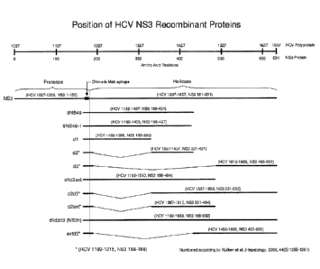
[0032] Figure 1 shows the position of HCV NS3 recombinant antigens of the
present invention.
DETAILED DESCRIPTION OF THE INVENTION
[0033] The present invention provides HCV combination immunoassays that
provide enhanced detection of exposure to HCV, by detecting both antibodies to
HCV
as is performed in conventional immunoassays, and by detecting HCV core
antigen that
may be present in the blood of individuals in the early stage of infection,
prior to the
development of antibodies to HCV. This invention meets the need in the art for
a
combination immunoassay for the simultaneous detection of both HCV antigens
and
anti-HCV antibodies in a sample in a single assay. The antigen/antibody
combination
assay methods rely on the identification and use of antigenic and immunogenic
HCV
antibodies and antigens that are present during the early stages of HCV
-12-
seroconversion, thereby increasing detection accuracy and reducing the
incidence of
false results during the window period.
[0034]
Biological samples that can be tested for HCV using the combination
assays of the present invention include any sample suspected to contain HCV
virions,
antigens or antibodies. The term "sample", as used herein, is used in its
broadest
sense. A "biological sample", as used herein, includes, but is not limited to,
any quantity
of a substance from a living thing or formerly living thing. Such living
things include, but
are not limited to, humans, mice, rats, monkeys, dogs, rabbits and other
animals. Such
substances include, but are not limited to, blood, (e.g., whole blood or
components
thereof), plasma, serum, urine, saliva, amniotic fluid, synovial fluid,
endothelial cells,
leukocytes, monocytes, other cells, organs, tissues, bone marrow, lymph nodes
and
spleen.
[0035] In the
anti-HCV antibody detection aspect of the combination assay at
least one (i.e., one or more) capture antigen is employed to bind and
therefore captures
anti-HCV antibodies present in the test sample. The capture antigens are
generally
antigenic peptides (containing one or more epitopes) derived from an HCV
protein
encoded by the HCV qenome. The sequence of the entire HCV qenome and the
encoded HCV polyprotein sequence are documented in GenBank (accession #M62321
and #AAA45676, respectively) and available to those skilled in the art.
Some
exemplary core antigens that could be used include antigens derived from the
DNA
binding domain (amino acids 1-125) of core protein. Still other preferred core
antigens
are derived from the lipid binding domain of core located at amino acid
residues 134-
171 of core protein (MGYIPLVGAPLGGAARALAHGVRVLEDGVNYATGNLPG )(SEQ ID NO: 89).
However, in
the present invention particularly preferred core antigens include antigens
derived from
core protein that comprise certain deletions or substitution in the known
epitope binding
regions for specific monoclonal antibodies such that monoclonal antibodies
used for
HCV core antigen detection would fail to detect these modified core antigens
but would
nonetheless detect complete core antigen from the test sample. Thus, these
novel
modified core antigens can be coated onto a solid phase support and/or used in
solution
phase to capture antibodies present in human serum or plasma that are directed
toward
the Core region of HCV but at the same time evade detection by the conjugate
antibody
Date Recue/Date Received 2020-06-18
CA 02906421 2015-09-14
WO 2014/158272 PCT/US2013/077504
-13-
used for the detection of Core Ag in an HCV Combo assay, but at the same time,
allow
detection of anti-Core antibodies that would also be expected to be in the
test sample
and identified in the same HCV Combo assay format. Preferred core antigens for
use in
the assays of the present invention comprise mutant core proteins that
comprise a
deletion of amino acids 34 and 48 and amino acids 115-121.
[0036] By using the novel core capture antigens described herein, the
present
invention overcomes a significant problem that is seen with the currently
available *Ac-
IDBA-c11-9/c11-14 conjugate that is used for the detection of core antigen in
an HCV
combination assay because the currently available core antigens used for
capture of
anti-core antibodies also react with detection antibodies designed to conduct
serological
detection of core antigen. Previously, constructs were made to obviate this
problem by
deletion of 5 amino acids (amino acids 32 33 and 34 for the C11-9 binding
region and
amino acids and residues 47 and 48 from the c11-14 binding region of core),
however,
these constructs yielded poorer anti-core antibody detection as these residues
are
highly immunogenic in anti-Core positive patients. The use of the core
antigens that are
described herein as capture antigens overcomes this problems due to their
design
which encompasses more minimal deletions that can successfully avoid detection
by
the *Ac-DBA c11-9/c11-14 conjugate but preserve or enhance detection of anti-
core
reactive specimens. In the combination assays of the present invention core
antigens
for the capture and detection of anti-HCV core antibodies advantageously
comprise
deletions of core amino acids sufficient for elimination of the binding of the
capture
antibody to the detection core antigen, for example, amino acids 115-121 are
deleted.
[0037] Definitions
[0038] The present invention provides reagents for the detection of HCV in
a test
sample. Preferably, this detection is achieved by the simultaneous detection
of both
HCV antigens and anti-HCV antibodies in the test sample. Throughout the
specification
certain terms are frequently used and as such the following section provides
additional
definitions of those terms. The term "antibody" (Ab) and "antibodies" (Abs)
refer to
monoclonal antibodies (mAb (singular) or mAbs (plural)), polyclonal antibodies
(pAbs
(plural)), multispecific antibodies, human antibodies, humanized antibodies
(fully or
CA 02906421 2015-09-14
WO 2014/158272 PCT/US2013/077504
-14-
partially humanized; a polypeptide comprising a modified variable region of a
human
antibody wherein a portion of the variable region has been substituted by the
corresponding sequence from a non-human sequence and wherein the modified
variable region is linked to at least part of the constant region of a human
antibody),
animal antibodies (such as, but not limited to, a bird (for example, a duck or
a goose), a
shark, a whale, and a mammal, including a non-primate (for example, a cow, a
pig, a
camel, a llama, a horse, a goat, a rabbit, a sheep, a hamster, a guinea pig, a
cat, a dog,
a rat, a mouse, etc.) or a non-human primate (for example, a monkey, a
chimpanzee,
etc.), recombinant antibodies, chimeric antibodies (cAb; a polypeptide
comprising all or
a part of the heavy and light chain variable regions of an antibody from one
host species
linked to at least part of the antibody constant regions from another host
species), single
chain antibodies, single domain antibodies, Fab fragments, F(ab') fragments,
Fab'-SH
fragments, F(ab')2 fragments, Fd fragments, Fv fragments, single-chain Fv
fragments
("scFv"), disulfide-linked Fv fragments ("sdFv"), dAb fragments, diabodies, an
isolated
complementarity determining region (CDR), and anti-idiotypic ("anti-Id")
antibodies,
bifunctional or dual-domain antibodies (e.g., dual variable domain antibodies,
or DVD-
IgGs), and functionally active, epitope-binding fragments (or antigenically
reactive
fragments) of any of the above. In particular, antibodies include
immunoglobulin
molecules and immunologically active (or antigenically reactive) fragments of
immunoglobulin molecules, namely, molecules that contain an analyte-binding
site as
further described in (n) herein, and variants as further described in (ac)
herein
lmmunoglobulin molecules can be of any type (for example, IgG, IgE, IgM, IgD,
IgA and
IgY), class (for example, IgG1, IgG2, IgG3, IgG4, IgA1 and IgA2), or subclass.
An
antibody, whose affinity (namely, KD, kd or ka) has been increased or improved
via the
screening of a combinatory antibody library that has been prepared using bio-
display, is
referred to as an "affinity maturated antibody." For simplicity sake, an
antibody against
an analyte is frequently referred to herein as being either an "anti-analyte
antibody" or
merely an "analyte antibody" (e.g., an anti-HCV antibody or an HCV antibody).
[0039] In the present invention the assay "component," "components," and
"at
least one component," refer generally to a capture antibody, a detection or
conjugate
antibody, a control, a calibrator, a series of calibrators, a sensitivity
panel, a container, a
CA 02906421 2015-09-14
WO 2014/158272 PCT/US2013/077504
-15-
buffer, a diluent, a salt, an enzyme, a co-factor for an enzyme, a detection
reagent, a
pretreatment reagent/solution, a substrate (e.g., as a solution), a stop
solution, and the
like that can be included in a kit for assay of a test sample, such as a
patient urine,
serum or plasma sample, in accordance with the methods described herein and
other
methods known in the art. Thus, in the context of the present disclosure, "at
least one
component," "component," and "components" can include a polypeptide as
described
herein, which is optionally immobilized on a solid support. Some components
can be in
solution or lyophilized for reconstitution for use in an assay.
[0040] In conducting the assays of the present invention, it may be useful
to use
a control. "Control" refers to a composition known to not contain anti-HCV
antibody
("negative control") or to contain anti-HCV antibody ("positive control"). A
positive
control can comprise a known concentration of anti-HCV antibody. "Control,"
"positive
control," and "calibrator" may be used interchangeably herein to refer to a
composition
comprising a known concentration of anti-HCV antibody. A "positive control"
can be
used to establish assay performance characteristics and is a useful indicator
of the
integrity of reagents (e.g., analytes).
[0041] The NS3 antigens of the present invention are useful in serological
assays
for the detection of anti-HCV antibodies in a test sample because such
antibodies
recognize epitopes contained within the NS3 antigens of the present invention.
"Epitope," "epitopes" and "epitopes of interest" refer to a site(s) on any
molecule (in this
case the NS3 antigens described herein) that is recognized and can bind to a
complementary site on a specific binding partner, such as an antibody or
antigenically
reactive fragment thereof. An epitope consists of the precise amino acid
residues of a
region of an antigen (or fragment thereof) known to bind to the complementary
site on
the specific binding partner. An antigenic fragment can contain more than one
epitope.
[0042] In the assays that are described herein, one or other component of
the
assay may comprise a detectable label. The terms "label" and "detectable
label" mean
a moiety attached to a specific binding partner, such as an antibody or an
analyte, to
render the reaction between members of a specific binding pair, such as an
antibody
and an analyte, detectable, and the specific binding partner, e.g., antibody
or analyte,
CA 02906421 2015-09-14
WO 2014/158272 PCT/US2013/077504
-16-
so labeled is referred to as "detectably labeled." A label can produce a
signal that is
detectable by visual or instrumental means. Various labels include signal-
producing
substances, such as chromogens, fluorescent compounds, chemiluminescent
compounds, radioactive compounds, and the like. Representative examples of
labels
include moieties that produce light, e.g., acridinium compounds, and moieties
that
produce fluorescence, e.g., fluorescein. Other labels are described herein. In
this
regard, the moiety itself may not be detectably labeled but may become
detectable
upon reaction with yet another moiety. Use of "detectably labeled" is intended
to
encompass the latter type of detectable labeling.
[0043] "Linking sequence" refers to a natural or artificial polypeptide
sequence
that is connected to one or more polypeptide sequences of interest (e.g., full-
length,
fragments, etc.). The term "connected" refers to the joining of the linking
sequence to
the polypeptide sequence of interest. Such polypeptide sequences are
preferably joined
by one or more peptide bonds. Linking sequences can have a length of from
about 4 to
about 50 amino acids. Preferably, the length of the linking sequence is from
about 6 to
about 30 amino acids. Natural linking sequences can be modified by amino acid
substitutions, additions, or deletions to create artificial linking sequences.
Exemplary
linking sequences include, but are not limited to: (i) Histidine residues (His
tags), such
as a 6xHis tag, which contains six histidine residues, are useful as linking
sequences to
facilitate the isolation and purification of polypeptides and antibodies of
interest. (ii)
Enterokinase cleavage sites, like His tags, are used in the isolation and
purification of
proteins and antibodies of interest. Often, enterokinase cleavage sites are
used
together with His tags in the isolation and purification of proteins and
antibodies of
interest. Various enterokinase cleavage sites are known in the art. (iii)
Miscellaneous
sequences can be used to link or connect the light and/or heavy chain variable
regions
of single chain variable region fragments. Examples of other linking sequences
can be
found in Bird et al., Science 242: 423-426 (1988); Huston et al., PNAS USA 85:
5879-
5883 (1988); and McCafferty et al., Nature 348: 552-554 (1990). Linking
sequences
also can be modified for additional functions, such as attachment of drugs or
attachment
to solid supports. In the context of the present disclosure, an mAb, for
example, can
contain a linking sequence, such as a His tag, an enterokinase cleavage site,
or both.
CA 02906421 2015-09-14
WO 2014/158272 PCT/US2013/077504
-17-
[0044] "Patient" and "subject" may be used interchangeably herein to refer
to an
animal, such as a bird (e.g., a duck or a goose), a shark, a whale, and a
mammal,
including a non-primate (for example, a cow, a pig, a camel, a llama, a horse,
a goat, a
rabbit, a sheep, a hamster, a guinea pig, a cat, a dog, a rat, and a mouse)
and a
primate (for example, a monkey, a chimpanzee, and a human). Preferably, the
patient
or subject is a human, such as a human at risk for HCV infection or a human
infected
with HCV.
[0045] In analysis of the results of the immunoassays described herein it
may be
useful to include certain levels of detection as cutoff levels. "Predetermined
cutoff" and
"predetermined level" refer generally to an assay cutoff value that is used to
assess
diagnostic/prognostic/therapeutic efficacy results by comparing the assay
results
against the predetermined cutoff/level, where the predetermined cutoff/level
already has
been linked or associated with various clinical parameters (e.g., severity of
disease,
progression/nonprogression/improvement, etc.). While the present disclosure
may
provide exemplary predetermined levels, it is well-known that cutoff values
may vary
depending on the nature of the immunoassay (e.g., antibodies employed, etc.).
It further
is well within the ordinary skill of one in the art to adapt the disclosure
herein for other
immunoassays to obtain immunoassay-specific cutoff values for those other
immunoassays based on this disclosure. Whereas the precise value of the
predetermined cutoff/level may vary between assays, the correlations as
described
herein should be generally applicable.
[0046] As described below, it may be desirable in some embodiments of the
invention to provide a pretreatment of the test sample. "Pretreatment
reagent," e.g.,
lysis, precipitation and/or solubilization reagent, as used in a diagnostic
assay as
described herein is one that lyses any cells and/or solubilizes any analyte
that is/are
present in a test sample. Pretreatment is not necessary for all samples, as
described
further herein. Among other things, solubilizing the analyte (i.e., anti-HCV
antibody)
entails release of the analyte from any endogenous binding proteins present in
the
sample. A pretreatment reagent may be homogeneous (not requiring a separation
step)
or heterogeneous (requiring a separation step). With use of a heterogeneous
pretreatment reagent there is removal of any precipitated analyte binding
proteins from
CA 02906421 2015-09-14
WO 2014/158272 PCT/US2013/077504
-18-
the test sample prior to proceeding to the next step of the assay. The
pretreatment
reagent optionally can comprise: (a) one or more solvents and salt, (b) one or
more
solvents, salt and detergent, (c) detergent, (d) detergent and salt, or (e)
any reagent or
combination of reagents appropriate for cell lysis and/or solubilization of
analyte.
[0047] The assays also may be subject to rigorous quality control. "Quality
control reagents" in the context of immunoassays and kits described herein,
include, but
are not limited to, calibrators, controls, and sensitivity panels. A
"calibrator" or
"standard" typically is used (e.g., one or more, such as a plurality) in order
to establish
calibration (standard) curves for interpolation of the concentration of an
analyte, such as
an antibody or an analyte. Alternatively, a single calibrator, which is near a
predetermined positive/negative cutoff, can be used. Multiple calibrators
(i.e., more than
one calibrator or a varying amount of calibrator(s)) can be used in
conjunction so as to
comprise a "sensitivity panel."
[0048] The terms "sample," "test sample," and "patient sample" may be used
interchangeably herein. The sample, such as a sample of urine, serum, plasma,
amniotic fluid, cerebrospinal fluid, placental cells or tissue, endothelial
cells, leukocytes,
or monocytes, can be used directly as obtained from a patient or can be pre-
treated,
such as by filtration, distillation, extraction, concentration,
centrifugation, inactivation of
interfering components, addition of reagents, and the like, to modify the
character of the
sample in some manner as discussed herein or otherwise as is known in the art.
Preferably, the sample is urine, serum or plasma.
[0049] In some assays, it may be desirable to provide calibration of the
assay.
"Series of calibrating compositions" refers to a plurality of compositions
comprising a
known concentration of anti-HCV antibody, wherein each of the compositions
differs
from the other compositions in the series by the concentration of anti-HCV
antibody.
[0050] Throughout the present specification, it is noted that the NS3
antigens
and/or other reagents may be bound to a solid support or solid phase, both of
which
terms are used interchangeably. The term "solid phase" refers to any material
that is
insoluble, or can be made insoluble by a subsequent reaction. The solid phase
can be
chosen for its intrinsic ability to attract and immobilize a capture agent.
Alternatively, the
CA 02906421 2015-09-14
WO 2014/158272 PCT/US2013/077504
-19-
solid phase can have affixed thereto a linking agent that has the ability to
attract and
immobilize the capture agent. The linking agent can, for example, include a
charged
substance that is oppositely charged with respect to the capture agent itself
or to a
charged substance conjugated to the capture agent. In general, the linking
agent can be
any binding partner (preferably specific) that is immobilized on (attached to)
the solid
phase and that has the ability to immobilize the capture agent through a
binding
reaction. The linking agent enables the indirect binding of the capture agent
to a solid
phase material before the performance of the assay or during the performance
of the
assay. The solid phase can, for example, be plastic, derivatized plastic,
magnetic or
non-magnetic metal, glass or silicon, including, for example, a test tube,
microtiter well,
sheet, bead, microparticle, chip, and other configurations known to those of
ordinary
skill in the art.
[0051] In certain descriptions of the assays described herein it may be
useful to
refer to either the NS3, NS4 or core antigen or the HCV antibody as a specific
binding
partner. "Specific binding partner" is a member of a specific binding pair. A
specific
binding pair comprises two different molecules, which specifically bind to
each other
through chemical or physical means. Therefore, in addition to antigen and
antibody
specific binding pairs of common immunoassays, other specific binding pairs
can
include biotin and avidin (or streptavidin), carbohydrates and lectins,
complementary
nucleotide sequences, effector and receptor molecules, cofactors and enzymes,
enzyme inhibitors and enzymes, and the like. Furthermore, specific binding
pairs can
include members that are analogs of the original specific binding members, for
example,
an analyte-analog. lmmunoreactive specific binding members include antigens,
antigen
fragments, and antibodies, including monoclonal and polyclonal antibodies as
well as
complexes, fragments, and variants (including fragments of variants) thereof,
whether
isolated or recombinantly produced. The term "specific" and "specificity" in
the context
of an interaction between members of a specific binding pair (e.g., an antigen
(or
fragment thereof) and an antibody (or antigenically reactive fragment
thereof)) refer to
the selective reactivity of the interaction. The phrase "specifically binds
to" and
analogous phrases refer to the ability of antibodies (or antigenically
reactive fragments
CA 02906421 2015-09-14
WO 2014/158272 PCT/US2013/077504
-20-
thereof) to bind specifically to a given antigen (or a fragment thereof) and
not bind
specifically to other entities.
[0052] Antigens For Use in the Present Invention
[0053] As described herein the present invention describes the detection of
a
combination of HCV antigens in one assay to advantageously provide a sensitive
and
selective detection of HCV in the test sample being assayed. In certain
preferred
embodiments, the combination assay further detects the presence of anti-HCV
antibodies. More particularly, the HCV antigens may be any antigen that is
typically
monitored in an HCV assay. Such antigens include but are not limited to core
antigen,
El, E2, NS2, NS3, N54 and N55 or distinct and independent portions of any one
of
core antigen, E1, E2, NS2, NS3, NS4 and NS5. Immunoassays for the detection of
such antigens individually are commercially available to those of skill in the
art and any
of the antigens used in such commercially available assays may readily be used
as
capture or detection antigens in the immunoassays of the present invention.
For
example, HCV NS3 protein and mutants thereof principally have to two main
protein
parts, the first corresponds to amino acids 1192-1457 per the HCV polyprotein
numbering of P26664 (Genbank, reproduced herein as SEQ ID NO:2; Choo et al.,
PNAS 1991) also known as C33 (as described originally by Chiron) or as
"9NB49H".
The second portion of the NS3 protein corresponds to amino acids 1192-1657
also
known as NS3 helicase or "NS3h." Antigens comprising all or portions of these
two
proteins can readily be used in the detection of anti-HCV antibodies in a test
sample.
For example, 033 is a well-known antigen derived from the NS3 protein of HCV
and
may readily be used herein as either the capture or detection antigen for the
detection
N53 antibodies in the combination immunoassays of the present invention.
[0054] Other NS3 derived antigens include those described in concurrently
filed
US Provisional Application No. 61/784,822 entitled "HCV NS3 Recombinant
Antigens
and Mutants Thereof for Improved Antibody Detection", Attorney Docket no.
03946-
26530U501. Such antigens are variant of the 033 and the NS3 helicase proteins
in
which the N-termini or C-termini sequences were modified. In some embodiments,
antigens were created that included cysteine to serine mutations. These
mutations
CA 02906421 2015-09-14
WO 2014/158272 PCT/US2013/077504
-21-
allowed for increased resistance of the antigen to oxidation thereby
preserving epitope
presentation and hence immunoreactivity. The cysteine to serine mutations also
allowed for site-specific modification of the protein (via chemical
conjugation using
maleimide reagents) by mutating only selected cysteine residues, e.g. those
deemed to
be unimportant for maintenance of immunoreactivity. Furthermore, at least some
of the
cysteine to serine substituted mutants disrupt the ability of full length
helicase enzyme
(HCV aa1192-1657) to bind nucleotide triphosphates (e.g. ATP). This maintains
the
protein in an open or extended conformation (see Gu & Rice, PNAS, 2010,
107:521-528
and references therein) and is shown in the present invention to produce
enhanced
i m mu noreactivity.
[0055] Exemplary NS3 antigens that may be used in of the present invention
are
shown in Table 1 herein below. In general, these NS3 antigens may be described
as
recombinant HCV NS3 antigen comprising a NS3 helicase sequence that comprises
each of domains I, ll and III of said helicase, wherein said antigen has
increased
immunoreactivity against HCV antibodies from serum as compared to 033 antigen,
wherein said recombinant HCV NS3 antigen comprises one or more of the
characteristics selected from the group consisting of: diminished ATP-binding
activity
as compared to the ATP-binding activity of wild-type NS3 helicase; diminished
ATPase
activity as compared to wild-type NS3 as compared to the ATP-binding activity
of wild-
type NS3 helicase, and increased redox stability as compared to the redox
stability of
wild-type NS3 helicase. More particularly, in the context of the present
invention, the
wild-type HCV NS3 comprises a sequence of SEQ ID NO: 87 and wherein the
recombinant antigen of the invention comprises at least one mutation as
compared to
the sequence of SEQ ID NO:87. Detailed description of production and testing
of these
antigens is provided in concurrently filed US Provisional Application No.
61/784,822,
entitled "HCV NS3 Recombinant Antigens and Mutants Thereof for Improved
Antibody
Detection", having Attorney Docket No. 03946-26530U301.
[0056] Table 1:
Antigen Antigen Sequence
designation
A K210N avdfipven lettmrspvf tdnssppvvp qsfqvahiha ptgsgNstkv
-22-
paayaaqgyk vlvinpsvaa
tlgfgaymsk ahgidpnirt
gvrtittgsp itystygkfl adggeggay
diiicdpchs
tdatsilgig tvldqaetag arlvvlatat
ppgsvtvphp
nieevalstt geipfygkai plevikggrh lifchskkkc
delaaklval ginavayyrg ldvsviptsg
dvvvvatdal
mtgytgdfds vidcrutcvtg tvdfsldptf
tietitlpqd
aysrtqrrgr tgrgkpgiyr fvapgerpsg
mfdssvlcec
_ _
ydagcawyel tpaettvrlr avmntpgipv
cqdhlefweg
vftglthida hflsqtkgsg enlpylvayq
atvcaraqap
_
ppswdqmwkc lirlkptlhg ptpllyrlga
vgneitlthp
vtkyimtcms adlevvt (SEQID NO: 109)
B S211A avdfipven lettmrspvf tdnssppvvp qsfqvahlha ptgsgkAtkv
paayaaqgyk vlvinpsvaa tlgfgaymsk ahgidpnirt
gvrtittgsp itystygkfl adggcsggay
diiicdechs
_ _
tdatsilgig tvldqaetag arlvvlatat
ppgsvtvphp
nieevalstt geipfygkai plevikggrh lifchskkkc
delaaklval ginavayyrg ldvsviptsg
dvvvvatdal
mtgytgdfds vidcntcvtq tvdfsldptf
tietitlpqd
avbiL4ILy1 Lylykpyiyi fvcipyLpy
malbbylcc
ydagcawyel tpaettvrlr avmntpglpv
cqdhlefweg
vftgithida hfisqtkgsg enlpylvayq
atvcaraqap
_
ppswdqmwkc lirlkptlhg ptpllyrlga
vgneitlthp
iTt icy; mtcm.c ad 1 P 7"µTi7 (SEQ ID NO: 110)
C T212E avdfipven lettmrspvf tdnssppvvp qsfqvahlha ptgsgksEkv
paayaaqgyk vlvinpsvaa tlgfgaymsk ahgidpnirt
gvrtittgsp itystygkfl adggcsggay
diiicdechs
_ _
tdatsilgig tvldgaetag arlvvlatat
ppgsvtvphp
nieevalstt geipfygkai plevikggrh lifchskkkc
deiaakival ginavayyrg ldvsviptsg
dvvvvatdal
mtgytgdfds videntcvtg tvdfsldptf
tietitlpqd
aysrtqrrgr tgrgkpgiyr fvapgerpsg
mfdssvlcec
_ _
ydagcawyel tpaettvrlr aymntpglpv
cqdhlefweg
vftglthida hflsgtkgsg enlpylvayq
atvcaraqap
_
ppswdqmwkc lirlkptlhg ptpllyrlga
vgneitlthp
vtkyimtcms adlevvt (SEQID NO: 111)
D Y241S, avdfipven lettmrspvf tdnssppvvp qsfqvahlha ptgsgkstkv
paayaaqgyk vlvinpsvaa tigfgaSmsk ahgidpnirt
gvrtittgsp itystygkfl adggcsggay
diiicdechs
_ _
tdatsilgig tvldqaetag arlvvlatat
ppgsvtvphp
nieevalstt geipfygkai plevikggrh iifchskkkc
delaaklval ginavayyrg ldvsviptsg
dvvvvatdal
Date Recue/Date Received 2020-06-18
-23-
mtgytgdfds vidcntcvtq tvdfsldptf
tietitlpqd
aysrtqrrgr tgrgkpgiyr fvapgerpsg
mfdssvlcec
ydagcawyel tpaettvrlr aymntpglpv
cqdhlefweg
vftglthida hflsqtkgsg enlpylvayq
atvcaraqap
_
ppswdqmwkc lirlkptlhg ptpllvrlga
voineitlthp
vtkyimtcms adlevvt (SEQID NO: 112)
E D290N avdfipven lettmrspvf tdnssppvvp qsfqvahlha ptgsgkstkv
paayaaqgyk vlvinpsvaa tlgfgaymsk ahgidpnirt
gvrtittgsp itystygkfl adggcsggay
diiicNechs
_ _
tdatsilgig tvldqaetag arlvvlatat
ppgsvtvphp
nieevalstt geipfygkai plevikggrh lifchskkkc
delaaklval ginavayyrg ldvsviptsg
dvvvvatdal
mtgytgdfds videntcvtg tvdfsldptf
tietitlpqd
aysrtqrrgr tgrgkpgiyr fvapgerpsg
mfdssvicec
_ _
ydagcawyel tpaettvrlr avmntpglpv
cqdhlefweg
vftglthida hflsqtkqsg enlpylvayq
atvcaraqap
_
ppswdqmwkc lirlkptlhg ptpllyrlga
vcineitithp
vtkyimtcms adlevvt(SEQIDNO:113)
F E2910 avdfipven leetmrspvf tdnssppvvp gsfgvahlha ptgsgkstkv
paayaaqgyk vlvinpsvaa tlgfgaymsk ahgidpnirt
gvrtittgsp itystygkfl adggcsggay
diiicdOgchs
_ _
tdatsilgig tvldqaetag arlvviatat
ppgsvtvphp
nieevalstt geipfygkai plevikggrh lifchskkkc
delaaklval ginavayyrg ldvsviptsg
dvvvvatdal
mtgytgdfds vidcntcvtq tvdfsldptf
tietitlpqd
aysrtqrrgr tgrgkpgiyr fvapgerpsg
mfdssvlcec
_ _
ydagcawyel tpaettvrlr avmntpgipv
cqdhlefweg
vftglthida hflsqtkgsg enlpylvayq
atvcaraqap
_
ppswdqmwkc lirlkptlhg ptpllyrlga
vqneitlthp
vtkyimtcms adlevvt (SEQIDNO:114)
G H293A avdfipven lettmrspvf tdnssppvvp gsfqvahlha ptgsgkstkv
paayaaggyk vlvinpsvaa tlgfgaymsk ahgidpnirt
gvrtittgsp itystygkfl adggcsggay
diiicdecAs
_ _
tdatsilgig tvldqaetag arlvvlatat
ppgsvtvphp
nieevalstt geipfygkai plevikggrh lifchskkkc
delaaklval ginavayyrg ldvsviptsg
dvvvvatdal
mtgytgdfds videntcvtg tvdfsldptf
tietitlpqd
aysrtqrrgr tgrgkpgiyr fvapgerpsg
mfdssvlcec
_ _
ydagcawyel tpaettvrlr aymntpglpv
cqdhlefweg
vfegithida hfisgtkgsg enipylvayq
atvcaraqap
_
ppswdqmwkc lirlkptlhg ptpllyrlga
vgneitlthp
Date Recue/Date Received 2020-06-18
-24-
vtkyimtcms adlevvt (SEQ ID NO: 115)
H T419G avdfipven lettmrspvf tdnssppvvp qsfqvahlha ptgsgkstkv
paayaaggyk vlvinpsvaa tlgfgaymsk ahgidpnirt
gvrtittgsp itystygkfl adggcsggay
diiicdechs
_ _
tdatsilgig tvldqaetag arlvvlatat
ppgsvtvphp
nieevalstt geipfygkai plevikggrh lifchskkke
delaaklval ginavayyrg ldvsviptsg
dvvvvatdal
mtgyGgdfds videntcvtg tvdfsldptf
tietitlpqd
aysrtqrrgr tgrgkpgiyr fvapgerpsg
mfdssvlcec
_ _
ydagcawyel tpaettvrlr aymntpglpv
cqdhlefweg
vftglthida hflsqtkgsg enlpylvayq
atvcaraqap
_
ppswdqmwkc lirlkptlhg ptpllvrlga
vgneitlthp
vtkyimtcms adlevvt (SEQIDNO:116)
1 Q460H avdfipven lettmrspvf tdnssppvvp qsfqvahlha ptgsgkstkv
paayaaggyk vlvinpsvaa tlgfgaymsk ahgidpnirt
gvrtittgsp itystygkfl adggcsggay
diiicdechs
_ _
tdatsilgig tvldqaetag arlvvlatat
ppgsvtvphp
nieevalstt geipfygkai plevikggrh lifchskkkc
delaaklval ginavayyrg ldvsviptsg
dvvvvatdal
mtgytgdfds vidcntcvtg tvdfsldptf
tietitlpqd
aysrtHrrgr tgrgkpgiyr fvapgerpsg
mfdssvlcec
_ _
ydagcawyel tpaettvrlr avmntpglpv
cqdhlefweg
,Jftglthida hflsgtkgsg enlpylvayg
atvcaragap
¨
ppswdqmwkc lirlkptlhg ptpllyrlga
vgneitlthp
vtkyimtcms adlevvt (SEQ ID NO: 117)
J R464A avdfipven lettmrspvf tdnssppvvp qsfqvahlha ptgsgkstkv
paayaaggyk vlvinpsvaa tlgfgaymsk ahgidpnirt
gvrtittgsp itystygkfl adggcsggay
diiicdechs
_ _
tdatsilgig tvldqaetag arlvvlatat
ppgsvtvphp
nieevalstt geipfygkai plevikggrh lifchskhke
delaaklval ginavayyrg ldvsviptsg
dvvvvatdal
mtgytgdfds videntcvtg tvdfsldptf
tietitlpqd
aysrtgrrgA tgrgkpgiyr fvapgerpsg
mfdssvlcec
_ _
ydagcawyel tpaettvrlr aymntpglpv
cqdhlefweg
vftglthida hflsqtkcisg enlpylvayq
atvcaraqap
_
ppswdqmwkc lirlkptlhg ptpllyrlga
vgneitlthp
vtkyimtcms adlevvt (SEQIDNO:118)
K R467K avdfipven lettmrspvf tdnssppvvp qsfqvahlha ptgsgkstkv
paayaaqgyk vlvinpsvaa tlgfgaymsk ahgidpnirt
gvrtittgsp itystygkfl adggcsggay
diiicdechs
_ _
tdatsilgig tvldqaetag arlvvlatat
ppgsvtvphp
Date Recue/Date Received 2020-06-18
-25-
nieevalstt geipfygkai plevikggrh lifchskkkc
delaaklval ginavayyrg ldvsviptsg
dvvvvatdal
mtgytgdfds vidcntcvtq tvdfsldptf
tietitlpqd
aysrtqrrgr tgKgkpgiyr fvapgerpsg
mfdssvlcec
_ _
ydagcawyel tpaettvrlr aymntpglpv
cqdhlefweg
vftglthida hflsqtkqsg enlpylvayq
atvcaraqap
ppswdqmwkc 1 irlkptlhg ptpllyrlga vqne it
lthp
vtkyimtcms adlevvt (SEQ ID NO: 119)
W501A avdfipven lettmrspvf tdnssppvvp gsfgvahlha ptgsgkstkv
paayaaggyk vlvinpsvaa tlgfgaymsk ahgidpnirt
gvrtittgsp itystygkfl adggcsggay
diiicdechs
_
tdatsilgig tvldqaetag arlvvlatat
ppgsvtvphp
nieevalstt geipfygkai plevikggrh lifchskkkc
delaaklval ginavayyrg ldvsviptsg
dvvvvatdal
mtgytgdfds videntcvtg tvdfsldptf
tietitlpqd
aysrtqrrgr tgrgkpgiyr fvapgerpsg
mfdssvlcec
_ _
ydagcaAyel tpaettvrlr aymntpglpv
cqdhlefweg
vftglthida hflsqtkgsg enlpylvayq
atvcaraqap
ppbwdgmwkc lillkpL1hy pLpllyLlya
vgiliLlLhp
vtkyimtcms adlevvt (SEQ ID NO: 120)
Any combination of two mutations selected from the group
consisting of K210N, S211A, T212E, Y241S, 0290N, E291Q, H293A,
T419G, Q460H, R464A, R467K and W501A
Any combination of three mutations selected from the group
consisting of K210N, S211A, T212E, Y241S, D290N, E2910, H293A,
T419G, Q460H, R464A, R467K and W501A
Any combination of four mutations selected from the group
consisting of K210N, S211A, T212E, Y241S, 0290N, E291Q, H293A,
T419G, Q460H, R464A, R467K and W501A
Any combination of five mutations selected from the group
consisting of K210N, S211A, T212E, Y241S, 0290N, E291Q, H293A,
T419G, Q460H, R464A, R467K and W501A
Any combination of six mutations selected from the group
consisting of K210N, S211A, T212E, Y241S, 0290N, E2910, H293A,
T419G, Q460H, R464A, R467K and W501A
Any combination of seven mutations selected from the group
consisting of K210N, S211A, T212E, Y241S, D290N, E2910, H293A,
1419G, 0460H, R464A, R467K and W501A
Any combination of eight mutations selected from the group
consisting of K210N, S211A, T212E, Y241S, 0290N, E291Q, H293A,
T419G, Q460H, R464A, R467K and W501A
Date Recue/Date Received 2020-06-18
-26-
Any combination of nine mutations selected from the group
consisting of K210N, S211A, T212E, Y241S, D290N, E2910, H293A,
T419G, 0460H, R464A, R467K and W501A
Any combination of ten mutations selected from the group
consisting of K210N, S211A, T212E, Y241S, 0290N, E291Q, H293A,
T419G, Q460H, R464A, R467K and W501A
V Any combination of eleven mutations selected from the group
consisting of K210N, S211A, T212E, Y241S, 0290N, E291Q, H293A,
T419G, Q460H, R464A, R467K and W501A
TA1 Any combination of twelve mutations selected from the group
consisting of K210N, S211A, T212E, Y241S, D290N, E2910, H293A,
T419G, Q460H, R464A, R467K and W501A
X avdfipven lettmrspvf tdnssppvvp qsfqvahlha ptgsgkstkv paayaaqgyk
vlvinpsvaa tlgfgaymsk ahgidpnirt gvrtittgsp itystygkfl
adggcsggay diiicdechs tdatsilgig tvldqaetag arlvvlatat ppgsvtvphp
nieevalstt geipfygkai
plevikggrh lifchskkkc delaaklval
ginavayyrg ldvsviptsg dvvvvatdal mtgytgdfds videntcvtg tvdfsldptf
tietitlpqd aysrtqrrgr tgrgkpgiyr fvapgerpsg mfdssvlcec ydagSawyel
tpaettvr1r aymntpglpv cqdnletweg vItgltnida nrisqtkqsg enipylvayq
atvcaraqap ppswdqmwkc lirlkptlhg ptpilyrlga vgneitlthp vtkyimtcms
adlevvt (SEQ ID NO: 121)
avdfipven lettmrspvf tdnssppvvp gsfqvahlha ptgsgkstkv paayaaqgyk
v1v1np3vaa tlgfgaymak ahgidpnirt gvrtittgop ityatygkfl
adggcsggay diiicdechs tdatsilgig tvldqaetag arlvvlatat ppgsvtvphp
nieevalstt geipfygkai
plevikggrh lifchskkkc delaaklval
ginavayyrg ldvsviptsg dvvvvatdal mtgytgdfds videntcvtg tvdfsldptf
tietitlpqd aysrtqrrgr tgrgkpgiyr fvapgerpsg mfdssvlcec ydagcawyel
tpaettvrlr aymntpglpv Sqdhlefweg vftglthida hflsqtkqsg enlpylvayq
atvcaraqap ppswdqmwkc lirlkptlhg ptpllyrlga vgneitlthp vtkyimtcms
adlevvt (SEQ ID NO: 122)
avdfipven lettmrspvf tdnssppvvp gsfqvahlha ptgsgkstkv paayaaqgyk
vlvinpsvaa tlgfgaymsk ahgidpnirt gvrtittgsp itystygkfl
adggcsggay diiicdeShs tdatsilgig tvldqaetag arlvvlatat ppgsvtvphp
nieevalstt geipfygkai
plevikggrh lifchskkkc delaaklval
ginavayyrg ldvsviptsg dvvvvatdal mtgytgdfds vidcntcvtq tvdfsldptf
tietitlpqd aysrtqrrgr tgrgkpgiyr fvapgerpsg mfdssvlcec ydagcawyel
tpaettvrir aymntpgipv cqdhiefweg vftgithida hfisqtkgsg enipylvayq
atvcaraqap ppswdgmwkc 1.irlkptlhg ptiollyriga vgneitithp vtkyimtems
adlevvt (SEQ ID NO: 123)
Date Recue/Date Received 2020-06-18
-27-
Al
avdfipven lettmrspvf tdnssppvvp gsfqvahlha ptgsgkstkv paayaaggyk
vlvinpsvaa tlgfgaymsk ahg]_dpnirt gvrtittgsp itystygkfl
adggcsggay diiicdechs tdatsilgig tvldqaetag arlvvlatat ppgsvtvphp
nieevalstt geipfygkai
plevikggrh lifShskkkc delaaklval
ginavayyrg ldvsviptsg dvvvvatdal mtgytgdfds videntcvtg tvdfsldptf
tietitlpqd aysrtqrrgr tgrgkpgivr fvapgerpsg mfdssvlcec ydagcawyel
tpaettvrlr aymntpglpv cqdhlefweg vftglthida hflsqtkcisg enlpylvayq
atvcaraqap ppswdqmwkc lirlkptlhg ptpllyrlga vgneitlthp vtkyimtcms
adlevvt (SEQ ID NO: 124)
A2 avdfipven lettmrspvf tdnssppvvp qsfqvahlha ptgsgkstkv paayaaggyk
vlvinpsvaa tlgfgaymsk ahgidpnirt gvrtittgsp itystygkfl
adggcsggay diiicdechs tdatsilgig tvldqaetag arlvvlatat ppgsvtvphp
nieevalstt geipfygkai
plevikggrh lifchskkkS delaaklval
gtnavayyrg ldvsviptsg dvvvvatdal mtgytgdfds vidcntcvtq tvdfsldptf
tietitlpqd aysrtqrrgr tgrgkpgiyr fvapgerpsg mfdssvlcec ydagcawyel
tpaettvrlr aymntpglpv cqdhlefweg vftglthida hflsqtkgsg enlpylvayg
atvcaraqap ppswdqmwkc lirlkptlhg ptpllyrlga vqneitlthp vtkyimtems
adlevvt (SEQ ID NO: 125)
Any combination of mutations of any of R-W In combination with
one two or three, four or five of the mutations shown in X, Y, Z,
Al, and A2.
[0057] In
other embodiments, another aspect of the combination immunoassay
detects the presence of antibodies to Core antigen. Some exemplary core
antigens that
could be used include antigens derived from the DNA binding domain (amino
acids 1-
125) of core protein. Still other preferred core antigens are derived from the
lipid
binding domain of core located at amino acid residues 134-171 of core protein
(MGYIPLVGAPLGGAARALAHGVRVLEDGVNYATGNLPG) (SEQ ID NO: 89). However, in the
present invention
particularly preferred core antigens include antigens derived from core
protein that
comprise certain deletions or substitution in the known epitope binding
regions for
specific monoclonal antibodies such that monoclonal antibodies used for HCV
core
antigen detection would fail to detect these modified core antigens but would
nonetheless detect complete core antigen from the test sample. Thus, these
novel
modified core antgens can be coated onto a solid phase support and/or used in
solution
phase to capture antibodies present in human serum or plasma that are directed
toward
the Core region of HCV but at the same time evade detection by the conjugate
antibody
Date Recue/Date Received 2020-06-18
CA 02906421 2015-09-14
WO 2014/158272 PCT/US2013/077504
-28-
used for the detection of Core antigen present in a test sample in an HCV
combination
immunoassay. Thus a combination immunoassay can be performed that detects both
Core antigen present in the test sample at the same time as detecting anti-
Core
antibodies that would also be expected to be in the test sample and identified
in the
same HCV Combo assay format. The Core antigens that can be used for the
purpose
of detecting anti-Core antibodies from the test sample preferably comprise
deletions of
amino acids 34 and 48 and amino acids 115-121 of Core antigen.
[0058] As noted herein throughout the methods of the invention typically
are
immunoassay methods. In exemplary embodiments, such methods include methods
for
isolating a molecule of interest (such as for example a specific antibody that
is present
in a test sample, or a specific antigen that may be present in the test
sample). In order
to facilitate such isolation, the molecule of interest comprises or is
attracted to a
purification tag that contacts a tag binding partner. The association of the
purification
tag and the tag binding partner thus may be used to separate the molecule of
interest
from a mixture of molecules. Purification tags can comprise moieties with the
same or
similar structures. In certain embodiments, the tagging moiety of an affinity
tag can be
associated with a functional tag directly by a single bond or via a linkage of
stable
chemical bonds, in linear, branched or cyclic arrangements, optionally
including single,
double, triple bond, aromatic carbon-carbon bonds, as well as carbon-nitrogen
bonds,
nitrogen-nitrogen bonds, carbon-oxygen bonds, carbon-sulfur bonds, phosphorus-
oxygen bonds, phosphorus-nitrogen bonds, and any combination thereof. In
certain
embodiments, the association between the tagging moiety and functional tag
comprises
ether, thioether, carboxamide, sulfonamide, urea or urethane moieties. In
preferred
embodiments, the linkage comprises a polyalkylene chain, i.e., a linear or
branched
arrangement of carbon-carbon bonds. In other embodiments, the linkage
comprises a
polyalkylene oxide chain, including a polyethylene glycol moiety. Examples, of
affinity
tags include, but are not limited to, biotin, digoxigenin (Dig), dinitrophenol
(DNP), zinc
fingers, fluorinated polymers, and polypeptide sequences such as polyhistidine
motifs.
[0059] The affinity tags are in some embodiments advantageously used to
isolate
the molecule of interest by relying on the binding or attraction of the
affinity tag and a
functional group that is attracted to or binds the affinity tag. In some
embodiments, solid
CA 02906421 2015-09-14
WO 2014/158272 PCT/US2013/077504
-29-
substrates having an affinity for the tag in that the solid substrate is
derivatized with the
tag binding partner. In some embodiments, the binding partner may be
immobilized on
an affinity substrate. The term "affinity substrate" can refer to an immobile
matrix or
support bound to a binding partner that is capable of forming a strong and
preferably
reversible interaction with the purification tag of a molecule. An affinity
substrate can
include a resin, a bead, a particle, a membrane, a gel. The binding partner
recognizes
or binds to the purification tag specifically. Specific binding partners will
depend on the
affinity tag, but include charged moieties and one member of a binding pair
such as
receptor-ligand, antibody-antigen, carbohydrate-lectin, and biotin-
streptavidin (or avidin,
neutravidin or an anti-biotin antibody).
[0060] In
specific and preferred embodiments, either the C or the N terminus of
any or all of the antigens used in the combination immunoassay may be
biotinylated or
may comprise a biotin binding moiety (e.g., avidin or streptavidin or
neutravidin or an
anti-biotin) as the affinity tag. These peptides are biotinylated or
avidin/streptavidin-
conjugated peptides and will serve as capture antigens. Likewise, the antigens
may
alternatively be labeled with a detection label in which case they will serve
as detection
antigens. The detection and capture antigens may have the same underlying
amino
acid sequence or alternatively, may have different sequences. In
exemplary
embodiments, the capture antigens are biotinylated at either the C or the N
terminus to
facilitate binding thereof to solid supports that have the biotin binding
partner (i.e., avidin
or streptavidin). For exemplary production purposes, the biotinylated peptides
are
recombinantly expressed in E. coli BL2L(DE3) cells via an IPTG induction
system at
25 C. In situ biotinylation at the C-terminal or N-terminal biotinylation is
accomplished
by co-transformation of the BL21(DE3) cells with the HCV expression plasmid
expressing the desired peptide and a second plasmid containing the BirA gene
from E.
coli (Weiss et al. (1994) Protein Expression & Purif, 14:751-755; Schatz et
al. (1993)
Biotechnology, 11:1138-1143). Purification of the recombinant proteins is
performed
using divalent cation chelators that are shown to prevent metal-catalyzed
oxidation and
aggregation of the protein. Protein stability is significantly improved when
EDTA or
related divalent cation chelator is added to the buffers used during
purification and to
the final storage buffer or buffers used in the immunoassay.
-30-
[0061] Antibodies for Use in the Combinations Assays
[0062] As discussed herein throughout the combination immunoassays
advantageously also determine the presence of one or more HCV antigens present
in
the test sample. In such embodiments, it will be desirable to use monoclonal
anti-HCV
antibodies to capture the antigen from the test sample and then use further
conjugate
antibodies to detect the presence of antigen that has been captured. There are
numerous commercially available antibodies that may be used in this endeavor.
Specifically, such antibodies preferably determine the presence of Core
antigen in the
test sample. Antibodies directed to Core antigen are known to those of skill
in the art
include, for example, those described in US Patent Publication No.
20120009196. In
addition, the present invention further contemplates that use of monoclonal
antibodies
described in concurrently filed US Patent Application No. 61/783,529, entitled
"HCV
Core Lipid Binding Domain Monoclonal Antibodies" Attorney Docket No. 03946-
26531US01 that is specifically immunoreactive with the lipid binding domain of
HCV
core antigen. More particularly, the HCV core antigen is amino acid residues
134-171
of HCV. In more particular embodiments, the antibody specifically binds at
least one
epitope formed by amino acid
sequence
MGYIPLVGAPLGGAARALAHGVRVLEDGVNYATGNLPG (SEQ ID NO: 89). In more specific
embodiments, the antibody is immunoreactive with an epitope formed by amino
acids
141-161, 134-154 and 151 to 171 of HCV core antigen.
[0063] In specific exemplary embodiments the antibodies used in the
combination
immunoassay are antibodies designed to detect HCV core protein or fragments
thereof
in a test sample. Such antibodies may detect the DNA binding domain, the lipid
binding
domain or indeed the complete Core protein. In some embodiments, the detection
antibody used in the immunoassay is directed to the lipid binding domain of
core
peptide and exemplary such antibodies are described in concurrently filed US
Provisional Application No. 61/783,529 entitled "HCV Core Lipid Binding Domain
Monoclonal Antibodies", Attorney Docket no. 03946-26531US01. In still other
embodiments, the anti-HCV Core antibodies used in the combination assays may
be for
example, C11-3, C11-7, C11-9, and C11-14 (as described in US Patent 6,727,092;
Morota, et al, J. Viral. Meth., 2009, 157:8-14).
Date Recue/Date Received 2020-06-18
CA 02906421 2015-09-14
WO 2014/158272 PCT/US2013/077504
-31-
[0064] In a specific assay of the present invention, the combination
immunoassay
at least detects core antigen as well detecting core antibodies in the test
sample. In
such embodiments, it becomes desirable, although not essential to ensure that
the
capture antigen that is designed to capture anti-Core one that preferably
comprise
certain deletions or substitution in the known epitope binding regions for
specific
monoclonal antibodies such that monoclonal antibodies used for HCV core
antigen
detection would fail to detect these modified core antigens but would
nonetheless detect
complete core antigen from the test sample. Exemplary anti-core antibodies to
be used
as capture antibodies include antibodies AOT3, 011-3, C11-7, C11-9, and 011-
14as
described in US Patent 6,727,092 as well as Morota, et al, J. Virol. Meth.,
2009, 157:8-
14.
[0065] lmmunodiagnostic Assays and Reagents
[0066] In particular embodiments, the antigens and antibodies described
above
are contemplated for use as immunodiagnostic reagents in combination
immunoassays
designed for the detection of multiple HCV components found in a test sample
suspected of having been infected with HCV. lmmunodiagnostic reagents (be they
antibodies or antigens) will be comprised of the above-described antigen
polypeptides
and antibodies (typically in combination) such that they can be used in a
combination
immunoassay designed for the detection of HCV antigens including but not
limited to
the NS3 region of HCV, the core antigen of HCV, the NS4 region of HCV or
combinations thereof as well as anti-HCV antibodies directed against one or
more of
these regions. For purposes of capture, the antigens and/or antibodies of
which the
immunodiagnostic reagent is comprised can be coated on a solid support such as
for
example, a microparticle, (e.g., magnetic particle), bead, test tube,
microtiter plate,
cuvette, membrane, scaffolding molecule, film, filter paper, disc or chip. In
this regard,
where the immunodiagnostic reagent comprises a combination of antigens (e.g.,
directed at different HCV proteins or different fragments of the same HCV
protein), the
antigens can be co-coated on the same solid support or can be on separate
solid
supports. Likewise, where the immunodiagnostic reagent comprises one or more
antibodies that will be used to capture one or more antigens from the test
sample, such
CA 02906421 2015-09-14
WO 2014/158272 PCT/US2013/077504
-32-
antibodies can be co-coated on the same solid support or can be on separate
solid
supports.
[0067] Notably, the immunodiagnostic reagent will include the antigens and
antibodies may be labeled with a detectable label or labeled with a specific
partner that
allows capture or detection. For example, the labels may be a detectable
label, such as
a fluorophore, radioactive moiety, enzyme, biotin/avidin label, chromophore,
chemiluminescent label, or the like. Such labels are described in further
detail infra.
[0068] Still further the invention contemplates the preparation of HCV
diagnostic
kits comprising the immunodiagnostic reagents described herein and
instructions for the
use of the immunodiagnostic reagents in combination immunoassays for
determining
the presence of HCV in a test sample by detecting the presence of two or more
HCV
proteins and/or anti-HCV antibodies in such a sample. For example, the kit can
comprise instructions for assaying the test sample for anti-HCV antibody
(e.g., an anti-
Core antibody in the test sample) by immunoassay. While preferred embodiments
employ chemiluminescent microparticle immunoassay for assaying the test
sample, it
should be understood that the antigens and antibodies used in the combination
immunoassays of the present invention may be used in any other immunoassay
formats
known to those of skill in the art for determining the presence of HCV in a
test sample.
The instructions can be in paper form or computer-readable form, such as a
disk, CD,
DVD, or the like. Alternatively or additionally, the kit can comprise a
calibrator or control,
e.g., purified, and optionally lyophilized, anti-HCV antibody or antigen,
and/or at least
one container (e.g., tube, microtiter plates or strips, which can be already
coated with
one or more of the capture components (antigens and/or antibodies) of the
combination
immunoassay) for conducting the assay, and/or a buffer, such as an assay
buffer or a
wash buffer, either one of which can be provided as a concentrated solution, a
substrate
solution for the detectable label (e.g., an enzymatic label), or a stop
solution. Preferably,
the kit comprises all components, i.e., reagents, standards, buffers,
diluents, etc., which
are necessary to perform the assay. In specific embodiments, it is preferred
that all the
components are individually presented in the kit such that the immunoassay may
be
performed as a capture-on-the-fly type combination immunoassay in which the
solid
support is coated with an agent that allows binding of the capturing moiety
(e.g., a
CA 02906421 2015-09-14
WO 2014/158272 PCT/US2013/077504
-33-
biotinylated antigen or a biotinylated antibody) and the kit further comprises
each of the
individual capture and detection antigen pairs and the biotinylated capture
antibodies in
one container and a second container provides the detection antibody
conjugate. The
instructions for conducting the assay also can include instructions for
generating a
standard curve or a reference standard for purposes of quantifying anti-HCV
antibody.
[0069] Any antibodies, which are provided in the kit, such as anti-1g6
antibodies
and anti-IgM antibodies, can also incorporate a detectable label, such as a
fluorophore,
radioactive moiety, enzyme, biotin/avidin label, chromophore, chemiluminescent
label,
or the like, or the kit can include reagents for labeling the antibodies or
reagents for
detecting the antibodies (e.g., detection antibodies) and/or for labeling the
analytes or
reagents for detecting the analyte. The antibodies, calibrators and/or
controls can be
provided in separate containers or pre-dispensed into an appropriate assay
format, for
example, into microtiter plates. In a preferred combination immunoassay there
are two
containers provided. In the first container is provided at least a first,
second and third
pair of antigens, wherein the first antigen in each pair is a capture antigen
from a given
HCV protein that is biotinylated and the second antigen in each pair is a
detection
antigen from the same protein as the first antigen but is labeled with a
detectable label
(e.g., it is acridinylated) as well as one or more biotinylated antibodies
designed for
detecting one or more HCV antigens from a test sample; and in the second
container is
provided the antibody that forms the conjugation partner for detection of the
antigen that
is captured by the biotinylated antibodies from the first container. It is
contemplated that
where there are multiple biotinylated antibodies in the first container, the
multiple
antibodies that form the conjugation partners may be present in a single
container or
individual containers for each different antigen detecting conjugate antibody.
[0070] Optionally, the kit includes quality control components (for
example,
sensitivity panels, calibrators, and positive controls). Preparation of
quality control
reagents is well-known in the art and is described on insert sheets for a
variety of
immunodiagnostic products. Sensitivity panel members optionally are used to
establish
assay performance characteristics, and further optionally are useful
indicators of the
integrity of the immunoassay kit reagents, and the standardization of assays.
CA 02906421 2015-09-14
WO 2014/158272 PCT/US2013/077504
-34-
[0071] The kit can also optionally include other reagents required to
conduct a
diagnostic assay or facilitate quality control evaluations, such as buffers,
salts,
enzymes, enzyme co-factors, substrates, detection reagents, and the like.
Other
components, such as buffers and solutions for the isolation and/or treatment
of a test
sample (e.g., pretreatment reagents), also can be included in the kit. The kit
can
additionally include one or more other controls. One or more of the components
of the
kit can be lyophilized, in which case the kit can further comprise reagents
suitable for
the reconstitution of the lyophilized components.
[0072] The various components of the kit optionally are provided in
suitable
containers as necessary, e.g., a microtiter plate. The kit can further include
containers
for holding or storing a sample (e.g., a container or cartridge for a sample).
Where
appropriate, the kit optionally also can contain reaction vessels, mixing
vessels, and
other components that facilitate the preparation of reagents or the test
sample. The kit
can also include one or more instrument for assisting with obtaining a test
sample, such
as a syringe, pipette, forceps, measured spoon, or the like.
[0073] In preferred embodiments, the detectable label is at least one
acridinium
compound. In such embodiments, the kit can comprise at least one acridinium-9-
carboxamide, at least one acridinium-9-carboxylate aryl ester, or any
combination
thereof. If the detectable label is at least one acridinium compound, the kit
also can
comprise a source of hydrogen peroxide, such as a buffer, solution, and/or at
least one
basic solution. It should be understood that in the immunodiagnostic reagent
the
antigens for antibody detection may be detectably labeled, and any antibodies
provided
in kit for use along with such reagents also may be detectably labeled.
[0074] If desired, the kit can contain a solid support phase, such as a
magnetic
particle, bead, test tube, microtiter plate, cuvette, membrane, scaffolding
molecule, film,
filter paper, disc or chip.
[0075] Method of Determining the Presence, Amount or Concentration of
HCV in a Test Sample
[0076] The present disclosure provides a combination immunoassay method for
determining the presence, amount or concentration of anti-HCV antibodies and
HCV
CA 02906421 2015-09-14
WO 2014/158272 PCT/US2013/077504
-35-
antigens in a test sample. Any suitable assay known in the art can be used in
such a
method as long as such an assay uses one or more of antigens for detecting HCV
antibodies and/or one or more anti-HCV antibodies for detecting one or more
HCV
antigens in the test sample. Examples of such assays include, but are not
limited to,
immunoassay, such as sandwich immunoassay (e.g., monoclonal-polyclonal
sandwich
immunoassays, including radioisotope detection (radioimmunoassay (RIA)) and
enzyme
detection (enzyme immunoassay (EIA) or enzyme-linked immunosorbent assay
(ELISA)
(e.g., Quantikine ELISA assays, R&D Systems, Minneapolis, Minn.)), competitive
inhibition immunoassay (e.g., forward and reverse), fluorescence polarization
immunoassay (FPIA), enzyme multiplied immunoassay technique (EMIT),
bioluminescence resonance energy transfer (BRET), and homogeneous
chemiluminescent assay, etc.
[0077] In
specific embodiments of the combination immunoassays, the
recombinant antigens (e.g., core, NS3 and NS4 antigens) may be used as capture
reagents (e.g., by using such antigens in which the amino ¨ or carboxy-
terminal of the
antigen comprises a biotin tag) or as a detection (conjugate) reagents in
which the
antigens are either directly or indirectly labeled with acridinium.
Indirect labeling
requires the use of acridinylated BSA covalently coupled to the free thiol of
unpaired
cysteine residues within a protein via SMCC-type linker. To facilitate such
indirect
labeling certain of the antigens used in the combination immunoassays of the
present
invention may readily be further modified to include additional cysteine
residues at the
C-terminus.
[0078]
Typically, immunoassays are performed in 1-step or 2-step format. Solid
phase reagents for capture of immune complexes formed in solution in the 1-
step assay
include anti-biotin monoclonal antibody, streptavidin or neutravidin to
capture the
biotinylated moiety (be it a biotinylated antigen for capture of an HCV
antibody or a
biotinylated antibody for the capture of an HCV protein/antigen in the test
sample).
[0079] In a
SELDI-based immunoassay, a capture reagent that specifically binds
anti-HCV-antibody or an HCV antigen is attached to the surface of a mass
spectrometry
probe, such as a pre-activated protein chip array. The anti-HCV antibody or
the antigen
CA 02906421 2015-09-14
WO 2014/158272 PCT/US2013/077504
-36-
is then specifically captured on the biochip, and the captured moiety is
detected by
mass spectrometry. Alternatively, the anti-HCV antibody can be eluted from the
capture
reagent and detected by traditional MALDI (matrix-assisted laser
desorption/ionization)
or by SELDI. A chemiluminescent microparticle immunoassay, in particular one
employing the ARCHITECT automated analyzer (Abbott Laboratories, Abbott Park,
III.), is an example of a preferred immunoassay in which a combination of
multiple
antigens (preferably antigens from two or more HCV proteins) as well as
multiple anti-
HCV antibodies may readily be employed. An agglutination assay, such as a
passive
hemagglutination assay, also can be used. In an agglutination assay an antigen-
antibody reaction is detected by agglutination or clumping. In a passive
hemagglutination assay, erythrocytes are coated with the antigen and the
coated
erythrocytes are used in the agglutination assay.
[0080] Methods well-known in the art for collecting, handling and
processing
urine, blood, serum and plasma, and other body fluids, are used in the
practice of the
present disclosure, for instance, when the immunodiagnostic reagents comprise
multiple antigens and/or in an anti-HCV antibody immunoassay kit. The test
sample can
comprise further moieties in addition to the polypeptide of interest, such as
antibodies,
antigens, haptens, hormones, drugs, enzymes, receptors, proteins, peptides,
polypeptides, oligonucleotides or polynucleotides. For example, the sample can
be a
whole blood sample obtained from a subject. It can be necessary or desired
that a test
sample, particularly whole blood, be treated prior to immunoassay as described
herein,
e.g., with a pretreatment reagent. Even in cases where pretreatment is not
necessary
(e.g., most urine samples), pretreatment optionally can be done for mere
convenience
(e.g., as part of a regimen on a commercial platform).
[0081] The pretreatment reagent can be any reagent appropriate for use with
the
combination immunoassay and kits of the invention. The pretreatment optionally
comprises: (a) one or more solvents (e.g., methanol and ethylene glycol) and
salt, (b)
one or more solvents, salt and detergent, (c) detergent, or (d) detergent and
salt.
Pretreatment reagents are known in the art, and such pretreatment can be
employed,
e.g., as used for assays on Abbott TDx, AxSYM , and ARCHITECT analyzers
(Abbott
Laboratories, Abbott Park, Ill.), as described in the literature (see, e.g.,
Yatscoff et al.,
-37-
Abbott TDx Monoclonal Antibody Assay Evaluated for Measuring Cyclosporine in
Whole
Blood, Olin. Chem. 36: 1969-1973 (1990), and Wallemacq et al., Evaluation of
the New
AxSYM Cyclosporine Assay: Comparison with TDx Monoclonal Whole Blood and EMIT
Cyclosporine Assays, Clin. Chem. 45: 432-435 (1999)), and/or as commercially
available. Additionally, pretreatment can be done as described in Abbott's
U.S. Pat. No.
5,135,875, European Pat. Pub. No. 0 471 293, U.S. Provisional Pat. App.
60/878,017,
filed Dec. 29, 2006, and U.S. Pat. App. Pub. No. 2008/0020401.
The pretreatment
reagent can be a heterogeneous agent or a homogeneous agent.
[0082] With use of a heterogeneous pretreatment reagent, the pretreatment
reagent precipitates analyte binding protein (e.g., protein that can bind to
anti-HCV
antibody or an antigen that can bind to an anti-HCV antibody form the present
in the
sample. Such a pretreatment step comprises removing any analyte binding
protein by
separating from the precipitated analyte binding protein the supernatant of
the mixture
formed by addition of the pretreatment agent to sample. In such an assay, the
supernatant of the mixture absent any binding protein is used in the assay,
proceeding
directly to the antibody capture step.
[0083] With use of a homogeneous pretreatment reagent there is no such
separation step. The entire mixture of test sample and pretreatment reagent
are
contacted with a labeled specific binding partner for anti-HCV antibody (i.e.,
an antigen)
or the labeled specific binding partner for the HCV antigen (i.e., an
antibody). The
pretreatment reagent employed for such an assay typically is diluted in the
pretreated
test sample mixture, either before or during capture by the first specific
binding partner.
Despite such dilution, a certain amount of the pretreatment reagent (for
example, 5 M
methanol and/or 0.6 methylene glycol) is still present (or remains) in the
test sample
mixture during capture.
[0084] In a heterogeneous format, after the test sample is obtained from a
subject, a first mixture is prepared. The mixture contains the test sample
being
assessed for anti-HCV antibodies and a first specific capture binding partner,
wherein
the first specific capture binding partner and any anti-HCV antibodies
contained in the
Date Recue/Date Received 2020-06-18
CA 02906421 2015-09-14
WO 2014/158272 PCT/US2013/077504
-38-
test sample form a first specific capture binding partner-anti-HCV antibody
complex.
The first specific capture binding partner may be any of a core antigen, an
NS3 antigen
or an NS3. Exemplary NS3 antigens used in the invention may be any one or more
of
the antigens shown in Table 1 herein above. Likewise, in the combination
assays of the
invention the mixture also contains a second and third specific capture
binding partner
and these second and third specific capture binding partners form second and
third
specific capture binding partner-anti-HCV antibody cornplexes with anti-HCV
antibodies
that are present in the test sample. Such second, third and fourth antigens
may be one
or more of at least one HCV antigen selected from the group consisting of core
antigen,
NS3, NS4, NS5, and portions thereof.
[0085] In addition the combination immunoassay may, and preferably does,
include at least one anti-HCV capture antibody that will form a specific
complex with a
fourth specific binding partner that is found in the test sample (i.e., an
antigen or HCV
protein that is found in the test sample) so as to form an anti-HCV antibody-
fourth
specific binding partner complex with the fourth antigen that is present in
the test
sample. Preferably, the fourth specific binding pair is one that detects Core
antigen in a
test sample, and hence the binding pair is an anti-Core antibody for detection
of the
fourth antigen (Core) in the test sample.
[0086] The order in which the test sample and the various specific binding
partners are added to form the mixture is not critical. In some embodiments,
the first,
second, and third specific capture binding partners (i.e., antigens) and the
anti-HCV
capture antibody are immobilized on a solid phase. In still other embodiments,
none of
these four components are immobilized but are instead all added at the same
time to
the test sample to effect capture onto the solid phase. The solid phase used
in the
combination immunoassay can be any solid phase known in the art, such as, but
not
limited to, a magnetic particle, a bead, a test tube, a microtiter plate, a
cuvette, a
membrane, a scaffolding molecule, a film, a filter paper, a disc and a chip.
[0087] After the immunocomplexes are formed between the first, second and
third specific capture binding partners and their respective anti-HCV
antibodies found in
the test sample, and the first anti-HCV capture antibodies (e.g., anti-Core)
and their
CA 02906421 2015-09-14
WO 2014/158272 PCT/US2013/077504
-39-
respective HCV antigens or HCV proteins found in the test sample, any unbound
anti-
HCV antibody or HCV antigen/protein is removed from the complex using any
technique
known in the art. For example, the unbound anti-HCV antibody or antigen can be
removed by washing. Desirably, however, the first, second and third specific
binding
partners and the anti-HCV antibodies are present in excess of any anti-HCV
antibody
and antigens, respectively present in the test sample, such that all anti-HCV
antibody
and antigens that are present in the test sample become bound by the first,
second, and
third specific binding partner and anti-HCV capture antibodies respectively.
[0088] After any unbound anti-HCV antibody and antigen is removed,
detection is
achieved by addition of a first specific detection binding partner to the
mixture to form a
first specific capture binding partner-anti-HCV antibody-first specific
detection binding
partner complex. The first specific detection binding partner is preferably a
combination
of an anti-IgG antibody and an anti-IgM antibody. Moreover, also preferably,
the first
specific detection binding partner is labeled with or contains a detectable
label as
described above. In specific embodiments, the first specific detection partner
may
instead or in addition be an antigen that binds the captured antibody.
Likewise, in the
combination assays of the invention the mixture also contains a second and
third
specific detection binding partner and these second and third specific
detection binding
partners form second or third specific capture binding partner-anti-HCV
antibody-
second or third specific detection binding partner complexes with the captured
anti-HCV
antibodies that are present in the test sample. Again, the second and third
specific
detection binding partners may be a combination of an anti-IgG antibody and an
anti-
IgM antibody. In specific embodiments, the second and third specific detection
partners
may instead or in addition be an antigen that binds the captured antibody.
Moreover,
also preferably, the second and third specific detection binding partners, be
they anti-
IgM or IgG antibodies or antigens, are labeled with or contains a detectable
label as
described above. In addition the combination immunoassay may, and preferably
does,
include at least one anti-HCV conjugate antibody that will form a specific
complex with
the captured antigen or HCV protein that is found in the test sample so as to
form a anti-
HCV antibody- fourth specific binding partner-anti-HCV conjugate antibody
complex
with the fourth antigen that captured from the test sample.
-40-
[0089] Any
suitable detectable label as is known in the art can be used as any
one or more of the detectable labels. For example, the detectable label can be
a
radioactive label (such as 3H, 1251, 35s, 14c, 32p, and 33.-1-), an enzymatic
label (such as
horseradish peroxidase, alkaline peroxidase, glucose 6-phosphate
dehydrogenase, and
the like), a chemiluminescent label (such as acridinium esters, thioesters, or
sulfonamides; luminol, isoluminol, phenanthridinium esters, and the like), a
fluorescent
label (such as fluorescein (e.g., 5-fluorescein, 6-carboxyfluorescein, 3'6-
carboxyfluorescein, 5(6)-carboxyfluorescein, 6-
hexachloro-fluorescein, 6-
tetrachlorofluorescein, fluorescein isothiocyanate, and the like)), rhodamine,
phycobiliproteins, R-phycoerythrin, quantum dots (e.g., zinc sulfide-capped
cadmium
selenide), a thermometric label, or an immuno-polymerase chain reaction label.
An
introduction to labels, labeling procedures and detection of labels is found
in Polak and
Van Noorden, Introduction to Immunocytochemistry, 2nd ed., Springer Verlag,
N.Y.
(1997), and in Haugland, Handbook of Fluorescent Probes and Research Chemicals
(1996), which is a combined handbook and catalogue published by Molecular
Probes,
Inc., Eugene, Oreg. A fluorescent label can be used in FPIA (see, e.g., U.S.
Pat. Nos.
5,593,896, 5,573,904, 5,496,925, 5,359,093, and 5,352,803).
An acridinium compound can be used as a
detectable label in a homogeneous chemiluminescent assay (see, e.g., Adamczyk
et
al., Bioorg. Med. Chem. Lett. 16: 1324-1328 (2006); Adamczyk et al., Bioorg.
Med.
Chem. Lett. 4: 2313-2317 (2004); Adamczyk et al., Biorg. Med. Chem. Lett. 14:
3917-
3921 (2004); and Adamczyk et al., Org. Lett. 5: 3779-3782 (2003)).
[0090] A
preferred acridinium compound is an acridinium-9-carboxamide.
Methods for preparing acridinium 9-carboxamides are described in Mattingly, J.
Biolumin. Chemilumin. 6:107-114 (1991); Adamczyk et al., J. Org. Chem. 63:
5636-
5639 (1998); Adamczyk et al., Tetrahedron 55: 10899-10914 (1999); Adamczyk et
al.,
Org. Lett. 1: 779-781 (1999); Adamczyk et al., Bioconjugate Chem. 11: 714-724
(2000);
Mattingly et al., In Luminescence Biotechnology: Instruments and Applications;
Dyke, K.
V. Ed.; CRC Press: Boca Raton, pp. 77-105 (2002); Adamczyk et al., Org. Lett.
5:3779-
3782 (2003); and U.S. Pat. Nos. 5,468,646, 5,543,524 and 5,783,699.
Date Recue/Date Received 2020-06-18
-41-
[0091]
Another preferred acridinium compound is an acridinium-9-carboxylate
aryl ester. An example of an acridinium-9-earboxylate aryl ester of formula II
is 10-
methyl-9-(phenoxycarbonyl)acridi niu m fluorosulfonate
(available from Cayman
Chemical, Ann Arbor, Mich.). Methods for preparing acridinium 9-carboxylate
aryl esters
are described in McCapra et al., Photochem. Photobiol. 4: 1111-21 (1965);
Razavi et
al., Luminescence 15: 245-249 (2000); Razavi et al., Luminescence 15: 239-244
(2000);
and U.S. Pat. No. 5,241,070.
Such acridinium-9-carboxylate aryl esters are
efficient chemiluminescent indicators for hydrogen peroxide produced in the
oxidation of
an analyte by at least one oxidase in terms of the intensity of the signal
and/or the
rapidity of the signal. The course of the chemiluminescent emission for the
acridinium-9-
carboxylate aryl ester is completed rapidly, i.e., in under 1 second, while
the acridinium-
9-carboxamide chemiluminescent emission extends over 2 seconds. Acridinium-9-
carboxylate aryl ester, however, loses its chemiluminescent properties in the
presence
of protein. Therefore, its use requires the absence of protein during signal
generation
and detection. Methods for separating or removing proteins in the sample are
well-
known to those skilled in the art and include, but are not limited to,
ultrafiltration,
extraction, precipitation, dialysis, chromatography, and/or digestion (see,
e.g., Wells,
High Throughput Bioanalytical Sample Preparation. Methods and Automation
Strategies, Elsevier (2003)). The amount of protein removed or separated from
the test
sample can be about 40%, about 45%, about 50%, about 55%, about 60%, about
65%,
about 70%, about 75%, about 80%, about 85%, about 90%, or about 95%. Further
details regarding acridinium-9-carboxylate aryl ester and its use are set
forth in U.S.
patent application Ser. No. 11/697,835, filed Apr. 9, 2007, and published on
Oct. 9,
2008, as U.S. Pat. App. Pub. No. 2008/0248493. Acridinium-9-carboxylate aryl
esters
can be dissolved in any suitable solvent, such as degassed anhydrous N,N-
dimethylformamide (DMF) or aqueous sodium cholate.
[0092]
Chemiluminescent assays can be performed in accordance with the
methods described in Adamczyk et al., Anal. Chim. Acta 579(1): 61-67 (2006).
While
any suitable assay format can be used, a microplate chemiluminometer (Mithras
LB-
940, Berthold Technologies U.S.A., LLC, Oak Ridge, Tenn.) enables the assay of
Date Recue/Date Received 2020-06-18
CA 02906421 2015-09-14
WO 2014/158272 PCT/US2013/077504
-42-
multiple samples of small volumes rapidly. The chemiluminometer can be
equipped with
multiple reagent injectors using 96-well black polystyrene microplates (Costar
#3792).
Each sample can be added into a separate well, followed by the
simultaneous/sequential addition of other reagents as determined by the type
of assay
employed. Desirably, the formation of pseudobases in neutral or basic
solutions
employing an acridinium aryl ester is avoided, such as by acidification. The
chemiluminescent response is then recorded well-by-well. In this regard, the
time for
recording the chemiluminescent response will depend, in part, on the delay
between the
addition of the reagents and the particular acridinium employed.
[0093] The order in which the test sample and the specific binding
partner(s) are
added to form the mixture for chemiluminescent assay is not critical. If the
first specific
capture binding partner is detectably labeled with an acridinium compound,
detectably
labeled first specific capture binding partner-anti-HCV antibody complexes
form.
Alternatively, if a first specific detection binding partner is used and the
first specific
detection binding partner is detectably labeled with an acridinium compound,
detectably
labeled first specific capture binding partner-anti-HCV antibody-first
specific detection
binding partner complexes form (similarly, for second and third complexes in
the
combination assays described above). Any unbound specific binding partner,
whether
labeled or unlabeled, can be removed from the mixture using any technique
known in
the art, such as washing.
[0094] Hydrogen peroxide can be generated in situ in the mixture or
provided or
supplied to the mixture before, simultaneously with, or after the addition of
an above-
described acridinium compound. Hydrogen peroxide can be generated in situ in a
number of ways such as would be apparent to one skilled in the art.
[0095] Alternatively, a source of hydrogen peroxide can be simply added to
the
mixture. For example, the source of the hydrogen peroxide can be one or more
buffers
or other solutions that are known to contain hydrogen peroxide. In this
regard, a solution
of hydrogen peroxide can simply be added.
[0096] Upon the simultaneous or subsequent addition of at least one basic
solution to the sample, a detectable signal, namely, a chemiluminescent
signal,
CA 02906421 2015-09-14
WO 2014/158272 PCT/US2013/077504
-43-
indicative of the presence of anti-HCV antibody (where capture is with an
antigen) or
antigen (where capture is with an antibody) is generated. The basic solution
contains at
least one base and has a pH greater than or equal to 10, preferably, greater
than or
equal to 12. Examples of basic solutions include, but are not limited to,
sodium
hydroxide, potassium hydroxide, calcium hydroxide, ammonium hydroxide,
magnesium
hydroxide, sodium carbonate, sodium bicarbonate, calcium hydroxide, calcium
carbonate, and calcium bicarbonate. The amount of basic solution added to the
sample
depends on the concentration of the basic solution. Based on the concentration
of the
basic solution used, one skilled in the art can easily determine the amount of
basic
solution to add to the sample.
[0097] The chemiluminescent signal that is generated can be detected using
routine techniques known to those skilled in the art. Based on the intensity
of the signal
generated, the amount of anti-HCV antibody and/or antigen in the sample can be
quantified. Specifically, the amount of anti-HCV antibody and/or in the sample
is
proportional to the intensity of the signal generated. The amount of anti-HCV
antibody
and/or antigen present can be quantified by comparing the amount of light
generated to
a standard curve for anti-HCV antibody and/or antigen or by comparison to a
reference
standard. The standard curve can be generated using serial dilutions or
solutions of
known concentrations of anti-HCV antibody by mass spectroscopy, gravimetric
methods, and other techniques known in the art.
[0098] Anti-HCV antibody and/or antigen immunoassays can be conducted using
any suitable format known in the art. Generally speaking, a sample being
tested for (for
example, suspected of containing) anti-HCV antibodies can be contacted with a
capture
antigen and at least one detection antibody (which can be a second detection
antibody
or a third detection antibody), such as labeled anti-IgG and anti-IgM
antibodies, either
simultaneously or sequentially and in any order. Similarly, the test for
presence of an
antigen can be contacted with a captured antibody which binds the antigen in
the test
sample and the bound antigen may then be detected by a detection antibody.
[0099] For example, the test sample can be first contacted with at least
one
capture antigen and then (sequentially) with at least one detection antibody.
CA 02906421 2015-09-14
WO 2014/158272 PCT/US2013/077504
-44-
Alternatively, the test sample can be first contacted with at least one
detection antibody
and then (sequentially) with at least one capture antibody. In yet another
alternative, the
test sample can be contacted simultaneously with a capture antigen and a
detection
antibody.
[00100] In the sandwich assay format, a sample suspected of containing anti-
HCV
antibodies (or a fragment thereof) is first brought into contact with an at
least one first
capture antigen under conditions that allow the formation of a first capture
antigen/anti-
HCV antibody complex. In the combination assay, the same is repeated or
simultaneously conducted with a second, third or more capture antigens. If
more than
one capture antigen is used, multiple first capture antigen/anti-HCV antibody
complexes
are formed. In a sandwich assay, the antigen(s), preferably, the at least one
capture
antigen, is/are used in molar excess amounts of the maximum amount of anti-HCV
antibodies expected in the test sample. For example, from about 5 iig to about
1 mg of
antigen per mL of buffer (e.g., microparticle coating buffer) can be used.
[00101] Competitive inhibition immunoassays, which are often used to
measure
small analytes, comprise sequential and classic formats. In a sequential
competitive
inhibition immunoassay the one or more capture antigen(s) (i.e., a
polypeptide, and
preferably a pair of polypeptides, as described herein) to an antibody of
interest (i.e., an
anti-HCV antibody) is/are coated onto a well of a microtiter plate. When the
sample
containing the antibody/antibodies of interest is added to the well, the
antibody of
interest binds to the capture antigen(s). After washing, a known amount of
labeled (e.g.,
biotin or horseradish peroxidase (HRP)) antibody is added to the well. A
substrate for an
enzymatic label is necessary to generate a signal. An example of a suitable
substrate
for HAP is 3,3',5,5'-tetramethylbenzidine (TMB). After washing, the signal
generated by
the labeled antibody is measured and is inversely proportional to the amount
of antibody
in the sample. In a classic competitive inhibition immunoassay antigen for an
antibody
of interest is coated onto a well of a microtiter plate. However, unlike the
sequential
competitive inhibition immunoassay, the sample containing the antibody of
interest (i.e.,
an anti-HCV antibody) and the labeled antibody are added to the well at the
same. Any
antibody in the sample competes with labeled antibody for binding to the
capture
CA 02906421 2015-09-14
WO 2014/158272 PCT/US2013/077504
-45-
antigen. After washing, the signal generated by the labeled analyte is
measured and is
inversely proportional to the amount of analyte in the sample.
[00102] Optionally, prior to contacting the test sample with the at least
one capture
antigen (for example, the first capture antigen), the at least one capture
antigen can be
bound to a solid support, which facilitates the separation of the first
antigen/anti-HCV
antibody complex from the test sample. The substrate to which the capture
antigen is
bound can be any suitable solid support or solid phase that facilitates
separation of the
capture antigen-anti-HCV antibody complex from the sample. Examples include a
well
of a plate, such as a microtiter plate, a test tube, a porous gel (e.g.,
silica gel, agarose,
dextran, or gelatin), a polymeric film (e.g., polyacrylamide), beads (e.g.,
polystyrene
beads or magnetic beads), a strip of a filter/membrane (e.g., nitrocellulose
or nylon),
microparticles (e.g., latex particles, magnetizable microparticles (e.g.,
microparticles
having ferric oxide or chromium oxide cores and homo- or hetero-polymeric
coats and
radii of about 1-1 0 microns). The substrate can comprise a suitable porous
material with
a suitable surface affinity to bind antigens and sufficient porosity to allow
access by
detection antibodies. A microporous material is generally preferred, although
a
gelatinous material in a hydrated state can be used. Such porous substrates
are
preferably in the form of sheets having a thickness of about 0.01 to about 0.5
mm,
preferably about 0.1 mm. While the pore size may vary quite a bit, preferably
the pore
size is from about 0.025 to about 15 microns, more preferably from about 0.15
to about
15 microns. The surface of such substrates can be activated by chemical
processes
that cause covalent linkage of an antibody to the substrate. Irreversible
binding,
generally by adsorption through hydrophobic forces, of the antigen to the
substrate
results; alternatively, a chemical coupling agent or other means can be used
to bind
covalently the antigen to the substrate, provided that such binding does not
interfere
with the ability of the antigen to bind to anti-HCV antibodies.
[00103] Alternatively, the anti-HCV antibody from the test sample can be
bound
with microparticles, which have been previously coated with antigen. If
desired, one or
more capture reagents, such as a pair of polypeptides as described herein,
each of
which can be bound by an anti-HCV antibody, can be attached to solid phases in
different physical or addressable locations (e.g., such as in a biochip
configuration (see,
CA 02906421 2015-09-14
WO 2014/158272 PCT/US2013/077504
-46-
e.g., U.S. Pat. No. 6,225,047, Intl Pat. App. Pub. No. WO 99/51773; U.S. Pat.
No.
6,329,209; Int'l Pat. App. Pub. No. WO 00/56934, and U.S. Pat. No. 5,242,828).
If the
capture reagent is attached to a mass spectrometry probe as the solid support,
the
amount of anti-HCV antibodies bound to the probe can be detected by laser
desorption
ionization mass spectrometry. Alternatively, a single column can be packed
with
different beads, which are derivatized with the one or more capture reagents,
thereby
capturing the anti-HCV antibody in a single place (see, antibody derivatized,
bead-
based technologies, e.g., the xMAP technology of Luminex (Austin, Tex.)).
[00104] After the test sample being assayed for anti-HCV antibodies is
brought
into contact with at least one capture antigen (for example, the first capture
antigen), the
mixture is incubated in order to allow for the formation of a first antigen
(or multiple
antigen)-anti-HCV antibody (or a fragment thereof) complex. The incubation can
be
carried out at a pH of from about 4.5 to about 10.0, at a temperature of from
about 2 C.
to about 45 C., and for a period from at least about one (1) minute to about
eighteen
(18) hours, preferably from about 1 to about 24 minutes, most preferably for
about 4 to
about 18 minutes. The immunoassay described herein can be conducted in one
step
(meaning the test sample, at least one capture antibody and at least one
detection
antibody are all added sequentially or simultaneously to a reaction vessel) or
in more
than one step, such as two steps, three steps, etc.
[00105] After or simultaneously with formation of the (first or multiple)
capture
antigen/anti-HCV antibody complex, the complex is then contacted with at least
one
detection antibody (under conditions which allow for the formation of a (first
or multiple)
capture antigen/anti-HCV antibody/first antibody detection complex). The at
least one
detection antibody can be the second, third, fourth, etc. antibodies used in
the
immunoassay. If the capture antigen/anti-HCV antibody complex is contacted
with more
than one detection antibody, then a (first or multiple) capture antigen/anti-
HCV
antibody/(multiple) detection antibody complex is formed. As with the capture
antigen
(e.g., the first capture antigen), when the at least second (and subsequent)
detection
antibody is brought into contact with the capture antigen/anti-HCV antibody
complex, a
period of incubation under conditions similar to those described above is
required for
the formation of the (first or multiple) capture antigen/anti-HCV
antibody/(second or
CA 02906421 2015-09-14
WO 2014/158272 PCT/US2013/077504
-47-
multiple) detection antibody complex. Preferably, at least one detection
antibody
contains a detectable label. The detectable label can be bound to the at least
one
detection antibody (e.g., the second detection antibody) prior to,
simultaneously with, or
after the formation of the (first or multiple) capture antigen/anti-HCV
antibody/(second or
multiple) detection antibody complex. Any detectable label known in the art
can be used
(see discussion above, including Polak and Van Noorden (1997) and Haugland
(1996)).
[00106] The
detectable label can be bound to the antibodies (or antigens which
may comprise detectable labels) either directly or through a coupling agent.
An example
of a coupling agent that can be used is EDAC (1-ethyl-3-(3-
dimethylaminopropyl)
carbodiimide, hydrochloride), which is commercially available from Sigma-
Aldrich, St.
Louis, Mo. Other coupling agents that can be used are known in the art.
Methods for
binding a detectable label to an antibody are known in the art. Additionally,
many
detectable labels can be purchased or synthesized that already contain end
groups that
facilitate the coupling of the detectable label to the antibody, such as CPSP-
Acridinium
Ester (i.e., 91N-tosyl-N-(3-carboxypropyl)]-10-(3-sulfopropyl)acridinium
carboxamide) or
SPSP-Acridinium Ester (i.e., N10-(3-
sulfopropyI)-N-(3-sulfopropy1)-acridiniu m-9-
carboxam ide).
[00107] The
(first or multiple) capture antigen/anti-HCV antibody/(second or
multiple) detection antibody complex can be, but does not have to be,
separated from
the remainder of the test sample prior to quantification of the label. For
example, if the
at least one capture antigen (e.g., the first capture antigen) is bound to a
solid support,
such as a well or a bead, separation can be accomplished by removing the fluid
(of the
test sample) from contact with the solid support. Alternatively, if the at
least first capture
antigen is bound to a solid support, it can be simultaneously contacted with
the anti-
HCV antibody-containing sample and the at least one second detection antibody
(or the
labeled detection antigen) to form a first (multiple) antigen/anti-HCV
antibody/second
(multiple) antibody (and/or labeled detection antigen) complex, followed by
removal of
the fluid (test sample) from contact with the solid support. If the at least
one first capture
antigen is not bound to a solid support, then the (first or multiple) capture
antigen/anti-
HCV antibody/(second or multiple) detection antibody (and/or detection antigen
for the
CA 02906421 2015-09-14
WO 2014/158272 PCT/US2013/077504
-48-
captured antibody) complex does not have to be removed from the test sample
for
quantification of the amount of the label.
[00108] After formation of the labeled capture antigen/anti-HCV
antibody/detection
antigen (and/or detection antibody) complex (e.g., the first capture
antigen/anti-HCV
antibody/first detection antigen complex optionally also with a second
detection
antibody), the amount of label in the complex is quantified using techniques
known in
the art. For example, if an enzymatic label is used, the labeled complex is
reacted with a
substrate for the label that gives a quantifiable reaction such as the
development of
color. If the label is a radioactive label, the label is quantified using a
scintillation
counter. If the label is a fluorescent label, the label is quantified by
stimulating the label
with a light of one color (which is known as the "excitation wavelength") and
detecting
another color (which is known as the "emission wavelength") that is emitted by
the label
in response to the stimulation. If the label is a chemiluminescent label, the
label is
quantified by detecting the light emitted either visually or by using
luminometers, x-ray
film, high speed photographic film, a CCD camera, etc. Once the amount of the
label in
the complex has been quantified, the concentration of anti-HCV antibody or
antigen in
the test sample is determined by use of a standard curve that has been
generated using
serial dilutions of anti-HCV antibody or antigens of known concentration.
Other than
using serial dilutions of anti-HCV antibodies or HCV antigens, the standard
curve can
be generated gravimetrically, by mass spectroscopy and by other techniques
known in
the art.
[00109] In a chemiluminescent microparticle assay employing the ARCHITECT
analyzer, the conjugate diluent pH should be about 6.0+/-0.2, the
microparticle coating
buffer should be maintained at room temperature (i.e., at about 17 to about 27
C.), the
microparticle coating buffer pH should be about 6.5+/-0.2, and the
microparticle diluent
pH should be about 6.5+/-0.2. Solids preferably are less than about 0.2%, such
as less
than about 0.15%, less than about 0.14%, less than about 0.13%, less than
about
0.12%, or less than about 0.11%, such as about 0.10%.
[00110] FPIAs are based on competitive binding immunoassay principles. A
fluorescently labeled compound, when excited by a linearly polarized light,
will emit
CA 02906421 2015-09-14
WO 2014/158272 PCT/US2013/077504
-49-
fluorescence having a degree of polarization inversely proportional to its
rate of rotation.
When a fluorescently labeled tracer-antibody complex is excited by a linearly
polarized
light, the emitted light remains highly polarized because the fluorophore is
constrained
from rotating between the time light is absorbed and the time light is
emitted. When a
"free" tracer compound (i.e., a compound that is not bound to an antibody) is
excited by
linearly polarized light, its rotation is much faster than the corresponding
tracer-antibody
conjugate produced in a competitive binding immunoassay. FPIAs are
advantageous
over RIAs inasmuch as there are no radioactive substances requiring special
handling
and disposal. In addition, FPIAs are homogeneous assays that can be easily and
rapidly performed.
[00111] Commercially available anti-HCV antibodies as well as anti-19G and
anti-
IgM antibodies can be used in the methods of assay and kits thereof.
Commercially
available antibodies include those available from Abnova (Walnut, Calif., and
Taiwan)
and GenWay Biotech, Inc. (San Diego, Calif.). See, also, European Pat. App.
EP2099825 A2 regarding the preparation of anti-HCV antibodies.
[00112] Any suitable control composition can be used in the anti-HCV
antibody
and HCV antigen combination immunoassays. The control composition generally
comprises anti-HCV antibodies and known antigens and any desirable additives.
[00113] Thus, in view of the above, a method of determining the presence,
amount, or concentration of anti-HCV antibodies or antigens in a test sample
is
provided. The method comprises assaying the test sample for anti-HCV
antibodies or
antigens by an assay:
(i) employing an immunodiagnostic reagent comprising at least an isolated
or purified polypeptide comprising HCV antigens, and at least one detectable
label, and
comparing a signal generated by the detectable label as a direct or indirect
indication of
the presence, amount or concentration of anti-HCV antibodies in the test
sample to a
signal generated as a direct or indirect indication of the presence, amount or
concentration of anti-HCV antibodies in a control or calibrator, which is
optionally part of
a series of calibrators in which each of the calibrators differs from the
other calibrators in
CA 02906421 2015-09-14
WO 2014/158272 PCT/US2013/077504
-50-
the series by the concentration of anti-HCV antibodies. The method can
comprise the
following steps:
(i) contacting the test sample with the immunodiagnostic reagent
comprising one of more recombinant HCV antigens so as to form first, second
and third
specific capture binding partner/anti-HCV antibody complexes with HCV
antibodies that
may be present in the test sample,
(ii) contacting the first, second and third specific capture binding
partner/first, second and third anti-HCV antibody complexes with at least one
detectably
labeled second specific binding partner for anti-HCV antibody (e.g., anti-IgG
antibody
and antidgM antibody or polypeptides as described herein) so as to form first
specific
binding partner/first, second and third anti-HCV antibody, respectively/second
specific
binding partner complexes, and
(iii) determining the presence, amount or concentration of anti-HCV
antibodies in the test sample by detecting or measuring the signal generated
by the
detectable label in the first specific binding partner/anti-HCV
antibody/second specific
binding partner complexes formed in (ii).
[00114] Optionally or preferably, in addition to, or instead of, use of the
anti-IgG
and IgM antibodies, the second step comprises addition of first, second and
third
detection antigens that will specifically bind the anti-HCV antibodies that
have been
specifically captured by the first, second and third capture antigens,
respectively so as
to form first specific binding partner/anti-HCV antibody/second specific
binding partner
complexes, and the third step comprises:
(iii) determining the presence, amount or concentration of anti-HCV
antibodies in the test sample by detecting or measuring the signal generated
by the
detectable label in the first, second and third specific capture binding
partner/first,
second and third anti-HCV antibodies/first, second and third specific
detection binding
partner complexes formed in (ii).
[00115] Alternatively, the method can comprise the following steps:
CA 02906421 2015-09-14
WO 2014/158272 PCT/US2013/077504
-51-
(i) contacting the test sample with the immunodiagnostic reagent
comprising one of more recombinant antigens and simultaneously or
sequentially, in
either order, contacting the test sample with at least one detectably labeled
second
specific binding partner, which can compete with anti-HCV antibody for binding
to the at
least one pair of first specific binding partners and which comprises
detectably labeled
anti-HCV antibodies, wherein any anti-HCV antibody present in the test sample
and the
at least one detectably labeled second specific binding partner compete with
each other
to form first specific binding partner/anti-HCV antibody complexes and first
specific
binding partner/second specific binding partner complexes, respectively, and
(ii) determining the presence, amount or concentration of anti-HCV
antibodies in the test sample by detecting or measuring the signal generated
by the
detectable label in the first specific binding partner/second specific binding
partner
complex formed in (ii), wherein the signal generated by the detectable label
in the first
specific binding partner/second specific binding partner complex is inversely
proportional to the amount or concentration of anti-HCV antibodies in the test
sample.
The recombinant antigens of which the immunodiagnostic reagent is comprised
can be
coated on microparticles. In this regard, the antigens of which the
immunodiagnostic
reagent is comprised can be co-coated on the same microparticles as additional
HCV
antigens. When the polypeptides of which the immunodiagnostic reagent is
comprised
are co-coated on the same microparticles (e.g., a microparticle suspension
containing
4% solids (4% weight/volume microparticles or 4 gr microparticles/100 mL
microparticle
suspension)), preferably the polypeptides are co-coated on the same
microparticles in a
ratio of about 1:2 to about 1:6, wherein, when the polypeptides are co-coated
on the
same microparticles in a ratio of about 1:2, the concentration of an isolated
or purified
antigen of the present invention (e.g., those described in Table 1) is at
least about 40
j.tg/mL and the concentration of the other isolated or purified polypeptide is
at least
about 80 iig/mL. If the test sample was obtained from a patient, the method
may further
comprise diagnosing, prognosticating, or assessing the efficacy of a
therapeutic/prophylactic treatment of the patient. If the method further
comprises
assessing the efficacy of a therapeutic/prophylactic treatment of the patient,
the method
optionally can further comprise modifying the therapeutic/prophylactic
treatment of the
CA 02906421 2015-09-14
WO 2014/158272 PCT/US2013/077504
-52-
patient as needed to improve efficacy. The method can be adapted for use in an
automated system or a semi-automated system.
[00116] Also, in view of the above, a method of determining the presence,
amount,
or concentration of anti-HCV antibodies or HCV antigens or proteins in a test
sample is
provided. The method comprises assaying the test sample by an assay:
(i) employing: an immunodiagnostic reagent comprising at least one HCV
antigen (and preferably two, three or more antigens) at least one detectable
label
(preferably each antigen being detectably labeled), and
(ii) comparing a signal generated by the detectable label as a direct or
indirect indication of the presence, amount or concentration of anti-HCV
antibodies in
the test sample to a signal generated as a direct or indirect indication of
the presence,
amount or concentration of anti-HCV antibodies in a control or calibrator,
which is
optionally part of a series of calibrators in which each of the calibrators
differs from the
other calibrators in the series by the concentration of anti-HCV antibodies.
The method
can comprise the following steps:
(i) contacting the test sample with the immunodiagnostic reagent
comprising at least one, two, three or more recombinant HCV antigens invention
so as
to form first specific capture binding partner/anti-HCV antibody complexes,
(ii) contacting the first specific capture binding partner/anti-HCV antibody
complexes with at least one detectably labeled second specific binding partner
for anti-
HCV antibody (e.g., anti-IgG antibody and anti-IgM antibody or labeled
antigens that
bind the anti-HCV antibodies) so as to form first specific binding
partner/anti-HCV
antibody/second specific binding partner complexes, and
(iii) determining the presence, amount or concentration of anti-HCV
antibodies in the test sample by detecting or measuring the signal generated
by the
detectable label in the first specific binding partner/anti-HCV
antibody/second specific
binding partner complexes formed in (ii). Alternatively, the method can
comprise the
following steps:
CA 02906421 2015-09-14
WO 2014/158272 PCT/US2013/077504
-53-
(i) contacting the test sample with the immunodiagnostic reagent
comprising at least one, two, three or more different HCV antigens and
simultaneously
or sequentially, in either order, contacting the test sample with at least one
detectably
labeled second specific binding partner, which can compete with anti-HCV
antibody for
binding to the at least one pair of first specific binding partners and which
comprises
delectably labeled anti-HCV antibodies, wherein any anti-HCV antibody present
in the
test sample and the at least one second specific binding partner compete with
each
other to form first specific binding partner/anti-HCV antibody complexes and a
first
specific binding partner/second specific binding partner complexes,
respectively, and
(ii) determining the presence, amount or concentration of anti-HCV
antibodies in the test sample by detecting or measuring the signal generated
by the
detectable label in the first specific binding partner/second specific binding
partner
complex formed in (ii), wherein the signal generated by the detectable label
in the first
specific binding partner/second specific binding partner complex is inversely
proportional to the amount or concentration of anti-HCV antibodies in the test
sample.
The polypeptides of which the immunodiagnostic reagent is comprised can be
coated
on microparticles. In this regard, the polypeptides of which the
immunodiagnostic
reagent is comprised can be co-coated on the same microparticles. When the
polypeptides of which the immunodiagnostic reagent is comprised are co-coated
on the
same microparticles (e.g., a microparticle suspension containing 4% solids (4%
weight/volume microparticles or 4 gr microparticles/100 mL microparticle
suspension)),
preferably the polypeptides are co-coated on the same microparticles in a
ratio of about
1:2 to about 1:6, wherein, when the polypeptides are co-coated on the same
microparticles in a ratio of about 1:2, the concentration of an isolated or
purified
polypeptide comprising the recombinant HCV antigen is at least about 401.tg/mL
and the
concentration of the other isolated or purified polypeptide is at least about
80 lag/mL. If
the test sample was obtained from a patient, the method can further comprise
diagnosing, prognosticating, or assessing the efficacy of a
therapeutic/prophylactic
treatment of the patient. If the method further comprises assessing the
efficacy of a
therapeutic/prophylactic treatment of the patient, the method optionally can
further
comprise modifying the therapeutic/prophylactic treatment of the patient as
needed to
CA 02906421 2015-09-14
WO 2014/158272 PCT/US2013/077504
-54-
improve efficacy. The method can be adapted for use in an automated system or
a
semi-automated system.
[00117] Generally, a predetermined level can be employed as a benchmark
against which to assess results obtained upon assaying a test sample for anti-
HCV
antibodies. Generally, in making such a comparison, the predetermined level is
obtained by running a particular assay a sufficient number of times and under
appropriate conditions such that a linkage or association of analyte presence,
amount or
concentration with a particular stage or endpoint of a disease, disorder or
condition
(e.g., preeclampsia or cardiovascular disease) or with particular indicia can
be made.
Typically, the predetermined level is obtained with assays of reference
subjects (or
populations of subjects).
[00118] In particular, with respect to a predetermined level as employed
for
monitoring disease progression and/or treatment, the amount or concentration
of anti-
HCV antibodies may be "unchanged," "favorable" (or "favorably altered"), or
"unfavorable" (or "unfavorably altered"). "Elevated" or "increased" refers to
an amount
or a concentration in a test sample that is higher than a typical or normal
level or range
(e.g., predetermined level), or is higher than another reference level or
range (e.g.,
earlier or baseline sample). The term "lowered" or "reduced" refers to an
amount or a
concentration in a test sample that is lower than a typical or normal level or
range (e.g.,
predetermined level), or is lower than another reference level or range (e.g.,
earlier or
baseline sample). The term "altered" refers to an amount or a concentration in
a sample
that is altered (increased or decreased) over a typical or normal level or
range (e.g.,
predetermined level), or over another reference level or range (e.g., earlier
or baseline
sample).
[00119] The typical or normal level or range for anti-HCV antibodies or HCV
antigens is defined in accordance with standard practice. Because the levels
of anti-
HCV antibodies and/or HCV antigens in some instances will be very low, a so-
called
altered level or alteration can be considered to have occurred when there is
any net
change as compared to the typical or normal level or range, or reference level
or range,
that cannot be explained by experimental error or sample variation. Thus, the
level
CA 02906421 2015-09-14
WO 2014/158272 PCT/US2013/077504
-55-
measured in a particular sample will be compared with the level or range of
levels
determined in similar samples from a so-called normal subject. In this
context, a "normal
subject" is an individual with no detectable hepatitis, for example, and a
"normal"
(sometimes termed "control") patient or population is/are one(s) that
exhibit(s) no
detectable hepatitis, for example. Furthermore, given that anti-HCV antibodies
and HCV
antigens are not routinely found at a high level in the majority of the human
population,
a "normal subject" can be considered an individual with no substantial
detectable
increased or elevated amount or concentration of anti-HCV antibodies or HCV
antigens,
and a "normal" (sometimes termed "control") patient or population is/are
one(s) that
exhibit(s) no substantial detectable increased or elevated amount or
concentration of
anti-HCV antibodies. An "apparently normal subject" is one in which anti-HCV
antibodies or HCV antigen has not been or is being assessed. The level of an
analyte is
said to be "elevated" when the analyte is normally undetectable (e.g., the
normal level is
zero, or within a range of from about 25 to about 75 percentiles of normal
populations),
but is detected in a test sample, as well as when the analyte is present in
the test
sample at a higher than normal level. Thus, inter alia, the disclosure
provides a method
of screening for a subject having, or at risk of having, hepatitis, for
example, as defined
herein.
[00120] Accordingly, the methods described herein also can be used to
determine
whether or not a subject has or is at risk of developing hepatitis.
Specifically, such a
method can comprise the steps of:
(a) determining the concentration or amount in a test sample from a
subject of anti-HCV antibodies and/or HCV antigens (e.g., using the methods
described
herein, or methods known in the art); and
(b) comparing the concentration or amount of anti-HCV antibodies and
HCV antigens determined in step (a) with a predetermined level, wherein, if
the
concentration or amount of anti-HCV antibodies and/or HCV antigens determined
in
step (a) is favorable with respect to a predetermined level, then the subject
is
determined not to have or be at risk for hepatitis. However, if the
concentration or
amount of anti-HCV antibodies and/or HCV antigens determined in step (a) is
CA 02906421 2015-09-14
WO 2014/158272 PCT/US2013/077504
-56-
unfavorable with respect to the predetermined level, then the subject is
determined to
have or be at risk for hepatitis.
[00121] Additionally, provided herein is method of monitoring the
progression of
disease in a subject. Optimally the method comprising the steps of:
(a) determining the concentration or amount in a test sample from a
subject of anti-HCV antibodies and/or HCV antigens;
(b) determining the concentration or amount in a later test sample from the
subject of anti-HCV antibodies and/or HCV antigens; and
(c) comparing the concentration or amount of anti-HCV antibodies and/or
HCV antigens as determined in step (b) with the concentration or amount of
anti-HCV
antibodies and/or HCV antigens determined in step (a), wherein if the
concentration or
amount determined in step (b) is unchanged or is unfavorable when compared to
the
concentration or amount of anti-HCV antibodies and/or antigens determined in
step (a),
then the disease in the subject is determined to have continued, progressed or
worsened. By comparison, if the concentration or amount of anti-HCV antibodies
and/or
antigens as determined in step (b) is favorable when compared to the
concentration or
amount of anti-HCV antibodies and/or antigens as determined in step (a), then
the
disease in the subject is determined to have discontinued, regressed or
improved
[00122] Optionally, the method further comprises comparing the
concentration or
amount of anti-HCV antibodies and/or HCV antigens as determined in step (b),
for
example, with a predetermined level. Further, optionally the method comprises
treating
the subject with one or more pharmaceutical compositions for a period of time
if the
comparison shows that the concentration or amount of anti-HCV antibodies
and/or anti-
HCV antigens as determined in step (b), for example, is unfavorably altered
with respect
to the predetermined level.
[00123] Still further, the methods can be used to monitor treatment in a
subject
receiving treatment with one or more pharmaceutical compositions.
Specifically, such
methods involve providing a first test sample from a subject before the
subject has been
administered one or more pharmaceutical compositions. Next, the concentration
or
CA 02906421 2015-09-14
WO 2014/158272 PCT/US2013/077504
-57-
amount in a first test sample from a subject of anti-HCV antibodies and/or HCV
antigens
is determined (e.g., using the methods described herein or as known in the
art). After
the concentration or amount of anti-HCV antibodies and/or HCV antigens is
determined,
optionally the concentration or amount of anti-HCV antibodies is then compared
with a
predetermined level. If the concentration or amount of anti-HCV antibodies
and/or HCV
antigens as determined in the first test sample is lower than the
predetermined level,
then the subject is not treated with one or more pharmaceutical compositions.
However,
if the concentration or amount of anti-HCV antibodies and/or HCV antigens as
determined in the first test sample is higher than the predetermined level,
then the
subject is treated with one or more pharmaceutical compositions for a period
of time.
The period of time that the subject is treated with the one or more
pharmaceutical
compositions can be determined by one skilled in the art (for example, the
period of
time can be from about seven (7) days to about two years, preferably from
about
fourteen (14) days to about one (1) year).
[00124] During the course of treatment with the one or more pharmaceutical
compositions, second and subsequent test samples are then obtained from the
subject.
The number of test samples and the time in which said test samples are
obtained from
the subject are not critical. For example, a second test sample could be
obtained seven
(7) days after the subject is first administered the one or more
pharmaceutical
compositions, a third test sample could be obtained two (2) weeks after the
subject is
first administered the one or more pharmaceutical compositions, a fourth test
sample
could be obtained three (3) weeks after the subject is first administered the
one or more
pharmaceutical compositions, a fifth test sample could be obtained four (4)
weeks after
the subject is first administered the one or more pharmaceutical compositions,
etc.
[00125] After each second or subsequent test sample is obtained from the
subject,
the concentration or amount of anti-HCV antibodies and/or HCV antigens is
determined
in the second or subsequent test sample is determined (e.g., using the methods
described herein or as known in the art). The concentration or amount of anti-
HCV
antibodies and/or HCV antigens as determined in each of the second and
subsequent
test samples is then compared with the concentration or amount of anti-HCV
antibodies
and/or HCV antigens as determined in the first test sample (e.g., the test
sample that
CA 02906421 2015-09-14
WO 2014/158272 PCT/US2013/077504
-58-
was originally optionally compared to the predetermined level). If the
concentration or
amount of anti-HCV antibodies and/or HCV antigens as determined in step (c) is
favorable when compared to the concentration or amount of anti-HCV antibodies
and/or
HCV antigens as determined in step (a), then the disease in the subject is
determined to
have discontinued, regressed or improved, and the subject should continue to
be
administered the one or pharmaceutical compositions of step (b). However, if
the
concentration or amount determined in step (c) is unchanged or is unfavorable
when
compared to the concentration or amount of anti-HCV antibodies and/or HCV
antigens
as determined in step (a), then the disease in the subject is determined to
have
continued, progressed or worsened, and the subject should be treated with a
higher
concentration of the one or more pharmaceutical compositions administered to
the
subject in step (b) or the subject should be treated with one or more
pharmaceutical
compositions that are different from the one or more pharmaceutical
compositions
administered to the subject in step (b). Specifically, the subject can be
treated with one
or more pharmaceutical compositions that are different from the one or more
pharmaceutical compositions that the subject had previously received to
decrease or
lower said subject's anti-HCV antibodies and/or HCV antigens level.
[00126] Generally, for assays in which repeat testing may be done (e.g.,
monitoring disease progression and/or response to treatment), a second or
subsequent
test sample is obtained at a period in time after the first test sample has
been obtained
from the subject. Specifically, a second test sample from the subject can be
obtained
minutes, hours, days, weeks or years after the first test sample has been
obtained from
the subject. For example, the second test sample can be obtained from the
subject at a
time period of about 1 minute, about 5 minutes, about 10 minutes, about 15
minutes,
about 30 minutes, about 45 minutes, about 60 minutes, about 2 hours, about 3
hours,
about 4 hours, about 5 hours, about 6 hours, about 7 hours, about 8 hours,
about 9
hours, about 10 hours, about 11 hours, about 12 hours, about 13 hours, about
14 hours,
about 15 hours, about 16 hours, about 17 hours, about 18 hours, about 19
hours, about
20 hours, about 21 hours, about 22 hours, about 23 hours, about 24 hours,
about 2
days, about 3 days, about 4 days, about 5 days, about 6 days, about 7 days,
about 2
weeks, about 3 weeks, about 4 weeks, about 5 weeks, about 6 weeks, about 7
weeks,
CA 02906421 2015-09-14
WO 2014/158272 PCT/US2013/077504
-59-
about 8 weeks, about 9 weeks, about 10 weeks, about 11 weeks, about 12 weeks,
about 13 weeks, about 14 weeks, about 15 weeks, about 16 weeks, about 17
weeks,
about 18 weeks, about 19 weeks, about 20 weeks, about 21 weeks, about 22
weeks,
about 23 weeks, about 24 weeks, about 25 weeks, about 26 weeks, about 27
weeks,
about 28 weeks, about 29 weeks, about 30 weeks, about 31 weeks, about 32
weeks,
about 33 weeks, about 34 weeks, about 35 weeks, about 36 weeks, about 37
weeks,
about 38 weeks, about 39 weeks, about 40 weeks, about 41 weeks, about 42
weeks,
about 43 weeks, about 44 weeks, about 45 weeks, about 46 weeks, about 47
weeks,
about 48 weeks, about 49 weeks, about 50 weeks, about 51 weeks, about 52
weeks,
about 1.5 years, about 2 years, about 2.5 years, about 3.0 years, about 3.5
years, about
4.0 years, about 4.5 years, about 5.0 years, about 5.5. years, about 6.0
years, about 6.5
years, about 7.0 years, about 7.5 years, about 8.0 years, about 8.5 years,
about 9.0
years, about 9.5 years or about 10.0 years after the first test sample from
the subject is
obtained. When used to monitor disease progression, the above assay can be
used to
monitor the progression of disease in subjects suffering from acute
conditions. Acute
conditions, also known as critical care conditions, refer to acute, life-
threatening
diseases or other critical medical conditions involving, for example, the
cardiovascular
system or excretory system. Typically, critical care conditions refer to those
conditions
requiring acute medical intervention in a hospital-based setting (including,
but not
limited to, the emergency room, intensive care unit, trauma center, or other
emergent
care setting) or administration by a paramedic or other field-based medical
personnel.
For critical care conditions, repeat monitoring is generally done within a
shorter time
frame, namely, minutes, hours or days (e.g., about 1 minute, about 5 minutes,
about 1 0
minutes, about 15 minutes, about 30 minutes, about 45 minutes, about 60
minutes,
about 2 hours, about 3 hours, about 4 hours, 4 about 5 hours, about 6 hours,
about 7
hours, about 8 hours, about 9 hours, about 10 hours, about 11 hours, about 12
hours,
about 13 hours, about 14 hours, about 15 hours, about 16 hours, about 17
hours, about
18 hours, about 19 hours, about 20 hours, about 21 hours, about 22 hours,
about 23
hours, about 24 hours, about 2 days, about 3 days, about 4 days, about 5 days,
about 6
days or about 7 days), and the initial assay likewise is generally done within
a shorter
timeframe, e.g., about minutes, hours or days of the onset of the disease or
condition.
CA 02906421 2015-09-14
WO 2014/158272 PCT/US2013/077504
-60-
[00127] The assays also can be used to monitor the progression of disease
in
subjects suffering from chronic or non-acute conditions. Non-critical care or,
non-acute
conditions, refers to conditions other than acute, life-threatening disease or
other critical
medical conditions involving, for example, the cardiovascular system and/or
excretory
system. Typically, non-acute conditions include those of longer-term or
chronic duration.
For non-acute conditions, repeat monitoring generally is done with a longer
timeframe,
e.g., hours, days, weeks, months or years (e.g., about 1 hour, about 2 hours,
about 3
hours, about 4 hours, about 5 hours, about 6 hours, about 7 hours, about 8
hours, about
9 hours, about 10 hours, about 11 hours, about 12 hours, about 13 hours, about
14
hours, about 15 hours, about 16 hours, about 17 hours, about 18 hours, about
19 hours,
about 20 hours, about 21 hours, about 22 hours, about 23 hours, about 24
hours, about
2 days, about 3 days, about 4 days, about 5 days, about 6 days, about 7 days,
about 2
weeks, about 3 weeks, about 4 weeks, about 5 weeks, about 6 weeks, about 7
weeks,
about 8 weeks, about 9 weeks, about 10 weeks, about 11 weeks, about 12 weeks,
about 13 weeks, about 14 weeks, about 15 weeks, about 16 weeks, about 17
weeks,
about 18 weeks, about 19 weeks, about 20 weeks, about 21 weeks, about 22
weeks,
about 23 weeks, about 24 weeks, about 25 weeks, about 26 weeks, about 27
weeks,
about 28 weeks, about 29 weeks, about 30 weeks, about 31 weeks, about 32
weeks,
about 33 weeks, about 34 weeks, about 35 weeks, about 36 weeks, about 37
weeks,
about 38 weeks, about 39 weeks, about 40 weeks, about 41 weeks, about 42
weeks,
about 43 weeks, about 44 weeks, about 45 weeks, about 46 weeks, about 47
weeks,
about 48 weeks, about 49 weeks, about 50 weeks, about 51 weeks, about 52
weeks,
about 1.5 years, about 2 years, about 2.5 years, about 3.0 years, about 3.5
years, about
4.0 years, about 4.5 years, about 5.0 years, about 5.5. years, about 6.0
years, about 6.5
years, about 7.0 years, about 7.5 years, about 8.0 years, about 8.5 years,
about 9.0
years, about 9.5 years or about 10.0 years), and the initial assay likewise
generally is
done within a longer time frame, e.g., about hours, days, months or years of
the onset
of the disease or condition.
[00128] Furthermore, the above assays can be performed using a first test
sample
obtained from a subject where the first test sample is obtained from one
source, such as
urine, serum or plasma. Optionally the above assays can then be repeated using
a
CA 02906421 2015-09-14
WO 2014/158272 PCT/US2013/077504
-61-
second test sample obtained from the subject where the second test sample is
obtained
from another source. For example, if the first test sample was obtained from
urine, the
second test sample can be obtained from serum or plasma. The results obtained
from
the assays using the first test sample and the second test sample can be
compared.
The comparison can be used to assess the status of a disease or condition in
the
subject.
[00129] Moreover, the present disclosure also relates to methods of
determining
whether a subject predisposed to or suffering from hepatitis will benefit from
treatment.
In particular, the disclosure relates to HCV companion diagnostic methods and
products. Thus, the method of "monitoring the treatment of disease in a
subject" as
described herein further optimally also can encompass selecting or identifying
candidates for therapy.
[00130] Thus, in particular embodiments, the disclosure also provides a
method of
determining whether a subject having, or at risk for, hepatitis is a candidate
for therapy.
Generally, the subject is one who has experienced some symptom of hepatitis or
who
has actually been diagnosed as having, or being at risk for, hepatitis and/or
who
demonstrates an unfavorable concentration or amount of anti-HCV antibodies or
a
fragment thereof and/or HCV antigens, as described herein.
[00131] The method optionally comprises an assay as described herein, where
analyte is assessed before and following treatment of a subject with one or
more
pharmaceutical compositions (e.g., particularly with a pharmaceutical related
to a
mechanism of action involving HCV), with immunosuppressive therapy, or by
immunoabsorption therapy, with anti-angiogenic therapy, or where analyte is
assessed
following such treatment and the concentration or the amount of analyte is
compared
against a predetermined level. An unfavorable concentration of amount of
analyte
observed following treatment confirms that the subject will not benefit from
receiving
further or continued treatment, whereas a favorable concentration or amount of
analyte
observed following treatment confirms that the subject will benefit from
receiving further
or continued treatment. This confirmation assists with management of clinical
studies,
and provision of improved patient care.
-62-
[00132] Adaptation of Kit and Method
[00133] The kit (or components thereof), as well as the method of
determining the
concentration of anti-HCV antibodies and/or HCV antigens in a test sample by
an
immunoassay as described herein, can be adapted for use in a variety of
automated
and semi-automated systems (including those wherein the solid phase comprises
a
microparticle), as described, e.g., in U.S. Pat. Nos. 5,089,424 and 5,006,309,
and as
commercially marketed, e.g., by Abbott Laboratories (Abbott Park, Ill.) as
ARCHITECT .
[00134] Some of the differences between an automated or semi-automated
system as compared to a non-automated system (e.g., ELISA) include the
substrate to
which the first specific binding partner (e.g., antigen) is attached (which
can impact
sandwich formation and analyte reactivity), and the length and timing of the
capture,
detection and/or any optional wash steps. Whereas a non-automated format such
as an
ELISA may require a relatively longer incubation time with sample and capture
reagent
(e.g., about 2 hours), an automated or semi-automated format (e.g., ARCHITECT
,
Abbott Laboratories) may have a relatively shorter incubation time (e.g.,
approximately
18 minutes for ARCHITECT ). Similarly, whereas a non-automated format such as
an
ELISA may incubate a detection antibody such as the conjugate reagent for a
relatively
longer incubation time (e.g., about 2 hours), an automated or semi-automated
format
(e.g., ARCHITECT()) may have a relatively shorter incubation time (e.g.,
approximately
4 minutes for the ARCHITECT ).
[00135] Other platforms available from Abbott Laboratories include, but are
not
limited to, AxSYMO, IMx(D (see, e.g., U.S. Pat. No. 5,294,404),
PRISM , EIA (bead), and Quantum.TM. II, as
well as other platforms. Additionally, the assays, kits and kit components can
be
employed in other formats, for example, on electrochemical or other hand-held
or point-
of-care assay systems. The present disclosure is, for example, applicable to
the
commercial Abbott Point of Care (i-STATO, Abbott Laboratories) electrochemical
immunoassay system that performs sandwich immunoassays lmmunosensors and their
methods of manufacture and operation in single-use test devices are described,
for
Date Recue/Date Received 2020-06-18
-63-
example in, U.S. Pat. No. 5,063,081, U.S. Pat. App. Pub. No. 2003/0170881,
U.S. Pat.
App. Pub. No. 2004/0018577, U.S. Pat. App. Pub. No. 2005/0054078, and U.S.
Pat.
App. Pub. No. 2006/0160164.
[00136] In particular, with regard to the adaptation of an assay to the I-
STATO
system, the following configuration is exemplary. A microfabricated silicon
chip is
manufactured with a pair of gold amperometric working electrodes and a silver-
silver
chloride reference electrode. On one of the working electrodes, polystyrene
beads (0.2
him diameter) with immobilized capture antibody are adhered to a polymer
coating of
patterned polyvinyl alcohol over the electrode. This chip is assembled into an
I-STAT
cartridge with a fluidics format suitable for immunoassay. On a portion of the
wall of the
sample-holding chamber of the cartridge there is a layer comprising the
detection
antibody labeled with alkaline phosphatase (or other label). Within the fluid
pouch of the
cartridge is an aqueous reagent that includes p-aminophenol phosphate.
[00137] In operation, a sample suspected of containing anti-HCV antibody
and/or
HCV antigens is added to the holding chamber of the test cartridge and the
cartridge is
inserted into the I-STATO reader. After the detection antibody or detectably
labeled
detection antigen has dissolved into the sample, a pump element within the
cartridge
forces the sample into a conduit containing the chip. Here it is oscillated to
promote
formation of the sandwich between the capture antigen (or capture antibody),
anti-HCV
antibody (or HCV antigen), and the labeled detection antibody (and/or
detection
antigen). In the penultimate step of the assay, fluid is forced out of the
pouch and into
the conduit to wash the sample off the chip and into a waste chamber. In the
final step
of the assay, the alkaline phosphatase label reacts with p-aminophenol
phosphate to
cleave the phosphate group and permit the liberated p-aminophenol to be
electrochemically oxidized at the working electrode. Based on the measured
current,
the reader is able to calculate the amount of anti-HCV antibody or HCV antigen
in the
sample by means of an embedded algorithm and factory-determined calibration
curve.
[00138] The methods and kits as described herein encompass other reagents
and
methods for carrying out the immunoassay. For instance, encompassed are
various
Date Recue/Date Received 2020-06-18
CA 02906421 2015-09-14
WO 2014/158272 PCT/US2013/077504
-64-
buffers such as are known in the art and/or which can be readily prepared or
optimized
to be employed, e.g., for washing, as a conjugate diluent, and/or as a
calibrator diluent.
An exemplary conjugate diluent is ARCHITECT conjugate diluent employed in
certain
kits (Abbott Laboratories, Abbott Park, Ill.) and
containing 2-(N-
morpholino)ethanesulfonic acid (MES), a salt, a protein blocker, an
antimicrobial agent,
and a detergent. An exemplary calibrator diluent is ARCHITECT human
calibrator
diluent employed in certain kits (Abbott Laboratories, Abbott Park, Ill.),
which comprises
a buffer containing MES, other salt, a protein blocker, and an antimicrobial
agent.
Additionally, as described in U.S. Patent Application No. 61/142,048 filed
Dec. 31, 2008,
and U.S. patent application Ser. No. 12/650,241, improved signal generation
may be
obtained, e.g., in an I-STAT0 cartridge format, using a nucleic acid sequence
linked to
the signal antibody as a signal amplifier.
[00139] EXAMPLES
[00140] Example 1: Cloning and expression of HCV NS3 9NB49H
[00141] The nucleotide sequence (Seq ID # 1) encoding amino acids 1192-1457
of
HCV -1 (Seq ID #) 2) was codon optimized for E. coli expression and cloned
into a
modified pET32a vector wherein the sequence encoding a thioredoxin fusion
protein
was eliminated, and replaced with Methionine (M). In addition, a carboxy-
terminal
hexahistidine tag was included to facilitate purification via immobilized
metal affinity
chromatography (IMAC). E. coli BL21 (DE3) cells were transformed with purified
plasmid
DNA and transformants screened. The resulting plasmid was designated p9NB49H
and
the protein expressed therefrom was designated as 9NB49H.
[00142] Protein expression was achieved by culturing the p9NB49H-
transformed
E. coli BL21(DE3) cells in terrific broth (TB) medium. Cells were grown in
shake flasks
to an OD600nm of 0.50 and then induced with 1mM IPTG and grown at 25-37 C for
approximately three hours until an OD600nm of approximately 3.5 was obtained.
Cells
were harvested by centrifugation, and suspended in lysis buffer (50 mM KPO4,
300 mM
KCI, 5 mM lmidazole, pH 8.0) supplemented with protease inhibitors. The cell
suspension was frozen and thawed, benzonase was added, and the cells were
lysed by
sonication on ice. The lysate was divided into soluble and insoluble fractions
by
-65-
centrifugation. SDS-PAGE revealed that the NS3 9NB49H protein was present in
the
soluble fraction. IMAC purification was performed on the lysate soluble
fraction using
the Native IMAC Buffer Kit and Profinity IMAC cartridge (BioRad) according to
the
manufacture's protocol. Buffer exchange of the purified protein into PBS
was
accomplished by a desalting column or by dialysis. All buffers used throughout
the
purification procedure contained 20 mM beta-mercaptoethanol (ME).
[00143] Example 2: Cloning and expression of HCV NS3 Nbt-9NB49H.
[00144] The nucleotide sequence encoding the NS3 9NB49H protein described
in
Example 1 was subcloned into a modified pET32a plasmid wherein the open
reading
frame encodes an amino-terminal biotinylation tag (MSGENDIFEAQKIEWHE) (SEQ ID
NO: 91)
with a GSGSNSM-linker (SEQ ID NO: 92) sequence upstream of the NS3-encoding
sequence followed by a
carboxyl-terminal hexahistidine tag followed by a stop codon. The resulting
plasmid was
designated pNbt-9NB49H. The biotinylation tag, described by Beckett et al.
(Protein
Science, 8(4):921-929, 1099) permits site-specific biotin incorporation via a
biotin ligasc
enzyme encoded by the E. coli BirA gene. E. coli BL21(DE3) cells were co-
transformed
with the pNbt-9NB49H expression plasmid and a second plasmid (pBirAcm)
expressing
the biotin ligase under control of an I PTG inducible promoter. Cells were
grown in shake
flasks at 37 C in Terrific Broth with biotin added to 0.050 rnIM final
concentration to an
OD600nm of 0.50 and then induced with 1mM IPTG and grown at 25 C overnight.
Cells
were then collected via centrifugation and resuspended in lysis buffer and
sonicated to
disrupt the cells. In some instances, to further ensure a high level of site-
specific
biotinylation, ATP and biotin were added to the lysed cells (3mM and 0.25 mM
final
concentrations, respectively) and incubated at room temperature for 2 hours.
Recombinant protein was then purified via IMAC as described in Example 1.
[00145] Example 3: Cloning and expression of HCV NS3 9NB49H-Cbt.
[00146] The nucleotide sequence encoding the NS3 9NB49H protein described
in
Example 1, was subcloned into a modified pET32a vector wherein the open
reading
frame encodes N-terminal methionine followed by N53 followed by a GSGSG-linker
(SEQ ID NO: 93) and a hexahistidine tag followed by a GG-linker and the
biotinylation tag
(GLNDIFEAOKI EWHE) (SEQ ID NO: 94) and finally the stop codon.The resulting
plasmid was
Date Recue/Date Received 2020-06-18
-66-
designated p9NB49H-Cbt. Protein expression and biotinylation was performed as
described in Examples land 2.
[00147] Example 4: Cloning and expression of HCV NS3h and variants
thereof.
[00148] Recombinant HCV NS3 helicase variants were constructed by using the
same amino terminus expressed by p9NB49H (i.e. amino acids 1192-1215 of the
HCV
polyprotein) fused to various regions of the HCV NS3 helicase as described in
the table
2 below and as shown in Figure 1. Nucleotide sequences encoding the helicase
constructs were cloned into a modified pET32a vector (minus thioredoxin
fusion) with
either a carboxyl-terminal GSGSG-hexahistidine tag (SEQ ID NO: 95) as
described in Example 1 or a
carboxyl-terminal GSGSG-hexahistidine-GG-biotinylation tag (SEQ ID NO: 96) as
described in Example
2. Protein expression with or without biotinylation and purification were
performed as
described in Examples 1 and 2.
[00149] Table 2
Region of Region of Plasmid Expressed Seq I D#
HCV HCV NS3 Designation Protein (nucleotide,
Polyprotein Designation amino acid)
1210-1058 190-032 pNS3h(+Cbt) NS3h 19,20
(helicase) (-
+Cbt)
[00150] Example 5: Fermentation, Protein Expression and Purification.
[00151] The N53 recombinant proteins (e.g. 9NB49H or NS3h or variants
thereof)
were expressed in E. coli BL21(DE3) cells cultured in 10L fermenters. An 120mL
seed
culture grown in a shake flask containing Superbroth (SB) Media (rich media
with
glycerol as a carbon source) was used to inoculate a 10L fermenter containing
SB
media. Cells were grown at 37 C until an optical density at 600nm of 8-12 was
reached.
Protein expression was induced by adding isopropyl I3-D-1-
thiogalactopyranoside
(IPTG) to a final concentration of 1 mM. The culture was then grown an
additional 4
hours at 25-37 C. Cells were then harvested from the fermenter and then passed
through a hollow fiber membrane filter to concentrate the harvest from the
starting
Date Recue/Date Received 2020-06-18
CA 02906421 2015-09-14
WO 2014/158272 PCT/US2013/077504
-67-
volume of 10L to 1-2 liters. The concentrated cells were then pelleted via
centrifugation,
the supernatant removed, and the resulting pellets were stored at -80 C until
used for
protein purification.
[00152] In vivo biotinylation of recombinant HCV NS3 proteins containing
either an
amino-terminal or carboxyl-terminal biotinylation tag sequence (see Examples 2
and 3)
was achieved by conducting fermentation as described above except that biotin
was
added to a final concentration of 0.05mM at the time of induction. The culture
was then
grown an additional 4 hours at 25-37 C and processed as described in the above
paragraph.
[00153] Frozen E. coil cell pellets containing expressed soluble HCV NS3
recombinant antigens were thawed then resuspended in chilled lysis buffer (40
mM
NaPO4, 300 mM NaCI, 1.5 mM MgCl2, 5% Glycerol, 5 mM beta-mercaptoethanol, pH
7.2) followed by lysis via continuous flow sonication at 0 C for 45 minutes.
After
centrifugation to remove insoluble material, GE nickel sepharose Fast Flow
resin was
added to the supernatant and incubated overnight at 2-8 C (shaking at 125
rpm). The
resin containing bound antigen was then washed under mild vacuum with wash
buffer
(40mM NaPO4, , pH 7.2, 500 mM NaCI, 1 mM EDTA, 20 mM imidizole, 5 mM beta-
mercaptoethanol) and bound antigen was eluted using buffer containing 40mM
NaPah
150 mM NaCI, 1 mM EDTA, 500 mM imidizole, 10 mM DTT, pH 7.2. The antigen was
further purified via anion exchange chromatography as follows: antigen was
bound to a
GE Q HP anion exchange resin in 20 mM Tris pH 8.4, followed by gradient
elution with
20 mM Tris, pH 8.4, 1 M NaCI, 5 mM EDTA. The eluted protein was then desalted
using a GE Sephadex G25 column into final buffer containing 10 mM Phosphate,
150
mM NaCI, 5 mM EDTA, pH 7.2. The purified NS3 protein was stored at -70 C.
[00154] Example 6: Preparation of Acridinium-Bovine Serum Albumin (Acr-
BSA).
[00155] A 30% solution (300 mg/mL) of bovine serum albumin (BSA) containing
0.1% sodium azide as preservative was purchased from a commercial source
(Proliant
Biologicals, Ankeny, IA). One milliliter (300 mg) of the 30% BSA solution was
diluted
with 2.0 mL of 0.1M PBS pH 8.0, transferred to a 0.5-3.0 mL Slide-A-Lyzer
dialysis
CA 02906421 2015-09-14
WO 2014/158272 PCT/US2013/077504
-68-
cassette (ThermoFisher, Waltham, MA) and dialyzed against 0.1M PBS pH 8.0 (2
exchanges, 600 mL/exchange) overnight at 2-8 oC. The concentration of the
dialyzed
BSA solution was 97.1 mg/mL based on UV absorbance at 280 nm. Two hundred
milligrams (2.060 mL, 3.0 umol, 1.0 mol equivalent) of the 97.1 mg/mL BSA
solution
was added to an amber glass vial containing 10.181 mL of 0.1M PBS pH 8Ø To
this
mixture was added 39 mg (1.092 mL, 45 umol, 15.0 mol equivalent) of SPSP-
acridinium
active ester in DMF [N,N-dimethylformamide. The reaction vial was capped; the
solution
was mixed by stirring at 350 rpm for 30 min, and then placed at room
temperature
overnight (20-26h). After incubation, free acridinium and aggregates were
removed
chromatographically (Sephacryl HR S-200 column, GE Healthsciences, PA) using
0.01M PBS/0.1 / CHAPS pH 6.3 running buffer. Fractions corresponding to
monomeric
Acr-BSA conjugate were pooled and characterized by UV spectrophotometry (240-
600
nm scan). Absorbance values at 280 nm and 370 nm were used to determine
protein
concentration and to calculate incorporation of acridinium per BSA molecule.
The
calculated protein concentration was 6.779 mg/mL with an average number of 6.2
acridiniums per BSA molecule.
[00156] Example 7: Preparation of Maleimide-Activated Acr-BSA.
[00157] Preparation of Maleimide-Activated Acr-BSA. Acr-BSA (Example 8;
13.5
milligrams, 202 nmoles, 1.0 mol equivalent) 1.99 mL in PBS/0.1 i CHAPS pH 6.3
was
added to an amber glass vial and treated with 0.254 mL of 0.4M phosphate/8 mM
EDTA/1.6% CHAPS pH 7.4 to adjust reaction pH to 7.4. To the homogeneous
solution
was added 0.040 mL (0.35 mg, 4.0 mole equivalents) of a fresh 0.02M aqueous
solution
of Succinimidyl 4-(N maleimidomethyl)-cyclohexane-1-carboxylate (Sulfo-SMCC,
Pierce Chemical Co., Rockford, Ill). The reaction vial was capped; the
solution was
stirred for 20 min without foaming and then allowed to incubate statically at
room
temperature for 60-90 minutes in the dark. The reaction mixture was desalted
to remove
unincorporated sulfo-SMCC by applying to a Zeba spin column (Pierce, Rockford,
Ill)
pre-equilibrated with 0.1M PBS/0.1% CHAPS/5 mM EDTA pH 6.7. The absorbance of
the eluted Acr-BSA-Mal reagent was measured at 280 and 370 nm to estimate
protein
concentration. The calculated protein concentration was 6.28 mg/mL. The Acr-
BSA-Mal
was used immediately in the conjugation of HCV N53 antigen.
CA 02906421 2015-09-14
WO 2014/158272 PCT/US2013/077504
-69-
[00158] Conjugation of Recombinant 9NB49H to Acr-BSA-Mal. Acr-BSA-Mal (5.6
milligrams, 84 nmoles, 2.0 mole equivalents) in 0.789 mL of 0.1M PBS/0.1%
CHAPS/5
mM EDTA pH 6.7 was added to a polypropylene tube. To this was added 1.2 mg
(1.3
mL, 42 nmoles, 1.0 mol equivalent) of recombinant 9NB49H antigen in 0.01M
PBS/5
mM EDTA pH 7.2. The solution was stirred for 30 min without foaming, and then
allowed to incubate statically at room temperature overnight in dark. The
conjugate was
purified either at this stage or after carboxymethylation of 9NB49H free
cysteines. In
the case of carboxymethylation, the crude conjugate solution was treated with
0.270 mL
of 0.5M phosphate buffer pH 11.0 to adjust pH to 8Ø The mixture was stirred
for 5 min,
then 0.94 mg (0.020 mL, 120 mole equivalents) of a fresh 0.25M iodoacetic
(IAA)
solution in 1N NaOH or 0.25M aqueous iodoacetaminde (IAM) was added under
mixing
to effect 9NB49H free Cys-carboxymethylation. The mixture was reacted
statically at
room temperature and dark for 60 min, and then passed thru a PD10 column
equilibrated in 0.01M PBS/0.1% CHAPS/5 mM EDTA pH 6.3 (3.0 mL elution volume).
[00159] The Acr-BSA-9NB49H conjugate protein concentration was determined
from the 280nm absorbance of the conjugate after subtracting the 280nm
absorbance
contributed by the Acr-BSA. The absorbance of a 1% (w/v) solution of 9NB49H of
0.52
was used to calculate the protein concentration. The 9NB49H concentration
calculated
as described was 0.406 mg/mL.
[00160] Example 8: Preparation of Acridinium-BSA-NS3h Conjugate.
[00161] Preparation of (LC)Maleimide-Activated Acr-BSA. Acr-BSA (Example 8;
3.0 mg, 0.443 mL, 45 nmol, or 1.0 mol equivalent) in PBS/0.1`)/0 CHAPS pH 6.3
was
added to an amber glass vial and treated with 0.058 ml of 0.4M phosphate/8 mM
EDTA/1.6% CHAPS pH 7.4 buffer to adjust the reaction pH to 7.4. To the
homogeneous solution was added 0.018 mL (0.080 mg, 180 nmoles, 4.0 mol
equivalent) of a fresh 0.01M solution of
Succinimidyl 4-(N-
maleimidomethyl)cyclohexane-1-carboxy-(6-amidocaproate) (Lon Chain or LC-SMCC,
Pierce Chemical Co., Rockford, Ill) in dimethylsulfoxide (DMSO, Sigma Aldrich,
St
Louis, MO). The reaction vial was capped; the solution was stirred for 20 min
without
foaming and then allowed to incubate statically at room temperature for 60
minutes in
CA 02906421 2015-09-14
WO 2014/158272 PCT/US2013/077504
-70-
dark. The reaction mixture was desalted to remove unincorporated LC-SMCC by
applying to a Zeba spin column (Pierce, Rockford, Ill) pre-equilibrated with
0.1M
PBS/0.1 i CHAPS/5 mM EDTA pH 6.7. The absorbance of the eluted Acr-BSA-Mal
reagent was measured at 280 and 370 nm to estimate protein concentration. The
calculated protein concentration was 5.25 mg/mL. The Acr-BSA-(LC)Mal was used
immediately in the next conjugation step.
[00162] Conjugation of Recombinant NS3h to Acr-BSA-(LC)Mal. 1.20 mL (3.12
mg) of a 2.6 mg/mL solution of NS3h in 0.025M phosphate/0.25M NaCl/5 mM beta-
mercaptoethano1/5 mM EDTA pH 8.0 was passed through a PD10 desalting column to
remove the beta-mercaptoethanol. The NS3h protein was eluted with 2.5 mL of
0.01M
PBS/5 mM EDTA pH 7.2 and the concentration of the eluent was calculated to be
2.9
mg/mL by absorbance at 280 nm. To a polypropylene tube were added 1.56 mg
(0.297
mL, 23.4 nmoles, 2.0 mol equivalent) of Acr-BSA-(LC)Mal in 0.1M PBS/0.1%
CHAPS/5
mM EDTA pH 6.7 followed by 0.60 mg (0.518 mL, 11.7 nmoles, 1.0 mol equivalent)
of
recombinant NS3h antigen in 0.01M PBS/5 mM EDTA pH 7.2. The solution was
stirred
for 30 min without foaming, and then allowed to incubate statically at room
temperature
overnight in dark. To the conjugate solution was added 0.093 mL of 0.5M
phosphate
buffer pH 11.0 to adjust mixture pH to 8Ø The mixture was stirred for 5 min,
then 0.56
mg (0.012 mL, 120 mole equivalent) of a fresh 0.25M iodoacetic (IAA,
Thermofisher
Scientific, Waltham, MA) solution in 1N NaOH was added under mixing to effect
NS3
free Cys-carboxymethylation. The mixture was reacted statically at room
temperature
and dark for 60 min, the final volume adjusted to 1.0 ml with 0.080 mL of
0.01M
PBS/0.1 i CHAPS/5 mM EDTA pH 6.3 and passed thru a PD10 column equilibrated
in
0.01M PBS/0.1% CHAPS/5 mM EDTA pH 6.3 (2.5 mL elution volume). The desalted
conjugate was next purified by SEC chromatography (TosoHaas G3000SWx1 column,
Toso Bioscience LLC, King of Prussia, PA) to remove undesired aggregates. The
Acr-
BSA-NS3h conjugate protein concentration was determined from the 280nm
absorbance of the conjugate after subtracting the 280nm absorbance contributed
by the
Acr-BSA. The absorbance of a 1% (w/v) solution of NS3h of 0.95 was used to
calculate
the protein concentration.
CA 02906421 2015-09-14
WO 2014/158272 PCT/US2013/077504
-71-
[00163] Example 9: Automated Magnetic
Microparticle-Based
Immunoassays.
[00164] The HCV NS3-derived proteins were tested for their ability to
detect anti-
HCV NS3 antibodies using an automated immunoanalyzer that utilizes
paramagnetic
microparticles and chemiluminescent conjugates (ARCHITECT system; Abbott
Laboratories; see "Bulk Reagent Random-Access Analyzer: ARCHITECT i2000" Frank
A. Quinn, pages 363-367. In The Immunoassay Handbook, Third Edition, edited by
David Ward, Nature Publishing Group, London, UK; U.S. Patent No. 5,795,784 and
U.S.
Patent No. 5,856,194). Assay formats examined included a 2-step format or a 1-
step
format. Assays can generally be described as comprising two formats: 2-step
and 1-
step (also described as 'pseudo' 1-step). In the 2-step format, human samples,
assay
specific diluent buffer and recombinant antigen coated paramagnetic
microparticles are
mixed into a reaction vessel, vortexed, and incubated for 18 min, wherein
antibodies
directed against the recombinant antigen are captured by the microparticles.
Following
this incubation, the microparticle/immune complexes are sequestered at the
side of the
reaction vessel using a magnet and the reaction supernatant is removed. The
microparticles are then washed with water/detergent solution. In the second
step,
antibodies from the sample bound to the microparticles are detected by
suspension and
incubation (4 min) of the particles in buffer containing acridinium-labeled
conjugate. The
conjugate may be an acridinium-labeled antibody directed against human
immunoglobulin(s) or an acridinium-labeled recombinant antigen. Incubation
with
conjugate is followed by a second wash step and finally an activation of the
acridinium
and simultaneous measurement of light output, which is proportional to the
amount of
conjugate bound onto the microparticles.
[00165] In the 1-step format, human samples, recombinant antigen coated
paramagnetic microparticles and an assay specific diluent buffer containing a
conjugate
comprised of acridinium-labeled recombinant antigen were mixed into a reaction
vessel.
Following an 18-minute incubation, wherein antibodies directed against the
recombinant
antigen were simultaneously captured by the magnetic microparticles and bound
to the
acridinium-labeled recombinant antigen. Subsequently, the microparticle/immune
complexes were sequestered at the side of the reaction vessel using a magnet
and
CA 02906421 2015-09-14
WO 2014/158272 PCT/US2013/077504
-72-
washed with a water/detergent mixture. Particles were then released from the
vessel
wall and suspended in diluent and incubated for 4 minutes. Incubation was
followed by
a second wash step and finally an activation of the acridinium and
simultaneous
measurement of light output, which was proportional to the amount of conjugate
bound
onto the microparticles.
[00166] Biotin-capture immunoassays. Biotin capture mediated immunoassays
on
the Architect analyzer used biotinylated NS3 protein (e,g, Nbt or Cbt as
described in
Example 2-6, or NS3 protein to which biotin has been coupled by chemical means
in a
non-site-specific manner) and a biotin capture protein (e.g. avidin,
Streptavidin,
Neutravidin, or anti-biotin antibody) coated paramagnetic particles. In this
format,
immune complexes formed between NS3 antibodies present in the sample and
biotinyl-
NS3 were captured onto the microparticle surface via the biotin capture
protein
immobilized onto the microparticle surface. A conjugate consisting of an
acridinylated
NS3 recombinant antigen can be added to the first step or the second step
(i.e.
following the capture step) to detect captured anti-NS3. Alternatively, an
anti-human
antibody acridinium conjugate can be added to the second step to detect
captured anti-
NS3.
[00167] Example 10: Immunoassay Formats.
[00168] The following human specimens were used:
[00169] Negative control sample is recalcified nonreactive human plasma
(nonreactive for HBsAg, and negative for anti-HCV, HIV-1 RNA or HIV-1 Ag,
anti-HIV-1 /HIV-2 and anti-HTLV-I/HTLV-11).
[00170] Positive control sample known as 'Panel B' is a human recalcifed
human
plasma sample reactive for a single anti-HCV marker as determined by Chiron
RIBA
HCV 3.0 SIA (2+ or greater c33 band intensity and nonreactive for other
bands). This
panel is diluted in recalcified nonreactive human plasma (nonreactive for
HBsAg, and
negative for anti-HCV, HIV-1 RNA or HIV-1 Ag, anti-HIV-1/HIV-2 and anti-HTLV-
I/HTLV-
II) containing disodium-EDTA and sodium azide.
CA 02906421 2015-09-14
WO 2014/158272 PCT/US2013/077504
-73-
[00171] Blood samples: A panel of commercially available human blood
samples,
referred to as seroconversion panels was obtained from SeraCare (Gaithersburg,
MD)
and Zeptometrix (Franklin, MA). The seroconversion panels consist of serial
blood
samples obtained from an individual who is negative for antibodies to HCV in
early
bleed dates, but reactive for antibodies in the later bleed dates.
Seroconversion panels
are utilized to determine the sensitivity of various antibody tests, and
antigen/antibody
tests. More sensitive tests detect exposure to HCV at an earlier time than
less sensitive
tests
[00172] Core Antigen specimen ST5 1:10 is human plasma that is HCV RNA
positive and HCV antibody negative and has been diluted 1:10 in recalcified
nonreactive
human plasma (nonreactive for HBsAg, and negative for anti HCV, HIV-1 RNA or
HIV-1
Ag, anti HIV 1/HIV-2 and anti-HTLV-I/HTLV-11) containing disodium-EDTA and
sodium
azide.
[00173] CAL is a recalcified human plasma reactive for antibody to HCV
core, NS3
and NS4 and diluted into recalcified nonreactive human plasma (nonreactive for
HBsAg,
and negative for anti HCV, HIV-1 RNA or HIV-1 Ag, anti HIV 1/HIV-2 and anti-
HTLV-
1/HTLV-11) containing disodium-EDTA and sodium azide.
[00174] Example 11: HCV Antigen/Antibody (Combo) Assay Format
[00175] Described herein is a method for detection of Hepatitis C (HCV)
core
antigen and antibody in a single reaction on the ARCHITECT immunoassay
platform
developed at Abbott Laboratories. A prototype chemiluminescent immunoassay was
developed for simultaneous detection of HCV core antigen and antibody to HCV
(anti-
HCV) in sera and plasma. The prototype combination assay is a 2-step (18'/4'),
3 bottle
assay on the ARCHITECT instrument platform. The HCV combo test provides
detection of human antibodies to the core, NS3 and NS4 proteins of HCV in
addition to
detection of HCV core antigen that may be present in the blood of HCV infected
individuals.
[00176] In the first step, the instrument dispenses 110 ul of specimen plus
50 ul of
the reaction mixture from bottle 1 plus 50 ul of streptavidin/neutravidin or
anti-biotin
paramagnetic microparticles from bottle 2 diluted in a detergent containing
microparticle
WO 2014/158272 PCT/US2013/077504
-74-
diluent (20 mM MES, pH 6.6, 0.15 M NaCI, 5 mM EDTA, 13.6% Sucrose, 0.1%
Nipasept, 0.0005% Quinolone, and 5 mM DTT & 5 mM glutathione and containing
24.3
mM 5B3-14 (N-Tetradecyl-N,N-dimethy1-3-ammonio-1-propanesulfonate). Bottle 1
contains a mixture of both biotin and acridinium labeled HCV specific reagents
(peptides, proteins, and antibodies as well as various detergents and buffers)
that
enable immune complex formation with HCV antibody or antigen present in the
serum
or plasma. Specifically, bottle 1 contains: Acridinylated - Core peptide 5 (aa
15-
68^34^48), Biotinylated - Core peptide 5 (aa 15-68^34A48), Acridinylated - NS3
recombinant antigen (9NB49H or NS3h), Biotinylated - N53 recombinant antigen
(9NB49H-Cbt or NS3h-Cbt), Acridinylated - NS4 peptide aa 1694-1735,
Biotinylated -
NS4 peptide aa 1694-1735, and Biotinylated - c11-7 monoclonal antibody in 80
mM
Bis-Tris, pH 6.3, 0.92 M NaCI, 8% Sucrose, 1.7% Dextran 2000, 3% BSA, 0.3%
TritonTm
X100, 0.04% Methylcellulose, 7 mM EDTA, 0.04% sodium azide). The first step of
the
reaction therefore includes 110 ul specimen plus 50 ul of the reaction mixture
from
bottle 1 plus 50 ul of streptavidinineutravidin/anti-biotin microparticles
from bottle 2 and
extends for 18 minutes - allowing various immune complexes to form.
[00177] The first step of the antibody detection assays are described as
follows.
Specifically, for anti-Core detection, one biotin labeled Core peptide and one
acridinium
labeled Core peptide need be present in the reaction mixture that can be bound
by anti-
Core antibody present in the specimen. This immune complex then binds to the
solid
phase coated with a biotin binding protein, in this case neutravidin, but
could
alternatively be streptavidin or anti-biotin. The process for the anti-NS3
reaction follows
that one biotin labeled NS3 protein plus one acridinium labeled NS3 protein
need be
present in the reaction mixture that can be bound by anti-NS3 antibody present
in the
specimen. Likewise for anti-N54, one biotin labeled NS4 peptide and one
acridinium
NS4 labeled peptide need be present in the reaction mixture that can be bound
by anti-
NS4 antibody present in the specimen.
[00178] The first step of the antigen detection assay is described as
follows. For
the Core antigen detection reaction, a biotin labeled monoclonal antibody (Mab
c11-7)
capable of binding to HCV Core antigen in serum or plasma is present in the
1st
Date Recue/Date Received 2021-07-28
WO 2014/158272 PCT/US2013/077504
-75-
reaction (bottle 1). This immune complex then binds to the solid phase, also
via the
biotin moiety.
[00179] The second step of the antibody and antigen reactions are as
follows.
After an 18 minute incubation step, the microparticles are washed to remove
unbound
reactants from the mixture. The microparticles are then incubated with the
conjugate
*Ac-DBA c11-9/c11-14 conjugate from bottle 3 diluted in buffered solution
containing
various detergents and proteins (80 mM Bis Tris, pH 6.3, 0.924 M NaCI, 3.0%
Sucrose,
5.0% Sorbitol, 7mM EDTA, 1.7% Dextran 2000, 0.8% PVSA (25% solution), 3.0%
BSA,
0.02% Benzethonium Chloride, 55,000 units/L Heparin Sodium, 0.2% Sodium
Flouride,
0.3% TritorrX-100, 0.3% Glycine, 0.2% SB3-12, 0.4% SB3-16, 0.2% SB3-18, 0.15%
CHAPS, 0.2% Saponin, 0.35% CTAB, 0.02% TTAB, 0.1% Sodium Azide, 0.1%
Nipasept, 1% A56620, 0.04% Methylcellulos). In this step, any immune-complexed
Core
antigen on the solid phase will be conjugated. After the 4 minute incubation
of the 2nd
step, the microparticles complete with labeled immune complexes intact are
again
washed and separated from unreacted components by a magnet. The reaction is
then
triggered and chemiluminescent signal generated from the acridinium-labeled
conjugates bound to the solid phase via immune complexes is read proportional
to the
amount of analyte that was present in the sample being tested.
[00180] Example 12: Core Peptide Design
[00181] Detection of HCV infection requires the use of multiple HCV
proteins for
antibody detection (including HCV core, NS3 and in some cases, NS4 and NS5
peptides or proteins). Detection of HCV core antigen requires the use of
antibodies that
bind to the HCV core antigen, and such antibodies may bind to the HCV core
protein
utilized in the antibody side of the HCV combo test. Research scientists have
identified
the amino acid sequences on the HCV core antigen that are targeted by the
antibodies
used in the core antigen test ¨ both for those utilized to capture the HCV
core antigen
(in this assay C-11-7), and those utilized to generate a signal (C11-9/C11-
14). For each
of the sites recognized by the antibodies, the amino acids comprising that
recognition
site must be modified by amino acid substitution or amino acid deletion.
Date Recue/Date Received 2021-07-28
-76-
PM 82] A modified Core peptide was generated by removing 5 amino acids
(amino acid residues 32-34, and 47-48) from the core protein, thusõ avoiding
recognition
by the two monoclonal (C11-14 and C11-9) used in the ARCHITECT HCV Core
Antigen
test. An unwanted result of removing these 5 amino acids is that human
antibody
response to the core protein was compromised by the loss of these amino acids.
L001831 Thus, a series of Core peptides were generated to .Aermine if
minimal
modifications can be made to the core protein ¨so that the minimally-modified
peptides
are not recognized by the antibodies utilized in the core antigen test - but
allow
adequate detection of antibodies to the core protein. These modified peptides
were
designed to restore some of the lost reactivity observed with the Core peptide
that had
five amino acids deleted, it was postulated that certain
deletions/substitutions in the
known epitope binding regions for the monoclonal antibody cl 1-9 (aa 29-37)
and cl 1-
14 (aa 45-49) could be designed that would evade detection by this conjugate.
[031311 841 The design of the Core peptides involves targeted amino add
deletions
and/or substitutions in distinct regions of the Core sequence of HCV whereby
these
deletion/substitutions successfully avoid detection by the 'Ac-DBA c11-9/c11-
14
conjugate used for detection of Core Ag in an HCV Combo assay,
[001851 Table 3
New HCV Core Peptides Synthesized aa 15-68
Peptide 1: TNRRPQDVKFPGGGQIVGGVYLLPRRGPRLGVRATRKTSERSQPRGRRQPIPKA
(SEQ ID NO: 97)
Peptide 2: TNRRPQDVKFPGGGQIV---YLLPRRGPRLGV¨TRKTSERSQPRGRRQPIPKA (SEQ ID
NO: 98)
Peptide 3: TNRRPQDVKFPGGGQIVGG-YLLPRRGPRLGV¨TRKTSERSQPRGRRQPIPKA (SEQ
ID NO: 99)
Peptide 4: TNRRPQDVKFPGGGQIVGG-YLLPRRGPRLGV-ATRKTSERSQPRGRRQPIPKA (SEQ
ID NO: 100)
Peptide 5: TNRRPQDVKFPGGGQIVGG-YLLPRRGPRLGVR-TRKTSERSQPRGRRQPWKA (SEQ
ID NO: 101)
Peptide 6: TNRRPQDVKFPGGGQIVGG-YT,I,PRRGPRI,GVIATRKTSFIRSQPRGRRQPIPK A (SEQ
ID NO: 102)
Date Recue/Date Received 2020-06-18
-76a-
Peptide 7: TNRRPQDVIUPGGGQIVGGGYLLPRRGPRLGV¨TRKTSERSQPRGRRQPIPKA (SEQ
ID NO: 103)
Peptide 8: TN RRPQD V KFPGGGQIV GGGYLLPRRGPRLGV -ATRKTSERSQPRGRRQPIPKA (SEQ
ID NO: 104)
Peptide 9: TNRRPQDVIUPGGGQIVGGGYLLPRRGPRLGVR-TRKTSERSQPRGRRQPIPKA (SEQ
ID NO: 105)
Peptide 10: TNRRPQDVIUPGGGQIVGGGYLLPRRGPRLGVIATRKTSERSQPRGRRQPIPKA
(SEQ ID NO: 106)
[00186] Each of
the newly synthesized Gore peptides was coated onto neutravidin
paramagnetic microparkles and probed vvith the *Ao-DBA c11-9/c1 1-14
conjugate. As
shown below (table 4), peptide 1 (intact sequence betweer amino acids 15 68)
provide high Eignai to no (SAN)
values when reacted with tha *Ac-DRA c11-9/o11-14
conjugate. NOTE: the negative controls include Innicroparticles that do NOT
contain
Date Recue/Date Received 2020-06-18
CA 02906421 2015-09-14
WO 2014/158272 PCT/US2013/077504
-77-
either on the solid phase or in liquid phase any HCV core epitope recognition
molecules. These negative controls produce low S/N (signal to noise) values.
The
positive control (6C37 coated microparticles) contains HCV recombinant protein
(compromising amino acids 1-150 of the HCV core protein) produces high S/N
values
due to its recognition by the *Ac-DBA c11-9/c11-14 conjugate.
[00187] Table 4
Detection by *Ac-c11-9/c11-14 Conjugate
Negative Positive
HIM/ Core Peptides
Controls Control
S/N Neut BSA 6C37 1 2 3 4 5 6 7 8 9 10
Summary uparts uparts uparts
Sample S/N S/N S/N S/N S/N S/N SIN S/N S/N S/N S/N S/N S/N
Architect
wash 1.0 1.1 18465.9 15176.8 1.6 0.5 10.0 1.1 2456.3 1695.2 2697.6
2666.0 10030.5
buffer
[00188] Peptide 1 above represents the intact amino acid sequence between
amino acids 15 - 68 that has been previously used to detect antibodies to the
HCV core
protein. Peptide 2 above has a total of 5 amino acids deleted, 3 of these
amino acids
(32, 33, and 34) representing part of the epitope recognition site for the C
11-9
monoclonal antibody, and 2 of these amino acids (47 and 48) representing the
epitope
recognition site for the C 11-14 monoclonal antibody. The signal for Peptide 1
is high
since it is recognized by the HCV core conjugate, *Ac-DBA c11-9/c11-14. The
S/N
values for peptides 4 and 6-10 have S/N values >3.0 and are not candidates for
use in
the HCV combination assay since they are also recognized by the core
conjugate. The
S/N values for Peptides 2, 3 and 5 are very low, similar to the S/N values
noted for the
negative control, and thus, are not recognized by the HCV core antigen
conjugate
thereby making their design useful for the HCV combo test.
[00189] Table 5. Immunoreactivity of core peptides 2, 3 and 5 with Human
Specimens
Indirect Anti-Human Assay - S/N Summary
Positive
Negative Controls HCV Core Peptides
Control
Neutravidi BSA 6C37 1 2 3 4 5
n uparts uparts uparts
Sample S/N S/N S/N S/N S/N S/N S/N S/N
CAL 2.5 2.9 205.3 24.4 9.5 21.4 15.2 13.1
CA 02906421 2015-09-14
WO 2014/158272 PCT/US2013/077504
-78-
[00190] Shown above in Table 5, peptides 2, 3 & 5 all show immunoreactivity
with
human specimens reactive for anti-HCV in an Indirect Assay format. The S/N
values for
the human samples containing antibodies to HCV were slightly higher with both
peptides 3 and 5 over that seen with peptide 2, but since peptide 5 contains
the most
minimal deletion of these two peptides (only aa's 34 and 48 are deleted),
peptide 5 was
chosen as the peptide of choice for HCV Combo development. Thus, peptide 5,
which
successfully avoids detection by the *Ac-DBA c11-9/c11-14 conjugate and is
immunoreactive for human specimens infected with HCV was considered as the
candidate peptide for HCV Combo.
[00191] Example 13: Monoclonal Antibodies
[00192] The HCV combo test utilizes three monoclonal antibodies (011-7, 011-
14,
and C11-9). Two of the monoclonal antibodies (011-7, 011-14) have been
previously
described in the Abbott US patent "Methods for the simultaneous detection of
HCV
Antigens and HCV antibodies". US patent 6,727,092 by Shah et al., issued April
27,
2004. However, the original disclosure of these two monoclonal antibodies was
cited in
US Patent 6,623,921 by Aoyagi et al., issued Sept 23, 2003. The third
monoclonal
antibody was disclosed in publications (Morota et al., J. Virol. Meth 157:8
(2009) and is
discussed in Patent Application number 20120009196 by Muerhoff, et al.
(publication
date 2012-01-12).
[00193] Example 14: Preparation of Microparticles.
[00194] The HCV Combo assay uses one type of paramagnetic microparticle
capable of capturing biotin-labeled proteins (streptavidin, neutravidin, and
anti-biotin).
Briefly, Dynal M270 Carboxylic Acid raw particles are washed with 2 exchanges
into
MES-Chaps Buffer, pH 5.5 (25 mM 2-(N-Morpholino)ethanesulfonic acid (MES),
0.1%
34(3-Cholamidopropyl)dimethylammonio]-1-propanesulfonate (Chaps)). Particles
are
pre-activated with EDAC (N-Ethyl-N1-(3-
dimethylaminopropyl)carbodiimide
hydrochloride) at 0.25 mg/mL for 30 minutes at 1% solids. Particles are washed
with 1
exchange into MES-Chaps Buffer, pH 5.5. Neutravidin/Streptavidin/anti-Biotin
Ab stock
solution is added to the particles at 0.40 mg/mL for 60 minutes at 1% solids.
Particles
are washed with 3 exchanges into PBS-Chaps Buffer, pH 7.2 (phosphate buffered
-79-
saline (PBS), 0.1% Chaps). Final particle concentration is 1% solids in PBS-
Chaps
Buffer, pH 7.2. These microparticles are subsequently diluted to 0.075% solids
in
micropartiole diluent (20 mM MES, pH 6.6, 0.15 M NaCI, 5 mM EDTA, 13.6%
Sucrose,
0.1% Nipasept, 0.0005% Quinolone, and 5 mM DTT & 1.54 g/L glutathione and
containing 24 mM SB3-14 (N-Tetradecyl-N,N-dimethy1-3-ammonio-1-
propanesulfonate).
[00195] Example 15: HCV Core peptide.
[00196] The synthetic peptides were manufactured by AnaSpec (Fremont, CA).
Purity level >95%.
Biotin-TNRRPQDVKFPGGGQIVGGYLLP RRGPRLGVRTRKTSERSQP RGRRQP I PKA
(SEQ ID NO: 101)
2. Acridinylated labeled HCV core peptide 5 was used as a conjugate for the
assay and
is represented as follows:
TN RRPQDVKFPGGGQIVGGYLLPRRG PRLGVRTRKTSERSQPRG RRQPIPKA
(SEQ ID NO: 101)
[00197] The acridinylation process for Core peptide 5 is described in
Example 18.
[00198] Example 16: NS4 Peptides ( amino acids 1694-1735)
[00199] These synthetic peptides were manufactured by Ana Spec (Fremont
CA).
Purity level >95%.
[00200] The HCV combo test utilized the HCV NS4 peptide in two forms:
1. Biotinylated NS4 peptide was captured on streptavidin coated
microparticles as follows:
Biotin ¨ I I PDREVLYREFDEM EECSQHLPYI EQGMMLAEQFKQKALGL (SEQ ID NO: 107)
2. Acridinylated labeled NS4 peptide was used as a conjugate for the
assay and is represented as follows:
Acridinium ¨ II P D REVLYREFD EMEECSQHLPYI EQG MMLAEQFKQKALGLC (SEQ ID NO:
108)
[00201] The acridinylation process for the NS4 is described in Example 17.
[00202] Example 17: Preparation of Acr-BSA-NS4 Peptide Conjugate
[00203] Thirteen milligrams (2.0 mL, 0.196 umol, 1.0 mol equivalent) of Acr-
BSA-
Mal in 0.1M PBS/0.1% CHAPS/5 mM EDTA pH 6.7 (from Example 2) was added to a
Date Recue/Date Received 2020-06-18
CA 02906421 2015-09-14
WO 2014/158272 PCT/US2013/077504
-80-
polypropylene tube. To this solution was added 0.100 mL (4.02 mg, 0.784 umol,
4.0
mol equivalent) of a fresh 41 mg/mL solution of C-terminal cysteine NS4
peptide
(AnaSpec, Fremont, CA) in dimethylsulfoxide (DMSO, Sigma Aldrich, St Louis,
MO).
The reaction vial was capped, the solution vortexed briefly without foaming
and allowed
to incubate at room temperature in dark overnight. The crude conjugate was
next
treated for about 30 min with a 0.25M mercaptoethylamine HCI (MEA) aqueous
solution
to a final 1.14 mM MEA reaction concentration to quench unreacted maleimide
groups.
The conjugate was immediately purified by SEC chromatography on a TosoHaas
G3000SW column (Tosoh Bioscience LLC., King of Prussia, PA) using 0.01M
PBS/0.1 /0 CHAPS pH 6.3. The fractions corresponding to the main conjugate
peak
were pooled. The absorbance of the conjugate pool was measured at 280 and 370
nm
and used to determine a corrected 280 nm absorbance value. The conjugate was
stored at -20 oC between uses.
[00204] Example 18: Preparation of Acr-BSA-Core Peptide Conjugate.
[00205] Six milligrams (1.01 mL, 90 nmoles, 1.0 mol equivalent) of Acr-BSA
in
0.1M PBS/0.1% CHAPS pH 6.3 (from Example 1) was added to a polypropylene tube.
To this solution was added 0.431 mL (4.31 mg, 22.5 umol, 250 mol equivalents)
of a
fresh 10 mg/mL 1-Ethyl-3-[3-dimethylaminopropyl]carbodiimide hydrochloride
(EDAC)
solution in water and 0.259 mL (2.58 mg, 22.5 umol, 250 mol equivalent) of a
fresh 10
mg/mL N-hydroxysuccinimide (NHS) solution in water. The mixture was vortexed
gently
and then allowed to react statically at room temperature and dark for 10 min.
To the
activated Acr-BSA conjugate solution was added 4.4 mg (0.881 mL, 0.72 umol, 8
mol
equivalent) of a fresh 5.0 mg/mL Core peptide (AnaSpec, Fremont, CA) solution
in
0.01M PBS pH 7.2. The solution was vortexed gently and allowed to react at
room
temperature in dark overnight. The conjugate was purified by SEC
chromatography on
a TosoHaas 33000SWx1 column (Tosoh Bioscience LLC., King of Prussia, PA) using
0.01M PBS/0.1% CHAPS pH 6.3 to remove aggregates. The fractions corresponding
to
the major conjugate peak were pooled. The absorbance of the Acr-BSa-NS4
peptide
conjugate pool was measured at 280 and 370 nm and used to determine a
corrected
280 nm absorbance value. The conjugate was stored at -20 oC between uses.
CA 02906421 2015-09-14
WO 2014/158272 PCT/US2013/077504
-81-
[00206] Example 19: Preparation of biotinylated C11-7 monoclonal antibody.
[00207] Thirteen milligrams (1.0 mL, 86.6 nmoles, 1.0 mol equivalent) of a
13.1
mg/mL solution of 011-7 monoclonal antibody (mAb) in 0.01M PBS pH 7.2 was
added
to an amber glass vial containing 0.916 mL of 0.01M PBS pH 7.2 buffer. To this
solution
was added 0.144 mL of 0.133M phosphate/0.376M NaCl/7.5% CHAPS pH 8.0 to adjust
the reaction pH to 7.4-7.5 and the mixture was stirred for 5 min without
foaming. To the
stirring 011-7 mAb solution was added 0.350 mg (0.100 mL, 433 nmoles, 5.0 mol
equivalent) of a 5.71 mg/mL solution of Chromalink Biotin (CLB, SoluLink, San
Diego,
CA) in anhydrous dimethylformamide (DMF, Sigma Aldrich, St Louis, MO). The
mixture was stirred for 30 min, then reacted statically at room temperature
overnight in
dark. The crude conjugate mixture was passes. The reaction mixture was
desalted to
remove unincorporated CLB biotin by passing thru a Zeba spin column (Pierce,
Rockford, Ill) equilibrated with 0.01M PBS/0.1% CHAPS pH 7.2. The absorbance
of the
eluted 011-7 mAb-CLB conjugate was measured at 280 and 354 nm to estimate
protein
concentration and calculate incorporation of biotin per antibody molecule. The
calculated protein concentration was 4.03 mg/mL with an average number of 4.12
biotins per 011-7 mAb molecule.
[00208] Example 20: Preparation of the Dextran-BSA.
[00209] 1.068 mL of a 100 mg/mL solution of sodium periodate (Sigma
Chemical
Co., St. Louis, MO) prepared in distilled water was added to a solution of
dextran that
was prepared by dissolving 117.48 mg of dextran (150,000 MW GPO Grade,
Pharmacosmos, Holbaek, Denmark) in 2.1 mL of distilled water and incubated in
a 23
C waterbath in the dark, with stirring for 120 minutes. At the end of the 120
minutes,
6.408 mL of a 55mg/mL solution of BSA (Proliant Biologicals, Boone, IA)
equilibrated in
150mM HEPBS (Sigma Chemical, St. Louis, MO) buffer, pH 8.9 was added to the
oxidized dextran solution and the reaction continued for an additional 120
minutes at
23 C in the dark. At the end of the incubation 1.06 g of borane-dimethylamine
complex
(97%, Sigma-Aldrich, St. Louis, MO) was added to the dextran-BSA solution for
60
minutes at 23 C in the dark followed by addition of 1.34 mL of a 0.65 M Tris-
HCI (Sigma
Chemical Co., St. Louis, MO)), pH 7.5 buffer for 16-20 hours at 23 C. The
resulting
CA 02906421 2015-09-14
WO 2014/158272 PCT/US2013/077504
-82-
solution was purified using a HiPrep Sephacryl S300 26/60 column (GE
Healthcare,
Uppsala, Sweden) that was equilibrated in PBS at 2.6 mL/min. The crude dextran-
BSA
was loaded onto the column and run at 2.6 mUmin. while monitoring the
absorbance at
280nm. 2.6 mL fractions were collected and the voided fractions were pooled.
The
pooled fractions were then concentrated to less than 10 mL using Amicon Ultra-
15
centrifugal concentrators (50,000 MWCO, EMD Millipore Corporation, Billerica,
MA).
The concentrated dextran-BSA was spiked with a solution of sodium azide and
CHAPS
(Sigma Chemical Co., St. Louis, MO) to a final concentration of 0.1% sodium
azide and
0.5% CHAPS. This solution was heat stressed in a 45 C oven for 7 days and
stored at
2-8 C prior to additional HiPrep Sephacryl S400 column purification. A HiPrep
Sephacryl S400 26/60 column (GE Healthcare, Uppsala, Sweden) was equilibrated
with
PBS at a flow rate of 2.6 mL/min. and the heat stressed dextran-BSA was loaded
onto
the column. Fractions were pooled in order to eliminate high molecular weight
aggregate and low molecular weight degradation products.
[00210] The
pooled fractions were concentrated to greater than 5 mg/mL using
Amicon Ultra-15 centrifugal concentrators (as above) and the solution stored
at 2-8 C
until used to prepare the conjugate.
[00211]
Example 21: Preparation of the C11-9/C11-14 Dextran-BSA Conjugate
[00212] 6 mg
of the purified, heat stressed dextran-BSA solution (from above) was
reacted with 1.62 mg of acridinium SPSP (9-E[414-oxo-4-(2,3,4,5,6-
pentafluorophenoxy)butyl]phenyl]sulfonylp-sulfopropyl)amino]carbony1]-10-(3-
sulfopropyl) in a conjugation buffer containing sodium phosphate, 150mM NaCI,
1mM
EDTA (Sigma Chemical Co., St. Louis, MO), 0.2% CHAPS (Sigma Chemical Co., St.
Louis, MO), pH 7.4. The reaction was allowed to proceed overnight at room
temperature in the dark. At the
end of the overnight reaction, 2.7 mg of
Sulfosuccinimidy1-4-(N-maleimidomethyl) cyclohexane-1-carboxylate (sSMCC,
Thermo-
Fisher Scientific, Rockford, IL) was added to the SPSP-dextran-BSA solution
and
incubation continued for 60 minutes at room temperature in the dark. The
unreacted
SPSP and sSMCC were removed by gel filtration using a column equilibrated with
a
buffer containing sodium phosphate, NaCI, 1mM EDTA, 0.5% CHAPS, pH 6Ø The
CA 02906421 2015-09-14
WO 2014/158272 PCT/US2013/077504
-83-
final solution was concentrated to greater than 10 mg/mL using Amicon Ultra-4
centrifugal concentrators (30,000 MWCO) and the absorbance at 280nm and 370nm
was determined.
[00213] 8.76 mg of a 2.5:1 (mg:mg) mixture of C11-9:C11-14 F(ab')2
fragments in
a conjugation buffer containing sodium phosphate, NaCI, 1mM EDTA, pH 6.0 was
equilibrated at 37 C in a waterbath. 0.39 mL of a 120mM solution of cysteamine
HCI
(Sigma-Aldrich, St. Louis, MO) prepared in sodium phosphate buffer containing
EDTA
at pH 6.0 was added to the temperature equilibrated antibody fragments and
incubated
at 37 C for 90 minutes. After reduction of the fragments, excess cysteamine
HCI was
removed by gel filtration using a column equilibrated with a buffer containing
sodium
phosphate, NaCI, 1mM EDTA, 0.5% CHAPS, pH 6.0 and the solution was
concentrated
to greater than 8 mg/mL using Amicon Ultra-4 centrifugal concentrators (10,000
MWCO). A final conjugation reaction containing 5 mg/mL of the SPSP and sSMCC
labeled dextran-BSA and 4 mg/mL of the reduced fragments was incubated in the
sodium phosphate, NaCI, 1mM EDTA, 0.5% CHAPS, pH 6.0 buffer at 2-8 C 16-24 in
the dark.
[00214] After blocking any unreacted maleimide groups with excess
cysteamine
HCI, the crude conjugation reaction was purified using a HiPrep Sephacryl S400
column
(GE Healthcare, Uppsala, Sweden) equilibrated in PBS with 0.1% CHAPS, pH 6.3.
Fractions were pooled from the main conjugate peak in order to eliminate high
molecular weight material and any unbound antibody fragments. The
concentration of
the conjugate was expressed as the amount of antibody fragments and was
determined
using the absorbance at 280nm and 370nm of the conjugate compared to the
absorbances of the SPSP and sSMCC labeled dextran-BSA.
[00215] Example 22: Assay Diluent Formulations.
[00216] In order to enable detection of Core antigen in the HCV Combo
format,
exposure of the Core capsid proteins is required. This exposure requires the
use of a
detergent present in either the outer ring bottle (bottle 1) or the middle
ring bottle (bottle
2), and this detergent can be of non-ionic classification and/or contain alkyl
chain
groups with amines. (Aoyagi et al: GO1N 33/576, WO 00/07023, Feb. 10, 2000).
CA 02906421 2015-09-14
WO 2014/158272 PCT/US2013/077504
-84-
[00217] The detergents required to detect HCV core antigen have a negative
impact on the ability of antibodies to bind to the NS3 protein utilized in the
HCV combo
assay. This loss of anti-NS3 signal is reproducible, and has been monitored
during the
assay development process using an anti-NS3 "only" sample that contains
antibodies to
NS3, but not to other HCV proteins. The sample utilized in our studies is
referred to as
Panel B, and is prepared by diluting a highly reactive sample in normal human
plasma
that is negative for antibodies to NS3. Panel B is diluted to contain a
moderate
reactivity, and serves as a surrogate marker for the capacity of the
immunoassay to
detect antibodies to NS3 in patient samples. In monitoring the anti-NS3
reactivity, the
signal to noise (S/N) ratio is utilized to denote relative reactivity, with
high S/N's being
desirable. Previous experience with anti-HCV assays has shown that a viable
antibody
assay should provide an S/N value of >20.
[00218] Example 23: Effect of detergents on HCV combo assay
[00219] Using the Combo format described in Example 11 (and all the capture
reagents of the combo assay described herein) , the data in Table 6 shows the
effect of
varying hydrocarbon chain length of zwitterionic detergent sulfobetaine (SB3)
in the
detection of anti-NS3 (Panel B) and core antigen in the HCV combo assay
format.
When no detergent is present in the reaction, Panel B detection is high (S/N =
39.6) but
detection of Core antigen is low (S/N = 3.8). When a hydrocarbon chain length
of 8
(SB3-8) is used in the reaction, both Panel B and Core antigen detection are
low. As the
hydrocarbon chain length is increased, particularly to 12 or 14, Core antigen
reactivity
improves. However, when the chain length is 1 6, both Core antigen detection
and Panel
B reactivity decline suggesting that the optimal hydrocarbon chain length to
strike a
suitable balance between Panel B detection and Core antigen detection appears
to be
12 to 14.
[00220] Table 6: Effect of different zwitterionic detergents in the
detection of anti-
NS3 and core antigen in the HCV combo assay format (S/N: Ratio of sample
rlu/negative plasma rlu).
CA 02906421 2015-09-14
WO 2014/158272 PCT/US2013/077504
-85-
[00221] Table 6:
control control control
control diluent control diluent+ SB3- control
diluent diluent + diluent+
- no detergents diluent+ SB3-8 10 + SB3-12 SB3-14 SB3-
16
samples S/N S/N S/N S/N S/N S/N
Panel B 39.6 9.8 36.9 26.9 26.4 15.2
core antigen
ST5 1:10 3.8 1.5 4.2 72.1 94.9 62.0
[00222] Table 7 shows the use of various detergents and their effect on
both Panel
B detection and Core antigen detection. The control diluent with no detergent
shows
good detection of Panel B (S/N = 63.9) but virtually no detection of Core
antigen (S/N =
2.2). Other detergents show moderate detection of both Panel B and Core
antigen
(C7Bz0). The best detection is seen with detergent SB3-14 where detection of
both
Panel B and Core antigen is the highest at an S/N of 71.7 and 81.9,
respectively.
[00223] Table 7:
contro contro contro
diluen diluen control diluen control control
t - no t + control control diluent + control t
diluent + diluent
deterg SB3- diluent + diluent +
Empigen diluent + +ASB- NDSB256 +NDSB201
ent 14 CHAPS C7Bz0 BB TSP16 16 sulfobetaine sulfobetaine
samples S/N S/N S/N S/N S/N S/N S/N S/N S/N
Panel B:
anti-NS3 63.9 71.7 46.8 56.4 32.5 58.3 35.2 46.2 26.8
core
antigen
ST51:10 2.2 81.9 5.1 56.2 40.4 10.3 43.6 1.9 1.8
[00224] Summary of detergents used in study: The detergents SB3-14, CHAPS,
C7Bz0 (3-(4-
Heptyl)pheny1-3-hydroxypropyl)dimethylammoniopropanesulfonate),
Empigen BB (EMPIGENO BB, Sigma-Aldrich), and ASB-16 (Amidosulfobetaine-16) are
classified as zwitterionic surfactants; they possess a neutral charge
resulting from the
presence of equal numbers of positive and negative charged chemical groups
within the
molecule. This group of detergents possesses the ability to solubilize
membrane
proteins (Sigmaalrich.com). TSP-16 is classified as a non-ionic surfactant,
which
contains an uncharged hydrophilic headgroup. The sulfobetaines, NDSB256
(Di methylbenzylam moniu m propane sulfonate; N-
phenyl-methyl-N,N-
CA 02906421 2015-09-14
WO 2014/158272 PCT/US2013/077504
-86-
dimethylammonium-propane-sulfonate), and N DSB201 (3-(1-
Pyridino)-1-propane
Sulfonate), are classified as non-detergent sulfobetaines which are
zwitterionic
compounds that can reduce aggregation and aid in refolding of proteins. They
are not
considered detergents because they cannot aggregate to form micelles.
[00225] Shown
in Table 8 is a titration of detergent SB3-14 from 0 to 100 mM in
the microparticle diluent (bottle 2, middle ring). The concentration of SB3-14
detergent
for optimal detection of both Panel B and Core antigen appears to be between
25 ¨ 75
mM, with acceptable Panel B (S/N>20) and core Ag (S/N>20) sensitivity.
[00226] Table 8:
control
control control control control control
control control diluent
diluent - diluent +
diluent + diluent + diluent + diluent + diluent + +
no 0.1mM 1mM 10mM 25mM 50mM 75mM 100mM
detergent SB3-14 SB3-14 SB3-14 SB3-14 SB3-14 SB3-14 SB3-14
samples S/N S/N S/N S/N S/N S/N S/N S/N
Panel B: anti-NS3 28.5 32.4 33.9 33.2 27.2 25.1 22.2
8.8
core antigen STS 1:10 4.1 4.5 4.4 8.1 35.8 38.3 43.7
56.8
[00227] Example 24: Assay Performance ¨ Placement of Detergent
[00228] Table
9 shows the HCV Combo assay performance on select
seroconversion panels where the detergent used for Core antigen detection is
placed in
the outer ring (bottle 1) or, alternatively, placed in the middle ring (bottle
2).
Performance remains roughly the same with the total number of bleeds detected
being
19/23.
[00229] Table
9: HCV Combo Assay Stability when 5B3-14 is in the outer ring
bottle (bottle 1) or the middle ring bottle (bottle 2)
CA 02906421 2015-09-14
WO 2014/158272
PCT/US2013/077504
-87-
[00230] Table 9:
HCV HCV
Antibody/Anti combo
RNA Anti-HCV gen blend
Days P Antibody Combination CoF -
Panel Sampl Bleed
from C Copies/ RIBA Data by Assay (HCV Detergen
e Date ml 3.0
0 R 6C37 Combo) - tin
(Vendor)
Assay Detergent in
Middle
Outer Ring ring
(bottle 1) (bottle 2)
S/C0 S/C0 S/C0
PHV- 1 6-Jan- 0 40,000 - 0.23 0.6 0.8
912 96 +
Genoty 2 10- 4 > - 0.16 3.4 17.6
pe 2b/3 Jan- + 500,000
96
3 13- 7 40,000 core 7.91 15.7
18.7
Jan- +
96
PHV- 1 31- 0 - BLD - 0.32 0.8 0.6
919 Dec-
99
2 7-Jan- 7 - BLD 0.48 0.7 0.8
00
Genoty 3 12- 12 - BLD - 0.26 0.7 0.5
pe 1a Jan-
00
4 25- 25 + 200,000 - 0.46 2.5 3.3
Jan-
00
28- 28 + 20,000 core/N 2.76 12.2 13.9
Jan- S3
00
6 1-Feb- 32 + 100,000 core/N 13.99 8.8 9.7
00 33
7 4-Feb- 35 + 100,000 core/N 13.90 5.7 6.0
00 S3
BCP 1 13- 0 246,000 0.12 1.6 2.3
6214 Jan- +
96
2 15- 2 181,000 - 0.12 3.2 7.1
Jan- +
96
Genoty 3 21- 8 241,000 - 0.09 3.9 4.7
pe la Jan-
96
4 23- 10 186,000 - 0.11 3.1 2.3
Jan- +
96
5 29- 16 290,000 0.10 2.1 4.0
Jan- +
96
6 31- 18 177,000 - 0.08 2.2 1.8
Jan- +
96
7 5-Feb- 23 312,000 - 0.27 3.0 3.5
96 +
8 7-Feb- 25 408,000 - 0.56 6.5 7.6
96 +
CA 02906421 2015-09-14
WO 2014/158272 PCT/US2013/077504
-88-
9 12- 30 290,000 NS3 3.51 3.1 4.6
Feb-
96
14- 32 632,000 NS3 4.44 3.5 4.6
Feb-
96
11 2-Mar- 49 228,000 NS3/N 13.07 1.8 2.7
96 S4
12 6-Mar- 53 228,000 NS3/N 13.21 2.5 3.9
96 S4
13 9-Mar- 56 193,000 NS3/N 13.00 4.1 4.7
96 S4
S/CO: 10 NC used for cutoff calculation
S/CO >1. 1.0 is considered reactive
[00231] Example 25: Assay Stability of SB3-14 in different reagent bottle
[00232] As mentioned above, the detergent(s) necessary for Core antigen
detection can be located either in the outer ring bottle (bottle 1) or in the
middle ring
bottle (bottle 2) with equivalent performance. However, stability testing over
a 62 day
period showed an approximate 67% drop in retention of rlu's for an NS3
specimen
(Panel B) when the detergent was kept in the outer ring bottle vs. no apparent
loss in rlu
retention when the detergent was moved into the middle ring bottle (Table 10).
[00233] Table 10: The Assay Stability of 5B3-14 in different reagent
bottle. RLU of
NS3 panel over time when reagent stored at 2 to 8 degree C.
[00234] Table 10:
SB3-14 in Outer Ring bottle 5B3-14
in Middle Ring bottle
RLU % retention RLU %
retention
Day 1 157668 157894
Day 3 144312 91.5% 158378 100.3%
Day 8 132705 84.2% 157401 99.7%
Day 15 103128 65.4% 162609 102.9%
Day 35 84415 53.5% 163996 103.8%
Day 62 51064 32.4% 161422 102.2%
CA 02906421 2015-09-14
WO 2014/158272 PCT/US2013/077504
-89-
[00235] Example 26: Performance of the HCV Combination Assay on
Seroconversion Panels.
[00236] A total of 9 seroconversion panels, PHV-907, PHV-909, PHV-912, PHV-
913, PHV-914, PHV-919 (commercially available from SeraCare) and BCP 6214, BCP
6229 and BCP 9044 (commercially available from ZeptoMetrix) were tested by an
anti-
HCV only assay (Abbott ARCHITECT LN6C37) and the HCV Combo Assay (described
above). The results are expressed in terms of S/CO (sample/cutoff) where an
S/CO of
1.0 or greater is considered reactive. As shown in Table 11, the HCV Combo
assay
detects evidence of infection in these panels earlier than that detected by
the Antibody
only assay (6C37). Shown below (Table 12) is the average window period
reduction in
days for seroconversion panels that were RNA positive on the 13' bleed of the
series.
The HCV Combo assay showed detection, on average, approximately 18.4 days
earlier
than the antibody only assay and roughly equivalent to that detected by RNA.
The
single seroconversion panel shown above that became RNA positive during the
course
of collection (PHV-919) shows detection by the HCV Combo assay at the same
time as
RNA and 3 days ahead of detection by the antibody only assay.
[00237] These data demonstrate the value of the HCV antigen/antibody Combo
test in detection of exposure to HCV earlier than antibody only tests.
[00238] Table 11:
Anti-HCV HCV
Antibody Antibody/Antigen
RNA Data by
Combination
Days Copies/ml 6C37 Assay
(HCV
Panel Sample Bleed Date from 0 PCR (Vendor) RIBA 3.0
Assay Combo)
S/CO S/CO
PHV-
907 1 6-Apr-96 0 + > 500,000 0.07
14.0
2 10-Apr-96 4 + > 500,000 0.06
24.8
Genotyp
e lb 3 13-Apr-96 7 + > 500,000 0.06
14.3
4 19-Apr-96 13 + > 500,000 core 0.46
7.4
24-Apr-96 18 + >500,000 core 2.37 3.1
6 27-Apr-96 21 + > 500,000 core/NS3 2.55
4.1
core,
NS3,
7 17-Sep-96 164 40,000 NS4 12.56
23.3
PHV-
909 1 18-Jan-96 0 10,000 0.12
8.6
CA 02906421 2015-09-14
WO 2014/158272 PCT/US2013/077504
-90-
Genotyp I
e3 2 15-Feb-96 28 + 40,000 core
1.37 3.3
3 17-Feb-96 30 + 20,000 core
1.13 2.7
PHV-
912 1 6-Jan-96 0 + 40,000 -
0.23 0.6
Genotyp
e 2b/3 2 10-Jan-96 4 + >500,000 -
0.16 3.4
3 13-Jan-96 7 + 40,000 core
7.91 15.7
PHV-
913 1 27-Feb-97 0 + > 500,000 -
0.07 13.7
2 1-Mar-97 2 + >500,000 -
0.23 15.0
Genotyp
e 2b 3 6-Mar-97 7 + >500,000 core
2.50 10.9
4 8-Mar-97 9 + >500,000 core
1.12 6.1
PHV-
914 1 9-Apr-97 , 0 + > 500,000 -
0.05 7.6
2 14-Apr-97, 5 + >500,000 -
0.05 11.3
Genotyp
e 2b 3 18-Apr-97 9 + >500,000 -
0.06 7.8
4 21-Apr-97 12 + > 500,000 -
0.10 7.7
25-Apr-97 16 + > 500,000 core 0.75 4.6
6 28-Apr-97 19 + > 500,000 core
2.24 6.3
7 3-May-97 24 + > 500,000 core
3.82 3.4
8 9-May-97 30 + 500,000
core/NS3 5.02 7.5
, 9 12-May-97 33 , + >
500,000 , core/NS3 , 7.84 13.7,
PHV-
919 1 31-Dec-99 0 BLD
0.32 0.8
2 7-Jan-00 7 BLD
0.48 0.7
Genotyp
e la 3 12-Jan-00 12 - BLD -
0.26 0.7
4 25-Jan-00 25 + 200,000 -
0.46 2.5
5 28-Jan-00 28 + 20,000 core/NS3
2.76 12.2
6 1-Feb-00 32 + 100,000 core/NS3
13.99 8.8
7 4-Feb-00 35 + 100,000 core/NS3
13.90 5.7
BCP
6214 1 13-Jan-96 o + 246,000 -
0.12 1.6
2 15-Jan-96 2 + 181,000 -
0.12 3.2
Genotyp
e la 3 21-Jan-96 8 + 241,000 -
0.09 3.9
4 23-Jan-96 10 + 186,000 -
0.11 3.1
5 29-Jan-96 16 + 290,000 -
0.10 2.1
6 31-Jan-96 18 + 177,000
0.08 2.2
7 5-Feb-96 23 + 312,000
0.27 3.0
8 7-Feb-96 25 + 408,000
0.56 6.5
9 12-Feb-96 30 + 290,000 NS3
3.51 3.1
14-Feb-96 32 + 632,000 NS3 4.44 3.5
11 2-Mar-96 49 + 228,000 NS3/N S4
13.07 1.8
12 6-Mar-96 53 + 228,000 NS3/N S4
13.21 2.5
13 9-Mar-96 56 + 193,000 NS3/N S4
13.00 4.1
BOP
6229 1 14-Nov-96 0 + >5,000,000 -
0.35 31.8
2 17-Nov-96 3 + >5,000,000 -
0.36 30.2
CA 02906421 2015-09-14
WO 2014/158272
PCT/US2013/077504
-91-
Genotyp ,
e la 3 21-Nov-96 7 + >5,000,000 -
0.18 23.8
4 24-Nov-96 10 + >5,000,000 -
0.42 38.3
1-Dec-96 17 + >5,000,000 - 1.22 20.9
6 4-Dec-96 20 + >5,000,000 -
1.56 27.5
7 8-Dec-96 24 + >5,000,000 NS3
2.65 17.3
8 12-Dec-96 28 + >5,000,000 NS3
7.02 15.7
BOP
9044 1 14-Apr-97 0 + -
0.07 26.0
2 18-Apr-97 4 + -
0.03 21.9
Genotyp
e la 3 1-May-97 17 + -
0.07 30.9
4 5-May-97 21 + -
0.62 33.4
5 9-May-97 25 + NS3
3.00 29.9
6 13-May-97 29 + NS3
5.58 24.9
BLD: Below limit of Detection
S/CO: 10 NC used for cutoff calculation
S/CO >1= 1.0 is considered reactive
[00239] Example 27:
[00240] Table 12 Window Period Reduction by HCV Combo Assay. Time (days) to
detection of HCV Ag or Ab in HCV seroconversion panels with HCV RNA detected
in
the 1' bleed.
[00241] Table 12:
First Day to Detection of:
IICV Combo-
RNA-Combo
Anti-HCV HCV Combo Ab
Panel Genotype RNA Differential
Assay Assay Differential
(Days)
(Days)
PHV-907 lb 0 18 0 0 18
PHV-909 3 0 28 0 0 28
PHV-912 2b/3 0 7 4 4 3
PHV-913 2b 0 7 0 0 7
PHV-914 2b 0 19 0 0 19
BCP 6214 la 0 30 0 0 30
BCP 6229 la 0 17 0 0 17
BCP 9044 la 0 25 0 0 25
Mean window period reduction 0.5 18.4
Average window period reduction by HCV Combo: 18.4 days
CA 02906421 2015-09-14
WO 2014/158272 PCT/US2013/077504
-92-
[00242] Example 28:
[00243] Table 13 shows that the highest number of seroconversion bleeds
detected by any format is by the Capture-on-the-Fly HCV Combo Assay format
with a
total number of 17 bleeds detected (out of a potential 21 bleeds). The 6037
antibody
only assay detected 11 bleeds while the Murex HCV Combo (MiDAS Report, Health
Protection Agency-Centre for Infections, Report PER06007, February, 2007.)
detected
9 bleeds. Table 14 shows the S/CO information on both seroconversion panels
shown
in table 13. Further, in Table 14, the S/CO values are shown for Panel B (anti-
NS3 only
sample). The Capture-on-the-fly format is more robust, S/CO of 6.19 vs. that
of 6C37 at
S/CO of 3-4 indicating that the Capture-on-the Fly format for HCV Combo is the
most
suitable assay format for detection of HCV seroconversion panels.
[00244] Table 13: Sensitivity comparison of different assay formats for 2
key
seroconversion panels
[00245] Table 13:
HCV
6C37 Anti- Murex HCV Combo in
HCV Only Combo Capture-on-
Assay Assay the-Fly
Format
BCP6212 8 2 9
BCP6213 3 7 8
Total Bleeds
11 9 17
Detected
Number of reactive bleeds from each seroconversion panel (10 NC used as
cutoff)
[00246] Table 14: S/CO information
Anti-HCV HCV combo HCV combo
RIBA Data
6C37 Murex CotF format
S/CO S/CO S/CO*
Panel B
NS3 3 - 4 6.19
(anti-NS3)
BCP6212-1 - 0.07 0.795 1.18
B0P6212-2 - 1.49 0.407 2.36
BCP6212-3 - 2.12 0.417 2.24
BCP6212-4 NS3 6.48 0.499 2.69
CA 02906421 2015-09-14
WO 2014/158272 PCT/US2013/077504
-93-
BCP 6212-5 NS3 7.97 0.489 6.96
Bap 6212-6 NS3 8.13 0.529 5.18
BCP 6212-7 NS3 8.17 0.509 4.95
BCP 6212-8 NS3 11.80 1.071 18.5
BCP 6212-9 NS3 12.23 1.245 19.5
BCP6213 -1 - 0.09 0.348 0.22
BCP 6213-2 - 0.09 0.371 0.22
BCP 6213-3 _ 0.11 0.470 0.20
BCP 6213-4 - 0.09 0.532 0.30
BCP 6213-5 - 0.08 0.969 1.27
BCP 6213-6 - 0.09 1.110 2.11
BCP 6213-7 - 0.09 2.233 3.52
BCP 6213-8 _ 0.08 5.609 4.69
BCP 6213-9 - 0.14 2.608 5.05
B0P6213-10 - 1.47 4.489 10.30
B0P6213-11 Core/NS3 10.48 9.192 12.27
BCP 6213-12 Core/NS3 10.30 6.860 6.13
S/CO*: 10 NC used for cutoff calculation
[00247] Example 29: Seroconversion Sensitivity of 9NB49H and NS3h.
[00248] The NS3 recombinant antigens 9NB49H (Acr-BSA-9NB49H and 9NB49H-
Cbt) and NS3h (NS3h-Cbt and Acr-BSA-NS3h) were examined in the HCV Ag/Ab
Combo format for their ability to detect antibodies among individual serum
samples from
a seroconversion panel from an HCV infected individual. The results are
expressed in
terms of S/CO (sample/cutoff) where samples with S/CO 1.0 are considered to be
reactive and samples with S/CO <1.0 are considered to be non-reactive. The
assay
using NS3h resulted in greater seroconversion sensitivity, i.e. most reactive
bleeds
detected with the highest S/CO values, as compared to the assay using 9NB49H
and
the Murex HCV Ag/Ab Combo.
CA 02906421 2015-09-14
WO 2014/158272 PCT/US2013/077504
-94-
[00249] Table15:
Panel Bleed ARCHITECT Murex
HCV Ag/Ab Combo HCV Ag/Ab Combo
Member Date Anti-HCV HCV Ag/Ab Combo (9NB49H) (NS3h)
S/CO S/CO S/CO S/CO
6228-1 20-Nov-96 0.03 0.58 nd 0.49
6228-2 22-Nov-96 0.03 0.42 0.19 0.23
6228-3 27-Nov-96 0.04 1.03 0.82 1.06
6228-4 29-Nov-96 0.03 0.58 0.31 0.35
6228-5 4-Dec-96 0.04 0.35 0.13 0.18
6228-6 6-Dec-96 0.03 0.30 0.08 0.11
6228-7 11-Dec-96 0.09 0.49 0.34 0.41
6228-8 14-Dec-96 0.10 0.61 0.42 0.67
6228-9 18-Dec-96 1.37 0.56 nd 3.04
6228-10 21-Dec-96 4.52 1.09 0.29
15.39
6228-11 26-Dec-96 6.62 1.67 0.39
17.01
6228-12 28-Dec-96 7.12 1.53 0.30
17.12
nd: not determined