Note: Descriptions are shown in the official language in which they were submitted.
DEMANDES OU BREVETS VOLUMINEUX
LA PRESENTE PARTIE DE CETTE DEMANDE OU CE BREVETS
COMPREND PLUS D'UN TOME.
CECI EST LE TOME 1 DE 2
NOTE: Pour les tomes additionels, veillez contacter le Bureau Canadien des
Brevets.
JUMBO APPLICATIONS / PATENTS
THIS SECTION OF THE APPLICATION / PATENT CONTAINS MORE
THAN ONE VOLUME.
THIS IS VOLUME 1 OF 2
NOTE: For additional volumes please contact the Canadian Patent Office.
CA 02906516 2015-09-30
, .
TITLE OF THE INVENTION
NUCLEIC ACIDS AND METHODS FOR THE DETECTION OF STREPTOCOCCUS
BACKGROUND OF THE INVENTION
Classical methods for the identification of microorganisms
Microorganisms are classically identified by their ability to utilize
different
substrates as a source of carbon and nitrogen through the use of biochemical
tests
such as the API2OETM system (bioMerieux). For susceptibility testing, clinical
microbiology laboratories use methods including disk diffusion, agar dilution
and
broth microdilution. Although identifications based on biochemical tests and
antibacterial susceptibility tests are cost-effective, generally two days are
required
to obtain preliminary results due to the necessity of two successive overnight
incubations to identify the bacteria from clinical specimens as well as to
determine
their susceptibility to antimicrobial agents. There are some commercially
available
automated systems (i.e. the MicroScanTM system from Dade Behring and the
VitekTM system from bioMerieux) which use sophisticated and expensive
apparatus for faster microbial identification and susceptibility testing
(Stager and
Davis, 1992, Clin. Microbiol. Rev. 5:302-327). These systems require shorter
incubation periods, thereby allowing most bacterial identifications and
susceptibility testing to be performed in less than 6 hours. Nevertheless,
these
I
CA 02906516 2015-09-30
WO 01/23604 PCT/CA00/01150
faster systems always require the primary isolation of the bacteria or fungi
as a
pure culture, a process which takes at least 18 hours for a pure culture or 2
days for
a mixed culture. So, the shortest time from sample reception to identification
of the
pathogen is around 24 hours. Moreover, fungi other than yeasts are often
difficult
or very slow to grow from clinical specimens. Identification must rely on
labor-
intensive techniques such as direct microscopic examination of the specimens
and
by direct and/or indirect immunological assays. Cultivation of most parasites
is
impractical in the clinical laboratory. Hence, microscopic examination of the
specimen, a few immunological tests and clinical symptoms are often the only
methods used for an identification that frequently remains presumptive.
The fastest bacterial identification system, the autoSCAN-Walk-AwayTM
system (Dade Behring) identifies both gram-negative and gram-positive
bacterial
species from standardized inoculum in as little as 2 hours and gives
susceptibility
patterns to most antibiotics in 5 to 6 hours. However, this system has a
particularly
high percentage (i.e. 3.3 to 40.5%) of non-conclusive identifications with
bacterial
species other than Enterobacteriaceae (Croize J., 1995, Lett. Infectiol.
10:109-113;
York et al., 1992, J. Clin. Microbiol. 30:2903-2910). For Enterobacteriaceae,
the
percentage of non-conclusive identifications was 2.7 to 11.4%. The list of
microorganisms identified by commercial systems based on classical
identification
methods is given in Table 15.
A wide variety of bacteria and fungi are routinely isolated and identified
from
clinical specimens in microbiology laboratories. Tables 1 and 2 give the
incidence
for the most commonly isolated bacterial and fungal pathogens from various
types
of clinical specimens. These pathogens are the main organisms associated with
nosocomial and community-acquired human infections and are therefore
considered the most clinically important.
Clinical specimens tested in clinical microbiology laboratories
2
CA 02906516 2015-09-30
WO 01/23604 PCT/CA00/01150
Most clinical specimens received in clinical microbiology laboratories are
urine and blood samples. At the microbiology laboratory of the Centre
Hospitalier
de l'Universite Laval (CHUL), urine and blood account for approximately 55%
and 30% of the specimens received, respectively (Table 3). The remaining 15%
of
clinical specimens comprise various biological fluids including sputum, pus,
cerebrospinal fluid, synovial fluid, and others (Table 3). Infections of the
urinary
tract, the respiratory tract and the bloodstream are usually of bacteria
etiology and
require antimicrobial therapy. In fact, all clinical samples received in the
clinical
microbiology laboratory are tested routinely for the identification of
bacteria and
antibiotic susceptibility.
Conventional pathogen identification from clinical specimens
Urine specimens
The search for pathogens in urine specimens is so preponderant in the routine
microbiology laboratory that a myriad of tests have been developed. However,
the
gold standard remains the classical semi-quantitative plate culture method in
which
1 I.LL of urine is streaked on agar plates and incubated for 18-24 hours.
Colonies
are then counted to determine the total number of colony forming units (CFU)
per
liter of urine. A bacterial urinary tract infection (UTI) is normally
associated with a
bacterial count of 107 CFU/L or more in urine. However, infections with less
than
107 CFU/L in urine are possible, particularly in patients with a high
incidence of
diseases or those catheterized (Stark and Maki, 1984, N. Engl. J. Med. 311:560-
564). Importantly, approximately 80% of urine specimens tested in clinical
microbiology laboratories are considered negative (i.e. bacterial count of
less than
107 CFU/L; Table 3). Urine specimens found positive by culture are further
characterized using standard biochemical tests to identify the bacterial
pathogen
and are also tested for susceptibility to antibiotics. The biochemical and
susceptibility testing normally require 18-24 hours of incubation.
3
CA 02906516 2015-09-30
WO 01/23604 PCT/CA00/01150
Accurate and rapid urine screening methods for bacterial pathogens would
allow a faster identification of negative specimens and a more efficient
treatment
and care management of patients. Several rapid identification methods
(UriscreenTM, UTIscreenTm, Flash TrackTm DNA probes and others) have been
compared to slower standard biochemical methods, which are based on culture of
the bacterial pathogens. Although much faster, these rapid tests showed low
sensitivities and poor specificities as well as a high number of false
negative and
false positive results (Koening et al., 1992, J. Clin. Microbiol. 30:342-345;
Pezzlo
et al., 1992, J. Clin. Microbiol. 30:640-684).
Blood specimens
The blood specimens received in the microbiology laboratory are always
submitted for culture. Blood culture systems may be manual, semi-automated or
completely automated. The BACTECTm system (from Becton Dickinson) and the
BacTAlertTm system (from Organon Teknika Corporation) are the two most widely
used automated blood culture systems. These systems incubate blood culture
bottles under optimal conditions for growth of most bacteria. Bacterial growth
is
monitored continuously to detect early positives by using highly sensitive
bacterial
growth detectors. Once growth is detected, a Gram stain is performed directly
from
the blood culture and then used to inoculate nutrient agar plates.
Subsequently,
bacterial identification and susceptibility testing are carried out from
isolated
bacterial colonies with automated systems as described previously. Blood
culture
bottles are normally reported as negative if no growth is detected after an
incubation of 6 to 7 days. Normally, the vast majority of blood cultures are
reported negative. For example, the percentage of negative blood cultures at
the
microbiology laboratory of the CHUL for the period February 1994-January 1995
= was 93.1% (Table 3).
Other clinical samples
4
CA 02906516 2015-09-30
WO 01/23604 PCT/CA00/01150
Upon receipt by the clinical microbiology laboratory, all body fluids other
than blood and urine that are from normally sterile sites (i.e. cerebrospinal,
synovial, pleural, pericardial and others) are processed for direct
microscopic
examination and subsequent culture. Again, most clinical samples are negative
for
culture (Table 3). In all these normally sterile sites, tests for the
universal detection
of algae, archaea, bacteria, fungi and parasites would be very useful.
Regarding clinical specimens which are not from sterile sites such as sputum
or stool specimens, the laboratory diagnosis by culture is more problematic
because of the contamination by the normal flora. The bacterial or fungal
pathogens potentially associated with the infection are grown and separated
from
the colonizing microbes using selective methods and then identified as
described
previously. Of course, the DNA-based universal detection of bacteria would not
be
useful for the diagnosis of bacterial infections at these non-sterile sites.
On the
other hand, DNA-based assays for species or genus or family or group detection
and identification as well as for the detection of antimicrobial agents
resistance
genes from these specimens would be very useful and would offer several
advantages over classical identification and susceptibility testing methods.
DNA-based assays with any specimen
There is an obvious need for rapid and accurate diagnostic tests for the
detection and identification of algae, archaea, bacteria, fungi and parasites
directly
from clinical specimens. DNA-based technologies are rapid and accurate and
offer
a great potential to improve the diagnosis of infectious diseases (Persing et
al.,
1993, Diagnostic Molecular Microbiology: Principles and Applications, American
Society for Microbiology, Washington, D.C.; Bergeron and Ouellette, 1995,
Infection 23:69-72; Bergeron and Ouellette, 1998, J Clin Microbiol. 36:2169-
72).
The DNA probes and amplification primers which are objects of the present
invention are applicable for the detection and identification of algae,
archaea,
bacteria, fungi, and parasites directly from any clinical specimen such as
blood,
CA 02906516 2015-09-30
WO 01/23604 PCT/CA00/01150
urine, sputum, cerebrospinal fluid, pus, genital and gastro-intestinal tracts,
skin or
any other type of specimens (Table 3). These assays are also applicable to
detection from microbial cultures (e.g. blood cultures, bacterial or fungal
colonies
on nutrient agar, or liquid cell cutures in nutrient broth). The DNA-based
tests
proposed in this invention are superior in terms of both rapidity and accuracy
to
standard biochemical methods currently used for routine diagnosis from any
clinical specimens in microbiology laboratories. Since these tests can be
performed
in one hour or less, they provide the clinician with new diagnostic tools
which
should contribute to a better management of patients with infectious diseases.
Specimens from sources other than humans (e.g. other primates, birds, plants,
mammals, farm animals, livestock, food products, environment such as water or
soil, and others) may also be tested with these assays.
A high percentage of culture-negative specimens
Among all the clinical specimens received for routine diagnosis,
approximately 80% of urine specimens and even more (around 95%) for other
types of normally sterile clinical specimens are negative for the presence of
bacterial pathogens (Table 3). It would also be desirable, in addition to
identify
bacteria at the species or genus or family or group level in a given specimen,
to
screen out the high proportion of negative clinical specimens with a DNA-based
test detecting the presence of any bacterium (i.e. universal bacterial
detection). As
disclosed in the present invention, such a screening test may be based on DNA
amplification by PCR of a highly conserved genetic target found in all
bacteria.
Specimens negative for bacteria would not be amplified by this assay. On the
other
hand, those that are positive for any bacterium would give a positive
amplification
signal. Similarly, highly conserved genes of fungi and parasites could serve
not
only to identify particular species or genus or family or group but also to
detect the
presence of any fungi or parasite in the specimen.
6
CA 02906516 2015-09-30
WO 01/23604 PCT/CA00/01150
Towards the development of rapid DNA-based diagnostic tests
A rapid diagnostic test should have a significant impact on the management of
infections. DNA probe and DNA amplification technologies offer several
advantages over conventional methods for the identification of pathogens and
antimicrobial agents resistance genes from clinical samples (Persing et al.,
1993,
Diagnostic Molecular Microbiology: Principles and Applications, American
Society for Microbiology, Washington, D.C.; Ehrlich and Greenberg, 1994, PCR-
based Diagnostics in Infectious Disease, Blackwell Scientific Publications,
Boston,
MA). There is no need for culture of the pathogens, hence the organisms can be
detected directly from clinical samples, thereby reducing the time associated
with
the isolation and identification of pathogens. Furthermore, DNA-based assays
are
more accurate for microbial identification than currently used phenotypic
identification systems which are based on biochemical tests and/or microscopic
examination. Commercially available DNA-based technologies are currently used
in clinical microbiology laboratories, mainly for the detection and
identification of
fastidious bacterial pathogens such as Mycobacterium tuberculosis, Chlamydia
trachomatis, Neisseria gonorrhoeae as well as for the detection of a variety
of
viruses (Tang Y. and Persing D. H., Molecular detection and identification of
microorganisms, In: P. Murray et al., 1999, Manual of Clinical Microbiology,
ASM press, 7th edition, Washington D.C.). There are also other commercially
available DNA-based 'assays which are used for culture confirmation assays.
Others have developed DNA-based tests for the detection and identification of
bacterial pathogens which are objects of the present invention, for example:
Staphylococcus sp. (US patent serial no. 5,437,978), Neisseria sp. (US patent
serial
no. 5,162,199 and European patent serial no. 0,337,896,131) and Listeria
monocytogenes (US patent serial nos. 5,389,513 and 5,089,386). However, the
diagnostic tests described in these patents are based either on rRNA genes or
on
genetic targets different from those described in the present invention. To
our
knowledge there are only four patents published by others mentioning the use
of
7
CA 02906516 2015-09-30
WO 01/23604 PCT/CA00/01150
any of the four highly conserved gene targets described in the present
invention for
diagnostic purposes (PCT international publication number W092/03455 and
W000/14274, European patent publication number 0 133 671 Bl, and European
patent publication number 0 133 288 A2). W092/03455 is focused on the
inhibition of Candida species for therapeutic purposes. It describes antisense
oligonucleotide probes hybridizing to Candida messenger RNA. Two of the
numerous mRNA proposed as targets are coding for translation elongation factor
1
(tefl) and the beta subunit of ATPase. DNA amplification or hybrization are
not
under the scope of their invention and although diagnostic use is briefly
mentioned
in the body of the application, no specific claim is made regarding
diagnostics.
W000/14274 describes the use of bacterial recA gene for identification and
speciation of bacteria of the Burkholderia cepacia complex. Specific claims
are
made on a method for obtaining nucleotide sequence information for the recA
gene
from the target bacteria and a following comparison with a standard library of
nucleotide sequence information (claim 1), and on the use of PCR for
amplification
of the recA gene in a sample of interest (claims 4 to 7, and 13). However, the
use
of a discriminatory restriction enzyme in a RFLP procedure is essential to
fulfill
the speciation and W000/14274 did not mention that multiple recA probes could
be used simultaneously. Patent EP 0 133 288 A2 describes and claims the use of
bacterial tuf (and fus) sequence for diagnostics based on hybridization of a
tuf (or
fus) probe with bacterial DNA. DNA amplification is not under the scope of EP
0
133 288 A2. Nowhere it is mentioned that multiple tuf (or fits) probes could
be
used simultaneously. No mention is made regarding speciation using tuf (or
fus)
DNA nucleic acids and/or sequences. The sensitivities of the tuf hybrizations
reported are 1x10⁶ bacteria or 1-100 ng of DNA. This is much less sensitive
than
what is achieved by our assays using nucleic acid amplification technologies.
Although there are phenotypic identification methods which have been used
for more than 125 years in clinical microbiology laboratories, these methods
do not
provide information fast enough to be useful in the initial management of
patients.
8
CA 02906516 2015-09-30
WO 01/23604 PCT/CA00/01150
There is a need to increase the speed of the diagnosis of commonly encountered
bacterial, fungal and parasitical infections. Besides being much faster, DNA-
based
diagnostic tests are more accurate than standard biochemical tests presently
used
for diagnosis because the microbial genotype (e.g. DNA level) is more stable
than
the phenotype (e.g. physiologic level).
Bacteria, fungi and parasites encompass numerous well-known microbial
pathogens. Other microorganisms could also be pathogens or associated with
human diseases. For example, achlorophylious algae of the Prototheca genus can
infect humans. Archae, especially methanogens, are present in the gut flora of
humans (Reeve, J.H., 1999, J. Bacteriol. 181:3613-3617). However, methanogens
have been associated to pathologic manifestations in the colon, vagina, and
mouth
(Belay et al., 1988, App!. Enviro. Microbiol. 54:600-603; Belay et al., 1990,
J.
Clin. Microbiol. 28:1666-1668; Weaver et al., 1986, Gut 27:698-704).
In addition to the identification of the infectious agent, it is often
desirable to
identify harmful toxins and/or to monitor the sensitivity of the microorganism
to
antimicrobial agents. As revealed in this invention, genetic identification of
the
microorganism could be performed simultaneously with toxin and antimicrobial
agents resistance genes.
Knowledge of the genomic sequences of algal, archaeal, bacterial, fungal
and parasitical species continuously increases as testified by the number of
sequences available from public databases such as GenBank. From the sequences
readily available from those public databases, there is no indication
therefrom as to
their potential for diagnostic purposes. For determining good candidates for
diagnostic purposes, one could select sequences for DNA-based assays for (i)
the
species-specific detection and identification of commonly encountered
bacterial,
fungal and parasitical pathogens, (ii) the genus-specific detection and
identification
of commonly encountered bacterial, fungal or parasitical pathogens, (iii) the
family-specific detection and identification of commonly encountered
bacterial,
fungal or parasitical pathogens, (iv) the group-specific detection and
identification
of commonly encountered bacterial, fungal or parasitical pathogens, (v) the
9
CA 02906516 2015-09-30
WO 01/23604 PCT/CA00/01150
universal detection of algal, archaeal, bacterial, fungal or parasitical
pathogens,
and/or (vi) the specific detection and identification of antimicrobial agents
resistance genes, and/or (vii) the specific detection and identification of
bacterial
toxin genes. All of the above types of DNA-based assays may be performed
directly from any type of clinical specimens or from a microbial culture.
In our assigned U.S. patent 6,001,564 and our W098/20157 patent
publication, we described DNA sequences suitable for (i) the species-specific
detection and identification of clinically important bacterial pathogens, (ii)
the
universal detection of bacteria, and (iii) the detection of antimicrobial
agents
resistance genes.
The W098/20157 patent publication describes proprietary tuf DNA sequences
as well as tuf sequences selected from public databases (in both cases,
fragments of
at least 100 base pairs), as well as oligonucleotide probes and amplification
primers derived from these sequences. All the nucleic acid sequences described
in
that patent publication can enter in the composition of diagnostic kits or
products
and methods capable of a) detecting the presence of bacteria and fungi b)
detecting
specifically at the species, genus, family or group levels, the presence of
bacteria
and fungi and antimicrobial agents resistance genes associated with these
pathogens. However, these methods and kits need to be improved, since the
ideal
kit and method should be capable of diagnosing close to 100% of microbial
pathogens and associated antimicrobial agents resistance genes and toxins
genes.
For example, infections caused by Enterococcus faecium have become a clinical
problem because of its resistance to many antibiotics. Both the detection of
these
bacteria and the evaluation of their resistance profiles are desirable.
Besides that,
novel DNA sequences (probes and primers) capable of recognizing the same and
other microbial pathogens or the same and additional antimicrobial agents
resistance genes are also desirable to aim at detecting more target genes and
complement our earlier patent applications.
The present invention improves the assigned application by disclosing new
proprietary tuf nucleic acids and/or sequences as well as describing new ways
to
CA 02906516 2015-09-30
WO 01/23604 PCT/CA00/01150
obtain tuf nucleic acids and/or sequences. In addition we disclose new
proprietary
atpD and recA nucleic acids and/or sequences. In addition, new uses of tuf,
atpD
and recA DNA nucleic acids and/or sequences selected from public databases
(Table 11) are disclosed.
Highly conserved genes for identification and diagnostics
Highly conserved genes are useful for identification of microorganisms. For
bacteria, the most studied genes for identification of microorganisms are the
universally conserved ribosomal RNA genes (rRNA). Among those, the principal
targets used for identification purposes are the small subunit (SSU) ribosomal
16S
rRNA genes (in prokaryotes) and 18S rRNA genes (in eukaryotes) (Reiman and
Persing, Genotyping Methods for Microbial Identification, In: D.H. Persing,
1996,
PCR Protocols for Emerging Infectious Diseases, ASM Press, Washington D.C.).
The rRNA genes are also the most commonly used targets for universal detection
of bacteria (Chen et al., 1988, FEMS Microbiol. Lett. 57:19-24; McCabe et al.,
1999, Mol. Genet. Metabol. 66:205-211) and fungi (Van Burik et al., 1998, J.
Clin.
Microbiol. 36:1169-1175).
However, it may be difficult to discriminate between closely related species
when using primers derived from the 16S rRNA. In some instances, 16S rRNA
sequence identity may not be sufficient to guarantee species identity (Fox et
al.,
1992, Int. J. Syst. Bacteriol. 42:166-170) and it has been shown that inter-
operon
sequence variation as well as strain to strain variation could undermine the
application of 16S rRNA for identification purposes (Clayton et al., 1995,
Int. J.
Syst. Bacteriol. 45:595-599). The heat shock proteins (HSP) are another family
of
very conserved proteins. These ubiquitous proteins in bacteria and eukaryotes
are
expressed in answer to external stress agents. One of the most described of
these
HSP is HSP 60. This protein is very conserved at the amino acid level, hence
it has
been useful for phylogenetic studies. Similar to 16S rRNA, it would be
difficult to
11
CA 02906516 2015-09-30
WO 01/23604 PCT/CA00/01150
discriminate between species using the HSP 60 nucleotide sequences as a
diagnostic tool. However, Goh et al. identified a highly conserved region
flanking
a variable region in HSP 60, which led to the design of universal primers
amplifying this variable region (Goh et al., US patent serial no. 5,708,160).
The
sequence variations in the resulting amplicons were found useful for the
design of
species-specific assays.
SUMMARY OF THE INVENTION
It is an object of the present invention to provide a specific, ubiquitous and
sensitive method using probes and/or amplification primers for determining the
presence and/or amount of nucleic acids:
- from any algal, archaeal, bacterial, fungal or parasitical species
in any sample suspected of containing said nucleic acids, and optionally,
- from specific microbial species or genera selected from the group
consisting of the species or genera listed in Table 4, and optionally,
- from an antimicrobial agents resistance gene selected from the group
consisting of the genes listed in Table 5, and optionally,
- from a toxin gene selected from the group consisting of the genes listed
in
Table 6,
wherein each of said nucleic acids or a variant or part thereof comprises a
selected target region hybridizable with said probes or primers;
said method comprising the steps of contacting said sample with said probes
or primers and detecting the presence and/or amount of hybridized probes or
amplified products as an indication of the presence and/or amount of said any
12
CA 02906516 2015-09-30
WO 01/23604 PCT/CA00/01150
microbial species, specific microbial species or genus or family or group and
antimicrobial agents resistance gene and/or toxin gene.
In a specific embodiment, a similar method directed to each specific
microbial species or genus or family or group detection and identification,
antimicrobial agents resistance genes detection, toxin genes detection, and
universal bacterial detection, separately, is provided.
In a more specific embodiment, the method makes use of DNA fragments
from conserved genes (proprietary sequences and sequences obtained from public
databases), selected for their capacity to sensitively, specifically and
ubiquitously
detect the targeted algal, archaeal, bacterial, fungal or parasitical nucleic
acids.
In a particularly preferred embodiment, oligonucleotides of at least 12
nucleotides in length have been derived from the longer DNA fragments, and are
used in the present method as probes or amplification primers. To be a good
diagnostic candidate, an oligonucleotide of at least 12 nucleotides should be
capable of hybridizing with nucleic acids from given microorganism(s), and
with
substantially all strains and representatives of said microorganism(s); said
oligonucleotide being species-, or genus-, or family-, or group-specific or
universal.
In another particularly preferred embodiment, oligonucleotides primers and
probes of at least 12 nucleotides in length are designed for their specificity
and
ubiquity based upon analysis of our databases of tuf, atpD and recA sequences.
These databases are generated using both proprietary and public sequence
information. Altogether, these databases form a sequence repertory useful for
the
design of primers and probes for the detection and identification ofalgal,
archaeal,
bacterial, fungal and parasitical microorganisms. The repertory can also be
subdivided into subrepertories for sequence analysis leading to the design of
various primers and probes.
The tuf, atpD and recA sequences databases as a product to assist the design
of oligonucleotides primers and probes for the detection and identification
ofalgal,
archaeal, bacterial, fungal and parasitical microorganisms are also covered.
13
CA 02906516 2015-09-30
WO 01/23604 PCT/CA00/01150
The proprietary oligonucleotides (probes and primers) are also another
object of this invention.
Diagnostic kits comprising probes or amplification primers such as those for
the detection of a microbial species or genus or family or phylum or group
selected
from the following list consisting of Abiotrophia adiacens, Acinetobacter
baumanii, Actinomycetae, Bacteroides, Cytophaga and Flexibacter phylum,
Bacteroides fragilis, Bordetella pertussis, Bordetella sp., Campylobacter
jejuni
and C. coli, Candida albicans, Candida dubliniensis, Candida glabrata, Candida
guilliermondii, Candida krusei, Candida lusitaniae, Candida parapsilosis,
Candida tropicalis, Candida zeylanoides, Candida sp., Chlamydia pneumoniae,
Chlamydia trachomatis, Clostridium sp., Corynebacterium sp., Crypococcus
neoformans, Cryptococcus sp., Cryptosporidium parvum, Entamoeba sp.,
Enterobacteriaceae group, Enterococcus casseliflavus-flavescens-gallinarum
group, Enterococcus faecalis, Enterococcus faecium, Enterococcus gallinarum,
Enterococcus sp., Escherichia coli and Shigella sp. group, Gemella sp.,
Giardia
sp., Haemophilus influenzae, Klebsiella pneumoniae, Legionella pneumophila,
Legionella sp., Leishmania sp., Mycobacteriaceae family, Mycoplasma
pneumoniae, Neisseria gonorrhoeae, platelets contaminants group (see Table
14),
Pseudomonas aeruginosa, Pseudomonads group, Staphylococcus aureus,
Staphylococcus epidermidis, Staphylococcus haemolyticus, Staphylococcus
hominis, Staphylococcus saprophyticus, Staphylococcus sp., Streptococcus
agalactiae, Streptococcus pneumoniae, Streptococcus pyogenes, Streptococcus
sp.,
Trypanosoma brucei, Trypanosoma cruzi, Trypanosoma sp., Trypanosomatidae
family, are also objects of the present invention.
Diagnostic kits further comprising probes or amplification primers for the
detection of an antimicrobial agents resistance gene selected from the group
listed
in Table 5 are also objects of this invention.
Diagnostic kits further comprising probes or amplification primers for the
detection of a toxin gene selected from the group listed in Table 6 are also
objects
of this invention.
14
CA 02906516 2015-09-30
WO 01/23604 PCT/CA00/01150
Diagnostic kits further comprising probes or amplification primers for the
detection of any other algal, archaeal, bacterial, fungal or parasitical
species than
those specifically listed herein, comprising or not comprising those for the
detection of the specific microbial species or genus or family or group listed
above,
and further comprising or not comprising probes and primers for the
antimicrobial
agents resistance genes listed in Table 5, and further comprising or not
comprising
probes and primers for the toxin genes listed in Table 6 are also objects of
this
invention.
In a preferred embodiment, such a kit allows for the separate or the
simultaneous detection and identification of the above-listed microbial
species or
genus or family or group; or universal detection of algae, archaea, bacteria,
fungi
or parasites; or antimicrobial agents resistance genes; or toxin genes; or for
the
detection of any microorganism (algae, archaea, bacteria, fungi or parasites).
In the above methods and kits, probes and primers are not limited to nucleic
acids and may include, but are not restricted to analogs of nucleotides such
as:
inosine, 3-nitropyrrole nucleosides (Nichols et al., 1994, Nature 369:492-
493),
Linked Nucleic Acids (LNA) (Koskin et al., 1998, Tetrahedron 54:3607-3630),
and Peptide Nucleic Acids (PNA) (Egholm et al., 1993, Nature 365:566-568).
In the above methods and kits, amplification reactions may include but are
not restricted to: a) polymerase chain reaction (PCR), b) ligase chain
reaction
(LCR), c) nucleic acid sequence-based amplification (NASBA), d) self-sustained
sequence replication (3SR), e) strand displacement amplification (SDA), 0
branched DNA signal amplification (bDNA), g) transcription-mediated
amplification (TMA), h) cycling probe technology (CPT), i) nested PCR, j)
multiplex PCR, k) solid phase amplification (SPA), 1) nuclease dependent
signal
amplification (NDSA), m) rolling circle amplification technology (RCA), n)
Anchored strand displacement amplification, o) Solid-phase (immobilized)
rolling
circle amplification.
In the above methods and kits, detection of the nucleic acids of target genes
may include real-time or post-amplification technologies. These detection
CA 02906516 2015-09-30
WO 01/23604 PCT/CA00/01150
technologies can include, but are not limited to, fluorescence resonance
energy
transfer (FRET)-based methods such as adjacent hybridization to FRET probes
(including probe-probe and probe-primer methods), TaqMan, Molecular Beacons,
scorpions, nanoparticle probes and Sunrise (Amplifluor). Other detection
methods
include target genes nucleic acids detection via immunological methods, solid
phase hybridization methods on filters, chips or any other solid support,
whether
the hybridization is monitored by fluorescence, chemiluminescence,
potentiometry,
mass spectrometry, plasmon resonance, polarimetry, colorimetry, or scanometry.
Sequencing, including sequencing by dideoxy termination or sequencing by
hybridization, e.g. sequencing using a DNA chip, is another possible method to
detect and identify the nucleic acids of target genes.
In a preferred embodiment, a PCR protocol is used for nucleic acid
amplification, in diagnostic method as well as in method of construction of a
repertory of nucleic acids and deduced sequences.
In a particularly preferred embodiment, a PCR protocol is provided,
comprising, an initial denaturation step of 1-3 minutes at 95 C, followed by
an
amplification cycle including a denaturation step of one second at 95 C and
an
annealing step of 30 seconds at 45-65 C, without any time allowed specifically
for
the elongation step. This PCR protocol has been standardized to be suitable
for
PCR reactions with most selected primer pairs, which greatly facilitates the
testing
because each clinical sample can be tested with universal, species-specific,
genus-
specific, antimicrobial agents resistance gene and toxin gene PCR primers
under
uniform cycling conditions. Furthermore, various combinations of primer pairs
may be used in multiplex PCR assays.
It is also an object of the present invention that tuf, atpD and recA
sequences
could serve as drug targets and these sequences and means to obtain them
revealed
in the present invention can assist the screening, design and modeling of
these
drugs.
It is also an object of the present invention that tuf, atpD and recA
sequences
could serve for vaccine purposes and these sequences and means to obtain them
16
CA 02906516 2015-09-30
WO 01/23604 PCT/CA00/01150
revealed in the present invention can assist the screening, design and
modeling of
these vaccines.
We aim at developing a universal DNA-based test or kit to screen out
rapidly samples which are free of algal, archaeal, bacterial, fungal or
parasitical
cells. This test could be used alone or combined with more specific
identification
tests to detect and identify the above algal and/or archaeal and/or bacterial
and/or
fungal and/or parasitical species and/or genera and/or family and/or group and
to
determine rapidly the bacterial resistance to antibiotics and/or presence of
bacterial
toxins. Although the sequences from the selected antimicrobial agents
resistance
genes are available from public databases and have been used to develop DNA-
based tests for their detection, our approach is unique because it represents
a major
improvement over current diagnostic methods based on bacterial cultures. Using
an
amplification method for the simultaneous or independent or sequential
microbial
detection-identification and antimicrobial resistance genes detection, there
is no
need for culturing the clinical sample prior to testing. Moreover, a modified
PCR
protocol has been developed to detect all target DNA sequences in
approximately
one hour under uniform amplification conditions. This procedure should save
lives
by optimizing treatment, should diminish antimicrobial agents resistance
because
less antibiotics will be prescribed, should reduce the use of broad spectrum
antibiotics which are expensive, decrease overall health care costs by
preventing or
shortening hospitalizations, and side effects of drugs, and decrease the time
and
costs associated with clinical laboratory testing.
In another embodiment, sequence repertories and ways to obtain them for
other gene targets are also an object of this invention, such is the case for
the hexA
nucleic acids and/or sequences of Streptococci.
In yet another embodiment, for the detection of mutations associated with
antibiotic resistance genes, we built repertories to distinguish between point
mutations reflecting only gene diversity and point mutations involved in
resistance.
Such repertories and ways to obtain them for pbpla, pbp2b and pbp2x genes of
sensitive and penicillin-resistant Streptoccoccus pneumoniae and also for gyrA
and
17
CA 02906516 2015-09-30
WO 01/23604 PCT/CA00/01150
parC gene fragments from various bacterial species are also an object of the
present invention.
The diagnostic kits, primers and probes mentioned above can be used to
identify algae, archaea, bacteria, fungi, parasites, antimicrobial agents
resistance
genes and toxin genes on any type of sample, whether said diagnostic kits,
primers
and probes are used for in vitro or in situ applications. The said samples may
include but are not limited to: any clinical sample, any environment sample,
any
microbial culture, any microbial colony, any tissue, and any cell line.
It is also an object of the present invention that said diagnostic kits,
primers
and probes can be used alone or in conjunction with any other assay suitable
to
identify microorganisms, including but not limited to: any immunoassay, any
enzymatic assay, any biochemical assay, any lysotypic assay, any serological
assay, any differential culture medium, any enrichment culture medium, any
selective culture medium, any specific assay medium, any identification
culture
medium, any enumeration cuture medium, any cellular stain, any culture on
specific cell lines, and any infectivity assay on animals.
In the methods and kits described herein below, the oligonucleotide probes
and amplification primers have been derived from larger sequences (i.e. DNA
fragments of at least 100 base pairs). All DNA fragments have been obtained
either
from proprietary fragments or from public databases. DNA fragments selected
from public databases are newly used in a method of detection according to the
present invention, since they have been selected for their diagnostic
potential.
In another embodiment, the amino acid sequences translated from the
repertory of tuf, atpD and recA nucleic acids and/or sequences are also an
object of
the present invention.
It is clear to the individual skilled in the art that other oligonucleotide
sequences appropriate for (i) the universal detection of algae, archaea,
bacteria,
fungi or parasites, (ii) the detection and identification of the above
microbial
species or genus or family or group, and (iii) the detection of antimicrobial
agents
resistance genes, and (iv) the detection of toxin genes, other than those
listed in
18
CA 02906516 2015-09-30
WO 01/23604 PCT/CA00/01150
Annexes I to III, XXI to XXII, XXXII to )(XXVII, )(XXIX to XLI, and XLIII to
LIV may also be derived from the proprietary fragments or selected public
database sequences. For example, the oligonucleotide primers or probes may be
shorter or longer than the ones chosen; they may also be selected anywhere
else in
the proprietary DNA fragments or in the sequences selected from public
databases;
they may be also variants of the same oligonucleotide. If the target DNA or a
variant thereof hybridizes to a given oligonucleotide, or if the target DNA or
a
variant thereof can be amplified by a given oligonucleotide PCR primer pair,
the
converse is also true; a given target DNA may hybridize to a variant
oligonucleotide probe or be amplified by a variant oligonucleotide PCR primer.
Alternatively, the oligonucleotides may be designed from any DNA fragment
sequences for use in amplification methods other than PCR. Consequently, the
core
of this invention is the identification of universal, species-specific, genus-
specific,
family-specific, group-specific, resistance gene-specific, toxin gene-specific
genomic or non-genomic DNA fragments which are used as a source of specific
and ubiquitous oligonucleotide probes and/or amplification primers. Although
the
selection and evaluation of oligonucleotides suitable for diagnostic purposes
requires much effort, it is quite possible for the individual skilled in the
art to
derive, from the selected DNA fragments, oligonucleotides other than the ones
listed in Annexes I to III, XXI to XXII, XXXII to XXXVII, XXXIX to XLI, and
XLIII to LIV which are suitable for diagnostic purposes. When a proprietary
fragment or a public databases sequence is selected for its specificity and
ubiquity,
it increases the probability that subsets thereof will also be specific and
ubiquitous.
Since a high percentage of clinical specimens are negative for bacteria
(Table 3), DNA fragments having a high potential for the selection of
universal
oligonucleotide probes or primers were selected from proprietary and public
database sequences. The amplification primers were selected from genes highly
conserved in algae, archaea, bacteria, fungi and parasites, and are used to
detect the
presence of any algal, archaeal, bacterial, fungal or parasitical pathogen in
clinical
specimens in order to determine rapidly whether it is positive or negative for
algae,
19
CA 02906516 2015-09-30
WO 01/23604 PCT/CA00/01150
archaea, bacteria, fungi or parasites. The selected genes, designated tuf
Jigs, atpD
and recA, encode respectively 2 proteins (elongation factors Tu and G)
involved in
the translational process during protein synthesis, a protein (beta subunit)
responsible for the catalytic activity of proton pump ATPase and a protein
responsible for the homologous recombination of genetic material. The
alignments
of tuf atpD and recA sequences used to derive the universal primers include
both
proprietary and public database sequences. The universal primer strategy
allows
the rapid screening of the numerous negative clinical specimens (around 80% of
the specimens received, see Table 3) submitted for microbiological testing.
Table 4 provides a list of the archaeal, bacterial, fungal and parasitical
species for which tuf and/or atpD and/or recA nucleic acids and/or sequences
are
revealed in the present invention. Tables 5 and 6 provide a list of
antimicrobial
agents resistance genes and toxin genes selected for diagnostic purposes.
Table 7
provides the origin of tuf atpD and recA nucleic acids and/or sequences listed
in
the sequence listing. Tables 8-10 and 12-14 provide lists of species used to
test the
specificity, ubiquity and sensitivity of some assays described in the
examples.
Table 11 provides a list of microbial species for which tuf and/or atpD and/or
recA
sequences are available in public databases. Table 15 lists the microorganisms
identified by commercial systems. Tables 16-18 are part of Example 42, whereas
Tables 19-20 are part of Example 43. Tables 21-22 illustrate Example 44,
whereas
Tables 23-25 illustrate Example 45.
CA 02906516 2015-09-30
WO 01/23604
PCT/CA00/01150
In accordance with the present invention is provided a method for generating
a repertory of nucleic acids of tuf, fus, atpD and/or recA genes from which
are derived probes or primers, or both, useful for the detection of one, more
than one related microorganisms, or substantially all microorganisms of a
group selected from algae, archaea, bacteria, fungi and parasites, which
comprises the step of:
amplifying the nucleic acids of a plurality of determined algal,
archaeal, bacterial, fungal and parasitical species with any combination of
the primer pairs defined in SEQ ID NOs.: 558-561, 562-574, 636-655, 664,
681-683, 696-697, 699-700, 708, 812-815, 911-917, 919-922, 935-938,
1203-1207, 1212-1213, 1221-1229, 1605-1606, 1974-1984, 1999- 2003,
2282-2285.
The terms "related microorganisms" are intended to cover microorganisms
that share a common evolutive profile up to the speciation e.g. those that
belong to a species, a genus, a family or a phyllum. The same terms are also
intended to cover a group of different species that are grouped for a specific
reason, for example, because they all have a common host tissue or cell. In
one specific example, a group of microorganims potentially found in platelet
preparations are grouped together and are considered "related" organisms
for the purpose of their simultaneous detection in that particular type of
sample.
The repertories per se of nucleic acids and of sequences derived therefrom
are also provided, as well as "gene banks" comprising these repertories.
For generating sequences of probes or primers, the above method is
reproduced or one may start from the sequence repertory or gene bank itself,
and the following steps are added:
aligning a subset of nucleic acid sequences of said repertory,
locating nucleic acid stretches that are present in the nucleic
acids of strains or representatives of said one, more than one related
microorganisms, or substantially all microorganisms of said group, and not
present in the nucleic acid sequences of other microorganisms, and
21
SUBSTITUTE SHEET (RULE 26)
CA 02906516 2015-09-30
WO 01/23604
PCT/CA00/01150
deriving consensus nucleic acid sequences useful as probes or
primers from said stretches.
Once the sequences of probes or primers are designed, they are converted
into real molecules by nucleic acid synthesis.
From the above methods and resulting repertories, probes and primers for
the universal detection of any one of alga, archaeon, bacterium, fungus and
parasite are obtainable.
More specifically, the following probes or primers having the sequence
defined in SEQ ID NOs.: 543, 556-574, 636-655, 658-661, 664, 681-683,
694, 696, 697, 699, 700, 708, 812-815, 911-917, 919-922, 935-938, 1203-
1207, 1212-1213, 1221-1229, 1605-1606, 1974-1984, 1999-2000, 2282-
2285 or any variant of at least 12 nucleotides capable of hybridizing with the
targeted microorganism(s) and these sequences and a diagnostic method
using the same are provided.
Further, probes or primers having specific and ubiquitous properties for the
detection and identification of any one of an algal, archaeal, bacterial,
fungal
and parasitital species, genus, family and group are also designed and
derived from the same methods and repertories.
More specifically, are provided definite probes or primers having specific and
ubiquitous properties for the detection and identification of microorganisms.
Indeed, a general method is provided for detecting the presence in a test
sample of any microorganism that is an alga, archaeum, bacterium, fungus or
parasite, which comprises:
a) putting in contact any test sample tuf or atpD or recA
sequences and nucleic acid primers and/or probes, said primers and/or
probes having been selected to be sufficiently complementary to hybridize to
22
CA 02906516 2015-09-30
WO 01/23604 PCT/CA00/01150
one or more tuf or atpD or recA sequences that are specific to said
microorganism:
b) allowing the primers and/or probes and any test sample tuf or
atpD or recA sequences to hybridize under specified conditions such as said
primers and/or probes hybridize to the tuf or atpD or recA sequences of said
microorganism and does not delectably hybridize to tuf or atpD or recA
sequences from other microorganisms; and,
c) testing for hybridization of said primers and/or probes to any
test sample tuf or atpD or recA sequences.
In the latter, step c) is based on a nucleic acid target amplification method,
or
on a signal amplification method.
The terms "sufficiently complementary" cover perfect and imperfect
complementarity.
In addition to the universal or the specific detection and/or identification
of
microorganisms, the simultaneous detection of antimicrobial agent resistance
gene or of a toxin gene is provided in compositions of matter as well as in
diagnostic methods. Such detection is brought by using probes or primers
having at least 12 nucleotides in length capable of hybridizing with an
antimicrobial agent resistance gene and/or toxin gene, a definite set thereof
being particularly provided.
Of course, any propriatory nucleic acid and nucleotide sequence derived
therefrom, and any variant of at least 12 nucleotides capable of a selective
hybridization with the following nucleic acids are within the scope of this
invention as well as derived recombinant vectors and hosts:
SEQ ID NOs.: 1-73, 75-241, 399-457, 498-529, 612-618, 621-624,
675, 677, 717-736, 779-792, 840-855, 865, 868-888, 897-910, 932, 967-989
992, 1266-1297, 1518-1526, 1561-1575, 1578-1580, 1662-1664, 1666-1667,
1669-1670, 1673-1683, 1685-1689, 1786-1843, 1874-1881, 1956-1960,
2183-2185, 2187-2188, 2193-2201, 2214-2249, 2255-2272, which are all tuf
sequences;
23
SUBSTITUTE SHEET (RULE 26)
CA 02906516 2015-09-30
WO 01/23604
PCT/CA00/01150
SEQ ID NOs.: 242-270, 272-398, 458-497, 530-538, 663, 667, 673-
676, 678-680, 737-778, 827-832, 834-839, 856-862, 866-867, 889-896, 929-
931, 941-966, 1245-1254, 1256-1265, 1527, 1576-1577, 1600-1604,1638-
1647, 1649-1660, 1671, 1684, 1844-1848, 1849-1865, 2189-2192, which are
all atpD sequences;
SEQ ID NOs.: 990-991, 1003, 1288-1289, 1714, 1756-1763, 1866-
1873 and 2202-2212, which are all recA sequences; and
SEQ ID NOs.: 1004-1075, 1255, 1607-1608, 1648, 1764-1785, 2013-
2014, 2056-2064, 2273-2280, which are antimicrobial agent resistance or
toxin gene sequences found to be suitable for the detection and identification
of microbial species.
To complement the following repertories, another one comprising hexA
nucleic acids and derived sequences have been construed through
amplification of nucleic acids of any streptococcal species with any
combination of primers SEQ ID NOs.: 1179, 1181, 1182 and 1184 to 1191.
From this particular repertory, primers and/or probes for detecting
Streptococcus pneumoniae have been designed and obtained. Particularly, a
nucleic acid sequence of at least 12 nucleotides capable of hybridizing with
Streptococcus pneumoniae and with any one of SEQ ID NOs.: 1184 to 1187
or with SEQ ID NOs.: 1179, 1180, 1181 or 1182 are provided.
The remarkable sequence diversity of nucleic acids that encode proteins also
provides diversity of peptide sequences which constitute another repertory
that is also within the scope of this invention. From the protein and nucleic
acid sequence repertories is derived a use therefrom for the design of a
therapeutic agent effective against a target microorganism, for example, an
antibiotic, a vaccine or a genic therapeutic agent.
Due to the constant evolution in the diagnostic methods, here is finally
provided a method for the identification of a microorganism in a test sample,
comprising the steps of:
a) obtaining a nucleic acid sequence from a tuf, fus, atpD,
and/or
recA genes of said microorganisms, and
24
SUBSTITUTE SHEET (RULE 26)
CA 02906516 2015-09-30
WO 01/23604
PCT/CA00/01150
b)
comparing said nucleic acid sequence with the nucleic acid
sequences of a bank as defined in claim 5, said repertory comprising a
nucleic acid sequence obtained from the nucleic acids of said
microorganism, whereby said microorganism is identify when there is a
match between the sequences.
In this method, any way by which the specified given sequence is obtained is
contemplated, and this sequence is simply compared to the sequences of a
bank or a repertory. If the comparison results in a match, e.g. if bank
comprises the nucleic acid sequence of interest, the identification of the
microorganism is provided.
SUBSTITUTE SHEET (RULE 26)
CA 02906516 2015-09-30
WO 01/23604 PCT/CA00/01150
DETAILED DESCRIPTION OF THE INVENTION
HIGHLY CONSERVED GENES AND THEIR USE TO GENERATE SPECIES-
SPECIFIC, GENUS-SPECIFIC, FAMILY-SPECIFIC, GROUP-SPECIFIC AND
UNIVERSAL NUCLEIC ACID PROBES AND AMPLIFICATION PRIMERS TO
RAPIDLY DETECT AND IDENTIFY ALGAL, ARCHAEAL, BACTERIAL,
FUNGAL AND PARASITICAL MICROORGANISMS FROM CLINICAL
SPECIMENS FOR DIAGNOSIS
The present inventors reasoned that comparing the published Haemophilus
influenzae and Mycoplasma genitalium genomes and searching for conserved
genes could provide targets to develop useful diagnostic primers and probes.
This
sequence comparison is highly informative as these two bacteria are distantly
related and most genes present in the minimal genome of M. genitalium are
likely
to be present in every bacterium. Therefore genes conserved between these two
bacteria are likely to be conserved in all other bacteria.
Following the genomic comparison, it was found that several protein-coding
genes were conserved in evolution. Highly conserved proteins included the
translation elongation factors G (EF-G) and Tu (EF-Tu) and the [3 subunit of
FOF1
type ATP-synthase, and to a lesser extent, the RecA recombinase. These four
proteins coding genes were selected amongst the 20 most conserved genes on the
basis that they all possess at least two highly conserved regions suitable for
the
design of universal amplification and sequencing primers. Moreover, within the
fragment amplified by these primers, highly conserved and more variable
regions
are also present hence suggesting it might be possible to rapidly obtain
sequence
information from various microbial species to design universal as well as
species-,
genus-, family-, or group-specific primers and probes of potential use for the
detection and identification and/or quantification of microorganisms.
26
CA 02906516 2015-09-30
WO 01/23604 PCT/CA00/01150
Translation elongation factors are members of a family of GTP-binding
proteins which intervene in the interactions of tRNA molecules with the
ribosome
machinery during essential steps of protein synthesis. The role of elongation
factor
Tu is to facilitate the binding of aminoacylated tRNA molecules to the A site
of the
ribosome. The eukaryotic, archaeal (archaebacterial) and algal homolog of EF-
Tu
is called elongation factor 1 alpha (EF-1a). All protein synthesis factors
originated
from a common ancestor via gene duplications and fusions (Cousineau et al.,
1997,
J. Mol. Evol. 45:661-670). In particular, elongation factor G (EF-G), although
having a functional role in promoting the translocation of aminoacyl-tRNA
molecules from the A site to the P site of the ribosome, shares sequence
homologies with EF-Tu and is thought to have arisen from the duplication and
fusion of an ancestor of the EF-Tu gene.
In addition, EF-Tu is known to be the target for antibiotics belonging to the
elfamycin's group as well as to other structural classes (Anborgh and
Parmeggiani,
1991, EMBO J. 10:779-784; Luiten etal., 1992, European patent application
serial
No. EP 0 466 251 Al). EF-G for its part, is the target of the antibiotic
fusidic acid.
In addition to its crucial activities in translation, EF-Tu has chaperone-like
functions in protein folding, protection against heat denaturation of proteins
and
interactions with unfolded proteins (Caldas et al., 1998, J. Biol. Chem
273:11478-
11482). Interestingly, a form of the EF-Tu protein has been identified as a
dominant component of the periplasm of Neisseria gonorrhoeae (Porcella et al.,
1996, Microbiology 142:2481-2489), hence suggesting that at least in some
bacterial species, EF-Tu might be an antigen with vaccine potential.
Fai type ATP-synthase belongs to a superfamily of proton-translocating
ATPases divided in three major families: P. V and F (Nelson and Taiz, 1989,
TIBS
14:113-116). P-ATPases (or E1-E2 type) operate via a phosphorylated
intermediate
and are not evolutionarily related to the other two families. V-ATPases (or
VoVi
type) are present on the vacuolar and other endomembranes of eukaryotes, on
the
plasma membrane of archaea (archaebacteria) and algae, and also on the plasma
membrane of some eubacteria especially species belonging to the order
27
CA 02906516 2015-09-30
WO 01/23604 PCT/CA00/01150
Spirochaetales as well as to the Chlamydiaceae and Deinococcaceae families. F-
ATPases (or Fai type) are found on the plasma membrane of most eubacteria, on
the inner membrane of mitochondria and on the thylakoid membrane of
chloroplasts. They function mainly in ATP synthesis. They are large multimeric
enzymes sharing numerous structural and functional features with the V-
ATPases.
F and V-type ATPases have diverged from a common ancestor in an event
preceding the appearance of eukaryotes. The 13 subunit of the F-ATPases is the
catalytic subunit and it possesses low but significant sequence homologies
with the
catalytic A subunit of V-ATPases.
The translation elongation factors EF-Tu, EF-G and EF-1 a, and the catalytic
subunit of F or V-types ATP-synthase, are highly conserved proteins sometimes
used for phylogenetic analysis and their genes are also known to be highly
conserved (Iwabe et al., 1989, Proc. Natl. Acad. Sci. USA 86:9355-9359,
Gogarten
et al., 1989, Proc. Natl. Acad. Sci. USA 86:6661-6665, Ludwig et al., 1993,
Antonie van Leeuwenhoek 64:285-305). A recent BLAST (Altschul et al., 1997, J.
Mol. Biol. 215:403-410) search performed by the present inventors on the
GenBank, European Molecular Biology Laboratory (EMBL), DNA Database of
Japan (DDBJ) and specific genome project databases indicated that throughout
bacteria, the EF-Tu and the 13 subunit of Fel type ATP-synthase genes may be
more conserved than other genes that are well conserved between H. influenzae
and M. genitalium.
The RecA recombinase is a multifunctional protein encoded by the recA
gene. It plays a central role in homologous recombination, it is critical for
the
repair of DNA damage and it is involved in the regulation of the SOS system by
promoting the proteolytic digestion of the LexA repressor. It is highly
conserved in
bacteria and could serve as a useful genetic marker to reconstruct bacterial
phylogeny (Miller and Kokjohn, 1990, Annu. Rev. Microbiol. 44:365-394).
Although RecA possesses some highly conserved sequence segments that we used
to design universal primers aimed at sequencing the recA fragments, it is
clearly
not as well conserved EF-G, EF-Tu and 13 subunit of FON type ATP-synthase.
28
CA 02906516 2015-09-30
WO 01/23604 PCT/CA00/01150
Hence, RecA may not be optimal for universal detection of bacteria with high
sensitivity but it was chosen because preliminary data indicated that EF-G, EF-
Tu
and 13 subunit of FOF1 type ATP-synthase may sometimes be too closely related
to
find specific primer pairs that could discriminate between certain very
closely
related species and genera. While RecA, EF-G, EF-Tu and 13 subunit of Fai type
ATP-synthase genes, possesses highly conserved regions suitable for the design
of
universal sequencing primers, the less conserved region between primers should
be
divergent enough to allow species-specific and genus-specific primers in those
cases.
Thus, as targets to design primers and probes for the genetic detection of
microorganisms, the present inventors have focused on the genes encoding these
four proteins: tuf, the gene for elongation factor Tu (EF-Tu); fus, the gene
for the
elongation factor G (EF-G); atpD, the gene for 13 subunit of FOFi type ATP-
synthase, and recA, the gene encoding the RecA recombinase. In several
bacterial
genomes tuf is often found in two highly similar duplicated copies named tufA
and
tufB (Filer and Furano, 1981, J. Bacteriol. 148:1006-1011, Sela et al., 1989,
J.
Bacteriol. 171:581-584). In some particular cases, more divergent copies of
the tuf
genes can exist in some bacterial species such as some actinomycetes (Luiten
et al.
European patent application publication No. EP 0 446 251 Al; Vijgenboom et
al.,
1994, Microbiology 140:983-998) and, as revealed as part of this invention, in
several enterococcal species. In several bacterial species, tuf is organized
in an
operon with its homolog gene for the elongation factor G (EF-G) encoded by the
fusA gene (Figure 3). This operon is often named the str operon. The tuf, fus,
atpD
and recA genes were chosen as they are well conserved in evolution and have
highly conserved stretches as well as more variable segments. Moreover, these
four
genes have eukaryotic orthologs which are described in the present invention
as
targets to identify fungi and parasites. The eukaryotic homolog of elongation
factor
Tu is called elongation factor 1-alpha (EF-1a) (gene name: tef, tell, efl, ef-
1 or
EF-1). In fungi, the gene for EF-1 a occurs sometimes in two or more highly
29
CA 02906516 2015-09-30
WO 01/23604 rI,tuuIu14.ju
similar duplicated copies (often named tefl, tef2, tef3...). In addition,
eukaryotes
have a copy of elongation factor Tu which is originating from their organelle
genome ancestry (gene name: tuft, tufM or tufA). For the purpose of the
current
invention, the genes for these four functionally and evolutionarily linked
elon-
gation factors (bacterial EF-Tu and EF-G, eukaryotic EF- 1 a, and organellar
EF-
Tu) will hereafter be designated as tuf nucleic acids and/or sequences . The
eukaryotic (mitochondrial) FFi type ATP-synthase beta subunit gene is named
atp2 in yeast. For the purpose of the current invention, the genes of
catalytic sub-
unit of either F or V-type ATP-synthase will hereafter be designated as atpD
nucleic acids and/or sequences . The eukaryotic homologs of RecA are
distributed
in two families, typified by the Rad51 and Dmc 1 proteins. Archaeal homologs
of
RecA are called RadA. For the purpose of the current invention, the genes
corres-
ponding to the latter proteins will hereafter be designated as recA nucleic
acids
and/or sequences .
In the description of this invention, the terms nucleic acids and
sequences might be used interchangeably. However, nucleic acids are
chemical entities while sequences are the pieces of information derived from
(inherent to) these nucleic acids . Both nucleic acids and sequences are
equiva-
lently valuable sources of information for the matter pertaining to this
invention.
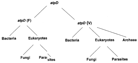
Analysis of multiple sequence alignments of tuf and atpD sequences
permitted the design of oligonucleotide primers (and probes) capable of
amplifying
(or hybridizing to) segments oftuf(and/orfus) and atpD genes from a wide
variety
of bacterial species (see Examples 1 to 4, 24 and 26, and Table 7). Sequencing
and
amplification primer pairs for tuf nucleic acids and/or sequences are listed
in
Annex I and hybridization probes are listed in Annexes III and XLVII.
Sequencing
and amplification primer pairs for atpD nucleic acids and/or sequences are
listed in
Annex II. Analysis of the main subdivisions of tuf and atpD sequences (see
Figures
1 and 2) permitted to design sequencing primers amplifying specifically each
of
these subdivisions. It should be noted that these sequencing primers could
also be
used as universal primers. However, since some of these sequencing primers
CA 02906516 2015-09-30
WO 01/23604 PCT/CA00/01150
include several variable sequence (degenerated) positions, their sensitivity
could be
lower than that of universal primers developed for diagnostic purposes.
Further
subdivisions could be done on the basis of the various phyla where these genes
are
encountered.
Similarly, analysis of multiple sequence alignments of recA sequences present
in the public databases permitted the design of oligonucleotide primers
capable of
amplifying segments of recA genes from a wide variety of bacterial species.
Sequencing and amplification primer pairs for recA sequences are listed in
Annex
XXI. The main subdivisions of recA nucleic acids and/or sequences comprise
recA,
radA, rad51 and dmcl. Further subdivisions could be done on the basis of the
various phyla where these genes are encountered.
The present inventor's strategy is to get as much sequence data information
from the four conserved genes (tuf, fus, atpD and recA). This ensemble of
sequence data forming a repertory (with subrepertories corresponding to each
target gene and their main sequence subdivisions) and then using the sequence
information of the sequence repertory (or subrepertories) to design primer
pairs
that could permit either universal detection of algae or archaea or bacteria
or fungi
or parasites, detection of a family or group of microorganism (e.g.
Enterobacteriaceae), detection of a genus (e.g. Streptococcus) or finally a
specific
species (e.g. Staphylococcus aureus). It should be noted that for the purpose
of the
present invention a group of microorganisms is defined depending on the needs
of
the particular diagnostic test. It does not need to respect i particular
taxonomical
grouping or phylum. See Example 12 where primers were designed to amplify a
group a bacteria consisting of the 17 major bacterial species encountered as
contaminants of platelet concentrates. Also remark that in that Example, the
primers are not only able to sensitively and rapidly detect at least the 17
important
bacterial species, but could also detect other species as well, as shown in
Table 14.
In these circumstances the primers shown in Example 12 are considered
universal
for platelet-contaminating bacteria. To develop an assay specific for the
latter, one
or more primers or probes specific to each species could be designed. Another
31
CA 02906516 2015-09-30
WO 01/23604 PCT/CA00/01150
example of primers and/or probes for group detection is given by the
Pseudomonad
group primers. These primers were designed based upon alignment of tuf
sequences from real Pseudomonas species as well as from former Pseudomonas
species such as Stenotrophomonas maltophilia. The resulting primers are able
to
amplify all Pseudomonas species tested as well as several species belonging to
different genera, hence as being specific for a group including Pseudomonas
and
other species, we defined that group as Pseudomonads, as several members were
former Pseudomonas.
For certain applications, it may be possible to develop a universal, group,
family or genus-specific reaction and to proceed to species identification
using
sequence information within the amplicon to design species-specific internal
probes or primers, or alternatively, to proceed directly by sequencing the
amplicon.
The various strategies will be discussed further below.
The ensembles formed by public and proprietary tuf atpD and recA nucleic
acids and/or sequences are used in a novel fashion so they constitute three
databases containing useful information for the identification of
microorganisms.
Sequence repertories of other gene targets were also built to solve some
specific identification problems especially for microbial species genetically
very
similar to each other such as E. coli and Shigella (see Example 23). Based on
tuf
atpD and recA sequences, Streptococcus pneumoniae is very difficult to
differentiate from the closely related species S. oralis and S. mitis.
Therefore, we
elected to built a sequence repertory from hexA sequences (Example 19), a gene
much more variable than our highly conserved tuf, atpD and recA nucleic acids
and/or sequences.
For the detection of mutations associated with antibiotic resistance genes, we
also built repertories to distinguish between point mutations reflecting only
gene
diversity and point mutations involved in resistance. This was done for pbpla,
pbp2b and pbp2x genes of penicillin-resistant and sensitive Streptoccoccus
pneumoniae (Example 18) and also for gyrA and parC gene fragments of various
bacterial species for which quinolone resistance is important to monitor.
32
CA 02906516 2015-09-30
WO 01/23604 PCT/CA00/01150
Oligonucleotide primers and probes design and synthesis
The tuf, fus, atpD and recA DNA fragments sequenced by us and/or selected
from public databases (GenBank and EMBL) were used to design oligonucleotides
primers and probes for diagnostic purposes. Multiple sequence alignments were
made using subsets of the tuf or atpD or recA sequences repertory. Subsets
were
chosen to encompass as much as possible of the targetted microorganism(s) DNA
sequence data and also include sequence data from phylogenetically related
microorganisms from which the targetted microorganism(s) should be
distinguished. Regions suitable for primers and probes should be conserved for
the
targetted microorganism(s) and divergent for the microorganisms from which the
targetted microorganism(s) should be distinguished. The large amount of tuf or
atpD or recA sequences data in our repertory permits to reduce trial and
errors in
obtaining specific and ubiquitous primers and probes. We also relied on the
corresponding peptide sequences of tuf, fus, atpD and recA nucleic acids
and/or
sequences to facilitate the identification of regions suitable for primers and
probes
design. As part of the design rules, all oligonucleotides (probes for
hybridization
and primers for DNA amplification by PCR) were evaluated for their suitability
for
hybridization or PCR amplification by computer analysis using standard
programs
(i.e. the Genetics Computer Group (GCG) programs and the primer analysis
software OligoTM 5.0). The potential suitability of the PCR primer pairs was
also
evaluated prior to the synthesis by verifying the absence of unwanted features
such
as long stretches of one nucleotide and a high proportion of G or C residues
at the
3' end (Persing et al., 1993, Diagnostic Molecular Microbiology: Principles
and
Applications, American Society for Microbiology, Washington, D.C.).
Oligonucleotide probes and amplification primers were synthesized using an
automated DNA synthesizer (Perkin-Elmer Corp., Applied Biosystems Division).
The oligonucleotide sequence of primers or probes may be derived from
either strand of the duplex DNA. The primers or probes may consist of the
bases
33
CA 02906516 2015-09-30
WO 01/23604 PCT/CA00/01150
A, G, C, or T or analogs and they may be degenerated at one or more chosen
nucleotide position(s). The primers or probes may be of any suitable length
and
may be selected anywhere within the DNA sequences from proprietary fragments
or from selected database sequences which are suitable for (i) the universal
detection of algae or archaea or bacteria or fungi or parasites, (ii) the
species-
specific detection and identification of any microorganism, including but not
limited to: Abiotrophia adiacens, Bacteroides fragilis, Bordetella pertussis,
Candida albicans, Candida dubliniensis, Candida glabrata, Candida
guilliermondii, Candida krusei, Candida lusitaniae, Candida parapsilosis,
Candida tropicalis, Candida zeylanoides, Campylobacter jejuni and C. coli,
Chlamydia pneumoniae, Chlamydia trachomatis, Cryptococcus neoformans,
Cryptosporidium parvum, Enterococcus faecalis, Enterococcus faecium,
Enterococcus gallina rum, Escherichia coli, Haemophilus influenzae, Legionella
pneumophila, Mycoplasma pneumoniae, Neisseria gonorrhoeae, Pseudomonas
aeruginosa, Staphylococcus aureus, Staphylococcus epidermidis, Staphylococcus
haemolyticus,
Staphylococcus hominis, Staphylococcus sap rophyticus,
Streptococcus agalactiae, Streptococcus pneumoniae, Trypanosoma brucei,
Trypanosoma cruzi, (iii) the genus-specific detection of Bordetella species,
Candida species, Clostridium species, Corynebacterium species, Cryptococcus
species, Entamoeba species, Enterococcus species, Gemella species, Giardia
species, Legionella species, Leishmania species, Staphylococcus species,
Streptococcus species, Trypanosoma species, (iv) the family-specific detection
of
Enterobacteriaceae family members, Mycobacteriaceae family members,
Trypanosomatidae family members, (v) the detection of Enterococcus
casseliflavus-flavescens-gallinarum group, Enterococcus, Gemella and
Abiotrophia adiacens group, Pseudomonads extended group, Platelet-
contaminating bacteria group, (vi) the detection of clinically important
antimicrobial agents resistance genes listed in Table 5, (vii) the detection
of
clinically important toxin genes listed in Table 6.
34
CA 02906516 2015-09-30
WO 01/23604 PCT/CA00/01150
Variants for a given target microbial gene are naturally occurring and are
attributable to sequence variation within that gene during evolution (Watson
et al.,
1987, Molecular Biology of the Gene, 4th ed., The Benjamin/Cummings Publishing
Company, Menlo Park, CA; Lewin, 1989, Genes IV, John Wiley & Sons, New
York, NY). For example, different strains of the same microbial species may
have
a single or more nucleotide variation(s) at the oligonucleotide hybridization
site.
The person skilled in the art is well aware of the existence of variant algal,
archaeal, bacterial, fungal or parasitical DNA nucleic acids and/or sequences
for a
specific gene and that the frequency of sequence variations depends on the
selective pressure during evolution on a given gene product. The detection of
a
variant sequence for a region between two PCR primers may be demonstrated by
sequencing the amplification product. In order to show the presence of
sequence
variants at the primer hybridization site, one has to amplify a larger DNA
target
with PCR primers outside that hybridization site. Sequencing of this larger
fragment will allow the detection of sequence variation at this site. A
similar
strategy may be applied to show variants at the hybridization site of a probe.
Insofar as the divergence of the target nucleic acids and/or sequences or a
part
thereof does not affect the specificity and ubiquity of the amplification
primers or
probes, variant microbial DNA is under the scope of this invention. Variants
of the
selected primers or probes may also be used to amplify or hybridize to a
variant
DNA.
Sequencing of tuf nucleic acids and/or sequences from a variety of archaeal,
bacterial, fungal and parasitical species
The nucleotide sequence of a portion of tuf nucleic acids and/or sequences
was determined for a variety of archaeal, bacterial, fungal and parasitical
species.
The amplification primers (SEQ ID NOs. 664 and 697), which amplify a tuf gene
portion of approximately 890 bp, were used along with newly designed
sequencing
primer pairs (See Annex I for the sequencing primers for tuf nucleic acids
and/or
CA 02906516 2015-09-30
WO 01/23604 PCT/CA00/01150
sequences). Most primer pairs can amplify different copies oftuf genes (tufA
and
tufB). This is not surprising since it is known that for several bacterial
species these
two genes are nearly identical. For example, the entire tufA and tufB genes
from E.
coli differ at only 13 nucleotide positions (Neidhardtet al., 1996,
Escherichia coli
and Salmonella: Cellular and Molecular Biology, 2nd ed., American Society for
Microbiology Press, Washington, D.C.). Similarly, some fungi are known to have
two nearly identical copies of tuf nucleic acids and/or sequences (EF-1a).
These
amplification primers are degenerated at several nucleotide positions and
contain
inosines in order to allow the amplification of a wide range oftuf nucleic
acids
and/or sequences. The strategy used to select these amplification primers is
similar
to that illustrated in Annex I for the selection of universal primers. The tuf
sequencing primers even sometimes amplified highly divergent copies oftuf
genes
(tufC) as illustrated in the case of some enterococcal species (SEQ ID NOs.:
73,
75, 76, 614 to 618, 621 and 987 to 989). To prove this, we have determined the
enterococcal tuf nucleic acids and/or sequences from PCR amplicons cloned into
a
plasmid vector. Using the sequence data from the cloned amplicons, we designed
new sequencing primers specific to the divergent (tufC) copy of
enterococci(SEQ
ID NOs.: 658-659 and 661) and then sequenced directly thetufC amplicons. The
amplification primers (SEQ ID NOs.: 543, 556, 557, 643-645, 660, 664, 694, 696
and 697) could be used to amplify the tuf nucleic acids and/or sequences from
any
bacterial species. The amplification primers (SEQ ID NOs.: 558, 559, 560, 653,
654, 655, 813, 815, 1974-1984, 1999-2003) could be used to amplify thetuf (EF-
1a) genes from any fungal and/or parasitical species. The amplification
primers
SEQ ID NOs. 1221-1228 could be used to amplify bacterial tuf nucleic acids
and/or sequences of the EF-G subdivision (fusA) (Figure 3). The amplification
primers SEQ ID NOs. 1224, and 1227-1229 could be used to amplify bacterialtuf
nucleic acids and/or sequences comprising the end of EF-G (fusA) and the
beginning of EF-Tu (tuf), including the intergenic region, as shown in Figure
3.
Most tuf fragments to be sequenced were amplified using the following
amplification protocol: One 1 of cell suspension (or of purified genomic DNA
36
CA 02906516 2015-09-30
WO 01/23604 rc 1/LAUU/U11J1/
0.1-100 ng/ 1) was transferred directly to 19 p,1 of a PCR reaction mixture.
Each
PCR reaction contained 50 mM KC1, 10 mM Tris-HC1 (pH 9.0), 0.1% Triton X-
100, 2.5 mM MgC12, 1 itY1 of each of the 2 primers, 200 AM of each of the four
dNTPs, 0.5 unit of Taq DNA polymerase (Promega Corp., Madison, WI). PCR
reactions were subjected to cycling using a PTC-200 thermal cycler (MJ
Research
Inc., Watertown, Mass.) as follows: 3 min at 94-96 C followed by 30-45 cycles
of
1 min at 95 C for the denaturation step, 1 min at 50-55 C for the annealing
step
and 1 min at 72 C for the extension step. Subsequently, twenty microliters of
the
PCR-amplified mixture were resolved by electrophoresis in a 1.5% agarose gel.
The amplicons were then visualized by staining with methylene blue (Flores et
al.,
1992, Biotechniques, 13:203-205). The size of the amplification products was
estimated by comparison with a 100-bp molecular weight ladder. The band
corresponding to the specific amplification product was excised from the
agarose
gel and purified using the QIAquickTM gel extraction kit (QIAGEN Inc.,
Chatsworth, CA). The gel-purified DNA fragment was then used directly in the
sequencing protocol. Both strands of the tuf genes amplification product were
sequenced by the dideoxynucleotide chain termination sequencing method by
using an Applied Biosystems automated DNA sequencer (model 377) with their
Big DyeTM Terminator Cycle Sequencing Ready Reaction Kit (Applied
Biosysterns, Foster City, CA). The sequencing reactions were performed by
using
the same amplification primers and 10 ng/100 bp of the gel-purified amplicon
per
reaction. For the sequencing of long amplicons such as those of eukaryotic tuf
(EF-
la) nucleic acids and/or sequences, we designed internal sequencing primers
(SEQ ID NOs.: 654, 655 and 813) to be able to obtain sequence data on both
strands for most of the fragment length. In order to ensure that the
determined
sequence did not contain errors attributable to the sequencing of PCR
artefacts, we
have sequenced two preparations of the gel-purified tuf amplification product
originating from two independent PCR amplifications. For most target microbial
species, the sequences determined for both amplicon preparations were
identical.
In case of discrepancies, amplicons from a third independent PCR amplification
37
CA 02906516 2015-09-30
WO 01/23604 PCT/CA00/01150
were sequenced. Furthermore, the sequences of both strands were 100%
complementary thereby confirming the high accuracy of the determined sequence.
The tuf nucleic acids and/or sequences determined using the above strategy are
described in the Sequence Listing. Table 7 gives the originating microbial
species
and the source for each tuf sequence in the Sequence Listing.
The alignment of the tuf sequences determined by us or selected from
databases revealed clearly that the length of the sequenced portion of the tuf
genes
is variable. There may be insertions or deletions of several amino acids. In
addition, in several fungi introns were observed. Intron nucleic acids and/or
sequences are part of tuf nucleic acids and/or sequences and could be useful
in the
design of species-specific primers and probes. This explains why the size of
the
sequenced tuf amplification products was variable from one fungal species to
another. Consequently, the nucleotide positions indicated on top of each of
Annexes IV to XX, XXIII to XXXI, XXXVIII and XLII do not correspond for
sequences having insertions or deletions.
It should also be noted that the various tuf nucleic acids and/or sequences
determined by us occasionally contain base ambiguities. These degenerated
nucleotides correspond to sequence variations between tufA and tufB genes (or
copies of the EF-G subdivision of tuf nucleic acids and/or sequences, or
copies of
EF-la subdivision of tuf nucleic acids and/or sequences for fungi and
parasites)
because the amplification primers amplify both tuf genes. These nucleotide
variations were not attributable to nucleotide misincorporations by the Taq
DNA
polymerase because the sequence of both strands was identical and also because
the sequences determined with both preparations of the gel-purified tuf
amplicons
obtained from two independent PCR amplifications were identical.
The selection of amplification primers from tuf nucleic acids and/or seauences
The tuf sequences determined by us or selected from public databases were
used to select PCR primers for universal detection of bacteria, as well as for
genus-
38
CA 02906516 2015-09-30
WO 01/23604 PCT/CA00/01150
specific, species-specific family-specific or group-specific detection and
identification. The strategy used to select these PCR primers was based on the
analysis of multiple sequence alignments of various tuf sequences. For more
details
about the selection of PCR primers from tuf sequences please refer to Examples
5,
7-14, 17, 22, 24, 28, 30-31, 33, 36, and 38-40, and to Annexes VI-IX, XI-XIX
and
X.XV.
Sequencing of atpD and recA nucleic acids and/or sequences from a variety of
archaeal, bacterial, fungal and parasitical species
The method used to obtain atpD and recA nucleic acids and/or sequences is
similar to that described above for tuf nucleic acids and/or sequences.
The selection of amplification primers from atpD or recA nucleic acids and/or
sequences
The comparison of the nucleotide sequence for the atpD or recA genes from
various archaeal, bacterial, fungal and parasitical species allowed the
selection of
PCR primers (refer to Examples 6, 13, 29, 34 and 37, and to Annexes IV, V, X,
and XX).
DNA amplification
For DNA amplification by the widely used PCR (polymerase chain reaction)
method, primer pairs were derived from proprietary DNA fragments or from
database sequences. Prior to synthesis, the potential primer pairs were
analyzed by
using the OligoTM 5.0 software to verify that they were good candidates for
PCR
amplification.
During DNA amplification by PCR, two oligonucleotide primers binding
respectively to each strand of the heat-denatured target DNA from the
microbial
39
CA 02906516 2015-09-30
WO 01/23604 PCT/CA00/01150
genome are used to amplify exponentially in vitro the target DNA by successive
thermal cycles allowing denaturation of the DNA, annealing of the primers and
synthesis of new targets at each cycle (Persing et al, 1993, Diagnostic
Molecular
Microbiology: Principles and Applications, American Society for Microbiology,
Washington, D.C.).
Briefly, the PCR protocols were as follows: Treated clinical specimens or
standardized bacterial or fungal or parasitical suspensions (see below) or
purified
genomic DNA from bacteria, fungi or parasites were amplified in a 20 Al PCR
reaction mixture. Each PCR reaction contained 50 mM KC1, 10 mM Tris-HC1 (pH
9.0), 2.5 mM MgC12, 0.4 AM of each primer, 200 AM of each of the four dNTPs
and 0.5 unit of Taq DNA polymerase (Promega) combined with the
TaqStartImantibody (Clontech Laboratories Inc., Palo Alto, CA). The
TaqStarti'm
antibody, which is a neutralizing monoclonal antibody to Taq DNA polymerase,
was added to all PCR reactions to enhance the specificity and the sensitivity
of the
amplifications (Kellogg et al., 1994, Biotechniques 16:1134-1137). The
treatment
of the clinical specimens varies with the type of specimen tested, since the
composition and the sensitivity level required are different for each specimen
type.
It consists in a rapid protocol to lyse the microbial cells and eliminate or
neutralize
PCR inhibitors. For amplification from bacterial or fungal or parasitical
cultures or
from purified genomic DNA, the samples were added directly to the PCR
amplification mixture without any pre-treatment step. An internal control was
derived from sequences not found in the target microorganisms or in the human
genome. The internal control was integrated into all amplification reactions
to
verify the efficiency of the PCR assays and to ensure that significant PCR
inhibition was absent. Alternatively, an internal control derived from rRNA
was
also useful to monitor the efficiency of microbial lysis protocols.
PCR reactions were then subjected to thermal cycling (3 min at 94-96 C
followed by 30 cycles of 1 second at 95 C for the denaturation step and 30
seconds
at 50-65 C for the annealing-extension step) using a PTC-200 thermal cycler
(MJ
Research Inc.). The number of cycles performed for the PCR assays varies
CA 02906516 2015-09-30
WO 01/23604 PCT/CA00/01150
according to the sensitivity level required. For example, the sensitivity
level
required for microbial detection directly from clinical specimens is higher
for
blood specimens than for urine specimens because the concentration of
microorganisms associated with a septicemia can be much lower than that
associated with a urinary tract infection. Consequently, more sensitive PCR
assays
having more thermal cycles are probably required for direct detection from
blood
specimens. Similarly, PCR assays performed directly from bacterial or fungal
or
parasitical cultures may be less sensitive than PCR assays performed directly
from
clinical specimens because the number of target organisms is normally much
lower
in clinical specimens than in microbial cultures.
The person skilled in the art of DNA amplification knows the existence of
other rapid amplification procedures such as ligase chain reaction (LCR),
transcription-mediated amplification (TMA), self-sustained sequence
replication
(3SR), nucleic acid sequence-based amplification (NASBA), strand displacement
amplification (SDA), branched DNA (bDNA), cycling probe technology (CPT),
solid phase amplification (SPA), rolling circle amplification technology
(RCA),
solid phase RCA, anchored SDA and nuclease dependent signal amplification
(NDSA) (Lee et al., 1997, Nucleic Acid Amplification Technologies: Application
to Disease Diagnosis, Eaton Publishing, Boston, MA; Persing et al., 1993,
Diagnostic Molecular Microbiology: Principles and Applications, American
Society for Microbiology, Washington, D.C.; Westin et al., 2000, Nat.
Biotechnol.
18:199-204). The scope of this invention is not limited to the use of
amplification
by PCR, but rather includes the use of any rapid nucleic acid amplification
method
or any other procedure which may be used to increase the sensitivity and/or
the
rapidity of nucleic acid-based diagnostic tests. The scope of the present
invention
also covers the use of any nucleic acids amplification and detection
technology
including real-time or post-amplification detection technologies, any
amplification
technology combined with detection, any hybridization nucleic acid chips or
arrays
technologies, any amplification chips or combination of amplification and
41
CA 02906516 2015-09-30
WO 01/23604 PCT/CA00/01150
hybridization chips technologies. Detection and identification by any
sequencing
method is also under the scope of the present invention.
Any oligonucleotide suitable for the amplification of nucleic acids by
approaches other than PCR or for DNA hybridization which are derived from the
species-specific, genus-specific and universal DNA fragments as well as from
selected antimicrobial agents resistance or toxin gene sequences included in
this
document are also under the scope of this invention.
Detection of amplification products
Classically, detection of amplification is performed by standard ethidium
bromide-stained agarose gel electrophoresis. It is clear that other methods
for the
detection of specific amplification products, which may be faster and more
practical for routine diagnosis, may be used. Such methods may be based on the
detection of fluorescence after or during amplification. One simple method for
monitoring amplified DNA is to measure its rate of formation by measuring the
increase in fluorescence of intercalating agents such as ethidium bromide or
SYBR Green I (Molecular Probes). If more specific detection is required,
fluorescence-based technologies can monitor the appearance of a specific
product
during the reaction. The use of dual-labeled fluorogenic probes such as in the
TaqManTm system (Applied Biosystems) which utilizes the 5'-3' exonuclease
activity of the Taq polymerase is a good example (Livak K.J. et al. 1995, PCR
Methods Appl. 4:357-362). TaqManTm can be performed during amplification and
this "real-time" detection can be done in a single closed tube hence
eliminating
post-PCR sample handling and consequently preventing the risk of amplicon
carryover. Several other fluorescence-based detection methods can be performed
in
real-time. Fluorescence resonance energy transfer (FRET) is the principle
behind
the use of adjacent hybridization probes (Wittwer, C.T. et al. 1997.
BioTechniques
22:130-138), molecular beacons (Tyagi S. and Kramer F.R. 1996. Nature
Biotechnology 14:303-308) and scorpions (Whitcomb et al. 1999. Nature
42
CA 02906516 2015-09-30
WO 01/23604 PCT/CA00/01150
Biotechnology 17:804-807). Adjacent hybridization probes are designed to be
internal to the amplification primers. The 3' end of one probe is labelled
with a
donor fluorophore while the 5' end of an adjacent probe is labelled with an
acceptor fluorophore. When the two probes are specifically hybridized in
closed
proximity (spaced by 1 to 5 nucleotides) the donor fluorophore which has been
excited by an external light source emits light that is absorbed by a second
acceptor
that emit more fluorescence and yields a FRET signal. Molecular beacons
possess
a stem-and-loop structure where the loop is the probe and at the bottom of the
stem
a fluorescent moiety is at one end while a quenching moiety is at the other
end.
The beacons undergo a fluorogenic conformational change when they hybridize to
their targets hence separating the fluorochrome from its quencher. The FRET
principle is also used in an air thermal cycler with a built-in fluorometer
(Wittwer,
C.T. et al. 1997. BioTechniques 22:130-138). The amplification and detection
are
extremely rapid as reactions are performed in capillaries: it takes only 18 mm
to
complete 45 cycles. Those techniques are suitable especially in the case where
few
pathogens are searched for. Boehringer-Roche Inc. sells the LightCyclerTM, and
Cepheid makes the SmartCycler. These two apparatus are capable of rapid cycle
PCR combined with fluorescent SYBR Green I or FRET detection. We recently
demonstrated in our laboratory, real-time detection of 10 CFU in less than 40
minutes using adjacent hybridization probes on the LightCyclerTM. Methods
based
on the detection of fluorescence are particularly promising for utilization in
routine
diagnosis as they are very rapid, quantitative and can be automated.
Microbial pathogens detection and identification may also be performed by
solid support or liquid hybridization using species-specific internal DNA
probes
hybridizing to an amplification product. Such probes may be generated from any
sequence from our repertory and designed to specifically hybridize to DNA
amplification products which are objects of the present invention.
Alternatively,
the internal probes for species or genus or family or group detection and
identification may be derived from the amplicons produced by a universal,
family-,
group-, genus- or species-specific amplification assay(s). The oligonucleotide
43
CA 02906516 2015-09-30
WO 01/23604 PCT/CA00/01150
probes may be labeled with biotin or with digoxigenin or with any other
reporter
molecule (for more details see below the section on hybrid capture).
Hybrization
on a solid support is amendable to miniaturization.
At present the oligonucleotide nucleic acid microarray technology is
appealing. Currently, available low to medium density arrays (Heller et al.,
An
integrated microelectronics hybridization system for genoinic research and
diagnostic applications. In: Harrison, D.J., and van den Berg, A., 1998, Micro
total
analysis systems '98, Kluwer Academic Publisher, Dordrecht.) could
specifically
capture fluorescent-labelled amplicons. Detection methods for hybridization
are
not limited to fluorescence; potentiometry, colorimetry and plasmon resonance
are
some examples of alternative detection methods. In addition to detection by
hybridization, nucleic acid microarrays could be used to perform rapid
sequencing
by hybridization. Mass spectrometry could also be applicable for rapid
identification of the amplicon or even for sequencing of the amplification
products
(Chiu and Cantor, 1999, Clinical Chemistry 45:1578; Berkenkamp et al., 1998,
Science 281:260).
For the future of our assay format, we also consider the major challenge of
molecular diagnostics tools, i.e.: integration of the major steps including
sample
preparation, genetic amplification, detection, data analysis and presentation
(Anderson et al., Advances in integrated genetic analysis. In: Harrison, D.J.,
and
van den Berg, A., 1998, Micro total analysis systems '98, Kluwer Academic
Publisher, Dordrecht.).
To ensure PCR efficiency, glycerol, dimethyl sulfoxide (DMSO) or other
related solvents can be used to increase the sensitivity of the PCR and to
overcome
problems associated with the amplification of a target DNA having a high GC
content or forming strong secondary structures (Dieffenbach and Dveksler,
1995,
PCR Primer: A Laboratory Manual, Cold Spring Harbor Laboratory Press,
Plainview, New York). The concentration ranges for glycerol and DMSO are 5-
15% (v/v) and 340% (v/v), respectively. For the PCR reaction mixture, the
concentration ranges for the amplification primers and MgC12 are 0.1-1.5 AM
and
44
CA 02906516 2015-09-30
WO 01/23604 PCT/CA00/01150
1.0-10.0 mM, respectively. Modifications of the standard PCR protocol using
external and nested primers (i.e. nested PCR) or using more than one primer
pair
(i.e. multiplex PCR) may also be used (Persing et al., 1993, Diagnostic
Molecular
Microbiology: Principles and Applications, American Society for Microbiology,
Washington, D.C.). For more details about the PCR protocols and amplicon
detection methods, see Examples.
Hybrid capture and chemiluminescence detection of amplification products
Hybridization and detection of amplicons by chemiluminescence were
adapted from Nikiforov et al. (1994, PCR Methods and Applications 3:285-291
and 1995, Anal. Biochem. 227:201-209 ) and from the DIGTM system protocol of
Boehringer Mannheim. Briefly, 50 1 of a 25 picomoles solution of capture
probe
diluted in EDC 11-ethy1-3-(3-dirriethylarninopropyl) carbodiimide
hydrochloride)
are immobilized in each well of 96-wells plates (MicroliteTm 2, Dynex) by
incubation overnight at room temperature. The next day, the plates are
incubated
with a solution of 1% BSA diluted into TNTw (10 mM Tris-HC1, pH 7.5; 150 mM
NaCl; 0.05% TweenTm 20) for 1 hour at 37 C. The plates are then washed on a
Wellwash AscentTM (Labsystems) with TNTw followed by Washing Buffer (100
mM maleic acid pH7.5; 150 mM NaCl; 0.3% TweenTm 20).
The amplicons were labelled with DIG-11-dUTP during PCR using the PCR
DIG Labelling Mix from Boehringer Mannheim according to the manufacturer's
instructions. Hybridization of the amplicons to the capture probes is
performed in
triplicate at stringent temperature (generally, probes are designed to allow
. hybrization at 55 C, the stringent temperature) for 30 minutes in 1.5 M
NaCl; 10
mM EDTA. It is followed by two washes in 2 X SSC; 0.1% SDS, then by four
washes in 0.1X SSC; 0.1% SDS at the stringent temperature (55 C). Detection
with 1,2 dioxetane chemiluminescent alkaline phosphatase substrates like CSPD
(Tropix Inc.) is performed according to the manufacturer's instructions but
with
shorter incubations times and a different antibody concentration. The plates
are
CA 02906516 2015-09-30
WO 01/23604 PCT/CA00/01150
agitated at each step, the blocking incubation is performed for only 5
minutes, the
anti-DIG-AP1 is used at a 1:1000 dilution, the incubation with antibody lasts
15
minutes, the plates are washed twice for only 5 minutes. Finally, after a 2
minutes
incubation into the detection buffer, the plates are incubated 5 minutes with
CSPD at room temperature followed by a 10 minutes incubation at 37 C without
agitation. Luminous signal detection is performed on a Dynex Microtiter Plate
Luminometer using RLU (Relative Light Units).
Specificity, ubiquity and sensitivity tests for oligonucleotide primers and
probes
The specificity of oligonucleotide primers and probes was tested by
amplification of DNA or by hybridization with bacterial or fungal or
parasitical
species selected from a panel comprising closely related species and species
sharing the same anatomo-pathological site (see Annexes and Examples). All of
the bacterial, fungal and parasitical species tested were likely to be
pathogens
associated with infections or potential contaminants which can be isolated
from
clinical specimens. Each target DNA could be released from microbial cells
using
standard chemical and/or physical treatments to lyse the cells (Sambrook et
al.,
1989, Molecular Cloning: A Laboratory Manual, 2nd ed., Cold Spring Harbor
Laboratory Press, Cold Spring Harbor, NY) or alternatively, genoinic DNA
purified with the GNOMETm DNA kit (Bio101, Vista, CA) was used.
Subsequently, the DNA was subjected to amplification with the primer pairs.
Specific primers or probes amplified only the target microbial species, genus,
family or group.
Oligonucleotides primers found to amplify specifically the target species,
genus, family or group were subsequently tested for their ubiquity by
amplification
(i.e. ubiquitous primers amplified efficiently most or all isolates of the
target
species or genus or family or group). Finally, the sensitivity of the primers
or
probes was determined by using 10-fold or 2-fold dilutions of purified genomic
DNA from the targeted microorganism. For most assays, sensitivity levels in
the
46
CA 02906516 2015-09-30
WO 01/23604 PCT/CA00/01150
range of 1-100 copies were obtained. The specificity, ubiquity and sensitivity
of
the PCR assays using the selected amplification primer pairs were tested
either
directly from cultures of microbial species or from purified microbial genomic
DNA.
Probes were tested in hybrid capture assays as described above. An
oligonucleotide probe was considered specific only when it hybridized solely
to
DNA from the species or genus or family or group from which it was selected.
Oligonucleotide probes found to be specific were subsequently tested for their
ubiquity (i.e. ubiquitous probes detected efficiently most or all isolates of
the target
species or genus or family or group) by hybridization to microbial DNAs from
different clinical isolates of the species or genus or family or group of
interest
including ATCC reference strains. Similarly, oligonucleotide primers and
probes
could be derived from antimicrobial agents resistance or toxin genes which are
objects of the present invention.
Reference strains
The reference strains used to build proprietary tuf, atpD and recA sequence
data subrepertories, as well as to test the amplification and hybridization
assays
were obtained from (i) the American Type Culture Collection (ATCC), (ii) the
Laboratoire de sante publique du Quebec (LSPQ), (iii) the Centers for Disease
Control and Prevention (CDC), (iv) the National Culture Type Collection (NCTC)
and (v) several other reference laboratories throughout the world. The
identity of
our reference strains was confirmed by phenotypic testing and reconfirmed by
analysis of tuf, atpD and recA sequences (see Example 13).
Antimicrobial agents resistance genes
Antimicrobial resistance complicates treatment and often leads to therapeutic
failures. Furthermore, overuse of antibiotics inevitably leads to the
emergence of
47
CA 02906516 2015-09-30
WO 01/23604 PCT/CA00/01150
microbial resistance. Our goal is to provide clinicians, in approximately one
hour,
the needed information to prescribe optimal treatments. Besides the rapid
identification of negative clinical specimens with DNA-based tests for
universal
algal, archaeal, bacterial, fungal or parasitical detection and the
identification of
the presence of a specific pathogen in the positive specimens with species-
and/or
genus- and/or family- and/or group-specific DNA-based tests, clinicians also
need
timely information about the ability of the microbial pathogen to resist
antibiotic
treatments. We feel that the most efficient strategy to evaluate rapidly
microbial
resistance to antimicrobials is to detect directly from the clinical specimens
the
most common and clinically important antimicrobial agents resistance genes
(i.e.
DNA-based tests for the specific detection of antimicrobial agents resistance
genes). Since the sequence from the most important and common antimicrobial
agents resistance genes are available from public databases, our strategy is
to use
the sequence from a portion or from the entire resistance gene to design
specific
oligonucleotide primers or probes which will be used as a basis for the
development of sensitive and rapid DNA-based tests. The list of each of the
antimicrobial agents resistance genes selected on the basis of their clinical
relevance (i.e. high incidence and importance) is given in Table 5;
descriptions of
the designed amplification primers and internal probes are given in Annexes
XXXIV-XXXVII, XXXIX, XLV, and L-LI. Our approach is unique because the
antimicrobial agents resistance genes detection and the microbial detection
and
identification can be performed simultaneously, or independently, or
sequentially
in multiplex or parallel or sequential assays under uniform PCR amplification
conditions. These amplifications can also be done separately.
Toxin genes
Toxin identification is often very important to prescribe optimal treatments.
Besides the rapid identification of negative clinical specimens with DNA-based
tests for universal bacterial detection and the identification of the presence
of a
48
CA 02906516 2015-09-30
WO 01/23604 PCT/CA00/01150
specific pathogen in the positive specimens with species- and/or genus- and/or
family- and/or group-specific DNA-based tests, clinicians sometimes need
timely
information about the ability of certain bacterial pathogens to produce
toxins.
Since the sequence from the most important and common bacterial toxin genes
are
available from public databases, our strategy is to use the sequence from a
portion
or from the entire toxin gene to design specific oligonucleotide primers or
probes
which will be used as a basis for the development of sensitive and rapid DNA-
based tests. The list of each of the bacterial toxin genes selected on the
basis of
their clinical relevance (i.e. high incidence and importance) is given in
Table 6;
descriptions of the designed amplification primers and internal probes are
given in
Annexes XXII, XXXII and XXXIII. Our approach is unique because the toxin
genes detection and the bacterial detection and identification can be
performed
simultaneously, or independently, or sequentially, in multiplex or parallel or
sequential assays under uniform PCR amplification conditions. These
amplifications can also be done separately.
Universal bacterial detection
In the routine microbiology laboratory, a high percentage of clinical
specimens sent for bacterial identification are negative by culture. Testing
clinical
samples with universal amplification primers or universal probes to detect the
presence of bacteria prior to specific identification and screening out the
numerous
negative specimens is thus useful as it reduces costs and may rapidly orient
the
clinical management of the patients. Several amplification primers and probes
were
therefore synthesized from highly conserved portions of bacterial sequences
from
the tuf, atpD and recA nucleic acids and/or sequences. The universal primers
selection was based on a multiple sequence alignment constructed with
sequences
from our repertory.
All computer analysis of amino acid and nucleotide sequences were
performed by using the GCG programs. Subsequently, optimal PCR primers for
49
CA 02906516 2015-09-30
WO 01/23604 PCT/CA00/01150
the universal amplification of bacteria were selected with the help of the
OligoTM
program. The selected primers are degenerated at several nucleotide positions
and
contain several inosines in order to allow the amplification of all clinically
relevant
bacterial species. Inosine is a nucleotide analog able to specifically bind to
any of
the four nucleotides A, C, G or T. Degenerated oligonucleotides consist of an
oligonucleotide mix having two or more of the four nucleotides A, C, G or T at
the
site of mismatches. The inclusion of inosine and/or of base ambiguities in the
amplification primers allow mismatch tolerance thereby permitting the
amplification of a wider array of target nucleotide sequences (Dieffenbach and
Dveksler, 1995 PCR Primer: A Laboratory Manual, Cold Spring Harbor
Laboratory Press, Plainview, NY).
The amplification conditions with the universal primers are very similar to
those used for the species- and genus-specific amplification assays except
that the
annealing temperature is slightly lower. The original universal PCR assay
described in our assigned W098/20157 (SEQ ID NOs. 23-24 of the latter
application) was specific and nearly ubiquitous for the detection of bacteria.
The
specificity for bacteria was verified by amplifying genomic DNA isolated from
the
12 fungal species as well as genomic DNA from Leishmania donovani,
Saccharomyces cerevisiae and human lymphocytes. None of the above eukaryotic
DNA preparations could be amplified by the universal assay, thereby suggesting
that this test is specific for bacteria. The ubiquity of the universal assay
was
verified by amplifying genomic DNAs from 116 reference strains which represent
95 of the most clinically relevant bacterial species. These species have been
selected from the bacterial species listed in Table 4. We found that at least
104 of
these strains could be amplified. However, the assay could be improved since
bacterial species which could not be amplified with the original tuf nucleic
acids
and/or sequences-based assay included species belonging, to the following
genera:
Corynebacterium (11 species) and Stenotrophomonas (1 species). Sequencing of
the tuf genes from these bacterial species and others has been performed in
the
scope of the present invention in order to improve the universal assay. This
CA 02906516 2015-09-30
WO 01/23604 Ill,AUU/U113U
sequencing data has been used to select new universal primers which may be
more
ubiquitous and more sensitive. Also, we improved our primer and probes design
strategy by taking into consideration the phylogeny observed in analysing our
repertory of tuf, atpD and recA sequences. Data from each of the 3 main
subrepertories (tuf, atpD and recA) was subjected to a basic phylogenic
analysis
using the Pileup command from version 10 of the GCG package (Genetics
Computer Group, inc.). This analysis indicated the main branches or phyla
reflecting the relationships between sequences. Instead of trying to design
primers
or probes able to hybridize to all phyla, we designed primers or probes able
to
hybridize to the main phyla while trying to use the largest phylum possible.
This
strategy should allow less degenerated primers hence improving sensitivity and
by
combining primers in a mutiplex assay, improve ubiquity. Universal primers SEQ
ID NOs. 643-645 based on tuf sequences have been designed to amplify most
pathogenic bacteria except Actinomyceteae, Clostridiaceae and the Cytophaga,
Flexibacter and Bacteroides phylum (pathogenic bacteria of this phylum include
mostly Bacteroides, Porphyromonas and Prevotella species). Primers to fill
these
gaps have been designed for Actinomyceteae (SEQ ID NOs. 646-648),
Clostridiaceae (SEQ ID NOs. 796-797, 808-811), and the Cytophaga, Flexibacter
and Bacteroides phylum (SEQ ID NOs. 649-651), also derived from tuf nucleic
acids and/or sequences. These primers sets could be used alone or in
conjuction to
render the universal assay more ubiquitous.
Universal primers derived from atpD sequences include SEQ ID NOs. 562-
565. Combination of these primers does not amplify human DNA but should
amplify almost all pathogenic bacterial species except proteobacteria
belonging to
the epsilon subdivision (Campylobacter and Helicobacter), the bacteria from
the
Cytophaga, Flexibacter and Bacteroides group and some actinomycetes and
corynebacteria. By analysing atpD sequences from the latter species, primers
and
probes to specifically fill these gaps could be designed and used in
conjuction with
primers SEQ ID NOs. 562-565, also derived from atpD nucleic acids and/or
sequences.
51
CA 02906516 2015-09-30
WO 01/23604 PCT/CA00/01150
In addition, universality of the assay could be expanded by mixing atpD
sequences-derived primers with tuf sequences-derived primers. Ultimately, even
recA sequences-derived primers could be added to fill some gaps in the
universal
assay.
It is important to note that the 95 bacterial species selected to test the
ubiquity
of the universal assay include all of the most clinically relevant bacterial
species
associated with a variety of human infections acquired in the community or in
hospitals (nosocomial infections). The most clinically important bacterial and
fungal pathogens are listed in Tables 1 and 2.
Amino acid sequences derived from tuf, atpD and recA nucleic acids and/or
sequences
The amino acid sequences translated from the repertory of tuf, atpD and recA
nucleic acids and/or sequences are also an object of the present invention.
The
amino acid sequence data will be particularly useful for homology modeling of
three-dimensional (3D) structure of the elongation factor Tu, elongation
factor G,
elongation factor la, ATPase subunit beta and RecA recombinase. For all these
proteins, at least one structure model has been published using X-ray
diffraction
data from crystals. Based on those structural informations it is possible to
use
computer sofware to build 3D model structures for any other protein having
peptide sequence homologies with the known structure (Greer, 1991, Methods in
Enzymology, 202:239-252; Taylor, 1994, Trends Biotechnol., 12(5):154-158;
Sali,
1995, CUIT. Opin. Biotechnol. 6:437-451; Sanchez and Sali, 1997, Cuff. Opin.
Struct. Biol. 7:206-214; Fischer and Eisenberg, 1999, Curr. Opin. Struct.
Biol.
9:208-211; Guex et al., 1999, Trends Biochem. Sci. 24: 364-367). Model
structures of target proteins are used for the design or to predict the
behavior of
ligands and inhibitors such as antibiotics. Since EF-Tu and EF-G are already
known as antibiotic targets (see above) and since the beta subunit of ATPase
and
RecA recombinase are essential to the survival of the microbial cells in
natural
52
CA 02906516 2015-09-30
WO 01/23604 PCT/CA00/01150
conditions of infection, all four proteins could be considered antibiotic
targets.
Sequence data, especially the new data generated by us could be very useful to
assist the creation of new antibiotic molecules with desired spectrum of
activity. In
addition, model structures could be used to improve protein function for
commercial purposes such as improving antibiotic production by microbial
strains
or increasing biomass.
53
CA 02906516 2015-09-30
WO 01/23604 PCT/CA00/01150
The following detailed embodiments and appended drawings are provided as
illustrative examples of his invention, with no intention to limit the scope
thereof.
DESCRIPTION OF THE DRAWINGS
Figures 1 and 2 illustrate the principal subdivisions of the tuf and atpD
sequences
repertories, respectively. For the design of primers and probes, depending on
the
needs, one may want to use the complete data set illustrated on the top of the
pyramid or use only a subset illustrated by the different branching points.
Smaller
subdivisions, representing groups, families, genus and species, could even be
made
to extend to the bottom of the pyramid. Because the tuf and atpD sequences are
highly conserved and evolved with each species, the design of primers and
probes
does not need to include all the sequences within the database or its
subdivisions.
As illustrated in Annexes IV to XX, XXIII to XXXI, XXXVIII and XLII,
depending on the use, sequences from a limited number of species can be
carefully
selected to represent: i) only the main phylogenetic branches from which the
intended probes and primers need to be differentiating, and ii) only the
species for
which they need to be matching. However, for ubiquity purposes, and especially
for primers and probes identifying large groups of species (genus, family,
group or
universal, or sequencing primers), the more data is included into the sequence
analysis, the better the probes and primers will be suitable for each
particular
intended use. Similarly, for specificity purposes, a larger data set (or
repertory)
ensures optimal primers and probes design by reducing the chance of employing
nonspecific oligonucleotides.
Figure 3 illustrates the approach used to design specific amplification
primers from
fusA as well as from the region between the end of fusA and the beginning of
tuf in
the streptomycin (str) operon (referred to as the fusA-tufintergenic spacer in
Table
7).
Figures 4 to 6 are illustrations to Example 42, whereas Figures 7 to 10
illustrate
Example 43. Figures 11 and 12 illustrate Example 44.
54
CA 02906516 2015-09-30
WO 01/23604 PCT/CA00/01150
FIGURE LEGENDS
Figure 3. Schematic organization of universal amplification primers (SEQ ID
NOs.
1221-1229) in the str operon. Amplicon sizes are given in bases pairs. Drawing
not
to scale, as the fusA-tuf intergenic spacer size varies depending on the
bacterial
species. Indicated amplicon lengths are for E. coli.
Figure 4. Abridged multiple amino acid sequence alignment of the partial tuf
gene
products from selected species illustrated using the program Alscript.
Residues
highly conserved in bacteria are boxed in grey and gaps are represented with
dots.
Residues in reverse print are unique to the enterococcal tufB as well as to
streptococcal and lactococcal tuf gene products. Numbering is based on E. coli
EF-
Tu and secondary structure elements of E. coli EF-Tu are represented by
cylinders
(a-helices) and arrows (13-strands).
Figure 5. Distance matrix tree of bacterial EF-Tu based on amino acid sequence
homology. The tree was constructed by the neighbor-joining method. The tree
was
rooted using archeal and eukaryotic EF- la genes as the outgroup. The scale
bar
represents 5% changes in amino acid sequence, as determined by taking the sum
of
all of the horizontal lines connecting two species.
Figure 6. Southern hybridization of Bg1111Xbal digested genomic DNAs of some
enterococci (except for E. casseliflavus and E. gallinarum whose genomic DNA
was digested with BamHIIPvull) using the tufA gene fragment of E. faecium as
probes. The sizes of hybridizing fragments are shown in kilobases. Strains
tested
are listed in Table 16.
CA 02906516 2015-09-30
WO 01/23604 PCT/CA00/01150
Figure 7. Pantoea and Tatumella species specific signature indel in atpD
genes.
The nucleotide positions given are for E. coli atpD sequence (GenBank
accession
no. V00267). Numbering starts from the first base of the initiation codon.
Figure 8: Trees based on sequence data from tuf (left side) and atpD (right
side).
The phylogenetic analysis was performed using the Neighbor-Joining method
calculated using= the Kimura two-parameter method. The value on each branch
indicates the occurence (%) of the branching order in 750 bootstrapped trees.
Figure 9: Phylogenetic tree of members of the family Enterobacteriaceae based
on
tuf (a), atpD (b), and 16S rDNA (c) genes. Trees were generated by neighbor-
joining method calculated using the Kimura two-parameter method. The value on
each branch is the percentage of bootstrap replications supporting the branch.
750
bootstrap replications were calculated.
Figure 10: Plot of tuf distances versus 16S rDNA distances (a), atpD distances
versus 16S rDNA distances (b), and atpD distances versus tuf distances (c).
Symbols: 0, distances between pairs of strains belonging to the same species;
*,
distances between E. coli strains and Shigella strains; 0, distances between
pairs
belonging to the same genus; s, distances between pairs belonging to different
genera; A, distances between pairs belonging to different families.
EXAMPLES AND ANNEXES
For sake of clarity, here is a list of Examples and Annexes:
Example 1: Sequencing of bacterial atpD (F-type and V-type) gene fragments.
Example 2: Sequencing of eukaryotic atpD (F-type and V-type) gene
fragments.
Example 3: Sequencing of eukaryotic tuf (EF-1) gene fragments.
56
CA 02906516 2015-09-30
WO 01/23604 PCT/CA00/01150
Example 4: Sequencing of eukaryotic tuf (organelle origin, M) gene fragments.
Example 5: Specific detection and identification of Streptococcus
agalactiae
using tuf sequences.
Example 6: Specific detection and identification of Streptococcus
agalactiae
using atpD sequences.
Example 7: Development of a PCR assay for detection and identification of
staphylococci at genus and species levels.
Example 8: Differentiating between the two closely related yeast species
Candida albicans and Candida dubliniensis.
Example 9: Specific detection and identification of Entamoeba histolytica.
Example 10: Sensitive detection and identification of Chlamydia trachomatis.
Example 11: Genus-specific detection and identification of enterococci.
Example 12: Detection and identification of the major bacterial platelets
contaminants using tuf sequences with a multiplex PCR test.
Example 13: The resolving power of the tuf and atpD sequences databases is
comparable to the biochemical methods for bacterial identification.
Example 14: Detection of group B streptococci from clinical specimens.
Example 15: Simultaneous detection and identification of Streptococcus
pyo genes and its pyrogenic exotoxin A.
Example 16: Real-time detection and identification of Shiga toxin-producing
bacteria.
Example 17: Development of a PCR assay for the detection and identification of
staphylococci at genus and species levels and its associated mecA
gene.
Example 18: Sequencing of pbpl a, pbp2b and pbp2x genes of Streptoccoccus
pneumoniae.
Example 19: Sequencing of hexA genes of Streptococcus species.
Example 20: Development of a multiplex PCR assay for the detection of
Streptococcus pneumoniae and its penicillin resistance genes.
57
CA 02906516 2015-09-30
WO 01/23604 PCT/CA00/01150
Example 21: Sequencing of the vancomycin resistance vanA, vanCl, vanC2 and
vanC3 genes.
Example 22: Development of a PCR assay for the detection and identification of
enterococci at genus and species levels and its associated resistance
genes vanA and vanB.
Example 23: Development of a multiplex PCR assay for detection and
identification of vancomycin-resistant Enterococcus faecalis,
Enterococcus faecium, Enterococcus gallinarum, Enterococcus
casseliflavus, and Enterococcus flavescens.
Example 24: Universal amplification involving the EF-G (fusA) subdivision of
tuf sequences.
Example 25: DNA fragment isolation from Staphylococcus sap rophyticus by
arbitrarily primed PCR.
Example 26: Sequencing of prokaryotic tuf gene fragments.
Example 27: Sequencing of procaryotic recA gene fragments.
Example 28: Specific detection and identification of Escherichia coli/Shigella
sp.
using tuf sequences.
Example 29: Specific detection and identification of Klebsiella pneumoniae
using atpD sequences.
Example 30: Specific detection and identification of Acinetobacter baumanii
using tuf sequences.
Example 31: Specific detection and identification of Neisseria gonorrhoeae
using tuf sequences.
Example 32: Sequencing of bacterial gyrA and parC gene fragments.
Example 33: Development of a PCR assay for the specific detection and
identification of Staphylococcus aureus and its quinolone resistance
genes gyrA and parC.
Example 34: Development of a PCR assay for the detection and identification of
Klebsiella pneumoniae and its quinolone resistance genes gyrA and
parC.
58
CA 02906516 2015-09-30
WO 01/23604 PCT/CA00/01150
Example 35: Development of a PCR assay for the detection and identification of
Streptococcus pneumoniae and its quinolone resistance genes gyrA
and parC.
Example 36: Detection of extended-spectrum TEM-type 13-lactamases in
Escherichia coli.
Example 37: Detection of extended-spectrum SHV-type 0-lactamases in
Klebsiella pneumoniae.
Example 38: Development of a PCR assay for the detection and identification of
Neisseria gonorrhoeae and its associated tetracycline resistance
gene tetM.
Example 39: Development of a PCR assay for the detection and identification of
Shigella sp. and their associated trimethoprim resistance gene
dhfrl a.
Example 40: Development of a PCR assay for the detection and identification of
Acinetobacter baumanii and its associated aminoglycoside
resistance gene aph(3')-V1a.
Example 41: Specific detection and identification of Bacteroides fragilis
using
atpD (V-type) sequences.
Example 42: Evidence for horizontal gene transfer in the evolution of the
elongation factor Tu in Enterococci.
Example 43: Elongation factor Tu (tuf) and the F-ATPase beta-subunit (atpD) as
phylogenetic tools for species of the family Enterobacteriaceae.
Example 44: Testing new pairs of PCR primers selected from two species-
specific genomic DNA fragments which are objects of US patent
6,001,564.
= Example 45: Testing modified versions of PCR primers derived from the
sequence of several primers which are objects of US patent
6,001,564.
59
CA 02906516 2015-09-30
WO 01/23604 PCT/CA00/01150
The various Annexes show the strategies used for the selection of a variety of
DNA amplification primers, nucleic acid hybridization probes and molecular
beacon internal probes:
(i) Annex I shows the amplification primers used for nucleic acid
amplification from tuf sequences.
(ii) Annex II shows the amplification primers used for nucleic acid
amplification from atpD sequences.
(iii) Annex III shows the internal hybridization probes for detection of
tuf
sequences.
(iv) Annex IV illustrates the strategy used for the selection of the
amplification primers specific for atpD sequences of the F-type.
(v) Annex V illustrates the strategy used for the selection of the
amplification
primers specific for atpD sequences of the V-type.
(vi) Annex VI illustrates the strategy used for the selection of the
amplification primers specific for the tuf sequences of organelle lineage
(M, the letter M is used to indicate that in most cases, the organelle is the
mitochondria).
(vii) Annex VII illustrates the strategy used for the selection of the
amplification primers specific for the tuf sequences of eukaryotes (EF-l).
(viii) Annex VIII illustrates the strategy for the selection of
Streptococcus
agalactiae-specific amplification primers from tuf sequences.
(ix) Annex IX illustrates the strategy for the selection of Streptococcus
agalactiae-specific hybridization probes from tuf sequences.
(x) Annex X illustrates the strategy for the selection of Streptococcus
agalactiae-specific amplification primers from atpD sequences.
(xi) Annex XI illustrates the strategy for the selection from tuf sequences
of
Candida albicansl dubliniensis-specific amplification primers, Candida
albicans-specific hybridization probe and Candida dubliniensis-specific
hybridization probe.
CA 02906516 2015-09-30
WO 01/23604 PCT/CA00/01150
(xii) Annex XII illustrates the strategy for the selection of
Staphylococcus-
specific amplification primers from tuf sequences.
(xiii) Annex XIII illustrates the strategy for the selection of the
Staphylococcus-
specific hybridization probe from tuf sequences.
(xiv) Annex XIV illustrates the strategy for the selection of
Staphylococcus
saprophyticus-specific and Staphylococcus haemolyticus-specific
hybridization probes from tuf sequences.
(xv) Annex XV illustrates the strategy for the selection of Staphylococcus
aureus-specific and Staphylococcus epidennidis-specific hybridization
probes from tuf sequences.
(xvi) Annex XVI illustrates the strategy for the selection of the
Staphylococcus
hominis-specific hybridization probe from tuf sequences.
(xvii) Annex XVII illustrates the strategy for the selection of the
Enterococcus-
specific amplification primers from tuf sequences.
(xviii) Annex XVIII illustrates the strategy for the selection of the
Enterococcus
faecalis-specific hybridization probe, of the Enterococcus faecium-
specific hybridization probe and of the Enterococcus casseliflavus-
flavescens-gallinarum group-specific hybridization probe from tuf
sequences.
(xix) Annex XIX illustrates the strategy for the selection of primers from
tuf
sequences for the identification of platelets contaminants.
(xx) Annex XX illustrates the strategy for the selection of the universal
amplification primers from atpD sequences.
(xxi) Annex XXI shows the amplification primers used for nucleic acid
amplification from recA sequences.
(xxii) Annex XXII shows the specific and ubiquitous primers for nucleic acid
amplification from speA sequences.
(xxiii) Annex XXIII illustrates the first strategy for the selection of
Streptococcus pyogenes-specific amplification primers from speA
sequences.
61
CA 02906516 2015-09-30
WO 01/23604 1/LAUU/U11U
(xxiv) Annex XXIV illustrates the second strategy for the selection of
Streptococcus pyogenes-specific amplification primers from speA
sequences.
(xxv) Annex XXV illustrates the strategy for the selection of Streptococcus
pyogenes-specific amplification primers from tuf sequences.
(xxvi) Annex XXVI illustrates the strategy for the selection of stx/-specific
amplification primers and hybridization probe.
(xxvii) Annex XXVII illustrates the strategy for the selection of stx2-
specific
amplification primers and hybridization probe.
(xxviii) Annex XXVIII illustrates the strategy for the selection of vanA-
specific
amplification primers from van sequences.
(xxix) Annex XXIX illustrates the strategy for the selection of vanB-specific
amplification primers from van sequences.
(xxx) Annex XXX illustrates the strategy for the selection of vanC-specific
amplification primers from vanC sequences.
(xxxi) Annex VOCI illustrates the strategy for the selection of Streptococcus
pneumoniae-specific amplification primers and hybridization probes from
pbp 1 a sequences.
(xxxii) Annex XXXII shows the specific and ubiquitous primers for nucleic acid
amplification from toxin gene sequences.
(xxxiii) Annex )(XXIII shows the molecular beacon internal hybridization
probes
for specific detection of toxin sequences.
(xxxiv) Annex XXXIV shows the specific and ubiquitous primers for nucleic acid
amplification from van sequences.
(xxxv) Annex XXXV shows the internal hybridization probes for specific
detection of van sequences.
(xxxvi) Annex XXXVI shows the specific and ubiquitous primers for nucleic acid
amplification from pbp sequences.
(xxxvii) Annex XXXVII shows the internal hybridization probes for specific
detection of pbp sequences.
62
CA 02906516 2015-09-30
WO 01/23604 PCT/CA00/01150
(xxxviii)Annex )(XXVIII illustrates the strategy for the selection of vanAB-
specific amplification primers and vanA- and vanB- specific hybridization
probes from van sequences.
(xxxix) Annex )(XXIX shows the internal hybridization probe for specific
detection of mecA.
(xl) Annex XL shows the specific and ubiquitous primers for nucleic acid
amplification from hexA sequences.
(xli) Annex XLI shows the internal hybridization probe for specific
detection
of hexA.
(xlii) Annex XLII illustrates the strategy for the selection of
Streptococcus
pneumoniae species-specific amplification primers and hybridization
probe from hexA sequences.
(xliii) Annex XLIII shows the specific and ubiquitous primers for nucleic acid
amplification from pcp sequences.
(xliv) Annex XLIV shows specific and ubiquitous primers for nucleic acid
amplification of S. saprophyticus sequences of unknown coding potential.
(xlv) Annex XLV shows the molecular beacon internal hybridization probes for
specific detection of antimicrobial agents resistance gene sequences.
(xlvi) Annex XLVI shows the molecular beacon internal hybridization krobe for
specific detection of S. aureus gene sequences of unknown coding
potential.
(xlvii) Annex XLVII shows the molecular beacon hybridization internal probe
for specific detection of tuf sequences.
(xlviii) Annex XLVIII shows the molecular beacon internal hybridization probes
for specific detection of ddl and mt/ sequences.
(xlix) Annex XLIX shows the internal hybridization probe for specific
detection
of S. aureus sequences of unknown coding potential.
(1) Annex L shows the amplification primers used for nucleic acid
amplification from antimicrobial agents resistance genes sequences.
63
CA 02906516 2015-09-30
WO 01/23604 PCT/CA00/01150
(ii) Annex LI shows the internal hybridization probes for specific
detection of
antimicrobial agents resistance genes sequences.
(lii) Annex LII shows the molecular beacon internal hybridization probes
for
specific detection of atpD sequences.
(liii) Annex LIII shows the internal hybridization probes for specific
detection
of atpD sequences.
(liv) Annex LIVI shows the internal hybridization probes for specific
detection
of ddl and mt/ sequences.
As shown in these Annexes, the selected amplification primers may contain
inosines and/or base ambiguities. Inosine is a nucleotide analog able to
specifically
bind to any of the four nucleotides A, C, G or T. Alternatively, degenerated
oligonucleotides which consist of an oligonucleotide mix having two or more of
the four nucleotides A, C, G or T at the site of mismatches were used. The
inclusion of inosine and/or of degeneracies in the amplification primers
allows
mismatch tolerance thereby permitting the amplification of a wider array of
target
nucleotide sequences (Dieffenbach and Dveksler, 1995 PCR Primer: A Laboratory
Manual, Cold Spring Harbor Laboratory Press, Plainview, New York).
EXAMPLES
EXAMPLE 1:
Sequencing of bacterial atpD (F-type and V-type) gene fragments. As shown in
Annex IV, the comparison of publicly available atpD (F-type) sequences from a
variety of bacterial species revealed conserved regions allowing the design of
PCR
primers able to amplify atpD sequences (F-type) from a wide range of bacterial
species. Using primers pairs SEQ ID NOs. 566 and 567, 566 and 814, 568 and
567,
570 and 567, 572 and 567, 569 and 567, 571 and 567, 700 and 567, it was
possible
to amplify and sequence atpD sequences SEQ ID NOs. 242-270, 272-398, 673-
64
CA 02906516 2015-09-30
WO 01/23604 PCT/CA00/01150
674, 737-767, 866-867, 942-955, 1245-1254, 1256-1265, 1527, 1576, 1577, 1600-
1604, 1640-1646, 1649, 1652, 1655, 1657, 1659-1660, 1671, 1844-1845, and
1849-1865.
Similarly, Annex V shows the strategy to design the PCR primers able to
amplify
atpD sequences of the V-type from a wide range of archaeal and bacterial
species.
Using primers SEQ ID NOs. 681-683, it was possible to amplify and sequence
atpD sequences SEQ ID NOs. 827-832, 929-931, 958 and 966. As the gene was
difficult to amplify for several species, additional amplification primers
were
designed inside the original amplicon (SEQ ID NOs. 1203-1207) in order to
obtain
sequence information for these species. Other primers (SEQ ID NO. 1212, 1213,
2282-2285) were also designed to amplify regions of the atpD gene (V-type) in
archaebacteria.
EXAMPLE 2:
Sequencing of eukaryotic atpD (F-type and V-type) gene fragments. The
comparison of publicly available atpD (F-type) sequences from a variety of
fungal
and parasitical species revealed conserved regions allowing the design of PCR
primers able to amplify atpD sequences from a wide range of fungal and
parasitical
species. Using primers pairs SEQ ID NOs. 568 and 573, 574 and 573, 574 and
708,
and 566 and 567, it was possible to amplify and sequence atpD sequences SEQ ID
NOs. 458-497, 530-538, 663, 667, 676, 678-680, 768-778, 856-862, 889-896, 941,
1638-1639, 1647, 1650-1651, 1653-1654, 1656, 1658, 1684, 1846-1848, and 2189-
2192.
In the same manner, the primers described in Annex V (SEQ ID NOs. 681-683)
could amplify the atpD (V-type) gene from various fungal and parasitical
species.
This strategy allowed to obtain SEQ ID NOs. 834-839, 956-957, and 959-965.
CA 02906516 2015-09-30
WO 01/23604 PCT/CA00/01150
EXAMPLE 3:
Sequencing of eukaryotic tuf (EF-1) gene fragments. As shown in Annex VII, the
comparison of publicly available tuf (EF-1) sequences from a variety of fungal
and
parasitical species revealed conserved regions allowing the design of PCR
primers
able to amplify tuf sequences from a wide range of fungal and parasitical
species.
Using primers pairs SEQ ID NOs. 558 and 559, 813 and 559, 558 and 815, 560
and 559, 653 and 559, 558 and 655, and 654 and 559, 1999 and 2000, 2001 and
2003, 2002 and 2003, it was possible to amplify and sequence tuf sequences SEQ
ID NOs. 399-457, 509-529, 622-624, 677, 779-790, 840-842, 865, 897-903, 1266-
1287, 1561-1571 and 1685.
EXAMPLE 4:
Sequencing of eukaryotic tuf (organelle origin, M) gene fragments. As shown in
Annex VI, the comparison of publicly available tuf (organelle origin, M)
sequences
from a variety of fungal and parasitical organelles revealed conserved regions
allowing the design of PCR primers able to amplify tuf sequences of several
organelles belonging to a wide range fungal and parasitical species. Using
primers
pairs SEQ ID NOs. 664 and ,652, 664 and 561, 911 and 914, 912 and 914, 913 and
915, 916 and 561, 664 and 917, it was possible to amplify and sequence tuf
sequences SEQ ID NOs. 498-508, 791-792, 843-855, 904-910, 1664, 1666-1667,
1669-1670, 1673-1683, 1686-1689, 1874-1876, 1879, 1956-1960, and 2193-2199.
EXAMPLE 5:
Specific detection and identification of Streptococcus agalactiae using tuf
sequences. As shown in Annex VIII, the comparison of tuf sequences from a
variety of bacterial species allowed the selection of PCR primers specific for
S.
agalactiae. The strategy used to design the PCR primers was based on the
analysis
66
CA 02906516 2015-09-30
WO 01/23604 PCT/CA00/01150
of a multiple sequence alignment of various tuf sequences. The multiple
sequence
alignment includes the tuf sequences of four bacterial strains from the target
species as well as tuf sequences from other species and bacterial genera,
especially
representatives of closely related species. A careful analysis of this=
alignment
allowed the selection of oligonucleotide sequences which are conserved within
the
target species but which discriminate sequences from other species and genera,
especially from the closely related species, thereby permitting the species-
specific,
ubiquitous and sensitive detection and identification of the target bacterial
species.
The chosen primer pair, oligos SEQ ID NO. 549 and SEQ ID NO. 550, gives an
amplification product of 252 bp. Standard PCR was carried out using 0.4 AM of
each primer, 2.5 rnM MgCl2, BSA 0.05 mM, 1X Taq Buffer (Promega), dNTP 0.2
rnM (Pharmacia), 0,5 U Taq DNA polymerase (Promega) coupled with TaqStartTm
antibody (Clontech Laboratories Inc., Palo Alto), 1 Al of genomic DNA sample
in
a final volume of 20 Al using a PTC-200 thermocycler (MJ Research Inc.). The
optimal cycling conditions for maximum sensitivity and specificity were 3
minutes
at 95 C for initial denaturation, then forty cycles of two steps consisting
of 1
second at 95 C and 30 seconds at 62 C, followed by terminal extension at 72
C
for 2 minutes. Detection of the PCR products was made by electrophoresis in
agarose gels (2 %) containing 0.25 ,1s/m1 of ethidium bromide.
Specificity of the assay was tested by adding into the PCR reactions, 0.1 ng
of
genomic DNA from each of the bacterial species listed in Table 8. Efficient
amplification was observed only for the 5 S. agalactiae strains listed. Of the
other
bacterial species, including 32 species representative of the vaginal flora
and 27
other streptococcal species, only S. acidominimus yielded amplification. The
signal
with 0.1 ng of S. acidominimus genomic DNA was weak and the detection limit
for
this species was 10 pg (corresponding to more than 4000 genome copies) while
the
detection limit for S. agalactiae was 2.5 fg (corresponding to one genome
copy) of
genomic DNA.
67
CA 02906516 2015-09-30
WO 01/23604 PCT/CA00/01150
To increase the specificity of the assay, internal probes were designed for
FRET
(Fluorescence Resonance Energy Transfer) detection using the LightCycler's'
(Idaho Technology). As illustrated in Annex IX, a multiple sequence alignment
of
streptococcal tuf sequence fragments corresponding to the 252 bp region
amplified
by primers SEQ ID NO. 549 and SEQ ID NO. 550, was used for the design of
internal probes TSagHF436 (SEQ ID NO. 582) and TSagHF465 (SEQ ID NO.
583). The region of the amplicon selected for internal probes contained
sequences
unique and specific to S. agalactiae. SEQ ID NO. 583, the more specific probe,
is
labelled with fluorescein in 3', while SEQ ID NO. 582, the less discriminant
probe,
is labelled with CY5 in 5' and blocked in 3' with a phosphate group. However,
since the FRET signal is only emitted if both probes are adjacently hybridized
on
the same target amplicon, detection is highly specific.
Real-time detection of PCR products using the LightCyclerTM was carried out
using 0.4 AM of each primer (SEQ ID NO. 549-550), 0.2 AM of each probe (SEQ
ID NO. 582-583), 2.5 mM MgC12, BSA 450 g/ml, 1X PC2 Buffer (AB Peptides,
St-Louis, MO), dNTP 0.2 mM (Pharmacia), 0.5 U KlenTaqlTm DNA polymerase
(AB Peptides) coupled with TaqStarirm antibody (Clontech Laboratories Inc.,
Palo
Alto), 0.7 Al of genomic DNA sample in a final volume of 7 Al using a
LightCycler thermocycler (Idaho Technology). The optimal cycling conditions
for
maximum sensitivity and specificity were 3 minutes at 94 C for initial
denaturation, then forty cycles of three steps consisting of 0 second (this
setting
meaning the LightCycler will reach the target temperature and stay at it for
its
minimal amount of time) at 94 C, 10 seconds at 64 C, 20 seconds at 72 C.
Amplification was monitored during each annealing steps using the fluorescence
ratio. The streptococcal species having close sequence homologies with the tuf
sequence of S. agalactiae (S: acidominimus, S. anginosus, S. bovis, S.
dysgalactiae,
S. equi, S. ferus, S. gordonii, S. interrnedius, S. parasanguis, S.
parauberis, S.
salivarius, S. sanguis, S. suis) as well as S. agalactiae were tested in the
68
CA 02906516 2015-09-30
WO 01/23604 PCT/CA00/01150
LightCycler with 0.07 ng of genomic DNA per reaction. Only S. agalactiae
yielded an amplification signal, hence demonstrating that the assay is species-
specific. With the LightCyclefm assay using the internal FRET probes, the
detection limit for S. agalactiae was 1-2 genome copies of genomic DNA.
EXAMPLE 6:
Specific detection and identification of Streptococcus agalactiae using atpD
sequences. As shown in Annex X, the comparison of atpD sequences from a
variety of bacterial species allowed the selection of PCR primers specific for
S.
agalactiae. The primer design strategy is similar to the strategy described in
the
preceding Example except that atpD sequences were used in the alignment.
Four primers were selected, ASag42 (SEQ ID NO. 627), ASag52 (SEQ ID NO.
628), ASag206 (SEQ ID NO. 625) and ASag371 (SEQ ID NO. 626). The
following combinations of these four primers give four amplicons; SEQ ID NO.
627 + SEQ ID NO. 625 = 190 bp, SEQ ID NO. 628 + SEQ ID NO. 625 = 180 bp,
SEQ ID NO. 627 + SEQ ID NO. 626 = 355 bp, and SEQ ID NO. 628 + SEQ ID
NO. 626 = 345 bp.
Standard PCR was carried out on PTC-200 thermocyclers (MJ Research Inc) using
0.4 AM of each primers pair, 2.5 mM MgC12, BSA 0.05 mM, 1X taq Buffer
(Promega), dNTP 0.2 mM (Pharmacia), 0.5 U Taq DNA polymerase (Promega)
coupled with TaqStarirm antibody (Clontech Laboratories Inc., Palo Alto), 1 1
of
genomic DNA sample in a fmal volume of 20 L. The optimal cycling conditions
for maximum sensitivity and specificity were adjusted for each primer pair.
Three
minutes at 95 C for initial denaturation, then forty cycles of two steps
consisting
of 1 second at 95 C and 30 seconds at the optimal annealing temperature
specified
below were followed by terminal extension at 72 C for 2 minutes. Detection of
the PCR products was made by electrophoresis in agarose gels (2 %) containing
69
CA 02906516 2015-09-30
WO 01/23604 PCT/CA00/01150
0.25 tg/m1 of ethidium bromide. Since atpD sequences are relatively more
specific
than tuf sequences, only the most closely related species namely, the
steptococcal
species listed in Table 9, were tested.
All four primer pairs only amplified the six S. agalactiae strains. With an
annealing temperature of 63 C, the primer pair SEQ ID NO. 627 + SEQ ID NO.
625 had a sensitivity of 1-5 fg (equivalent to 1-2 genome copies). At 55 C,
the
primer pair SEQ ID NO. 628 + SEQ ID NO. 625 had a sensitivity of 2.5 fg
(equivalent to 1 genome copy). At 60 C, the primer pair SEQ ID NO. 627 + SEQ
ID NO. 626 had a sensitivity of 10 fg (equivalent to 4 genome copies). At 58
C,
the primer pair SEQ ID NO. 628 + SEQ ID NO. 626 had a sensitivity of 2.5-5 fg
(equivalent to 1-2 genome copies). This proves that all four primer pairs can
detect
S. agalactiae with high specificity and sensitivity. Together with Example 5,
this
example demonstrates that both tuf and atpD sequences are suitable and
flexible
targets for the identification of microorganisms at the species level. The
fact that 4
different primer pairs based on atpD sequences led to efficient and specific
amplification of S. agalactiae demonstrates that the challenge is to find
target
genes suitable for diagnostic purposes, rather than finding primer pairs from
these
target sequences.
EXAMPLE 7:
Development of a PCR assay for detection and identification of staphylococci
at
genus and species levels.
Materials and Methods
Bacterial strains. The specificity of the PCR assay was verified by using a
panel
of ATCC (America Type Culture Collection) and DSMZ (Deutsche Sammlung
von Milcroorganismen und ZellIculturen GmbH; German Collection of
CA 02906516 2015-09-30
WO 01/23604 PCT/CA00/01150
Microorganisms and Cell Cultures) reference strains consisting of 33 gram-
negative and 47 gram-positive bacterial species (Table 12). In addition, 295
clinical isolates representing 11 different species of staphylococci from the
microbiology laboratory of the Centre Hospitalier Universitaire de Quebec,
Pavilion Centre Hospitalier de l'Universite Laval (CHUL) (Ste-Foy, Quebec,
Canada) were also tested to further validate the Staphylococcus-specific PCR
assay. These strains were all identified by using (i) conventional methods or
(ii) the
automated MicroScan Autoscan-4 system equipped with the Positive BP Combo
Panel Type 6 (Dade Diagnostics, Mississauga, Ontario, Canada). Bacterial
strains
from frozen stocks kept at ¨80 C in brain heart infusion (Bill) broth
containing
10% glycerol were cultured on sheep blood agar or in BHI broth (Quelab
Laboratories Inc, Montreal, Quebec, Canada).
PCR primers and internal probes. Based on multiple sequence alignments,
regions of the tuf gene unique to staphylococci were identified.
Staphylococcus-
specific PCR primers TStaG422 (SEQ ID NO. 553) and TStaG765 (SEQ ID NO.
575) were derived from these regions (Annex XII). These PCR primers are
displaced
by two nucleotide positions compared to original Staphylococcus-specific PCR
primers described in our patent publication W098/20157 (SEQ ID NOs. 17 and 20
in
the said patent publication). These modifications were done to ensure
specificity and
ubiquity of the primer pair, in the light of new tuf sequence data revealed in
the
present patent application for several additional staphylococcal species and
strains.
Similarly, sequence alignment analysis were performed to design genus and
species-specific internal probes (see Annexes XIII to XVI). Two internal
probes
specific for Staphylococcus (SEQ ID NOs. 605-606), five specific for S. aureus
(SEQ
ID NOs. 584-588), five specific for S. epidennidis (SEQ ID NO. 589-593), two
specific for S. haemolyticus (SEQ ID NOs. 594-595), three specific for S.
hominis
(SEQ ID NOs. 596-598), four specific for S. saprophyticus (SEQ ID NOs. 599-601
and 695), and two specific for coagulase-negative Staphylococcus species
including
71
CA 02906516 2015-09-30
WO 01/23604 PCT/CA00/01150
S. epidennidis, S. hominis, S. saprophyticus, S. auricularis, S. capitis, S.
haemolyticus, S. lugdunensis, S. simulans, S. cohnii and S. warneri (SEQ ID
NOs.
1175-1176) were designed. The range of mismatches between the Staphylococcus-
specific 371-bp amplicon and each of the 20-mer species-specific internal
probes was
from 1 to 5, in the middle of the probe when possible. No mismatches were
present
in the two Staphylococcus-specific probes for the 11 species analyzed: S.
aureus, S.
auricularis, S. capitis, S. cohnii, S. epidennidis, S. haemolyticus, S.
hominis, S.
lugdunensis, S. saprophyticus, S. simulans and S. warneri. In order to verify
the
intra-specific sequence conservation of the nucleotide sequence, sequences
were
obtained for the 371-bp amplicon from five unrelated ATCC and clinical strains
for
each of the species S. aureus, S. epidermidis, S. haemolyticus, S. hominis and
S.
saprophyticus. The OligoTM (version 5.0) primer analysis software (National
Biosciences, Plymouth, Minn.) was used to confirm the absence of self-
complementary regions within and between the primers or probes. When required,
the primers contained inosines or degenerated nucleotides at one or more
variable
positions. Oligonucleotide primers and probes were synthesized on a model 394
DNA synthesizer (Applied Biosystems, Mississauga, Ontario, Canada). Detection
of
the hybridization was performed with the DIG-labeled dUTP incorporated during
amplification with the Staphylococcus-specific PCR assay, and the
hybridization
signal was detected with a luminometer (Dynex Technologies) as described above
in
the section on luminescent detection of amplification products. Annexes XIII
to XVI
illustrate the strategy for the selection of several internal probes.
PCR amplification. For all bacterial species, amplification was performed from
purified genomic DNA or from a bacterial suspension whose turbidity was
adjusted
to that of a 0.5 McFarland standard, which corresponds to approximately 1.5 x
108
bacteria per ml. One nanogram of genomic DNA or 1 I of the standardized
bacterial
suspension was transferred directly to a 19 Ill PCR mixture. Each PCR reaction
contained 50 mM KC1, 10 mM Tris-HC1 (pH 9.0), 0.1% Triton X-100, 2.5 mM
72
CA 02906516 2015-09-30
WO 01/23604 PCT/CA00/01150
MgC12, 0.2 M (each) of the two Staphylococcus genus-specific primers (SEQ ID
NOs. 553 and 575), 200 11M (each) of the four deoxynucleoside triphosphates
(Pharmacia Biotech), 3.311g/111 bovine serum albumin (BSA) (Sigma-Aldrich
Canada
Ltd, Oakville, Ontario, Canada), and 0.5 U Taq polymerase (Promega) coupled
with
TaqStartTm Antibody (Clontech). The PCR amplification was performed as
follows:
3 min. at 94 C for initial denaturation, then forty cycles of two steps
consisting of 1
second at 95 C and 30 seconds at 55 C, plus a terminal extension at 72 C
for 2
minutes. Detection of the PCR products was made by electrophoresis in agarose
gels
(2 %) containing 0.25 Wm' of ethidium bromide. Visualization of the PCR
products
was made under UV at 254 nm.
For determination of the sensitivities of the PCR assays, two-fold dilutions
of
purified genomic DNA were used to determine the minimal number of genome
copies which can be detected.
Results
Amplifications with the Staphylococcus genus-specific PCR assay. The
specificity of the assay was assessed by performing 30-cycle and 40-cycle PCR
amplifications with the panel of gram-positive (47 species from 8 genera) and
gram-
negative (33 species from 22 genera) bacterial species listed in Table 12. The
PCR
assay was able to detect efficiently 27 of 27 staphylococcal species tested in
both 30-
cycle and 40-cycle regimens. For 30-cycle PCR, all bacterial species tested
other
than staphylococci were negative. For 40-cycle PCR, Enterococcus faecalis and
Macrococcus caseolyticus were slightly positive for the Staphylococcus-
specific
PCR assay. The other species tested remained negative. Ubiquity tests
performed on
a collection of 295 clinical isolates provided by the microbiology laboratory
of the
Centre Hospitalier Universitaire de Quebec, Pavillon Centre Hospitalier de
l'Universite Laval (CHUL), including Staphylococcus aureus (n=34), S.
auricularis
(n=2), S. capitis (n=19), S. cohnii (n=5), S. epidennidis (n=18), S.
haemolyticus
73
CA 02906516 2015-09-30
WO 01/23604 PCT/CA00/01150
(n=21), S. hominis (n=73), S. lugdunensis (n=17), S. saprophyticus (n=6), S
simulans (n=3), S. warneri (n=32) and Staphylococcus sp. (n=65), showed a
unifonr
amplification signal with the 30-cycle PCR assays and a perfect relation
between the
genotype and classical identification schemes.
The sensitivity of the Staphylococcus-specific assay with 30-cycle and 40-
cycle
PCR protocols was determined by using purified genomic DNA from the 11
staphylococcal species previously mentioned. For PCR with 30 cycles, a
detection
limit of 50 copies of genomic DNA was consistently obtained. In order to
enhance
the sensitivity of the assay, the number of cycles was increased. For 40-cycle
PCR
assays, the detection limit was lowered to a range of 5-10 genome copies,
depending
on the staphylococcal species tested.
Hybridization between the Staphylococcus-specific 371-bp amplicon and
species-specific or genus-specific internal probes. Inter-species polymorphism
was sufficient to generate species-specific internal probes for each of the
principal
species involved in human diseases (S. aureus, S. epidermidis, S.
haemolyticus, S.
hominis and S. saprophyticus). In order to verify the intra-species sequence
conservation of the nucleotide sequence, sequence comparisons were performed
on
the 371-bp amplicon from five unrelated ATCC and clinical strains for each of
the
principal staphylococcal species: S. aureus, S. epidermidis, S. haemolyticus,
S.
hominis and S. saprophyticus. Results showed a high level of conservation of
nucleotide sequence between different unrelated strains from the same species.
This sequence information allowed the development of staphylococcal species
identification assays using species-specific internal probes hybridizing to
the 371-
bp amplicon. These assays are specific and ubiquitous for those five
staphylococcal species. In addition to the species-specific internal probes,
the
genus-specific internals probes were able to recognize all or most
Staphylococcus
species tested.
74
CA 02906516 2015-09-30
WO 01/23604 PCT/CA00/01150
EXAMPLE 8:
Differentiating between the two closely related yeast species Candida albicans
and
Candida dubliniensis. It is often useful for the clinician to be able to
differentiate
between two very closely related species of microorganisms. Candida albicans
is
the most important cause of invasive human mycose. In recent years, a very
closely
related species, Candida dubliniensis, was isolated in immunosuppressed
patients.
These two species are difficult to distinguish by classic biochemical methods.
This
example demonstrates the use of tuf sequences to differentiate Candida
albicans
and Candida dubliniensis. PCR primers SEQ ID NOs. 11-12, from previous patent
publication W098/20157, were selected for their ability to specifically
amplify a
tuf (elongation factor 1 alpha type) fragment from both species (see Annex XI
for
primer positions). Within this tuf fragment, a region differentiating C.
albicans and
C. dubliniensis by two nucleotides was selected and used to design two
internal
probes (see Annex XI for probe design, SEQ ID NOs. 577 and 578) specific for
each species. Amplification of genomic DNA from C. albicans and C.
dubliniensis
was carried out using DIG-11-dUTP as described above in the section on
chemiluminescent detection of amplification products. Internal probes SEQ ID
NOs. 577 and 578 were immobilized on the bottom of individual microtiter
plates
and hybridization was carried out as described above in the above section on
chemiluminescent detection of amplification products. Luminometer data showed
that the amplicon from C. albicans hybridized only to probe SEQ ID NO. 577
while the amplicon from C. dubliniensis hybridized only to probe SEQ ID NO.
578, thereby demonstrating that each probe was species-specific.
EXAMPLE 9:
Specific identification of Entamoeba histolvtica. Upon analysis of tuf
(elongation
factor 1 alpha) sequence data, it was possible to find four regions where
CA 02906516 2015-09-30
WO 01/23604 PCT/CAOU/0115U
Entamoeba histolytica sequences remained conserved while other parasitical and
eukaryotic species have diverged. Primers TEntG38 (SEQ ID NO. 703), TEntG442
(SEQ ID NO. 704), TEntG534 (SEQ ID NO. 705), and TEntG768 (SEQ ID NO.
706) were designed so that SEQ ID NO. 703 could be paired with the three other
primers. On PTC-200 thermocyclers (MI Research), the cycling conditions for
initial sensitivity and specificity testing were 3 min. at 94 C for initial
denaturation, then forty cycles of two steps consisting of 1 second at 95 C
and 30
seconds at 55 C, followed by terminal extension at 72 C for 2 minutes.
Detection
of the PCR products was made by electrophoresis in agarose gels (2 %)
containing
0.25 ps/m1 of ethidium bromide. The three primer pairs could detect the
equivalent
of less than 200 E. histolytica genome copies. Specificity was tested using
0.5 ng
of purified genomic DNA from a panel of microorganisms including Babesia
bovis, Babesia microtti, Candida albi cans, Crithidia fasciculata, Leishmania
major, Leishmania hertigi and Neospora caninum. Only E. histolytica DNA could
be amplified, thereby suggesting that the assay was species-specific.
EXAMPLE 10:
Sensitive identification of Chlamydia trachomatis. Upon analysis of tuf
sequence
data, it was possible to find two regions where Chlamydia trachomatis
sequences
remained conserved while other species have diverged. Primers Ctr82 (SEQ ID
NO. 554) and Ctr249 (SEQ ID NO. 555) were designed. With the PTC-200
thermocyclers (MJ Research), the optimal cycling conditions for maximum
sensitivity and specificity were determined to be 3 min. at 94 C for initial
denaturation, then forty cycles of two steps consisting of 1 second at 95 C
and 30
seconds at 60 C, followed by terminal extension at 72 C for 2 minutes.
Detection
of the PCR products was made by electrophoresis in agarose gels (2 %)
containing
0.25 g/m1 of ethidium bromide. The assay could detect the equivalent of 8 C.
trachomatis genome copies. Specificity was tested with 0.1 ng of purified
genomic
DNA from a panel of microorganisms including 22 species commonly encountered
76
CA 02906516 2015-09-30
WO 01/23604 PCT/CA00/01150
in the vaginal flora (Bacillus subtilis, Bacteroides fragilis, Candida
albicans,
Clostridium difficile, Corynebacterium cervicis, Corynebacterium urealyticum,
Enterococcus faecalis, Enterococcus faecium, Escherichia coli, Fusobacterium
nucleatum, Gardnerella vaginalis, Haemophilus influenzae, Klebsiella oxytoca,
Lactobacillus acidophilus, Peptococcus niger, Peptostreptococcus prevotii,
Polphyromonas asaccharolytica, Prevotella melaninogenica, Propionibacterium
acnes, Staphylococcus aureus, Streptococcus acidominimus, and Streptococcus
agalactiae). Only C. trachomatis DNA could be amplified, thereby suggesting
that
the assay was species-specific.
EXAMPLE 11:
Genus-specific detection and identification of enterococci. Upon analysis of
tuf
sequence data and comparison with the repertory of tuf sequences, it was
possible
to find two regions where Enterococcus sequences remained conserved while
other
genera have diverged (Annex XVII). Primer pair Encg313dF and Encg599c (SEQ
ID NOs. 1137 and 1136) was tested for its specificity by using purified
genomic
DNA from a panel of bacteria listed in Table 10. Using the PTC-200
thermocycler
(MJ Research), the optimal cycling conditions for maximum sensitivity and
specificity were determined to be 3 min. at 94 C for initial denaturation,
then forty
cycles of two steps consisting of 1 second at 95 C and 30 seconds at 55 C,
followed by terminal extension at 72 C for 2 minutes. Detection of the PCR
products was made by electrophoresis in agarose gels (2 %) containing 0.25
tg/m1
of ethidium bromide. Visualization of the PCR products was made under UV at
254 nm. The 18 enterococcal species listed in Table 10 were all amplified
efficiently. The only other species amplified were Abiotrophia adiacens,
Gemella
haemolysans and Gemella morbillorum, three gram-positive species. Sensitivity
tested with several strains of E. casseliflavus, E. faecium, E. faecalis, E.
flavescens
and E. gallinarum and with one strain of each other Enterococcus species
listed in
Table 10 ranged from 1 to 10 copies of genomic DNA. The sequence variation
77
CA 02906516 2015-09-30
WO 01/23604 PCT/CA00/01150
within the 308-bp amplicon was sufficient so that internal probes could be
used to
speciate the amplicon and differenciate enterococci from Abiotrophia adiacens,
Gemella haemolysans and Gemella morbillorum, thereby allowing to achieve
excellent specificity. Species-specific internal probes were generated for
each of
the clinically important species, E. faecalis (SEQ ID NO. 1174), E. faecium
(SEQ
ID NO. 602), and the group including E. casseliflavus, E. flavescens and E.
gallinarum (SEQ ID NO. 1122) (Annex XVIII). The species-specific internal
probes were able to differentiate their respective Enterococcus species from
all
other Enterococcus species. These assays are sensitive, specific and
ubiquitous for
those five Enterococcus species.
EXAMPLE 12:
Identification of the major bacterial platelets contaminants using tuf
sequences
with a multiplex PCR test. Blood platelets preparations need to be monitored
for
bacterial contaminations. The tuf sequences of 17 important bacterial
contaminants
of platelets were aligned. As shown in Annex XIX, analysis of these sequences
allowed the design of PCR primers. Since in the case of contamination of
platelet
concentrates, detecting all species (not just the more frequently encountered
ones)
is desirable, perfect specificity of primers was not an issue in the design.
However,
sensitivity is important. That is why, to avoid having to put too much
degeneracy,
only the most frequent contaminants were included in primer design, knowing
that
the selected primers would anyway be able to amplify more species than the 17
used in the design because they target highly conserved regions of tuf
sequences.
Oligonucleotide sequences which are conserved in these 17 major bacterial
contaminants of platelet concentrates were chosen (oligos Tplaq 769 and Tplaq
991, respectively SEQ ID NOs. 636 and 637) thereby permitting the detection of
these bacterial species. However, sensitivity was slightly deficient with
staphylococci. To ensure maximal sensitivity in the detection of all the more
frequent bacterial contaminants, a multiplex assay also including
oligonucleotide
78
CA 02906516 2015-09-30
WO 01/23604 PCT/CA00/01150
primers targetting the Staphylococcus genera (oligos Stag 422, SEQ ID NO. 553;
and Stag 765, SEQ ID NO. 575) was developed. The bacterial species detected
with the assay are listed in Table 14.
The primer pairs, oligos SEQ ID NO. 636 and SEQ ID NO. 637 that give an
amplification product of 245 pb, and oligos SEQ ID NO. 553 and SEQ ID NO. 575
that give an amplification product of 368 pb, were used simultaneously in the
multiplex PCR assay. Detection of these PCR products was made on the
LightCycler thermocycler (Idaho Technology) using SYBR Green I (Molecular
Probe Inc.). SYBR Green I is a fluorescent dye that binds specifically to
double-
stranded DNA.
Fluorogenic detection of PCR products with the LightCycler was carried out
using
1.0 AM of both Tplaq primers (SEQ ID NOs. 636-637) and 0.4 AM of both TStaG
primers (SEQ ID NOs. 553 and 575), 2.5 mM MgC12, BSA 7.5 AM , dNTP 0.2
mM (Pharmacia), 10 mM Tris-HC1 (pH 8.3), 50 mM KC1, 0.5 U Taq DNA
polymerase (Boerhinger Mannheim) coupled with TaqStarti'm antibody (Clontech),
and 0.07 ng of genornic DNA sample in a final volume of 7 Al. The optimal
cycling conditions for maximum sensitivity and specificity were 1 minute at 94
C
for initial denaturation, then forty-five cycles of three steps consisting of
0 second
at 95 C, 5 seconds at 60 C and 9 seconds at 72 C. Amplification was
monitored
during each elongation cycle by measuring the level of SYBR Green I. However,
real analysis takes place after PCR. Melting curves are done for each sample
and
transformation of the melting peak allows determination of Tm. Thus primer-
dimer
and specific PCR product are discriminated. With this assay, all prominent
bacterial contaminants of platelet concentrates listed in Annex XIX and Table
14
were detected. Sensitivity tests were performed on the 9 most frequent
bacterial
contaminants of platelets. The detection limit was less than 20 genome copies
for
E. cloacae, B. cereus, S. choleraesuis and S. marcescens; less than 15 genome
copies for P. aeruginosa; and 2 to 3 copies were detected for S. aureus, S.
79
CA 02906516 2015-09-30
WO 01/23604 PCT/CA00/01150
epidermidis, E. coli and K. pneumoniae. Further refinements of assay
conditions
should increase sensitivity levels.
EXAMPLE 13:
The resolving power of the tuf and atpD sequences databases is comparable to
the
biochemical methods for bacterial identification. The present gold standard
for
bacterial identification is mainly based on key morphological traits and
batteries of
biochemical tests. Here we demonstrate that the use of tuf and atpD sequences
combined with simple phylogenetic analysis of databases formed by these
sequences is comparable to the gold standard. In the process of acquiring data
for
the tuf sequences, we sequenced the tuf gene of a strain that was given to us
labelled as Staphylococcus hominis ATCC 35982. That tuf sequence (SEQ ID NO.
192) was incorporated into the tuf sequences database and subjected to a basic
phylogenic analysis using the Pileup command from version 10 of the GCG
package (Genetics Computer Group). This analysis indicated that SEQ ID NO. 192
is not associated with other S. hominis strains but rather with the S. warneri
strains.
The ATCC 35982 strain was sent to the reference laboratory of the Laboratoire
de
sante publique du Quebec (LSPQ). They used the classic identification scheme
for
staphylococci (Kloos and Schleifer, 1975., J. Clin. Microbiol. 1:82-88). Their
results shown that although the colonial morphology could correspond to S.
hominis, the more precise biochemical assays did not. These assays included
discriminant mannitol, mannose and ribose acidification tests as well as rapid
and
dense growth in deep thioglycolate agar. The LSPQ report identified strain
ATCC
35982 as S. warneri which confirms our database analysis. The same thing
happened for S. warneri (SEQ ID NO. 187) which had initially been identified
as
S. haemolyticus by a routine clinical laboratory using a low resolving power
automated system (MicroScan, AutoScan-4Tm). Again, the tuf and LSPQ analysis
agreed on its identification as S. warneri. In numerous other instances, in
the
course of acquiring tuf and atpD sequence data from various species and
genera,
CA 02906516 2015-09-30
WO 01/23604 PCT/CA00/01150
analysis of our tuf and/or atpD sequence databases permitted the exact
identification of mislabelled or erroneously identified strains. These results
clearly
demonstrate the usefulness and the high resolving power of our sequence-based
identification assays using the tuf and atpD sequences databases.
EXAMPLE 14:
Detection of group B streptococci from clinical specimens.
Introduction
Streptococcus agalactiae, the group B streptococcus (GBS), is responsible for
a severe illness affecting neonate infants. The bacterium is passed from the
healthy
carrier mother to the baby during delivery. To prevent this infection, it is
recommended to treat expectant mothers susceptible of carrying GBS in their
vaginal/anal flora. Carrier status is often a transient condition and rigorous
monitoring requires cultures and classic bacterial identification weeks before
delivery. To improve the detection and identification of GBS we developped a
rapid, specific and sensitive PCR test fast enough to be performed right at
delivery.
Materials and Methods
GBS clinical specimens. A total of 66 duplicate vaginal/anal swabs were
collected from 41 consenting pregnant women admitted for delivery at the
Centre
Hospitalier Universitaire de Quebec, Pavilion Saint-Francois d'Assise
following
the CDC recommendations. The samples were obtained either before or after
rupture of membranes. The swab samples were tested at the Centre de Recherche
en Infectiologie de l'Universite Laval within 24 hours of collection. Upon
receipt,
one swab was cut and then the tip of the swab was added to GNS selective broth
for identification of group B streptococci (GBS) by the standard culture
methods
81
CA 02906516 2015-09-30
WO 01/23604 PCT/CA00/01150
recommended by the CDC. The other swab was processed following the
instruction of the IDI DNA extraction kit (Infectio Diagnotics (IDI) Inc.)
prior to
PCR amplification.
Oligonucleotides. PCR primers, Tsag340 (SEQ ID NO. 549) and Tsag552
(SEQ ID NO. 550) complementary to the regions of the tuf gene unique for GBS
were designed based upon a multiple sequence alignment using our repertory of
tuf
sequences. Oligo primer analysis software (version 5.0) (National Biosciences)
was used to analyse primers annealing temperature, secondary structure
potential
as well as mispriming and dimerization potential. The primers were synthesized
using a model 391 DNA synthesizer (Applied Biosystems).
A pair of fluorescently labeled adjacent hybridization probes Sag465-F (SEQ
ID NO. 583) and Sag436-C (SEQ ID NO. 582) were synthesized and purified by
Operon Technologies. They were designed to meet the recommendations of the
manufacturer (Idaho Technology) and based upon multiple sequence alignment
analysis using our repertory of tuf sequences to be specific and ubiquitous
for
GBS. These adjacent probes, which are separated by one nucleotide, allow
fluorescence resonance energy transfer (FRET), generating an increased
fluorescence signal when both hybridized simultaneously to their target
sequences.
The probe SEQ ID NO. 583 was labeled with FITC in 3 prime while SEQ ID NO.
582 was labeled with Cy5 in 5 prime. The Cy5-labeled probes contained a 3'-
blocking phosphate group to prevent extension of the probes during the PCR
reactions.
PCR amplification. Conventional amplifications were performed either from
2 Al of a purified genomic DNA preparation or cell lysates of vaginal/anal
specimens. The 20 Al PCR mixture contained 0.4 AM of each GBS-specific primer
(SEQ ID NOs. 549-550), 200 AM of each deoxyribonucleotide (Pharmacia
Biotech), 10 mM Tris-HC1 (pH 9.0), 50 mM KC1, 0.1% Triton X-100, 2.5 mM
MgC12, 3.3 mg/m1 bovine serum albumin (BSA) (Sigma), and 0.5 U of Taq
polymerase (Promega) combined with the TaqStartIm antibody (Clontech). The
TaqStartTm antibody, which is a neutralizing monoclonal antibody of Taq DNA
82
CA 02906516 2015-09-30
WO 01/23604 PCT/CA00/01150
polymerase, was added to all PCR reactions to enhance the efficiency of the
amplification. The PCR mixtures were subjected to thermal cycling (3 min at 95
C and then 40 cycles of 1 s at 95 C, and 30 s at 62 C with a 2-min final
extension at 72 C) with a PTC-200 DNA Engine thermocycler (MJ research). The
PCR-amplified reaction mixture was resolved by agarose gel electrophoresis.
The LightCyclerTm PCR amplifications were performed with 1 Al of a
purified genomic DNA preparation or cell lysates of vaginal/anal specimens.
The
10A1 amplification mixture consisted of 0.4 AM each GBS-specific primer (SEQ
ID NOs. 549-550), 200 AM each dNTP, 0.2 AM each fluorescently labeled probe
(SEQ ID NOs. 582-583), 300 /2g/ml BSA (Sigma), and 1 Al of 10x PC2 buffer
(containing 50 mM Tris-HC1 (pH 9.1), 16 mM ammonium sulfate, 3.5 mM Mg2+,
and 150 Ag/ml BSA) and 0.5 U KlenTaqlTm (AB Peptides) coupled with
TaqStartTm antibody (Clontech). KlenTaq 1 Tm is a highly active and more heat-
stable DNA polymerase without 5'-exonuclease activity. This prevents
hydrolysis
of hybridized probes by the 5' to 3' exonuclease activity. A volume of 7 Al of
the
PCR mixture was transferred into a composite capillary tube (Idaho
Technology).
The tubes were then centrifuged to move the reaction mixture to the tips of
the
capillaries and then cleaned with optical-grade methanol. Subsequently the
capillaries were loaded into the carousel of a LC32 LightCyclerTm (Idaho
Technology), an instrument that combines rapid-cycle PCR with fluorescence
analysis for continuous monitoring during amplification. The PCR reaction
mixtures were subjected to a denaturation step at 94 C for 3 mM followed by
45
cycles of 0 s at 94 C, 20 s at 64 C and 10 s at 72 C with a temperature
transition
rate of 20 C/s. Fluorescence signals were obtained at each cycle by
sequentially
positioning each capillary on the carousel at the focus of optical elements
affiliated
to the built-in fluorimeter for 100 milliseconds. Complete amplification and
analysis required about 35 min.
Specificity and sensitivity tests. The specificity of the conventional and
LightCyclerTm PCR assays was verified by using purified genomic DNA (0.1
ng/reaction) from a battery of ATCC reference strains representing 35
clinically
83
CA 02906516 2015-09-30
WO 01/23604 PCT/CA00/01150
relevant gram-positive species (Abiotrophia defectiva ATCC 49176,
Bifidobacterium breve ATCC 15700, Clostridium difficile ATCC 9689,
Corynebacterium urealyticum ATCC 43042, Enterococcus casseliflavus ATCC
25788, Enterococcus durans ATCC 19432, Enterococcus faecalis ATCC 29212,
Enterococcus faecium ATCC 19434, Enterococcus gallinarum ATCC 49573,
Enterococcus raffinosus ATCC 49427, Lactobacillus reuteri ATCC 23273,
Lactococcus lactis ATCC 19435, Listeria monocytogenes ATCC 15313,
Peptococcus niger ATCC 27731, Peptostreptococcus anaerobius ATCC 27337,
Peptostreptococcus prevotii ATCC 9321, Staphylococcus aureus ATCC 25923,
Staphylococcus epidennidis ATCC 14990, Staphylococcus haemolyticus ATCC
29970, Staphylococcus saprophyticus ATCC 15305, Streptococcus agalactiae
ATCC 27591, Streptococcus anginosus ATCC 33397, Streptococcus bovis ATCC
33317, Streptococcus constellatus ATCC 27823, Streptococcus dysgalactiae
ATCC 43078, Streptococcus gordonii ATCC 10558, Streptococcus mitis ATCC
33399, Streptococcus mutans ATCC 25175, Streptococcus oralis ATCC 35037,
Streptococcus parauberis ATCC 6631, Streptococcus pneumoniae ATCC 6303,
Streptococcus pyogenes ATCC 19615, Streptococcus salivarius ATCC 7073,
Streptococcus sanguinis ATCC 10556, Streptococcus uberis ATCC 19436). These
microbial species included 15 species of streptococci and many members of the
normal vaginal and anal floras. In addition, 40 GBS isolates of human origin,
whose identification was confirmed by a latex agglutination test (Streptex,
Murex),
were also used to evaluate the ubiquity of the assay.
For determination of the sensitivities (i.e., the minimal number of genome
copies that could be detected) for conventional and LightCycleirm PCR assays,
serial 10-fold or 2-fold dilutions of purified genomic DNA from 5 GBS ATCC
strains were used.
Results
Evaluation of the GBS-specific conventional and LightCycleirm PCR
assays. The specificity of the two assays demonstrated that only DNAs from.
GBS
84
CA 02906516 2015-09-30
WO 01/23604 PCT/CA00/01150
strains could be amplified. Both PCR assays did not amplify DNAs from any
other
bacterial species tested including 14 streptococcal species other than GBS as
well
as phylogenetically related species belonging to the genera Enterococcus,
Peptostreptococcus and Lactococcus. Important members of the vaginal or anal
flora, including coagulase-negative staphylococci, Lactobacillus sp., and
Bacteriodes sp. were also negative with the GBS-specific PCR assay. The
LightCyclerTM PCR assays detected only GBS DNA by producing an increased
fluorescence signal which was interpreted as a positive PCR result. Both PCR
methods were able to amplify all of 40 GBS clinical isolates, showing a
perfect
correlation with the phenotypic identification methods.
The sensitivity of the assay was determined by using purified genomic DNA
from the 5 ATCC strains of GBS. The detection limit for all of these 5 strains
was
one genome copy of GBS. The detection limit of the assay with the
LightCyclerTm
was 3.5 fg of genomic DNA (corresponding to 1-2 genome copies of GBS). These
results confirmed the high sensitivity of our GBS-specific PCR assay.
Direct Detection of GBS from vaginal/anal specimens. Among 66
vaginal/anal specimens tested, 11 were positive for GBS by both culture and
PCR.
There was one sample positive by culture only. The sensitivity of both PCR
methods with vaginal/anal specimens for identifying colonization status in
pregnant women at delivery was 91.7% when compared to culture results. The
specificity and positive predictive values were both 100% and the negative
predictive value was 97.8%. The time for obtaining results was approximately
45
min for LightCyclerTm PCR, approximately 100 min for conventional PCR and 48
hours for culture.
Conclusion
We have developed two PCR assays (conventional and LightCyderi) for the
detection of GBS, which are specific (i.e., no amplification of DNA from a
variety
of bacterial species other than GBS) and sensitive (i.e., able to detect
around 1
CA 02906516 2015-09-30
WO 01/23604 PCT/CA00/01150
genome copy for several reference ATCC strains of GBS). Both PCR assays are
able to detect GBS directly from vaginal/anal specimens in a very short
turnaround
time. Using the real-time PCR assay on LightCyclerTm, we can detect GBS
carriage in pregnant women at delivery within 45 minutes.
EXAMPLE 15:
Simultaneous detection and identification of Streptococcus pyo genes and its
pyrogenic exotoxin A. The rapid detection of Streptococcus pyogenes and of its
pyrogenic exotoxin A is of clinical importance. We developed a multiplex assay
which permits the detection of strains of S. pyogenes carrying the pyrogenic
toxin
A gene, which is associated with scarlet fever and other pathologies. In order
to
specifically detect S. pyogenes, nucleotide sequences of the pyrrolidone
carboxylyl
peptidase (pcp) gene were aligned to design PCR primers Spy291 (SEQ ID NO.
1211) and Spy473 (SEQ ID NO. 1210). Next, we designed primers for the specific
detection of the pyrogenic exotoxin A. Nucleotide sequences of the speA gene,
carried on the bacteriophage T12, were aligned as shown in Annex XXIII to
design
PCR primers Spytx814 (SEQ ID NO. 994) and Spytx 927 (SEQ ID NO. 995).
The primer pairs: oligos SEQ ID NOs. 1210-1211, yielding an amplification
product of 207 bp, and oligos SEQ ID NOs. 994-995, yielding an amplification
product of 135 bp, were used in a multiplex PCR assay.
PCR amplification was carried out using 0.4 AM of both pairs of primers, 2.5
mM
MgC12, BSA 0.05 AM , dNTP 0.2 AM (Pharmacia), 10mM Tris-HC1 (pH 9.0),
0.1% Triton X-100, 2.5 mM MgC12, 0.5 U Taq DNA polymerase (Promega)
coupled with TaqStartni antibody (Clontech Laboratories Inc.), and 1 Al of
genomic DNA sample in a final volume of 20 Al. PCR amplification was
performed using a PTC-200 thermal cycler (MJ Research). The optimal cycling
conditions for maximum specificity and sensitivity were 3 minutes at 94 C for
86
CA 02906516 2015-09-30
WO 01/23604 PCT/CA00/01150
initial denaturation, then forty cycles of two steps consisting of 1 second at
95 C
and 30 seconds at 63 C, followed by a final step of 2 minutes at 72 C.
Detection
of the PCR products was made by electrophoresis in agarose gels (2 %)
containing
0.25 i1g/m1 of ethidium bromide. Visualization of the PCR products was made
under UV at 254 nm.
The detection limit was less than 5 genome copies for both S. pyo genes and
its
pyrogenic exotoxin A. The assay was specific for pyrogenic exotoxin A-
producing
S. pyogenes: strains of the 27 other species of Streptococcus tested, as well
as 20
strains of various gram-positive and gram-negative bacterial species were all
negative.
A similar approach was used to design an alternative set of speA-specific
primers
(SEQ ID NOs. 996 to 998, see Annex XXIV). In addition, another set of primers
based on the tuf gene (SEQ ID NOs. 999 to 1001, see Annex XXV) could be used
to specifically detect Streptococcus pyogenes.
EXAMPLE 16:
Real-time detection and identification of Shiga toxin-producing bacteria.
Shiga
toxin-producing Escherichia coli and Shigella dysenteriae cause bloody
diarrhea.
Currently, identification relies mainly on the phenotypic identification of S.
dysenteriae and E. coli serotype 0157:H7. However, other serotypes of E. coli
are
increasingly found to be producers of type 1 and/or type 2 Shiga toxins. Two
pairs
of PCR primers targeting highly conserved regions present in each of the Shiga
toxin genes six/ and stx2 were designed to amplify all variants of those genes
(see
Annexes XXVI and XXVII). The first primer pair, oligonucleotides 1SLT224
(SEQ ID NO. 1081) and 1SLT385 (SEQ ID NO. 1080), yields an amplification
product of 186 bp from the stx] gene. For this amplicon, the 1SLTB1-Fam (SEQ
ID NO. 1084) molecular beacon was designed for the specific detection of stx]
87
CA 02906516 2015-09-30
WO 01/23604 PCT/CA00/01150
using the fluorescent label 6-carboxy-fluorescein. The 1S1tS1-FAM (SEQ ID NO.
2012) molecular scorpion was also designed as an alternate way for the
specific
detection of sup A second pair of PCR primers, oligonucleotides 2SLT537 (SEQ
ID NO. 1078) and 2SLT678b (SEQ ID NO. 1079), yields an amplification product
of 160 bp from the stx2 gene. Molecular beacon 2SLTB1-Tet (SEQ ID NO. 1085)
was designed for the specific detection of stx2 using the fluorescent label 5-
tetrachloro-fluorescein. Both primer pairs were combined in a multiplex PCR
assay.
PCR amplification was carried out using 0.8 AM of primer pair SEQ ID NOs.
1080-1081, 0.5 AM of primer pair SEQ ID NOs. 1078-1079, 0.3 AM of each
molecular beacon, 8 mM MgC12, 490 Ag/mL BSA, 0.2 mM dNTPs (Pharmacia),
50 mM Tris-HC1, 16 mM NH4SO4, lx TaqMaster (Eppendorf), 2.5 U KlenTaql
DNA polymerase (AB Peptides) coupled with TaqStartTm antibody (Clontech
Laboratories Inc.), and 1 Al of genomic DNA sample in a final volume of 25 Al.
PCR amplification was performed using a SmartCycler thermal cycler (Cepheid).
The optimal cycling conditions for maximum sensitivity and specificity were 60
seconds at 95 C for initial denaturation, then 45 cycles of three steps
consisting of
seconds at 95 C, 15 seconds at 56 C and 5 seconds at 72 C. Detection of the
PCR products was made in real-time by measuring the fluorescent signal emitted
by the molecular beacon when it hybridizes to its target at the end of the
annealing
step at 56 C.
=
The detection limit was the equivalent of less than 5 genome copies. The assay
was
specific for the detection of both toxins, as demonstrated by the perfect
correlation
between PCR results and the phenotypic characterization performed using
antibodies specific for each Shiga toxin type. The assay was successfully
performed on several Shiga toxin-producing strains isolated from various
geographic areas of the world, including 10 0157:H7 E. coli, 5 non-0157:H7 E.
coli and 4 S. dysenteriae.
88
CA 02906516 2015-09-30
WO 01/23604 PCT/CA00/01150
EXAMPLE 17:
Development of a PCR assay for the detection and identification of
staphylococci
at genus and species levels and its associated mecA gene. The Staphylococcus-
specific PCR primers described in Example 7 (SEQ ID NOs. 553 and 575) were
used in multiplex with the mecA-specific PCR primers and the S. aureus-
specific
primers described in our assigned US patent no. 5,994,066 (SEQ ID NOs. 261 and
262 for mecA and SEQ ID NOs. 152 and 153 for S.aureus in the said patent).
Sequence alignment analysis of 10 publicly available mecA gene sequences
allowed to design an internal probe specific to mecA (SEQ ID NO. 1177). An
internal probe was also designed for the S. aureus-specific amplicon (SEQ ID
NO
1234). PCR amplification and agarose gel electrophoresis of the amplified
products
were performed as described in Example 7, with the exception that 0.4 AM
(each)
of the two Staphylococcus-specific primers (SEQ ID NOs. 553 and 575) and 0.4
itM (each) of the mecA-specific primers and 0.4 p.M (each) of the S. aureus-
specific primers were used in the PCR mixture. The specificity of the
multiplex
assay with 40-cycle PCR protocols was verified by using purified genomic DNA
from five methicillin-resistant and fifteen methicillin-sensitive
staphylococcal
strains. The sensitivity of the multiplex assay with 40-cycle PCR protocols
was
determined by using purified genomic DNA from twenty-three - methicillin-
resistant and twenty-eight methicillin-sensitive staphylococcal strains. The
detection limit was 2 to 10 genome copies of genomic DNA, depending on the
staphylococcal species tested. Furthermore, the mecA-specific internal probe,
the S.
aureus-specific internal probe and the coagulase-negative staphylococci-
specific
internal probe (described in Example 7) were able to recognize twenty-three
methicillin-resistant staphylococcal strains and twenty-eight methicillin-
sensitive
staphylococcal strains with high sensitivity and specificity.
89
CA 02906516 2015-09-30
WO 01/23604 PCT/CA00/01150
The format of the assay is not limited to the one described above. A person
skilled
in the art could adapt the assay for different formats such as PCR with real-
time
detection using molecular beacon probes. Molecular beacon probes designed to
be
used in this assay include, but are not limited to, SEQ ID NO. 1232 for
detection of
the S. aureus-specific amplicon, SEQ ID NO. 1233 for detection of coagulase-
negative staphylococci and SEQ ID NO. 1231 for detection of mecA.
Alternatively, a multiplex PCR assay containing the Staphylococcus-specific
PCR
primers described in Example 7 (SEQ ID NOs. 553 and 575) and the mecA-
specific PCR primers described in our assigned US patent no. 5,994,066 (SEQ ID
=
NOs. 261 and 262 in the said patent) were developed. PCR amplification and
agarose gel electrophoresis of the amplified products were performed as
described
in Example 7, with the exception that 0.4 itM (each) of the Staphylococcus-
specific
primers (SEQ ID NOs. 553 and 575) and 0.4 AM (each) of the mecA-specific
primers described in our assigned US patent no. 5,994,066 (SEQ ID NOs. 261 and
262 in the said patent) were used in the PCR mixture. The sensitivity of the
multiplex assay with 40-cycle PCR protocols was determined by using purified
genomic DNA from two methicillin-resistant and five methicillin-sensitive
staphylococcal strains. The detection limit was 2 to 5 copies of genomic DNA,
depending on the staphylococcal species tested. The specificity of the
multiplex
PCR assay coupled with capture-probe hybridization was tested with two strains
of
methicillin-resistant S. aureus, two strains of methicillin-sensitive S.
aureus and
seven strains of methicillin-sensitive coagulase-negative staphylococci. The
mecA-
specific internal probe (SEQ ID NO. 1177) and the S. aureus-specific internal
probe (SEQ ID NO. 587) described in Example 7 were able to recognize all the
strains with high specificity showing a perfect correlation with
susceptibility to
methicillin. The sensitivity of the PCR assay coupled with capture-probe
hybridization was tested with one strain of methicillin-resistant S. aureus.
The
detection limit was around 10 copies of genomic DNA.
CA 02906516 2015-09-30
WO 01/23604 PCT/CA00/01150
EXAMPLE 18:
Sequencing of pbpla, pbp2b and pbp2x genes of Strwtoccoccus pneumoniae.
Penicillin resistance in Streptococcus pneumoniae involves the sequential
alteration of up to five penicillin-binding proteins (PBPs) 1A, 1B, 2A, 2X and
2B
in such a way that their affinity is greatly reduce toward the antibiotic
molecule.
The altered PBP genes have arisen as the result of interspecies recombination
events from related streptococcal species. Among the PBPs usually found in S.
pneumoniae, PBPs 1A, 2B, and 2X play the most important role in the
development of penicillin resistance. Alterations in PBP 2B and 2X mediate low-
level resistance to penicillin while additional alterations in PBP 1A plays a
significant role in full penicillin resistance.
In order to generate a database for pbp sequences that can be used for design
of
primers and/or probes for the specific and ubiquitous detection of 13-lactam
resistance in S. pneumoniae, pbpla, pbp2b and pbp2x DNA fragments sequenced
by us or selected from public databases (GenBank and EMBL) from a variety of
S.
pneumoniae strains were used to design oligonucleotide primers. This database
is
essential for the design of specific and ubiquitous primers and/or probes for
detection of 13-lactam resistance in S. pneumoniae since the altered PBP 1A,
PBP
2B and PBP 2X of 13-lactam resistant S. pneumoniae are encoded by mosaic genes
with numerous sequence variations among resistant isolates. The PCR primers
were located in conserved regions of pbp genes and were able to amplify pbpla,
pbp2b, and pbp2x sequences of several strains of S. pneumoniae having various
levels of resistance to penicillin and third-generation cephalosporins. Using
primer
pairs SEQ ID NOs. 1125 and 1126, SEQ ID NOs. 1142 and 1143, SEQ ID NOs.
1146 and 1147, it was possible to amplify and determine pbpla sequences SEQ ID
NOs. 1004-1018, 1648, 2056-2060 and 2062-2064, pbp2b sequences SEQ ID NOs.
1019-1033, and pbp2x sequences SEQ ID NOs. 1034-1048. Six other PCR primers
91
CA 02906516 2015-09-30
WO 01/23604 PCT/CA00/01150
(SEQ ID NOs. 1127-1128, 1144-1145, 1148-1149) were also designed and used to
complete the sequencing of pbpl a, pbp2b and pbp2x amplification products. The
described primers (SEQ ID NOs. 1125 and 1126, SEQ ID NOs. 1142 and 1143,
SEQ ID NOs. 1146 and 1147, SEQ ID NOs. 1127-1128, 1144-1145, 1148-1149)
represent a powerful tool for generating new pbp sequences for design of
primers
and/or probes for detection of 13-lactam resistance in S. pneumoniae.
EXAMPLE 19:
Sequencing of hexA genes of Streptococcus species. The hexA sequence of S.
pneumoniae described in our assigned US patent no. 5,994,066 (SEQ ID NO. 31 in
the said patent, SEQ ID NO. 1183 in the present application) allowed the
design of
a PCR primer (SEQ ID NO. 1182) which was used with primer Spn1401 described
in our assigned US patent no. 5,994,066 (SEQ ID NO. 156 in the said patent,
SEQ
ID NO. 1179 in the present application) to generate a database for hexA
sequences
that can be used to design primers and/or probes for the specific
identification and
detection of S. pneumoniae (Annex XLII). Using primers SEQ ID NO. 1179 and
SEQ ID NO. 1182 (Annex XLII), it was possible to amplify and determine the
hexA sequence from S. pneumoniae (4 strains) (SEQ ID NOs. 1184-1187), S. mitis
(three strains) (SEQ ID NOs. 1189-1191) and S. oralis (SEQ ID NO. 1188).
EXAMPLE 20:
Development of multiplex PCR assays coupled with capture probe hybridization
for the detection and identification of Streptococcus pneumoniae and
its_penicillin
resistance genes.
Two different assays were developed to identify S. pneumoniae and its
susceptibility to penicillin.
92
CA 02906516 2015-09-30
WO 01/23604 PCT/CA00/01150
ASSAY I:
Bacterial strains. The specificity of the multiplex PCR assay was verified by
using a panel of ATCC (American Type Culture Collection) reference strains
consisting of 33 gram-negative and 67 gram-positive bacterial species (Table
13).
In addition, a total of 98 strains of S. pneumoniae, 16 strains of S. mitis
and 3
strains of S. oralis from the American Type Culture Collection, the
microbiology
laboratory of the Centre Hospitalier Universitaire de Quebec, Pavilion Centre
Hospitalier de l'Universite Laval (CHUL), (Ste-Foy, Quebec, Canada), the
Laboratoire de sante publique du Quebec, (Sainte-Anne-de-Bellevue, Quebec,
Canada), the Sunnybrook and Women's College Health Sciences Centre (Toronto,
Canada), the Infectious Diseases Section, Department of Veterans Affairs
Medical
Center, (Houston, USA) were also tested to further validate the Streptococcus
pneumoniae-specific PCR assay. The penicillin MICs (minimal inhibitory
concentrations) were measured by the broth dilution method according to the
= recommended protocol of NCCLS.
PCR primers and internal probes. The analysis of hexA sequences from a variety
of streptococcal species from the publicly avalaible hexA sequence and from
the
database described in Example 19 (SEQ ID NOs. 1184-1191) allowed the selection
of a PCR primer specific to S. pneumoniae, SEQ ID NO. 1181. This primer was
used with the S. pneumoniae-specific primer SEQ ID NO. 1179 to generate an
amplification product of 241 bp (Annex XLII). The PCR primer SEQ ID NO. 1181
is located 127 nucleotides downstream on the hexA sequence compared to the
original S. pneumoniae-specific PCR primer Spn1515 described in our assigned
US patent no. 5,994,066 (SEQ ID NO. 157 in the said patent). These
modifications
were done to ensure the design of the S. pneumoniae-specific internal probe
according to the new hexA sequences of several streptococcal species from the
database described in Example 19 (SEQ ID NOs. 1184-1191).
93
CA 02906516 2015-09-30
WO 01/23604 PCT/CA00/01150
The analysis ofpbpla sequences from S. pneumoniae strains with various levels
of
penicillin resistance from public databases and from the database described in
Example 18 allowed the identification of amino acid substitutions Ile-459 to
Met
and Ser-462 to Ala that occur in isolates with high-level penicillin
resistance
(MICs 1 g/rn1), and amino acid substitutions Ser-575 to Thr, Gln-576 to Gly
and
Phe-577 to Tyr that are common to all penicillin-resistant isolates with MICs
0.25 Kg/ml. As shown in Annex XXXI, PCR primer pair SEQ ID NOs. 1130
and 1131 were designed to detect high-level penicillin resistance (MICs 1
g/ml),
whereas PCR primer pair SEQ ID NOs. 1129 and 1131 were designed to detect
intermediate- and high-level penicillin resistance (MICs 0.25 g/m1).
The analysis of hexA sequences from the publicly avalaible hexA sequence and
from the database described in Example 19 allowed the design of an internal
probe
specific to S. pneumoniae (SEQ ID NO. 1180) (Annex XLII). The range of
mismatches between the S. pneumoniae-specific 241-bp amplicon was from 2 to 5,
in the middle of the 19-bp probe. The analysis of pbpla sequences from public
databases and from the database described in Example 18 allowed the design of
five internal probes containing all possible mutations to detect the high-
level
penicillin resistance 383-bp amplicon (SEQ ID NOs. 1197, 1217-1220).
Alternatively, two other internal probes (SEQ ID NOs. 2024-2025) can also be
used to detect the high-level penicillin resistance 383-bp amplicon. Five
internal
probes containing all possible mutations to detect the 157-bp amplicon which
includes intermediate- and high-level penicillin resistance were also designed
(SEQ ID NOs. 1094, 1192-1193, 1214 and 1216). Design and synthesis of primers
and probes, and detection of the probe hybridization were performed as
described
in Example 7. Annex XXXI illustrates one of the internal probe for detection
of the
high-level penicillin resistance 383-bp amplicon (SEQ ID NO. 1197) and one of
the internal probe for detection of the intermediate- and high-level
penicillin
resistance 157-bp amplicon (SEQ ID NO. 1193).
94
CA 02906516 2015-09-30
WO 01/23604 PCT/CA00/01150
PCR amplification. For all bacterial species, amplification was performed from
purified genomic DNA using a PTC-200 thermocycler (MJ Research). 1 Al of
genomic DNA at 0.1 ng/ 1, or 1 pi of a bacterial lysate, was transferred to a
19
PCR mixture. Each PCR reaction contained 50 mM KC1, 10 mM Tris-HCI (H 9.0),
0.1% Triton X-100, 2.5 mM MgC12, 0.1 AM (each) of the S. pneumoniae-specific
primers SEQ ID NO. 1179 and SEQ ID NO. 1181, 0.2 AM of primer SEQ ID NO.
1129, 0.7 AM of primer SEQ ID NO. 1131, and 0.6 ILM of primer SEQ ID NO.
1130, 0.05 mM bovine serum albumin (BSA), and 0.5 U Taq polymerase
(Promega) coupled with TaqStartTm antibody. In order to generate Digoxigenin
(DIG)-labeled amplicons for capture probe hybridization, 0.1X PCR DIG labeling
four deoxynucleoside triphosphates mix (Boehringer Mannheim GmbH) was used
for amplification.
For determination of the sensitivitiy of the PCR assays, 10-fold dilutions of
purified genomic DNA were used to determine the minimal number of genome
copies which can be detected.
Capture probe hybridization. The DIG¨labeled amplicons were hybridized to the
capture probes bound to 96-well plates. The plates were incubated with anti-
DIG-
alkaline phosphatase and the chemilutninescence was measured by using a
luminometer (MLX, Dynex Technologies Inc.) after incubation with CSPD and
recorded as Relative Light Unit (RLU). The RLU ratio of tested sample with and
without captures probes was then calculated. A ratio 2.0 was defined as a
positive hybridization signal. All reactions were performed in duplicate.
Results
Amplifications with the multiplex PCR assay. The specificity of the assay was
assessed by performing 40-cycle PCR amplifications with the panel of gram-
positive (67 species from 12 genera) and gram-negative (33 species from 17
CA 02906516 2015-09-30
WO 01/23604 PCT/CA00/01150
genera) bacterial species listed in Table 13. All bacterial species tested
other than
S. pneumoniae were negative except S. mitis and S. oralis. Ubiquity tests were
performed using a collection of 98 S. pneumoniae strains including high-level
penicillin resistance (n=53), intermediate resistance (n=12) and sensitive
(n=33)
strains. There was a perfect correlation between PCR and standard
susceptibility
testing for 33 penicillin-sensitive isolates. Among 12 S. pneumoniae isolates
with
intermediate penicillin resistance based on susceptibility testing, 11 had
intermediate resistance based on PCR, but one S. pneumoniae isolate with
penicillin MIC of 0.25 lighnl showed a high-level penicillin resistance based
on
genotyping. Among 53 isolates with high-level penicillin resistance based on
susceptibility testing, 51 had high-level penicillin resistance based on PCR
but two
isolates with penicillin MIC > 1 gg/m1 showed an intermediate penicillin
resistance
based on genotyping. In general, there was a good correlation between the
genotype and classical culture method for bacterial identification and
susceptibility
testing.
The sensitivity of the S. pneumoniae-specific assay with 40-cycle PCR
protocols
was determined by using purified genomic DNA from 9 isolates of S. pneumoniae.
The detection limit was around 10 copies of genomic DNA for all of them.
Post-PCR hybridization with internal probes. The specificity of the multiplex
PCR assay coupled with capture-probe hybridization was tested with 98 strains
of
S. pneumoniae, 16 strains of S. mitis and 3 strains of S. oralis. The internal
probe
specific to S. pneumoniae (SEQ ID NO. 1180) detected all 98 S. pneunoniae
strains but did not hybridize to the S. mitis and S. oralis amplicons. The
five
internal probes specific to the high-level resistance amplicon (SEQ ID NOs.
1197,
1217-1220) detected all amplification patterns corresponding to high-level
resistance. The two S. pneumoniae strains with penicillin MIC > 1 g/m1 that
showed an intermediate penicillin resistance based on PCR amplification were
also
intermediate resistance based on probe hybridization. Similarly, among 12
strains
96
CA 02906516 2015-09-30
WO 01/23604 PCT/CA00/01150
with intermediate-penicillin resistance based on susceptibility testing, 11
showed
intermediate-penicillin resistance based on hybridization with the five
internal
probes specific to the intermediate and high-level resistance amplicon (SEQ ID
NOs. 1094, 1192-1193, 1214 and 1216). The strain described above having a
penicillin MIC of 0.25 ptg/m1 which was high-level penicillin resistance based
on
PCR amplification was also high-level resistance based on probe hybridization.
In
summary, the combination of the multiplex PCR and hybridization assays results
in a highly specific test for the detection of penicillin-resistant
Streptococcus
pneumoniae.
ASSAY II:
Bacterial strains. The specificity of the multiplex PCR assay was verified by
using the same strains as those used for the development of Assay I. The
penicillin
MICs (minimal inhibitory concentrations) were measured by the broth dilution
method according to the recommended protocol of NCCLS.
PCR primers and internal probes. The analysis of pbpla sequences from S.
pneumoniae strains with various levels of penicillin resistance from public
databases and from the database described in Example 18 allowed the design of
two primers located in the constant region of pbpla. PCR primer pair (SEQ ID
NOs. 2015 and 2016) was designed to amplify a 888-bp variable region of pbpla
from all S. pneumoniae strains. A series of internal probes were designed for
identification of the pbpla mutations associated with penicillin resistance in
S.
pneumoniae. For detection of high-level penicillin resistance (MICs
11.tg/m1),
three internal probes were designed (SEQ ID NOs. 2017-2019). Alternaltively,
ten
other internal probes were designed that can also be used for detection of
high-
level resistance within the 888-bp pbpla amplicon: (1) three internal probes
for
identification of the amino acid substitutions Thr-371 to Ser or Ala within
the
motif S370TMK (SEQ ID NOs. 2031-2033); (2) two internal probes for detection
97
CA 02906516 2015-09-30
WO 01/23604 PCT/CA00/01150
of the amino acid substitutions Ile-459 to Met and Ser-462 to Ala near the
motif
S428RN (SEQ ID NOs. 1135 and 2026); (3) two internal probes for identification
of the amino acid substitutions Asn-443 to Asp (SEQ ID NOs. 1134 and 2027);
and (4) three internal probes for detection of all sequence variations within
another
region (SEQ ID NOs. 2028-2030). For detection of high-level and intermediate
penicillin resistance (MICs 0.25 gimp, four internal probes were designed
(SEQ ID NOs. 2020-2023). Alternatively, six other internal probes were
designed
for detection of the four consecutive amino acid substitutions T574SQF to
A574TGY near the motif K557TG (SEQ ID NOs. 2034-2039) that can also be
used for detection of intermediate- and high-level resistance within the 888-
bp
pbpl a amplicon.
PCR amplification. For all bacterial species, amplification was performed from
purified genomic DNA using a PTC-200 thermocycler (MJ Research). 1 id of
genomic DNA at 0.1 ng/ 1, or 1 Al of a bacterial lysate, was transferred to a
19 1
PCR mixture. Each PCR reaction contained 50 mM KC1, 10 mM Tris-HC1 (pH
9.0), 0.1% Triton X-100, 2.5 mM MgC12, 0.08 AM (each) of the S. pneumoniae-
specific primers SEQ ID NO. 1179 and SEQ ID NO. 1181, 0.4 ILM of the pbpla-
specific primer SEQ ID NO. 2015, 1.2 AM of pbp/a-specific primer SEQ ID NO.
2016, 0.05 mM bovine serum albumin (BSA), and 0.5 U Taq polymerase
(Promega) coupled with TaqStartTm antibody. In order to generate Digoxigenin
(DIG)-labeled amplicons for capture probe hybridization, 0.1X PCR DIG labeling
four deoxynucleoside triphosphates mix (Boehringer Mannheim GmbH) was used
for amplification.
For determination of the sensitivities of the PCR assays, 10-fold dilutions of
purified genomic DNA were used to determine the minimal number of genome
copies which can be detected.
98
CA 02906516 2015-09-30
WO 01/23604 PCT/CA00/01150
Capture probe hybridization. The DIG¨labeled amplicons were hybridized to the
capture probes bound to 96-well plates as described for Assay I.
Results
Amplifications with the multiplex PCR assay. The specificity of the assay was
assessed by performing 40-cycle PCR amplifications with the panel of gram-
positive (67 species from 12 genera) and gram-negative (33 species from 17
genera) bacterial species listed in Table 13. All bacterial species tested
other than
S. pneumoniae were negative except S. mitis and S. oralis. Ubiquity tests were
performed using a collection of 98 S. pneumoniae strains including high-level
penicillin resistance (n=53), intermediate resistance (n=12) and sensitive
(n=33)
strains. All the above S. pneumoniae strains produced the 888-bp amplicon
corresponding to pbpl a and the 241-bp fragment corresponding to hexA.
The sensitivity of the S. pneumoniae-specific assay with 40-cycle PCR
protocols
was determined by using purified genomic DNA from 9 isolates of S. pneumoniae.
The detection limit was around 10 copies of genomic DNA for all of them.
Post-PCR hybridization with internal probes. The specificity of the multiplex
PCR assay coupled with capture-probe hybridization was tested with 98 strains
of
S. pneumoniae, 16 strains of S. mitis and 3 strains of S. oralis. The internal
probe
specific to S. pneumoniae (SEQ ID NO. 1180) detected all 98 S. pneunoniae
strains but did not hybridize to the S. mitis and S. oralis amplicons. The
three
internal probes (SEQ ID NOs 2017-2019) specific to high-level resistance
detected
all the 43 strains with high-level penicillin resistance based on
susceptibility
testing. Among 12 isolates with intermediate-penicillin resistance based on
susceptibility testing, 11 showed intermediate-penicillin resistance based on
hybridization with 4 internal probes (SEQ ID NOs. 2020-2023) and one strain
99
CA 02906516 2015-09-30
WO 01/23604 PCT/CA00/01150
having penicillin MIC of 0.25 g/ml was misclassified as high-level penicillin
resistance. In summary, the combination of the multiplex PCR and hybridization
assays results in a highly specific test for the detection of penicillin-
resistant
Streptococcus pneumoniae.
EXAMPLE 21:
Sequencing of the vancomycin resistance vanA, van Cl, vanC2 and vanC3 genes.
The publicly available sequences of the vanH-vanA-vanX-vanY locus of
transposon
Tn1546 from E. faecalis, van Cl sequence from one strain of E. gallinarum,
vanC2
and vanC3 sequences from a variety of E. casseliflavus and E. flavescens
strains,
respectively, allowed the design of PCR primers able to amplify the vanA,
vanCl,
vanC2 and vanC3 sequences of several Enterococcus species. Using primer pairs
van6877 and van9106 (SEQ ID NOs. 1150 and 1155), vanC1-122 and vanC1-1315
(SEQ ID NOs. 1110 and 1109), and vanC2C3-1 and vanC2C3-1064 (SEQ ID NOs.
1108 and 1107), it was possible to amplify and determine vanA sequences SEQ ID
NOs. 1049-1057, vanC1 sequences SEQ ID NOs. 1058-1059, vanC2 sequences
SEQ ID NOs. 1060-1063 and vanC3 sequences SEQ ID NOs. 1064-1066,
respectively. Four other PCR primers (SEQ ID NOs. 1151-1154) were also
designed and used to complete the sequencing of vanA amplification products.
EXAMPLE 22:
Development of a PCR assay for the detection and identification of enterococci
at
genus and species levels and its associated resistance genes vanA and vanB.
The
comparison of vanA and vanB sequences revealed conserved regions allowing the
design of PCR primers specific to both vanA and vanB sequences (Annex
XXXVIII). The PCR primer pair vanAB459 and vanAB83OR (SEQ ID NOs. 1112
and 1111) was used in multiplex with the Enterococcus-specific primers
Encg313dF and Encg599c (SEQ ID NOs. 1137 and 1136) described in Example
100
CA 02906516 2015-09-30
WO 01/23604 PCT/CA00/01150
11. Sequence alignment analysis of vanA and vanB sequences revealed regions
suitable for the design of internal probes specific to vanA (SEQ ID NO. 1170)
and
vanB (SEQ ID NO. 1171). PCR amplification and agarose gel electropheresis of
the amplified products were performed as described in Example 11. The optimal
cycling conditions for maximum sensitivity and specificity were found to be 3
min.
at 94 C, followed by forty cycles of two steps consisting of 1 second at 95
C and
30 seconds at 62 C, plus a terminal extension at 72 C for 2 minutes. The
specificity of the multiplex assay with 40-cycle PCR was verified by using 0.1
nanogram of purified genomic DNA from a panel of bacteria listed in Table 10.
The sensitivity of the multiplex assay with 40-cycle PCR was verified with
three
strains of E. casseliflavus, eight strains of E. gallinarum, two strains of E.
flavescens, two vancomycin-resistant strains of E. faecalis and one vancomycin-
sensitive strain of E. faecalis, three vancomycin-resistant strains of E.
faecium, one
vancomycin-sensitive strain of E. faecium and one strain of each of the other
enterococcal species listed in Table 10. The detection limit was 1 to 10
copies of
genomic DNA, depending on the enterococcal species tested. The vanA- and vanB-
specific internal probes (SEQ ID NOs. 1170 and 1171), as well as the E.
faecalis-
and E. faecium-specific internal probes (SEQ ID NOs. 1174 and 602) and the
internal probe specific to the group including E. casseliflavus, E. gallinarum
and E.
flavescens (SEQ ID NO. 1122) described in Example 11, were able to recognize
vancomycin-resistant enterococcal species with high sensitivity, specificity
and
ubiquity showing a perfect correlation between the genotypic and phenotypic
analysis.
The format of the assay is not limited to the one described above. A person
skilled
in the art could adapt the assay for different formats such as PCR with real-
time
detection using molecular beacon probes: Molecular beacon probes designed to
be
used in this assay include, but are not limited to, SEQ ID NO. 1236 for the
detection of E. faecalis, SEQ ID NO. 1235 for the detection of E. faecium, SEQ
ID
NO. 1240 for the detection of vanA, and SEQ ID NO. 1241 for the detection of
vanB.
101
CA 02906516 2015-09-30
WO 01/23604 PCT/CA00/01150
EXAMPLE 23:
Development of a multiplex PCR assay for detection and identification of
vancomycin-resistant Enterococcus faecalis, Enterococcus faecium and the group
including Enterococcus gallinarum, Enterococcus casseliflavus, and
Enterococcus
flavescens. The analysis of vanA and vanB sequences revealed conserved regions
allowing design of a PCR primer pair (SEQ ID NOs. 1089 and 1090) specific to
vanA sequences (Annex XXVIII) and a PCR primer pair (SEQ ID NOs. 1095 and
1096) specific to vanB sequences (Annex XXIX). The vanA-specific PCR primer
pair (SEQ ID NOs. 1089 and 1090) was used in multiplex with the vanB-specific
PCR primer pair described in our assigned US patent 5,994,066 (SEQ ID NOs.
1095 and 1096 in the present patent and SEQ ID NOs. 231 and 232 in the said
patent). The comparison of van Cl, vanC2 and vanC3 sequences revealed
conserved regions allowing design of PCR primers (SEQ ID NOs. 1101 and 1102)
able to generate a 158-bp amplicon specific to the group including E.
gallinarum,
E. casseliflavus and E. flavescens (Annex XXX). The van C-specific PCR primer
pair (SEQ ID NOs. 1101 and 1102) was used in multiplex with the E. faecalis-
specific PCR primer pair described in our assigned US patent 5,994,066 (SEQ ID
NOs. 40 and 41 in the said patent) and with the E. faecium-specific PCR primer
pair described in our patent publication W098/20157 (SEQ ID NOs. 1 and 2 in
the
said publication). For both multiplexes, the optimal cycling conditions for
maximum sensitivity and specificity were found to be 3 min. at 94 C, followed
by
forty cycles of two steps consisting of 1 second at 95 C and 30 seconds at 58
C,
plus a terminal extension at 72 C for 2 minutes. Detection of the PCR
products
was made by electrophoresis in agarose gels (2 %) containing 0.25 Ag/m1 of
ethidium bromide. The vanA-specific PCR primer pair (SEQ ID NOs. 1089 and
1090), the vanB-specific primer pair (SEQ ID NOs. 1095 and 1096) and the vanC-
specific primer pair (SEQ ID NOs. 1101 and 1102) were tested for their
specificity
by using 0.1 nanogram of purified genomic DNA from a panel of 5 vancomycin-
102
CA 02906516 2015-09-30
WO 01/23604 PCT/CA00/01150
sensitive Enterococcus species, 3 vancomycin-resistant Enterococcus species,
13
other gram-positive bacteria and one gram-negative bacterium. Specificity
tests
were performed with the E. faecium-specific PCR primer pair described in our
patent publication W098/20157 (SEQ ID NOs. 1 and 2 in the said publication)
and
with the E. faecalis-specific PCR primer pair described in our assigned US
patent
5,994,066 (SEQ ID NOs. 40 and 41 in the said patent) on a panel of 37 gram-
positive bacterial species. All Enterococcus strains were amplified with high
specificity showing a perfect correlation between the genotypic and phenotypic
analysis. The sensitivity of the assays was determined for several strains of
E.
gallinarum, E. casseliflavus, E. flavescens and vancomycin-resistant E.
faecalis
and E. faecium. Using each of the E. faecalis- and E. faecium-specific PCR
primer
pairs as well as vanA-, vanB- and vanC-specific PCR primers used alone or in
multiplex as described above, the sensitivity ranged from 1 to 10 copies of
genomic DNA.
The format of the assay is not limited to the one described above. A person
skilled
in the art could adapt the assay for different formats such as PCR with real-
time
detection using molecular beacon probes. Molecular beacon probes designed to
be
used in this assay include, but are not limited to, SEQ ID NO. 1238 for the
detection of E. faecalis, SEQ ID NO. 1237 for the detection of E. faecium, SEQ
ID
NO. 1239 for the detection of vanA, and SEQ ID NO. 1241 for the detection of
vanB.
Alternatively, another PCR assay was developed for the detection of vancomycin-
resistant E. faecium and vancomycin-resistant E. faecalis. This assay included
two
multiplex: (1) the first multiplex contained the vanA-specific primer pair
(SEQ ID
NOs. 1090-1091) and the vanB-specific PCR primer pair described in our
assigned
US patent 5,994,066 (SEQ ID NOs. 1095 and 1096 in the present patent and SEQ
ID NOs. 231 and 232 in the said patent), and (2) the second multiplex
contained
the E. faecalis-specific PCR primer pair described in our assigned US patent
5,994,066 (SEQ ID NOs. 40 and 41 in the said patent) and the E. faecium-
specific
PCR primer pair described in our patent publication W098/20157 (SEQ ID NOs. 1
103
CA 02906516 2015-09-30
WO 01/23604 PCT/CA00/01150
and 2 in the said publication). For both multiplexes, the optimal cycling
conditions
for maximum sensitivity and specificity were found to be 3 min. at 94 C,
followed
by forty cycles of two steps consisting of 1 second at 95 C and 30 seconds at
58
C, plus a terminal extension at 72 C for 2 minutes. Detection of the PCR
products was made by electrophoresis in agarose gels (2 %) containing 0.25
ptg/m1
of ethidium bromide. The two multiplexes were tested for their specificity by
using
0.1 nanogram of purified genornic DNA from a panel of two vancomycin-sensitive
E. faecalis strains, two vancomycin-resistant E. faecalis strains, two
vancomycin-
sensitive E. faecium strains, two vancomycin-resistant E. faecium strains, 16
other
enterococcal species and 31 other gram-positive bacterial species. All the E.
faecium and E. faecalis strains were amplified with high specificty showing a
perfect correlation between the genotypic analysis and the susceptibility to
glycopeptide antibiotics (vancomycin and teicoplanin). The sensitivity of the
assay
was determined for two vancomycin-resistant E. faecalis strains and two
vancomycin-resistant E. faecium strains. The detection limit was 5 copies of
genomic DNA for all the strains.
This multiplex PCR assay was coupled with capture-probe hybridization. Four
internal probes were designed: one specific to the vanA amplicon (SEQ ID NO.
2292), one specific to the vanB amplicon (SEQ ID NO. 2294), one specific to
the
E. faecalis amplicon (SEQ ID NO. 2291) and one specific to the E. faecium
amplicon (SEQ ID NO. 2287). Each of the internal probes detected their
specific
amplicons with high specificity and sensitivity.
EXAMPLE 24:
Universal amplification involving the EF-G (fusA) subdivision of tuf
sequences. As
shown in Figure 3, primers SEQ ID NOs. 1228 and 1229 were designed to amplify
the region between the end of fusA and the beginning of tuf genes in the str
operon.
Genomic DNAs from a panel of 35 strains were tested for PCR amplification with
those primers. In the initial experiment, the following strains showed a
positive
104
CA 02906516 2015-09-30
WO 01/23604 PCT/CA00/01150
result: Abiotrophia adiacens ATCC 49175, Abiotrophia defectiva ATCC 49176,
Bacillus subtilis ATCC 27370, Closridium difficile ATCC 9689, Enterococcus
avium ATCC 14025, Enterococcus casseliflavus ATCC 25788, Enterococcus
cecorum ATCC 43198, Enterococcus faecalis ATCC 29212, Enterococcus
faecium ATCC 19434, Enterococcus flavescens ATCC 49996, Enterococcus
gallinarum ATCC 49573, Enterococcus solitarius ATCC 49428, Escherichia coli
ATCC 11775, Haemophilus influenzae ATCC 9006, Lactobacillus acidophilus
ATCC 4356, Peptococcus niger ATCC 27731, Proteus mirabilis ATCC 25933,
Staphylococcus aureus ATCC 43300, Staphylococcus auricularis ATCC 33753,
Staphylococcus capitis ATCC 27840, Staphylococcus epidemidis ATCC 14990,
Staphylococcus haemolyticus ATCC 29970, Staphylococcus hominis ATCC
27844, Staphylococcus lugdunensis ATCC 43809, Staphylococcus sap rophyticus
ATCC 15305, Staphylococcus simulans ATCC 27848, and Staphylococcus
warneri ATCC 27836. This primer pair could amplify additional bacterial
species;
however, there was no amplification for some species, suggesting that the PCR
cycling conditions could be optimized or the primers modified. For example,
SEQ
ID NO. 1227 was designed to amplify a broader range of species.
In addition to other possible primer combinations to amplify the region
covering
fusA and tuf, Figure 3 illustrates the positions of amplification primers SEQ
ID
NOs. 1221-1227 which could be used for universal amplification offusA
segments.
All of the above mentioned primers (SEQ ID NOs. 1221-1229) could be useful for
the universal and/or the specific detection of bacteria.
Moreover, different combinations of primers SEQ ID NOs. 1221-1229, sometimes
in combination with tuf sequencing primer SEQ ID NO. 697, were used to
sequence portions of the str operon, including the intergenic region. In this
manner,
the following sequences were generated: SEQ ID NOs. 1518-1526, 1578-1580,
1786-1821, 1822-1834, 1838-1843, 2184, 2187, 2188, 2214-2249, and 2255-2269.
EXAMPLE 25:
105
CA 02906516 2015-09-30
WO 01/23604 PCT/CA00/01150
DNA fragment isolation from Staphylococcus saprophyticus by arbitrarily primed
PCR. DNA sequences of unknown coding potential for the species-specific
detection and identification of Staphylococcus saprophyticus were obtained by
the
method of arbitrarily primed PCR (AP-PCR).
AP-PCR is a method which can be used to generate specific DNA probes for
microorganisms (Fani et al., 1993, Molecular Ecology 2:243-250). A description
of the AP-PCR protocol used to isolate a species-specific genomic DNA fragment
from Staphylococcus saprophyticus follows. Twenty different oligonucleotide
primers of 10 nucleotides in length (all included in the AP-PCR kit OPAD
(Operon
Technologies, Inc., Alameda, CA)) were tested systematically with DNAs from 5
bacterial strains of Staphylococcus saprophyticus as well as with bacterial
strains
of 27 other staphylococcal (non-S. saprophyticus) species. For all bacterial
species,
amplification was performed directly from one AL (0.1 ng/AL) of purified
genomic
DNA. The 25 LL PCR reaction mixture contained 50 mM KC1, 10 mM Tris-HC1
(PH 9.0), 0.1% Triton X-100, 2.5 mM MgCl2, 1.2 AM of only one of the 20
different AP-PCR primers OPAD, 200 AM of each of the four dNTPs, 0.5 U of
Taq DNA polymerase (Promega Corp., Madison, Wis.) coupled with TaqStarti'm
antibody (Clontech Laboratories Inc., Palo Alto, CA). PCR reactions were
subjected to cycling using a MJ Research PTC-200 thermal cycler as follows: 3
min at 96 C followed by 42 cycles of 1 min at 94 C for the denaturation
step, 1
min at 31 C for the annealing step and 2 min at 72 C for the extension step.
A
final extension step of 7 min at 72 C followed the 42 cycles to ensure
complete
extension of PCR products. Subsequently, twenty microliters of the PCR-
amplified
mixture were resolved by electrophoresis on a 1.5 % agarose gel containing
0.25
ttg/m1 of ethidium bromide. The size of the amplification products was
estimated
by comparison with a 50-bp molecular weight ladder.
Amplification patterns specific for Staphylococcus saprophyticus were observed
with the AP-PCR primer OPAD-16 (sequence: 5'-AACGGGCGTC-3').
Amplification with this primer consistently showed a band corresponding to a
106
CA 02906516 2015-09-30
WO 01/23604 PCT/CA00/01150
DNA fragment of approximately 380 bp for all Staphylococcus saprophyticus
strains tested but not for any of the other staphylococcal species tested.
The band corresponding to the 380 bp amplicon, specific and ubiquitous for S.
saprophyticus based on AP-PCR, was excised from the agarose gel and purified
using the QIAquickTm gel extraction kit (QIAGEN Inc.). The gel-purified DNA
fragment was cloned into the T/A cloning site of the pCR 2.1Tm plasmid vector
(Invitrogen Inc.) using T4 DNA ligase (New England BioLabs). Recombinant
plasmids were transformed into E. coli DH5a competent cells using standard
procedures. All reactions were performed according to the manufacturer's
instructions. Plasmid DNA isolation was done by the method of Birnboim and
Doly (Nucleic Acid Res., 1979, 7:1513-1523) for small-scale preparations. All
plasmid DNA preparations were digested with the EcoRI restriction endonuclease
to ensure the presence of the approximately 380 bp AP-PCR insert into the
plasmid. Subsequently, a large-scale and highly purified plasmid DNA
preparation
was performed from two selected clones shown to carry the AP-PCR insert by
using the QIAGEN plasmid purification kit (midi format). These large-scale
plasmid preparations were used for automated DNA sequencing.
The 380 bp nucleotide sequence was determined for three strains of S.
saprophyticus (SEQ ID NOs. 74, 1093, and 1198). Both, strands of the AP-PCR
insert from the two selected clones were sequenced by the dideoxynucleotide
chain
termination sequencing method with SP6 and T7 sequencing primers by using the
Applied Biosystems automated DNA sequencer (model 373A) with their PRISMTm
SequenaseRmi Terminator Double-stranded DNA Sequencing Kit (Applied
Biosystems, Foster City, CA).
Optimal species-specific amplification primers (SEQ ID NOs. 1208 and 1209)
have been selected from the sequenced AP-PCR Staphylococcus saprophyticus
DNA fragments with the help of the primer analysis software Oligol'm 5.0
(National BioSciences Inc.). The selected primers were tested in PCR assays to
verify their specificity and ubiquity. Data obtained with DNA preparations
from
reference ATCC strains of 49 gram-positive and 31 gram-negative bacterial
107
CA 02906516 2015-09-30
WO 01/23604 PCT/CA00/01150
species, including 28 different staphylococcal species, indicate that the
selected
primer pairs are specific for Staphylococcus saprophyticus since no
amplification
signal has been observed with DNAs from the other staphylococcal or bacterial
species tested. This assay was able to amplify efficiently DNA from all 60
strains
of S. saprophyticus from various origins tested. The sensitivity level
achieved for
three S. saprophyticus reference ATCC strains was around 6 genome copies.
EXAMPLE 26:
Sequencing of prokaryotic tuf gene fragments. The comparison of publicly
available tuf sequences from a variety of bacterial species revealed conserved
regions, allowing the design of PCR primers able to amplify tuf sequences from
a
wide range of bacterial species. Using primer pair SEQ ID NOs. 664 and 697, it
was possible to amplify and determine tuf sequences SEQ ID NOs.: 1-73, 75-241,
607-618, 621, 662, 675, 717-736, 868-888, 932, 967-989, 992, 1002, 1572-1575,
1662-1663, 1715-1733, 1835-1837, 1877-1878, 1880-1881, 2183, 2185, 2200,
2201, and 2270-2272.
EXAMPLE 27:
Sequencing of procaryotic recA gene fragments. The comparison of publicly
available recA sequences from a variety of bacterial species revealed
conserved
regions, allowing the design of PCR primers able to amplify recA sequences
from
a wide range of bacterial species. Using primer pairs SEQ ID NOs. 921-922 and
1605-1606, it was possible to amplify and determine recA sequences SEQ ID
NOs.: 990-991, 1003, 1288-1289, 1714, 1756-1763, 1866-1873 and 2202-2212.
EXAMPLE 28:
108
CA 02906516 2015-09-30
WO 01/23604 PCT/CA00/01150
Specific detection and identification of Escherichia coli/Shisella sp. using
tuf
sequences. The analysis of tuf sequences from a variety of bacterial species
allowed the selection of PCR primers (SEQ ID NOs. 1661 and 1665) and of an
internal probe (SEQ ID NO. 2168) specific to Escherichia coli/Shigella sp. The
strategy used to design the PCR primers was based on the analysis of a
multiple
sequence alignment of various tuf sequences. The multiple sequence alignment
included the tuf sequences of Escherichia coli/Shigella sp. as well as tuf
sequences
from other species and bacterial genera, especially representatives of closely
related species. A careful analysis of this alignment allowed the selection of
oligonucleotide sequences which are conserved within the target species but
which
discriminate sequences from other species, especially from the closely related
species, thereby permitting the species-specific and ubiquitous detection and
identification of the target bacterial species.
The chosen primer pair, oligos SEQ ID NOs. 1661 and 1665, gives an
amplification product of 219 bp. Standard PCR was carried out using 0.4 AM of
each primer, 2.5 mM MgC12, BSA 0.05 mM, 50 mM KC1, 10 mM Tris-HC1 (pH
9.0), 0.1 % Triton X-100, dNTPs 0.2 mM (Pharmacia), 0,5 U Taq DNA
polymerase (Promega) coupled with TaqStartTm antibody (Clontech Laboratories
Inc.), 1 1 of genomic DNA sample in a final volume of 20 Al using a PTC-200
thermocycler (MJ Research). The optimal cycling conditions for maximum
sensitivity and specificity were 3 minutes at 95 C for initial denaturation,
then
forty cycles of two steps consisting of 1 second at 95 C and 30 seconds at 60
C,
followed by terminal extension at 72 C for 2 minutes. Detection of the PCR
products was made by electrophoresis in agarose gels (2 %) containing 0.25
/1g/ml
of ethidium bromide. Visualization of the PCR products was made under UV at
254 nm.
Specificity of the assay was tested by adding to the PCR reactions 0.1 ng of
genomic DNA from each of the following bacterial species: Escherichia coli (7
109
CA 02906516 2015-09-30
WO 01/23604 PCT/CA00/01150
strains), Shigella sonnei, Shigella flexneri, Shigella dysenteriae, Salmonella
typhimyurium, Salmonella typhi, Salmonella enteritidis, Tatumella ptyseos,
Klebsiella pneumoniae (2 strains), Enterobacter aero genes, Citrobacter
farmeri,
Campylobacter jejuni, Serratia marcescens. Amplification was observed only for
the Escherichia coli and Shigella sp. strains listed and Escherichia
fergusonii. The
sensitivity of the assay with 40-cycle PCR was verified with one strain of E.
coli
and three strains of Shigella sp. The detection limit for E. coli and Shigella
sp. was
1 to 10 copies of genomic DNA, depending on the strains tested.
EXAMPLE 29:
Specific detection and identification of Klebsiella pneumoniae using atpD
sequences. The analysis of atpD sequences from a variety of bacterial species
allowed the selection of PCR primers specific to K. pneumoniae. The primer
design strategy is similar to the strategy described in Example 28 except that
atpD
sequences were used in the alignment.
Two K pneumoniae-specific primers were selected, (SEQ ID NOs. 1331 and
1332) which give an amplification product of 115 bp. Standard PCR was carried
out on PTC-200 thermocyclers (MJ Research) using 0.4 tiM of each primer as
described in Example 28. The optimal cycling conditions for maximum
sensitivity
and specificity were as follow: three minutes at 95 C for initial
denaturation, then
forty cycles of two steps consisting of 1 second at 95 C and 30 seconds at 55
C,
followed by terminal extension at 72 C for 2 minutes.
Specificity of the assay was tested by adding to the PCR reactions 0.1 ng of
genomic DNA from each of the following bacterial species: Klebsiella
pneumoniae
(2 strains), Klebsiella orrzitholytica, Klebsiella oxytoca (2 strains),
Klebsiella
planticola, Klebsiella terrigena, Citrobacter freundii, Escherichia coli,
Salmonella
cholerasuis typhi, Serratia marcescens, Enterobacter aero genes, Proteus
vulgaris,
110
CA 02906516 2015-09-30
WO 01/23604 PCT/CA00/01150
Kluyvera ascorbata, Kluyvera georgiana, Kluyvera cryocrescens and Yersinia
enterolitica. Amplification was detected for the two K. pneumoniae strains, K.
planticola, K. terrigena and the three Kluyvera species tested. Analysis of
the
multiple alignment sequence of the atpD gene allowed the design of an internal
probe SEQ ID NO. 2167 which can discrimate Klebsiella pneumoniae from other
Klebsiella sp. and Kluyvera sp. The sensitivity of the assay with 40-cycle PCR
was
verified with one strain of K. pneumoniae. The detection limit for K.
pneumoniae
was around 10 copies of genomic DNA.
EXAMPLE 30:
Specific detection and identification of Acinetobacter baumannii using atpD
sequences. The analysis of atpD sequences from a variety of bacterial species
allowed the selection of PCR primers specific to Acinetobacter baumannii. The
primer design strategy is similar to the strategy described in Example 28.
Two A. baumannii-specific primers were selected, SEQ ID NOs. 1690 and 1691,
which give an amplification product of 233 bp. Standard PCR was carried out on
PTC-200 thermocyclers (MJ Research) using 0.4 M of each primer as described
in Example 28. The optimal cycling conditions for maximum sensitivity and
specificity were as follow: three minutes at 95 C for initial denaturation,
then
forty cycles of two steps consisting of 1 second at 95 C and 30 seconds at 60
C,
followed by terminal extension at 72 C for 2 minutes.
Specificity of the assay was tested by adding to the PCR reactions 0.1 ng of
genomic DNA from each of the following bacterial species: Acinetobacter
baumannii (3 strains), Acinetobacter anitratus, Acinetobacter lwoffi, Serratia
marcescens, Enterobacter cloacae, Enterococcus faecalis, Pseudomonas
aeruginosa, Psychrobacter phenylpyruvicus, Neisseria gonorrheoae, Haemophilus
haemoliticus, Yersinia enterolitica, Proteus vulgaris, Eikenella corrodens,
111
CA 02906516 2015-09-30
WO 01/23604 PCT/CA00/01150
Escherichia coli. Amplification was detected only for A. baumannii, A
anitratus
and A. /wo.ffi. The sensitivity of the assay with 40-cycle PCR was verified
with two
strains of A. baumannii. The detection limit for the two A. baumannii strains
tested'
was 5 copies of genomic DNA. Analysis of the multiple alignment sequence of
the
atpD gene allowed the design of a A. baumannii-specific internal probe (SEQ ID
NO. 2169).
EXAMPLE 31:
Specific detection and identification of Neisseria gonorrhoeae using tuf
sequences.
The analysis of tuf sequences from a variety of bacterial species allowed the
selection of PCR primers specific to Neisseria gonorrhoeae. The primer design
strategy is similar to the strategy described in Example 28.
Two N. gonorrhoeae-specific primers were selected, SEQ ID NOs. 551 and 552,
which give an amplification product of 139 bp. PCR amplification was carried
out
on PTC-200 thermocyclers (MJ Research) using 0.4 AM of each primer as
described in Example 28. The optimal cycling conditions for maximum
sensitivity
and specificity were as follow: three minutes at 95 C for initial
denaturation, then
forty cycles of two steps consisting of 1 second at 95 C and 30 seconds at 65
C,
followed by terminal extension at 72 C for 2 minutes.
Specificity of the assay was tested by adding into the PCR reactions, 0.1 ng
of
genomic DNA from each of the following bacterial species: Neisseria
gonorrhoeae
(19 strains), Neisseria meningitidis (2 strains), Neisseria lactamica,
Neisseria
flavescens, Neisseria animalis, Neisseria canis, Neisseria cuniculi, Neisseria
elongata, Neisseria mucosa, Neisseria polysaccharea, Neisseria sicca,
Neisseria
subflava, Neisseria weaveri. Amplification was detected only for N.
gonorrhoeae,
N. sicca and N. polysaccharea. The sensitivity of the assay with 40-cycle PCR
was
verified with two strains of N. gonorrhoeae. The detection limit for the N.
112
CA 02906516 2015-09-30
WO 01/23604 PCT/CA00/01150
gonorrhoeae strains tested was 5 copies of genomic DNA. Analysis of the
multiple
alignment sequence of the tuf gene allowed the design of an internal probe,
SEQ
ID NO. 2166, which can discriminate N. gonorrhoeae from N. sicca and N.
polysaccharea.
EXAMPLE 32:
Sequencing of bacte_riAtsvrA and parC gene fragments. Sequencing of bacterial
gyrA and parC fragments. One of the major mechanism of resistance to quinolone
in various bacterial species is mediated by target changes (DNA gyrase and/or
topoisomerase IV). These enzymes control DNA topology and are vital for
chromosome function and replication. Each of these enzymes is a tetramer
composed of two subunits: GyrA and GyrB forming A2B2 complex in DNA
gyrase; and ParC and ParE forming and C2E2 complex in DNA topoisomerase IV.
It has been shown that they are hotspots, called the quinolone-resitance-
determining region (QRDR) for mutations within gyrA that encodes for the GyrA
subunit of DNA gyrase and within parC that encodes the parC subunit of
topoisomerase IV.
In order to generate a database for gyrA and parC sequences that can be used
for
design of primers and/or probes for the specific detection of quinolone
resistance
in various bacterial species, gyrA and parC DNA fragments selected from public
database (GenBanK and EMBL) from a variety of bacterial species were used to
design oligonucleotide primers.
Using primer pair SEQ ID NOs. 1297 and 1298, it was possible to amplify and
determine gyrA sequences from Klebsiella .oxytoca (SEQ ID NO. 1764),
Klebsiella
pneumoniae subsp. ozaneae (SEQ ID NO. 1765), Klebsiella planticola (SEQ ID
NO. 1766), Klebsiella pneumoniae (SEQ ID NO. 1767), Klebsiella pneumoniae
subsp. pneumoniae (two strains) (SEQ ID NOs. 1768-1769), Klebsiella
113
CA 02906516 2015-09-30
WO 01/23604 PCT/CA00/01150
pneumoniae subsp. rhinoscleromatis (SEQ ID NO. 1770), Klebsiella terrigena
(SEQ ID NO. 1771), Kluyvera ascorbata (SEQ ID NO. 2013), Kluyvera georgiana
(SEQ ID NO. 2014) and Escherichia coli (4 strains) (SEQ ID NOs. 2277-2280).
Using primer pair SEQ ID NOs. 1291 and 1292, it was possible to amplify and
determine gyrA sequences from Legionella pneumophila subsp. pneumophila (SEQ
ID NO. 1772), Proteus mirabilis (SEQ ID NO. 1773), Providencia rettgeri (SEQ
ID NO. 1774), Proteus vulgaris (SEQ ID NO. 1775) and Yersinia enterolitica
(SEQ ID NO. 1776). Using primer pair SEQ ID NOs. 1340 and 1341, it was
possible to amplify and determine gyrA sequence from Staphylococcus aureus
(SEQ ID NO. 1255).
Using primers SEQ ID NOs. 1318 and 1319, it was possible to amplify and
determine parC sequences from K. oxytoca (two strains) (SEQ ID NOs. 1777-
1778), Klebsiella pneumoniae subsp. ozaenae (SEQ ID NO. 1779), Klebsiella
planticola (SEQ ID NO. 1780), Klebsiella pneumoniae (SEQ ID NO. 1781),
Klebsiella pneumoniae subsp. pneumoniae (two strains) (SEQ ID NOs. 1782-
1783), Klebsiella pneumoniae subsp. rhinoscleromatis (SEQ ID NO. 1784) and
Klebsiella terrigena (SEQ ID NO. 1785).
EXAMPLE 33:
Development of a PCR assay for the specific detection and identification of
Staphylococcus aureus and its quinolone resistance genes gyrA and parC. The
analysis of gyrA and parC sequences from a variety of bacterial species
revealed
conserved regions allowing the design of PCR primers specific to the quinolone-
resistance-determining region (QRDR) of gyrA and parC from Staphylococcus
aureus. PCR primer pair SEQ ID NOs. 1340 and 1341 was designed to amplify the
gyrA sequence of S. aureus, whereas PCR primer pair SEQ ID NOs. 1342 and
1343 was designed to amplify S. aureus parC. The comparison of gyrA and parC
sequences from S. aureus strains with various levels of quinolone resistance
114
CA 02906516 2015-09-30
WO 01/23604 PCT/CA00/01150
allowed the identification of amino acid substitutions Ser-84 to Leu, Glu-88
to Gly
or Lys in the GyrA subunit of DNA gyrase encoded by gyrA and amino acid
changes Ser-80 to Phe or Tyr and Ala-116 to Glu in the ParC subunit of
topoisomerase IV encoded by parC. These amino acid substitutions in GyrA and
ParC subunits occur in isolates with intermediate- or high-level quinolone
resistance. Internal probes for the specific detection of wild-type S. aureus
gyrA
(SEQ ID NO. 1940) and wild-type S. aureus parC (SEQ ID NO. 1941) as well as
internal probes for the specific detection of each of the gyrA (SEQ ID NOs.
1333-
1335) and parC mutations identified in quinolone-resistant S. aureus (SEQ ID
NOs. 1336-1339) were designed.
The gyrA- and parC-specific primer pairs (SEQ ID NOs. 1340-1341 and SEQ ID
NOs. 1342-1343) were used in multiplex. PCR amplification was carried out on
PTC-200 thermocyclers (MJ Research) using 0.3, 0.3, 0.6 and 0.6 AM of each
primers, respectively, as described in Example 28. The optimal cycling
conditions
for maximum sensitivity and specificity were 3 minutes at 95 C for initial
denaturation, then forty cycles of two steps consisting of 1 second at 95 C
and 30
seconds at 62 C, followed by terminal extension at 72 C for 2 minutes.
Detection
of the PCR products was made by electrophoresis in agarose gels (2 %)
containing
0.25 Ag/m1 of ethidium bromide. The specificity of the multiplex assay with 40-
cycle PCR was verified by using 0.1 ng of purified genomic DNA from a panel of
gram-positive bacteria. The list included the following: Abiotrophia adiacens,
Abiotrophia defectiva, Bacillus cereus, Bacillus mycoides, Enterococcus
faecalis
(2 strains), Enterococcus flavescens, Gemella morbillorum, Lactococcus lactis,
Listeria innocua, Listeria monocyto genes, Staphylococcus aureus (5 strains),
Staphylococcus auricalis, Staphylococcus cap itis subsp.
urealyticus,
Staphylococcus carrzosus, Staphylococcus chromo genes, Staphylococcus
epiderrnidis (3 strains), Staphylococcus gallinarum, Staphylococcus
haemolyticus
(2 strains), Staphylococcus hominis, Staphylococcus hominis subsp hominis,
Staphylococcuslentus, Staphylococcus lugdunensis,
Staphylococcus
115
CA 02906516 2015-09-30
WO 01/23604 PCT/CA00/01150
saccharolyticus, Staphylococcus saprophyticus (3 strains), Staphylococcus
simulans, Staphylococcus warneri, Staphylococcus xylosus, Streptococcus
agalactiae, Streptococcus pneumoniae. Strong amplification of both gyrA and
parC genes was only detected for the S. aureus strains tested. The sensitivity
of the
multiplex assay with 40-cycle PCR was verified with one quinolone-sensitive
and
four quinolone-resistant strains of S. aureus. The detection limit was 2 to 10
copies
of genomic DNA, depending on the strains tested.
Detection of the hybridization with the internal probes was performed as
described
in Example 7. The internal probes specific to wild-type gyrA and parC of S.
aureus
and to the gyrA and parC variants of S. aureus were able to recognize two
quinolone-resistant and one quinolone-sensitive S. aureus strains showing a
perfect
correlation with the susceptibility to quinolones.
The complete assay for the specific detection of S. aureus and its
susceptibility to
quinolone contains the Staphylococcus-specific primers (SEQ ID NOs. 553 and
.
575) described in Example 7 and the multiplex containing the S. aureus gyrA-
and
parC-specific primer pairs (SEQ ID NOs. 1340-1341 and SEQ ID NOs. 1342-
1343). Amplification is coupled with post-PCR hybridization with the internal
probe specific to S. aureus (SEQ ID NO. 587) described in Example 7 and the
internal probes specific to wild-type S. aureus gyrA and parC (SEQ ID NOs.
1940-
1941) and to the S. aureus gyrA and parC variants (SEQ ID NOs. 1333-1338).
An assay was also developed for the detection of quinolone-resistant S. aureus
using the SmartCycler (Cepheid). Real-time detection is based on the use of S.
aureus parC-specific primers (SEQ ID NOs. 1342 and 1343) and the
Staphylococcus-specific primers (SEQ ID NOs. 553 and 575) described in
Example 7. Internal probes were designed for molecular beacon detection of the
wild-type S. aureus parC (SEQ ID NO.1939), for detection of the Ser-80 to Tyr
or
116
CA 02906516 2015-09-30
WO 01/23604 PCT/CA00/01150
Phe amino acid substitutions in the ParC subunit encoded by S. aureus parC
(SEQ
ID NOs. 1938 and 1955) and for detection of S. aureus (SEQ ID NO. 2282) .
EXAMPLE 34:
Development of a PCR assay for the detection and identification of Klebsiella
pneumoniae and its quinolone resistance genes gyrA and parC. The analysis of
gyrA and parC sequences from a variety of bacterial species from the public
databases and from the database described in Example 32 revealed conserved
regions allowing the design of PCR primers specific to the quinolone-
resistance-
determining region (QRDR) of gyrA and parC from K pneumoniae. PCR primer
pair SEQ ID NOs. 1936 and 1937, or pair SEQ ID NOs. 1937 and 1942, were
designed to amplify the gyrA sequence of K pneumoniae, whereas PCR primer
pair SEQ ID NOs. 1934 and 1935 was designed to amplify K pneumoniae parC
sequence. An alternative pair, SEQ ID NOs. 1935 and 1936, can also amplify K
pneumoniae parC. The comparison of gyrA and parC sequences from K.
pneumoniae strains with various levels of quinolone resistance allowed the
identification of amino acid substitutions Ser-83 to Tyr or Phe and Asp-87 to
Gly
or Ala and Asp-87 to Asn in the GyrA subunit of DNA gyrase encoded by gyrA
and amino acid changes Ser-80 to Ile or Arg and Glu-84 to Gly or Lys in the
ParC
subunit of topoisomerase IV encoded by parC. These amino acid substitutions in
the GyrA and ParC subunits occur in isolates with intermediate- or high-level
quinolone resistance. Internal probes for the specific detection of wild-type
K
pneumoniae gyrA (SEQ ID NO. 1943) and wild-type K. pneumoniae parC (SEQ
ID NO. 1944) as well as internal probes for the specific detection of each of
the
gyrA (SEQ ID NOs. 1945-1949) and parC mutations identified in quinolone-
resistant K. pneumoniae (SEQ ID NOs. 1950-1953) were designed.
Two multiplex using the K pneumoniae gyrA- and parC-specific primer pairs were
used: the first multiplex contained K. pneumoniae gyrA-specific primers (SEQ
ID
117
CA 02906516 2015-09-30
WO 01/23604 PCT/CA00/01150
NOs. 1937 and 1942) and K. pneumoniae parC-specific primers (SEQ ID NOs.
1934 and 1935) and the second multiplex contained K. pneumoniae gyrA/parC-
specific primer (SEQ ID NOs. 1936), K. pneumoniae gyrA-specific primer (SEQ
ID NO. 1937) and K. pneumoniae parC-specific primer (SEQ ID NO. 1935).
Standard PCR was carried out on PTC-200 thermocyclers (MJ Research) using for
the first multiplex 0.6, 0.6, 0.4, 0.4 iM of each primer, respectively, and
for the
second multiplex 0.8, 0.4, 0.4 AM of each primer, respectively. PCR
amplification
and agarose gel electrophoresis of the amplified products were performed as
described in Example 28. The optimal cycling conditions for maximum
sensitivity
and specificity were 3 minutes at 95 C for initial denaturation, then forty
cycles of
two steps consisting of 1 second at 95 C and 30 seconds at 62 C, followed by
terminal extension at 72 C for 2 minutes. The specificity of the two
multiplex
assays with 40-cycle PCR was verified by using 0.1 ng of purified genomic DNA
from a panel of gram-negative bacteria. The list included: Acinetobacter
baumannii, Citrobacter freundii, Eikenella corrodens, Enterobacter aerogenes,
Enterobacter cancerogenes, Enterobacter cloacae, Escherichia coli (10
strains),
Haemophilus influenzae, Klebsiella pneumoniae, Klebsiella ornitholytica,
Klebsiella oxytoca (2 strains), Klebsiella planticola, Klebsiella terrigena,
Kluyvera
ascorbata, Kluyvera cryocrescens, Kluyvera georgiana, Neisseria gonorrhoeae,
Proteus mirabilis, Proteus vulgaris, Pseudomonas aeruginosa, Salmonella
choleraesuis subsp. typhimurium, Salmonella enteritidis, Serratia
liquefaciens,
Serratia marcescens and Yersinia enterocolytica. For both multiplex, strong
amplification of both gyrA and parC was observed only for the K. pneumoniae
strain tested. The sensitivity of the two multiplex assays with 40-cycle PCR
was
verified with one quinolone-sensitive strain of K. pneumoniae. The detection
limit
was around 10 copies of genomic DNA.
The complete assay for the specific detection of K. pneumoniae and its
susceptibility to quinolone contains the Klebsiella-specific primers (SEQ ID
NOs.
1331 and 1332) described in Example 29 and either the multiplex containing the
K.
118
CA 02906516 2015-09-30
WO 01/23604 PCT/CA00/01150
pneumoniae gyrA- and parC-specific primers (SEQ ID NOs. 1935, 1936, 1937) or
the multiplex containing the K. pneumoniae gyrA- and parC-specific primers
(SEQ
ID NOs. 1934, 1937, 1939, 1942). Amplification is coupled with post-PCR
hybridization with the internal probe specific to K. pneumoniae (SEQ ID NO.
2167) described in Example 29 and the internal probes specific to wild-type K.
pneumoniae gyrA and parC (SEQ ID NOs. 1943, 1944) and to the K pneumoniae
gyrA and parC variants (SEQ ID NOs. 1945-1949 and 1950-1953).
An assay was also developed for the detection of quinolone-resistant K
pneumoniae using the SmartCycler (Cepheid). Real-time detection is based on
the
use of resistant K. pneumoniae gyrA-specific primers (SEQ ID NOs. 1936 and
1937) and the K. pneumoniae-specific primers (SEQ ID NOs. 1331 and 1332)
described in Example 29. Internal probes were designed for molecular beacon
detection of the wild-type K. pneumoniae gyrA (SEQ ID NO. 2251), for detection
of the Ser-83 to Tyr or Phe and/or Asp-87 to Gly or Asn in the GyrA subunit of
DNA gyrase encoded by gyrA (SEQ ID NOs. 2250) and for detection of K.
pneumoniae (SEQ ID NO. 2281).
EXAMPLE 35:
Development of a PCR assay for detection and identification of S. pneumoniae
and
its quinolone resistance genes gyrA and parC. The analysis of gyrA and parC
sequences from a variety of bacterial species revealed conserved regions
allowing
the design of PCR primers able to amplify the quinolone-resistance-determining
region (QRDR) of gyrA and parC from all S. pneumoniae strains. PCR primer pair
SEQ ID NOs. 2040 and 2041 was designed to amplify the QRDR of S.
pneumoniae gyrA, whereas PCR primer pair SEQ ID NOs. 2044 and 2045 was
designed to amplify the QRDR of S. pneumoniae parC. The comparison of gyrA
and parC sequences from S. pneumoniae strains with various levels of quinolone
resistance allowed the identification of amino acid substitutions Ser-81 to
Phe or
119
CA 02906516 2015-09-30
WO 01/23604 PCT/CA00/01150
Tyr in the GyrA subunit of DNA gyrase encoded by gyrA and amino acid changes
Ser-79 to Phe in the ParC subunit of topoisomerase IV encoded by parC. These
amino acid substitutions in the GyrA and ParC subunits occur in isolates with
intermediate- or high-level quinolone resistance. Internal probes for the
specific
detection of each of the gyrA (SEQ ID NOs. 2042 and 2043) and parC (SEQ ID
NO. 2046) mutations identified in quinolone-resistant S. pneumoniae were
designed.
For all bacterial species, amplification was performed from purified genomic
DNA. 1 Al of genomic DNA at 0.1 ng/AL was transferred directly to a 19 pl PCR
mixture. Each PCR reaction contained 50 mM KC1, 10 mM Tris-HC1 (pH 9.0),
0.1% Triton X-100, 2.5 mM MgCl2, 0.4 AM (each) of the above primers SEQ ID
NOs. 2040, 2041, 2044 and 2045, 0.05 mM bovine serum albumin (BSA) and 0.5
U Taq polymerase coupled with TaqStartTm antibody. The optimal cycling
conditions for maximum sensitivity and specificity were 3 minutes at 95 C for
initial denaturation, then forty cycles of two steps consisting of 1 second at
95 C
and 30 seconds at 58 C, followed by terminal extension at 72 C for 2
minutes. In
order to generate Digoxigenin (DIG)-labeled amplicons for capture probe
hybridization, 0.1X PCR DIG labeling four deoxynucleoside triphosphates mix
(Boehringer Mannheim GmbH) was used for amplification.
The DIG¨labeled amplicons were hybridized to the capture probes bound to 96-
well plates. The plates were incubated with anti-DIG-alkaline phosphatase and
the
chemiluminescence was measured by using a luminometer (MLX, Dynex
Technologies Inc.) after incubation with CSPD and recorded as Relative Light
Unit
(RLU). The RLU ratio of tested sample with and without captures probes was
then
calculated. A ratio 2.0 was defined as a positive hybridization signal. All
reactions were performed in duplicate.
120
CA 02906516 2015-09-30
WO 01/23604 PCT/CA00/01150
The specificity of the multiplex assay with 40-cycle PCR was verified by using
0.1
ng of purified genomic DNA from a panel of bacteria listed in Table 13. Strong
amplification of both gyrA and parC was detected only for the S. pneumoniae
strains tested. Weak amplification of both gyrA and parC genes was detected
for
Staphylococcus simulans. The detection limit tested with purified genomic DNA
from 5 strains of S. pneumoniae was 1 to 10 genome copies. In addition, 5
quinolone-resistant and 2 quinolone-sensitive clinical isolates of S.
pneumoniae
were tested to further validate the developed multiplex PCR coupled with
capture
probe hybridization assays. There was a perfect correlation between detection
of S.
pneumoniae gyrA and parC mutations and the susceptibility to quinolone.
The complete assay for the specific detection of S. pneumoniae and its
susceptibility to quinolone contains the S. pneumoniae-specific primers (SEQ
ID
NOs. 1179 and 1181) described in Exemple 20 and the multiplex containing the
S.
pneumoniae gyrA-specific and parC-specific primer pairs (SEQ ID NOS. 2040 and
2041 and SEQ ID NOs. 2044 and 2045). Amplification is coupled with post-PCR
hybridization with the internal probe specific to S. pneumoniae (SEQ ID NO.
1180) described in Example and the internal probes specific to each of the S.
pneumoniae gyrA and parC variants (SEQ ID NOs. 2042, 2043 and 2046).
EXAMPLE 36:
Detection of extended-spectrum TEM-type P-lactamases in Escherichia coll. The
analysis of TEM sequences which confer resistance to third-generation
cephalosporins and to f3-lactamase inhibitors allowed the identification of
amino
acid substitutions Met-69 to Ile or Leu or Val, Ser-130 to Gly, Arg-164 to Ser
or
His, Gly-238 to Ser, Glu-240 to Lys and Arg-244 to Ser or Cys or Thr or His or
Leu. PCR primers SEQ ID NOs. 1907 and 1908 were designed to amplify TEM
sequences. Internal probes for the specific detection of wild-type TEM (SEQ ID
NO. 2141) and for each of the amino acid substitutions (SEQ ID NOs. 1909-1926)
identified in TEM variants were designed to detect resistance to third-
generation
121
CA 02906516 2015-09-30
WO 01/23604
PCT/CA00/01150
cephalosporins and to 0-lactamase inhibitors. Design and synthesis of primers
and
probes, and detection of the hybridization were performed as described in
Example
7.
For all bacterial species, amplification was performed from purified genomic
DNA. One 1 of genomic DNA at 0.1ng/ 1 was transferred directly to a 19 PCR
mixture. Each PCR reaction contained 50 mM KC1, 10 mM Tris-HC1 (pH 9.0);
0.1% Triton X-100, 2.5 mM MgC12, 0.4 p,M of the TEM-specific primers SEQ ID
NOs. 1907 and 1908, 200 M (each) of the four deoxynucleoside triphosphates,
0.05 mM bovine serum albumin (BSA) and 0.5 U Taq polymerase (Promega)
coupled with TaqStartml antibody. PCR amplification and agarose gel analysis
of
the amplified products were performed as described in Example 28. The optimal
cycling conditions for maximum sensitivity and specificity were 3 minutes at
95
C for initial denaturation, then forty cycles of three steps consisting of 5
seconds
at 95 C, 30 seconds at 55 C and 30 seconds at 72 C, followed by terminal
extension at 72 C for 2 minutes. =
The specificity of the TEM-specific primers with 40-cycle PCR was verified by
using 0.1 ng of purified genomic from the following bacteria: three third-
generation cephalosporin-resistant Escherichia coli strains (one with TEM-10,
one
with TEM-28 and the other with TEM-49), two third-generation cephalosporin-
sensitive Escherichia coli strain (one with 'TEM-1 and the other without
'TEM), one
third-generation cephalosporin-resistant Klebsiella pneumoniae strain (with
TEM-
47), and one 0-lactamase-inhibitor-resistant Proteus mirabilis strain (with
TEM-
39). Amplification with the TEM-specific primers was detected only for strains
containing TEM.
The sensitivity of the assay with 40-cycle PCR was verified with three E. coli
strains containing TEM-1 or TEM-10 or TEM-49, one K. pneumoniae strain
containing TEM-47 and one P. mirabilis strain containing TEM-39. The detection
122
CA 02906516 2015-09-30
WO 01/23604 1/l,AUU/Ulinu
limit was 5 to 100 copies of genomic DNA, depending on the TEM-containing
strains tested.
The TEM-specific primers SEQ ID NOs. 1907 and 1908 were used in multiplex
with the Escherichia coli/Shigella sp.¨specific primers SEQ ID NOs. 1661 and
1665 described in Example 28 to allow the complete identification of
Escherichia
coli/Shigella sp. and the susceptibility to 13-lactams. PCR amplification with
0.4
AM of each of the primers and agarose gel analysis of the amplified products
was
performed as described above.
The specificity of the multiplex with 40-cycle PCR was verified by using 0.1
ng of
purified genomic DNA from the following bacteria: three third-generation
cephalosporin-resistant Escherichia coli strains (one with TEM-10, one with
TEM-
28 and the other with TEM-49), two third-generation cephalosporin-sensitive
Escherichia coli strain (one with TEM-1 and the other without TEM), one third-
generation cephalosporin-resistant Klebsiella pneumoniae strain (with TEM-47),
and one 13-lactamase-inhibitor-resistant Proteus mirabilis strain (with TEM-
39).
The multiplex was highly specific to Escherichia coli strains containing TEM.
The complete assay for detection of TEM-type 13-1actamases in E. coli includes
PCR amplification using the multiplex containing the TEM-specific primers (SEQ
ID NOs. 1907 and 1908) and the Escherichia coli/Shigella sp.-specific primers
(SEQ ID NOs. 1661 and 1665) coupled with post PCR-hybridization with the
internal probes specific to wild-type TEM (SEQ ID NO. 2141) and to the 'TEM
variants (SEQ ID NOs. 1909-1926).
EXAMPLE 37:
Detection of extended-spectrum SHV-typef3-lactamases in Klebsiella pneumoniae.
The comparison of SHV sequences, which confer resistance to third-generation
123
CA 02906516 2015-09-30
WO 01/23604 1%., I/UAW/011M
cephalosporins and to 13-lactamase inhibitors, allowed the identification of
amino
acid substitutions Ser-130 to Gly, Asp-179 to Ala or Asn, Gly-238 to Ser , and
Glu-240 to Lys. PCR primer pair SEQ ID NOs. 1884 and 1885 was designed to
amplify SHV sequences. Internal probes for the specific identification of wild-
type
SHV (SEQ ID NO. 1896) and for each of the amino acid substitutions (SEQ ID
NOs. 1886-1895 and 1897-1898) identified in SHV variants were designed to
detect resistance to third-generation cephalosporins and to 13-lactamase
inhibitors.
Design and synthesis of primers and probes, and detection of the hybridization
were performed as described in Example 7.
For all bacterial species, amplification was performed from purified genomic
DNA. One Al of of genomic DNA at 0.1ng/A1 was transferred directly to a 19 Al
PCR mixture. Each PCR reaction contained 50 mM KC1, 10 mM Tris-HC1 (pH
9.0), 0.1% Triton X-100, 2.5 mM MgC12, 0.4 AM of the SHV-specific primers
SEQ ID NO. 1884 and 1885, 200 AM (each) of the four deoxynucleoside
triphosphates, 0.05 mM bovine serum albumin (BSA) and 0.5 U Taq polymerase
(Promega) coupled with TaqStartTm antibody. PCR amplification and agarose gel
analysis of the amplified products were performed as described in Example 28.
The optimal cycling conditions for maximum sensitivity and specificity were 3
minutes at 95 C for initial denaturation, then forty cycles of three steps
consisting
of 5 seconds at 95 C, 30 seconds at 55 C and 30 seconds at 72 C, followed
by
terminal extension at 72 C for 2 minutes.
The specificity of the SHV-specific primers with 40-cycle PCR was verified by
using 0.1 ng of purified genomic from the following bacteria: two third-
generation
cephalosporin-resistant Klebsiella pneumoniae strains (one with SHV-2a and the
other with SHV-12), one third-generation cephalosporin-sensitive Klebsiella
pneumoniae strain (with SHV-1), two third-generation cephalosporin-resistant
Escherichia coli strains (one with SHV-8 and the other with SHV-7), and two
third-generation cephalosporin-sensitive Escherichia coli strains (one with
SHV-1
124
CA 02906516 2015-09-30
WO 01/23604 PCT/CA00/01150
and the other without any SHV). Amplification with the SHV-specific primers
was
detected only for strains containing SHV.
The sensitivity of the assay with 40-cycle PCR was verified with four strains
containing SHV. The detection limit was 10 to 100 copies of genomic DNA,
depending on the SHV-containing strains tested.
The amplification was coupled with post-PCR hybridization with the internal
probes specific for identification of wild-type SHV (SEQ ID NO. 1896). and for
each of the amino acid substitutions (SEQ ID NOs. 1886-1895 and 1897-1898)
identified in SHV variants. The specificity of the probes was verified with
six
strains containing various SHV enzymes, one Klebsiella pneumoniae strain
containing SHV-1, one Klebsiella pneumoniae strain containing SHV-2a, one
Klebsiella pneumoniae strain containing SHV-12, one Escherichia coli strain
containing SHV-1, one Escherichia coli strain containing SHV-7 and one
Escherichia coli strain containing SHV-8. The probes correctly detected each
of
the SHV genes and their specific mutations. There was a perfect correlation
between the SHV genotype of the strains and the susceptibility to 0-lactam
antibiotics.
The SHV-specific primers SEQ ID NOs. 1884 and 1885 were used in multiplex
with the K. pneumoniae¨specific primers SEQ ID NOs. 1331 and 1332 described
in Example 29 to allow the complete identification of K. pneumoniae and the
susceptibility to 11-lactams. PCR amplification with 0.4 M of each of the
primers
and agarose gel analysis of the amplified products were performed as described
above.
The specificity of the multiplex with 40-cycle PCR was verified by using 0.1
ng ,of
purified genomic DNA from the following bacteria: three K. pneumoniae strains
containing SHV-1, one Klebsiella pneumoniae strain containing SHV-2a, one
125
CA 02906516 2015-09-30
WO 01/23604 PC1ICAUUIU115U
Klebsiella pneumoniae strain containing SHV-12, one K. rhinosclerornatis
strain
containing SHV-1, one Escherichia coli strain without SHV. The multiplex was
highly specific to Klebsiella pneumoniae strain containing SHV.
EXAMPLE 38:
Development of a PCR assay for the detection and identification of Neisseria
gonorrhoeae and its associated tetracycline resistance gene tetM. The analysis
of
publicly available tetM sequences revealed conserved regions allowing the
design
of PCR primers specific to tetM sequences. The PCR primer pair SEQ ID NOs.
1588 and 1589 was used in multiplex with the Neisseria gonorrhoeae-specific
primers SEQ ID NOs. 551 and 552 described in Example 31. Sequence alignment
analysis of tetM sequences revealed regions suitable for the design of an
internal
probe specific to tetM (SEQ ID NO. 2254). PCR amplification was carried out on
PTC-200 thermocyclers (MJ Research) using 0.4 AM of each primer pair as
described in Example 28. The optimal cycling conditions for maximum
sensitivity
and specificity were as follow: three minutes at 95 C for initial
denaturation, then
forty cycles of two steps consisting of 1 second at 95 C and 30 seconds at 60
C,
followed by terminal extension at 72 C for 2 minutes.
The specificity of the multiplex PCR assay with 40-cycle PCR was verified by
using 0.1 ng of purified genornic DNA from the following bacteria: two
tetracycline-resistant Escherichia coli strains (one containing the
tetracycline-
resistant gene tetB and the other containing the tetracycline-resistant gene
tetC),
one tetracycline-resistant Pseudomonas aeruginosa strain (containing the
tetracycline-resistant gene tetA), nine tetracycline-resistant Neisseria
gonorrhoeae
strains, two tetracycline-sensitive Neisseria meningitidis strains, one
tetracycline-
sensitive Neisseria polysaccharea strain, one tetracycline-sensitive Neisseria
sicca
strain and one tetracycline-sensitive Neisseria subflava strain. Amplification
with
both the tetM-specific and Neisseria gonorrhoeae-specific primers was detected
126
CA 02906516 2015-09-30
WO 01/23604 PC,
1/C,A00/1111nU
only for N. gonorrhoeae strains containing tetM. There was a weak
amplification
signal using Neisseria gonorrhoeae-specific primers for the following species:
Neisseria sicca, Neisseria polysaccharea and Neisseria meningitidis. There was
a
perfect correlation between the tetM genotype and the tetracycline
susceptibility
pattern of the Neisseria gonorrhoeae strains tested. The internal probe
specific to
N. gonorrhoeae SEQ ID NO. 2166 described in Example 31 can discriminate
Neisseria gonorrhoeae from the other Neisseria sp.
The sensitivity of the assay with 40-cycle PCR was verified with two
tetracycline
resistant strains of N. gonorrhoeae. The detection limit was 5 copies of
genornic
DNA for both strains.
EXAMPLE 39:
Development of a PCR assay for the detection and identification of Shigella
sp.
and their associated trimethoprim resistance gene dhfrIa. The analysis of
publicly
available dhfrla and other dhfr sequences revealed regions allowing the design
of
PCR primers specific to dhfrIa sequences. The PCR primer pair (SEQ ID NOs.
1459 and 1460) was used in multiplex with the Escherichia coli/Shigella sp.-
specific primers SEQ ID NOs. 1661 and 1665 described in Example 28. Sequence
alignment analysis of dhfrIa sequences revealed regions suitable for the
design of
an internal probe specific to dhfrIa (SEQ ID NO. 2253). PCR amplification and
agarose gel analysis of the amplified products were performed as described in
Example 28 with an annealing temperature of 60 C. The specificity of the
multiplex assay with 40-cycle PCR was verified by using 0.1 ng of purified
genomic DNA from a panel of bacteria. The list included the following
trimethoprim-sensitive strains, Salmonella typhimyurium, Salmonella typhi,
Salmonella enteritidis, Tatumella ptyseos, Klebsiella pneumoniae, Enterobacter
aerogenes, Citrobacter farmeri, Campylobacter jejuni, Serratia marcescens,
Shigella dysenteriae, Shigella flexneri, Shigella sonnei, six trimethoprim-
resistant
Escherichia coli strains (containing dhfrIa or dhfrV or dhfrVII or dhfrXII or
127
CA 02906516 2015-09-30
WO 01/23604 PC 1 /UAIJIMUI
dhfrXIII or dhfrXV), four trimethoprim-resistant strains containing dhfrIa
(Shigella
sonnei, Shigella flexneri, Shigella dysenteriae and Escherichia coli). There
was a
perfect correlation between the dhfrIa genotype and the trimethoprim
susceptibility
pattern of the Escherichia coli and Shigella sp. strains tested. The dhfrla
primers
were specific to the dhfrla gene and did not amplify any of the other
trimethoprim-
resistant dhfr genes tested. The sensitivity of the multiplex assay with 40-
cycle
PCR was verified with three strains of trimethoprim-resistant strains of
Shigella sp.
The detection limit was 5 to 10 genome copies of DNA, depending on the
Shigella
sp. strains tested.
EXAMPLE 40:
Development of a PCR assay for the detection and identification of
Acinetobacter
baumannii and its associated aminoglycoside resistance gene aph( 3 ')-Via.
The
comparison of publicly available aph(3')-Vla sequence revealed regions
allowing
the design of PCR primers specific to aph( 3 ' )-Vla . The PCR primer pair
(SEQ ID
NOs. 1404 and 1405) was used in multiplex with the Acinetobacter baurnannii-
specific primers SEQ ID NOs. 1692 and 1693 described in Example 30. Analysis
of the aph(3')-Vla sequence revealed region suitable for the design of an
internal
probe specific to aph( 3 ' )-VI a (SEQ ID NO. 2252). PCR amplification and
agarose
gel analysis of the amplified products were performed as described in Example
28.
The specificity of the multiplex assay with 40-cycle PCR was verified by using
0.1
ng of purified genomic DNA from a panel of bacteria including: two
aminoglycoside-resistant A. baumanni strains (containing aph(3')-VIa), one
aminoglycoside-sensitive A. baumani strain, one of each of the following
aminoglycoside-resistant bacteria, one Serratia marcescens strain containing
the
aminoglycoside-resistant gene aacC1, one Serratia marcescens strain containing
the aminoglycoside-resistant gene aacC4, one Enterobacter cloacae strain
containing the aminoglycoside-resistant gene aacC2, one Enterococcus faecalis
containing the aminoglycoside-resistant gene aacA-aphD, one Pseudomonas
128
CA 02906516 2015-09-30
WO 01/23604 lil,AUUM113U
aeruginosa strain containing the aminoglycoside-resistant gene aac6Ila and one
of
each of the following arninoglycoside-sensitive bacterial species,
Acinetobacter
anitratus, Acinetobacter lwoffi, Psychobbacter phenylpyruvian, Neisseria
gonorrhoeae, Haemophilus haemolyticus, Haemophilus influenzae, Yersinia
enterolitica, Proteus vulgaris, Eikenella corrodens, Escherichia coli. There
was a
perfect correlation between the aph( 3 ' )-Vla genotype and the aminoglycoside
susuceptibility pattern of the A. baumannii strains tested. The aph(3')-V/a-
specific
primers were specific to the aph( 3 ' )-VIa gene and did not amplify any of
the other
aminoglycoside-resistant genes tested. The sensitivity of the multiplex assay
with
40-cycle PCR was verified with two strains of aminoglycoside-resistant strains
of
A. baumannii. The detection limit was 5 genome copies of DNA for both A.
baumannii strains tested.
EXAMPLE 41:
Specific identification of Bacteroides fragilis using atpD (V-type)
sequences.The
comparison of atpD (V-type) sequences from a variety of bacterial species
allowed
the selection of PCR primers for Bacteroides fragilis. The strategy used to
design
the PCR primers was based on the analysis of a multiple sequence alignement of
various atpD sequences from B. fragilis, as well as atpD sequences from the
related species B. dispar, bacterial genera and archaea, especially
representatives
with phylogenetically related atpD sequences. A careful analysis of this
alignment
allowed the selection of oligonucleotide sequences which are conserved within
the
target species but which discriminate sequences from other species, especially
from closely related species B. dispar, thereby permitting the species-
specific and
ubiquitous detection and identification of the target bacterial species.
The chosen primer pair, SEQ ID NOs. 2134-2135, produces an amplification
product of 231 bp. Standard PCR was carried out on PTC-200 thermocyclers (MJ
Research Inc.) using 0.4 M of each primers pair as described in Example 28.
The
129
CA 02906516 2015-09-30
WO 01/23604 PC 1/CA00/01150
_ _ _ _ _ - "IP
=== === OP
optimal cycling conditions for maximum sensitivity and specificity were as
follows: three minutes at 95 C for initial denaturation, then forty cycles of
two
steps consisting of 1 second at 95 C and 30 seconds at 60 C, followed by
terminal
extension at 72 C for 2 minutes.
The format of this assay is not limited to the one described above. A person
skilled
in the art could adapt the assay for different formats such as PCR with real-
time
detection using molecular beacon probes. Molecular beacon probes designed to
be
used in this assay include, but are not limited to, SEQ ID NO. 2136 for the
detection of the B. fragilis amplicon.
EXAMPLE 42:
Evidence for horizontal gene transfer in the evolution of the elongation
factor Tu in
Enterococci.
ABSTRACT
The elongation factor Tu, encoded by tuf genes, is a GTP binding protein that
plays a central role in protein synthesis. One to three tuf genes per genome
are
present depending on the bacterial species. Most low G+C gram-positive
bacteria
carry only one tuf gene. We have designed degenerate PCR primers derived from
consensus sequences of the tuf gene to amplify partial tuf sequences from 17
enterococcal species and other phylogenetically related species. The amplified
DNA fragments were sequenced either by direct sequencing or by sequencing
cloned inserts containing putative amplicons. Two different tuf genes (tufA
and
tuf13) were found in 11 enterococcal species, including Enterococcus avium, E.
casseliflavus, E. dispar, E. durans, E. faecium, E. gallinarum, E. hirae, E.
malodoratus, E. mundtii, E. pseudoavium, and E. raffinosus. For the other six
enterococcal species (E. cecorum, E. columbae, E. faecalis, E. sulfureus, E.
130
CA 02906516 2015-09-30
WO 01/23604 PCT/CA00/01150
saccharolyticus, and E. solitarius), only the tufA gene was present. Based on
16S
rRNA gene sequence analysis, the 11 species having two tuf genes all share a
common ancestor, while the six species having only one copy diverged from the
enterococcal lineage before that common ancestor. The presence of one or two
copies of the tuf gene in enterococci was confirmed by Southern hybridization.
Phylogenetic analysis of tuf sequences demonstrated that the enterococcal tufA
gene branches with the Bacillus, Listeria and Staphylococcus genera, while the
enterococcal tufB gene clusters with the genera Streptococcus and Lactococcus.
Primary structure analysis showed that four amino acid residues within the
sequenced regions are conserved and unique to the enterococcal tufB genes and
the
tuf genes of streptococci and L. lactis. The data suggest that an ancestral
streptococcus or a streptococcus-related species may have horizontally
transferred
a tuf gene to the common ancestor of the 11 enterococcal species which now
carry
two tuf genes.
INTRODUCTION
The elongation factor Tu (EF-Tu) is a GTP binding protein playing a central
role
in protein synthesis. It mediates the recognition and transport of aminoacyl-
tRNAs
and their positioning to the A-site of the ribosome. The highly conserved
function
and ubiquitous distribution render the elongation factor a valuable
phylogenetic
marker among eubacteria and even throughout the archaebacterial and eukaryotic
kingdoms. The tuf genes encoding elongation factor Tu are present in various
copy
numbers per bacterial genome. Most gram-negative bacteria contain two tuf
genes.
As found in Escherichia coli, the two genes, while being almost identical in
sequence, are located in different parts of the bacterial chromosome. However,
recently completed microbial genomes revealed that only one tuf gene is found
in
Helicobacter pylori as well as in some obligate parasitic bacteria, such as
Borrelia
burgdoiferi, Rickettsia prowazekii, and Treponema pallidum, and in some
cyanobacteria. In most gram-positive bacteria studied so far, only one tuf
gene was
found. However, Southern hybridization showed that there are two tuf genes in
131
CA 02906516 2015-09-30
WO 01/23604 M1/CAN/01150
some clostridia as well as in Streptomyces coelicolor and S. lividans. Up to
three
tuf-like genes have been identified in S. ramocissimus.
Although massive prokaryotic gene transfer is suggested to be one of the
factors
responsible for the evolution of bacterial genomes, the genes encoding
components
of the translation machinery are thought to be highly conserved and difficult
to be
transferred horizontally due to the complexity of their interactions. However,
a few
recent studies demonstrated evidence that horizontal gene transfer has also
occurred in the evolution of some genes coding for the translation apparatus,
namely, 16S rRNA and some aminoacyl-tRNA synthetases. No further data
suggest that such a mechanism is involved in the evolution of the elongation
factors. Previous studies concluded that the two copies of tuf genes in the
genomes
of some bacteria resulted from an ancient event of gene duplication. Moreover,
a
study of the tuf gene in R. prowazekii suggested that intrachromosomal
recombination has taken place in the evolution of the genome of this organism.
To date, little is known about the tuf genes of enterococcal species. In this
study,
we analyzed partial sequences of tuf genes in 17 enterococcal species, namely,
E.
avium, E. casseliflavus, E. cecorum, E. columbae, E. dispar, E. durans, E.
faecalis,
E. faecium, E. gallinarum, E. hirae, E. malodoratus, E. mundtii, E.
pseudoavium,
E. raffinosus, E. saccharolyticus, E. solitarius, and E. sulfureus. We report
here the
presence of two divergent copies of tuf genes in 11 of these enterococcal
species.
The 6 other species carried a single tuf gene. The evolutionary implications
are
discussed.
MATERIALS AND METHODS
Bacterial strains. Seventeen enterococcal strains and other gram-positive
bacterial strains obtained from the American Type Culture Collection (ATCC,
Manassas, Va.) were used in this study (Table 16). All strains were grown on
sheep
blood agar or in brain-heart infusion broth prior to DNA isolation.
132
CA 02906516 2015-09-30
WO 01/23604 1/CA00/01150
DNA isolation. Bacterial DNAs were prepared using the G NOME DNA
extraction kit (Bio101, Vista, Calif.) as previously described.
Sequencing of putative tuf genes. In order to obtain the tuf gene sequences of
enterococci and other gram-positive bacteria, two sequencing approaches were
used: 1) sequencing of cloned PCR products and 2) direct sequencing of PCR
products. A pair of degenerate primers (SEQ ID NOs. 664 and 697) were used to
amplify an 886-bp portion of the tuf genes from enterococcal species and other
gram-positive bacteria as previously described. For E. avium, E.
casseliflavus, E.
dispar, E. durans, E. faecium, E. gallinarum, E. hirae, E. mundtii, E.
pseudoavium,
and E. raffinosus, the amplicons were cloned using the Original TA cloning kit
(Invitrogen, Carlsbad, Calif.) as previously described. Five clones for each
species
were selected for sequencing. For E. cecorum, E. faecalis, E. saccharolyticus,
and
E. solitarius as well as the other gram-positive bacteria, the sequences of
the 886-
bp amplicons were obtained by direct sequencing. Based on the results obtained
from the earlier rounds of sequencing, two pairs of primers were designed for
obtaining the partial tuf sequences from the other enterococcal species by
direct
sequencing. One pair of primers (SEQ ID NOs. 543 and 660) were used to amplify
the enterococcal tuf gene fragments from E. columbae, E. malodoratus, and E.
sulfureus. Another pair of primers (SEQ ID NOs. 664 and 661) were used to
amplify the second tuf gene fragments from E. avium, E. malodoratus, and E.
pseudoavium.
Prior to direct sequencing, PCR products were electrophoresed on 1% agarose
gel at 120V for 2 hours. The gel was then stained with 0.02% methylene blue
for
30 minutes and washed twice with autoclaved distilled water for 15 minutes.
The
gel slices containing PCR products of the expected sizes were cut out and
purified
with the QIAquick gel extraction kit (QIAgen Inc., Mississauga, Ontario,
Canada)
according to the manufacturer's instructions. PCR mixtures for sequencing were
prepared as described previously. DNA sequencing was carried out with the Big
Dye Tm Terminator Ready Reaction cycle sequencing kit using a 377 DNA
sequencer (PE Applied Biosystems, Foster City, Calif.). Both strands of the
133
CA 02906516 2015-09-30
WO 01/23604 1/C,AUU/Ullm
amplified DNA were sequenced. The sequence data were verified using the
SequencerTM 3.0 software (Gene Codes Corp., Ann Arbor, Mich.).
Sequence analysis and phylogenetic study. Nucleotide sequences of the tuf
genes and their respective flanking regions for E. faecalis, Staphylococcus
aureus,
and Streptococcus pneumoniae, were retrieved from the TIGR microbial genome
database and S. pyo genes from the University of Oklahoma database. DNA
sequences and deduced protein sequences obtained in this study were compared
with those in all publicly available databases using the BLAST and FASTA
programs. Unless specified, sequence analysis was conducted with the programs
from GCG package (Version 10; Genetics Computer Group, Madison, Wisc.).
Sequence alignment of the tuf genes from 74 species representing all three
kingdoms of life (Tables 16 and 17) were carried out by use of Pileup and
corrected upon visual analysis. The N- and C-termini extremities of the
sequences
were trimmed to yield a common block of 201 amino acids sequences and
equivocal residues were removed. Phylogenetic analysis was performed with the
aid of PAUP 4.0b4 written by Dr. David L. Swofford (Sinauer Associates, Inc.,
Publishers, Sunderland, Mass.). The distance matrix and maximum parsimony
were used to generate phylogenetic trees and bootstrap resampling procedures
were performed using 500 and 100 replications in each analysis, respectively.
Protein structure analysis. The crystal structures of (i)Thermus aquaticus EF-
Tu in complex with Phe-tRNAPhe and a GTP analog and (ii) E. coli EF-Tu in
complex with GDP served as templates for constructing the equivalent models
for
enterococcal EF-Tu. Homology modeling of protein structure was performed using
the SWISS-MODEL server and inspected using the SWISS-PDB viewer version
3.1.
Southern hybridization. In a previous study, we amplified and cloned an 803-
bp PCR product of the tuf gene fragment from E. faecium. Two divergent
sequences of the inserts, which we assumed to be tufA and tufB genes, were
obtained. The recombinant plasmid carrying either tufA or tuf73 sequence was
used
to generate two probes labeled with Digoxigenin (DIG)-11-dUTP by PCR
134
CA 02906516 2015-09-30
WO 01/23604 PCT/CA00/01150
incorporation following the instructions of the manufacturer (Boehringer
Mannheim, Laval, Quebec, Canada). Enterococcal genomic DNA samples (1-2 g)
were digested to completion with restriction endonucleases B gill and Xbal as
=
recommended by the supplier (Amersham Pharmacia Biotech, Mississauga,
Ontario, Canada). These restriction enzymes were chosen because no restriction
sites were observed within the amplified tuf gene fragments of most
enterococci. =
Southern blotting and filter hybridization were performed using positively
charged
nylon membranes (Boehringer Mannheim) and Quildlyb hybridization solution
(Stratagene Cloning Systems, La Jolla, Calif.) according to the manufacturers'
instructions with modifications. Twenty 111 of each digestion were
electrophoresed
for 2 h at 120V on a 0.8% agarose gel. The DNA fragments were denatured with
0.5 M NaOH and transferred by Southern blotting onto a positively charged
nylon
membrane (Boehringer Mannheim). The filters were pre-hybridized for 15 min and
then hybridized for 2 h in the Quildlyb solution at 68 C with either DIG-
labeled
probe. Posthybridization washings were performed twice with 0.5x SSC, 1% SDS
at room temperature for 15 min and twice in the same solution at 60 C for 15
min.
Detection of bound probes was achieved using disodium 3- (4-methoxyspiro (1,2-
dioxetane-3,2'- (5'-chloro) tricyclo(3,3.1.133) decan)-4-y1) phenyl phosphate
(CSPD) (Boehringer Mannheim) as specified by the manufacturer.
GenBank submission. The GenBank accession numbers for partial tuf gene
sequences generated in this study are given in Table 16.
RESULTS
Sequencing and nucleotide sequence analysis. In this study, all gram-positive
bacteria other than enterococci yielded a single tuf sequence of 886 bp using
primers SEQ ID NOs. 664 and 697 (Table 16). Each of four enterococcal species
including E. cecorum, E. faecalis, E. saccharolyticus, and E. solitarius also
yielded
one 886-bp tuf sequence. On the other hand, for E. avium, E. casseliflavus, E.
dispar, E. durans, E. faecium, E. gallinarum, E. hirae, E. mundtii, E.
pseudoavium,
135
CA 02906516 2015-09-30
WO 01/23604 PCT/CA00/01150
and E. raffinosus, direct sequencing of the 886-bp fragments revealed
overlapping
= peaks according to their sequence chromatograms, suggesting the presence
of
additional copies of the tuf gene. Therefore, the tuf gene fragments of these
10
species were cloned first and then sequenced. Sequencing data revealed that
two
different types of tuf sequences (tufA and tufB) are found in eight of these
species
including E. casseliflavus, E. dispar, E. durans, E. faecium, E. gallinarum,
E.
hirae, E. mundtii, and E. raffinosus. Five clones from E. avium and E.
pseudoavium yielded only a single tuf sequence. These new sequence data
allowed
the design of new primers specific for the enterococcal tufA or tuff?
sequences.
Primers SEQ ID NOs. 543 and 660 were designed to amplify only enterococcal
tufA sequences and a 694-bp fragment was amplified from all 17 enterococcal
species. The 694-bp sequences of tufA genes from E. columbae, E. malodoratus,
and E. sulfureus were obtained by direct sequencing using these primers.
Primers
SEQ ID NOs. 664 and 661 were designed for the amplification of 730-bp portion
of tufB genes and yielded the expected fragments from 11 enterococcal species,
including E. malodoratus and the 10 enterococcal species in which
heterogeneous
tuf sequences were initially found. The sequences of the tufB fragments for E.
avium, E. malodoratus and E. pseudoavium were determined by direct sequencing
using the primers SEQ ID NOs. 664 and 661. Overall, tufA gene fragments were
obtained from all 17 enterococcal species but tufB gene fragments were
obtained
with only 11 enterococcal species (Table 16).
The identities between tufA and tufB for each enterococcal species were 68-79%
at the nucleotide level and 81 to 89% at the amino acid level. The tufA gene
is
highly conserved among all enterococcal species with identities varying from
87%
to 99% for DNA and 93% to 99% for amino acid sequences, while the identities
among tufB genes of enterococci varies from 77% to 92% for DNA and 91% to
99% for amino acid sequences, indicating their different origins and evolution
(Table 18). Since E. solitarius has been transferred to the genus
Tetragenococcus,
which is also a low G+C gram-positive bacterium, our sequence comparison did
not include this species as an enterococcus. G+C content of enterococcal tufA
136
CA 02906516 2015-09-30
WO 01/23604 PCT/CA00/01150
sequences ranged from 40.8% to 43.1%, while that of enterococcal tufB
sequences
varied from 37.8% to 46.3%. Based on amino acid sequence comparison, the
enterococcal tufA gene products share higher identities with those of
Abiotrophia
adiacens, Bacillus subtilis, Listeria monocytogenes, S. aureus, and S.
epidermidis.
On the other hand, the enterococcal tufB gene products share higher
percentages of
amino acid identity with the tuf genes of S. pneumoniae, S. pyo genes and
Lactococcus lactis (Table 18).
In order to elucidate whether the two enterococcal tuf sequences encode
genuine
EF-Tu, the deduced amino acid sequences of both genes were aligned with other
EF-Tu sequences available in SWISSPROT (Release 38). Sequence alignment
demonstrated that both gene products are highly conserved and carry all
conserved
residues present in this portion of prokaryotic EF-Tu (Figure 4). Therefore,
it
appears that both gene products could fulfill the function of EF-Tu. The
partial tuf
gene sequences encode the portion of EF-Tu from residues 117 to 317, numbered
as in E. coli. This portion makes up of the last four a-helices and two 13-
strands of
domain I, the entire domain II and the N-terminal part of domain III on the
basis of
the determined structures of E. coli EF-Tu.
Based on the deduced amino acid sequences, the enterococcal tufB genes have
unique conserved residues Lys129, Leu140, Ser230, and Asp234 (E. coli
numbering) that are also conserved in streptococci and L. lactis, but not in
the
other bacteria (Figure 4). All these residues are located in loops except for
Ser230.
In other bacteria the residue Ser230 is substituted for highly conserved Thr,
which
is the 5th residue of the third 13-strand of domain II. This region is
partially
responsible for the interaction between the EF-Tu and aminoacyl-tRNA by the
formation of a deep pocket for any of the 20 naturally occurring amino acids.
According to our three-dimensional model (data not illustrated), the
substitution
Thr230--)Ser in domain II of EF-Tu may have little impact on the capability of
the
pocket to accommodate any amino acid. However, the high conservation of Thr230
comparing to the unique Ser substitution found only in streptococci and 11
enterococci could suggest a subtle functional role for this residue.
137
CA 02906516 2015-09-30
WO 01/23604 PCT/CA00/01150
The tuf gene sequences obtained for E. faecalis, S. aureus, S. pneumoniae and
S.
pyo genes were compared with their respective incomplete genome sequence.
Contigs with more than 99% identity were identified. Analysis of the E.
faecalis
genome data revealed that the single E. faecalis tuf gene is located within an
str
operon where tuf is preceded by flu that encodes the elongation factor G. This
str
operon is present in S. aureus and B. subtilis but not in the two
streptococcal
genomes examined. The 700-bp or so sequence upstream the S. pneumoniae tuf
gene has no homology with any known gene sequences. In S. pyo genes, the gene
upstream of tuf is similar to a cell division gene,ftsW, suggesting that the
tuf genes
in streptococci are not arranged in a str operon.
Phylogenetic analysis. Phylogenetic analysis of the tuf amino acid sequences
with representatives of eubacteria, archeabacteria, and eukaryotes using
neighbor-
joining and maximum parsimony methods showed three major clusters
representing the three kingdoms of life. Both methods gave similar topologies
consistent with the rIZNA gene data (data not shown). Within the bacterial
clade,
the tree is polyphyletic but tufA genes from all enterococcal species always
clustered with those from other low G+C gram-positive bacteria (except for
streptococci and lactococci), while the tufB genes of the 11 enterococcal
species
form a distinct cluster with streptococci and L. lactis (Figure 5). Duplicated
genes
from the same organism do not cluster together, thereby, not suggesting
evolution
by recent gene duplication.
Southern hybridization. Southern hybridization of Bg1111Xbal digested
genomic DNA from 12 enterococcal species tested with the tufA probe (DIG-
labeled tufA fragment from E. faecium) yielded two bands of different sizes in
9
species, which also carried two divergent tuf sequences according to their
sequencing data. For E. faecalis and E. solitarius, a single band was observed
indicating that one tuf gene is present (Figure 6). A single band was also
found
when digested genomic DNA from S. aureus, S. pneumoniae, and S. pyogenes
were hybridized with the tufA probe (data not shown). For E. faecium, the
presence
of three bands can be explained by the existence of a Xbal restriction site in
the
138
CA 02906516 2015-09-30
WO 01/23604 PC, I/UAW/1111M)
middle of the tufA sequence, which was confirmed by sequencing data.
Hybridization with the tufB probe (DIG-labeled tufB fragment of E. faecium)
showed a banding profile similar to the one obtained with the tufA probe (data
not
shown).
DISCUSSION
In this study, we have shown that two divergent copies of genes encoding the
elongation factor Tu are present in some enterococcal species. Sequence data
revealed that both genes are highly conserved at the amino acid level. One
copy
(tufA) is present in all enterococcal species, while the other (tuff?) is
present only in
11 of the 17 enterococcal species studied. Based on 16S rRNA sequence
analysis,
these 11 species are members of three different enterococcal subgroups (E.
avium,
E. faecium, and E. gallinarum species groups) and a distinct species (E.
dispar).
Moreover, 16S rDNA phylogeny suggests that these 11 species possessing 2 tuf
genes all share a common ancestor before they further evolved to become the
modern species. Since the six other species having only one copy diverged from
the enterococcal lineage before that common ancestor, it appears that the
presence
of one tuf gene in these six species is not attributable to gene loss.
Two clusters of low G+C gram-positive bacteria were observed in the
phylogenetic tree of the tuf genes: one contains a majority of low G+C gram-
positive bacteria and the other contains lactococci and streptococci. This is
similar
to the finding on the basis of pbylogenetic analysis of the 16S rRNA gene and
the
hrcA gene coding for a unique heat-shock regulatory protein. The enterococcal
tufA genes branched with most of the low G+C gram-positive bacteria,
suggesting
that they originated from a common ancestor. On the other hand, the
enterococcal
tufB genes branched with the genera Streptococcus and Lactococcus that form a
distinct lineage separated from other low G+C gram-positive bacteria (Figure
5).
The finding that these EF-Tu proteins share some conserved amino acid residues
unique to this branch also supports the idea that they may share a common
ancestor. Although these conserved residues might result from convergent
139
CA 02906516 2015-09-30
WO 01/23604 PCT/CA00/01150
evolution upon a specialized function, such convergence at the sequence level,
even for a few residues, seems to be rare, making it an unlikely event.
Moreover,
no currently known selective pressure, if any, would account for keeping one
versus two tuf genes in bacteria. The G+C contents of enterococcal tufA and
tuffi
sequences are similar, indicating that they both originated from low G+C gram-
positive bacteria, in accordance with the phylogenetic analysis.
The tuf genes are present in various copy numbers in different bacteria.
Furthermore, the two tuf genes are normally associated with characteristic
flanking
genes. The two tuf gene copies commonly encountered within gram-negative
bacteria are part of the bacterial str operon and tRNA-tuf73 operon,
respectively.
The arrangement of tufA in the str operon was also found in a variety of
bacteria,
including Thermotoga maritima, the most ancient bacteria sequenced so far,
Aquifex aeolicus, cyanobacteria, Bacillus sp., Micrococcus luteus,
Mycobacterium
tuberculosis, and Streptomyces sp. Furthermore, the tRNA-tufB operon has also
been identified in Aquifex aeolicus, The rmus the rmophilus, and Chlamydia
trachomatis. The two widespread tuf gene arrangements argue in favor of their
ancient origins. It is noteworthy that most obligate intracellular parasites,
such as
Mycoplasma sp., R. prowazekii, B. burgdoiferi, and T. pallidum, contain only
one
tuf gene. Their flanking sequences are distinct from the two conserved
patterns as a
result of selection for effective propagation by an extensive reduction in
genome
size by intragenomic recombination and rearrangement.
Most gram-positive bacteria with low G+C content sequenced to date contain
only a single copy of the tuf gene as a part of the str operon. This is the
case for B.
subtilis, S. aureus and E. faecalis. PCR amplification using a primer
targeting a
conserved region of the fus gene and the tufA-specific primer SEQ ID NO. 660,
but
not the tufB-specific primer SEQ ID NO. 661, yielded the expected amplicons
for
all 17 enterococcal species tested, indicating the presence of the fus-tuf
organization in all enterococci (data not shown). However, in the genomes of
S.
pneumoniae and S. pyogenes, the sequences flanking the tuf genes varies
although
the tuf gene itself remains highly conserved. The enterococcal tufB genes are
140
CA 02906516 2015-09-30
WU U1/236114 PC1 /CAUU/U1150
clustered with streptococci, but at present we do not have enough data to
identify
the genes flanking the enterococcal tufB genes. Furthermore, the functional
role of
the enterococcal tufB genes remains unknown. One can only postulate that the
two
divergent gene copies are expressed under different conditions.
The amino acid sequence identities between the enterococcal tufA and tufB
genes are lower than either i) those between the enterococcal tufA and the tuf
genes
from other low G+C gram-positive bacteria (streptococci and lactococci
excluded)
or ii) those between the enterococcal tufB and streptococcal and lactococcal
tuf
genes. These findings suggest that the enterococcal tufA genes share a common
ancestor with other low G+C gram-positive bacteria via the simple scheme of
vertical evolution, while the enterococcal tufB genes are more closely related
to
those of streptococci and lactococci. The facts that some enterococci possess
an
additional= tuf gene and that the single streptococcal tuf gene is not
clustered with
other low G+C gram-positive bacteria cannot be explained by the mechanism of
gene duplication or intrachromosomal recombination. According to sequence and
phylogenetic analysis, we propose that the presence of the additional copy of
the
tuf genes in 11 enterococcal species is due to horizontal gene transfer. The
common ancestor of the 11 enterococcal species now carrying tufB genes
acquired
a tuf gene from an ancestral streptococcus or a streptococcus-related species
during
enterococcal evolution through gene transfer before the diversification of
modern
enterococci. Further study of the flanking regions of the gene may provide
more
clues for the origin and function of this gene in enterococci.
Recent studies of genes and genomes have demonstrated that considerable
horizontal transfer occurred in the evolution of aminoacyl-tRNA synthetases in
all
three kingdoms of life. The heterogeneity of 16S rRNA is also attributable to
horizontal gene transfer in some bacteria, such as Streptomyces,
Thermomonospora
chromogena and Mycobacterium celatum. In this study, we provide the first
example in support of a likely horizontal transfer of the tuf gene encoding
the
elongation factor Tu. This may be an exception since stringent functional
constraints do not allow for frequent horizontal transfer of the tuf gene as
with
141
CA 02906516 2015-09-30
WO 01/23604 PCT/CAOU/01150
other genes. However, enterococcal tuf genes should not be the only such
exception as we have noticed that the phylogeny of Streptomyces tuf genes is
equally or more complex than that of enterococci. For example, the three tuf-
like
genes in a high G+C gram-positive bacterium, S. ramocissimus, branched with
the
tuf genes of phylogenetically divergent groups of bacteria (Figure 5). Another
example may be the tuf genes in clostridia, which represent a phylogenetically
very
broad range of organisms and form a plethora of lines and groups of various
complexities and depths. Four species belonging to three different clusters
within
the genus Clostridium have been shown by Southern hybridization to carry two
copies of the tuf gene. Further sequence data and phylogenetic analysis may
help
interpreting the evolution of the elongation factor Tu in these gram-positive
bacteria. Since the tuf genes and 16S rRNA genes are often used for
phylogenetic
study, the existence of duplicate genes originating from horizontal gene
transfer
may alter the phylogeny of microorganisms when the laterally acquired copy of
the
gene is used for such analysis. Hence, caution should be taken in interpreting
phylogenetic data. In addition, the two tuf genes in enterococci have evolved
separately and are distantly related to each other phylogenetically. The
enterococcal tufB genes are less conserved and unique to the 11 enterococcal
species only. We previously demonstrated that the enterococcal tufA genes
could
serve as a target to develop a DNA-based assay for identification of
enterococci.
The enterococcal tufB genes would also be useful in identification of these 11
enterococcal species.
EXAMPLE 43:
Elongation Factor Tu (tuf) and the F-ATPase beta-subunit (atpD) as
phylogenetic
tools for species of the family Enterobacteriaceae.
SUMMARY
142
CA 02906516 2015-09-30
WO 01/23604 MT/LAM/0115U
The phylogeny of enterobacterial species commonly found in clinical samples
was
analyzed by comparing partial sequences of their elongation factor Tu (tuf)
genes
and their F-ATPase beta-subunit (atpD) genes. A 884-bp fragment for tuf and a
884- or 871-bp fragment for atpD were sequenced for 88 strains of 72 species
from
25 enterobacterial genera. The atpD sequence analysis revealed a specific
indel to
Pantoea and Tatumella species showing for the first time a tight phylogenetic
affiliation between these two genera. Comprehensive tuf and atpD phylogenetic
trees were constructed and are in agreement with each other. Monophyletic
genera
are Yersinia, Pantoea, Edwardsiella, Cedecea, Salmonella, Serratia, Proteus,
and
Providencia. Analogous trees were obtained based on available 16S rDNA
sequences from databases. tuf and atpD phylogenies are in agreement with the
16S
rDNA analysis despite the smaller resolution power for the latter. In fact,
distance
comparisons revealed that tuf and atpD genes provide a better resolution for
pairs
of species belonging to the family Enterobacteriaceae. However, 16S rDNA
distances are better resolved for pairs of species belonging to different
families. In
conclusion, tuf and atpD conserved genes are sufficiently divergent to
discriminate
different species inside the family Enterobacteriaceae and offer potential for
the
development of diagnostic tests based on DNA to identify enterobacterial
species.
INTRODUCTION
Members of the family Enterobacteriaceae are facultatively anaerobic gram-
negative rods, catalase-positive and oxydase-positive (Brenner, 1984). They
are
found in soil, water, plants, and in animals from insects to man. Many
enterobacteria are opportunistic pathogens. In fact, members of this family
are
responsible for about 50 % of nosocomial infections in the United States
(Brenner,
1984). Therefore, this family is of considerable clinical importance.
Major classification studies on the family Enterobacteriaceae are based on
phenotypic traits (Brenner et al., 1999; Brenner et al., 1980; Dickey &
Zumoff,
143
CA 02906516 2015-09-30
W() 01/23604 1/UAUWV113t/
1988; Farmer III et at., 1980; Farmer III et at., 1985b; Farmer III et al.,
1985a)
such as biochemical reactions and physiological characteristics. However,
phenotypically distinct strains may be closely related by genotypic criteria
and may
belong to the same genospecies (Bercovier et al., 1980; Hartl & Dykhuizen,
1984).
Also, phenotypically close strains (biogroups) may belong to different
genospecies,
like Klebsiella pneumoniae and Enterobacter aerogenes (Brenner, 1984) for
example. Consequently, identification and classification of certain species
may be
ambiguous with techniques based on phenotypic tests (Janda et al., 1999; Kitch
et
al., 1994; Sharma et al., 1990).
More advances in the classification of members of the family
Enterobacteriaceae
have come from DNA-DNA hybridization studies (Brenner et al., 1993; Brenner et
al., 1986; Brenner, et al., 1980; Farmer III, et al., 1980; Farmer III, et
al., 1985b;
Izard et al., 1981; Steigerwalt et al., 1976). Furthermore, the phylogenetic
significance of bacterial classification based on 16S rDNA sequences has been
recognized by many workers (Stackebrandt & Goebel, 1994; Wayne et al., 1987).
However, members of the family Enterobacteriaceae have not been subjected to
extensive phylogenetic analysis of 16S rDNA (Sproer et al., 1999). In fact,
this
molecule was not thought to solve taxonomic problems concerning closely
related
species because of its very high degree of conservation (Brenner, 1992;
Sproer, et
al., 1999). Another drawback of the 16S rDNA gene is that it is found in
several
copies within the genome (seven in Escherichia coli and Salmonella
typhimurium)
(Hill & Hamish, 1981). Due to sequence divergence between the gene copies,
direct sequencing of PCR products is often not suitable to achieve a
representative
sequence (Cilia et al., 1996; Hill & Harnish, 1981). Other genes such as gap
and
ompA (Lawrence et al., 1991), ToB (Mollet et at., 1997), and infB (Hedegaard
et
al., 1999) were used to resolve the phylogeny of enterobacteria. However, none
of
these studies covered an extensive number of species.
144
CA 02906516 2015-09-30
W 1/1/23004 MI/CAM/0115U
tuf and atpD are the genes encoding the elongation factor Tu (EF-Tu) and the F-
ATPase beta-subunit, respectively. EF-Tu is involved in peptide chain
formation
(Ludwig et al., 1990). The two copies of the tuf gene (tufA and tufB) found in
enterobacteria (Sela et al., 1989) share high identity level (99 %) in
Salmonella
typhimurium and in E. coli. The recombination phenomenon could explain
sequence homogenization between the two copies (Abdulkarim & Hughes, 1996;
Grunberg-Manago, 1996). F-ATPase is present on the plasma membranes of
eubacteria (Nelson & Taiz, 1989). It functions mainly in ATP synthesis (Nelson
&
Taiz, 1989) and the beta-subunit contains the catalytic site of the enzyme. EF-
Tu
and F-ATPase are highly conserved throughout evolution and shows functional
constancy (Amann et al., 1988; Ludwig, et al., 1990). Recently, phylogenies
based
on protein sequences from EF-Tu and F-ATPase beta-subunit showed good
agreement with each other and with the rDNA data (Ludwig et al., 1993).
We elected to sequence 884-bp fragments of tuf and atpD from 88 clinically
relevant enterobacterial strains representing 72 species from 25 genera. These
sequences were used to create phylogenetic trees that were compared with 16S
rDNA trees. These trees revealed good agreement with each others and
demonstrated the high resolution of tuf and atpD phylogenies at the species
level.
MATERIALS AND METHODS
Bacterial strains and genomic material. All bacterial strains used in this
study
were obtained from the American Type Culture Collection (ATCC) or the
Deutsche Sammlung von Mikroorganismen und Zellkulturen GmbH (DSMZ).
These enterobacteria can all be recovered from clinical specimens, but not all
are
pathogens. Whenever possible, we choose type strains. Identification of all
strains
was confirmed by classical biochemical tests using the automated system
MicroScan WalkAway-96 system equipped with a Negative BP Combo Panel
Type 15 (Dade Behring Canada). Genomic DNA was purified using the G NOME
145
CA 02906516 2015-09-30
WO 01/23604 PC 1 /CAUU/t11
InU
DNA kit (Bio 101). Genomic DNA from Yersinia pestis was kindly provided by
Dr. Robert R. Brubaker. Strains used in this study and their descriptions are
shown
in Table 19.
PCR primers. The eubacterial tuf and atpD gene sequences available from public
databases were analyzed using the GCG package (version 8.0) (Genetics Computer
Group). Based on multiple sequence alignments, two highly conserved regions
were chosen for each genes, and PCR primers were derived from these regions
with the help of Oligo primer analysis software (version 5.0) (National
Biosciences). A second 5' primer was design to amplify the gene atpD for few
enterobacteria difficult to amplifiy with the first primer set. When required,
the
primers contained inosines or degeneracies to account for variable positions.
Oligonucleotide primers were synthesized with a model 394 DNAJRNA
synthesizer (PE Applied Biosystems). PCR primers used in this study are listed
in
Table 20.
DNA sequencing. An 884-bp portion of the tuf gene and an 884-bp portion (or
alternatively an 871-bp portion for a few enterobacterial strains) of the atpD
gene
were sequenced for all enterobacteria listed in the first strain column of
Table 19.
Amplification was performed with 4 ng of genomic DNA. The 40-pd PCR
mixtures used to generate PCR products for sequencing contained 1.0 AM each
primer, 200 AM each deoxyribonucleoside triphosphate (Pharmacia Biotech), 10
mM Tris-HC1 (pH 9.0 at 25 C), 50 mM KC1, 0.1 % (w/v) Triton X-100, 2.5 mM
MgC12, 0.05 mM BSA, 0.3 U of Taq DNA polymerase (Promega) coupled with
TaqStarirm. antibody (Clontech Laboratories). The TaqStartTm neutralizing
monoclonal antibody for Taq DNA polymerase was added to all PCR mixtures to
enhance efficiency of amplification (Kellogg et al., 1994). The PCR mixtures
were
subjected to thermal cycling (3 min at 95 C and then 35 cycles of 1 min at 95
C,
1 min at 55 C for tuf or 50 C for atpD, and 1 min at 72 C, with a 7-min
final
extension at 72 C) using a PTC-200 DNA Engine thermocycler (MJ Research).
146
CA 02906516 2015-09-30
WO 01/23604 rt. 1
il.iiI/U/k/1101/
PCR products having the predicted sizes were recovered from an agarose gel
stained for 15 min with 0.02 % of methylene blue followed by washing in
sterile
distilled water for 15 min twice (Flores et al., 1992). Subsequently, PCR
products
having the predicted sizes were recovered from gels using the QIAquick gel
extraction kit (QIAGEN).
Both strands of the purified amplicons were sequenced using the ABI Prism
BigDye Terminator Cycle Sequencing Ready Reaction Kit (PE Applied
Biosystems) on an automated DNA sequencer (Model 377). Amplicons from two
independant PCR amplifications were sequenced for each strain to ensure the
absence of sequencing errors attributable to nucleotide miscorporations by the
Tag
DNA polymerase. Sequence assembly was performed with the aid of Sequencher
3.0 software (Gene Codes).
Phylogenetic analysis. Multiple sequence alignments were performed using
PileUp from the GCG package (Version 10.0) (Genetics Computer Group) and
checked by eye with the editor SeqLab to edit sequences if necessary and to
note
which regions were to be excluded for phylogenetic analysis. Vibrio cholerae
and
Shewanella putrefaciens were used as outgroups. Bootstrap subsets (750 sets)
and
phylogenetic trees were generated with the Neighbor Joining algorithm from Dr.
David Swofford's PAUP (Phylogenetic Analysis Using Parsimony) Software
version 4.0b4 (Sinauer Associates) and with tree-bisection branch-swapping.
The
distance model used was Kimura (1980) two-parameter. Relative rate test was
performed with the aid of Phyltest program version 2.0 (c).
RESULTS AND DISCUSSION
DNA amplification, sequencing and sequence alignments
A PCR product of the expected size of 884 bp was obtained for tuf and of 884
or
871 bp for atpD from all bacterial strains tested. After subtracting for
biased
147
CA 02906516 2015-09-30
WO 01/23604 1/UAIM/U11nu
primer regions and ambiguous single strand data, sequences of at least 721 bp
for
tuf and 713 bp for atpD were submitted to phylogenetic analyses. These
sequences
were aligned with tuf and atpD sequences available in databases to verify that
the
nucleotide sequences indeed encoded a part of tested genes. Gaps were excluded
to
perform phylogenetic analysis.
Signature sequences
From the sequence alignments obtained from both tested genes, only one
insertion
was observed. This five amino acids insertion is located between the positions
325
and 326 of atpD gene of E. coli strain K-12 (Saraste et al., 1981) and can be
considered a signature sequence of Tatumella ptyseos and Pantoea species (Fig.
7).
The presence of a conserved indel of defined length and sequence and flanked
by
conserved regions could suggest a common ancestor, particularly when members
of a given taxa share this indel (Gupta, 1998). To our knowledge, high
relatedness
between the genera Tatumella and Pantoea is demonstrated for the first time.
Enterobacter agglomerans ATCC 27989 sequence does not possess the five amino
acid indel (Fig. 7). This indel could represent a useful marker to help
resolve the
Enterobacter agglomerans and Pantoea classification. Indeed, the transfer of
Enterobacter agglomerans to Pantoea agglomerans was proposed in 1989 by
Gavini et al. (Gavini et al., 1989). However, some strains are provisionally
classified as Pantoea sp. until their interrelatedness is elucidated (Gavini,
et al.,
1989). Since the transfer was proposed, the change of nomenclature has not yet
been made for all Enterobacter agglomerans in the ATCC database. The absence
of the five amino acids indel suggests that some strains of Enterobacter
agglomerans most likely do not belong to the genus Pantoea.
148
CA 02906516 2015-09-30
WO 01/23604 rci,i.juuiuiit)
Phylogenetic trees based on partial tuf sequences, atpD sequences, and
published 16S rDNA data of members of the Enterobacteriaceae.
Representative trees constructed from tuf and atpD sequences with the neighbor-
joining method are shown in Fig. 8. The phylogenetic trees generated from
partial
tuf sequences and atpD sequences are very similar. Nevertheless, atpD tree
shows
more monophyletic groups corresponding to species that belong to the same
genus.
These groups are more consistent with the actual taxonomy. For both genes,
some
genera are not monophyletic. These results support previous phylogenies based
on
the genes gap and ompA (Lawrence, et al., 1991), rpoB (Mollet, et al., 1997),
and
inf73 (Hedegaard, et al., 1999) which all showed that the genera Escherichia
and
Klebsiella are polyphyletic. There were few differences in branching between
tuf
and atpD genes.
Even though Pantoea agglomerans and Pantoea dispersa indels were excluded for
phylogenetic analysis, these two species grouped together and were distant
from
Enterobacter agglomerans ATCC 27989, adding another evidence that the latter
species is heterogenous and that not all members of this species belong to the
genus Pantoea. In fact, the E. agglomerans strain ATCC 27989 exhibits branch
lengths similar to others Enterobacter species with both genes. Therefore, we
suggest that this strain belong to the genus Enterobacter until further
reclassification of that genus.
tuf and atpD trees exhibit very short genetic distances between taxa belonging
to
the same genetic species including species segregated for clinical
considerations.
This first concern E. coli and Shigella species that were confirmed to be the
same
genetic species by hybridization studies (Brenner et al., 1972; Brenner etal.,
1972;
Brenner et al., 1982) and phylogenies based on 16S rDNA (Wang et al., 1997)
and
lpoB genes (Monet, et al., 1997). Hybridization studies (Bercovier, et at.,
1980)
and phylogeny based on 16S rDNA genes (Ibrahim et al., 1994) demonstrated also
that Yersinia pestis and Y. pseudotuberculosis are the same genetic species.
Among
149
CA 02906516 2015-09-30
WO 01/23604 FL,
1/LAUU/1111311
Yersinia pestis and Y. pseudotuberculosis, the three Klebsiella pneumoniae
subspecies, E. coli-Shigella species, and Salmonella choleraesuis subspecies,
Salmonella is a less tightly knit species than the other genetic species. The
same is
true for E. coli and Shigella species.
Escherichia fergusonii is very close to E. coli-Shigella genetic species. This
observation is corroborated by 16S rDNA phylogeny (McLaughlin et al., 2000)
but
not by DNA hybridization values. In fact, E. fergusonii is only 49% to 63%
related
to E. coli-Shigella (Farmer III, et al., 1985b). It was previously observed
that very
recently diverged species may not be recognizable based on 16S rDNA sequences
although DNA hybridization established them as different species (Fox et al.,
1992). Therefore, E. fergusonii could be a new "quasi-species".
atpD phylogeny revealed Salmonella subspecies divisions consistent with the
actual taxonomy. This result was already observed by Christensen et al.
(Christensen & Olsen, 1998). Nevertheless, tuf partial sequences discriminate
less
than atpD between Salmonella subspecies.
Overall, tuf and atpD phylogenies exhibit enough divergence between species to
ensure efficient discrimination. Therefore, it could be easy to distinguish
phenotypically close enterobacteria belonging to different genetic species
such as
Klebsiella pneumoniae and Enterobacter aero genes.
Phylogenetic relationships between Salmonella, E. coli and C. freundii are not
well
defined. 16S rDNA and 23S rDNA sequence data reveals a closer relationship
between Salmonella and E. coli than between Salmonella and C. freundii
(Christensen et al., 1998), while DNA homology studies (Selander et al., 1996)
and infB phylogeny (Hedegaard, et al., 1999) showed that Salmonella is more
closely related to C. freundii than to E. coli. In that regard, tuf and atpD
phylogenies are coherent with 16S rDNA and 23S rDNA sequence analysis.
150
CA 02906516 2015-09-30
WO 01/23604 Pt I/CAM/111M
Phylogenetic analyses were also performed using amino acids sequences. tuf
tree
based on amino acids is characterized by a better resolution between taxa
outgroup
and taxa ingroup (enterobacteria) than tree based on nucleic acids whereas
atpD
trees based on amino acids and nucleic acids give almost the same resolution
between taxa outgroup and ingroup (data not shown).
Relative rate test (or two cluster test (Takezaki et al., 1995)) evaluates if
evolution
is constant between two taxa. Before to apply the test, the topology of a tree
is
determined by tree-building method without the assumption of rate constancy.
Therefore, two taxa (or two groups of taxa) are compared with a third taxon
that is
an outgroup of the first two taxa (Takezaki, et al., 1995). Few pairs of taxa
that
exhibited a great difference between their branch lengths at particular nodes=
were
chosen to perform the test. This test reveals that tuf and atpD are not
constant in
their evolution within the family Enterobacteriaceae. For tuf, for example,
the
hypothesis of rate constancy is rejected (Z value higher than 1.96) between
Yersinia species. The same is true for Proteus species. For atpD, for example,
evolution is not constant between Proteus species, between Proteus species and
Providencia species, and between Yersinia species and Escherichia coli. For
16S
rDNA, for example, evolution is not constant between two E. coli, between E.
coli
and Enterobacter aero genes, and between E. coli and Proteus vulgaris. These
results suggest that tuf, atpD and 16S rDNA could not serve as a molecular
clock
for the entire family Enterobacteriaceae.
Since the number and the nature of taxa can influence topology of trees,
phylogenetic trees from tuf and atpD were reconstructed using sequences
corresponding to strains for which 16S rDNA genes were published in GenEMBL.
These trees were similar to those generated using 16S rDNA (Fig. 9).
Nevertheless,
16S rDNA tree gave poorer resolution power than tuf and atpD gene trees.
Indeed,
these latter exhibited less multifurcation (polytomy) than the 16S rDNA tree.
151
CA 02906516 2015-09-30
WU VI/L.301N Ft. 1 /UAUO/U115U
Comparison of distances based on tuf, atpD, and 16S rDNA data.
tuf, atpD, and 16S rDNA distances (i.e. the number of differences per
nucleotide
site) were compared with each other for each pair of strains. We found that
the tuf
and atpD distances were respectively 2.268 0-965 and 2.927 0.896 times
larger
than 16S rDNA distances (Fig. 10a and b). atpD distances were 1.445 0.570
times larger than tuf distances (Fig. 10c). Figure 10 also shows that the tuf,
atpD,
and 16S rDNA distances between members of different species of the same genus
(0.053 0.034, 0.060 0.020, and 0.024 0.010, respectively) were in mean
smaller than the distances between members of different genera belonging to
the
same family (0.103 0-053, 0.129 0.051, and 0.044 0.013, respectively).
However, the overlap exhibits with standard deviations add to a focus of
evidences
that some enterobacterial genera are not well defined (Brenner, 1984). In
fact,
many distances for pairs of species especially belonging to the genera
Escherichia,
Shigella, Enterobacter, Citrobacter, Klebsiella, and Kluyvera overlap
distances for
pairs of species belonging to the same genus (Fig. 10). For example, distances
for
pairs composed by species of Citrobacter and species of Klebsiella overlap
distances for pairs composed by two Citrobacter or by two Klebsiella.
Observing the distance distributions, 16S rDNA distances reveal a clear
separation
between the families Enterobacteriaceae and Vibrionaceae despite the fact that
the
family Vibrionaceae is genetically very close to the Enterobacteriaceae (Fig.
10a
and b). Nevertheless, tuf and atpD show higher discriminating power below the
family level (Fig. 10a and b).
There were some discrepancies in the relative distances for the same pairs of
taxa
between the two genes studied. First, distances between Yersinia species are
at
least two times lower for atpD than for tuf (Fig. 10c). Also, distances at the
family
level (between Enterobacteriaceae and Vibrionaceae) show that
Enterobacteriaceae is a tightlier knit family with atpD gene (Proteus genus
152
CA 02906516 2015-09-30
WO 01/23604 PC 1 /UM/U/1111m
excepted) than with tuf gene. Both genes well delineate taxa belonging to the
same
species. There is one exception with atpD: Klebsiella planticola and K
omithinolithica belong to the same genus but fit with taxa belonging to the
same
species (Fig. 10a and c). These two species are also very close genotypically
with
tuf gene. This suggest that Klebsiella planticola and K. omithinolithica could
be
two newborn species. tuf and atpD genes exhibit, little distances between
Escherichia fergusonii and E. coli-Shigella species. Unfortunately, comparison
with 16S rDNA could not be achieved because the E. fergusonii 16S rDNA
sequence is not yet accessible in GenEMBL database. Therefore, the majority of
phenotypically close enterobacteria could be easily discriminated
genotypically
using tuf and atpD gene sequences.
In conclusion, tuf and atpD genes exhibit phylogenies consistent with 16S rDNA
genes phylogeny. For example, they reveal that the family Enterobacteriaceae
is
monophyletic. Moreover, tuf and atpD distances provide a higher discriminating
power than 16S rDNA distances. In fact, tuf and atpD genes discriminate well
between different genospecies and are conserved between strains of the same
genetic species in such a way that primers and molecular probes for diagnostic
purposes could be designed. Preliminary studies support these observations and
diagnostic tests based on tuf and atpD sequence data to identify
enterobacteria are
currently under development.
EXAMPLE 44:
Testing new pairs of PCR primers selected from two species-specific genomic
DNA fragments which are objects of our assigned US patent 6,001,564
Objective: The goal of these experiments is to demonstrate that it is
relatively easy
for a person skilled in the art to firid other PCR primer pairs from the
species-specific
153
CA 02906516 2015-09-30
WO 01/23604 ft, 1 A.,AUU/U1
fragments used as targets for detection and identification of a variety of
microorganisms. In fact, we wish to prove that the PCR primers previously
tested by
our group and which are objects of the present patent application are not the
only
possible good choices for diagnostic purposes. For this example, we used
diagnostic
targets described in our assigned US patent 6,001,564.
Experimental strategy: We have selected randomly two species-specific genomic
DNA fragments for this experiment. The first one is the 705-bp fragment
specific to =
Staphylococcus epidermidis (SEQ ID NO: 36 from US patent 6,001,564) while the
second one is the 466-bp fragment specific to Moraxella catarrhalis (SEQ ID
NO:
29 from US patent 6,001,564). Subsequently, we have selected from these two
fragments a number of PCR primer pairs other than those previously tested. We
have
chosen 5 new primer pairs from each of these two sequences which are well
dispersed along the DNA fragment (Figures 11 and 12). We have tested these
primers for their specificity and compared them with the original primers
previously
tested. For the specificity tests, we have tested all bacterial species
closely related to
the target species based on phylogenetic analysis with three conserved genes
(rRNA
genes, tuf and atpD ). The rational for selecting a restricted number of
bacterial
species to evaluate the specificity of the new primer pairs is based on the
fact that the
lack of specificity of a DNA-based assay is attributable to the detection of
closely
related species which are more similar at the nucleotide level. Based on the
phylogenetic analysis, we have selected (i) species from the closely related
genus
Staphylococcus, Enterococcus, Streptococcus and Listeria to test the
specificity of
the S. epidermidis-specific PCR assays and (ii) species from the closely
related genus
Moraxella, Kingella and Neisseria to test the specificity of the M.
catarrhalis-
specific PCR assays.
Materials and methods
154
CA 02906516 2015-09-30
WO 01/23604 PCT/CA00/01150
Bacterial strains. All bacterial strains used for these experiments were
obtained
from the American Type Culture Collection (ATCC, Rockville, MD).
Genomic DNA isolation. Genomic DNA was purified from the ATCC reference
strains by using the G-nome DNA kit (Bio 101 Inc., Vista, CA).
Oligonucleotide design and synthesis. PCR primers were designed with the help
of
the OligoTm primer analysis software Version 4.0 (National Biosciences Inc.,
Plymouth, Minn.) and synthesized using a model 391 DNA synthesizer (Applied
Biosystems, Foster City, CA).
PCR assays. All PCR assays were performed by using genornic DNA purified from
reference strains obtained from the ATCC. One Al of purified DNA preparation
(containing 0.01 to 1 ng of DNA per 1) was added directly into the PCR
reaction
mixture. The 20 AL PCR reactions contained fmal concentrations of 50 mM KC1,
10
mM Tris-HC1 (pH 9.0), 0.1% Triton X-100, 2.5 mM MgC12, 0.4 M of each primer,
200 iLM of each of the four dNTPs and 0.5 unit of Taq DNA polymerase (Promega,
Madison, WI) combined with the TaqStartTm antibody (Clontech Laboratories
Ina.,
Palo Alto, CA). An internal control was integrated into all amplification
reactions to
verify the efficiency of the amplification reaction as well as to ensure that
significant
PCR inhibition was absent. Primers amplifying a region of 252 bp from a
control
plasmid added to each amplification reaction were used to provide the internal
control. PCR reactions were then subjected to thermal cycling (3 min at 95 C
followed by 30 cycles of 1 second at 95 C for the denaturation step and 30
seconds at
50 to 65 C for the annealing-extension step) using a PTC-200 thermal cycler
(MJ
Research Inc., Watertown, MA). PCR amplification products were then analyzed
by
standard agarose gel (2%) electrophoresis. Amplification products were
visualized in
agarose gels containing 0.25 p.g/mL of ethidium bromide under UV at 254 mil
Results
155
CA 02906516 2015-09-30
WO 01/23604 FL 1 /LAMM M
Tables 21 and 22 show the results of specificity tests with the 5 new primer
pairs
selected from SEQ ID NO: 29 (specific to M. catarrhalis from US patent
6,001,564)
and SEQ ID NO: 36 (specific to S. epidennidis from US patent 6,001,564),
respectively. In order to evaluate the performance of these new primers pairs,
we
compared them in parallel with the original primer pairs previously tested.
For M. catarrhalis, all of the 5 selected PCR primer pairs were specific for
the target
species because none of the closely related species could be amplified (Table
21). In
fact, the comparison with the original primer pair SEQ ID NO: 118 + SEQ ID NO:
119 (from US patent 6,001,564) revaled that all new pairs showed identical
results in
terms of specificity and sensitivity thereby suggesting their suitability for
diagnostic
purposes.
For S. epidermidis, 4 of the 5 selected PCR primer pairs were specific for the
target
species (Table 22). It should be noted that for 3 of these four primer pairs
the
annealing temperature had to be increased from 55 C to 60 or 65 C to attain
specificity for S. epidermidis. Again the comparison with the original primer
pair
SEQ ID NO: 145 + SEQ ID NO: 146 (from US patent 6,001,564) revealed that these
four primer pairs were as good as the original pair. Increasing the annealing
temperature for the PCR amplification is well known by persons skilled in the
art to
be a very effective way to improve the specificity of a PCR assay (Persing et
al.,
1993, Diagnostic Molecular Microbiology: Principles and Applications, American
Society for Microbiology, Washington, D.C.; Ehrlich and Greenberg, 1994, PCR-
based Diagnostics in Infectious Disease, Blackwell Scientific Publications,
Boston,
MA). In fact, those skilled in the art are well aware of the fact that the
annealing
temperature is critical for the optimization of PCR assays. Only the primer
pair
VBsep3 + VBsep4 amplified bacterial species other than S. epidermidis
including the
staphylococcal species S. cap itis, S. cohnii, S. aureus, S. haemolyticus and
S. hominis
(Table 22). For this non-specific primer pair, increasing the annealing
temperature
156
CA 02906516 2015-09-30
WO 01/23604 1/LAW1/011U
from 55 to 65 C was not sufficient to attain the desired specificity. One
possible
explanation for the fact that it appears sligthly easier to select species-
specific
primers for M. catarrhalis than for S. epidermidis is that M. catarrhalis is
more
isolated in phylogenetic trees than S. epidennidis. The large number of
coagulase
negative staphylococcal species such as S. epidermidis is largely responsible
for this
phylogenetic clustering.
Conclusion
These experiment clearly show that it is relatively easy for a person skilled
in the art
to select, from the species-specific DNA fragments selected as target for
identification, PCR primer pairs suitable for diagnostic purposes other than
those
previously tested. The amplification conditions can be optimize by modifying
critical
variables such as the annealing temperature to attain the desired specificity
and
sensitivity. Consequently, we consider that it is legitimate to claim any
possible
primer sequences selected from the species-specific fragment and that it would
be
unfair to grant only the claims dealing with the primer pairs previously
tested. By
extrapolation, these results strongly suggest that it is also relatively easy
for a person
skilled in the art to select, from the species-specific DNA fragments, DNA
probes
suitable for diagnostic purposes other than those previously tested.
EXAMPLE 45:
Testing modified versions of PCR primers derived from the sequence of several
primers which are objects of US patent 6,001,564.
Objective: The purpose of this project is to verify the efficiency of
amplification by
modified PCR primers derived from primers previously tested. The types of
primer
modifications to be tested include (i) variation of the sequence at one or
more
nucleotide positions and (ii) increasing or reducing the length of the
primers. For this
example, we used diagnostic targets described in US patent 6,001,564.
157
CA 02906516 2015-09-30
WO 01/23604 PCT/CA00/01150
Experimental strategy:
a) Testing primers with nucleotide changes
We have designed 13 new primers which are derived from the S. epidermidis-
specific SEQ ID NO: 146 from US patent 6,001,564 (Table 23). These primers
have
been modified at one or more nucleotide positions. As shown in Table 23, the
nucleotide changes were introduced all along the primer sequence. Furthermore,
instead of modifying the primer at any nucleotide position, the nucleotide
changes
were introduced at the third position of each codon to better reflect
potential genetic
variations in vivo. It should be noted that no nucleotide changes were
introduced at
the 3' end of the oligonucleotide primers because those skilled in the art are
well
aware of the fact that mimatches at the 3' end should be avoided (Persing et
al.,
1993, Diagnostic Molecular Microbiology: Principles and Applications, American
Society for Microbiology, Washington, D.C.). All of these modified primers
were
tested in PCR assays in combination with SEQ ID NO: 145 from US patent
6,001,564 and the efficiency of the amplification was compared with the
original
primer pair SEQ ID NO: 145 + SEQ ID NO: 146 previously tested in US patent
6,001,564.
b) Testing shorter or longer versions of primers
We have designed shorter and longer versions of the original S. epidermidis-
specific
PCR primer pair SEQ ID NO: 145 + 146 from US patent 6,001,564 (Table 24) as
well as shorter versions of the original P. aeruginosa-specific primer pair
SEQ ID
NO: 83 + 84 from US patent 6,001,564 (Table 25). As shown in Tables 24 and 25,
both primers of each pair were shortened or lengthen to the same length.
Again, those
skilled in the art know that the melting temperature of both primers from a
pair
should be similar to avoid preferential binding at one primer binding site
which is
158
CA 02906516 2015-09-30
WO 01/23604 rt- it_d-turuturi
lour
detrimental in PCR (Persing et al., 1993, Diagnostic Molecular Microbiology:
Principles and Applications, American Society for Microbiology, Washington,
D.C.;
Ehrlich and Greenberg, 1994, PCR-based Diagnostics in Infectious Disease,
Blackwell Scientific Publications, Boston, MA). All of these shorter or longer
primer
versions were tested in PCR assays and the efficiency of the amplification was
compared with the original primer pair SEQ ID NOs 145 and 146.
Materials and methods
See the Materials and methods section of Example 44.
Results
a) Testing primers with nucleotide changes
The results of the PCR assays with the 13 modified versions of SEQ ID NO: 146
from US patent 6,001,564 are shown in Table 23. The 8 modified primers having
a
single nucleotide variation showed an efficiency of amplification identical to
the
original primer pair based on testing with 3 different dilutions of genomic
DNA. The
four primers having two nucleotide variations and primer VBmut12 having 3
nucleotide changes also showed PCR results identical to those obtained with
the
original pair. Finally, primer VBmut13 with four nucleotide changes showed a
reduction in sensitivity by approximately one log as compared with the
original
primer pair. However, reducing the annealing temperature from 55 to 50 C gave
an
efficiency of amplification very similar to that observed with the original
primer pair
(Table 23). In fact, reducing the annealing temperature of PCR cycles
represents an
effective way to reduce the stringency of hybridization for the primers and
consequently allows the binding of probes with mismatches (Persing et al.,
1993,
Diagnostic Molecular Microbiology: Principles and Applications, American
Society
for Microbiology, Washington, D.C.). Subsequently, we have confirmed the
159
CA 02906516 2015-09-30
WO 01/23604 PC I/CAN/0115U
specificity of the PCR assays with each of these 13 modified versions of SEQ
ID
NO: 146 from US patent 6,001,564 by performing amplifications from all
bacterial
species closely related to S. epidermidis which are listed in Table 22.
b) Testing shorter or longer versions of primers
For these experiments, two primer pairs were selected: i) SEQ ID NO: 145 + 146
from US patent 6,001,564 (specific to S. epidermidis) which are AT rich and
SEQ
ID NO: 83 + 84 (specific to P. aeruginosa) which are GC rich. For the AT rich
sequence, primers of 15 to 30 nucleotide in length were designed (Table 24)
while
for the GC rich sequences, primers of 13 to 19 nucleotide in length were
designed
(Table 25).
Table 24 shows that, for an annealing temperature of 55 C, the 30- 25-, 20-
and 17-
nucleotide versions of SEQ ID NO: 145 and 146 from US patent 6,001,564 all
showed identical results as compared with the original primer pair except that
the 17-
nucleotide version amplified slightly less efficiently the S. epidermidis DNA.
Reducing the annealing temperature from 55 to 45 C for the 17-nucleotide
version
allowed to increase the amplification efficiency to a level very similar to
that with the
original primer pair (SEQ ID NO: 145 + 146 from US patent 6,001,564).
Regarding
the 15-nucleotide version, there was amplification of S. epidermidis DNA only
when
the annealing temperature was reduced to 45 C. Under those PCR conditions the
assay remained S. epidermidis-specific but the amplification signal with S.
epidermidis DNA was sligthly lower as compared with the original primer pair.
Subsequently, we have further confirmed the specificity of the shorter or
longer
versions by amplifying DNA from all bacterial species closely related to S.
epidermidis which are listed in Table 22.
Table 25 shows that, for an annealing temperature of 55 C, all shorter
versions of
SEQ ID NO: 83 and 84 from US patent 6,001,564 showed identical PCR results as
160
CA 02906516 2015-09-30
WO 01/23604 PCT/CA00/01150
compared with the original primer pair. As expected, these results show that
it is
simpler to reduce the length of GC rich as compared with AT rich. This is
attributable to the fact that GC binding is more stable than AT binding.
Conclusion
a) Testing primers with nucleotide changes
The above experiments clearly show that PCR primers may be modified at one or
more nucleotide positions without affecting the specificity and the
sensitivity of the
PCR assay. These results strongly suggest that a given oligonucleotide can
detect
variant genomic sequences from the target species. In fact, the nucleotide
changes in
the selected primers were purposely introduced at the third position of each
codon to
mimic nucleotide variation in genomic DNA. Thus we conclude that it is
justified to
claim "a variant thereof' for i) the SEQ IDs of the fragments and
oligonucleotides
which are object of the present patent application and ii) genomic variants of
the
target species.
b) Testing shorter or longer versions of primers
The above experiments clearly show that PCR primers may be shorter or longer
without affecting the specificity and the sensitivity of the PCR assay. We
have
showed that oligonucleotides ranging in sizes from 13 to 30 nucleotides may be
as
specific and sensitive as the original primer pair from which they were
derived.
Consequently, these results suggest that it is not exaggerated to claim
sequences
having at least 12 nucleotide in length.
161
CA 02906516 2015-09-30
The scope of the claims should not be limited by the preferred embodiments set
forth in the examples, but should be given the broadest interpretation
consistent with the
description as a whole.
162
CA 02906516 2015-09-30
WO 01/23604
PCT/CA00/01150
Table 1. Distribution (%) of nosocomial pathogens for various
human infections in USA
1
(1990-1992) .
. ,
2 3 4
Pathogen UTI SSI BSI Pneumonia
CSF
5
Escherichia coli 27 9 5 4
2
Staphylococcus aureus 2 21 17 21
2
Staphylococcus epidermidis 2 6 20 0
1
Enterococcus faecalis 16 12 9 2
0
Enterococcus faecium 1 1 0 0 0
Pseudomonas aeruginosa 12 9 3 18
0
Klebsiella pneumoniae 7 3 4 9
0
Proteus mirabilis 5 3 1 2
0
Streptococcus pneumoniae 0 0 3 1
18
Group B Streptococci 1 1 2 1 6
Other streptococci 3 5 2 1
3
Haemophilus influenzae 0 0 0 6
45
Neisseria meningitidis 0 0 0 0
14
Listeria monocyto genes 0 0 0 0
3
Other enterococci 1 1 0 0 0
Other staphylococci 2 8 13 2
0
Candida albicans 9 3 5 5
0
Other Candida 2 1 3 1
0
Enterobacter sp. 5 7 4 12
2
Acinetobacter sp. 1 1 2 4 2
Citrobacter sp. 2 1 1 1
0
Serratia marcescens 1 1 1 3
1
Other Klebsiella 1 1 1 2
1
Others 0 6 4 5
0
1
Data recorded by the National Nosocomial Infections Surveillance (NNIS) from
80 hospitals
(Emori and Gaynes, 1993, Clin. Microbial. Rev., 6:428-442).
2
Urinary tract infection.
3
Surgical site infection.
4 Bloodstream infection.
5
Cerebrospinal fluid.
163
SUBSTITUTE SHEET (RULE 26)
CA 02906516 2015-09-30
WO 01/23604 PCT/CA00/01150
Table 2. Distribution (%) of bloodstream infection pathogens in Quebec
(1995),
Canada (1992), UK (1969-1988) and USA (1990-1992).
Organism Quebeci Canada2
UK3
USA4
Community- Hospital- Hospital-
acquired acquired acquired
E. coli i 15.6
r 53.8 24.8 20.3 5.0
S. epidermigis and
other CoNS 25.8- 0.5 7.2
31.0
S. aureus 9.6- 9.7 19.4
16.0
S. pneumoniae 6.3- 22.5 2.2 -
E. faecalis 3.0 - 1.0 4.2 -
E. faecium 2.6 - 0.2 0.5 -
Enterococcus sp. - - - - 9.0
H. influenzae 1.5 - 3.4 0.4 -
P. aeruginosa 1.5 8.2 1.0 8.2 3.0
K. pneumoniae 3.0 11.2 3.0 9.2 4.0
P. mirabilis - 3.9 2.8 5.3 1.0
S. pyogenes - - 1.9 0.9 -
Enterobacter sp. 4.1 5.5 0.5 2.3 4.0
Candida sp. 8.5 - - 1.0 8.0
Others 18.5 17.4 28.7 18.9
19.0
1
Data obtained for 270 isolates collected at the Centre Hospitalier de
l'Universite Laval (CHUL)
during a 5 month period (May to October 1995).
2
Data from 10 hospitals throughout Canada representing 941 gram-negative
isolates.
(Chamberland et aL, 1992, Clin. Infect. Dis., 15:615-628).
3
Data from a 20-year study (1969-1988) for nearly 4000 isolates. (Eykyn et al.,
1990, J.
Antimicrob. Chemother., Suppl. C, 25:41-58).
4
Data recorded by the National Nosocomial Infections Surveillance (NNIS) from
80 hospitals
(Emori and Gaynes, 1993, Clin. MicrobioL Rev., 6:428-442).
5
Coagulase-negative staphylococci.
164
SUBSTITUTE SHEET (RULE 26)
CA 02906516 2015-09-30
WO 01/23604 PCT/CA00/01150
Table 3. Distribution of positive and negative clinical specimens tested at
the
microbiology laboratory of the CHUL (February 1994- January 1995).
Clinical specimens No. of samples % of
positive % of negative
and/or sites tested (%) specimens
specimens
Urine 17,981 (54.5) 19.4 80.6
Blood culture/marrow 10,010 (30.4) 6.9 93.1
Sputum 1,266 (3.8) 68.4 31.6
Superficial pus 1,136 (3.5) 72.3 27.7
Cerebrospinal fluid 553 (1.7) 1.0 99.0
Synovial fluid 523 (1.6) 2.7 97.3
Respiratory tract 502 (1.5) 56.6 43.4
Deep pus 473 (1.4) 56.8 43.2
Ears 289 (0.9) 47.1 52.9
Pleural and pericardial fluid 132 (0.4) 1.0 99.0
Peritoneal fluid 101(0.3) 28.6 71.4
Total: 32,966 (100.0) 20.0 80.0
165
SUBSTITUTE SHEET (RULE 26)
CA 02906516 2015-09-30
WO 01/23604 PCT/CA00/01150
Table 4. Example of microbial species for which tuf and/or atpD
and/or recA nucleic acids and/or
sequences are used in the present invention.
Bacterial species
Abiotrophia adiacens Brevibacterium flavum
Abiotrophia defectiva Brevundimonas diminuta
Achromobacter xylosoxidans subsp. denitnficans 65 Buchnera aphidicola
Acetobacterium woodi Budvicia aquatica
Acetobacter aceti Burkholderia cepacia
Acetobacter altoacetigenes Burkholderia mallei
Acetobacter polyoxogenes Burkholderia pseudomallei
Acholeplasma laidlawii 70 Buttiauxella agrestis
Acidothennus cellulolyticus Butyrivibrio fibrisolvens
Acidiphilum facilis Campylobacter coli
Acinetobacter baumannii Campylobacter curvus
Acinetobacter calcoaceticus Campylobacter fetus subsp.
fetus
Acinetobacter lwojfii 75 Campylobacter fetus
subsp. venerealis
Actinomyces meyeri Campylobacter gracilis
Aerococcus viridans Campylobacter jejuni
Aeromonas hydrophila Campylobacter jejuni subsp.
doylei
Aeromonas salmonicida Campylobacter jejuni subsp.
jejuni
= Agrobacterium radiobacter 80
Campylobacter lari
Agrobacterium tumefaciens Campylobacter rectus
Alcaligenes faecalis subsp. faecalis Campylobacter sputorum subsp.
sputorum
Allochromatium vinosum Campylobacter upsaliensis
Anabaena variabilis Cedecea davisae
Anacystis nidulans 85 Cedecea lapagei
Anaerorhabdus furcosus Cedecea neteri
Aquifex aeolicus Chlamydia pneumoniae
Aquifex pyrophilus Chlamydia psittaci
Arcanobacterium haemolyticum Chlamydia trachomatis
Archaeoglobus fulgidus 90 Chlorobium vibriofonne
Azotobacter vinelandii Chloroflexus aurantiacus
Bacillus anthracis Chryseobacterium
meningosepticum
Bacillus cereus Citrobacter amalonaticus
Bacillus firmus Citrobacter braakii
Bacillus halodurans 95 Citrobacter farmeri
Bacillus megaterium Citrobacter freundii
Bacillus mycoides Citrobacter koseri
Bacillus pseudomycoides Citrobacter sedlalcii
Bacillus stearothermophilus Citrobacter werkmanii
Bacillus subtilis 100 Citrobacter youngae
Bacillus thuringiensis Clostridium acetobutylicum
Bacillus weihenstephanensis Clostridium beijerinckii
Bacteroides distasonis Clostridium bifermentans
Bacteroides fragilis Clostridium botulinum
Bacteroides forsythus 105 Clostridium difficile
Bacteroides ovatus Clostridium innocuum
Bacteroides vulgatus Clostridium histolyticum
Bartonella henselae Clostridium novyi
Bifidobacterium adolescentis Clostridium septicum
Bifidobacterium breve 110 Clostridium perfringens
Bifidobacterium dentium Clostridium ramoswn
Bifidobacterium longum Clostridium sordellii
Blastochloris viridis Clostridium tertium
Borrelia burgdorferi Clostridium tetani
Bordetella pertussis 115 Comamonas acidovorans
Bordetella bronchiseptica Corynebacterium accolens
Brucella abort= Corynebacterium bovis
Brevibacterium linens Corynebacterium cervicis
166
SUBSTITUTE SHEET (RULE 26)
CA 02906516 2015-09-30
WO 01/23604 PCT/CA00/01150
Table 4. Example of microbial species for which tuf and/or atpD and/or
recA nucleic acids and/or
sequences are used in the present invention (continued).
Bacterial species (continued)
Corynebacterium diphtheriae Eubacterium lentum
Cotynebacterium flavescens 65 Eubacterium nodatum
Corynebacterium genitalium Ewingella americana
Corynebacterium glutamicum Francisella tularensis
Cotynebacterium jeikeium Frankia alni
Corynebacterium kutscheri Fervidobacterium islandicum
Corynebacterium minutissimum 70 Fibrobacter succinogenes
Corynebacterium tnycetoides Flavobacterium ferrigeneum
Corynebacterium pseudodiphtheriticum Flexistipes sinusarabici
Corynebacterium pseudogenitalium Fusobacterium gonidiaformans
Corynebacterium pseudotuberculosis Fusobacterium necrophorum subsp.
necrophorum
Colynebacterium renale 75 Fusobacterium nucleatum subsp.
polymotphum
Corynebacterium striatum Gardnerella vaginalis
Corynebacterium ulcerans Gemella haemolysans
Corynebacterium urealyticum Gemella morbillorum
Corynebacterium xerosis Globicatella sanguis
Coxiella burnetii 80 Gloeobacter violaceus
Cytophaga lytica Gloeothece sp.
Deinococcus radiodurans Gluconobacter oxydans
Deinonema sp. Haemophilus actinomycetemcomitans
Edwardsiella hoshinae Haemophilus aphrophilus
Edwardsiella tarda 85 Haemophilus ducreyi
Ehrlichia canis Haemophilus haemolyticus
Ehrlichia risticii Haemophilus influenzae
Eikenella corrodens Haemophilus parahaemolyticus
Enterobacter aerogenes Haemophilus parainfluenzae
Enterobacter agglomerans 90 Haemophilus paraphrophilus
Enterobacter amni genus Haemophilus segnis
Enterobacter asburiae Hafnia alvei
Enterobacter cancerogenus Halobacterium marismortui
Enterobacter cloacae Halobacterium salinarum
Enterobacter gergoviae 95 Haloferax volcanii
Enterobacter hormaechei Helicobacter pylori
Enterobacter sakazakii Herpetoshiphon aurantiaczis
Enterococcus avium Kingella kingae
Enterococcus casseliflavus Klebsiella ornithinolytica
Enterococcus cecorum 100 Klebsiella o.xytoca
Enterococcus columbae Klebsiella planticola
Enterococcus dispar Klebsiella pneumoniae subsp.
ozaenae
Enterococcus durans Klebsiella pneumoniae subsp. pneumoniae
Enterococcus faecalis Klebsiella pneumoniae subsp.
Enterococcus faecium 105 rhinoscleromatis
Enterococcus flavescens Klebsiella terrigena
Enterococcus gallinarum Kluyvera ascorbata
Enterococcus hirae Kluyvera ctyocrescens
Enterococcus malodoratus Kluyvera georgiana
Enterococcus mundtii 110 Kocuria kristinae
Enterococcus pseudoavium Lactobacillus acidophilus
Enterococcus raffinosus Lactobacillus garvieae
Enterococcus saccharolyticus Lactobacillus paracasei
Enterococcus solitarius Lactobacillus casei subsp. casei
Enterococcus sulfureus 115 Lactococcus garvieae
Erwinia atnylovora Lactococcus lactis
Erwinia carotovora Lactococcus lactis subsp. lactis
Escherichia coli Leclercia adecarboxylata
Escherichia fergusonii Legionella micdadei
Escherichia hermannii
Escherichia vulneris
167
SUBSTITUTE SHEET (RULE 261
CA 02906516 2015-09-30
WO 01/23604 PUT/CAN/01150
Table 4. Example of microbial species for which tuf and/or atpD and/or
recA nucleic acids and/or
sequences are used in the present invention (continued).
Bacterial species (continued)
Legionella pneumophila subsp. pneumophila Neisseria gonorrhoeae
Leminorella grimontii Neisseria lactamica
Leminorella richardii 65 Neisseria meningitidis
Leptospira biflexa Neisseria mucosa
Leptospira interrogans Neisseria perflava
Leuconostoc mesenteroides subsp. Neisseria pharyngis var. flava
dextranicum Neisseria polysaccharea
Listeria innocua 70 Neisseria sicca
Listeria ivanovil Neisseria subflava
Listeria monocytogenes Neisseria weaveri
Listeria seeligeri Obesumbacterium proteus
Macrococcus caseolyticus Ochrobactrum anthropi
Magnetospirillum magnetotacticum 75 Pantoea agglomerans
Megamonas hypermegale Pantoea dispersa
Methanobacterium thermoautotrophicum Paracoccus denitnficans
Methanococcus jannaschii Pasteurella multocida
Methanococcus vannielii Pectinatus frisingensis
Methanosarcina barkeri 80 Peptococcus niger
Methanosarcina jannaschii Peptostreptococcus anaerobius
Methylobacillus flagellatum Peptostreptococcus
asaccharolyticus
Methylomonas clara Peptostreptococcus prevotii
Micrococcus luteus Phormidium ectocarpi
Micrococcus lylae 85 Pirellula marina
Mitsuokella multacidus Planobispora rosea
Mobiluncus curtisii subsp. holmesii Plesiomonas shigelloides
Moellerella the rmoacetica Plectonema boryanum
Moellerella wisconsensis Porphyromonas asaccharolytica
Moorella thennoacetica 90 Porphyromonas gingivalis
Moraxella catarrhalis Pragia fontium
Moraxella osloensis Prevotella buccalis
Morganella morganii subsp. morganii Prevotella melaninogenica
Mycobacterium avium Prevotella oralis
Mycobacterium bovis 95 Prevotella ruminocola
Mycobacterium gordonae Prochlorothrix hollandica
Mycobacterium kansasii Propionibacterium acnes
Mycobacterium leprae Propionigenium modestum
Mycobacterium terrae Proteus mirabilis
Mycobacterium tuberculosis 100 Proteus penned
Mycoplasma capricolum Proteus vulgaris
Mycoplasma gallisepticum Providencia alcalifaciens
Mycoplasma genitalium Providencia rettgeri
Mycoplasma hominis Providencia rustigianii
Mycoplasma pirum 105 Providencia stuartii
Mycoplasma mycoides Pseudomonas aer-uginosa
Mycoplasma pneumoniae Pseudomonas fluorescens
Mycoplasma pulmonis Pseudomonas putida
Mycoplasma salivarium Pseudomonas stutzeri
Myxococcus xanthus 110 Psychrobacter phenylpyruvicum
Neisseria animalis Pyrococcus abyssi
Neisseria canis Rahnella aquatilis
Neisseria cinerea Rickettsia prowazekii
Neisseria cuniculi Rhizobium leguminosarum
Neisseria elongata subsp. elongata 115 Rhizobium phaseoli
Neisseria elongata subsp. intermedia Rhodobacter capsulatus
Neisseria flava Rhodobacter sphaeroides
Neisseria flavescens
168
SUBSTITUTE SHEET (RULE 26)
CA 02906516 2015-09-30
WO 01/23604
PCT/CA00/01150
Table 4. Example of microbial species for which tuf and/or atpD and/or recA
nucleic acids and/or
sequences are used in the present invention (continued).
Bacterial species (continued)
Rhodopseudomonas palustris Streptococcus gordonii
Rhodospirillum rubrum 65 Streptococcus macacae
Ruminococcus albus Streptococcus mitis
Ruminococcus bromii Streptococcus mutans
Salmonella bongori Streptococcus oralis
Salmonella choleraesuis subsp. arizonae Streptococcus parasanguinis
Salmonella choleraesuis subsp 70 Streptococcus pneumoniae
choleraesuis Streptococcus pyogenes
Salmonella choleraesuis subsp. Streptococcus rani
diarizonae Streptococcus salivarius
Salmonella choleraesuis subsp. Streptococcus salivarius subsp.
thermophilus
houtenae 75 Streptococcus sanguinis
Salmonella choleraesuis subsp. indica Streptococcus sobrinus
Salmonella choleraesuis subsp. salamae Streptococcus suis
Serpulina hyodysenteriae Streptococcus uberis
Serratia ficaria Streptococcus vestibularis
Serratia fonticola 80 Streptomyces anbofaciens
Serratia grimesii Streptomyces aureofaciens
Serratia liquefaciens Streptomyces cinnamoneus
Serratia marcescens Streptomyces coelicolor
Serratia odonfera Streptomyces collinus
Serratia plymuthica 85 Streptomyces lividans
Serratia rubidaea Streptomyces netropsis
Shewanella putrefaciens Streptomyces ramocissimus
Shigella boydii Streptomyces rimosus
Shigella dysenteriae Streptomyces venezuelae
Shigella flexneri 90 Succinivibrio dextrinosolvens
Shigella sonnei Synechococcus sp.
Sinorhizobium meliloti Synechocystis sp.
Spirochaeta aurantia Tatumella ptyseos
Staphylococcus aureus Taxeobacter occealus
Staphylococcus aureus subsp. aureus 95 Tetragenococcus halophilus
Staphylococcus auricularis Tizennoplasma acidophilum
Staphylococcus capitis subsp. capitis Thermotoga maritima
Staphylococcus cohnii subsp. cohnii Thermus aquaticus
Staphylococcus epidermidis Thermus thermophilus
Staphylococcus haemolyticus 100 Thiobacillus ferrooxidans
Staphylococcus hominis Thiomonas cuprina
Staphylococcus hominis subsp. hominis Trabulsiella guamensis
Staphylococcus lugdunensis Treponema pallidum
Staphylococcus saprophyticus Ureaplasma urealyticum
Staphylococcus sciuri subsp. sciuri 105 Veillonella parvula
Staphylococcus simulans Vibrio alginolyticus
Staphylococcus warneri Vibrio anguillarum
Stigmatella aurantiaca Vibrio cholerae
Stenotrophomonas maltophilia Vibrio mimicus
Streptococcus acidominimus 110 Wolinella succino genes
Streptococcus agalactiae Xanthomonas citri
Streptococcus anginosus Xanthomonas ozyzae
Streptococcus bovis Xenorhabdus bovieni
Streptococcus cricetus Xenorhabdus nematophilus
Streptococcus cristatus 115 Yersinia bercovieri
Streptococcus downei Yersinia enterocolitica
Streptococcus dysgalactiae Yersinia frederiksensii
Streptococcus equi subsp. equi Yersinia intennedia
Streptococcus ferus Yersinia pestis
169
SUBSTITUTE SHEET (RULE 26)
CA 02906516 2015-09-30
WO 01/23604 rt. 1/liWUR/IIDU
Table 4. Example of microbial species for which tuf and/or atpD and/or
recA nucleic acids and/or
sequences are used in the present invention (continued).
Bacterial species (continued)
Yersinia pseudotuberculosis
Yersinia rohdei
Yokenella regensburgei
Zoogloea ramigera
170
SUBSTITUTE SHEET (RULE 26)
CA 02906516 2015-09-30
WO 01/23604 1/LAUU/U113U
Table 4. Example of microbial species for which tuf and/or atpD and/or
recA nucleic acids and/or
sequences are used in the present invention (continued).
Fungal species
Absidia corymbifera Fusarium monilifonne
Absidia glauca Fusarium oxysporum
Alternaria alternata 65 Fusarium solani
Ancula adeninivorans Geotrichum sp.
Aspergillus flavus Histoplasma capsulatum
Aspergillus fumigatus Hortaea werneckii
Aspergillus nidulans Issatchenkia orientalis Kudrjanzev
Aspergillus niger 70 Kluyveromyces lactis
Aspergillus oryzae Malassezia furfur
Aspergillus terreus Malassezia pachydermatis
Aspergillus versicolor Malbranchea filamentosa
Aureobasidium pullulans Metschnikowia pulcherrima
Basidiobolus ranarum 75 Microsporum audouinii
Bipolaris hawaiiensis Microsporum canis
Bilophila wadsworthia Mucor circinelloides
Blastoschizomyces capitatus Neurospora crassa
Blastomyces dermatitidis Paecilomyces lilacinus
Candida albicans 80 Paracoccidioides brasiliensis
Candida catenulata Penicillium marneffei
Candida dubliniensis Phialaphora verrucosa
Candida famata Pichia anomala
Candida glabrata Piedraia hortai
Candida guilliermondii 85 Podospora anserina
Candida haemulonii Podospora curvicolla
Candida inconspicua Puccinia graminis
Candida kefyr Pseudallescheria boydii
Candida krusei Reclinomonas americana
Candida lambica 90 Rhizomucor racemosus
Candida lusitaniae Rhizopus oryzae
Candida norvegica Rhodotorula minuta
Candida norvegensis Rhodotorula mucilaginosa
Candida parapsilosis Saccharomyces cerevisiae
Candida rugosa 95 Saksenaea vasiformis
Candida sphaerica Schizosaccharomyces pombe
Candida tropicalis Scopulariopsis koningii
Candida utilis Sordaria macrospora
Candida viswanathii Sporobolomyces salmonicolor
Candida zeylanoides 100 Sporothrix schenckii
Cladophialophora carrionii Stephanoascus ciferrii
Coccidioides immitis Syncephalastrum racemosum
Coprinus cinereus Trichoderma reesei
Cryptococcus albidus Trichophyton mentagrophytes
Cryptococcus humicolus 105 Trichophyton rubrum
Cryptococcus laurentii Trichophyton tonsurans
Cryptococcus neoformans Trichosporon cutaneum
Cunninghamella bertholletiae Ustilago maydis
Curvularia lunata Wangiella dermatitidis
Emericella nidulans 110 Yarrowia lipolytica
Emmonsia parva
Eremothecium gossypii
Exophiala dermatitidis
Exophiala jeanselmei
Exophiala moniliae
Exserohilum rostratum
Eremothecium gossypii
Fonsecaea pedrosoi
171
SUBSTITUTE SHEET (RULE 26)
CA 02906516 2015-09-30
WO 01/23604 ru UH111//1/ 1 IOU
Table 4. Example of microbial species for which tuf and/or atpD and/or
recA nucleic acids and/or
sequences are used in the present invention (continued).
Parasitical species
Babesia bigemina
Babesia bovis
Babesia microti
Blastocystis hominis
Crithidia fasciculata
Cryptosporidium parvum
Entamoeba histolytica
Giardia lamblia
Kentrophoros sp.
Leishmania aethiopica
Leishmania amazonensis
Leishmania braziliensis
Leishmania donovani
Leishmania infantum
Leishmania enriettii
Leishmania gerbilli
Leishmania guyanensis
Leishmania hertigi
Leishmania major
Leishmania mexicana
Leishmania parzamensis
Leishmania tarentolae
Leishmania tropica
Neospora caninum
Onchocerca vol vulus
Plasmodium berghei
Plasmodium falciparum
Plasmodium knowlesi
Porphyra purpurea
Toxoplasma gondii
Treponema pallidum
Trichomonas terzax
Trichomorzas vaginalis
Trypanosoma brucei
Trypanosoma brucei subsp. brucei
Trypanosoma congolense
Trypanosoma cruzi
172
SUBSTITUTE SHEET (RULE 26)
CA 02906516 2015-09-30
WO 01/23604 PC 1/LAUO/U1 IN)
Table 5.Antimicrobial agents resistance genes selected for diagnostic
purposes.
Gene Antimicrobial agent
Bacterial ACCESSION NO. SEQ ID NO.
aac(3)-lb 2 Aminoglycosides Enterobacteriaceae L06157
Pseudomonads
aac(3)-11b 2 Aminoglycosides Enterobacteriaceae, M97172
Pseudomonads
aac(3)-IVa 2 Aminoglycosides Enterobacteriaceae X01385
aac(3)-VIa 2 Aminoglycosides Enterobacteriaceae, M88012
Pseudomonads
aac(2')-la 2 Aminoglycosides Enterobacteriaceae, X04555
Pseudomonads
aac(6')-aph(2") 2 Aminoglycosides
Enterococcus sp., 83-86 3
Staphylococcus sp.
aac(6')-1a, 2 Aminoglycosides Enterobacteriaceae, M18967
Pseudomonads
aac(6')-k 2 Aminoglycosides Enterobacteriaceae, M94066
Pseudomonads
aac(6')-Ha 2 Aminoglycosides Pseudomonads 112 4
aadB [ant(r)-la 2j Aminoglycosides Enterobacteriaceae 53-54 3
aacCI [aac(3)-la Aminoglycosides Pseudomonads 55-56 3
aacC2 [aac(3)-Ila z] Aminoglycosides
Pseudomonads 57-58 3
aacC3 [aac(3)-Ill 22] Aminoglycosides
Pseudomonads 59-60 3
aacA4 [aac(6')-lb ] Aminoglycosides Pseudomonads 65-66 3
ant(3")-la 2 Aminoglycosides Enterobacteriaceae, X02340
Enterococcus sp., M10241
Staphylococcus sp.
ant(4')-Ia 2 Aminoglycosides Staphylococcus sp. V01282
aph(3')-la 2 Aminoglycosides Enterobacteriaceae, J01839
Pseudomonads
aph(3')-11a 2 Aminoglycosides Enterobacteriaceae, V00618
Pseudomonads
aph(3)-11Ia 2 Aminoglycosides Enterococcus sp., V01547
Staphylococcus sp.
aph(3)-VIa 2 Aminoglycosides Enterobacteriaceae, X07753
Pseudomonads
IpsL 2 Streptomycin M. tuberculosis, X80120
M. avium complex U14749
X70995
L08011
blaoxA 5'6 B-lactams Enterobacteriaceae, Y10693 110 4
Pseudomonads A1238349
A1009819
X06046
X03037
X07260
U13880
X75562
AF034958
J03427
Z22590
U59183
L38523
U63835
AF043100
AF060206
U85514
AF043381
AF024602
AF064820
blaROB 5 B-lactams Haemophilus sp. 45-48 3
Pasteurella sp.
_____________________________________________________________________
173
SUBSTITUTE SHEET (RULE 26)
CA 02906516 2015-09-30
WO 01/23604 Ft 1/1AUU/U11311
Table S. Antimicrobial agents resistance genes selected for diagnostic
purposes (continued).
Gene Antimicrobial agent Bacterial
ACCESSION NO. SEQ ID NO.
b/aslly 5,6 B-lactams Enterobacteriacea, AF124984 41-
44 3
Pseudomonas aeruginosa AF148850
M59181
X98099
M33655
AF148851
X53433
L47119
AF074954
X53817
AF096930
X55640
Y11069
U20270
U92041
582452
X98101
X98105
AF164577
AJ011428
AF116855
AB023477
AF293345
AF227204
AF208796
AF132290
b/aTEm 5,6 6-lactams Enterobacteriaceae, AF012911 37-
40 3
Neisseria sp., U48775
Haemophilus sp. AF093512
AF052748
X64523
Y13612
X57972
AF157413
U31280
U36911
U48775
V00613
X97254
A1012256
X04515
AF126482
U09188
M88143
Y14574
AF188200
A1251946
Y17581
Y17582
Y17583
M88143
U37195
Y17584
X64523
U95363
Y10279
Y10280
Y10281
AF027199
AF104441
AF104442
AF062386
X57972
AF047171
AF188199
AF157553
AF190694
AF190695
AF190693
AF190692
174
SUBSTITUTE SHEET (RULE 26)
CA 02906516 2015-09-30
WO 01/23604 PUI/CAUU/0115U
Table 5. Antimicrobial agents resistance genes selected for diagnostic
purposes (continued).
Gene Antimicrobial agent Bacterial
ACCESSION NO. SEQ ID NO.
blacARB 5 B-lactams Pseudomonas sp., J05162
Enterobacteriaceae S46063
M69058
U14749
D86225
D13210
Z18955
AF071555
AF153200
AF030945
bla ax.m_ 5 B-1actams Enterobacteriaceae X92506
bla crx_m_2 5 B-lactams Enterobacteriaceae X92507
bla cmy_27 B-lactams Enterobacteriaceae X91840
AJ007826
AJ011293
AJ011291
Y17716
Y16783
Y16781
Y15130
U77414
S83226
Y15412
X78117
bla/MP 5 B-lactams Enterobacteriaceae, AJ223604
Pseudomonas aeruginosa S71932
D50438
D29636
X98393
AB010417
D78375
b/apER4 5 B-lactams Enterobacteriaceae, Z21957
Pseudomodanaceae
btapER_2 7 B-lactams Enterobacteriaceae X93314
biaz12 B-lactams Enterococcus sp., 111 4
Staphylococcus sp.
mecAl2 B-lactams Staphylococcus sp. 97-98 3
175
SUBSTITUTE SHEET (RULE 26)
CA 02906516 2015-09-30
WO 01/23604 FL iii,Auumintfr
Table S. Antimicrobial agents resistance genes selected for diagnostic
purposes (continued).
Gene Antimicrobial agent Bacterial ACCESSION NO. SEQ ID NO.
pbpla13 B-lactams Streptococcus pneumoniae 1004-1018,
M90527 1648,2056-
2064,
X67872 2273-2276
AB006868
AB006874
X67873
AB006878
AB006875
AB006877
AB006879
AF046237
AF046235
AF026431
AF046232
AF046233
AF046236
X67871
Z49095
AF046234
AB006873
X67866
X67868
AB006870
AB006869
AB006872
X67870
AB006871
X67867
X67869
AB006876
AF046230
AF046238
Z49094
pbp2b 13 B-lactams Streptococcus pneumoniae 1019-1033
X16022
M25516
M25518
M25515
U20071
U20084
U20082
U20067
U20079
Z22185
U20072
pbp2b 13 I3-lactams Streptococcus pneumoniae U20083
U20081
M25522
U20075
U20070
U20077
U20068
Z22184
U20069
U20078
M25521
M25525
M25519
Z21981
M25523
M25526
U20076
U20074
M25520
M25517
M25524
Z22230
U20073
U20080
_______________________________________________________________________
176
SUBSTITUTE SHEET (RULE 26)
CA 02906516 2015-09-30
WO 01/23604 FL
1/UAUUMIIM
Table 5. Antimicrobial agents resistance genes selected for diagnostic
purposes (continued).
Gene Antimicrobial agent
Bacterial ACCESSION NO. SEQ ID NO.
pbp2x 13 B-lactams Streptococcus pneumoniae 1034-
1048
X16367
X65135
AB011204
AB011209
AB011199
AB011200
AB011201
AB011202
AB011198
AB011208
AB011205
AB015852
AB011210
AB015849
AB015850
AB015851
AB015847
AB015846
AB011207
AB015848
Z49096
int -lactams, Enterobacteriaceae, 99-
102 3
trimethoprim
sui aminoglycosides, Pseudomonads 103-106 3
antiseptic,
chlorarnphenicol
ennA 14 Macrolides, Staphylococcus sp.
113 4
lincosamides,
streptogramin B
ermB 14 Macrolides, Enterobacteriaceae,
114 4
Staphylococcus sp.
lincosamides, Enterococcus sp.
streptogramin B Streptococcus sp.
ennC 14 Macrolides, Enterobacteriaceae, 115 4
lincosamides, Staphylococcus sp.
streptogramin B
ereA 12 Macrolides Enterobacteriaceae, M11277
Staphylococcus sp. E01199
AF099140
ereB 12 Macrolides Enterobacteriaceae A15097
Staphylococcus sp. X03988
msrA 12 Macrolides Staphylococcus sp.
77-80 3
mefA, mefE 8 Macrolides Streptococcus sp. U70055
U83667
mphA 8 Macrolides Enterobacteriaceae, D16251
Staphylococcus sp. U34344
U36578
linA/linA ' 9 Lincosamides Staphylococcus sp. J03947
M14039
A15070
E01245
linB 10 Lincosamides Enterococcus faecium AF110130
AJ238249
vga 15 Streptrogramin Staphylococcus sp. M90056
89-90 3
= U82085
vgb 15 Streptrogramin Staphylococcus sp. M36022
M20219
AF015628
177
SUBSTITUTE SHEET (RULE 26)
CA 02906516 2015-09-30
WO 01/23604 pc: IICAUU/U I IN)
Table 5. Antimicrobial agents resistance genes selected for diagnostic
purposes (continued).
Gene Antimicrobial agent
Bacterial ACCESSION NO. SEQ ID NO.
vat 15 Streptrogramin Staphylococcus sp. L07778 87-88 3
vatB 15 Streptrogramin Staphylococcus sp. U19459
L38809
satA 15 Streptrogramin Enterococcus faecium L12033 81-82
3
mupA 12 Mupirocin Staphylococcus aureus X75439
X59478
X59477
gyrA 16 Quinolones Gram-positive and X95718 1255, 1607-
1608,
gram-negative bacteria X06744 1764-1776,
X57174 2013-2014,
X16817 2277-2280
X71437
AF065152
AF060881
D32252
parC/grIA 16 Quinolones Gram-positive and AB005036 1777-1785
gram-negative bacteria AF056287
X95717
AF129764
AB017811
AF065152
parE/grIB 16 Quinolones Gram-positive bacteria X95717
AF065153
AF058920
norA 16 Quinolones Staphylococcus sp. D90119
M80252
M97169
mexR gutIB) 16 Quinolones Pseudomonas aeruginosa U23763
nfxB 1 Quinolones Pseudomonas aeruginosa X65646
cat 12 Chloramphenicol Gram-positive and M55620
gram-negative bacteria X15100
A24651
M28717
A00568
A00569
X74948
Y00723
A24362
A00569
M93113
M62822
M58516
V01277
X02166
M77169
X53796
J01841
X07848
ppflo-like Chloramphenicol AF071555
embB 11 Ethambutol Mycobacterium tuberculosis U68480
pncA 17 Pyrazinamide Mycobacterium tuberculosis U59967
rpoB 17 Rifampin Mycobacterium tuberculosis AF055891
AF055892
S71246
L27989
AF055893
inhA 17 Isoniazid Mycobacterium tuberculosis AF106077
U02492
178
SUBSTITUTE SHEET (RULE 26)
CA 02906516 2015-09-30
WO 01/23604 FL, 1/l,AUll/U1 inu
Table 5. Antimicrobial agents resistance genes selected for diagnostic
purposes (continued).
Gene Antimicrobial agent
Bacterial ACCESSION NO. SEQ ID NO.
vanA 12 Vancomycin Enterococcus sp. 67-703
1049-1057
vanB 12 Vancomycin Enterococcus sp. 1164
vanC1 12 Vancomycin Enterococcus gallinarum 1174
1058-1059
vanC2 12 Vancomycin Enterococcus casseliflavus 1060-1063
U94521
U94522
U94523
U94524
U94525
L29638
vanC3 12 Vancomycin Enterococcus flavescens 1064-1066
L29639
U72706
vanD 18 Vancomycin Enterococcus faecium AF130997
vanE 12 Vancomycin Enterococcus faecium AF136925
tetB 19 Tetracycline Gram-negative bacteria J01830
AF162223
AP000342
S83213
U81141
V00611
tetM 19 Tetracycline Gram-negative and X52632
Gram-positive bacteria AF116348
U50983
X92947
M211136
U08812
X04388
sul 1120 Sulfonamides Gram-negative bacteria M36657
AF017389
AF017391
dhfrIa 20 Trimethoprim Gram-negative bacteria AJ238350
x17477
K00052
U09476
X00926
dhfrlb 20 Trimethoprim Gram-negative bacteria Z50805
Z50804
anft.v 20 Trimethoprim Gram-negative bacteria X12868
dhfrvi 20 Trimethoprim Gram-negative bacteria Z86002
dhfrVII 20 Trimethoprim Gram-negative bacteria U31119
AF139109
X58425
dhfrVIII 2 Trimethoprim Gram-negative bacteria U10186
U09273
dhfrIX 20 Trimethoprim Gram-negative bacteria X57730
dhfrX11 2 Trimethoprim Gram-negative bacteria Z21672
AF175203
AF180731
M84522
dhfrX111 2 Trimethoprim Gram-negative bacteria Z50802
dhfrXV 20 Trimethoprim Gram-negative bacteria Z83331
dhfrxvi/20 Trimethoprim Gram-negative bacteria AF170088
AF180469
AF169041
179
SUBSTITUTE SHEET (RULE 26)
CA 02906516 2015-09-30
WO 01/23604 PC 1 /lAtIU/111 nu
Table 5. Antimicrobial agents resistance genes selected for diagnostic
purposes (continued).
Gene Antimicrobial agent Bacterial ACCESSION
NO. SEQ ID NO.
dfrA 20 Trimethoprim Staphylococcus sp. AF045472
U40259
AF051916
X13290
Y07536
Z16422
148233
1 Bacteria having high incidence for the specified antibiotic resistance
gene. The presence of the
antibiotic resistance genes in other bacteria is not excluded.
2 Shaw, K. J., P. N. Rather, R. S. Hare, and G. H. Miller. 1993.
Molecular genetics of aminoglycoside
resistance genes and familial relationships of the aminoglycoside-modifying
enzymes. Microbiol.
Rev. 57:138-163.
3 Antibiotic resistance genes from our assigned US patent no. 6,001,564
for which we have selected
PCR primer pairs.
4 These SEQ ID NOs. refer to a previous patent (publication W098/20157).
5 Bush, K., G.A. Jacoby and A. Medeiros. 1995. A functional
classification scheme for 13-lactamase
and its correlation with molecular structure. Antimicrob. Agents. Chemother.
39:1211-1233.
6 Nucleotide mutations in blasHV, blaTEm, and blaom, are associated with
extended-spectrum fl-
lactamase or inhibitor-resistant 11-lactamase.
7 Bauerfeind, A., Y. Chong, and K. Lee. 1998. Plasmid-encoded AmpC beta-
lactamases: how far
have we gone 10 ears after discovery? Yonsei Med. J. 39:520-525.
8 Sutcliffe, J., T. Grebe, A. Tait-Kamradt, and L. Wondrack. 1996. Detection
of erythromycin-resistant
determinants by PCR. Antimicrob. Agent Chemother. 40:2562-2566.
9 Leclerc, R., A., Brisson-Noël, J. Duval, and P. Courvalin. 1991.
Phenotypic expression and genetic
heterogeneity of lincosamide inactivation in Staphylococcus sp. Antimicrob.
Agents. Chemother.
31:1887-1891.
10 Bozdogan, B., L. Berrezouga, M.-S. Kuo, D. A. Yurek, K. A. Farley, B. J.
Stockman, and R.
Leclercq. 1999. A new gene, linB, conferring resistance to lincosamides by
nucleotidylation in
Enterococcus faecium HM1025. Antimicrob. Agents. Chemother. 43:925-929.
11 Cockerill III, F.R. 1999. Genetic methods for assessing antimicrobial
resistance. Antimicrob. Agents.
Chemother. 43:199-212.
12 Tenover, F. C., T. Popovic, and 0 Olsvik. 1996. Genetic methods for
detecting antibacterial
resistance genes. pp. 1368-1378. In Murray, P. R., E. J. Baron, M. A. Pfaller,
F. C. Tenover, R. H.
Yolken (eds). Manual of clinical microbiology. 6th ed., ASM Press, Washington,
D.C. USA
13 Dowson, C. G., T. J. Tracey, and B. G. Spratt. 1994. Origin and molecular
epidemiology of
penicillin-binding-protein-mediated resistance to B-lactam antibiotics. Trends
Molec. Microbio1.2:
361-366.
14 Jensen, L. B., N. Frimodt-Moller, F. M. Aarestrup. 1999. Presence of erm
gene classes in Gram-
positive bacteria of animal and human origin in Denmark. FEMS Microbiol.
170:151-158.
15 Thal, L. A., and M. J. Zervos. 1999. Occurrence and epidemiology of
resistance to virginimycin and
streptrogramins. J. Antimicrob. Chemother. 43:171-176-
16 Martinez J. L., A. Alonso, J. M. Gomez-Gomez, and F. Baquero. 1998.
Quinolone resistance by
mutations in chromosomal gyrase genes. Just the tip of the iceberg? J.
Antimicrob. Chemother.
42:683-688
17 Cockerill III, F.R. 1999. Genetic methods for assessing antimicrobial
resistance. Antimicrob. Agents.
Chemother. 43:199-212.
18 Casadewall, B. and P. Courvalin. 1999 Characterization of the vanD
glycopeptide resistance gene
cluster from Enterococcus faecium BM 4339. J. Bacteriol. 181:3644-3648.
19 Roberts, M.C. 1999. Genetic mobility and distribution of tetracycline
resistance determinants.Ciba
Found. Symp. 207:206-222.
20 Huovinen, P., L. Sundstretm, G. Swedberg, and 0. SkOld. 1995. Trimethoprim
and sulfonamide
resistance.Antimicrob. Agent Chemother. 39:279-289.
180
SUBSTITUTE SHEET (RULE 26)
CA 02906516 2015-09-30
WO 01/23604 FL 1/UAIRJ/U1131/
Table 6. List of bacterial toxins selected for diagnostic purposes.
Organism Toxin Accession number
Actinobacillus actinomycetemcomitans Cytolethal distending toxin (cdtA, cdtB,
cdtC) AF006830
Leukotoxin (lDA) M27399
Actinomyces pyogenes Hemolysin (pyolysin) U84782
Aeromonas hydrophila Aerolysin (aerA) M16495
Haemolysin (hlyA) U81555
Cytotonic enterotoxin (alt) L77573
Bacillus anthracis Anthrax toxin (cya) M23179
Bacillus cereus Enterotoxin (bceT) D17312
AF192766, AF192767
Enterotoxic hemolysin BL AJ237785
Non-haemolytic enterotoxins A,B and C (nhe) Y19005
Bacillus mycoides Hemolytic enterotoxin HBL AJ243150 to
AJ243153
Bacillus pseudomycoides Hemolytic enterotoxin HBL AJ243154 to
AJ243156
Bacteroides fragilis Enterotoxin (bftP) U67735
Matrix metalloprotease/enterotoxin (fragilysin) S75941, AF038459
Metalloprotease toxin-2 U90931
AF081785
Metalloprotease toxin-3 AF056297
Bordetella bronchiseptica Adenylate cyclase hemolysin (cyaA) Z37112,
U22953
Dermonecrotic toxin (dnt) U59687
AB020025
Bordetella pertussis Pertussis toxin (Si subunit, (ox) AJ006151
AJ006153
AJ006155
AJ006157
AJ006159
AJ007363
M14378, M16494
AJ007364
M13223
X16347
Adenyl cyclase (cya) 18323
Dermonecrotic toxin (dnt) U10527
Campylobacter jejuni Cytolethal distending toxin (cdtA, cdtB, cdtC)
U51121
Citrobacter freundii Shiga-like toxin (s/t-IIcA) X67514, S53206
Clostridium botulinum Botulism toxin (BoNT) (A,B,E and F serotypes
X52066, X52088
are neurotoxic for humans; the other serotypes X73423
have not been considered) M30196
X70814
X70819
X71343
Z11934
X70817
M81186
X70818
X70815
X62089
X62683
S76749
X81714
X70816
181
SUBSTITUTE SHEET (RULE 26)
CA 02906516 2015-09-30
WO 01/23604 1/LA1/0/011U
Table 6. List of bacterial toxins selected for diagnostic purposes
(continued).
Organism Toxin Accession number
Clostridium botulinum (continued) X70820
X70281
L35496
M92906
Clostridium difficile A toxin (enterotoxin) (tcdA) (cdtA) AB012304
AF053400
Y12616
X51797
X17194
M30307
B toxin (cytotoxin) (toxB) (cdtB) Z23277
X53138
Clostridium perfringens Alpha (phospholipase C) (cpa) L43545
L43546
L43547
L43548
X13608
X17300
D10248
Beta (dermonecrotic protein) (cpb) L13198
X83275
L77965
Enterotoxin (cpe) AJ000766
M98037
X81849
X71844
Y16009
Enterotoxin pseudogene (not expressed) AF037328
AF037329
AF037330
Epsilon toxin (etxD) M80837
M95206
X60694
Iota (Ia and lb) X73562
Lambda (metalloprotease) D45904
Theta (perfringolysin 0) M36704
Clostridium sordellii Cytotoxin L X82638
Clostridium tetani Tetanos toxin X06214
X04436
Corynebacterium diphtheriae Diphtheriae toxin X00703
Corynebacterium pseudotuberculosis Phospholipase C
A21336
Eikenella corrodens lysine decarboxylase (cadA) U89166
Enterobacter cloacae Shiga-like toxin II Z50754, U33502
Enterococcus faecalis Cytolysin B (cy1B) M38052
Escherichia coli (EHEC) Hemolysin toxin (hlyA and ehxA) AF043471
X94129
X79839
X86087
AB011549
AF074613
182
SUBSTITUTE SHEET (RULE 26)
CA 02906516 2015-09-30
WO 01/23604 PC 1 itAl10/1.111M
Table 6. List of bacterial toxins selected for diagnostic purposes
(continued).
Organism Toxin Accession number
Escherichia coli (EHEC) Shiga-like (Vero cytotoxin) (stx) X81418,
M36727
M14107, E03962
M10133, E03959
M12863, X07865
X81417, Y10775
X81416, Z50754
X81415, X67515
Z36900, AF043627
L11078, M19473
L04539, M17358
L11079, M19437
X65949, M24352
M21534, X07903
M29153, Z36899
Z37725
Z36901
X61283
AB017524
U72191
X61283
Escherichia coli (ETEC) Enterotoxin (heat-labile) (eltB) M17874
M17873
J01605
AB011677
Enterotoxin (heat-stable) (astA) (estAl) L11241
M58746
M29255
V00612
J01831
Escherichia coli (other) Cytolethal-distending toxin U03293
(cdt) (3 genes) U04208
U89305
Cytotoxic necrotizing factor 1 (cnfl) U42629
Microcin 24 (mVS) U47048
Autotransporter enterotoxin (Pet) (cytotoxin) AF056581
Haemophilus ducreyi Cytolethal distending toxin (cdtA, cdtB, cdtC)
U53215
Helicobacter pylori Vacuolating toxin (vacA) U07145
U80067
U80068
=
AF077938
AF077939
AF077940
AF077941
Legionella pneumophila Structural toxin protein (rtxA) AF057703
Listeria monocytogenes Listeriolysin 0 (lisA, hlyA) X15127
M24199
X60035
U25452
U25443
U25446
U25449
Pasteurella multocida Mitogenic toxin (dermonecrotic toxin) X57775,
Z28388
X51512
X52478
Proteus mirabilis Hemolysin (hpmA) M30186
Pseudomonas aeruginosa Cytotoxin (Enterotoxin A) X14956
Salmonella typhimurium Calmodulin-sensitive adenylate cyalase toxin (cya)
AF060869
Cytolysin (salmolysin) (slyA) U03842
Enterotoxin (stn) L16014
183
SUBSTITUTE SHEET (RULE 26)
CA 02906516 2015-09-30
WO 01/23604 PC 1 /CAUll/U11M1
Table 6. List of bacterial toxins selected for diagnostic purposes
(continued).
Organism Toxin Accession number
Serratia marcescens Hemolysin (shlA) M22618
Shigella dysenteriae type 1 Shiga toxin (stxA and stxB) X07903,
M32511
M19437
M24352, M21947
Shigellaflexneri ShET2 enterotoxin (senA) Z54211
Z47381
Enterotoxin 1 (setlA and set1B) U35656
Hemolysin E (hlyE, clyA, sheA) AF200955
Shigella sonnei Shiga toxin (stxA and stxB) AJ132761
Sphingomonas paucimobilis Beta-hemolysin (hlyA) L01270
Staphylococcus aureus Gamma-hemolysin (hlg2) D42143
L01055
Enterotoxin U93688
Enterotoxin A (sea) L22565, L22566
M18970
Enterotoxin B M11118
Enterotoxin Cl (entC1) X05815
Enterotoxin C2 (entC2) P34071
Enterotoxin C3 (entC3) X51661
Enterotoxin D (sed) M94872
Enterotoxin E M21319
Enterotoxin G (seg) AF064773
Enterotoxin H (seh) U11702
Enterotoxin I (set) AF064774
Enterotoxin J AF053140
Exfoliative toxin A (ETA, Epidermolytic toxin A) M17347
M17357
L25372, M20371
Exfoliative toxin B (ETB) M17348, M13775
Leukocidin R (F and S component, lukF and lukS; X64389, S53213
Hemolysin B and C) X72700
L01055
Toxic shock syndrome toxin 1 (TSST-1, X01645
alpha toxin, alpha hemolysin) M90536
J02615
U93688
Staphylococcus epidermidis Delta toxin (hid) AF068634
Staphylococcus intermedius Enterotoxin 1 U91526
Leukocidin R (F and S component, lukF and lukS; X79188
synergohymenotropic toxin)
Streptococcus pneumoniae Pneumolysin X52474
___________________________________________________________________
184
SUBSTITUTE SHEET (RULE 26)
CA 02906516 2015-09-30
WO 01/23604 PCT/CA09/01150
Table 6. List of bacterial toxins selected for diagnostic purposes
(continued).
Organism Toxin Accession number
Streptococcus pyogenes Streptococcus pyrogenic exotoxin A (speA)
X61553 to X61573
X03929
U40453, M19350
Pyrogenic exotoxin B (speB) U63134
M86905, M35110
Vibrio cholerae Cholerae toxin (ctxA and ctxB subunits)
X00171
X76390
X58786
X58785, S55782
D30052
D30053
K02679
AF175708
Accessory cholera enterotoxin (ace) Z22569, AF175708
Heat-stable enterotoxin (sto) X74108, M85198
M97591, L03220
Zonula occludens toxin (zot) M83563, AF175708
Vibrio parahaemolyticus Thermostable direct hemolysin (tdh) S67841
Vibrio vulnificus Cytolysin (vvhA) M34670
Yersinia enterocolitica Heat-stable enterotoxin (yst) U09235, X65999
Heat-stable enterotoxin type B (ystB) D88145
Heat-stable enterotoxin type C (ystC) D63578
Yersinia kristensenii Enterotoxin X69218
Yersinia pestis Toxin X92727
185
SUBSTITUTE SHEET (RULE 26)
CA 02906516 2015-09-30
WO 01/23604 Ft,
1/1,A00/01 nu
Table 7. Origin of the nucleic acids and/or sequences in the sequence listing.
SEQ ID NO. Archaeal, bacterial, fungal or parasitical species
Source Gene*
1 Acinetobacter baumannii This patent tuf
2 Actinomyces meyeri This patent tuf
3 Aerococcus viridans This patent tuf
4 Achromobacter xylosoxidans subsp. denitrfficans This
patent tuf
5 Anaerorhabdus furcosus This patent tuf
6 Bacillus anthracis This patent tuf
7 Bacillus cereus This patent tuf
8 Bacteroides distasonis This patent tuf
9 Enterococcus casseliflavus This patent tuf
10 Staphylococcus saprophyticus This patent tuf
11 Bacteroides ovatus This patent tuf
12 Bartonella henselae This patent tuf
13 Bifidobacterium adolescentis This patent tuf
14 Bifidobacterium dentium This patent tuf
15 Brucella abortus This patent tuf
16 Burkholderia cepacia This patent tuf
17 Cedecea davisae This patent tuf
18 Cedecea neteri This patent tuf
19 Cedecea lapagei This patent tuf
20 Chlamydia pneumoniae This patent tuf
21 Chlamydia psittaci This patent tuf
22 Chlamydia trachomatis This patent = tuf
23 Chryseobacterium meningosepticum This patent tuf
24 Citrobacter amalonaticus This patent tuf
25 Citrobacter braakii This patent tuf
26 Citrobacter koseri This patent tuf
27 Citrobacter farmeri This patent tuf
28 Citrobacter freundii This patent tuf
29 Citrobacter sedlakii This patent tuf
30 Citrobacter werkmanii This patent tuf
31 Citrobacter youngae This patent tuf
32 Clostridium petfringens This patent tuf
33 Comamonas acidovorans This patent tuf
34 Corynebacterium bovis This patent tuf
35 Corynebacterium cervicis This patent tuf
36 Corynebacterium flavescens This patent tuf
37 Corynebacterium kutscheri This patent tuf
38 Corynebacterium minutissimum This patent tuf
39 Corynebacterium mycetoides This patent tuf
40 Corynebacterium pseudogenitalium This patent tuf
41 Corynebacterium renale This patent tuf
42 Corynebacterium ulcer= This patent tuf
43 Corynebacterium urealyticum This patent tuf
44 Corynebacterium xerosis This patent tuf
45 Coxiella burnetii This patent tuf
46 Edwardsiella hoshinae This patent tuf
47 Edwardsiella tarda This patent tuf
48 Eikenella corrodens This patent tuf
49 Enterobacter aerogenes This patent tuf
50 Enterobacter agglomerans This patent tuf
51 Enterobacter amnigenus This patent tuf
52 Enterobacter asburiae This patent tuf
53 Enterobacter cancerogenus This patent tuf
54 Enterobacter cloacae This patent tuf
55 Enterobacter gergoviae This patent tuf
56 Enterobacter hormaechei This patent tuf
57 Enterobacter sakazakii This patent tuf
= 58 Enterococcus
casseliflavus This patent tuf
59 Enterococcus cecorwn This patent tuf
60 Enterococcus dispar This patent tuf
61 Enterococcus durans This patent tuf
186
SUBSTITUTE SHEET (RULE 26)
CA 02906516 2015-09-30
WO 01/23604 l'C'1 /CA110/011511
Table 7. Origin of the nucleic acids and/or sequences in the
sequence listing (continued).
SEQ ID NO. Archaeal, bacterial, fungal or parasitical species
Source Gene*
62 Enterococcus faecalis This patent tuf
63 Enterococcus faecalis This patent tuf
64 Enterococcus faecium This patent tuf
65 Enterococcus flavescens This patent tuf
66 Enterococcus gallinarum This patent tuf
67 Enterococcus hirae This patent tuf
68 Enterococcus mundtii This patent tuf
69 Enterococcus pseudoavium This patent tuf
70 Enterococcus rajfinosus This patent tuf
71 Enterococcus saccharolyticus This patent tuf
72 Enterococcus solitarius This patent tuf
73 Enterococcus casseliflavus This patent tuf (C)
74 Staphylococcus saprophyticus This patent unknown
75 Enterococcus flavescens This patent tuf (C)
76 Enterococcus gallinarum This patent tuf (c)
77 Ehrlichia canis This patent tuf
78 Escherichia coli This patent tuf
79 Escherichia fergusonii This patent tuf
80 Escherichia hermannii This patent tuf
81 Escherichia vulneris This patent tuf
82 Eubacterium lentum This patent tuf
83 Eubacterium nodatiun This patent tuf
84 Ewingella americana This patent tuf
85 Francisella tularensis This patent tuf
86 Fusobacterium nucleatum subsp. polymorphum This patent
tuf
87 Gemella haemolysans This patent tuf
88 Gemella morbillorum This patent tuf
89 Haemophilus actinomycetemcomitatzs This patent tuf
90 Haemophilus aphrophilus This patent tuf
91 Haemophilus ducreyi This patent tie
92 Haemophilus haemolyticus This patent tuf
93 Haemophilus parahaemolyticus This patent tuf
94 Haemophilus parainfluenzae This patent tuf
95 Haemophilia paraphrophilus This patent tuf
96 Haemophilus segnis This patent tuf
97 Hafnia alvei This patent tuf
98 Kingella kingae This patent tuf
99 Klebsiella ornithinolytica This patent tuf
100 Klebsiella oxytoca This patent tuf
101 Klebsiella plcuuicola This patent tuf
102 Klebsiella pneumoniae subsp. ozaenae This patent tuf
103 Klebsiella pneumoniae pneumoniae This patent tuf
104 Klebsiella pneumoniae subsp. rhinoscleromatis This
patent tuf
105 Kluyvera ascorbata This patent tuf
106 Kluyvera cryocrescens This patent tuf
107 Kluyvera georgiana This patent tuf
108 Lactobacillus casei subsp. casei This patent tuf
109 Lactococcus lactis subsp. lactis This patent tuf
110 Leclercia adecarboxylata This patent tuf
111 Legionella micdadei This patent tuf
112 Legionella pneumophila subsp. pneumophila This patent tuf
113 Leminorella grimontii This patent tuf
114 Leminorella richardii This patent tuf
115 Leptospira interrogans This patent tuf
116 Megamonas hypermegale This patent tuf
117 Mitsuokella multacidus This patent tuf
118 Mobiluncus curtisii subsp. holmesii This patent tuf
119 Moellerella wisconsensis This patent tuf
120 Moraxella catarrhalis This patent tuf
121 Morganella morganii subsp. morganii This patent tuf
122 Mycobacterium tuberculosis This patent tuf
187
SUBSTITUTE SHEET (RULE 26)
CA 02906516 2015-09-30
WO 01/23604 PC1/CAUO/U1150
Table 7. Origin of the nucleic acids and/or sequences in the sequence
listing (continued).
SEQ ID NO. Archaeal, bacterial, fungal
or parasitical species Source Gene*
123 Neisseria cinerea This patent tuf
124 Neisseria elongata subsp. elongata This patent tuf
125 Neisseria flavescens This patent tuf
126 Neisseria gonorrhoeae This patent tuf
127 Neisseria lactamica This patent tuf
128 Neisseria meningitidis This patent tuf
129 Neisseria mucosa This patent tuf
130 Neisseria sicca This patent tuf
131 Neisseria subflava This patent tuf
132 Neisseria weavefi This patent tuf
133 Ochrobactrum anthropi This patent tuf
134 Pantoea agglomerans This patent tuf
135 Pantoea dispersa This patent tuf
136 Pasteurella multocida This patent tuf
137 Peptostreptococcus anaerobius This patent tuf
138 Peptostreptococcus asaccharolyticus This patent tuf
139 Peptostreptococcus prevotii This patent nil'
140 Porphyromonas asaccharolytica This patent tuf
141 Porphyromonas gingivalis This patent tuf
142 Pragia fontium This patent tuf
143 Prevotella melaninogenica This patent tuf
144 Prevotella oralis This patent tuf
145 Propionibacterium acnes This patent tuf
146 Proteus mirabilis This patent tuf
147 Proteus penneri This patent III
148 Proteus vulgaris This patent tuf
149 Providencia alcalifaciens This patent tuf
150 Providencia rettgeri This patent tuf
151 Providencia rustigianii This patent tuf
152 Providencia stuartii This patent tuf
153 Pseudomonas aeruginosa This patent tuf
154 Pseudomonas fluorescens This patent tuf
155 Pseudomonas stutzeri This patent tuf
156 Psychrobacter phenylpyruvicum This patent tuf
157 Rahnella aquatilis This patent tuf
158 Salmonella choleraesuis subs p.arizonae This patent tuf
159 Salmonella choleraesuis subsp. choleraesuis This patent tuf
serotype Choleraesuis
160 Salmonella choleraesuis subsp. diarizonae This patent tuf
161 Salmonella choleraesuis subsp. choleraesuis This patent tuf
serotype Heidelberg
162 Salmonella choleraesuis subsp. houtenae This patent tuf
163 Salmonella choleraesuis subsp. indica This patent tuf
164 Salmonella choleraesuis subsp. salamae This patent tuf
165 Salmonella choleraesuis subsp. choleraesuis serotype Typhi This
patent tuf
166 Serratia fonticola This patent tuf
167 Serratia liquefaciens This patent tuf
168 Serratia marcescens This patent tuf
169 Serratia odonfera This patent tuf
170 Serratia plymuthica This patent tuf
171 Serratia rubidaea This patent tuf
172 Shigella boydii This patent tuf
173 Shigella dysetuefiae This patent tuf
174 Shigella flexneri This patent tuf
175 Shigella sonnei This patent tuf
176 Staphylococcus aureus This patent tuf
177 Staphylococcus aureus This patent tuf
178 Staphylococcus aureus This patent tuf
179 Staphylococcus aureus This patent tuf
180 Staphylococcus aureus subsp. aureus This patent tuf
181 Staphylococcus auricularis This patent tuf
182 Staphylococcus capitis subsp. capitis This patent tuf
188
SUBSTITUTE SHEET (RULE 26)
CA 02906516 2015-09-30
WO 01/23604
PUTA,A00/01150
Table 7. Origin of the nucleic acids and/or sequences in the sequence
listing (continued). -
SEQ ID NO. Archaeal, bacterial, fungal or parasitical species
Source Gene*
183 Macrococcus caseolyticus This patent tuf
184 Staphylococcus cohnii subsp. cohnii This patent tuf
185 Staphylococcus epidemidis This patent tuf
186 Staphylococcus haemolyticus This patent tuf
187 Staphylococcus warneri This patent tuf
188 Staphylococcus haemolyticus This patent tuf
189 Staphylococcus haemolyticus This patent tuf
190 Staphylococcus haemolyticus This patent tuf
191 Staphylococcus hominis subsp. hominis This patent tuf
192 Staphylococcus warneri This patent tuf
193 Staphylococcus hominis This patent tuf
194 Staphylococcus hominis This patent tuf
195 Staphylococcus hominis This patent tuf
196 Staphylococcus hominis This patent tuf
197 Staphylococcus lugdunensis This patent tuf
198 Staphylococcus saprophyticus This patent tuf
199 Staphylococcus saprophyticus This patent tuf
200 Staphylococcus saprophyticus This patent tuf
201 Staphylococcus sciuri subsp. sciuri This patent tuf
202 Staphylococcus warneri This patent tuf
203 Staphylococcus warneri This patent tuf
204 Bifidobacterium longum This patent tuf
205 Stenotrophomonas maltophilia This patent tuf
206 Streptococcus acidominimus This patent tuf
207 Streptococcus agalactiae This patent tuf
208 Streptococcus agalactiae This patent tuf
209 Streptococcus agalactiae This patent tuf
210 Streptococcus agalactiae This patent tuf
211 Streptococcus anginosus This patent tuf
212 Streptococcus bovis This patent tuf
213 Streptococcus anginosus This patent tuf
214 Streptococcus cricetus This patent tuf
215 Streptococcus cri status This patent tuf
216 Streptococcus downei This patent tuf
217 Streptococcus dysgalactiae This patent tuf
218 Streptococcus equi subsp. equi This patent tuf
219 Streptococcus ferus This patent tuf
220 Streptococcus gordonii This patent tuf
221 Streptococcus anginosus This patent tuf
222 Streptococcus macacae This patent tuf
223 Streptococcus gordonii This patent tuf
224 Streptococcus mutans This patent tuf
225 Streptococcus parasanguinis This patent tuf
226 Streptococcus rata This patent tuf
227 Streptococcus sanguinis This patent tuf
228 Streptococcus sobrinus This patent tuf
229 Streptococcus suis This patent tuf
230 Streptococcus uberis This patent tuf
231 Streptococcus vestibularis This patent tuf
232 Tatumella ptyseos This patent tuf
233 Trabulsiella guamensis This patent tuf
234 Veillonella parvula This patent tuf
235 Yersinia enterocolitica This patent tuf
236 Yersinia frederiksenii This patent tuf
237 Yersinia intermedia This patent tuf
238 Yersinia pestis This patent tuf
239 Yersinia pseudotuberculosis This patent tuf
240 Yersinia rohdei This patent tuf
241 Yokenella regensburgei This patent tuf
242 Achromobacter xylosoxidans subsp. denitrtficans This
patent atpD
243 Acinetobacter baumannii This patent atpD
244 Acinetobacter lwoffii This patent atpD
189
SUBSTITUTE SHEET (RULE 26)
CA 02906516 2015-09-30
WO 01/23604 PC T/CA00/01150
Table 7. Origin of the nucleic acids and/or sequences in the sequence
listing (continued).
SEQ ID NO. Archaeal, bacterial, fungal or parasitical species
Source Gene*
245 Staphylococcus saprophyticus This patent atpD
246 Alcaligenes faecalis subsp. faecalis This patent atpD
247 Bacillus anthracis This patent atpD
248 Bacillus cereus This patent atpD
249 Bacteroides distasonis This patent atpD
250 Bacteroides ovatus This patent atpD
251 Leclercia adecarboxylata This patent atpD
252 Stenotrophomonas maltophilia This patent atpD
253 Bartonella henselae This patent atpD
254 Bifidobacterium adolescentis This patent atpD
255 Brucella abortus This patent atpD
256 Cedecea davisae This patent atpD
257 Cedecea lapagei This patent atpD
258 Cedecea neteri This patent atpD
259 Chryseobacterium meningosepticum This patent atpD
260 Citrobacter amalonaticus This patent atpD
261 Citrobacter braaldi This patent atpD
262 Citrobacter koseri This patent atpD
263 Citrobacter farmeri This patent atpD
264 Citrobacter freundii This patent atpD
265 Citrobacter koseri This patent atpD
266 Citrobacter sedlakii This patent atpD
267 Citrobacter werkmanii This patent atpD
268 Citrobacter youngae This patent atpD
269 Clostridium innocuum This patent atpD
270 Clostridium petfringens This patent atpD
272 Corynebacterium diphthetiae This patent atpD
273 Corynebacterium pseudodiphtheriticum This patent atpD
274 Corynebacterium ulcerans This patent atpD
275 Corynebacterium urealyticum This patent atpD
276 Coxiella burnetii This patent atpD
277 Edwardsiella hoshinae This patent atpD
278 Edwardsiella tarda This patent atpD
279 Eikenella corrodens This patent atpD
280 Enterobacter agglomerans This patent atpD
281 Enterobacter amnigenus This patent atpD
282 Enterobacter asburiae This patent atpD
283 Enterobacter cancerogenus This patent atpD
284 Enterobacter cloacae This patent alp')
285 Enterobacter gergoviae This patent atpD
286 Enterobacter hormaechei This patent atpD
287 Enterobacter sakazakii This patent atpD
288 Enterococcus avium This patent atpD
289 Enterococcus casseliflavus This patent atpD
290 Enterococcus durans This patent atpD
291 Enterococcus faecalis This patent atpD
292 Enterococcus faecium This patent atpD
293 Enterococcus gallinarum This patent atpD
294 Enterococcus saccharolyticus This patent atpD
295 Escherichia fergusonii This patent atpD
296 Escherichia hennannii This patent atpD
297 Escherichia vulneris This patent atpD
298 Eubacterium lentum This patent atpD
299 Ewingella americana This patent atpD
300 Francisella tularensis This patent atpD
301 Fusobacterium gonidiaformans This patent atpD
302 Fusobacterium necrophorum subsp. necrophorum This patent
atpD
303 Fusobacterium nucleatum subsp. polymorphum This patent
atpD
304 Gardnerella vaginalis This patent atpD
305 Gemella haemolysans This patent atpD
306 Gemella morbillorum This patent atpD
190
SUBSTITUTE SHEET (RULE 26)
CA 02906516 2015-09-30
WO 01/23604
PCT/CA00/01150
Table 7. Origin of the nucleic acids and/or sequences in the sequence
listing (continued).
SEQ ID NO. Archaeal, bacterial, fungal or parasitical species
Source Gene*
307 Haemophilus ducreyi This patent atpD
308 Haemophilus haemolyticus This patent atpD
309 Haemophilus parahaemolyticus This patent atpD
310 Haemophilus parainfluenzae This patent atpD
311 Hafnia alvei This patent atpD
312 Kingella kingae This patent atpD
313 Klebsiella pneumoniae subsp. ozaenae This patent atpD
314 Klebsiella ornithinolytica This patent atpD
315 Klebsiella oxytoca This patent atpD
316 Klebsiella planticola This patent atpD
317 Klebsiella pneumoniae subsp. pneumoniae This patent atpD
318 Kluyvera ascorbata This patent atpD
319 Kluyvera cryocrescens This patent atpD
320 Kluyvera georgiana This patent atpD
321 Lactobacillus acidophilus This patent atpD
322 Legionella pneumophila subsp. pneumophila This patent atpD
323 Leminorella grimontii This patent atpD
324 Listeria monocytogenes This patent atpD
325 Micrococcus lylae This patent atpD
326 Moellerella wisconsensis This patent atpD
327 Moraxella catarrhalis This patent atpD
328 Morcccella osloensis This patent atpD
329 Morganella morganii subsp. morganii This patent atpD
330 Pantoea agglomerans This patent atpD
331 Pantoea dispersa This patent atpD
332 Pasteurella multocida This patent aq,L,
333 Pragia fontium This patent atpD
334 Proteus mirabilis This patent atpD
335 Proteus vulgaris This patent atpD
336 Providencia alcalifaciens This patent atpD
337 Providencia rettgeri This patent atpD
338 Providencia rustigianii This patent atpD
339 Providencia stuartii This patent atpD
340 Psychrobacter phenylpyruvicum This patent atpD
341 Rahnella aquatilis This patent atpD
342 Salmonella choleraesuis subsp. arizonae This patent atpD
343 Salmonella choleraesuis subsp. choleraesuis This patent
atpD
serotype Choleraesuis
344 Salmonella choleraesuis subsp. diarizonae This patent
atpD
345 Salmonella choleraesuis subsp. houtenae This patent
atpD
346 Salmonella choleraesuis subsp. indica This patent atpD
347 Salmonella choleraesuis subsp. choleraesuis This patent
cmyD
serotype Paratyphi A
348 Salmonella choleraesuis subsp. choleraesuis This patent
atpD
serotype Paratyphi B
349 Salmonella choleraesuis subsp. salamae This patent atpD
350 Salmonella choleraesuis subsp. choleraesuis serotype Typhi
This patent atpD
351 Salmonella choleraesuis subsp. choleraesuis This patent
atpD
serotype Typhimurium
352 Salmonella choleraesuis subsp. choleraesuis This patent
atpD
serotype Virchow
353 Serratia ficaria This patent atpD
354 Serratia fonticola This patent atpD
355 Serratia grimesii This patent atpD
356 Serratia liquefaciens This patent atpD
357 Serratia marcescens This patent atpD
358 Serratia odonfera This patent atpD
359 Serratia plymuthica This patent atpD
360 Serratia mbidaea This patent atpD
361 Pseudomonas putida This patent atpD
362 Shigella boydii This patent atpD
363 Shigella dysenteriae This patent atpD
191
SUBSTITUTE SHEET (RULE 26)
CA 02906516 2015-09-30
WO 01/23604 PCT/CA00/01150
Table 7. Origin of the nucleic acids and/or sequences in the sequence
listing (continued).
SEQ ID NO. Archaeal, bacterial, fungal or
parasitical species SourceGene*
364 Shigella flexneri This patent atpD
365 Shigella sonnei This patent atpD
366 Staphylococcus aureus This patent atpD
367 Staphylococcus auricularis This patent atpD
368 Staphylococcus capitis subsp. capitis This patent atpD
369 Staphylococcus cohnii subsp. cohnii This patent atpD
370 Staphylococcus epidermidis This patent atpD
371 Staphylococcus haemolyticus This patent atpD
372 Staphylococcus hominis subsp. hominis This patent atpD
373 Staphylococcus hominis This patent atpD
374 Staphylococcus lugdunensis This patent atpD
375 Staphylococcus saprophyticus This patent atpD
376 Staphylococcus simulans This patent atpD
377 Staphylococcus warneri This patent atpD
378 Streptococcus acidominimus This patent atpD
379 Streptococcus agalactiae This patent t4rD
380 Streptococcus agalactiae This patent atpD
381 Streptococcus agalactiae This patent atpD
382 Streptococcus agalactiae This patent atpD
383 Streptococcus agalactiae This patent atpD
384 Streptococcus dysgalactiae This patent atpD
385 Streptococcus equi subsp. equi This patent atpD
386 Streptococcus anginosus This patent atpD
387 Streptococcus salivarius This patent atpD
388 Streptococcus suis This patent atpD
389 Streptococcus uberis This patent atpD
390 Tatumella ptyseos This patent atpD
391 Trabulsiella guamensis This patent atpD
392 Yersinia bercovieri This patent atpD
393 Yersinia enterocolitica This patent atpD
394 Yersinia frederiksenii This patent atpD
395 Yersinia intermedia This patent atpD
396 Yersinia pseudotuberculosis This patent atpD
397 Yersinia rohdei This patent atpD
398 Yokenella regensburgei This patent atpD
399 Yarrowia lipolytica This patent tuf (EF-1)
400 Absidia corymbifera This patent tuf (EF-1)
401 Alternaria alterrzata This patent tuf (EF-1)
402 Aspergillus flavus This patent tuf (EF-1)
403 Aspergillus fumigatus This patent tuf (EF-1)
404 Aspergillus fumigatus This patent tuf (EF-1)
405 Aspergillus niger This patent tuf (EF-1)
406 Blastoschizomyces capitatus This patent tuf (EF-1)
407 Candida albi cans This patent tuf (EF-1)
408 Candida albicans This patent tuf (EF-1)
409 Candida albi cans This patent tuf (EF-1)
410 Candida albi cans This patent tuf (EF-1)
411 Candida albi cans This patent tuf (EF-1)
412 Candida dubliniensis This patent tuf (EF-1)
413 Candida catenulata This patent tuf (EF-1)
414 Candida dubliniensis This patent tuf (EF-1)
415 Candida dubliniensis This patent tuf (EF-1)
416 Candida famata This patent tuf (EF-1)
417 Candida glabrata W098/20157 tuf (EF-1)
418 Candida guilliermondii This patent tuf (EF-1)
419 Candida haemulonii This patent tuf (EF-1)
420 Candida inconspicua This patent tuf (EF-1)
421 Candida kefyr This patent tuf (EF-1)
422 Candida krusei W098/20157 tuf (EF-1)
423 Candida lambica This patent tuf (EF-1)
424 Candida lusitaniae This patent tuf (EF-1)
425 Candida norvegensis This patent tuf (EF-1)
192
SUBSTITUTE SHEET (RULE 26)
CA 02906516 2015-09-30
WO 01/23604 PCT/CA00/01150
Table 7. Origin of the nucleic acids and/or sequences in the sequence
listing (continued).
SEQ ID NO. Archaeal, bacterial, fungal or parasitical species
Source Gene*
426 Candida parapsilosis W098/20157 tuf (EF-1)
427 Candida rugosa This patent tuf (EF-
1)
428 Candida sphaerica This patent tuf (EF-
1)
429 Candida tropicalis W098/20157 tuf (EF-
1)
430 Candida utilis This patent tuf (EF-
1)
431 Candida viswanathii This patent tuf (EF-1)
432 Candida zeylanoides This patent tuf (EF-
1)
433 Coccidioides immitis This patent tuf (EF-
1)
434 Cryptococcus albidus This patent tuf (EF-
1)
435 Exophiala jeanselmei This patent tuf (EF-
1)
436 Fusarium oxysporum This patent tuf (EF-1)
437 Geotri chum sp. This patent tuf (EF-
1)
438 Histoplasma capsulatum This patent tuf (EF-
1)
439 Issatchenkia orientalis Kudrjanzev This patent tuf
(EF-1)
440 Malassezia fwfur This patent tuf (EF-
1)
441 Malassezia pachydermatis This patent tuf (EF-1)
442 Malbranchea filamentosa This patent tuf (EF-
1)
443 Metschnikowia pulcherrima This patent tuf (EF-
1)
444 Paecilomyces lilacinus This patent tuf (EF-
1)
445 Paracoccidioides brasiliensis This patent tuf (EF-
1)
446 Penicillium marneffei This patent tuf (EF-1)
447 Pichia anomala This patent tuf (EF-
1)
448 Pichia anomala This patent tuf (EF-
1)
449 Pseudallescheria boydii This patent tuf (EF-
1)
450 Rhizopus oryzae This patent tuf (EF-
1)
451 Rhodotorula minuta This patent tuf (EF-1)
452 Sporobolomyces salmonicolor This patent tuf (EF-
1)
453 Sporothrix schenckii This patent tuf (EF-
1)
454 Stephanoascus ciferrii This patent tuf (EF -
1)
455 Trichophyton menwgrophytes This patent tuf (EF-
1)
456 Trichosporon cutaneum This patent tuf (EF-1)
457 Wangiella dermatitidis This patent tuf (EF-
1)
458 Aspergillus fi4migatus This patent atpD
459 Blastoschizomyces capitatus This patent atpD
460 Candida albi cans This patent atpD
461 Candida dubliniensis This patent atpD
462 Candida famata This patent atpD
463 Candida glabrata This patent atpD
464 Candida guilliermondii This patent atpD
465 Candida haemulonii This patent atpD
466 Candida inconspicua This patent atpD
467 Candida kefyr This patent atpD
468 Candida krusei This patent atpD
469 Candida lambica This patent atpD
470 Candida lusitaniae This patent atpD
471 Candida norvegensis This patent atpD
472 Candida parapsilosis This patent atpD
473 Candida rugosa This patent atpD
474 Candida sphaerica This patent atpD
475 Candida tropicalis This patent atpD
476 Candida utilis This patent atpD
477 Candida viswanathii This patent atpD
478 Candida zeylanoides This patent atpD
479 CocCidioides immitis This patent atpD
480 Oyptococcus albidus This patent atpD
481 Fusarium oxysporum This patent atpD
482 Geotrichum sp. This patent atpD
483 Histoplasma capsulatum This patent atpD
484 Malassezia fwfur This patent atpD
485 Malassezia pachydennatis This patent atpD
486 Metschnikowia pulcherrima This patent atpD
487 Penicillium marneffei This patent atpD
193
SUBSTITUTE SHEET (RULE 26)
CA 02906516 2015-09-30
WO 01/23604 PCT/CA00/01150
Table 7. Origin of the nucleic acids and/or sequences in the sequence
listing (continued).
SEQ ID NO. Archaeal, bacterial, fungal or parasitical species
Source Gene*
488 Pichia anomala This patent atpD
489 Pichia anomala This patent atpD
490 Rhodotorula minuta This patent atpD
491 Rhodotorula mucilaginosa This patent atpD
492 Sporobolomyces salmonicolor This patent atpD
493 Sporothrix schenckii This patent atpD
494 Stephanoascus ciferrii This patent atpD
495 Trichophyton mentagrophytes This patent atpD
496 Wangiella derrnatitidis This patent atpD
497 Yarrowia lipolytica This patent atpD
498 Aspergillus fumigatus This patent tuf (M)
499 Blastoschizomyces capitatus This patent tuf (M)
500 Candida rugosa This patent tuf (M)
501 Coccidioides immitis This patent tuf (n)
502 Fusarium oxysporum This patent tuf (M)
503 Histoplasma capsulatum This patent tuf (M)
504 Paracoccidioides brasiliensis This patent tuf (M)
505 Penicillium marneffei This patent Mi. (M)
506 Pichia anomala This patent tuf (M)
507 Trichophyton mentagrophytes This patent tuf (m)
508 Yarrowia lipolytica This patent tuf (M)
509 Babesia bigemina This patent tuf (EF-
1)
510 Babesia bovis This patent tuf (EF-
1)
511 Crithidia fasciculata This patent tuf (EF-
1)
512 Entamoeba histolytica This patent tuf (EF-
1)
513 Giardia lamblia This patent tuf (EF-1)
514 Leishmania tropica This patent tuf (EF-
1)
515 Leishmania aethiopica This patent tuf (EF-
1)
516 Leishmania tropica This patent tuf (EF-
1)
517 Leishmania donovani This patent tuf (EF-
1)
518 Leishmania infantum This patent tuf (EF-1)
519 Leishmania enriettii This patent tuf (EF-
1)
520 Leishmania gerbilli This patent tuf (EF-
1)
521 Leishmania hertigi This patent tuf (EF-
1)
522 Leishmania major This patent tuf (EF-
1)
523 Leishmania amazonensis This patent tuf (EF-1)
524 Leishmania mexicana This patent tuf (EF-
1)
525 Leishmania tarentolae This patent tuf (EF-
1)
526 Leishmania tropica This patent tuf (EF-
1)
527 Neospora caninum This patent tuf (EF-
1)
528 Triciwmonas vaginalis This patent tuf (EF-1)
529 Trypanosoma brucei subsp. brucei This patent tuf (EF-
1)
530 Crithidia fasciculata This patent atpD
531 Leishmania tropica This patent atpD
532 Leishmania aethiopica This patent atpD
533 Leishmania donovani This patent atpD
534 Leishmania infantum This patent atpD
535 Leishmania gerbilli This patent atpD
536 Leishmania hertigi This patent atpD
537 Leishmania major This patent atpD
538 Leishmania amazonensis This patent atpD
607 Enterococcus faecalis W098/20157 tuf
608 Enterococcus faecium W098/20157 tuf
609 Enterococcus gallinarum W098/20157 tuf
610 Haemophilus influenzae W098/20157 tuf
611 Staphylococcus epidermidis W098/20157 tuf
612 Salmonella choleraesuis subsp. choleraesuis This patent
tuf
serotype Paratyphi A
613 Serratia ficaria This patent tuf
614 Enterococcus malodoratus This patent tuf (C)
615 Enterococcus durans This patent tuf (C)
616 Enterococcus pseudoavium This patent ruf (C)
194
SUBSTITUTE SHEET (RULE 26)
CA 02906516 2015-09-30
WO 01/23604 PCT/CA00/01150
Table 7. Origin of the nucleic acids and/or sequences in the sequence
listing (continued).
SEQ ID NO. Archaeal, bacterial, fungal
or parasitical species Source Gene*
617 Enterococcus dispar This patent tuf (C)
618 Enterococcus avium This patent tuf (C)
619 Saccharomyces cerevisiae Database tuf (M)
621 Enterococcus faecium This patent tuf (C)
622 Saccharomyces cerevisiae This patent tuf (EF-1)
623 Cryptococcus neoformans This patent tuf (EF-1)
624 Candida albi cans W098/20157 tuf (EF-1)
662 Corynebacterium diphtheriae W098/20157 tuf
663 Candida catenulata This patent atpD
665 Saccharomyces cerevisiae Database tuf (EF-1)
666 Saccharomyces cerevisiae Database atpD
667 Trypanosoma cruzi This patent atpD
668 Corynebacterium glutamicum Database tuf
669 Escherichia coil Database atpD
670 Helicobacter pylori Database atpD
671 Clostridium acetobutylicum Database atpD
672 Cytophaga lytica Database atpD
673 Ehrlichia risticii This patent atpD
674 Vibrio cholerae This patent atpD
675 Vibrio cholerae This patent tuf
676 Leishmania enriettii This patent atpD
677 Babesia microti This patent tuf (EF-1)
678 Cryptococcus neoformans This patent atpD
679 Cryptococcus neoformans This patent atpD
680 Cunninghamella bertholletiae This patent atpD
684 Candida tropicalis Database atpD (V)
685 Enterococcus hirae Database atpD (V)
686 Chlamydia pneumoniae Database atpD (V)
687 Halobacterium salinarum Database atpD (V)
688 Homo sapiens Database atpD (V)
689 Plasmodium falciparum Database atpD (V)
690 Saccharomyces cerevisiae Database atpD (V)
691 Schizosaccharomyces pombe Database atpD (V)
692 Trypanosoma congolense Database atpD (V)
693 Thermus thermophilus Database atpD (V)
698 Escherichia coli W098/20157 tuf
709 Borrelia burgdorferi Database atpD (V)
710 Treponema pallidum Database atpD (V)
711 Chlamydia trachomatis Genome project atpD (V)
712 Enterococcus faecalis Genome project atpD (V)
713 Methanosarcina barkeri Database atpD (V)
714 Methanococcus jannaschii Database atpD (V)
715 Porphyromonas gingivalis Genome project atpD (V)
716 Streptococcus pneumoniae Genome project atpD (V)
717 Burkholderia mallei This patent tuf
718 Burkholderia pseudomallei This patent tuf
719 Clostridium beijerinckii This patent tuf
720 Clostridium innocuum This patent tuf
721 Clostridium novyi This patent tuf
722 Clostridium septicum This patent tuf
723 Clostridium tertium This patent tuf
724 Clostridium tetani This patent tuf
725 Enterococcus malodoratus This patent tuf
726 Enterococcus sulfureus This patent tuf
727 Lactococcus garvieae This patent tuf
728 Mycoplasma pirum This patent tuf
'729 Mycoplasma salivarium This patent tuf
730 Neisseria polysaccharea This patent tuf
731 Salmonella choleraesuis subsp. choleraesuis This patent tuf
serotype Enteritidis
195
SUBSTITUTE SHEET (RULE 26)
CA 02906516 2015-09-30
WO 01/23604 PCT/CA00/01150
Table 7. Origin of the nucleic acids and/or sequences in the sequence
listing (continued).
SEQ ID NO. Archaeal, bacterial,
fungal or parasitical species Source Gene*
732 Salmonella choleraesuis subsp. choleraesuis This patent tuf
serotype Gallinarum
733 Salmonella choleraesuis subsp. choleraesuis This patent tuf
serotype Paratyphi B
734 Salmonella choleraesuis subsp. choleraesuis This patent tuf
serotype Virchow
735 Serratia grimesii This patent tuf
736 Clostridium Officile This patent tuf
737 Burkholderia pseudomallei This patent atpD
738 Clostridium bzfermentans This patent atpD
739 Clostridium beijerinckii This patent atpD
740 Clostridium dtOicile This patent atpD
741 Clostridium ramosum This patent atpD
742 Clostridium septicum This patent atpD
743 Clostridium tertium This patent atpD
744 Comamonas acidovorans This patent atpD
745 Klebsiella pneumoniae subsp. rhinoscleromatis This patent atpD
746 Neisseria canis This patent atpD
747 Neisseria cinerea This patent atpD
748 Neisseria cuniculi This patent atpD
749 Neisseria elongata subsp. elongata This patent atpD
750 Neisseria flavescens This patent atpD
751 Neisseria gonorrhoeae This patent atpD
752 Neisseria gonorrhoeae This patent atpD
753 Neisseria lactamica This patent atpD
754 Neisseria meningitidis This patent atpD
755 Neisseria mucosa This patent atpD
756 Neisseria subjlava This patent atpD
757 Neisseria weaveri This patent atpD
758 Neisseria animalis This patent atpD
759 Proteus penneri This patent atpD
760 Salmonella choleraesuis subsp. choleraesuis This patent atpD
serotype Enteritidis
761 Yersinia pestis This patent atpD
762 Burkholderia mallei This patent atpD
763 Clostridium sordellii This patent atpD
764 Clostridium novyi This patent atpD
765 Clostridium botulinum This patent atpD
766 Clostridium histolyticum This patent atpD
767 Peptostreptococcus prevotii This patent atpD
768 Absidia corymbifera This patent atpD
769 Alternaria alternata This patent atpD
770 Aspergillus flavus This patent atpD
771 Mucor circinelloides This patent atpD
772 Piedraia hortai This patent atpD
773 Pseudallescheria boydii This patent atpD
774 Rhizopus oryzae This patent atpD
775 Scopulariopsis koningii This patent atpD
776 Trichophyton mentagrophytes This patent atpD
777 Trichophyton tonsurans This patent atpD
778 Trichosporon cutaneum This patent atpD
779 Cladophialophora carrionii This patent tuf (EF-1)
780 Cunninghamella bertholletiae This patent tuf (EF-1)
781 Curvularia lunata This patent tuf (EF-1)
782 Fonsecaea pedrosoi This patent tuf (EF-1)
783 Microsporum audouinii This patent tuf (EF-1)
784 Mucor circinelloides This patent tuf (EF-1)
785 Phialophora verrucosa This patent tuf (EF-1)
786 Saksenaea vasiformis This patent tuf (EF-1)
787 Syncephalast rum racemosum This patent tuf (EF-1)
788 Trichophyton tonsurans This patent tuf (EF-1)
789 Trichophyton mentagrophytes This patent tuf (EF-1)
196
SUBSTITUTE SHEET (RULE 26)
CA 02906516 2015-09-30
WO 01/23604 PCT/CA00/01150
Table 7. Origin of the nucleic acids and/or sequences in the sequence
listing (continued).
SEQ ID NO. Archaeal, bacterial, fungal or parasitical species
Source Gene*
790 Bipolaris hawaiiensis This patent tuf (EF-1)
791 Aspergillus fumigatus This patent tuf (M)
792 Trichophyton mentagrophytes This patent tuf (M)
827 Clostridium novyi This patent atpD (V)
828 Clostridium difficile This patent atpD (V)
829 Clostridium septic= This patent atpD (V)
830 Clostridium botulinum This patent atpD (V)
831 Clostridium perfringens This patent atpD (V)
832 Clostridium tetani This patent atpD (V)
833 Streptococcus pyogenes Database atpD (v)
834 Babesia bovis This patent atpD (V)
835 Cryptosporidium parvum This patent atpD (V)
836 Leishmania infantum This patent atpD (V)
837 Leishmania major This patent atpD (V)
838 Leishmania tarentolae This patent atpD (V)
839 Trypanosoma brucei This patent atpD (V)
840 Ttypanosoma cruzi This patent tuf (EF-1)
841 Trypanosoma cruzi This patent tuf (EF-1)
842 Trypanosoma cruzi This patent tuf (EF-1)
843 Babesia bovis This patent Mi. (m)
844 Leishmania aethiopica This patent tuf (M)
845 Leishmania amazonensis This patent tuf (M)
846 Leishmania donovani This patent tuf (M)
847 Leishmania infantum This patent tuf (M)
848 Leishmania enriettii This patent tuf (M)
849 Leishmania gerbilli This patent tuf (M)
850 Leishmania major This patent tuf (M)
851 Leishmania mexicana This patent tuf (M)
852 Leishnzania tarentolae This patent tuf (M)
853 Trypanosoma cruzi This patent tuf (m)
854 Trypanosoma cruzi This patent tuf (M)
855 Trypanosoma cruzi This patent tuf (M)
856 Babesia bigemina This patent atpD
857 Babesia bovis This patent atpD
858 Babesia microti This patent atpD
859 Leishmania guyanensis This patent atpD
860 Leishmania mexicana This patent atpD
861 Leishmania tropica This patent atpD
862 Leishmania tropica This patent atpD
863 Bordetella pertussis Database tuf
864 Trypanosoma brucei brucei Database tuf (EF-1)
865 Ctyptosporidium parvum This patent tuf (EF-1)
866 Staphylococcus saprophyticus This patent atpD
867 Zoogloea ramigera This patent atpD
868 Staphylococcus saprophyticus This patent tuf
869 Enterococcus casseliflavus This patent tuf
870 Enterococcus casseliflavus This patent tuf
871 Enterococcus flavescens This patent tuf
872 Enterococcus gallinarum This patent tuf
873 Enterococcus gallinarum This patent tuf
874 Staphylococcus haemolyticus This patent tuf
875 Staphylococcus epidermidis This patent tuf
876 Staphylococcus epidennidis This patent tuf
877 Staphylococcus epidermidis This patent tuf
878 Staphylococcus epidermidis This patent tuf
879 Enterococcus gallinarum This patent tuf
880 Pseudomonas aeruginosa This patent tuf
881 Enterococcus casseliflavus This patent tuf
882 Enterococcus casseliflavus This patent tuf
883 Enterococcus faecalis This patent tuf
884 Enterococcus faecalis This patent tuf
885 Enterococcus faecium This patent lui
197
SUBSTITUTE SHEET (RULE 26)
CA 02906516 2015-09-30
WO 01/23604 YU 1/LAUWU1131/
Table 7. Origin of the nucleic acids and/or sequences in the sequence
listing (continued).
SEQ ID NO. Archaeal, bacterial, fungal or parasitical species Source
Gene*
886 Enterococcus faecium This patent tuf
887 Zoogloea ramigera This patent tuf
888 Enterococcus faecalis This patent tuf
889 Aspergillus fumigatus This patent atpD
890 Penicillium marneffei This patent atpD
891 Paecilomyces lilacinus This patent atpD
892 Penicillium marneffei This patent atpD
893 Sporothrix schenckii This patent atpD
894 Malbranchea filamentosa This patent atpD
895 Paecilomyces lilacinus This patent atpD
896 Aspergillus niger This patent atpD
897 Aspergillus fumigatus This patent tuf (EF-1)
898 Penicillium marneffei This patent tuf (EF-1)
899 Piedraia hortai This patent tuf (EF-1)
900 Paecilomyces lilacinus This patent tuf (EF-1)
901 Paracoccidioides brasiliensis This patent tuf (EF-1)
902 Sporothrix schenckii This patent tuf (EF-1)
903 Penicillium marneffei This patent tuf (EF-1)
904 Curvularia lunata This patent tuf (M)
905 Aspergillus niger This patent tuf (M)
906 Bipolaris hawaiiensis This patent tuf (M)
907 Aspergillus flavus This patent tuf (M)
908 Alternaria alternata This patent hi (M)
909 Penicillium marneffei This patent tuf (M)
910 Penicillium marneffei This patent tuf (M)
918 Escherichia coli Database recA
929 Bacteroides fragilis This patent atpD (V)
930 Bacteroides distasonis This patent atpD (V)
931 Porphyromonas asaccharolytica This patent atpD (V)
932 Listeria monocytogenes This patent tuf
939 Saccharomyces cerevisiae Database recA (Rad51)
940 Saccharomyces cerevisiae Database recA (Dmc 1)
941 Coptococcus humicolus This patent atpD
942 Escherichia coli This patent atpD
943 Escherichia coli This patent atpD
944 Escherichia coli This patent atpD
945 Escherichia coli This patent atpD
946 Neisseria polysaccharea This patent atpD
947 Neisseria sicca This patent atpD
948 Streptococcus mitis This patent atpD
949 Streptococcus mitis This patent atpD
950 Streptococcus mitis This patent atpD
951 Streptococcus oralis This patent atpD
952 Streptococcus pneumoniae This patent atpD
953 Streptococcus pneumoniae This patent atpD
954 Streptococcus pneumoniae This patent atpD
955 Streptococcus pneumoniae This patent atpD
956 Babesia microti This patent atpD (V)
957 Entamoeba histolytica This patent atpD (V)
958 Fusobacterium nucleatum subsp. polymolphum This patent
atpD (v)
959 Leishmania aethiopica This patent atpD (V)
960 Leishmania tropica This patent atpD (V)
961 Leishmania guyanensis This patent atpD (V)
962 Leishmania donovani This patent atpD (V)
963 Leishmania hertigi This patent atpD (v)
964 Leishmania mexicana This patent atpD (V)
965 Leishmania tropica This patent atpD (V)
966 Peptostreptococcus anaerobius This patent atpD (V)
967 Bordetella pertussis This patent tuf
968 Bordetella pertussis This patent tuf
969 Enterococcus columbae This patent tuf
198
SUBSTITUTE SHEET (RULE 26)
CA 02906516 2015-09-30
WO 01/23604 PC l'/CAUU/Ullsti
Table 7. Origin of the nucleic acids and/or sequences in the sequence
listing (continued).
SEQ ID NO. Archaeal, bacterial, fungal or parasitical species
Source Gene*
970 Enterococcus flavescens This patent tuf
971 Streptococcus pneumoniae This patent tuf
972 Escherichia coli This patent tuf
= 973 Escherichia coli
This patent tuf
974 Escherichia coli This patent tuf
975 Escherichia coli This patent tuf
976 Mycobacterium avium This patent tuf
977 Streptococcus pneumoniae This patent tuf
978 Mycobacterium gordonae This patent tuf
979 Streptococcus pneumoniae This patent tuf
980 Mycobacterium tuberculosis This patent tuf
981 Staphylococcus warneri This patent tuf
982 Streptococcus mitis This patent tuf
983 Streptococcus mitis This patent Mi.
984 Streptococcus mitis This patent tuf
985 Streptococcus oralis This patent tuf
986 Streptococcus pneumoniae This patent tuf
987 Enterococcus hirae This patent tuf
(C)
988 Enterococcus mundtii This patent tuf
(C)
989 Enterococcus raffinosus This patent tuf
(C)
990 Bacillus anthracis This patent recA
991 Prevotella melaninogenica This patent recA
992 Enterococcus casseliflavus This patent tuf
993 Streptococcus pyogenes Database speA
1002 Streptococcus pyogenes W098/20157 tuf
1003 Bacillus cereus This patent recA
1004 Streptococcus pneumoniae This patent pbpla
1005 Streptococcus pneumoniae This patent pbpla
1006 Streptococcus pneumoniae This patent pbpla
1007 Streptococcus pneumoniae This patent pbpla
1008 Streptococcus pneumoniae This patent pbpla
1009 Streptococcus pneumoniae This patent pbpla
1010 Streptococcus pneumoniae This patent pbpla
1011 Streptococcus pneumoniae This patent pbpla
1012 Streptococcus pneumoniae This patent pbpla
1013 Streptococcus pneumoniae This patent pbpla
1014 Streptococcus pneumoniae This patent pbpla
1015 Streptococcus pneumoniae This patent pbpla
1016 Streptococcus pneumoniae This patent pbpla
1017 Streptococcus pneumoniae This patent pbpla
1018 Streptococcus pneumoniae This patent pbpla
1019 Streptococcus pneumoniae This patent pbp2b
1020 Streptococcus pneumoniae This patent pbp2b
1021 Streptococcus pneumoniae This patent pbp2b
1022 Streptococcus pneumoniae This patent pbp2b
1023 Streptococcus pneumoniae This patent pbp2b
1024 Streptococcus pneumoniae This patent pbp2b
1025 Streptococcus pneumoniae This patent pbp2b
1026 Streptococcus pneumoniae This patent pbp2b
1027 Streptococcus pneumoniae This patent pbp2b
1028 Streptococcus pneumoniae This patent pbp2b
1029 Streptococcus pneumoniae This patent pbp2b
1030 Streptococcus pneumoniae This patent pbp2b
1031 Streptococcus pneumoniae This patent pbp2b
1032 Streptococcus pneumoniae This patent pbp2b
1033 Streptococcus pneumoniae This patent pbp2b
1034 Streptococcus pneumoniae This patent pbp2x
1035 Streptococcus pneumoniae This patent pbp2x
1036 Streptococcus pneumoniae This patent pbp2x
1037 Streptococcus pneumoniae This patent pbp2x
___________________________________________________________________
199
SUBSTITUTE SHEET (RULE 26)
CA 02906516 2015-09-30
WO 01/23604 PCT/CA00/01150
Table 7. Origin of the nucleic acids and/or sequences in the sequence
listing (continued).
SEQ ID NO. Archaeal, bacterial, fungal or parasitical species
Source Gene*
1038 Streptococcus pneumoniae This patent pbp2x
1039 Streptococcus pneumoniae This patent pbp2x
1040 Streptococcus pneumoniae This patent pbp2x
1041 Streptococcus pneumoniae This patent pbp2x
1042 Streptococcus pneumoniae This patent pbp2x
1043 Streptococcus pneumoniae This patent pbp2x
1044 Streptococcus pneumoniae This patent pbp2x
1045 Streptococcus pneumoniae This patent pbp2x
1046 Streptococcus pneumoniae This patent pbp2x
1047 Streptococcus pneumoniae This patent pbp2x
1048 Streptococcus pneumoniae This patent pbp2x
1049 Enterococcus faecium This patent vanA
1050 Enterococcus gallinarum This patent vanA
1051 Enterococcus faecium This patent vanA
1052 Enterococcus faecium This patent vanA
1053 Enterococcus faecium This patent vanA
1054 Enterococcus faecalis This patent vanA
1055 Enterococcus gallinarum This patent vanA
1056 Enterococcus faecium This patent vanA
1057 Enterococcus flavescens This patent vanA
1058 Enterococcus gallinarum This patent van Cl
1059 Enterococcus gallinarum This patent van
Cl
1060 Enterococcus casseliflavus This patent vanC2
1061 Enterococcus casseliflavus This patent vanC2
1062 Enterococcus casseliflavus This patent vanC2
1063 Enterococcus casseliflavus This patent vanC2
1064 Enterococcus flavescens This patent vanC3
1065 Enterococcus flavescens This patent vanC3
1066 Enterococcus flavescens This patent vanC3
1067 Enterococcus faecium This patent vanXY
1068 Enterococcus faecium This patent vanXY
1069 Enterococcus faeciutn This patent vanXY
1070 Enterococcus faecalis This patent vanXY
1071 Enterococcus gallinarum This patent vanXY
1072 Enterococcus faecium This patent
vatz.7CY
1073 Enterococcus flavescens This patent vanXY
1074 Enterococcus faecium This patent vanXY
1075 Enterococcus gallinarum This patent vanXY
1076 Escherichia con Database six,
1077 Escherichia coli Database str2
1093 Staphylococcus saprophyticus This patent unknown
1117 Enterococcus faecium Database vanB
1138 Enterococcus gallinarum Database vanC1
1139 Enterococcus faecium Database vanA
1140 Enterococcus casseliflavus Database vanC2
1141 Enterococcus faecium Database vanHAXY
1169 Streptococcus pneumoniae Database pbpla
1172 Streptococcus pneumoniae Database pbp2b
1173 Streptococcus pneumoniae Database pbp2x
1178 Staphylococcus aureus Database mecA
1183 Streptococcus pneumoniae Database hexA
1184 Streptococcus pneumoniae This patent hexA
1185 Streptococcus pnewnoniae This patent hexA
1186 Streptococcus pneumoniae This patent hexA
1187 Streptococcus pneumoniae This patent hexA
200
SUBSTITUTE SHEET (RULE 26)
CA 02906516 2015-09-30
WO 01/23604
PCT/CA00/01150
Table 7. Origin of the nucleic acids and/or sequences In the
sequence listing (continued).
SEQ ID NO. Archaeal, bacterial, fungal or parasitical species
Source Gene*
1188 Streptococcus oralis This patent hexA
1189 Streptococcus mitis This patent hexA
1190 Streptococcus mitis This patent hexA
1191 Streptococcus mitis This patent hexA
1198 Staphylococcus saprophyticus This patent unknown
1215 Streptococcus pyogenes Database pcp
1230 Escherichia coil Database tuf (EF-G)
1242 Enterococcus faecium Database ddl
1243 Enterococcus faecalis Database mtlF, mtID
1244 Staphylococcus aureus subsp. aureus This patent unknown
1245 Bacillus anthracis This patent atpD
1246 Bacillus mycoides This patent atpD
1247 Bacillus thuringiensis This patent atpD
1248 Bacillus thuringiensis This patent atpD
1249 Bacillus thuringiensis This patent atpD
1250 Bacillus weihenstephanensis This patent atpD
1251 Bacillus thuringiensis This patent atpD
1252 Bacillus thuringiensis This patent atpD
1253 Bacillus cereus This patent atpD
1254 Bacillus cereus This patent atpD
1255 Staphylococcus aureus This patent gyrA
1256 Bacillus weihenstephanensis This patent atpD
1257 Bacillus anthracis This patent atpD
1258 Bacillus thuringiensis This patent atpD
1259 Bacillus cereus This patent atpD
1260 Bacillus cereus This patent atpD
1261 Bacillus thuringiensis This patent atpD
1262 Bacillus thuringiensis This patent atpD
1263 Bacillus thuringiensis This patent atpD
1264 Bacillus thuringiensis This patent atpD
1265 Bacillus anthracis This patent atpD
1266 Paracoccidioides brasiliensis This patent tuf (EF-1)
1267 Blastomyces dermatitidis This patent tuf (EF-1)
1268 Histoplasma capsulatum This patent tuf (EF-1)
1269 Trichophyton rubrum This patent tuf (EF-1)
1270 Microsporum canis This patent tuf (EF-1)
1271 Aspergillus versicolor This patent tuf (EF-1)
1272 Exophiala moniliae This patent tuf (EF-1)
,
1273 Hortaea wemeckii This patent tuf (EF-1)
1274 Fusarium solani This patent tuf (EF-1)
1275 Aureobasidium pullulans This patent tuf (EF-1)
1276 Blastomyces dermatitidis This patent tuf (EF-1)
1277 Exophiala dermatitidis This patent tuf (EF-1)
1278 Fusarium monilifonne This patent tuf (EF-1)
1279 Aspergillus terreus This patent tuf (EF-1)
1280 Aspergillus fumigatus This patent tuf (EF-1)
1281 Cryptococcus laurentii This patent tuf (EF-1)
1282 Emmonsia parva This patent tuf (EF-1)
1283 Fusarium solani This patent tuf (EF-1)
1284 Sporothrix schenckii This patent tuf (EF-1)
1285 Aspergillus nidulans This patent tuf (EF-1)
1286 Cladophialophora carrionii This patent tuf (EF-1)
1287 Exserohilum rostratum This patent tuf (EF-1)
1288 Bacillus thuringiensis This patent recA
1289 Bacillus thuringiensis This patent recA
1299 Staphylococcus aureus Database gyrA
1300 Escherichia coli Database gyrA
1307 Staphylococcus aureus Database gyrB
1320 Escherichia coil Database parC (grIA)
1321 Staphylococcus aureus Database parC (griA)
1328 Staphylococcus aureus Database parE (grIB)
201
CA 02906516 2015-09-30
WO 01/23604 PCT/CA00/01150
Table 7. Origin of the nucleic acids and/or sequences in the
sequence listing (continued).
SEO ID NO. Archaeal, bacterial, fungal or parasitical species
Source Gene*
1348 unidentified bacterium Database aac2la
1351 Pseudomonas aeruginosa Database aac3lb
1356 Serratia marcescens Database aac31Ib
1361 Escherichia coil Database aac31Va
1366 Enterobacter cloacae Database aac3Via
1371 Citrobacter koseri Database aac6la
1376 Serratia marcescens Database aac6Ic
1381 Escherichia coil Database ant3la
1386 Staphylococcus aureus Database ant4la
1391 Escherichia coil Database aph3la
1396 Escherichia coli Database aph3lla
1401 Enterococcus faecalis Database aph311Ia
1406 Acinetobacter baumannii Database aph3Via
1411 Pseudomonas aeruginosa Database blaCARB
1416 Klebsiella pneumoniae Database blaCMY-2
1423 Escherichia coil Database blaCTX-M-1
1428 = Salmonella choleraesuis
subsp. choleraesuis serotype Database blaCTX-M-2
Typhimurium
1433 Pseudomonas aeruginosa Database blaIMP
1438 Escherichia coli Database bla0X/12
1439 Pseudomonas aeruginosa Database bla0XA10
1442 Pseudomonas aeruginosa Database blaPER1
1445 Salmonella choleraesuis subsp. choleraesuis .serotype Database
blaPER2
Typhimurium
1452 Staphylococcus epidermidis Database dfrA
1461 Escherichia coil Database dhfria
1470 Escherichia coil Database dhfrlb
1475 Escherichia coli Database dhfrV
1480 Proteus mirabilis Database dhfrVI
1489 Escherichia coli Database dhfrVil
1494 Escherichia coli Database dhfrVIII
1499 Escherichia coil Database dhfrIX
1504 Escherichia coil Database dhfrXII
1507 Escherichia coil Database dhfrXIII
1512 Escherichia coil Database dhfrXV
1517 Escherichia coil Database dhfrX VII
15.18 Acinetobacter Iwoffii This patent fusA
1519 Acinetobacter iwoffii This patent fusA-tuf
spacer
1520 Acinetobacter Iwoffii This patent tuf
1521 Haemophilus influenzae This patent fusA
1522 Haemophilus influenzae This patent fusA-tuf
spacer
1523 Haemophilus influenzae This patent tuf
1524 Proteus mirabilis This patent fusA
1525 Proteus mirabilis This patent fusA-tuf
spacer
1526 Proteus mirabilis This patent tuf
1527 Campylobacter curvus This patent atpD
1530 Escherichia coil Database ereA
1535 Escherichia col! Database ereB
1540 Staphylococcus haemolyticus Database linA
1545 Enterococcus faecium Database linB
1548 = Streptococcus pyogenes
Database mefA
1551 Streptococcus pneumoniae Database mefE
1560 Escherichia coil Database mphA
1561 Candida albicans This patent tuf(EF-1)
1562 Candida dubliniensis This patent tuf (EF-1)
1563 Candida famata This patent tuf (EF-1)
1564 Candida glabrata This patent tuf (EF-1)
1565 Candida guilliermondii This patent tuf (EF-1)
1566 Candida haemulonii This patent tuf (EF-1)
1567 Candida kefyr This patent tuf (EF-1)
1568 Candida lusitaniae This patent tuf (EF-1)
202
CA 02906516 2015-09-30
WO 01/23604 PCT/CA00/01150
Table 7. Origin of the nucleic acids and/or sequences in the
sequence listing (continued).
SEQ ID NO. Archaeal, bacterial, fungal or parasitical species
Source Gene*
1569 Candida sphaerica This patent tuf (EF-1)
1570 Candida tropicalis This patent tuf (EF-1)
1571 Candida viswanathii This patent tuf (EF-1)
1572 Alcaligenes faecalis subsp. faecalis This patent tuf
1573 Prevotella buccalis This patent tuf
1574 Succinivibtio dextrinosolvens This patent tuf
1575 Tetragenococcus halophilus This patent tuf
1576 Campylobacter jejuni subsp. jejuni This patent atpD
1577 Campylobacter rectus This patent atpD
1578 Enterococcus casseliflavus This patent fusA
1579 Enterococcus gallinarum This patent fusA
1580 Streptococcus mitis This patent fusA
1585 Enterococcus faecium Database satG
1590 Cloning vector pFW16 Database tetM
1594 Enterococcus faecium Database vanD
1599 Enterococcus faecalis Database vanE
1600 Campylobacter jejunisubsp. doylei This patent atpD
1601 Enterococcus sulfureus This patent atpD
1602 Enterococcus solitarius This patent atpD
1603 Campylobacter sputomm subsp. sputorum This patent atpD
1604 Enterococcus pseudoavium This patent atpD
1607 Klebsiella omithinolytica This patent gyrA
1608 Klebsiella oxytoca This patent gyrA
1613 Staphylococcus aureus Database vatB
1618 Staphylococcus cohnii Database vatC
1623 Staphylococcus aureus Database vga
1628 Staphylococcus aureus Database vgaB
1633 Staphylococcus aureus Database vgb
1638 Aspergillus fumigatus This patent atpD
1639 Aspergillus fumigatus This patent atpD
1640 Bacillus mycoides This patent atpD
1641 Bacillus mycoides This patent atpD
1642 Bacillus mycoides This patent atpD
1643 Bacillus pseudomycoides This patent atpD
1644 Bacillus pseudomycoides This patent atpD
1645 Budvicia aquatica This patent atpD
1646 Buttiauxella agrestis This patent atpD
1647 Candida norvegica This patent atpD
1648 Streptococcus pneumoniae This patent pbpla
1649 Campylobacter /ad This patent atpD
1650 Coccidioides immitis This patent atpD
1651 Emmonsia patva This patent atpD
1652 Erwinia amylovora This patent atpD
1653 Fonsecaea pedrosoi This patent atpD
1654 Fusatium monilifonne This patent atpD
1655 Klebsiella oxytoca This patent atpD
1656 Microsporum audouinii This patent atpD
1657 Obesumbacterium proteus This patent atpD
1658 Paracoccidioides brasiliensis This patent atpD
1659 Plesiomonas shigelloides This patent atpD
1660 Shewanella putrefaciens This patent atpD
1662 Campylobacter curvus This patent tuf
1663 Campylobacter rectus This patent tuf
1664 Fonsecaea pedrosoi This patent tuf
1666 Microsporum audouinii This patent tuf
1667 Piedraia hortai This patent tuf
1668 Escherichia coli Database tuf
1669 Saksenaea vasiformis This patent tuf
1670 Trichophyton tonsurans This patent tuf
1671 Enterobacter aerogenes This patent atpD
1672 Bordetella pertussis Database atpD
1673 Arcanobacterium haemolyticum This patent tuf
203
CA 02906516 2015-09-30
WO 01/23604
PCT/CA00/01150
Table 7. Origin of the nucleic acids and/or sequences in the
sequence listing (continued).
SEQ ID NO. Archaeal, bacterial, fungal or parasitical species
Source Gene*
1674 Butyrivibrio fibrisolvens This patent tuf
1675 Campylobacter jejuni subsp. doylei This patent
tuf
=
1676 Campylobacter lad This patent
tuf
1677 Campylobacter sputorurn subsp. sputorum This patent
tuf
1678 Campylobacter upsaliensis This patent
tuf
1679 Globicatella sanguis This patent tuf
1680 Lactobacillus acidophilus This patent
tuf
1681 Leuconostoc mesenteroides subsp. dextranicum This
patent tuf
1682 Prevotella buccalis This patent
tuf
1683 Ruminococcus bromii This patent
tuf
1684 Paracoccidioides brasilierisis This patent atpD
1685 Candida norvegica This patent
tuf (EF-1)
1686 Aspergillus nidulans This patent
tuf
1687 Aspergillus terreus This patent
tuf
1688 Candida norvegica This patent
tuf
1689 Candida parapsilosis This patent tuf
1702 Streptococcus gordonii W098/20157
recA
1703 Streptococcus mutans W098/20157 recA
1704 Streptococcus pneumoniae W098/20157
recA
1705 Streptococcus pyogenes W098/20157
recA
1706 Streptococcus salivarius subsp. thermophilus W098/20157
recA
1707 Escherichia coli W098/20157 oxa
1708 Enterococcus faecalis W098/20157
blaZ
1709 Pseudomonas aeruginosa W098/20157
aac6'-lla
1710 Staphylococcus aureus W098/20157
ermA
1711 Escherichia coil W098/20157 ermB
1712 Staphylococcus aureus W098/20157 . ermC
1713 Enterococcus faecalis W098/20157
vanB
1714 Campylobacter jejuni subsp. jejuni This patent
recA
1715 Abiotrophia adiacens W098/20157 tuf
1716 Abiotrophia defectiva W098/20157 tuf
1717 Corynebacterium accolens W098/20157 tuf
1718 Corynebacterium genitalium W098/20157 tuf
1719 Corynebacterium jeikeium W098/20157 tuf
1720 Corynebacterium pseudodiphtheriticum W098/20157 tuf
1721 Corynebacterium striatum W098/20157 tuf
1722 Enterococcus avium W098/20157 tuf
1723 Gardnerella vagina/is W098/20157 tuf
=
1724 Listeria innocua W098/20157 tuf
1725 Listeria ivanovii W098/20157 tuf
1726 Listeria monocytogenes W098/20157 tuf
1727 Listeria seeligeri W098/20157 tuf
1728 Staphylococcus aureus W098/20157 tuf
1729 Staphylococcus saprophyticus W098/20157 tuf
1730 Staphylococcus simulans W098/20157 tuf
1731 Streptococcus agalactiae W098/20157 tuf
1732 Streptococcus pneumoniae W098/20157 tuf
1733 Streptococcus saliva rius W098/20157 tuf
1734 Agrobacterium radiobacter W098/20157 tuf
1735 Bacillus subtilis W098/20157 tuf
1736 Bacteroides fragilis W098/20157 tuf
1737 Borrelia burgdorferi W098/20157 tuf
1738 Brevibacterium linens W098/20157 tuf
1739 Chlamydia trachomatis W098/20157 tuf
1740 Fibrobacter succinogenes W098/20157 tuf
1741 Flavobacterium ferrugineum W098/20157 tuf
1742 Helicobacter pylori W098/20157 tuf
1743 Micrococcus luteus W098/20157 tuf
1744 Mycobacterium tuberculosis W098/20157 tuf
1745 Mycoplasma genitalium W098/20157 tuf
1746 Neisseria gonorrhoeae W098/20157 tuf
204
CA 02906516 2015-09-30
WO 01/23604 PCT/CA00/01150
_
Table 7. Origin of the nucleic acids and/or sequences in the
sequence listing (continued).
SEQ ID NO. Archaeal, bacterial, fungal or parasitical species
Source Gene*
1747 Rickettsia prowazekii W098/20157 tuf
1748 Salmonella choleraesuis subsp. choleraesuis W098/20157 tuf
serotype Typhimurium
1749 Shewanella putrefaciens W098/20157 tuf
1750 Stigma tella aurantiaca W098/20157 tuf
1751 Thiomonas cuprina W098/20157 tuf
1752 Treponema paffidum W098/20157 tuf
1753 Ureaplasma urealyticum W098/20157 tuf
1754 Wanes succinogenes W098/20157 tuf
1755 Burkholderia cepacia W098/20157 tuf
1756 Bacillus anthracis This patent recA
1757 Bacillus anthracis This patent recA
1758 Bacillus cereus This patent recA
1759 Bacillus cereus This patent recA
1760 Bacillus mycoides This patent recA
1761 Bacillus pseudomycoides This patent recA
1762 Bacillus thutingiensis This patent recA
1763 Bacillus thuringiensis This patent recA
1764 Klebsiella oxytoca This patent gyrA
1765 Klebsiella pneumoniae subsp. ozaenae This patent gyrA
1766 Klebsiella planticola This patent gyrA
1767 Klebsiella pneumoniae This patent gyrA
1768 Klebsiella pneumoniae subsp. pneumoniae This patent gyrA
1769 Klebsiella pneumoniae subsp. pneumoniae This patent gyrA
1770 Klebsiella pneumoniae subsp. rhinoscleromatis This patent
gyrA
1771 Klebsiella terrigena This patent gyrA
1772 Legionella pneumophila subsp. pneumophila This patent gyrA
1773 Proteus mirabilis This patent gyrA
1774 Providencia rettgeri This patent gyrA
1775 Proteus vulgatis This patent gyrA
1776 Yersinia enterocolitica This patent gyrA
1777 Klebsiella oxytoca This patent parC (grIA)
1778 Klebsiella oxytoca This patent parC (grIA)
1779 Klebsiella pneumoniae subsp. ozaenae This patent parC (grIA)
1780 Klebsiella planticola This patent parC (grIA)
1781 Klebsiella pneumoniae This patent parC (grIA)
1782 Klebsiella pneumoniae subsp. pneumoniae This patent parC (grIA)
1783 Klebsiella pneumoniae subsp. pneumoniae This patent parC (grIA)
1784 Klebsiella pneumoniae subsp. rhinoscleromatis This patent
parC (grIA)
1785 Klebsiella teffigena This patent parC (grIA)
1786 Bacillus cereus This patent fusA
1787 Bacillus cereus This patent fusA
1788 Bacillus anthracis This patent fusA
1789 Bacillus cereus This patent fusA
1790 Bacillus anthracis This patent fusA
1791 Bacillus pseudomycoides This patent fusA
.
1792 Bacillus cereus This patent fusA
1793 Bacillus anthracis This patent fusA
1794 Bacillus cereus This patent fusA
1795 Bacillus weihenstephanensis This patent fusA
1796 Bacillus mycoides This patent fusA
1797 Bacillus thuringiensis This patent fusA
1798 Bacillus weihenstephanensis This patent fusA-tuf
spacer
1799 Bacillus thutingiensis This patent fusA-tuf
spacer
1800 Bacillus anthracis This patent fusA-tuf
spacer
1801 Bacillus pseudomycoides This patent fusA-tuf
spacer
1802 Bacillus anthracis This patent fusA-tuf
spacer
1803 Bacillus cereus This patent fusA-tuf
spacer
1804 Bacillus cereus This patent fusA-tuf
spacer
1805 Bacillus mycoides This patent fusA-tuf
spacer
1806 Bacillus cereus This patent fusA-tuf
spacer
205
CA 02906516 2015-09-30
WO 01/23604
1'C"1/CAt10/U11ntl
Table 7. Origin of the nucleic acids and/or sequences in the
sequence listing (continued).
SEQ ID NO. Archaeal, bacterial, fungal or parasitical species
Source Gene*
1807 Bacillus cereus This patent fusA-tuf
spacer
1808 Bacillus cereus This patent
fusA-tuf spacer
1809 Bacillus anthracis This patent
fusA-tuf spacer
1810 Bacillus mycoides This patent
tuf
1811 Bacillus thuringiensis This patent
tuf
1812 Bacillus cereus This patent tuf
1813 Bacillus weihenstephanensis This patent
tuf
1814 Bacillus anthracis This patent
tuf
1815 Bacillus cereus This patent
tuf
1816 Bacillus cereus This patent
tuf
1817 Bacillus anthracis This patent tuf
1818 Bacillus corpus This patent
tuf
1819 Bacillus anthracis This patent
tuf
1820 Bacillus pseudomycoides This patent
tuf
1821 Bacillus cereus This patent
tuf
1822 Streptococcus oralis This patent fusA
1823 Budvicia aquatica This patent
fusA
1824 Buttiauxella agrestis This patent
fusA
1825 Klebsiella oxytoca This patent
fusA
1826 Plesiomonas shigelloides This patent
fusA
1827 Shewanella putrefaciens This patent fusA
1828 Obesumbacterium proteus This patent
fusA
1829 Klebsiella oxytoca This patent
fusA-tuf spacer
1830 Budvicia aquatica This patent
fusA-tuf spacer
1831 Plesiomonas shigelloides This patent
fusA-tuf spacer
1832 Obesumbacterium proteus This patent fusA-tuf
spacer
1833 She wanella putrefaciens This patent
fusA-tuf spacer
1834 Buttiauxella agrestis This patent
fusA-tuf spacer
1835 Campylobacter coli This patent
tuf
1836 Campylobacter fetus subsp. fetus This patent
tuf
1837 Campylobacter fetus subsp. venerealis This patent tuf
1838 Buttiauxella agrestis This patent
tuf
1839 Klebsiella oxytoca This patent
tuf
1840 Plesiomonas shigelloides This patent
tuf
1841 Shewanella putrefaciens This patent
tuf
1842 Obesumbacterium proteus This patent tuf
1843 Budvicia aquatica This patent
tuf
1844 Abiotrophia adiacens This patent
atpD
1845 Arcanobacterium haemolyticum This patent
atpD
1846 Basidiobolus ranarum This patent
atpD
1847 Blastomyces dermatitidis This patent atpD
1848 Blastomyces dermatitidis This patent
atpD
1849 Campylobacter coli This patent
atpD
1850 Campylobacter fetus subsp. fetus This patent
atpD
1851 Campylobacter fetus subsp. venerealis This patent
atpD
1852 Campylobacter gracilis This patent atpD
1853 Campylobacter jejuni subsp. jejuni This patent
atpD
1854 Enterococcus cecorum This patent
atpD
1855 Enterococcus columbae This patent
atpD
1856 Enterococcus dispar This patent
atpD
1857 Enterococcus malodoratus This patent atpD
1858 Enterococcus mundtii This patent
atpD
1859 Enterococcus raffinosus This patent
atpD
1860 Globicatella sanguis This patent
atpD
1861 Lactococcus garvieae This patent
atpD
1862 Lactococcus lactis This patent atpD
1863 Listeria ivanovii This patent
atpD
1864 Succinivibtio dextrinosolvens This patent
atpD
1865 Tetragenococcus halophilus This patent
atpD
1866 Campylobacter fetus subsp. fetus This patent
recA
1867 Campylobacter fetus subsp. venerealis This patent recA
1868 Campylobacterjejuni subsp. jejuni This patent
recA
206
CA 02906516 2015-09-30
WO 01/23604 l'U'l /CAUU/U1IM
Table 7. Origin of the nucleic acids and/or sequences in the
sequence listing (continued).
SEO ID NO. Archaean bacterial, fungal or parasitical species Source
Gene*
1869 Enterococcus avium This patent recA
1870 Enterococcus faecium This patent recA
1871 Listeria monocytogenes This patent recA
1872 Streptococcus mitis This patent recA
1873 Streptococcus oralis This patent recA
1874 Aspergillus fumigatus This patent tuf (M)
1875 Aspergillus versicolor This patent tuf (M)
1876 Basidiobolus ranarum This patent tuf (M)
1877 Campylobacter gracilis This patent tuf
1878 Campylobacter jejuni subsp. jejuni This patent tuf
1879 Coccidioides immitis This patent tuf (M)
1880 Erwinia amylovora This patent tuf
1881 Salmonella choleraesuis subsp. choleraesuis serotype This patent
tuf
Typhimurium
1899 Klebsiella pneumoniae Database blaSHV
1900 Klebsiella pneumoniae Database blaSHV
1901 Escherichia coil Database blaSHV
1902 Klebsiella pneumoniae Database blaSHV
1903 Klebsiella pneumoniae Database blaSHV
1904 Escherichia coil Database blaSHV
1905 Pseudomonas aeruginosa Database blaSHV
1927 Neisseria meningitidis Database blaTEM
1928 Escherichia coil Database blaTEM
1929 Klebsiella oxytoca Database blaTEM =
1930 Escherichia coli Database blaTEM
1931 Escherichia coli Database blaTEM
1932 Escherichia coil Database blaTEM
1933 Escherichia coil Database blaTEM
1954 Klebsiella pneumoniae subsp. pneumoniae Database gyrA
1956 Candida inconspicua This patent tuf (M)
1957 Candida utilis This patent tuf (M)
1958 Candida zeylanoides This patent tuf (M)
1959 Candida catenulata This patent tuf (M)
1960 Candida krusei This patent tuf (M)
1965 Plasmid pGS05 Database sun/
1970 Transposon Tn10 Database tetB
1985 Cryptococcus neoformans Database tuf (EF-1)
1986 Cryptococcus neoformans Database tuf (EF-1)
1987 Saccharomyces cerevisiae Database tuf (EF-1)
1988 Saccharomyces cerevisiae Database tuf (EF-1)
1989 Eremothecium gossypii Database tuf (EF-1)
1990 Eremothecium gossypii Database tuf (EF-1)
1991 Aspergillus oryzae Database tuf (EF-1)
1992 Aureobasidium pullulans Database tuf (EF-1)
1993 Histoplasma capsulatum Database tuf (EF-1)
1994 Neurospora crassa Database tuf (EF-1)
1995 Podospora anserina Database tuf (EF-1)
1996 Podospora curvicolla Database tuf (EF-1)
1997 Sordaria macrospora Database tuf (EF-1)
1998 Trichoderma reesei Database tuf (EF-1)
2004 Candida albicans Database tuf (M)
2005 Schizosaccharomyces pombe Database tuf (M)
2010 Klebsiella pneumoniae Database blaTEM
2011 Klebsiella pneumoniae Database blaTEM
2013 Kluyvera ascorbata This patent gyrA
2014 Kluyvera georgiana This patent gyrA
2047 Streptococcus pneumoniae Database pbp1A
2048 Streptococcus pneumoniae Database pbp1A
2049 Streptococcus pneumoniae Database pbp1A
. 207
CA 02906516 2015-09-30
W0111/23694 rl, 1 /
ldikl/11/111 IOU
Table 7. Origin of the nucleic acids and/or sequences in the
sequence listing (continued).
SEO ID NO. Archaeal, bacterial, fungal or parasitical species
Source Gene*
2050 Streptococcus pneumoniae Database pbpl A
2051 Streptococcus pneumoniae Database pbpl A
2052 Streptococcus pneumoniae Database pbpl A
2053 Streptococcus pneumoniae Database pbpl A
2054 Streptococcus pneumoniae Database gyrA
2055 Streptococcus pneumoniae Database parC
2056 Streptococcus pneumoniae This patent pbplA
2057 Streptococcus pneumoniae This patent pbpl A
2058 Streptococcus pneumoniae This patent pbpl A
2059 Streptococcus pneumoniae This patent pbpl A
2060 Streptococcus pneumoniae This patent pbpl A
2061 Streptococcus pneumoniae This patent pbplA
2062 Streptococcus pneumoniae This patent pbpl A
2063 Streptococcus pneumoniae This patent pbpl A
2064 Streptococcus pneumoniae This patent pbpl A
2072 Mycobacterium tuberculosis Database rpoB
2097 Mycoplaerna pneumoniae Database tuf
2101 Mycobacterium tuberculosis Database inhA
2105 Mycobacterium tuberculosis Database embB
2129 Clostridium difficile Database cdtA
2130 Clostridium difficile Database cdtB
2137 Pseudomonas putida Genome project tuf
2138 Pseudomonas aeruginosa Genome project tuf
2139 Campylobacter jejuni Database atpD
2140 Streptococcus pneumoniae Database pbpl a
2144 Staphylococcus aureus Database mupA
2147 Escherichia coli Database catl
2150 Escherichia coli Database cat!!
2153 Shigella flexneri Database cat!!!
2156 Clostridium perfringens Database catP
2159 Staphylococcus aureus Database cat
2162 Staphylococcus aureus Database cat
2165 Salmonella typhimurium Database ppflo-like
2183 Alcaligenes faecalis subsp. faecalis This patent tuf
2184 Campylobacter coli This patent fusA
2185 Succinivibrio dextrinosolvens This patent tuf
2186 Tetragenococcus halophilus This patent tuf
2187 Campylobacter jejuni subsp. jejuni This patent fusA
2188 Campylobacter jejuni subsp. jejuni This patent fusA
2189 Leishmania guyanensis This patent atpD
2190 Trypanosoma brucei brucei This patent atpD
2191 Aspergillus nidulans This patent atpD
2192 Leishmania panamensis This patent atpD
2193 Aspergillus nidulans This patent tuf (M)
2194 Aureobasidium pullulans This patent tuf (M)
2195 Emmonsia parva This patent tuf (M)
2196 Exserohilum rostratum This patent tuf (M)
2197 Fusarium monilifonne This patent tuf (M)
2198 Fusarium solani This patent tuf (M)
2199 Histoplasma capsulatum This patent tuf (M)
2200 Kocuria kristinae This patent tuf
2201 Vibrio mimicus This patent tuf
2202 Citrobacter freundii This patent recA
2203 Clostridium botulinum This patent recA
2204 Francisella tularensis This patent recA
2205 Peptostreptococcus anaerobius This patent recA
2206 Peptostreptococcus asaccharolyticus This patent recA
2207 Providencia stuartii This patent recA
208
CA 02906516 2015-09-30
WO 01/23604 PCT/CA00/01150
Table 7. Origin of the nucleic acids and/or sequences in the
sequence listing (continued).
SEQ ID NO. Archaeal, bacterial, fungal or parasitical species
Source Gene*
2208 Salmonella choleraesuis subsp. choleraesuis
serotype Paratyphi A This patent
recA
2209 Salmonella choleraesuis subsp. choleraesuis
serotype Typhimurium This patent
recA
2210 Staphylococcus saprophyticus This patent recA
2211 Yersinia pseudotuberculosis This patent recA
2212 Zoogloea ramigera This patent recA
2214 Abiotrophia adiacens This patent fusA
2215 Acinetobacter baumannii This patent fusA
2216 Actinomyces meyeri This patent fusA
2217 Clostridium difficile This patent fusA
2218 Corynebacterium diphtheriae This patent fusA
2219 Enterobacter cloacae - This patent fusA
2220 Klebsiella pneumoniae subsp. pneumoniae This patent fusA
2221 Listeria monocytogenes This patent fusA
2222 Mycobacterium avium This patent fusA
2223 Mycobacterium gordonae This patent fusA
2224 Mycobacterium kansash This patent fusA
2225 Mycobacterium terrae This patent fusA
2226 Neisseria polysaccharea This patent fusA
2227 Staphylococcus epidermidis This patent fusA
2228 Staphylococcus haemolyticus This patent fusA
2229 Succinivibrio dextrinosolvens This patent fusA
2230 Tetragenococcus halophilus This patent fusA
2231 Veillonella parvula This patent fusA
2232 Yersinia pseudotuberculosis This patent fusA
2233 Zoogloea ramigera This patent fusA
2234 Aeromonas hydrophila This patent fusA
2235 Abiotrophia adiacens This patent fusA-tuf
spacer
2236 Acinetobacter baumannii This patent fusA-tuf
spacer
2237 Actinomyces meyeri This patent fusA-tuf
spacer
2238 Clostridium difficile This patent fusA-tuf
spacer
2239 Corynebacterium diphtheriae This patent fusA-tuf
spacer
2240 Enterobacter cloacae This patent fusA-tuf
spacer
2241 Klebsiella pneumoniae subsp. pneumoniae This patent fusA-tuf
spacer
2242 Listeria monocytogenes. This patent fusA-tuf
spacer
2243 Mycobacterium avium This patent fusA-tuf
spacer
2244 Mycobacterium gordonae This patent fusA-tuf
spacer
2245 Mycobacterium kansasii This patent fusA-tuf
spacer
2246 Mycobacterium terrae This patent fusA-tuf
spacer
2247 Neisseria polysaccharea This patent fusA-tuf
spacer
2248 Staphylococcus epidermidis This patent fusA-tuf
spacer
2249 Staphylococcus haemolyticus This patent fusA-tuf
spacer
2255 Abiotrophia adiacens This patent tuf
2256 Acinetobacter baumannii This patent tuf
2257 Actinomyces meyeri This patent tuf
2258 Clostridium difficile This patent tuf
2259 Corynebacterium diphtheriae This patent tuf
2260 Enterobacter cloacae This patent tuf
2261 Klebsiella pneumoniae subsp. pneumonia() This patent tuf
2262 Listeria monocytogenes This patent tuf
2263 Mycobacterium avium This patent tuf
2264 Mycobacterium gordonae This patent tuf
2265 Mycobacterium kansash This patent tuf
2266 Mycobacterium terrae This patent tut
2267 Neisseria polysaccharea This patent tuf
2268 Staphylococcus epidermidis This patent tuf
2269 Staphylococcus haemolyticus This patent tuf
2270 Aeromonas hydrophila This patent tuf
2271 Bilophila wadsworthia This patent tuf
2272 Brevundimonas diminuta This patent tuf
2273 Streptococcus mitis This patent pbpl a
209
CA 02906516 2015-09-30
WO 01/23604
r...,1/....,......¨
Table 7. Origin of the nucleic acids and/or sequences in the
sequence listing (continued).
SEO ID NO. Archaeal, bacterial, fungal or parasitical species
Source Gene*
2274 Streptococcus mitis This patent pbpl a
2275 Streptococcus mitis This patent
pbpla
2276 Streptococcus oralis This patent
pbpla
2277 Escherichia coil This patent
gyrA
2278 Escherichia coil This patent
gyrA
2279 Escherichia coli This patent gyrA
2280 Escherichia coil This patent
gyrA
2288 Enterococcus faecium Database ddl
2293 Enterococcus faecium Database vanA
2296 Enterococcus faecalis Database vanB
' tuf indicates tuf sequences, tuf (C) indicates tuf sequences divergent from
main (usually A and B) copies of the
elongation factor-Tu, tuf (EF-1) indicates tuf sequences of the eukaryotic
type (elongation factor 1a), tuf (M) indicates
tuf sequences from organellar (mostly mitochondrial) origin.
fusA indicates fusA sequences; fusA -tuf spacer indicates the intergenic
region between fusA and tut
atpD indicates atpD sequences ot the F-type, atpD (V) indicates atpD sequences
of the V-type.
recA indicates recA sequences, recA(Rad51) indicates rad51 sequences or
homologs and recA(Dmc1) indicates dmcl
sequences or homologs.
'
210
CA 02906516 2015-09-30
WO 01/23604 PC 1/CAUU/01150
Table 8. Bacterial species used to test the specificity of the Streptococcus
ay/act/se-specific
amplification primers derived from tuf sequences.
Strain Reference number Strain Reference number
Streptococcus acidominimus ATCC 51726
Bacteroides caccae ATCC 43185
Streptococcus agalactiae ATCC 12403
Bacteroides vulgatus ATCC 8482
Streptococcus agalactiae ATCC 12973
Bacteroides fragilis ATCC 25285
Streptococcus agalactiae ATCC 13813 Candida albicans
ATCC 11006
Streptococcus agalactiae ATCC 27591
Clostridium innoculum ATCC 14501
Streptococcus agalactiae CDCs 1073
Clostridium ramosum ATCC 25582
Streptococcus anginosus ATCC 27335 Lactobacillus casei
subsp. casei ATCC 393
Streptococcus anginosus ATCC 33397
Clostridium septicum ATCC 12464
Streptococcus bovis ATCC 33317 Corynebacterium
cervicis NCTC 10604
Streptococcus anginosus ATCC 27823
Corynebacterium genital/urn ATCC 33031
Streptococcus cricetus ATCC 19642 Corynebacterium
urealyticum ATCC 43042
Streptococcus cristatus ATCC 51100
Enterococcus faecalis ATCC 29212
Streptococcus downei ATCC 33748
Enterococcus faecium ATCC 19434
Streptococcus dysgalactiae ATCC 43078 Eubacterium
lentum ATCC 43055
Streptococcus equi subsp. equi ATCC 9528 Eubacterium nodutum ATCC
33099
Streptococcus ferus ATCC 33477
Gardnerella vagina/is ATCC 14018
Streptococcus gordonii ATCC 10558
Lactobacillus acidophilus ATCC 4356
Streptococcus macacae ATCC 35911
Lactobacillus crispatus ATCC 33820
Streptococcus mitis ATCC 49456 Lactobacillus
gasseri ATCC 33323
Streptococcus mutans ATCC 25175
Lactobacillus johnsonii ATCC 33200
Streptococcus oralis ATCC 35037 Lactococcus lactis
subsp. lactis ATCC 19435
Streptococcus parasanguinis ATCC 15912 Lactococcus lactis
subsp. /actis ATCC 11454
Streptococcus parauberis DSM 6631 Listeria innocua ATCC
33090
Streptococcus pneumoniae ATCC 27336 Micrococcus
luteus ATCC 9341
Streptococcus pyogenes ATCC 19615
Escherichia coli ATCC 25922
Streptococcus ratti ATCC 19645
Micrococcus lylae ATCC 27566
Streptococcus saliva rius ATCC 7073 Porphyromonas
asaccharolytica ATCC 25260
Streptococcus sanguinis ATCC 10556
Prevotella corporis ATCC 33547
Streptococcus sobrinus ATCC 27352 Prevotella
melanogenica ATCC 25845
'
Streptococcus suis ATCC 43765
Staphylococcus aureus ATCC 13301
Streptococcus uberis ATCC 19436
Staphylococcus epidermidis ATCC 14990
Streptococcus vestubularis ATCC 49124 Staphylococcus
saprophyticus ATCC 15305
=
211
CA 02906516 2015-09-30
WO 01/23604 rl., 1 lUtillll/l111JUP
Table 9. Bacterial species used to test the specificity of the Streptococcus
ages/act/se-specific
amplification primers derived from atpD sequences.
Strain Reference number Strain Reference
number
Streptococcus acidominimus ATCC 51726 Streptococcus gordonii
ATCC 10558
Streptococcus agalactiae ATCC 12400 Streptococcus macacae
ATCC 35911
Streptococcus agalactiae ATCC 12403 Streptococcus mitis
ATCC 49456
Streptococcus agalactiae ATCC 12973
Streptococcus mutans ATCC 25175
Streptococcus agalactiae ATCC 13813 Streptococcus oralis
ATCC 35037
Streptococcus agalactiae ATCC 27591 Streptococcus parasanguinis
ATCC 15912
Streptococcus agalactiae CDCs-1073 Streptococcus parauberis
DSM 6631
Streptococcus anginosus ATCC 27335 Streptococcus pneumoniae
ATCC 27336
Streptococcus anginosus ATCC 27823
Streptococcus pyogenes ATCC 19615
Streptococcus bovis ATCC 33317 Streptococcus ratti
ATCC 19645
Streptococcus cricetus ATCC 19642 Streptococcus saliva rius
ATCC 7073
Streptococcus cristatus ATCC 51100 Streptococcus sanguinis
ATCC 10556
Streptococcus downei ATCC 33748 Streptococcus sobrinus
ATCC 27352
Streptococcus dysgalactiae ATCC 43078
Streptococcus suis ATCC 43765
Streptococcus equi subsp. equi ATCC 9528 Streptococcus uberis
ATCC 19436
Streptococcus ferus ATCC 33477 Streptococcus vestibularis
ATCC 49124
212
CA 02906516 2015-09-30
WO 01/23604 PCT/CA00/01150
Table 10. Bacterial species used to test the specificity of the Enterococcus-
specific amplification
primers derived from tuf sequences.
Strain Reference number Strain Reference
number
Gram-positive species (n=74)
Abiotrophia adiacens ATCC 49176 Listeria innocua
ATCC 33090
Abiotrophia defectiva ATCC 49175 Listeria ivanovii
ATCC 19119
Bacillus cereus ATCC 14579 Listeria moinocytogenes
ATCC 15313
Bacillus subtilis ATCC 27370 Listeria seeligeri
ATCC 35967
Bifidobacterium adolescentis ATCC 27534
Micrococcus luteus ATCC 9341
Bifidobacterium breve ATCC 15700 Pediococcus acidilacti
ATCC 33314
Bifidobacterium dentium ATCC 27534 Pediococcus pentosaceus
ATCC 33316
Bifidobacterium longum ATCC 15707 Peptococcus niger
ATCC 27731
Clostridium perfringens ATCC 3124
Peptostreptococcus anaerobius ATCC 27337
Clostridium septicum ATCC 12464
Peptostreptococcus indolicus ATCC 29247
Corynebacterium aquaticus ATCC 14665 Peptostreptococcus micros
ATCC 33270
Corynebacterium ATCC 10700 Propionibacterium acnes
ATCC 6919
pseudodiphtheriticum Staphylococcus aureus
ATCC 43300
Enterococcus avium ATCC 14025 Staphylococcus capitis
ATCC 27840
Enterococcus casseliflavus ATCC 25788
Staphylococcus epidermidis ATCC 14990
Enterococcus cecorum ATCC 43199
Staphylococcus haemolyticus ATCC 29970
Enterococcus columbae ATCC 51263 Staphylococcus hominis
ATCC 27844
Enterococcus dispar ATCC 51266 Staphylococcus lugdunensis
ATCC 43809
Enterococcus durans ATCC 19432
Staphylococcus saprophyticus ATCC 15305
Enterococcus faecalis ATCC 29212 Staphylococcus simulans ATCC
27848
Enterococcus faecium ATCC 19434 Staphylococcus warner/
ATCC 27836
Enterococcus flavescens ATCC 49996 Streptococcus agalactiae
ATCC 13813
Enterococcus gallinarum ATCC 49573 Streptococcus anginosus
ATCC 33397
Enterococcus hirae ATCC 8044 Streptococcus bovis
ATCC 33317
Enterococcus malodoratus ATCC 43197
Streptococcus constellatus ATCC 27823
Enterococcus mundtii ATCC 43186 Streptococcus cristatus
ATCC 51100
Enterococcus pseudoavium ATCC 49372 Streptococcus intermedius
ATCC 27335
Enterococcus raffinosus ATCC 49427 Streptococcus mitis
ATCC 49456
Enterococcus saccharolyticus ATCC 43076 Streptococcus mitis
ATCC 3639
Enterococcus solitarius ATCC 49428
Streptococcus mutans ATCC 27175
Enterococcus sulfureus ATCC 49903 Streptococcus parasanguinis
ATCC 15912
Eubacterium lentum ATCC 49903 Streptococcus pneumoniae
ATCC 27736
Gemella haemolysans ATCC 10379 Streptococcus pneumoniae
ATCC 6303
Gemella morbillorum ATCC 27842 Streptococcus pyogenes
ATCC 19615
Lactobacillus acidophilus ATCC 4356
Streptococcus saliva rius ATCC 7073
Leuconostoc mesenteroides ATCC 19225 Streptococcus sanguinis
ATCC 10556
Listeria gray! ATCC 19120 Streptococcus suis
ATCC 43765
Listeria gray! ATCC 19123
li /
CA 02906516 2015-09-30
WO 01/23604 PCT/CA00/01150
Table 10. Bacterial species used to test the specificity of the Enterococcus-
specific amplification
primers derived from tuf sequences (continued).
Strain Reference number Strain
Reference number
Gram-negative species (n=39)
Acidominococcus fermentans ATCC 2508 = Hafnia alvei
ATCC 13337
Acinetobacter baumannii ATCC 19606 Klebsiella oxytoca
ATCC 13182
Alcaligenes faecalis ATCC 8750 Meganomonas hypermegas
ATCC 25560
Anaerobiospirillum ATCC 29305 Mitsukoella muftiacidus
ATCC 27723
succiniproducens Moraxella catarrhalis ATCC
43628
Anaerorhabdus furcosus ATCC 25662 Morganella morganii
ATCC 25830
Bacteroides distasonis ATCC 8503 Neisseria meningitidis
ATCC 13077
Bacteroides thetaiota omicron ATCC 29741 Pasteurella aerogenes
ATCC 27883
Bacteroides vulgatus ATCC 8482 Proteus vulgaris
ATCC 13315
Bordetella pertussis LSPQ 3702 Providencia alcalifaciens ATCC
9886
Bulkholderia cepacia LSPO 2217 Providencia rettgeri
ATCC 9250
Butyvibrio fibrinosolvens ATCC 19171 Pseudomonas aeruginosa
ATCC 27853
Cardiobacterium hominis ATCC 15826 Salmonella typhimurium
ATCC 14028
Citrobacter freundii ATCC 8090 Serratia marcescens
ATCC 13880
Desulfovibrio vulgaris ATCC 29579
Shigella flexneri ATCC 12022
Edwardsiellae tarda ATCC 15947 Shigella sonnei
ATCC 29930
Enterobacter cloacae ATCC 13047 Succinivibrio dextrinosolvens
ATCC 19716
Escherichia coil ATCC 25922 Tissierella praeacuta
ATCC 25539
Fusobacterium russii ATCC 25533 Veillonella parvula
ATCC 10790
Haemophilus influenzae ATCC 9007
Yersinia enterocolitica ATCC 9610
214
CA 02906516 2015-09-30
WO 01/23604
PCT/CA00/01150
Table 11. Microbial species for which tuf and/or atpD and/or recA
sequences are available in
public databases.
Species Strain Accession number Coding
gene*
tuf sequences
Bacteria
Actinobacillus actinomycetemcomitans HK1651 Genome project2 tuf
Actinobacillus actinomycetemcomitans HK1651 Genome project2 tuf (EF-
G)
Agrobacterium tumefaciens X99673 tuf
Agrobacterium tumefaciens X99673 tuf (EF-
G)
Agrobacterium tumefaciens X99674 tuf
Anacystis nidulans PCC 6301 X17442 tuf
Aquifex aeolicus VF5 AE000669 tuf
Aquifex aeolicus VF5 AE000669 tuf (EF-
G)
Aquifex pyrophilus Genome project2 tuf (EF-
G)
Aquifex pyrophilus Y15787 tuf
Bacillus anthracis Ames Genome project2 tuf
Bacillus anthracis Ames Genome project2 tuf (EF-
G)
Bacillus halodurans C-125 AB017508 tuf
Bacillus halodurans C-125 AB017508 tuf (EF-
G)
Bacillus stearothermophilus CCM 2184 AJ000260 tuf
Bacillus subtilis 168 D64127 tuf
Bacillus subtilis 168 D64127 tuf (EF-
G)
Bacillus subtilis DSM 10 Z99104 tuf
Bacillus subtilis DSM 10 Z99104 tuf (EF-
G)
Bacteroides forsythus ATCC 43037 AB035466 tuf
Bacteroides fragilis DSM 1151 1 tuf
Bordetella bronchiseptica RB50 Genome project2 tuf
Bordetella pertussis Tohama 1 Genome project2 tuf
Bordetella perlussis Tohama 1 Genome project2 tuf (EF-
G)
Borrelia burdorgferi B31 U78193 tuf
Borrelia burgdorteri AE001155 tuf (EF-G)
Brevibacterium linens DSM 20425 X76863 tuf
Buchnera aphidicola Ap Y12307 tuf
Burkholderia pseudomallei K96243 Genome project2 tuf (EF-
G)
Campylobacter jejuni NCTC 11168 Y17167 tuf
Campylobacter jejuni NCTC 11168 CJ11168X2 tuf (EF-G)
Chlamydia pneumoniae CWL029 AE001592 tuf
Chlamydia pneumoniae CWL029 AE001639 tuf (EF-
G)
Chlamydia trachomatis M74221 tuf
Chlamydia trachomatis D/UW-3/CX AE001317 tuf (EF-
G)
Chlamydia trachomatis D/UW-3/CX AE001305 tuf
Chlamydia trachomatis F/IC-Cal-13 122216 tuf
Chlorobium vibrioforme DSM 263 X77033 tuf
Chloroflexus aurantiacus DSM 636 X76865 tuf
Clostridium acetobutylicum ATCC 824 Genome project2 tuf
Clostridium difficile 630 Genome project2 tuf
Clostridium difficile 630 Genome project2 tuf (EF-
G)
Corynebacterium diphtheriae NCTC 13129 Genome project2 tuf
Corynebacterium diphtheriae NCTC 13129 Genome project2 tuf (EF-
G)
Corynebacterium glutamicum ASO 19 X77034 tuf
Corynebacterium glutamicum MJ-233 E09634 tuf
Coxieffa bumetii Nine Mile phase I AF136604
tuf
Cytophaga lytica DSM 2039 X77035 tuf
Deinococcus radiodurans R1 AE001891 tuf (EF-
G)
Deinococcus radiodurans Al AE180092 tuf
215
CA 02906516 2015-09-30
WO 01/23604
PCT/CA00/01150
Table 11. Microbial species for which tuf and/or atpD and/or recA
sequences are available In
public databases (continued).
Species Strain Accession number Coding
gene*
Deinococcus radiodurans R1 AE002041 tuf
.1
Deinonema sp. tuf
Eikenefia corrodens ATCC 23834 Z12610 tuf
Eikenella corrodens ATCC 23834 Z12610 tuf(EF-
G)
Enterococcus faecalis Genome project2 tuf (EF-G)
Escherichia coil J01690 tuf
Escherichia coil J01717 tuf
Escherichia coil X00415 tuf (EF-
G)
Escherichia coil X57091 tuf
Escherichia coil K-12 MG1655 U00006 tuf
Escherichia coil K-12 MG1655 U00096 tuf
Escherichia coil K-12 MG1655 AE000410 tuf (EF-
G)
Fervidobacterium islandicum DSM 5733 Y15788 tuf
Fibrobacter succinogenes S85 X76866 tuf
Flavobacterium ferrigeneum DSM 13524 X76867 tuf
Flexistipes sin usarabici X59461 tuf
Gloeobacter violaceus PCC 7421 U09433 tuf
Gloeothece sp. PCC 6501 U09434 tuf
Haemophilus actinomycetemcomitans HK1651 Genome project2 tuf
Haemophilus ducreyi 35000 AF087414 tuf (EF-G)
Haemophilus influenzae Rd U32739 tuf
Haemophilus influenzae Rd U32746 tuf
Haemophilus influenzae Rd U32739 tuf (EF-
G)
Helicobacter pylori 26695 AE000511 tuf
Helicobacter pylori J99 AE001539 tuf (EF-G)
Helicobacter pylori J99 AE001541 tuf
Herpetosiphon aurantiacus Hpga1 X76868 tuf
Klebsiella pneumoniae M6H 78578 Genome project2 tuf
Klebsiella pneumoniae M6H 78578 Genome project2 tuf (EF-
G)
Lactobacillus paracasei E13922 tuf
Legionella pneumophila Philadelphia-1 Genome project2 tuf
Leptospira interrogans AF115283 tuf
Leptospira interrogans AF115283 tuf (EF-
G)
Micrococcus luteus IFO 3333 M17788 tuf (EF-
G)
Micrococcus luteus IFO 3333 M17788 tuf
Moraxella sp. TAC II 25 AJ249258 tuf
Mycobacterium avium 104 Genome project2 tuf
Mycobacterium avium 104 Genome project2 tuf (EF-
G)
Mycobacterium bovis AF2122/97 Genome project2 tuf
Mycobacterium bovis AF2122/97 Genome project2 tuf (EF-G)
Mycobacterium leprae L13276 tuf
Mycobacterium leprae Z14314 tuf
Mycobacterium leprae Z14314 tuf (EF-
G)
Mycobacterium leprae Thai 53 D13869 tuf
Mycobacterium tuberculosis Erdmann S40925 tuf
Mycobacterium tuberculosis H37Rv AL021943 tuf (EF-
G)
Mycobacterium tuberculosis H37Rv Z84395 tuf
Mycobacterium tuberculosis y42 AD000005 tuf
Mycobacterium tuberculosis CSU#93 Genome project2 tuf
Mycobacterium tuberculosis CSU#93 Genome project2 tuf (EF-G)
Mycoplasma capricolum PG-31 X16462 tuf
Mycoplasma genitalium G37 U39732 tuf
Mycoplasma genitalium G37 U39689 tuf (EF-
G)
Mycoplasma hominis X57136 tuf
Mycoplasma hominis PG21 M57675 tuf
216
CA 02906516 2015-09-30
WO 01/23604
PCT/CA00/01150
Table 11. Microbial species for which tuf and/or atpD and/or recA
sequences are available in
public databases (continued).
Species Strain Accession number Coding
gene*
Mycoplasma pneumoniae M129 AE000019 tuf
Mycoplasma pneumoniae M129 AE000058 tuf (EF-
G)
Neisseria gonorrhoeae MS11 L36380 tuf
Neisseria gonorrhoeae MS11 L36380 tuf (EF-
G)
Neisseria meningitidis Z2491 Genome project2 tuf (EF-G)
Neisseria meningitidis Z2491 Genome project2 tuf
Pasteurella multocida Pm70 Genome project2 tuf
Peptococcus niger DSM 20745 X76869 tuf
Phormidium ectocarpi PCC 7375 U09443 tuf
Planobispora rosea ATCC 53773 U67308 tuf
Planobispora rosea ATCC 53733 X98830 tuf
Planobispora rosea ATCC 53733 X98830 tuf (EF-
G)
Plectonema boryanum PCC 73110 U09444 tuf
Porphyromonas gingivalis W83 = Genome project2 tuf
Porphyromonas gingivalis W83 Genome project2 tuf (EF-G)
Porphyromonas gingivalis FDC 381 AB035461 tuf
Porphyromonas gingivalis W83 AB035462 tuf
Porphyromonas gingivalis SUNY 1021 AB035463 tuf
Porphyromonas gingivalis A7A1-28 AB035464 tuf
Porphyromonas gingivalis ATCC 33277 AB035465 tuf
Porphyromonas gingivalis ATCC 33277 AB035471 tuf (EF-
G)
Prochlorothrix hollandica U09445 tuf
Pseudomonas aeruginosa PAO-1 Genome project2 tuf
Pseudomonas putida Genome project2 tuf
Rickettsia prowazekii Madrid E AJ235272 tuf
Rickettsia prowazekii Madrid E AJ235270 tuf (EF-
G)
Rickettsia prowazekii Madrid E Z54171 tuf (EF-
G)
Salmonella choleraesuis subsp.
choleraesuis serotype Typhimurium X64591 tuf (EF-
G)
Salmonella choleraesuis subsp.
choleraesuis serotype Typhimurium LT2 trpE91 X55116 tuf
Salmonella choleraesuis subsp.
choleraesuis serotype Typhimurium LT2 trpE91 X55117 tuf
Serpulina hyodysenteriae B204 U51635 tuf
Serratia marcescens AF058451 tuf
Shewanella putrefaciens DSM 50426 .1 tuf
Shewanella putrefaciens MR-1 Genome project2 tuf
.
Spirocha eta aurantia DSM 1902 X76874 tuf
Staphylococcus aureus AJ237696 tuf (EF-
G)
Staphylococcus aureus EMRSA-16 Genome project2 tuf
Staphylococcus aureus NCTC 8325 Genome project2 tuf
Staphylococcus aureus COL Genome project2 tuf
Staphylococcus aureus EMRSA-16 Genome project2 tuf (EF-
G)
Stigmatella aura ntiaca DW4 X82820 tuf
Stigmatella aurantiaca Sg al X76870 tuf
Streptococcus mutans GS-5 Kuramitsu U75481 tuf
Streptococcus mutans UAB159 Genome project2 tuf
Streptococcus oralis NTCC 11427 P331701 tuf
Streptococcus pyogenes Genome project2 tuf (EF-
G)
Streptococcus pyogenes M1-GAS Genome project2 tuf
Streptomyces aureofaciens ATCC 10762 AF007125 tuf
Streptomyces cinnamoneus Tue89 X98831 tuf
Streptomyces coelicolor A3(2) AL031013 tuf (EF-
G)
Streptomyces coelicolor A3(2) X77039 tuf (EF-
G)
Streptomyces coelicolor M145 X77039 tuf
217
CA 02906516 2015-09-30
WO 01/23604
PCT/CA00/01150
Table 11. Microbial species for which tuf and/or atpD and/or recA
sequences are available in
public databases (continued).
Species Strain Accession number Coding
gene*
Streptomyces coffinus BSM 40733 S79408 tuf
Streptomyces netropsis Tu1063 AF153618 tuf
Streptomyces ramocissimus X67057 tuf
Streptomyces ramocissimus X67058 tuf
Streptomyces ramocissimus X67057 tuf (EF-G)
Synechococcus sp. PCC 6301 X17442 tuf (EF-
G)
Synechococcus sp. PCC 6301 X17442 tuf
Synechocystis sp. PCC 6803 D90913 tuf (EF-
G)
Synechocystis sp. PCC 6803 D90913 tuf
Synechocystis sp. PCC 6803 X65159 tuf (EF-G)
Taxeobacter occealus Myx 2105 X77036 tuf
Thermotoga maritima Genome project2 tuf (EF-
G)
Thermotoga maritima M27479 tuf
Thermus aquaticus EP 00276 X66322 tuf
Thermus the rmophilus HB8 X16278 tuf (EF-G)
Thermus thermophilus HB8 X05977 tuf
Thermus the rmophilus HB8 X06657 tuf
Thiomonas cuprina DSM 5495 U78300 tuf
Thiomonas cuprina DSM 5495 U78300 tuf (EF-
G)
Thiomonas cuprina Hoe5 X76871 tuf
Treponema denticola Genome project2 tuf
Treponema denticola Genome project2 tuf (EF-
G)
Treponema paffidum AE001202 tuf
Treponema paffidum AE001222 tuf (EF-
G)
Treponema pallidum AE001248 tuf (EF-G)
Ureaplasma urealyticum ATCC 33697 Z34275 tuf
Ureaplasma urealyticum serovar 3 biovar 1 AE002151
tuf
Ureaplasma urealyticum serovar 3 biovar 1 AE002151
tuf (EF-G)
Vibrio cholerae N16961 Genome project2 tuf
Wolinella succinogenes DSM 1740 X76872 tuf
Yersinia pestis CO-92 Genome project2 tuf
Yersinia pestis CO-92 Genome project2 tuf (EF-
G)
Archaebacteria
Archaeoglobus fulgidus Genome project2 tuf (EF-
G)
Halobacterium marismortui X.16677 tuf
Methanobacterium thermoautrophicum delta H AE000877 tuf
Methanococcus jannaschii ATCC 43067 U67486 tuf
Methanococcus vannieffi X05698 tuf
Pyrococcus abyss! Orsay AJ248285 tuf
Thermoplasma acidophilum DSM 1728 X53866 tuf
Fungi
Absidia glauca CBS 101.48 X54730 tuf (EF-
1)
Arxula adeninivorans Ls3 Z47379 tuf (EF-
1)
Aspergillus otyzae KBN616 AB007770 tuf (EF-1)
Aureobasidium pullulans R106 U19723 tuf (EF-
1)
Candida albicans SC5314 Genome project2 tuf (M)
Candida albicans SC5314 M29934 tuf (EF-
1)
Candida albicansSC5314 M29935 tuf (EF-
1)
Cryptococcus neoformans B3501 U81803 tuf (EF-1)
218
CA 02906516 2015-09-30
WO 01/23604
PCT/CA00/01150
Table 11. Microbial species for which tuf and/or atpD and/or recA
sequences are available in
public databases (continued).
Species Strain Accession number Coding
gene*
Cryptococcus neoformans M1-106 U81804 tuf (EF-
1)
Eremothecium gossypii ATCC 10895 X73978 tuf (EF-
1)
Eremothecium gossypii A29820 tuf (EF-
1)
Fusarium oxysporum NRRL 26037 AF008498 tuf (EF-
1)
Histoplasma capsulatum 186AS U14100 tuf (EF-1)
Podospora anserina X74799 tuf (EF-
1)
Podospora curvicolla VLV X96614 tuf (EF-
1)
Prototheca wickerhamii 263-11 AJ245645 tuf (EF-
1)
Puccinia graminis race 32 X73529 tuf (EF-
1)
Reclinomonas americana ATCC 50394 AF007261 tuf (M)
Rhizomucor racemosus ATCC 1216B X17475 tuf (EF-
1)
Rhizomucor racemosus ATCC 1216B J02605 tuf (EF-
1)
Rhizomucor racemosus ATCC 1216B X17476 tuf (EF-
1)
Rhodotorula mucllaginosa AF016239 tuf (EF-
1)
Saccharomyces cerevisiae K00428 tuf (M)
Saccharomyces cerevisiae M59369 tuf (EF-
G)
Saccharomyces cerevisiae X00779 tuf (EF-
1)
Saccharomyces cerevisiae X01638 tuf (EF-
1)
Saccharomyces cerevisiae M10992 tuf (EF-
1)
Saccharomyces cerevisiae Alpha S288 X78993 tuf (EF-1)
Saccharomyces cerevisiae M15666 tuf (EF-
1)
Saccharomyces cerevisiae Z35987 tuf (EF-
1)
Saccharomyces cerevisiae S288C (AB972) U51033 tuf (EF-
1)
Schizophyllum commune 1-40 X94913 tuf (EF-
1)
Schizosaccharomyces pombe 972h- AL021816 tuf (EF-1)
Schizosaccharomyces pombe 972h- AL021813 tuf (EF-
1)
Schizosaccharomyces pombe 972h- D82571 tuf (EF-
1)
Schizosaccharomyces pombe U42189 tuf (EF-
1)
Schizosaccharomyces pombe PR745 D89112 tuf (EF-
1)
Sordaria macrospora 000 X96615 tuf (EF-1)
Trichoderma reesei QM9414 Z23012 tuf (EF-
1)
Yarrowia hpolytica AF054510 tuf (EF-
1)
Parasites
Blastocystis hominis HE87-1 D64080 tuf (EF-
1)
Ctyptosporidium parvum U69697 tuf (EF-
1)
Eimeria tenella LS18 A1755521 tuf (EF-
1)
Entamoeba histolyfica HM1:1MSS X83565 tuf (EF-1)
Entamoeba histolytica NIH 200 M92073 tuf (EF-
1)
Giardia lamblia D14342 tuf (EF-
1)
Kentrophoros sp. AF056101 tuf (EF-
1)
Leishmania amazonensis I FLA/BR/67/PH8 M92653 tuf (EF-
1)
Leishmania braziliensis U72244 tuf (EF-1)
Onchocerca volvulus M64333 tuf (EF-
1)
Porphyra purpurea Avonport U08844 tuf (EF-
1)
Plasmodium berghei ANKA AJ224150 tuf (EF-
1)
Plasmodium falciparum K1 X60488 tuf (EF-
1)
Plasmodium knowlesi line H AJ224153 tuf (EF-1)
Toxoplasma gondii RH Y11431 tuf (EF-
1)
Trichomonas tenax ATCC 30207 D78479 tuf (EF-
1)
Trypanosoma brucei LVH/75/ U10562 tuf (EF-
1)
USAMRU-K/18
Ttypanosoma cruzi Y L76077 tuf (EF-1)
219
CA 02906516 2015-09-30
WO 01/23604
PCT/CA00/01150
Table 11. Microbial species for which tuf and/or atpD and/or recA
sequences are available In
public databases (continued).
Species Strain Accession number Coding
gene*
Human and plants
Arabidopsis thaliana Columbia X89227 tuf (EF-
1)
Glycine max Ceresia X89058 tuf (EF-1)
Glycine max Ceresia Y15107 tuf (EF-
1)
Glycine max Ceresia Y15108 tuf (EF-
1)
Glycine max Maple Arrow X66062 tuf (EF-
1)
Homo sapiens X03558 tuf (EF-
1)
Pyramimonas disomata AB008010 tuf
atpD sequences
Bacteria
Acetobacterium wood! DSM 1030 U10505 atpD
Actinobacillus actinomycetemcomitans HK1651 Genome project2 atpD
Bacillus anthracis Ames Genome project2 atpD
Bacillus firmus 0F4 M60117 atpD
Bacillus megaterium OM B1551 M20255 atpD
Bacillus stearothermophilus 038058 atpD
Bacillus stearothermophilus IF01035 038060 atpD
Bacillus subtilis 168 Z28592 atpD
Bacteroides fragilis DSM 2151 M22247 atpD
Bordetella bronchiseptica RB50 Genome project2 atpD
Bordetella pertussis Tohama 1 Genome project2 atpD
Borrelia burgdorferi B31 AE001122 atpD (V)
Burkholderia cepacia DSM50181 X76877 atpD
Burkholderia pseudomallei K96243 Genome project2 atpD
Campylobacter jejuni NCTC 11168 CJ11168X1 atpD
Chlamydia pneumoniae Genome project2 atpD (V)
Chlamydia trachomatis MoPn Genome project2 atpD (V)
Chlorobium vibrioforme DSM 263 X76873 atpD
Citrobacter freundii JE0503 AF037156 atpD
Clostridium acetobutylicum ATCC 824 Genome project2 atpD
Clostridium acetobutylicum DSM 792 AF101055 atpD
Clostridium difficile 630 Genome project2 atpD
Corynebacterium diphtheriae NCTC13129 Genome project2 atpD
Corynebacterium glutamicum ASO 19 X76875 atpD
Corynebacterium glutamicum MJ-233 E09634 atpD
Cytophaga lytica DSM 2039 M22535 atpD
Enterobacter aerogenes DSM 30053 3 atpD
Enterococcus faeces V583 Genome project2 atpD (V)
Enterococcus hirae M90060 atpD
Enterococcus hirae ATCC 9790 D17462 atpD (V)
Escherichia coil J01594 atpD
Escherichia coil M25464 atpD
Escherichia coil V00267 atpD
Escherichia coil V00311 atpD
Escherichia coil K12 MG1655 L10328 atpD
Flavobacterium ferrugineum DSM 13524 -3 atpD
Haemophilus actinomycetemcomitans Genome project2 atpD
Haemophilus influenzae Rd U32730 atpD
Helicobacter pylori NCTC 11638 AF004014 atpD
220
CA 02906516 2015-09-30
WO 01/23604
PCT/CA00/01150
Table 11. Microbial species for which tut and/or atpD and/or recA
sequences are available In
public databases (continued).
Species Strain Accession number Coding
gene*
Helicobacter pylori 26695 Genome project2 atpD
Helicobacter pylori J99 Genome project2 atpD
Klebsiella pneumoniae M6H 78578 Genome project2 atpD
Lactobacillus casei DSM 20021 X64542 atpD
Legionella pneumophila Philadelphia-1 Genome project2 atpD
Moore/la thermoacetica ATCC 39073 U64318 atpD
Mycobacterium avium 104 Genome project2 atpD
Mycobacterium bovis AF2122/97 Genome project2 atpD
Mycobacterium leprae U15186 atpD
Mycobacterium leprae Genome project2 atpD
Mycobacterium tuberculosis H37Rv Z73419 atpD
Mycobacterium tuberculosis CSU#93 Genome project2 atpD
Mycoplasma gallisepticum X64256 atpD
Mycoplasma genitalium G37 U39725 atpD
Mycoplasma pneumoniae M129 U43738 atpD
Neisseria gonorrhoeae FA 1090 Genome project2 atpD
Neisseria meningitidis Z2491 Genome project2 atpD
Pasteurella muftocida Pm70 Genome project2 atpD
Pectinatus frisingensis DSM 20465 X64543 atpD
Peptococcus niger DSM 20475 X76878 atpD
Pirellula marina IFAM 1313 X57204 atpD
Porphyromonas gingivalis W83 Genome project2 atpD (V)
Propionigenium modestum DSM 2376 X58461 atpD
Pseudomonas aeruginosa PA01 Genome project2 atpD
Pseudomonas putida Genome project2 atpD
Rhodobacter capsulatus B100 X99599 atpD
Rhodospirillum rubrum X02499 atpD
Rickettsia pro wazekii F-12 AF036246 atpD
Rickettsia pro wazekii Madrid Genome project2 atpD
Ruminococcus albus 7ATCC AB006151 atpD
Salmonella bongori JE04162 AF037155 atpD
Salmonella bongori BR1859 AF037154 atpD
Salmonella choleraesuis S83769 AF037146 atpD
subsp. arizonae
Salmonella choleraesuis u24 AF037147 atpD
subsp. arizonae
Salmonella choleraesuis subsp. K228 AF037140 atpD
choleraesuis serotype Dublin
Salmonella choleraesuis subsp. K771 AF037139 atpD
choleraesuis serotype Dublin
Salmonella choleraesuis subsp. D1v36-86 AF037142 atpD
choleraesuis serotype Infant's
Salmonella choleraesuis subsp. Div95-86 AF037143 atpD
choleraesuis serotype Tennessee
Salmonella choleraesuis subsp. LT2 AF037141 atpD
choleraesuis serotype Typhimurium
Salmonella choleraesuis DS210/89 AF037149 atpD
subsp. diarizonae
Salmonella choleraesuis JE0307 AF037148 atpD
subsp. diarizonae
Salmonella choleraesuis S109671 AF037150 atpD
subsp. diarizonae
Salmonella choleraesuis S84366 AF037151 atpD
subsp. houtenae
Salmonella choleraesuis S84098 AF037152 atpD
221
CA 02906516 2015-09-30
WO 01/23604
PCT/CA00/01150
Table 11. Microbial species for which tuf and/or atpD and/or recA
sequences are available in
public databases (continued).
Species Strain Accession number Coding
gene*
subsp. houtenae
Salmonella choleraesuis BR2047 AF037153 atpD
subsp. indica
Salmonella choleraesuis NSC72 AF037144 atpD
subsp. salamae
Salmonella choleraesuis S114655 AF037145 atpD
subsp. salamae
Shewanella putrefaciens MR-1 Genome project2 atpD
Staphylococcus aureus COL Genome project2 atpD
Stigmata/la aurantiaca Sga1 X76879 atpD
Streptococcus bovis JB-1 AB009314 atpD
Streptococcus mutans GS-5 U31170 atpD
Streptococcus mutans UAB159 Genome project2 atpD
Streptococcus pneumoniae Type 4 Genome project2 atpD (V)
Streptococcus pneumoniae Type 4 Genome project2 atpD
Streptococcus pyogenes M1-GAS Genome project2 atpD (V)
Streptococcus pyogenes M1-GAS Genome project2 atpD
Streptococcus sanguinis 10904 AF001955 atpD
Streptomyces lividans 1326 Z22606 atpD
The rmus thermophilus HB8 D63799 atpD (V)
Thiobacillus ferrooxidans ATCC 33020 M81087 atpD
Treponema pallidum Nichols AE001228 atpD (V)
Vibrio alginolyticus X16050 atpD
Vibrio cholerae N16961 Genome project2 atpD
Wolinella succinogenes DSM 1470 X76880 atpD
Yersinia enterocolitica NCTC 10460 AF037157 atpD
Yersinia pestis 00-92 Genome project2 atpD
Archaebacteria
Archaeoglobus fulgidus DSM 4304 AE001023 atpD (V)
Halobacterium salinarum S56356 atpD (V)
Halo ferax volcanii WA 340 X79516 atpD
Methanococcus jannaschii DSM 2661 U67477 atpD (V)
Methanosarcina barkeri DSM 800 J04836 atpD (V)
Fungi
Candida albicans SC5314 Genome project2 atpD
Candida tropicalis M64984 atpD (V)
Kluyveromyces lactis 2359/152 U37764 atpD
Neurospora crassa X53720 atpD
Saccharomyces cerevisiae M12082 atpD
Saccharomyces cerevisiae X2180-1A J05409 atpD (V)
Schizosaccharomyces pombe 972 h- S47814 atpD (V)
Schizosaccharomyces pombe 972 h- M57956 atpD
Parasites
Giardia lamblia WB U18938 atpD
Plasmodium falciparum 3D7 L08200 atpD (V)
Ttypanosoma congolense IL3000 Z25814 atpD (10
222
CA 02906516 2015-09-30
WO 01/23604
PCT/CA00/01150
Table 11. Microbial species for which tuf and/or atpD and/or recA
sequences are available in
public databases (continued).
Species Strain Accession number Coding
gene*
Human and plants
Homo sapiens L09234 atpD (V)
Homo sapiens M27132 atpD
recA sequences
Bacteria
Acetobacter aceti no. 1023 S60630 recA
Acetobacter altoacetigenes MH-24 E05290 recA
Acetobacter polyoxogenes NBI 1028 D13183 recA
Acholeplasma laidlawii 8195 M81465 recA
Acidiphilium facilis ATCC 35904 D16538 recA
Acidothermus cellulolyticus ATCC 43068 AJ006705 recA
Acinetobacter calcoaceticus BD413/ADP1 L26100 recA
Actinobacillus actinomycetemcomitans HK1651 Genome project2 recA
Aeromonas salmonicida A449 U83688 recA
Agrobqcterium tumefaciens C58 L07902 recA
Allochromatium vinosum AJ000677 recA
Aquifex aeolicus VF5 AE000775 recA
Aquifex pyrophilus Kol5a L23135 recA
Azotobacter vinelandii S96898 recA
Bacillus stearothermophilus 10 Genome project2 recA
Bacillus subtilis PB1831 U87792 recA
Bacillus subtilis 168 Z99112 recA
Bacteroides fragilis M63029 recA
Bifidobacterium breve NCFB 2258 AF094756 recA
Blastochloris viridis DSM 133 AF022175 recA
Bordetella pertussis 165 X53457 recA
Bordetella pertussis Tohama I Genome project2 recA
Borrelia burgdorferi Sh-2-82 U23457 recA
Borrelia burgdorferi B31 AE001124 recA
Brevibacterium flavum MJ-233 E10390 recA
Brucella abortus 2308 L00679 recA
Burkholderia cepacia ATCC 17616 U70431 recA
Burkholderia cepacia 090120 recA
Burkholderia pseudomallei K96243 Genome project2 recA
Campylobacter fetus subsp. fetus 230 AF020677 recA
Campylobacter jejuni 81-176 U03121 recA
Campylobacter jejuni NCTC 11168 AL139079 recA
Chlamydia trachomatis L2 U16739 recA
Chlamydia trachomatis D/UW-3/CX AE001335 recA
Chlamydophila pneumoniae CW L029 AE001658 recA
Chloroflexus aura ntiacus J-10-fl AF037259 recA
Clostridium acetobutylicum M94057 recA
Clostridium perfringens 13 U61497 recA
Corynebacterium diphtheriae NCTC13129 Genome project2 recA
Corynebacterium glutamicum AS019 U14965 recA
Corynebacterium pseudotuberculosis C231 U30387 recA
Deinococcus radiodurans KD8301 AB005471 recA
Deinococcus radiodurans Al U01876 recA
223
CA 02906516 2015-09-30
WO 01/23604
PCT/CA00/01150
Table 11. Microbial species for which tuf and/or atpD and/or recA
sequences are available In
public databases (continued).
Species Strain Accession number Coding
gene*
Enterobacter agglomerans 339 L03291 recA
Enterococcus faecalis OGIX M81466 recA
Erwinia carotovora X55554 recA
Escherichia coil J01672 recA
Escherichia coil X55552 recA
Escherichia coil K-12 AE000354 recA
Frankia alni Ar13 AJ006707 recA
Gluconobacter oxydans U21001 recA
Haemophilus influenzae Rd U32687 recA
Haemophilus influenzae Rd U32741 recA
Haemophilus influenzae Rd L07529 recA
Helicobacter pylori 69A Z35478 recA
Helicobacter pylori 26695 AE000536 recA
Helicobacter pylori J99 AE001453 recA
Klebsiella pneumoniae M6H 78578 Genome project2 recA
Lactococcus lactis ML3 M88106 recA
Legionella pneumophila X55453 recA
Leptospira biflexa serovar patoc U32625 recA
Leptospira interrogans serovar pomona U29169 recA
.
Magnetospirillum magnetotacticum MS-1 X17371 recA
Methylobacillus flagellatus MFK1 M35325 recA
Methylomonas clara ATCC 31226 X59514 recA
Mycobacterium avium 104 Genome project2 recA
Mycobacterium bovis AF122/97 Genome project2 recA
Mycobacterium leprae X73822 recA
Mycobacterium tuberculosis H37Rv X58485 recA
Mycobacterium tuberculosis CSU#93 Genome project2 recA
Mycoplasma genitalium G37 U39717 recA
Mycoplasma mycoides ' GM9 L22073 recA
Mycoplasma pneumoniae ATCC 29342 MPAE000033 recA
Mycoplasma pulmonis KD735 L22074 recA
Myxococcus xanthus L40368 recA
Myxococcus xanthus L40367 recA
Neisseria animalis NCTC 10212 U57910 recA
Neisseria cinerea LCDC 81-176 AJ223869 recA
Neisseria cinerea LNP 1646 U57906 recA
Neisseria cinema NCTC 10294 AJ223871 recA
Neisseria cinerea Vedros M601 AJ223870 recA
Neisseria elongate CCUG 2131 AJ223882 recA
Neissetia elongate CCUG 4165A AJ223880 recA
Neisseria elongate NCTC 10660 AJ223881 recA
Neisseria elongate NCTC 11050 AJ223878 recA =
.
Neisseria elongate NHITCC 2376 AJ223877 recA
Neisseria elongate CCUG 4557 AJ223879 recA
subsp. interrnedia
Neisseria flava Bangor 9 AJ223873 recA
Neisseria flavescens LNP 444 U57907 recA
Neisseria gonorrhoeae CH95 U57902 recA
Neisseria gonorrhoeae FA19 X64842 recA
Neisseria gonorrhoeae MS11 X17374 recA
Neisseria gonorrhoeae Genome project2 recA
Neisseria lactamica CCUC 7757 AJ223866 recA
Neisseria lactamica CCUG 7852 Y11819 recA
Neisseria lactamica LCDC 77-143 Y11818 recA
Neisseria lactamica LCDC 80-111 AJ223864 recA
224
CA 02906516 2015-09-30
WO 01/23604
PCT/CA00/01150
Table 11. Microbial species for which tuf and/or atpD and/or recA
sequences are available In
public databases (continued).
Species Strain Accession number Coding
gene*
Neisseria lactamica LCDC 845 AJ223865 recA
Neisseria lactamica NCTC 10617 U57905 recA
Neisseria lactamica NCTC 10618 AJ223863 recA
Neisseria meningitidis 44/46 X64849 recA
Neisseria meningitidis Bangor 13 AJ223868 recA
Neisseria meningitidis HF116 X64848 recA
Neisseria meningitidis HF130 X64844 recA
Neisseria meningitidis HF46 X64847 recA
Neisseria meningitidis M470 X64850 recA
Neisseria meningitidis N94I1 X64846 recA
Neisseria meningitidis NCTC 8249 AJ223867 recA
Neisseria meningitidis P63 X64845 recA
Neisseria meningitidis S3446 U57903 recA
Neisseria meningitidis FAM18 Genome project2 recA
Neisseria mucosa LNP 405 U57908 recA
Neisseria mucosa Vedros M1801 AJ223875 recA
Neisseria perflava CCUG 17915 AJ223876 recA
Neisseria perflava LCDC 85402 AJ223862 recA
Neisseria pharyngis var. flava NCTC 4590 U57909 recA
Neisseria polysaccharea CCUG 18031 Y11815 recA
Neisseria polysaccharea CCUG 24845 Y11816 recA
Neisseria polysaccharea CCUG 24846 Y11814 recA
Neisseria polysaccharea INS MA 3008 Y11817 recA
Neisseria polysaccharea NCTC 11858 U57904 recA
Neisseria sicca NRL 30016 AJ223872 recA
Neisseria subflava NRL 30017 AJ223874 recA
Paracoccus denitrificans DSM 413 U59631 recA
Pasteurella multocida X99324 recA
Porphyromonas gingivalis W83 U70054 recA
Prevotella ruminicola JCM 8958 U61227 recA
Proteus mirabilis pG1300 X14870 recA
Proteus vulgaris X55555 recA
Pseudomonas aeruginosa X05691 recA
Pseudomonas aeruginosa PAM 7 X52261 recA
Pseudomonas aeruginosa PA012 013090 recA
Pseudomonas fluorescens OE 28.3 M96558 recA
Pseudomonas putida L12684 recA
Pseudomonas putida PpS145 U70864 recA
Rhizobium leguminosarum VF39 X59956 recA
biovar viciae
Rhizobium phaseoli CNPAF512 X62479 recA
Rhodobacter capsulatus J50 X82183 recA
Rhodobacter sphaeroides 2.4.1 X72705 recA
Rhodopseudomonas palustris N 7 084467 recA
Rickettsia prowazekii Madrid E AJ235273 recA
Rickettsia prowazekii Madrid E U01959 recA
Serratia marcescens M22935 recA
Shigella flexneri X55553 recA
Shigefta sonnei KNIH104S AF101227 recA
Sinorhizobium meliloti 2011 X59957 recA
Staphylococcus aureus L25893 recA
Streptococcus gordonft Challis V288 L20574 recA
Streptococcus mutans UA96 M81468 recA
Streptococcus mutans GS-5 M61897 recA
Streptococcus pneumoniae Z17307 recA
225
CA 02906516 2015-09-30
WO 01/23604
PCT/CA00/01150
Table 11. Microbial species for which tuf and/or atpD and/or recA
sequences are available in
public databases (continued).
Species Strain Accession number Coding
gene*
Streptococcus pneumoniae R800 Z34303 recA
Streptococcus pyogenes NZ131 U21934 recA
Streptococcus pyogenes D471 M81469 recA
Streptococcus saliva rius M94062 recA
subsp. thermophilus
Streptomyces ambofaciens DSM 40697 230324 recA
Streptomyces coelicolor A3(2) AL020958 recA
Streptomyces lividans TK24 X76076 recA
Streptomyces rimosus R6 X94233 recA
Streptomyces venezuelae ATCC10712 U04837 recA
Synechococcus sp. PR6 M29495 recA
Synechocystis sp. PCC6803 D90917 recA
Thermotoga maritima L23425 recA
Thermotoga maritima AE001823 recA
Thermus aquaticus L20095 recA
Thermus the rmophilus HB8 D17392 recA
Thiobacillus ferrooxidans M26933 recA
Treponema denticola Genome project2 recA
Treponema paNdum Nichols AE001243 recA
Vibrio anguillarum M80525 recA
Vibrio cholerae 017 X71969 recA
Vibrio cholerae 2740-80 U10162 recA
Vibrio cholerae 569B L42384 recA
Vibrio cholerae M549 AF117881 recA
Vibrio cholerae M553 AF117882 recA
Vibrio cholerae M645 AF117883 recA
Vibrio cholerae M793 AF117878 recA
Vibrio cholerae M794 AF117880 recA
Vibrio cholerae M967 AF117879 recA
Xanthomonas citri XW47 AF006590 recA
Xanthomonas oryzae AF013600 recA
Xenorhabdus bovienii 1228/1 U87924 recA
Xenorhabdus nematophilus AN6 AF127333 recA
Yersinia pestis 231 X75336 recA
Yersinia pestis 00-92 Genome project2 recA
Fungi, parasites, human and plants
Anabaena variabilis ATCC 29413 M29680 recA
Arabidopsis thaliana U43652 recA
(Rad51)
Candida albicans U39808 recA
(Dmc1)
Coprinus cinereus Okayama-7 U21905 recA
(Rad51)
Emericella nidulans Z80341 recA
(Rad51)
Gallus gallus L09655 recA (Rad51)
Homo sapiens D13804 recA
(Rad51)
Homo sapiens D63882 recA
(Dmc1)
Leishmania major Friedlin AF062379 recA
(Rad51)
Leishmania major Friedlin AF062380 recA
(Dmc1)
Mus musculus D58419 recA (Dmc1)
Neurospora crassa 74-0R23-1A D29638 recA
(Rad51)
Saccharomyces cerevisiae D10023 recA
(Rad51)
Schizosaccharomyces pombe Z22691 recA
(Rad51)
Schizosaccharomyces pombe 972h- AL021817 recA
(Dmc1)
Tetrahymena therrnophila PB9R AF064516 recA (Rad51)
226
CA 02906516 2015-09-30
WO 01/23604
PCT/CA00/01150
Table 11. Microbial species for which tuf and/or atpD and/or recA
sequences are available in
public databases (continued).
Species Strain Accession number
Coding gene*
Trypanosome brucei stock 427 Y13144
recA (Rad51)
Ustilago maydis U62484
recA (Rad51)
Xenopus laevis 038488
recA (Rad51)
Xenopus laevis 038489 recA (Rad51)
* tuf indicates tuf sequences, including tuf genes, fusA genes and fusA-tuf
intergenic spacers.
tuf (C) indicates tuf sequences divergent from main (usually A and B) copies
of the elongation factor-Tu
tuf (EF-1) indicates tuf sequences of the eukaryotic type (elongation factor
1a)
tuf (M) indicates tuf sequences from organellar (mostly mitochondria!) origin
atpD indicates atpD sequences ot the F-type
atpD (V) indicates atpD sequences of the V-Type
recA indicates recA sequences
recA (Rad51) indicates rad51 sequences or homologs
recA (Dmc1) indicates dmc1 sequences or homologs
1 Nucleotides sequences published in Arch. Microbiol. 1990 153:241-247
2 These sequences are from theTIGR database (http://www.tigr.org/tdb/tdb.html)
3 Nucleotides sequences published in FEMS Microbiology Letters 1988 50:101-106
227
CA 02906516 2015-09-30
WO 01/23604 YU I/LAM/011M
Table 12. Bacterial species used to test the specificity of the Staphylococcus-
specific amplification
primers derived from tuf sequences.
Strain Reference number Strain Reference
number
Staphylococcal species (n=27) Other Gram-positive bacteria
(n=20)
Staphylococcus arlettae ATCC 43957
Bacillus subtilis ATCC 27370
Staphylococcus aureus ATCC 35844
Enterococcus avium ATCC 14025
subsp. anaerobius
Staphylococcus aureus ATCC 43300
Enterococcus durans ATCC 19432
subsp. aureus
Staphylococcus auricularis ATCC 33753
Enterococcus faecalis ATCC 19433
Staphylococcus capitis ATCC 27840
Enterococcus faecium ATCC 19434
subsp. capitis
Staphylococcus caprae ATCC 35538
Enterococcus flavescens ATCC 49996
Staphylococcus camosus ATCC 51365
Enterococcus gallinarum ATCC 49573
Staphylococcus chromogenes ATCC 43764 Lactobacillus acidophilus
ATCC 4356
Staphylococcus cohnii DSM 20260
Lactococcus lactis ATCC 11454
subsp. urealyticum
Staphylococcus delphini ATCC 49171
Listeria innocua ATCC 33090
Staphylococcus epidermidis ATCC 14990
Listeria ivanovii ATCC 19119
Staphylococcus equorum ATCC 43958
Listeria monocytogenes ATCC 15313
Staphylococcus fells ATCC 49168
Macrococcus caseolyticus ATCC 13548
Staphylococcus gallinarum ATCC 35539
Streptococcus agalactiae ATCC 13813
Staphylococcus haemolyticus ATCC 29970 Streptococcus anginosus
ATCC 33397
Staphylococcus hominis ATCC 27844
Streptococcus bovis ATCC 33317
Staphylococcus hyicus ATCC 11249
Streptococcus mutans ATCC 25175
Staphylococcus intermedius ATCC 29663
Streptococcus pneumoniae ATCC 6303
Staphylococcus kloosis ATCC 43959
Streptococcus pyogenes ATCC 19615
Staphylococcus lentus ATCC 29070
Streptococcus salivarius ATCC 7073
Staphylococcus lugdunensis ATCC 43809
Staphylococcus saprophyticus ATCC 15305
Staphylococcus schleiferi ATCC 49545
subsp. coagulans
Staphylococcus sciuri ATCC 29060
subsp. sciuri
Staphylococcus simulans ATCC 27848
Staphylococcus warner! ATCC 27836
Staphylococcus xylosus ATCC 29971
Gram-negative bacteria (n=33)
Acinetobacter baumannii ATCC 19606
Morganella morganii ATCC 25830
Bacteroides distasonis ATCC 8503
Neisseria gonorrhoeae ATCC 35201
Bacteroides fragilis ATCC 25285
Neisseria meningitidis ATCC 13077
Bulkholderia cepacia ATCC 25416
Proteus mirabilis ATCC 25933
Bordetella pertussis ATCC 9797
Proteus vulgaris ATCC 13315
Citrobacter freundii ATCC 8090
Providencia rettgeri ATCC 9250
Enterobacter aerogenes ATCC 13048
Providencia stuattii ATCC 29914
Enterobacter cloacae ATCC 13047
Pseudomonas aeruginosa ATCC 27853
Escherichia coil ATCC 25922
Pseudomonas fluorencens ATCC 13525
Haemophilus influenzae ATCC 8907
Salmonella choleraesuis = ATCC 7001
Haemophilus parahaemolyticus ATCC 10014 Salmonella typhimurium
ATCC 14028
Haemophilus parainfluenzae ATCC 7901
Serratia marcescens ATCC 8100
Hafnia alvei ATCC 13337
Shigella flexneri ATCC 12022
Kin gella indologenes ATCC 25869
Shigella sonnei ATCC 29930
Klebsiella oxytoca ATCC 13182
Stenotrophomonas maftophilia ATCC 13843
Klebsiella pneumoniae ATCC 13883
Yersinia enterocolitica ATCC 9610
Moraxella catarrhalis ATCC 25240
228
CA 02906516 2015-09-30
WO 01/23604 PC"1 /1-A00/U1150
Table 13. Bacterial species used to test the specificity of the penicillin-
resistant Streptococcus
pneumoniae assay.
Strain Reference number Strain Reference number
Gram-positive species (n=67)
Abiotrophia adiacens ATCC 49175 Staphylococcus hominis
ATCC 27844
Abiotrophia defectiva ATCC 49176 Staphylococcus lugdunensis
ATCC 43809
Actinomyces pyogenes ATCC 19411 Staphylococcus saprophyticus
ATCC 15305
Bacillus anthracis ATCC 4229 Staphylococcus simulans ATCC
27848
Bacillus cereus ATCC 14579 Staphylococcus. wameri
ATCC 27836
Bifidobacterium breve ATCC 15700 Streptococcus acidominimus
ATCC 51726
Clostridium difficile ATCC 9689 Streptococcus agalactiae
ATCC 12403
Enterococcus avium ATCC 14025 Streptococcus anginosus
ATCC 33397
Enterococcus casseliflavus ATCC 25788
Streptococcus bovis ATCC 33317
Enterococcus dispar ATCC 51266 Streptococcus constellatus
ATCC 27823
Enterococcus durans ATCC 19432 Streptococcus cricetus
ATCC 19624
Enterococcus faecalis ATCC 29212 Streptococcus cristatus
ATCC 51100
Enterococcus faecium ATCC 19434 Streptococcus downei
ATCC 33748
Enterococcus flavescens ATCC 49996
Streptococcus dysgalactiae ATCC 43078
Enterococcus gallinarum ATCC 49573 Streptococcus equi
ATCC 9528
Enterococcus hirae ATCC 8043 Streptococcus ferus
ATCC 33477
Enterococcus mundtii ATCC 43186 Streptococcus gordonii
ATCC 10558
Enterococcus raffinosus ATCC 49427 Streptococcus intennedius
ATCC 27335
Lactobacillus lactis ATCC 19435 Streptococcus mitis ATCC
903
Lactobacillus monocylogenes ATCC 15313 Streptococcus mitis
LSPQ 2583
Mobiluncus curtisii ATCC 35242 Streptococcus mitis
ATCC 49456
Peptococcus niger ATCC 27731 Streptococcus mutans
ATCC 27175
Peptostreptococcus scones ATCC 6919 Streptococcus oralis
ATCC 10557
Peptostreptococcus anaerobius ATCC 27337 Streptococcus oralis ATCC
9811
Peptostreptococcus ATCC 2639 Streptococcus oralis
ATCC 35037
asaccharolyticus Streptococcus parasanguinis
ATCC 15912
Peptostreptococcus lactolyticus ATCC 51172 Streptococcus parauberis
ATCC 6631
Peptostreptococcus ma gnus ATCC 15794 Streptococcus rattus
ATCC 15912
Peptostreptococcus prevotii ATCC 9321
Streptococcus saliva rius ATCC 7073
Peptostreptococcus tetradius ATCC 35098 Streptococcus sanguinis
ATCC10556
Staphylococcus aureus ATCC 25923 Streptococcus suis
ATCC 43765
Staphylococcus capitis ATCC 27840 Streptococcus uberis
ATCC 19436
Staphylococcus epidermidis ATCC 14990 Streptococcus vestibularis
ATCC 49124
Staphylococcus haemolyticus ATCC 29970
Gram-negative species (n=33)
Actinetobacter baumannii ATCC 19606 Moraxella morganii
ATCC 13077
Bordetella pertussis ATCC 9797 Neisseria gonorrhoeae
ATCC 35201
Citrobacter divers us ATCC 27028 Neisseria meningitidis
ATCC 13077
Citrobacter freundii ATCC 8090 Proteus mirabilis ATCC
25933
Enterobacter aerogenes ATCC 13048 Proteus vulgaris
ATCC 13315
Enterobacter agglomerans ATCC 27155 Providencia alcalifaciens
ATCC 9886
Enterobacter cloacae ATCC 13047 Providencia rettgeri
ATCC 9250
Escherichia coil ATCC 25922 Providencia rustigianii
ATCC 33673
Haemophilus ducreyi ATCC 33940 Providencia stuartii ATCC
33672
Haemophilus haemolyticus ATCC 33390 Pseudomonas aeruginosa
ATCC 35554
Haemophilus influenzae ATCC 9007 Pseudomonas fluorescens
ATCC 13525
Haemophilus parainfluenzae ATCC 7901 Pseudomonas stutzeri
ATCC 17588
Hafnia alvei ATCC 13337 Salmonella typhimurium
ATCC 14028
Klebsiella oxytoca ATCC 13182 Serratia marcescens ATCC
13880
Klebsiella pneumoniae ATCC 13883 Shigella flexneri
ATCC 12022
Moraxella atlantae ATCC 29525 Yersina enterocolitica
ATCC 9610
Moraxella catarrhalis ATCC 43628
229
CA 02906516 2015-09-30
WO 01/23604 PCT/CA00/01150
Table 14. Bacterial species (n=104) detected by the platelet contaminants
assay. Bold characters
indicate the major bacterial contaminants found in platelet concentrates.
5 Abiotrophia adiacens Klebsiella oxytoca Staphylococcus
simulans
Abiotrophia defectiva Kiebsiella pneumoniae Staphylococcus
wameri
Acinetobacter baumannfi Leg/one/la pneumophila Stenotrophomonas
maltophilia
Acinetobacter Iwoffi Megamonas hypermegale 80 Streptococcus
acidominimus
Aerococcus viridans 45 Moraxella atlantae Streptococcus
agalactiae
Bacillus anthracis Moraxella catarrhalis Streptococcus
anginosus
Bacillus cereus Morganella morganii Streptococcus
bovis
Bacillus subtilis Neisseria gonorrheae Streptococcus
constellatus
Bruce/la abortus Neisseria meningitidis 85 Streptococcus
cricetus
Burkholderia cepacia 50 Pasteurella aerogenes Streptococcus
cristatus
Citrobacter dive rsus Pasteurella multocida Streptococcus
dysgalactiae
Citrobacter freundii Peptostreptococcus magnus Streptococcus aqui
Enterobacter aerogenes Proteus mirabilis Streptococcus
ferus
Enterobacter agglomerans Providencia alcalifaciens 90 Streptococcus
gordonii
Enterobacter cloacae 55 Providencia reffgeri Streptococcus
intermedius
Enterococcus avium Providencia rustigianfi Streptococcus macacae
Enterococcus casseliflavus Providencia stuartii Streptococcus
mitis
Enterococcus dispar Pseudomonas aeruglnosa Streptococcus
mutans
Enterococcus durans Pseudomonas fluorescens 95 Streptococcus
oralis
Enterococcus faecafis 60 Pseudomonas stutzeri Streptococcus
parasanguinis
Enterococcus faecium Salmonella bongori Streptococcus
parauberis
Enterococcus flavescens Salmonella choleraesuis Streptococcus
pneumoniae
Enterococcus gallinarum Salmonella enteritidis Streptococcus
pyogenes
Enterococcus mundtii Salmonella gallinarum 100 Streptococcus
ratti
Enterococcus raffinosus 65 Salmonella typhimurium Streptococcus
salivarlus
Enterococcus sofitarius Serratia liquefaciens Streptococcus
sanguinis
Escherichia coil Serratia marcescens Streptococcus
sobrinus
Gemella morbillorum Shigella flexneri Streptococcus
uberis
Haemophilus ducreyi Shigella sonnei 105 Streptococcus
vestibularis
Haemophilus haemolyticus 70 Staphylococcus aureus Vibrio cholerae
Haemophilus influenzae Staphylococcus capitis Yersinia
enterocolitica
Haemophilus Staphylococcus epldermidis Yersinia pestis
parahaemolyticus Staphylococcus haemolyticus Yersinla
pseudotuberculosis
Haemophilus parainfluenzae Staphylococcus hominis
Hafnia alvei 75 Staphylococcus lugdunensis
Kingella kingae Staphylococcus saprophyticus
230
CA 02906516 2015-09-30
WO 01/23604 l'UT/CAUWutnit
Table 15. Microorganismiblentified by commercial systems'.
__________________ ¨ ________________________________________________________
Abiotrophia adacens (Streptococcus 75 Alcaligenes
xylosoxidans subsp. Brevibacterium species
acgacens) xylosoxidans 150 Brevundimonas
(Pseudomonas)
Abiotrophla defectiva (Streptococcus Alloiococcus otitis
diminuta
defectivus) Anaerobiospirillum succiniciproducens
Brevundimonas (Pseudomonas)
Achromobacter species Anaerovibrio lipolytica vesicularis
Acidaminococcus fermentans 80 Arachnia propionica Brevundimonas species
Acinetobacter alcaligenes Arcanobacterium (Actinomyces) 155 Brochothrix
thennosphacta
Acinetobacter anitratus bemardiae Brucella abortus
Acinetobacter baumannii Arcanobacterium (Actinomyces) Brucella can's
Acinetobacter calcoaceticus pyogenes Brucella melitensis
Acinetobacter calcoaceticus biovar 85 Arcanobacterium
haemolyticum Brucella ovis
anitratus Arcobacter cryaerophilus 160 Brucella species
Acinetobacter calcoaceticus biovar (Campylobacter
ciyaerophila) Bruce& suls
!worn Arthrobacter globiformis Budvicla aquatica
Acinetobacter genomospecies Arthrobacter species Burkholderia
(Pseudomonas) cepacia
Acinetobacter haemolyticus 90 Arxiozyma telluris (Torulopsis
Burlcholderia (Pseudomonas) gladioli
Acinetobacter johnsonfi pintolopesii) 165 Burkholdetia
(Pseudomonas) mallei
Acinetobacter junii Atopobium minutum (Lactobacillus Burkholderia
(Pseudomonas)
Acinetobacter Mai minutus) pseudomallei
Acinetobacter ractioresi,stens Aureobacterium species Butltholderia
species
Acinetobacter species 95 Bacillus amyloliquefaciens Buttlauxella
agrestis
Actinobacillus actinomycetemcomitans Bacillus anthracis 170
Campylobacter coil
Actinobacillus capsulatus Bacillus badius Campylobacter
concisus
Actinobacillus equufi Bacillus cereus Campylobacter fetus
Actlnobacillus hominis Bacillus circulans Campylobacter fetus subsp.
fetus
Actinobacillus lignieresil 100 Bacillus coagulans Campylobacter fetus
subsp.
Actinobacillus pleuropneumoniae Bacillus litmus 175
venerealis
Actinobacillus species Bacillus lentus Campylobacter
hyointestinalis
Actinobacillus suis Bacillus lichenifomfis Campylobacter jejuni
subsp. doylei
Actinobacillus ureae Bacillus megaton= Campylobacter jejuni subsp.
jejunl
Actinomyces bovis 105 Bacillus mycoides Campylobacter lari
Actinomyces israelii Bacillus pant othenticus 180 Campylobacter
lari subsp. UPTC
Actinomyces meyeri Bacillus pumilus Campylobacter
mucosalis
Actinomyces naeslundii Bacillus species Campylobacter species
Actinomyces neuii subsp. anitratus Bacillus sphaericus Campylobacter
sputomm
Actinomyces neuii subsp. neuii 110 Bacillus
stearothermophilus Campylobacter sputorum subsp.
Actinomyces odontolyticus Bacillus subtilis 185 bubulus
Actinomyces pyogenes Bacillus thuringiensis Campylobacter
sputorum subsp.
Actinomyces radingae Bacteroides caccae fecalis
Actinomyces species Bacteroides capillosus Campylobacter sputorum
subsp.
Actinomyces tuncensis 115 Bacteroides distasonis sputorum
Actinomyces viscosus Bacteroides eggerthil 190 Campylobacter
upsaliensis
Aerococcus species Bacteroides fragilis Candida (Clavispora)
lusitaniae
Aerococcus viridans Bacteroides merdae Candida (Pichia)
guilliennondii
Aeromonas caviae Bacteroides ovatus Candida (Torulopsis) glabrata
Aeromonas hydrophila 120 Bacteroides species Candida albicans ,
Aeromonas hydrophila group Bacteroldes splanchnicus 195 Candida boidinfi
Aeromonas jandaei Bacteroides stercoris Canada catenulata
Aeromonas salmonicida Bacteroides thetaiotaamicron Candida ciferrii
Aeromonas salmonicida subsp. Bacteroides uniformis
Candida colliculosa
achromogenes 125 Bacteroides ureolyticus (B. corrodens)
Candida conglobata
Aeromonas salmonicida subsp. Bacteroides vulgatus 200 Candida cutvata
(Cryptococcus
masoucida Bergeyella (Weeksella) zoohelcum curvatus)
Aeromonas salmonicida subsp. Bifidobacterium adolescentis Candida
dattila
salmonicida Bifidobacterium bifio'um Candida dublinlensis
Aeromonas schubertii 130 Bifidobacterium breve Candida tamale
Aeromonas sobria Bifidobacterium dentium 205 Candida globose
Aeromonas species Bilidobacterium infantis Candida hellenica
Aeromonas trota Bifidobactenum species Candida holmii
Aeromonas veronii Blastoschizomyces (Dipodascus) Candida hunficola
Aeromonas veronii biovar sobria 135 capitatus
Candida inconspicua
Aeromonas veronii biovar veronfi Bordetella avium 210
Candida lntermedia
Agrobacterium radiobacter Bordetella bronchiseptica Candida kefyr
Agrobacterium species . Bordetella parapertussls Candida &use,
Agrobacterium tumefaciens Bordetella pertussis Candida lambica
Alcaligenes denitrificans 140 Bordetella species Candida magnoliae
Alcaligenes faecalis Borrelia species 215 Candida marls
Alcaligenes odorans Branhamella (Moraxella) cataffhalis Candida
melibiosica
Alcaligenes odorans (Alcaligenes Branhamella species
Candida membranaefaciens
faecalis) Brevibacillus brews Candida norvegensis
Alcaligenes species 145 BrevibacIllus laterosporus Candida norvegica
Alcaligenes xylosoxidans Brevibacterium casei 220 Candida parapsilosis
Alcaligenes xylosoxidans subsp. Brevibacterium
epidermidis Candida paratropicalis
denitrificans Brevibactetium linens Candida pelliculoss
231
CA 02906516 2015-09-30
WO 01/23604 PC1/CAUU/011U
Table 15. Microorganism :ntified by commercial systems (continued)
Candida pseudotropicalis Clostridium hastiforme Corynebacterium
urealyticum (group
Candida pulchenima 80 Clostridium histolyticum D2)
Candida ravautil Clostridium innocuum Corynebacterium
xerosis
Candida rugosa Clostridium limosum 160 Cryptococcus albidus
Candida sake Clostridium novyi Cryptococcus ater
Candida silvicola Clostridium nowi A Cryptococcus cereanus
Candida species 85 Clostridium paraputrificum = Cryptococcus
gastricus
Candida sphaerica Clostridium perfringens Cryptococcus
humicolus
Candida stellatoidea Clostridium putrificum 165 Cryptococcus
lactativorus
Candida tenuis Clostridium ramosum Cryptococcus laurentii
Candida tropicalis Clostridium septicum Cryptococcus luteolus
Candida utilis 90 Clostridium sordellii Cryptococcus
melibiosum
Candida valida Clostridium species Cryptococcus
neoformans
Candida 1411i Clostridium sphenoideS 170 Cryptococcus
species
Candida viswanathil Clostridium sporogenes Cryptococcus ferrous
Candida zeylanoides Clostridium subterminale Cryptococcus
unigutfulatus
Capnocytophaga gingivalis 95 Clostridium terlium Debaryomyces hansenil
Capnocytophaga ochracea Clostridium tetani Debaryomyces marama
Capnocytophaga species Clostridium tyrobutyricum 175 Debaryomyces
polymorphus
Capnocytophaga sputigena Comamonas (Pseudornonas) Debaryomyces species
Cardiobacterium hominis acidovorans Dermabacter hominis
Camobacterium divergens 100 Comamonas (Pseudomonas) Dermacoccus
(Micrococcus)
Camobacterium piscicola testosteroni nishinomiyaensis
CDC group ED-2 Comamonas species 180 Dietzia species
CDC group EF4 (Pasteurella sp.) Corynebacterium accolens Edwardsiella
hoshinae
CDC group EF-4A Corynebacterium aferrnentans Edwardsiella
ictaluri
CDC group EF-4B 105 Corynebacterium amycolatum Edwardsiella
species
CDC group EQ-Z Corynebacterlum aquatIcum Ectwardsiella tarda
CDC group HB-5 Corynebacterlum argentoratense 185 Eikenella
corrodens
CDC group II K-2 Corynebacterium awls Empedobacter brevis
(Flavobacterium
CDC group IV C-2 (Bordetella-like) Corynebacterium bovis
breve)
CDC group MS 110 Corynebacterium coyleae Enterobacter
aerogenes
CDC group M6 Corynebacterium cystitidis Enterobacter
agglomerans
Cedecea devisee Corynebacterium diphtheriae 190 Enterobacter
amnigenus
Cedecea lapagel Corynebacterium diphtheriae biotype Enterobacter
amnigenus asburiae
Cedecea neteri belfanti (CDC enteric group
17)
Cedecea species 115 Corynebacterium diphtheriae biotype
Enterobacter amnigenus biogroup 1
Cellulomonas (Oerskovia) turbata gravis
Enterobacter amnigenus biogroup 2
Cellulomonas species Corynebacterium diphtheriae biotype 195
Enterobacter asburiae
Chlamyrfia species intermedius Enterobacter cancerogenus
Chromobacterium vlolaceum Corynebacterium diphtheriae biotype
Enterobacter cloacae
Chryseobacterium (Flavobacterium) 120 mills
Enterobacter gergoviae
indologenes Corynebacterium flavescens Enterobacter
hormaechel
Chryseobacterium (Flavobacterium) Corynebacterium
glucuronolyticum 200 Enterobacter intermedius
meningosepticum Corynebacterium glucuronolyticum- Enterobacter
sakazakii
Chryseobacterium gleum smarmier Enterobacter species
Chryseobacterium species 125 Corynebacterium group A Enterobacter
taylorae
Chryseomonas lndologenes Corynebacterium group A-4 Enterobacter
taylorae (CDC enteric
Citeromyces matritensis Corynebacterium group A-5 205 group 19)
Citrobacter tunalonaticus Corynebacterium group ANF Enterococcus
(Streptococcus)
Citrobacter braakii Corynebacterium group B cecorum
Citrobacter diversus 130 Corynebacterium group B-3 Enterococcus
(Streptococcus) faecalis
Citrobacter farineri Corynebacterium group F (Group 1))
Citrobacter freundii Corynebacterium group F-1 210 Enterococcus
(Streptococcus)
Citrobacter freundii complex Corynebacterium group F-2 faeclun(Group D)
Citrobacter koseri Corynebacterium group G Enterococcus
(Streptococcus)
Citrobacter sedlakii 135 Corynebacterium group 0-1 saccharolyticus
Citrobacter species Corynebacterium group 0-2 Enterococcus evium
(Group D)
Citrobacter werkmanfi Corynebacterium group! 215 Enterococcus
casseliflavus
Citrobacter youngae Corynebacterium group 1-2 (Steptococcus faecium
subsp.
Clostridium acetobutylicum Corynebacterium jeikeium (group JK)
casselifiavus)
Clostridium barati 140 Corynebacterium kutscheri (C. Enterococcus
durans (Streptococcus
Clostridium befierinckii murium) Mackin) subsp.
durans) (Group D)
Clostridium blfermentans Corynebacterium macginleyi 220 Enterococcus
gallinarum
Clostridium botulinum Corynebacterium minutissimum Enterococcus hirae
Clostridium botufinum (NP) B&F Corynebacterium pilosum Enterococcus
malodoratus
Clostridium botullnum (NP) E 145 Corynebacterium
propinquum Enterococcus mundtil
Clostridium botulinum (P) A&H Corynebacterium Enterococcus
nafflnosus
Clostridium botulinum (P) F pseudodiphtheriticum 225 Enterococcus
species
Clostridium botulinum G1 Corynebacterium pseudotuberculosis Erwinia
amylovora
Clostridium botulinum 02 Corynebacterium pyogenes Erwinia carotovora
Clostridium butyricum 150 Corynebacterium renal Erwinia carotovora
subsp. atroseptica
Clostridium cadaveric Corynebacterium renale group &winks
carotovora subsp.
Clostridium chauvoel Corynebacterium seminale 230 befavasculorum
Clostridium clostridifforine Corynebacterium species Erwinia carotovora
subsp. carotovora
Clostridium difficile Corynebacterium striatum (C. Ervtinia
chrysanthemi
Clostridium Wax 155 flaviclum) Etwinia cypripedil
Clostridium glycolicum Corynebacterium ulcerans Erwinie mallotivora
232
CA 02906516 2015-09-30
WO 01/23604 PUI/LAUU/Ulnu
-
Table 15. Microorganismi entified by commercial systems (continued)
Erwinia nigrifluens VII Lactobacillus
paracasei subsp.
Erwinia quercina 80 Haemophilus parainfluenzae biotype paracasei
Erwinia rhaponticl VIII Lactobacillus pen
tosus
Erwinia rubrifaciens Haemophilus paraphrohaemolyticus 160
Lactobacillus plantarum
Erwinia salicis Haemophilus paraphrophilus Lactobacillus saliva
rius
Erwinia species Haemophilus segnis Lactobacillus
sativarius var. salicinius
Erysipelothrix rhusiopathiae 85 Haemophilus somnus
Lactobacillus species
Erysipelothrix species Haemophilus species Lactococcus
diacitilactis
Escherichia blattae Hafnia alvel 165 Lactococcus garvieae
10. Escherichia coil Hanseniaspora gulillermondil Lactococcus
lactis subsp. cremoris
Escherichia coil A-D Hanseniaspora uvarum Lactococcus lactis
subsp. dlacitilactis
Escherichia coil 0157:H7 90 Hanseniaspora valbyensis Lactococcus lactis
subsp. hordnlae
Eschefichia fergusonii Hansenula anomala Lactococcus lactis
subsp. lactis
Eschefichia hermannii Hansenula holstii 170 Lactococcus plantarum
Escherichia species Hansenula polymorpha Lactococcus raffinolactis
Escherichla vulneris Helicobacter (Campylobacter) cinaedi
Leciercia adecarboxylata
Eubacterium aerofaciens 95 Helicobacter (Campylobacter) Legionella
species
Eubacterium alactolyticum fennelliae Leminorella species
Eubacterium lentum Helicobacter (Campylobacter) pylori 175
Leptospira species
Eubacterium limosum Issatchenkia orientalis Leptotrichia buccalis
Eubacterium species Kingella denitrificans Leuconostoc
(Weissella)
Ewingella americana 100 Kingella indologenes parwnesenteroides
Filobasktiella neoformans Kingella kingae Leuconostoc camOsum
Filobasidium floriforme Kingella species 180 Leuconostoc citreum
Filobasidium uniguttulatum Klebsiella omithinolytica
Leuconostoc gelidum
Flavimonas oryzihabitans Klebsiella oxytoca Leuconostoc lactis
Flavobacterium gleum 105 Klebsiella planticola Leuconostoc
mesenteroides
Flavobacterium indologenes Klebsiella pneumoniae subsp. Leuconostoc
mesenteroides subsp.
Flavobacterium odoratum ozaenae 185 cremoris
Flavobacterium species Klebsiella pneumoniae subsp. Leuconostoc
mesenteroides subsp.
Francisella novicida pneumoniae dextranicum
Francisella philomiragia 110 Klebsiella pneumoniae subsp. Leuconostoc
mesenteroides subsp.
Francisella species rhinoscleromatis mesenteroides
Francis ella tularensis Klebsiella species 190 Leuconostoc species
Fusobacterium mortiferum Klebsiella terrigena Listeria gray!
Fusobacterium necrogenes Kloeckera apiculata Listeria innocua
Fusobacterium necrophorum 115 Kloeckera apis Listeria ivanovii
Fusobacterium nucleatum Kloeckera japonica Listeria
monocytogenes
Fusobacterium species Kloeckera species 195 Listeria murrayi
=
Fusobacterium varium Kluyvera ascorbata Listeria seeligeri
Gafficya species Kluyvera cryocrescens Lister's species
Gardnereb vagina& 120 Kluyvera species Listeria wetshimeri
Gernella haemolysans Kluyveromyces lactis Megasphaera elsdenii
Game/la morbillorum Kluyveromyces marxianus 200 Methylobacterium
mesophilicum
Cornelia species Kluyveromyces thermotolerans Metschnikowia
pulchenima
"
Geotrichum candidum Kocuria (Micrococcus) kristinae
Microbacterium species
Geotrichum fermentans 125 Kocuria (Micrococcus) rosea Micrococcus
luteus
Geotrichum penIcIllarum Kocuria(Micrococcus) varians Micrococcus
lylae
Geotrichum penicillatum Koserella trabutsil 205 Micrococcus species
Geotrichum species Kytococcus (Micrococcus) sedentarius Mobiluncus
curtisil
*Cordons species Lactobacillus (Weissella) viridescens
Mobiluncus mu/lens
Haemophilus aegyptius 130 Lactobacillus A Mobiluncus species
Haemophilus aphrophilus Lactobacillus acidophilus Moellerella
wisconsensis
Haemophilus ducreyi Lactobacillus B 210 Moraxella
(Branhamella) catanhalis
Haemophilus haemoglobinophilus Lactobacillus brevis
Moraxella atfantae
Haemophilus haemolyticus Lactobacillus buchnerf Moraxella bovis
'
Haemophilus influenzae 135 Lactobacillus casei Moraxella lacunata
Haemophilus influenzae biotype I Lactobacillus easel
subsp. case! Moraxella nonliquefaciens
Haemophilus influenzae biotype II Lactobacillus casei
subsp. lactosus 215 Moraxella osloensis
Haemophilus influenzae biotype III Lactobacillus casei subsp.
rhamnosus Moraxella phenylpyruvica
Haemophilus influenzae biotype IV Lactobacillus
catenafortnIs Moraxella species
Haemophllus influenzae biotype V 140 Lactobadllus
colloblosus Morganella morgenii
Haemophilus influenzae biotype VI Lactobacillus
collinoides Morganella morganii subsp. morganii
Haemophilus influenzae biotype VII Lactobacillus coprophilus
220 Morganella morganii subsp. sibonii
Haemophilus influenzae biotype VIII Lactobacillus crispatus
Mycobacterium africanum
Haemophilus paragallinarum Lactobacillus curvatus Mycobacterium
asiaticum
Haemophilus parahaemolyticus 145 Lactobacillus
delbareckii subsp. Mycobacterium ax/urn
Haemophilus parainffuenzae bulgaricus Mycobacterium bovis
Haemophilus parainfluenzae biotype I LactobacNus delbrueckii
subsp. 225 Mycobacterium chelonae
Haemophilus parainfluenzae biotype II delbrueckii
Mycobacterium fortuitum
Haemophilus parainfluenzae biotype Lactobacillus
delbrueckiisubsp. lactis Mycobacterium gordonae
III 150 Lactobacillus fermentum Mycobacterium
kansasii
Haemophilus parainfluenzae biotype Lactobacillus
fructivorans Mycobacterium malmoense
IV Lactobadllus heiveticus 230 Mycobacterium
marinum
Haemophltus parainfluenzae biotype V Lactobacillus helveticus
subsp. jugurtl Mycobacterium phlei
Haemophilus parainfluenzae biotype Lactobacillus
jensenii Mycobacterium scrofulaceum
Vi 155 Lactobacillus lindneri Mycobacterium
smegmatis
Haemophilus parainfluenzae biotype Lactobacillus minutus
Mycobacterium species
233 .
CA 02906516 2015-09-30
WO 01/23604 PC'l /1.A111E0 1
nu
Table 15. Microorganism ntlfied by commercial systems (continued)1
Mycobacterium tuberculosis Pichia fermentans Saocharomyces exiguus
Mycobacterium ulcerans 80 Pichia membranaefaciens Saccharomyces
kluyverii
Mycobacterium xenopi Pichia norvegensis Saccharomyces species
Mycoplasma fennentans Pichia ohmeri 160 Sakaguchia dacryoides
Mycoplasma hominis Pichia spa rtinae (Rhodosporidium
dacryoidum)
Mycoplasma orate Pichia species Salmonella arizonae
Mycoplasma pneumoniae 85 Pleslomonas shigelloides Salmonella
choleraesuis
Mycoplasma species PorphyromonaS asaccharolytica Salmonella
enterifidis
Myroides species Porphyromonas endodontalis 165 Salmonella
gallinarum
NeIsseria cinerea Porphyromonas ginglvalls Salmonella paratyphi A
Neisseria elongata subsp. elan gala Porphyromonas !evil
Salmonella paratyphl B
Neisseria flaws 90 Prevotella (Bacteroides) buccae Salmonella
pullorum
Neisseria flavescens Prevotella (Bacteroides) buccalis Salmonella
species
Neisseria gonorrhoea& Prevotella (Bacteroides) corporis 170
Salmonella typhi
Neisseria lactamica Prevotella (Bacteroides) denticola Salmonella
typhimurium
Neissetia meningitidis Prevotella (Bacteroides) loescheii Salmonella
typhisuis
Neisseria mucosa 95 Prevotella (Bacteroides) malls
Salmonella/Arizona
Nelsseria perflava Prevotella (Bacteroides)disiens Serratia
ficaria
Neisseria polysaccharea Prevotella (Bacteroides)oris 175 Serratia
font/cola
Neisserie saprophytes Prevotella bivia (Bacteroides bivius) Serratia
grimesii
Neisseria sicca Prevotella intermedia (Bacteroides Serratia
liquefaciens
Neisseria subflava 100 intermedlus) Serratia marcescens
Neisseria weaveri Prevotella melanlnogenlca Serratia odorifera
Neisseria weaveri (CDC group M5) (Bacteroides
melaninogenicus) 180 Serratia odorifera type 1
Nocardia species Prevotella ruminicola Serratia odorifera type 2
Ochrobactrum anthropi Propionibacterium acnes Serratia plymuthica
Oerskovia species 105 Propionibacterium avidum Serratia
proteamaculans
Oerskovia xanthineolytica Propionibacterium granulosum Samba
proteamaculans subsp.
Ofigella (Moraxella) urethra/is Propionibacterium
propionicum 185 proteamaculans
Ofigella species Propionibacterium species Serratia proteamaculans
subsp.
Oligella ureolytica Proteus mirabilis quinovora
Peon/bacillus alvei 110 Proteus penned Serratia rubidaea
Paenibacillus macerans Proteus species Samba species
Paenibacillus polymyxa Proteus vulgaris 190 She wanella
(Pseudomonas,
Pantoea agglomerans Prototheca species Alteromonas) putrefaciens
Pantoea ananas (Erwinla uredovora) Prototheca wickerhamk
Shigella boy&
Pantoea dispersa 115 Prototheca zopfii Shigella dysenteriae
Pantoea species Providencia alcalifaciens Shigella flexneri
Pantoea stewartii Providencia heimbachae 195 Shigella sonnei
Pasteurella (Haemophilus) avium Providencia rettgeri
Shigella species
Pasteurella aerogenes Providencia rustigianii Sphingobacterium
multivorum
Pasteurella gallinarum 120 Providencia species Sphingobacterium
species
Pasteurella haemolytica Providencia stuartii Sphingobacterium
spiritivorum
Pasteurella haemoiyticus Providencia stuartii urea + 200
Sphingobacterium thalpophilum
Pasteurella mukocida Pseudomonas (Chryseomonas) Sphingomonas
(Pseudomonas)
Pasteurella multocida SF luteola paucimobilis
Pasteurella multocida subsp. 125 Pseudomonas
acidovorans Sporidiobolus salmonicolor
multocida Pseudomonas aeruginosa Sporobolomyces 'Maus
Pasteurella multoclda subsp. septIca Pseudomonas alcaligenes
205 Sporobolomyces salmonicolor
Pasteurella pneumotropica Pseudomonas cepacia
Sporobolomyces species
Pasteurella species Pseudomonas chlorora,ohis (P. Staphylococcus
(Peptococcus)
Pasteurella ureae 130 aureofaciens) saccharolyticus
Pediococcus acid/lactic! Pseudomonas fluorescens Staphylococcus
arlettae
Pediococcus damnosus Pseudomonas fluorescens group 210
Staphylococcus aureus
Pediococcus pentosaceus Pseudomonas mendocina Staphylococcus aureus
(Coagulase-
Pediococcus species Pseudomonas pseudoalcallgenes negative)
Peptococcus niger 135 Pseudomonas putida Staphylococcus
auricularis
Peptococcus species Pseudomonas species Staphylococcus
capitis
Peptostreptococcus anaeroblus Pseudomonas stutzeri 215 Staphylococcus
capitis subsp. capitis
Peptostreptococcus asaccharolyticus Pseudomonas testosteronl
Staphylococcus wapitis subsp.
Peptostreptococcus indolicus Pseudomonas veslcularis ureolyticus
Peptostreptococcus ma gnus 140 Pseudoramibacter (Eubacterium)
Staphylococcus caprae
Peptostreptococcus micros alactolyticus Staphylococcus
camosus
Peptostreptococcus patvulus Psychrobacter (Moraxella) 220 Staphylococcus
caseolyticus
Peptostreptococcus prevotii phenylpyruvicus
Staphylococcus chromogenes
Peptostreptococcus productus Rahnella aqua/ills Staphylococcus cohnil
Peptostreptococcus species 145 Ralstonia (Pseudomonas, Staphylococcus
cohnii subsp. cohnii
Peptostreptococcus totted/us Burkholderki) pickettii Staphylococcus
cohnii subsp.
Phaecoccomyces exophialiae Rhodococcus (Corynebacterium) equi 225
urealyticum
Photobacterium damselae Rhodococcus species Staphylococcus epidermidis
Pichia (Hansenula) anomala Rhodosporidium toruloides Staphylococcus
equorum
Pichia (Hansenula) jadinii 150 Rhodotorula glutinis Staphylococcus
gallinarum
Pichla (Hansenula) petersonli Rhodotorula minuta Staphylococcus
haemolyticus
Pichia angusta (Hansenula Rhodotorula mucilaginosa (R. rubra) 230
Staphylococcus hoMinis
polymorpha) Rhodotorula species Staphylococcus hominis
subsp.
Pichla carsonli (P. vino Rickettsia species hominis
Pichia etchellsii 155 Rothia dentocariosa Staphylococcus
hominis subsp.
Pichia fatinosa Saccharomyces cerevisiae novobiosepticus
234
CA 02906516 2015-09-30
WO 01/23604 PC 1/1-
AllU/U113U
Table 15. Microorganisms identified by commercial systems (continued)1.
60 Streptococcus Gamma (non). Tetragenococcus
(Pediococcus)
Staphylococcus hyicus hemolytic 120 halophilus
Staphylococcus intermedius Streptococcus gordonii Torulaspora
delbrueckii
Staphylococcus kloosii Streptococcus Group B (Saccharomyces
roses)
Staphylococcus lentus Streptococcus Group C Torulopsis candida
Staphylococcus lugdunensis 65 Streptococcus Group D Torulopsis
haemulonii
Staphylococcus saprophyticus Streptococcus Group E 125 Torulopsis
inconspicua
Staphylococcus schleiferi Streptococcus Group F Treponema species
Staphylococcus sciuri Streptococcus Group G Trichosporon
asahii
Staphylococcus simulans Streptococcus Group L Trichosporon asteroides
Staphylococcus species 70 Streptococcus Group P Trichosporon
beigeffi
Staphylococcus wameri Streptococcus Group U 130 Ttichosporon
cutaneum
Staphylococcus xylosus Streptococcus intermedius Trichosporon inkin
Stenotrophomonas (Xanthomonas) Streptococcus intermedius Ttichosporon
mucoides
maltophilia (Streptococcus milled II) Trichosporon ovoides
Stephanoascus ciferrii 75 Streptococcus intermedius (viridans
Trichosporon pullulans
Stomatococcus mucilaginosus Streptococcus) 135 Trichosporon
species
Streptococcus acidominimus Streptococcus miller group Turicella
otitidis
Streptococcus agalactiae Streptococcus mills Ureaplasma species
Streptococcus agalactiae (Group B) Streptococcus mitis (viridans
Ureaplasma urealyticum
Streptococcus agalactiae hemolytic 80 Streptococcus)
Veillonella parvula (V. alcalescens)
Streptococcus agalactiae non- Streptococcus mitis group 140 Veilionella
species
hemolytic Streptococcus mutans Vibrio
alginolyticus
Streptococcus alactolyticus Streptococcus mutans (viridans Vibrio
cholerae
Streptococcus anginosus Streptococcus) Vibrio damsels
Streptococcus anginosus (Group D, 85 Streptococcus
oralis Vibrio fiuvialis
nonenterococci) Streptococcus parasanguis 145 Vibrio
fumissii
Streptococcus beta-hemolytic group A Streptococcus
pneumoniae Vibrio harveyl
Streptococcus beta-hemolytic non- Streptococcus
porcinus Vibrio hollisae
group A or B Streptococcus pyogenes Vibrio metschnikovii
Streptococcus beta-hemolytic non- 90 Streptococcus
pyogenes (Group A) Vibrio mimicus
group A Streptococcus salivarius 150 Vibrio
parahaemolyticus
Streptococcus beta-hemolytic Streptococcus saliva rius (viridans Vibrio
species
Streptococcus bovis (Group D, Streptococcus) Vibrio species SF
nonenterococci) Streptococcus sativartus subsp. Vibrio
vulnificus
Streptococcus bovis I 95 salivarius Weeksella
(Bergeyila) virosa
Streptococcus bovis ll Streptococcus saliva rius subsp. 155
Weeksella species
Streptococcus canis thermophilus Weeksella virosa
Streptococcus constellatus Streptococcus sanguis Williopsis
(Hansenula) satumus
Streptococcus constellatus Streptococcus sanguis I (viridans Xanthomonas
campestris
(Streptococcus milled I) 100 Streptococcus) Xanthomonas
species
Streptococcus constellatus (viridans Streptococcus sanguis
II 160 Yarrowia (Candida) lipolytica
Streptococcus) Streptococcus sanguis II (*Wens Yersinia
aldovae
Streptococcus downei Streptococcus) Yersinia
enterocolitica
Streptococcus dysgalactiae subsp. Streptococcus sobrinus Yersinia
enterocolitica group
dysgalactiae 105 Streptococcus species Yersinia
frederiksenii
Streptococcus dysgalactiae subsp. Streptococcus suis I
165 Yersinia intennedia
equisimffis Streptococcus suis ll Yersinia
intermedius
Streptococcus equi (Group C/Group G Streptococcus
ubetis Yersinia kristensenii
Streptococcus) Streptococcus uberis (viridans Yersinia pestis
Streptococcus equi subsp. equl 110 Streptococcus)
Yersinia pseudotuberculosis
Streptococcus equi subsp. Streptococcus vestlbularis 170 Yersinia
pseudotuberculosis SF
zooepidemicus Streptococcus zooepidemicus Yersinia rucked
Streptococcus equinus Streptococcus zooepidemicus (Group Yersinia
species
Streptococcus equinus (Group D, C) Yokenefia regensburgei
nonenterococci) 115 Streptomyces somaliensis Yokenella
regensburgel (Koserella
Streptococcus equisimilis Streptomyces species 175 trabulsiO
Streptococcus equisimulis (Group Suttonella
(Kingella) indologenes Zygoascus hellenicus
C/Group G Streptococcus) Tatumella ptyseos Zygosaccharomyces
species
1 The list includes microorganisms that may be identified by API
identification test systems and VITEK
automated identification system from bioMOrieux Inc., or by the MicroScane -
WalkAway automated
systems from Dade Behring. Identification relies on classical identification
methods using batteries of
biochemical and other phenotypical tests.
235
CA 02906516 2015-09-30
WO 01/23604
Table 16. tuf gene sequences obtained In our laboratory (Example 42).
Species Strain no. Gene GenBank Accession no.
Abiotrophia adiacens ATCC49175 tuf AF124224
Enterococcus avium ATCC14025 tufA AF124220
tufB AF274715
Enterococcus casseliflavus ATCC25788 tufA AF274716
tufB AF274717
Enterococcus cecorum ATCC43198 tuf AF274718
Enterococcus columbae ATCC51263 tuf AF274719
Enterococcus dispar ATCC51266 tufA AF274720
tufB AF274721
Enterococcus durans ATCC19432 tufA AF274722
tufB AF274723
Enterococcus faecalis ATCC29212 tuf AF124221
Enterococcus faecium ATCC 19434 tufA AF124222
tufB AF274724
Enterococcus gallinarum ATCC49573 tufA AF124223
tufB AF274725
Enterococcus hinae ATCC8043 tufA AF274726
tufB AF274727
Enterococcus malocloratus ATCC43197 tufA AF274728
tufB AF274729
Enterococcus mundtii ATCC43186 tufA AF274730
tufB AF274731
Enterococcus pseudoavium ATCC49372 tufA AF274732
tufB AF274733
Enterecoccus raffinosus ATCC49427 tufA AF274734
tufB AF274735
Enterococcus saccharolyticus ATCC43076 tuf AF274736
Enterococcus solitarius ATCC49428 tuf AF274737
Enterococcus suffureus ATCC49903 tuf AF274738
Lactococcus lactis ATCC11154 tuf AF274745
Listetia monocytogenes ATCC15313 tuf AF274746
Listeria seeligeri ATCC35967 tuf AF274747
Staphylococcus aureus ATCC25923 tuf AF274739
Staphylococcus epidermidis ATCC14990 tuf AF274740
Streptococcus mutans ATCC25175 tuf AF274741
Streptococcus pneumoniae ATCC6303 tuf AF274742
Streptococcus pyogenes ATCC19615 tuf AF274743
Streptococcus suis ATCC43765 tuf AF274744
Corresponding sequence ID NO. for the above ATCC strains are given in table 7.
236
CA 02906516 2015-09-30
WO 01/23604 FL
I/LAMM/113U
Table 17. tuf gene sequences selected from databases for Example 42.
Species Gene Accession no.
Agrobacterium tumefaciens tufA X99673
tufB X99674
Anacystis nidulans tuf X17442
Aquifex aeolicus tufA AE000657
tufB AE000657
Bacillus stearothermophilus tuf AJ000260
Bacillus subtilis tuf AL009126
Bacteroides fragilis tuf P33165
= Borrelia burgdorferi __ tuf __ AE000783
Brevibacterium linens tuf X76863
Bulkholderia cepacia tuf P33167
Campylobacterjejuni tufB Y17167
Chlamydia pneumoniae tuf AE001363
Chlamydia trachomatis tuf M74221
Corynebacterium glutamicum tuf X77034
Cytophaga lytica tuf X77035
Deinococcus radiodurans tuf - AE000513
Escherichia coil tufA J01690
tufB J01717
Fervidobacterium islandicum tuf Y15788
Haemophilus influenzae tufA L42023
tufB L42023
Helicobacter pylori tuf AE000511
Homo sapiens (Human) EF-1 a X03558
Methanococcus jannaschfi EF-la U67486
Mycobacterium leprae tuf D13869
Mycobacterium tuberculosis tuf X63539
Mycoplasma genitalium tuf L43967
Mycoplasma pneumoniae tuf U00089
Neisseria gonorrhoeae tufA L36380
Nicotiana tabacum (Tobacco) EF-1a U04632
Peptococcus niger tuf X76869
Planobispora rosea . tuf 1 U67308
Saccharomyces cerevisiae (Yeast) EF-la X00779
Salmonella typhimurium tufA X55116
tufB X55117
Shewanella putrefaciens tuf P33169
Spirocha eta aurantia tuf X76874
Spirulina platensis tufA X15646
Streptomyces aureofaciens tuf I AF007125
Streptomyces cinnamoneus tuf I X98831
Streptomyces coelicolor tuf 1= X77039
tuf3 X77040
Streptomyces coffinus tuf I S79408
Streptomyces ramocissimus tufl X67057
tuf2 X67058
tuf3 X67059
Synechocystis sp. tuf AB001339
Taxeobacter ocellatus tuf X77036
Thermotoga maritima tuf AE000512
Thermus aquaticus tuf X66322
Thermus thermophilus tuf X06657
Thiobacillus cuprinus tuf U78300
Treponema paffidum tuf AE000520
Wolinella succinogenes tuf X76872
* Sequence data were obtained from GenBank, EMBL, and SWISSPROT databases.
Genes were designated as
appeared in the references.
237
CA 02906516 2015-09-30
WO 01/23604
1'C:11CAUU/U11JU
Table 18. Nucleotide and amino acid sequence identities of EF-Tu between
different enterococci
and other low G+C gram-positive bacteria.
The upper right triangle represents the deduced amino acid sequence identities
of gram-positive bacterial
EF-Tu, while the lower left triangle represents the DNA sequence identities of
the corresponding tuf genes.
The sequence identities between different enterococcal tufA genes are boxed
while those between
enterococcal tufB genes are shaded.
Becterial iv/ gene
1 2 3 4 5 6 7 9 loll 12 13 14 15 16 17 la 19 20 21 22 23 24 26 26 27 29
29 30 31 32 33 34 35 36 37 U 39
l.E.,umMA
98 98 96 ee 96 96 97 95 98 99 95 95 96 94 96 93 86 87 95 86 86 86 86 85 86
87 86 92 91 90 90 90 92 84 85 84 82 B3
2. E. ussetemus 50A
90 97 96 98 99 96 95 96 96 96 95 95 96 96 94 93 87 88 88 87 87 86 87 87
67 88 88 94 91 90 91 91 92 86 87 85 85 95
3. E. cespal fulA
93 90 95 95 06 95 96 95 97 97 91 90 95 96 95 93 86 87 85 67 07 96 87 86
87 87 87 93 BO 89 90 90 92 85 86 84 85 84
4. E. grant UM
90 89 90 98 96 99 93 99 95 96 90 91 94 95 94 92 87 87 88 86 116 85 86 87
87 88 a7 94 90 90 90 90 91 85 86 84 84 94
LE.045UmWM 89 90 e9 96
96 98 93 98 95 96 89 91 88 94 93 92 87 88 88 66 87 87 8$ 87 87 88 67 94 92
91 91 9i 93 80 86 64 64 84
O. E. gaNnetum 81A
90 97 89 B9 89 96 93 95 96 96 88 89 89 96 93 92 87 87 86 87 87 67 86 87
87 88 87 93 92 00 90 90 93 85 86 94 83 84
7. E. twee MA 90 90 es 99 98 89
93 99 95 96 91 91 89 95 94 92 86 87 86 85 86 85 86 86 87 87 87 94 BO 90 90
90 91 85 86 84 84 84
&E. rnaioderalus MA
96 91 94 90 89 90 89 92 97 97 89 89 90 93 96 92 86 85 82 85 85 85 85 83
85 86 88 92 90 88 88 89 91 83 84 83 83 82
9.E.'mmEMA 89 89 88 96 93 99 96 BB
94 95 Be 90 88 94 94 92 87 87 88 86 96 85 86 87 87 88 87 94 90 89 90 B9 91
85 86 84 ea 04
10.E. pseudornium lutA 97 P2 93 90 89 91 es 97 89
98 90 90 91 95 coe a4 87 57 88 87 87 SO 87 se 87 80 88 93 90 89 90 90 91 85
88 65 85 64
11.E. raffinosus tutA
97 91 93 90 89 89 89 97 88 97 91 BD 93 94 96 93 86 87 65 86 96 85 86 85
87 87 87 93 89 89 90 89 91 84 85 84 84 83
12E4:o:creme4A 90 90 95 96 96 95 95 92 95 95 95
98 95 93 93 93 88 88 87 87 87 86 86 89 87 89 69 93 90 90 91 91 93 86 88 84
85 84
13.E. cambia tulA
90 90 95 96 97 96 96 93 95 95 95 97 95 94 92 92 89 88 136 87 88 88 87 87
87 89 89 94 92 91 91 92 93 86 86 85 86 85
1&E.hsMA 91 91 BO 89 96 97 94 94 94 95 96 90 89
94 94 93 87 e7 88 87 87 88 88 87 87 Be 87 93 91 89 90 91 93 86 88 86 85 85
ULE. saccharolytian tufA 91 9i 91 90 87 90 89 91 89 92 91 89 89 92
94 92 86 87 85 87 86 84 86 85 87 87 87 92 90 89 89 88 90 84 85 84 84 84
ILE,sulIurivstuM
91 a9 90 91 se 98 90 91 89 92 91 es 89 91 94 91 85 84 81 84 85 84 84 81
84 85 tal 91 90 87 88 89 91 62 83 83 82 82
17.E. actioatite tuf
83 84 83 83 84 83 82 84 83 84 84 84 83 64 83 83 88 87 811 87 87. 66 87 88
88 88 89 92 91 89 90 90 91 86 85 85 85 64
ILE. avitim tufa
77 77 78 78 76 77 78 76 n 78 77 78 78 78 77 76 77: 793 93-:04 44. 94, 92 98
93 99 97 87 86 87 86 85 86 89 88 87 85 86
19.E. eassetreevut tvet
71 72 72 72 70 72 72 70 71 72 72 72 70 72 72 68 72 79 = 93 95 .95 .9695 93
95 94 94 87 86 88 88 84 85 90 90 89 88 86
20.E. dtpar tufa
76 78 77 n 77 77 77 16 77 76 77 77 77 77 78 75 78,82 79 -, 91:91 92 91 94 92
93 =93 85 B3 85 85 62 84 $9 89 87 87 86
21.E. Atoms fula
77 78 78 78 78 77 79 77 76 77 78 77 n 78 711 75 75.83 80 82 98i 95 .97 54
97 95 94 87 66 88 88 84 85 90 91 89 88 89
22.E. trews MB
76 75 76 76 75 77 76 76 75 75 76 77 77 77 76 74 74 80=78" 79 88 = .98: 97.
95 87 95 94 a7 87 85 86 84 86 90 90 89 87 87
23.E. gametum tuf9
72 73 72 73 72 74 72 71 72 72 72 72 72 73 73 72 72 78 81 77 81 412 94 94
95 95 94 85 87 89 89 84 86 90 90 89 87 88
24.E. hero tufe 75 74 76 75 75 75 75 75 78 76 75 74 74 74 75 72 74.80 79 TO
86 83 79 93 97 =93 04 87 85 86 88 83 85 89 90 88 Be 87
25.E. maiodornha NM
76 76 76 77 77 77 77 74 77 78 76 77 75 77 77 73 78 90 79 63 -81 80 77 79
93 99 97: 87 86 87 67 85 86 88 89 87 85 86
26.E.mundtri8,f9
74 74 74 75 73 74 74 74 74 74 74 74 74 75 74 71 73.90 BO 78 85 10 80 84 80
94 94. 87 86 88 88 84 86 90 90 89 88 89
27.E. psevalavnon luf9 77 77 78 77 78 78 77 77 76 78 78 77 77 78 78 77 78 91
60 85 ea ei le 80 91 80 98 88 87 88 87 85 87 90 89 98 66 87
2LE.i88iwsZ&i9
78 79 79 78 77 77 78 78 77 79 79 78 78 78 79 77 7990 79 84 84 91. 77,80 90
81 92. ' 87 85 87 88 84 66 90 89 ee 88 87
29.A. adracerts tut
88 87 87 86 88 95 86 99 86 88 88 87 88 88 88 90 82 77 70 76 77 76 71 73 77
73 78 ss90 88 89 90 91 85 86 84 85 63
30.8. Subbi9 81 BO 79 79 80 80 79 79 79 80 81 eo 81 el 80 78 78 73 69 73 73
71 70 71 72 71 74 74 78 91 92 90 90 82 82 63 82 04
31.L.nionocyrtfl6les WI 82 81 82 82 82 82 82 el 01 el 82 81 91 81 81 79 79 76
71 76 7$ 75 73 71 75 73 79 76 79 82 99 8990 94 84 St 84 94
32.L seolgan tut
82 81 82 82 82 81 82 81 82 81 62 81 82 80 81 79 79 76 71 76 75 74 73 75 75
73 77 76 79 82 99 88 91 84 85 85 84 85
33.S.aweustul 84 84 83 83 83 84 94 62 64 133 84 86 86 84 82 8i 79 75 89 75
75 73 69 72 74 72 74 74 93 79 81 el 98 81 82 82 80 82
34.5, epodentede fa
93 85 83 84 83 94 84 82 84 83 83 ee 87 85 83 82 79 75 69 75 76 73 68 72 74
72 74 75 81 79 82 81 94 83 83 83 83 93
35.5 mutans tut 78 77 76 76 78 77 76 75 76 78 76 77 78 76 7574 78 79 72 77
78 77 74 75 78 75 78 81 77 75 75 77 74 73 97 96 94 98
36.S. pneumonia() tuf
76 77 76 77 77 77 77 75 78 76 76 77 76 77 75 74 75 76 72 76 78 76 73 74 77
75 75 78 75 76 77 76 74 74 87 96 96 89
37.S. prgenes tuf
78 77 76 77 76 75 77 74 77 76 75 78 75 77 75 73 75 74 7i 75 78 75 73 74 75
75 75 77 76 77 76 76 73 72 87 113 94 89
38.5. sus tel 74 78 76 76 74 75 76 74 78 76 77 17 75 78 76 73 75 74 71 75
78 74 70 74 75 73 73 77 77 77 77 77 72 73 88 93 91 88
iactit tut 75 76 75 76 75 75 76 75 76 76 76 77 75 78 75 72 74 75 72 75 77
76 71 76 74 75 75 75 75 75 77 76 74 74 90 83 82 01
=
=
238
CA 02906516 2015-09-30
WO 01/23604
1'Cl/CAUU/U1IM
Table 19. Strains analyzed in Example 43.
Taxon Strain* Straint 16S rDNA sequence
accession number
Cedecea ATCC 33431T
Cedecea lapagei ATCC 33432T
Cedecea neteri ATCC 33855T
Citrobacter amalonaticus ATCC 25405T CDC 9020-77T AF025370
Citrobacter braakii ATCC 43162
CDC 080-58T AF025368
Citrobacter farmeri ATCC 51112T CDC 2991-81T AF025371
Citrobacter freundii ATCC 8090T DSM 30039T AJ233408
Citrobacter koseri ATCC 27156T
Citrobacter sedlakii ATCC 51115T CDC 4696-86T AF025364
Citrobacter werkmanii ATCC 51114T CDC 0876-58T AF025373
Citrobacter youngae ATCC 29935T
Edwardsiella hoshinae ATCC 33379T
Edwardsiella tarda ATCC 15947T
CDC 4411-68 AF015259
Enterobacter aerogenes ATCC 13048T JCM 1235T AB004750
Enterobacter agglomerans ATCC 27989
Enterobacter amnigenus ATCC 33072T JCM 1237T AB004749
Enterobacter asburiae ATCC 35953T JCM 6051T AB004744
Enterobacter cancerogenus ATCC 35317T
Enterobacter cloacae ATCC 13047T
Enterobacter gergoviae ATCC 33028T JCM 1234T AB004748
Enterobacter hormaechei ATCC 49162T
Enterobacter sakazakii ATCC 29544T JCM 1233T AB004746
Escherichia colt ATCC 11775T ATCC 11775T X80725
Escherichia colt ATCC 25922 ATCC 25922 X80724
Escherichia coil (ETEC) ATCC 35401
Escherichia coil (0157:H7) ATCC 43895 ATCC 43895
Z83205
Escherichia fergusonii ATCC 35469T
Escherichia hermanii ATCC 33650T
Eschetichia vulneris ATCC 33821T ATCC 33821T X80734
Ewingella americana ATCC 33852T
NCPPB 3905 X88848
Hafnia alvei ATCC 13337T ATCC 13337T M59155
Klebsiella omithinolytica ATCC 31898
CIP 103.364 U78182
Klebsiella oxytoca ATCC 33496
ATCC 13182T U78183
Klebsiella plant/cola ATCC 33531T JCM 7251T AB004755
Klebsiella pneumoniae
subsp. pneumoniae ATCC 13883T DSM 30104T AJ233420
subsp. ozaenae ATCC 11296T ATCC 11296T Y17654
subsp. rhinoscleromatis ATCC 13884T
239
CA 02906516 2015-09-30
WO 01/23604
PCT/CA00/01150
Table 19. Strains analyzed in Example 43 (continued).
Taxon Strain* Straint 16$ rDNA sequence
accession number
Kluyvera ascorloata ATCC 33433T
ATCC 14236 Y07650
Kluyvera ayocrescens ATCC 33435T
Kluyvera georgiana ATCC 51603T
Leclercia adecarboxylata ATCC 23216T
Leminorella grimontii ATCC 33999T DSM 5078T AJ233421
Moellerella wisconsensis ATCC 350171
Morganella morganii ATCC 25830T
Pantoea agglomerans ATCC 27155T DSM 3493T AJ233423
Pantoea disperse ATCC 14589T
Plesiomonas shigellokles ATCC 14029T
Pragia fontium ATCC 49100T DSM 5563T AJ233424
Proteus mirabilis ATCC 25933
Proteus penneri ATCC 33519T
Proteus vulgaris ATCC 13315T DSM 30118T AJ233425
Providencia alcalifaciens ATCC 9886T
Providencia rettgeti ATCC 9250
Providencia rustigianii ATCC 33673T
Providencia stuartii ATCC 33672
Rahnella aquatilis ATCC 33071T DSM 4594T AJ233426
Salmonella cholera esuis
subsp. arizonae ATCC 13314T
subsp. choleraesuis
serotype Choleraesuis ATCC 7001
serotype Enteritidist ATCC 13076T
SE22 SE22
serotype Gallinarum ATCC 9184
serotype Heidelberg ATCC 8326
serotype Paratyphi A ATCC 9150
serotype Paratyphi B ATCC 8759
serotype Typhit ATCC 10749
Still U88545
serotype Typhimuriumt ATCC 14028
serotype Virchow ATCC 51955
subsp. diatizonae ATCC 43973T
subsp. houtenae ATCC 43974T
subsp. indica ATCC 43976T
subsp. salamae ATCC 43972T
Serratia fonticola DSM 4576T DSM 45761 AJ233429
Serratia grimesii ATCC 14460T DSM 30063T AJ233430
Serratia liquefaciens ATCC 27592T
Seffatia marcescens ATCC 13880T DSM 30121T AJ233431
Serratia odorifera ATCC 33077T DSM 4582T AJ233432
Serratia plymuthica DSM 4540T DSM 4540T AJ233433
Serratia n.rbidaea DSM 4480T DSM 4480T AJ233436
Shigella boydii ATCC 9207 ATCC 9207 X96965
Shigella dysenteriae ATCC 11835
ATCC 13313T X96966
ATCC 25931 X96964
240
CA 02906516 2015-09-30
WO 01/23604 MT/CAM/01150
Table 19. Strains analyzed In Example 43 (continued).
Taxon Strain* Straint 16S rDNA sequence
accession number
Shigella flexneri ATCC 12022 ATCC 12022 X96963
Shigella sonnei ATCC 29930T
Tatumella plyseos ATCC 33301T DSM 5000T AJ233437
Trabulsiella guamensis ATCC 49490T
Yersinia enterocolitica ATCC 9610T ATCC 9610T M59292
Yersinia frederiksenii ATCC 33641T
Yersinia intermedia ATCC 29909T
Yersinia pestis RRB KIM D27
ATCC 19428T X75274
Yersinia pseudotuberculosis ATCC 29833T
Yersinia rohdei ATCC 43380T ER-2935T X75276
Shewanella putrefaciens ATCC 8071T
Vibrio cholerae ATCC 25870
ATCC 14035T X74695
T Type strain
*Strains used in this study for sequencing of partial tuf and atpD genes. SEO
ID NOs. for tuf and atpD sequences
corresponding to the above reference strains are given in table 7.
tStrains used in other studies for sequencing of 16S rDNA gene. When both
strain numbers are on the same row, both
strains are considered to be the same although strain numbers may be
different.
$Phylogenetic serotypes considered species by the Bacteriological Code (1990
Revision).
=
241
CA 02906516 2015-09-30
WO 01/23604 PC
1/LAUU/U1M
Table 20. PCR primer pairs used in this study
= Primer Sequence Nucleotide
Amplicon
SEO ID NO. positions* length (bp)
tuf
664 5'-AAYATGATIACIGGIGCIGCICARATGGA- 271-299 884
3'
697 5'-CCIACIGTICKICCRCCYTCRCG-3' 1132-1156
atpD
568 5'-RTIATIGGIGCIGTIRTIGAYGT-3' 25-47 884
567 5'-TCRTCIGCIGGIACRTAIAYIGCYTG-3' 883-908
700 5'-TIRTIGAYGTCGARTTCCCTCARG-3' 38-61 871
567 5'-TCRTCIGCIGGIACRTAIAYIGCYTG-3' 883-908
*The nucleotide positions given are for E. coil tuf and atpD sequences
(GenBank accession no. AE000410
and V00267, respectively). Numbering starts from the first base of the
initiation codon.
242
Table 21. Selection of M. catarrhalls-specific primer pairs from SEQ ID NO:
291(466 pb DNA fragment) other than those previously tested2.
0
co of
e
Z
o) f44
c"
Amplicon
.6) f., 4=.
RI
Primer Sequence size L.- t..,
41: 2 2 g, t. al , c .6
(bp) 41 a a a a lu e 13 & g e a 8 u
8
R, w ,e g .0 es co 03 m 4.-- si 41 ex t 8
kok 0 k k k k k "Zy"
F. 0 e 0 a PI Itl) tx
0 1....
0 ce
0
SEQ ID NO:118 CGCTGACGGCTTGTTTGTACCA
----118 +3
0
SEQ ID NO:119 - TGTTTTGAGCTTTTT-ATTTTTTGA
_
VBmcat1 TGCTTAAGATTCACTCTGCCATTTT
0
_ 93 +
+ - - - - - - - - - - -
1..)
ko
V8mcat2 TAAGTCGETGAC6-6-C-TTGTTT
0
VBmcat3 CCTGCACCACAAGTCATCAT
01
--- -- 140
1-,
0,
VBmcat4 AATTCACCAACAATGTCAAAGC
,-)
o. VBmcat5 AATGATAACCAGTCAAGCAAGC
. iv
0
J.)
1-,
219
VBmcat6 GGTGCATGGTGATTTGTAAAA
1
_
- 0
VBmcat7 GTGTGCGTTCACTTTTACAAAT
ko
1
VBmcat8 GGTGTTAAGCTGATGATGAGAG
¨ 160 + + - - - . . . _ . _ . . w
0
-
VBmcat9 TGACCATGCACACCCTTATT
- 167
VBmcat10 TCATTGGGATGAAAGTATCGTT
_
'SEQ ID NO. from US patent 6,001,564.
2 All PCR assays were performed with 1 ng of purified genomic DNA by using an
annealing temperature of 55 C and 30 cycles of amplification. The genomic DNA
from the various
ot
bacterial species above was always isolated from reference strains obtained
from ATCC. r
0.
3 All positive results showed a strong amplification signal with genomic DNA
from the target species M. catarrhalis. -cl
C'
c
c
=
=
u
c
,
Table 22. Selection of S. epkiermidls.specifIc primer pairs from SEQ ID NO:
361 (705 pb DNA fragment) other than those previously tested.
__
0
13 'a
vi 9
a e
...
0 . o
t..)
3
c,
,E
Amplioon:13-Ck. 1
7:110 t vz, .. g
U C,1 tr. I:74 e e o o4,
Primer Sequence (all 25 nucleotides) size 0 0
c.1 u 00 .c co 0 e --.... 2 ol
co co e Of CI (I II el fa fa 4) 4.1. a a,
it 4 4 .g.
(bp) Siu lu aaaatiaaaa :s.-.- CI I/ ea t " " " e
oc, oco o o o o o o o
oo, ON 0 0 0 0 0 0 0 0 o o o 43 u u 0 o o o ¨
a 4' CI cel 8 c't 8 88 8 8 8 8 8 c* '4 8 ta e % g a r
1-..- ¨
>. b..
. c c 0
c -0
0 .
0 a. ck. 4,
Q. 0
0. 0. Q. 0. 0. 0. C. 11. 0. 0. -6 L) t a .P. lb 0
3 i-
cii g
3 t- .
E
(r) < to elt to to to en to u) ta m u) u) 43 10 10 10 ..1 16 60 ti) ,1
SEO ID NO:145 ATCAAAAAGTTGGCGAACCTTTTCA
0
_
SEO ID NO:146 CAAAAGAGC¨GTG-aG-AiaAGTA-T-CA- - 125 +3 +
........................................... 55
N.)
VBsep3 CATAGTCTGATTGCTCAAAGTCTTG + + -
-- - 4- ............................... 55 ko
208
_______________________________________________________________________________
_________________________________________________ 4 ¨..-0
VBsep4 GCGAATAGTGAACTACATTCTGTTG + 4.
........................................ 60 0,
.
. , . . . . . . 1-,
VBsep5 CACGCTCTTTTGCAATTTCCATTGA + + +
+ + - + + ............................. 55 0,
¨ 208 ---'
1..)
s.) VBsep6 GAAGCAAATATTCAAAATG-EACCAG- 4. 4.
4. + + _ t 4. 4. - _ - NT NT NT NT- NT NT NT NT NT 65 0 VBsep7
AAAGTCTTTTGCTTCTTCAGATTCA - _
+ + -
- - - + - - - + ----------------------- 55 1-,
(xi
_
VBsep8 GTGTTCACAGGTATGGATGCTCTTA 177 4. 4.
NT NT - NT - NT. _ 4 NT NT NT NT NT NT NT MINT 60 0
_____ --_ -*
- 4.........-4-=-....... .* 1'
4. +
NT NT - NT - NT. - . NT NT NT NT NT NT NT NT NT 65 w
0
,
VBsep9 GAGCATCCATACCTGTGAACACAGA +
+¨______ - - - - + - + + .................. 55
_______________________________________________________________________________
___________________________ __,
153 4. 4.
NT NT .NT + NT + - - NT NT NT NT NT NT NT NT NT 60
VBsep10 TTTTCCAATTACAAGAGACATCAGT
¨ _________
4. 4.
__ tNT NT - NT _ NT - - - NT NT NT NT NT NT1NTINT 't=Th
L
_
VBsep11 TTTGAATTCGCATGTACTTTGTTTG
- ---
135 + + .............................. 55
VBsep12 CCCCGGGTTCGAAATCGATAAAAAG
-
t
4
I SEO ID NO. from US patent 6,001,564.
r
*
-
2 All PCR assays were performed with 1 ng of purified genomic DNA by using an
annealing temperature of 55 to 65 C and 30 cycles of amplification. The
genomic DNA from the r
various bacterial species above was always isolated from reference strains
obtained from ATCC.
c
3 All positive results showed a strong amplification signal with genomic DNA
from the target species S. epidermidis. The instensity of the positive
amplification signal with species c
other than S. epidetmidis was variable.
0-
u
c
NT = not tested.
Table 23. Influence of nucleotide variation(s) on the efficiency of the
PCR amplification: Example with SEQ ID NO: 146 from S. eptclermidis.
0
o
(a
.
o
t..4
0
0
0
0
Staphyloccus epktermidis2
o
ATCC 14990 c z
Q. e.
(1) as
Number of 50 C
55 C 50 C
Primer' Sequence (all 25 nucleotides) mutation 1
1 0,1 0,01 __ 1
SEQ ID NO:145 ATCAAAAAGTTGGCGAACCTTTTCA 0
SEQ ID NO:146 CAAAAGAGCGTGGAGAAAAGTATCA 0 3+4
3+ 2+ + - o
VBmut1 CAAAAGAGCGTGGAGAAAAGTAPCA 1 3+
3+ 2+ + - 0
iv
VBm ut2 CAAAAGAGCGTGGAGAAA*ATCA 1 3+
3+ 2+ + - ko
0
0,
VBmut3 CAAAAGAGCGTGGAGAgAAGTATCA 1 3+
3+ 2+ + - 01
1-,
,
! VBmut4 ______ CAAAAGAGCGTGGE1GAAAAGTATCA¨ ________ 1 3+
3+ 2+ + - 0,
iv
1 VBmut5 CAAAAGAGCGEIGGAGAAAAGTATCA 1 3+
3+ 2+ + - 0
1-,
VBmut6 CAAAAGAECGTGGAGAAAAGTATCA 1 3+
3+ 2+ + - 01
1
0
VBmut7 CAAAgGAGCGTGGAGAAAAGTATCA 1 3+
3+ 2+ + - ko
1
VBmut8 ClilAAAGAGCGTGGAGAAAAGTATCA 1 3+
3+ 2+ + - w
0
VBmut9 ____________________ CAAAAGAGCGTGGAGAUAAGTAHCA 2 3+
3+ 2+ + -
____________________________________________________________________________ --
, _____
VBmut10 CAAAAGAGCGKIGGAGAIJAAGTATCA 2 3+
3+ 2+ + -
VBmut11 CAAAMGAGatiGGAGAAAAGTATCA 2 3+
3+ 2+ + -
-
_______________________________________________________________________________
___ -4 ____
VBmut12 CAMOGAGCGTGGffiGAAAAGTAEICA 3 3+
3+ 2+ + -
VBmut13 CAAA GAGCGEIGGAGARAAGTAECA 4 3+
2+ + - 7
u:
1 All PCR tests were performed with SEQ ID NO:145 without modification
combined with SEQ ID NO:146 or 13 modified versions of SEQ ID NO:146. Boxed
nucleotides r!
1-!
indicate changes in SEQ ID NO:146. All SEC) ID NOs. are from US patent
6,001,564.
-
2 The tests with S. epidermidis were performed by using an annealing
temperature of 55 C with 1, 0,1 and 0,01 ng of purified genomic DNA or at 50 C
with 1 ng of purified
genomic DNA.
c
c
3 The tests with S. aurous were performed only at 50 C with 1 ng of genomic
DNA. =
=
C/
4 The intensity of the positive amplification signal was quantified as
follows: 3+ = strong signal, 2+ = intermediate signal and + = weak signal.
c
Table 24. Effect of the primer length on the efficiency of the PCR
amplification': Example with the AT-rich SEQ ID NO: 1452 and SEQ ID NO: 1462
from
S. epidermidis.
0
o
,.-
,
k..)
61
t...
z
c"
0
4:.
V,
Staphylococcus
na
* 0
ch
st
G
epidermidis3
e 74.. ...
ATCC 14990
z
0 0
3
O
0 0
z
u
u 0 0
O
CI 0 0
O
0 0 0
CA 0 0 C.)
O
a a a
)..
a. a. a. a.
45 C
55 C ca co co co
Length
_______________________________________________________________________________
________________________________________________ 0
Primer Sequence (nt) 1
0,1 0,01 1 0,1 0,01
45 55 45 I 55 45 55 45 55 iv
ko
0
_______________________________________________________________________________
__________________________________________ _ 0,
VBsep301
ATATCATCAAAAAGTTGGCGAACCTTTTCA
30 01
I¨,
NT NT NT 4+ 3+ 2+ NT - NT - NT - NT -
0,
t,) VBsep302
AATTGCAAAAGAGCGTGGAGAAAAGTATCA 30
tv
ON SEQ ID NO:145
ATCAAAAAGTTGGCGAACCTTTTCA
25 0
4+5 3+ 2+ 4+ 3+ 2+ - - - - + - - -
1-,
01
SEQ ID NO:146
CAAAAGAGCGTGGAGAAAAGTATCA
25 1
0
VBsep201
AAAGTTGGCGAACCTTTTCA
20 ko
,
______________________________________________________________________ NT NT
NT 4+ 3+ 2+ NT -NT - NT - NT -
VBsep202
GAGCGTGGAGAAAAGTATCA
20 w
0
__ VBsep171 _
GTTGGCGAACCTTTTCA 17
4+ 3+ 2+ 3+ 2+ + - - - - - - - -
-\Tesep1-72--- CGTG-
GAGAAAAGTATC¨A 17
VBsep151 TGGCGAACCTTTTCA
15
VBsep152 =
TGGAGAAAAGTATCA --.-1-5 3+ 2+ + - - - - - - - - - - -
1-c
I All PCR tests were performed using an annealing temperature of 45 or 55 C
and 30 cycles of amplification. en
1-
2 All SEQ ID NOs. in this Table are from US patent 6,001,546.
3 The tests with S. epidermidis were made with 1, 0,1 and 0,01 ng of purified
genomic DNA. C
4 The tests with all other bacterial species were made only with 1 ng of
purified genomic DNA. c
c
..
5 The intensity of the positive amplification signal was quantified as
follows: 4+ = very strong signal, 3+ = strong signal, 2+ = intermediate signal
and + = weak signal. =-
ur
NT= not tested.
c
Table 25. Effect of the primer length on the efficiency of the PCR
amplification': Example with the GC-rich SEO ID NO: 832 and SE0 ID NO: 842
from P. aeruginosa.
0
1¨
Co4
C1'
0
.1=,
VI
.03
*
=....
=G
õ...41
.5%
Pseudomonas 1$
o.
o
-6
aeruginosa3
a)
43
li 0 E
ATCC 35554
to = E
e
:=1, ,,, :-..... -Z
0
04 Of ce =-= ce$
..-= 0 :e. 0 .e co
en 0 0 c c ck.
to
co 0. ft-- eb va
e t; 0 ...--
-
--
E 0 o
0 0 -
E 13 13 2- IC Z
i.,
Primer Sequence
Length 1 0,1 0,01
I¨,
.
_______________________________________________________________________________
______________________________________________________ 0)
16..) SEO ID NO 83 CGAGCGGGTGGTGTTCATC 19
tv
41. ---- ¨
¨ 2+ + - - - - - - - o
--1 SEC) ID NO 84 CAAGTCGTCGTCGGAGGGA 19
01
1
Pse554-16a CGAGCGGGTGGTGTTC 16
0
ko
1
----
2+ + - - - - - - -
Pse674-16a GTCGTCGTCGGAGGGA 16
w
0
Pse554-13b GCGGGTGGTGTTC 13
------2+ + - - - - - - -
Pse674-13a GTCGTCGGAGGGA 13
'All PCR tests were performed using an annealing temperature of 55 C and 30
cycles of amplification.
2 All SEO ID NOs. in this Table are from US patent 6,001,546.
oc
3 The tests with P. aeruginosa were made with 1, 0,1 and 0,01 ng of purified
genomic DNA. r
0-
-
4 The tests with all other bacterial species were made only with 1 ng of
purified genomic DNA. r
5 The intensity of the positive amplification signal was quantified as
follows: 2+ = strong signal and + = moderately strong signal. c
c
c
i.
u
c
CA 02906516 2015-09-30
WO 01/23604 rt., 1
ik.Avunri iDu
Annex I: Specific and ubiquitous primers for nucleic acid
amplification (tuf sequences).
Originating DNA fragment
SEQ ID NO. Nucleotide sequence SEQ ID
Nucleotide
NO. position
Bacterial species: Acinetobacter baumannii
1692 5'-GGT GAG AAC TGT GGT ATC TTA CTT 1 478-
501
1693a 5'-CAT TTC AAC GCC TTC TTT CAA CTG 1 691-
714
Bacterial species: Chlamydia pneumoniae
630 5'-CGG AGC TAT CCT AGT CGT TTC A 20
2-23
629a 5'-AAG TTC CAT CTC AAC AAG GTC AAT A 20 146-
170
2085 5'-CAA ACT AAA GAA CAT ATC TTG CTA 20 45-
68
2086a 5'-ATA TAA TTT GCA TCA CCT TCA AG 20 237-
259
2087 5'-TCA GCT CGT GGG ATT AGG AGA G 20 431-
452
2088a 5'-AGG CTT CAC GCT GTT AGG CTG A 20 584-
605
Bacterial species: Chlamydia trachomatis
554 5'-GTT CCT TAC ATC GTT GTT TTT CTC 22 82-
105
555a 5 -TCT CGA ACT TTC TCT ATG TAT GCA 22 249-
272
Parasitical species: Cryptosporidium parvum
798 5'-TGG TTG TCC CAC CCG ATC GTT T 865 158-
179
804a 5'-CCT GGG ACG GCC TCT GGC AT 865 664-
683
799 5'-ACC TGT GAA TAC AAG CAA TCT 865 280-
300
805a 5'-CTC TTG TCC ATC TTA GCA GT 865 895-
914
800 5'-GAT GAA ATC TTC AAC GAA GTT GAT 865 307-
330
806a 5'-AGC ATC ACC AGA CTT GAT AAG 865 946-966
801 5'-ACA ACA CCG AGA AGA TCC CA 865 353-
372
803a 5'-ACT TCA GTG GTA ACA CCA GC 865 616-
635
802 5'-TTG CCA TTT CTG GTT TCG TT 865 377-396
807a 5'-AAA GTG GCT TCA AAG GTT GC 865
981-1000
Bacterial species: Enterococcus faecium
1696 5'-ATG TTC CTG TAG TTG CTG GA 64 189-208
1697a 5'-TTT CTT CAG CAA TAC CAA CAA C 64 422-
443
Bacterial species: Klebsiella pneumoniae
1329 5'-TGT AGA GCG CGG TAT CAT CAA AGT A 103 352-377
1330a 5'-AGA TTC GAA CTT GGT GTG CGG G 103 559-
571
a These sequences are from the complementary DNA strand of the sequence of the
originating fragment given in the Sequence Listing.
248
DEMANDES OU BREVETS VOLUMINEUX
LA PRESENTE PARTIE DE CETTE DEMANDE OU CE BREVETS
COMPREND PLUS D'UN TOME.
CECI EST LE TOME 1 DE 2
NOTE: Pour les tomes additionels, veillez contacter le Bureau Canadien des
Brevets.
JUMBO APPLICATIONS / PATENTS
THIS SECTION OF THE APPLICATION / PATENT CONTAINS MORE
THAN ONE VOLUME.
THIS IS VOLUME 1 OF 2
NOTE: For additional volumes please contact the Canadian Patent Office.