Note: Descriptions are shown in the official language in which they were submitted.
CA 02906631 2017-02-20
UNICONDYLAR TIBIAL KNEE IMPLANT
[0001]
BACKGROUND OF THE INVENTION
[0002] The present invention relates generally to orthopedic
implants. In particular, the present invention is discussed in
connection with the tibial component of a unicondylar knee
implant system, although the invention is not limited to just
that type of component.
[0003] Orthopedic knee implant systems have been used for many
years to treat patients with knee joints that have been damaged
by trauma or disease, such as osteoarthritis, rheumatoid
arthritis, and avascular neurosis. A
knee arthroplasty
procedure generally involves resecting, cutting, or resurfacing
the damaged sections of the knee and replacing them with an
endoprosthetic or implant.
[0004] Most knee implant systems are tricompartmental or total
implants and the surgical procedure used with such implants is
commonly known as total knee arthroplasty. These implants are
known as tricompartmental implants because they are used when
the knee joint is prepared to receive an implant by resurfacing
or resecting the three articulating compartments, i.e., the
medial .and lateral femorotibial and the patellofemoral
surfaces.
Regardless of the type of implant used,
arthroplasties generally require the bone to be specifically
prepared to receive a corresponding implant by resecting,
cutting, resurfacing, or otherwise deforming the bone to accept
the implant.
[0005] Unicondylar or unicompartmental knee implants have
become of great interest in the orthopedic industry due to
their less invasive nature and the maintaining of the other
1
CA 02906631 2015-09-14
WO 2014/143740 PCT/US2014/027827
healthy knee compartments.
Unicondylar knees typically
resurface or resect the medial or lateral femorotibial
articulating surfaces thus allowing preservation of the other
compartments not suffering from damage due to trauma or
disease.
[0006] Historically, orthopedic devices have been mated with
host bone by cementing them in place using methyl methacrylate,
generally termed bone cement. The
use of bone cement in
attaching a prosthesis within or onto a prepared bone provides
an excellent immediate fixation but has various disadvantages
that appear over time. Physical loads are repeatedly applied
to the implant over its life. If bone cement is used to secure
a unicompartmental knee prosthesis, the bone cement may fatigue
and fracture under the repeated loading. In some instances,
degradation of the bone cement integrity may cause the device
to become loose, thereby necessitating replacement. Old bone
cement must be removed from the host bone as part of the
implant replacement procedure. This procedure can be complex,
time consuming and potentially destructive to healthy bone
structures surrounding the implant. Furthermore, conventional
bone cement is cured after it has been dispensed into the
patient's joint. Loose undetected cement fragments can remain
in the joint space and, with patient mobility over time,
increase the degradation rate of articulating implant surfaces.
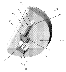
[0007] More recently, the development of orthopedic implant
designs has moved towards satisfying the requirements of high
demand patients.
Patients today require more from their
implants, and because patients are living longer, they require
implants that to last longer. Accordingly, developments have
been made in materials used to make orthopedic implants to
improve implant longevity, such as highly porous metals that
improve biological bone fixation. These implants are generally
termed press-fit or cementless.
2
CA 02906631 2015-09-14
WO 2014/143740 PCT/US2014/027827
[0008] Recognizing the disadvantages of cement fixation
techniques, prior art devices have also been developed that
utilize other mechanical attachment means to join an implant to
bone for immediate stabilization.
Although various implant
surface treatments intended to bond with bone biologically for
long term stable attachment have proven successful, an initial
fixation and stabilization is required before the bone growth
can occur. A simple technique of mechanically securing an
implant, is to affix it within the bone with screws or other
mechanical fasteners. However, due to the nature of the bone
surrounding the surgical site, and other limiting factors such
as artery location and the like, screws can only be applied in
certain limited regions. The
use of a screw for implant
fixation should be considered only as an option by the surgeon
depending upon implant placement and bone quality.
[0009] Therefore, there exists a need for an improved implant
design that provides both short term and long term fixation and
stabilization.
BRIEF SUMMARY OF THE INVENTION
[0010] The present invention is described below in connection
with the preferred embodiment unicondylar tibial implant.
However, the present invention has applicability to other
orthopedic implants, including unicondylar femoral implants and
even total implants. For
instance, the below description of
the present invention is provided for a tibial implant to be
used on the medial condyle. However, the preferred embodiment
can also be used on the lateral condyle, and when utilized in
such a manner would have some features reversed in orientation.
A description of the medial component features of the tibial
implant is provided only for simplification.
[0011] In accordance with a preferred embodiment, the present
invention provides for a unicondylar tibial implant. The
tibial implant includes a tibial keel positioned on a surface
of the tibial implant to be submerged into prepared bone with a
3
CA 02906631 2015-09-14
WO 2014/143740 PCT/US2014/027827
first projection extending along its lengthwise direction and a
second projection extending along a direction perpendicular to
the first projection. The first projection may be interrupted
by a void to allow clearance for another implant or instrument.
The second projection intersects the first projection. The
tibial implant can be fabricated from a metal, a polymer, a
biodegradable material, a porous metal material, or
combinations thereof. The
device as described could be
produced through additive manufacturing techniques such as
direct metal laser sintering.
[0012] The tibial keel is configured as an anterior-posterior
projection with an intersecting keel segment that extends about
a medial-lateral direction. The tibial keel is comprised of a
solid material on a bone interfacing leading edge of the tibial
keel i.e., a solid end portion, with the tibial keel having a
porous material between the tibial tray and the solid end
portion of the tibial keel. The tibial implant can optionally
include a bone screw to secure the tibial implant to bone.
[0013] In accordance with another preferred embodiment, the
present invention provides for a unicondylar tibial implant
having a tibial keel configured as an anterior-posterior
projection with at its most anterior aspect being an
intersecting keel in the medial-lateral direction. The tibial
keel is comprised of a solid material on a leading edge of the
keel and porous material between the tibial tray and the solid
end portion of the keel, and smaller protrusions on the medial
facing portion of the tibial keel at the intersection of the
tibial keel and tibial tray. The tibial implant is fabricated
from a metal, a polymer and/or a biodegradable material. The
tibial implant can optionally include a bone screw to secure
the tibial implant to bone.
[0014] In accordance with yet another preferred embodiment, the
present invention provides for a unicondylar tibial implant
having a tibial keel configured as an anterior-posterior
4
CA 02906631 2015-09-14
WO 2014/143740 PCT/US2014/027827
projection with at its most anterior aspect being an
intersecting keel in the medial-lateral direction. The tibial
keel is comprised of a solid material on the leading edge of
the keel and porous material between the tibial tray and a
solid end portion of the keel being implanted into an
interference-fit created by an undersized preparation in the
bone. The tibial implant is fabricated from a metal, a polymer
and/or a biodegradable material. The
tibial implant can
optionally include a bone screw to secure the tibial implant to
bone.
[0015] In accordance with another preferred embodiment, the
present invention provides for a unicondylar tibial implant
having a tibial keel configured as an anterior-posterior
projection with at its most anterior aspect being an
intersecting keel in the medial-lateral direction. The tibial
keel is comprised of a solid material on a leading edge of the
keel and porous material between the tibial tray and a solid
end portion of the keel, and smaller protrusions on the medial
facing portion of the keel at the intersection of the tibial
keel and tibial tray where the protrusions preferentially force
the tibial implant into the bone prepared about a resected mid-
tibial eminence. The
tibial implant is implanted into an
interference fit created by an undersized preparation in the
bone. The tibial implant is fabricated from a metal, a polymer
and/or a biodegradable material. The
tibial implant can
optionally include a bone screw to secure the tibial implant to
bone.
[0016] In accordance with yet another preferred embodiment, the
present invention provides for a keel for a unicondylar tibial
implant. The
keel is connected to the tibial tray of the
tibial implant and includes smaller protrusions on a medial
facing portion of the keel at an intersection of the keel and
the tibial tray where the protrusions push the tibial implant
into the bone prepared about a resected tibial eminence. The
CA 02906631 2015-09-14
WO 2014/143740 PCT/US2014/027827
keel is fabricated from a metal, a polymer and/or a
biodegradable material. The
tibial implant can optionally
include a bone screw to secure the tibial implant to bone.
[0017] In accordance with another preferred embodiment, the
present invention provides for a unicondylar tibial implant
having a tibial tray with a porous keel and protrusions
extending from the keel. The
tibial tray accepts a
polyethylene tibial bearing having an articulating surface for
articulating with a femoral component. The tibial bearing can
be a modular polyethylene tibial bearing. The
tibial implant
and tibial bearing can also be formed as a monoblock component.
Alternatively, the tibial tray with a porous keel can be formed
out of a singular biomaterial which is also used to form the
tibial bearing. The
tibial implant can optionally include a
bone screw to secure the tibial implant to bone.
[0018] In accordance with yet another preferred embodiment, the
present invention provides for a unicondylar tibial implant
having at least one section of material that in its normal
state forms at least one uninterrupted surface of the implant
that is separable from the greater bulk of the tibial implant
in a predictable shape defined by the presence of a shear
section. The shear section of material when removed exposes a
passageway for at least one additional implant, such as a bone
screw. The removal of the shear section also exposes a
passageway for surgical instrumentation, for the application of
osteobiologic materials or for the application of bone cement.
[0019] In accordance with another preferred embodiment, the
present invention provides for the ornamental design of a
unicondylar tibial implant as shown and described in the
figures below.
[0020] Another embodiment of the present invention is an
orthopedic implant for replacing a portion of a bone including
a bone contacting surface and a keel extending from the bone
contacting surface. The keel includes a first projection with
6
CA 02906631 2015-09-14
WO 2014/143740 PCT/US2014/027827
a first longitudinal axis and a second projection with a second
projection with a second longitudinal axis. The first and
second longitudinal axes are oriented orthogonally to each
other. The hole may be configured to accept a bone screw at a
plurality of different angles, and the first and second
projections may be separated from each other by the hole. The
hole may include a plug removable upon the application of a
force. At
least one fin may be associated with the first
projection and extend oblique to the first longitudinal axis.
That fin may be shaped to engage the bone, and/or configured to
enter into an unprepared portion of the bone. At
least one
extension may be associated with the second projection and
extend oblique to the second longitudinal axis. That extension
may be shaped to engage the bone, and/or frictionally engage
the bone. The
implant may further include a porous portion
adapted to allow for the bone to grow therein. The
porous
portion may cover at least a portion of the bone contacting
surface and at least a portion of the keel, and the keel may
include a solid portion at a distal end of the keel. The
porous portion may define a first porous surface and at least
one boundary strut extending from the surface in a first
direction. The boundary strut may extend any angle, including
from 0 to 10 degrees from normal to the first porous surface.
The implant may also further include a third projection, as
well as a bearing component attachable to the implant. In
certain embodiments, the implant is a unicondylar tibial
baseplate, and a kit including the implant may include at least
one other implant.
[0021] Yet another embodiment of the present invention is a
tibial baseplate including a bone contacting surface having
anterior, posterior, medial and lateral sides, a first
projection extending from the bone contacting surface and
having a first length extending in a first direction between
the anterior and posterior ends, a second projection extending
7
CA 02906631 2015-09-14
WO 2014/143740 PCT/US2014/027827
from the bone contacting surface and having a second length
extending in a second direction between the medial and lateral
sides, an aperture for receiving a bone screw and a porous
material for promoting bone ingrowth, the porous material at
least partially covering the bone contacting surface, the first
projection and the second projection. The
baseplate may
further include a third projection. The
porous material may
define a plurality of boundary struts extending from the bone
contacting surface in a first direction at between 0 to 10
degrees from normal to the bone contacting surface. The first
and second projections may be separated from each other by the
aperture. The
aperture may be configured to accept a bone
screw at a plurality of different angles, and may include a
plug removable upon the application of a force. At least one
fin or extension may be associated with at least one of the
first and second projections, where the fin is configured to
enter into an unprepared portion of the bone and the extension
frictionally engages the bone. A solid portion may be included
at distal ends of the first and second projections.
[0022] A still further embodiment is a tibial baseplate
including a bone contacting surface having anterior, posterior,
medial and lateral sides, a first projection extending from the
bone contacting surface and having a first length extending in
a first direction between the anterior and posterior ends, a
second projection extending from the bone contacting surface
and having a second length extending in a second direction
between the medial and lateral sides, an aperture for receiving
a bone screw, a plug at least partially covering the aperture,
the plug being removable upon the application of a force and a
porous material for promoting bone ingrowth, the porous
material at least partially covering the bone contacting
surface, the first projection and the second projection,
wherein the porous material defines a plurality of boundary
8
CA 02906631 2015-09-14
WO 2014/143740 PCT/US2014/027827
struts extending from the bone contacting surface from 0 to 10
degrees from normal to the bone contacting surface.
BRIEF DESCRIPTION OF THE DRAWINGS
[0023] The foregoing summary, as well as the following detailed
description of the preferred embodiments of the invention, will
be better understood when read in conjunction with the appended
drawings. For the purpose of illustrating the invention, there
are shown in the drawings embodiments which are presently
preferred. It should be understood, however, that the
invention is not limited to the precise arrangements and
instrumentalities shown.
[0024] Referring to the figures, wherein like reference
numerals represent like parts throughout the several views:
[0025] Fig. 1 is a top perspective view of a unicondylar tibial
implant assembly in accordance with a preferred embodiment of
the present invention.
[0026] Fig. 2 is a bottom perspective view of the unicondylar
tibial implant of Fig. 1.
[0027] Fig. 3 is a top view of the unicondylar tibial implant
of Fig. 1.
[0028] Fig. 4 is a side view of the unicondylar tibial implant
of Fig. 1.
[0029] Fig. 5 is a bottom view of the unicondylar tibial
implant of Fig. 1.
[0030] Fig. 6 is a front view of the unicondylar tibial implant
of Fig. 1.
[0031] Fig. 7 is an opposite side view of that shown in Fig. 4.
[0032] Fig. 8 is a rear view of the unicondylar tibial implant
of Fig. 1.
[0033] Fig. 9 is a bottom perspective view of a unicondylar
tibial implant of the tibial implant assembly of Figs. 1-8;
[0034] Fig. 10 is another bottom perspective view of the
unicondylar tibial implant of Fig. 9.
9
CA 02906631 2015-09-14
WO 2014/143740 PCT/US2014/027827
[0035] Fig. 11 is a top perspective view of the unicondylar
tibial implant of Fig. 9.
[0036] Fig. 12 is another top perspective view of the
unicondylar tibial implant of Fig. 9.
[0037] Fig. 13 is a top view of the unicondylar tibial implant
of Fig. 9.
[0038] Fig. 14 is a side view of the unicondylar tibial implant
of Fig. 9.
[0039] Fig. 15 is a bottom view of the unicondylar tibial
implant of Fig. 9.
[0040] Fig. 16 is a front view of the unicondylar tibial
implant of Fig. 9.
[0041] Fig. 17 is an opposite side view of that shown in Fig.
15.
[0042] Fig. 18 is a rear view of the unicondylar tibial implant
of Fig. 9.
[0043] Fig. 19 is a side view of the unicondylar tibial implant
of Figs. 9-18 with a bone screw positioned within a through
hole of the tibial implant.
[0044] Fig. 20 is a rear view of the assembly of Fig. 19.
[0045] Figs. 21-29 are highly magnified photographic images of
from a bottom perspective of a porous portion of the
unicondylar tibial implant of Fig. 9.
[0046] Fig. 30 is a side view of a unicondylar tibial implant
in accordance with another embodiment of the present invention.
[0047] Fig. 31 is a top perspective view of the unicondylar
tibial implant of Fig. 30.
[0048] Fig. 32 is a side view of the unicondylar tibial implant
of Fig. 30.
[0049] Fig. 33 is a bottom perspective view of the unicondylar
tibial implant of Fig. 30.
[0050] Fig. 34 is a side view of a unicondylar tibial implant
in accordance with yet another embodiment of the present
invention.
CA 02906631 2015-09-14
WO 2014/143740 PCT/US2014/027827
[0051] Fig. 35 is a bottom view of the unicondylar tibial
implant of Fig. 34.
[0052] Fig. 36 is a side view of a unicondylar tibial implant
in accordance with a further embodiment of the present
invention.
[0053] Fig. 37 is a bottom perspective view of the unicondylar
tibial implant of Fig. 36.
[0054] Fig. 38 is a bottom view of the unicondylar tibial
implant of Fig. 36.
[0055] Fig. 39 is a bottom perspective view of a unicondylar
tibial implant according to another embodiment of the present
invention.
[0056] Fig. 40 is another bottom perspective view of the
unicondylar tibial implant of Fig. 39.
DETAILED DESCRIPTION OF THE INVENTION
[0057] When referring to specific directions in the following
discussion of certain implantable devices, it should be
understood that such directions are described with regard to
the implantable device's orientation and position during
exemplary application to the human body. Thus, as used herein,
the term "proximal" means close to the heart and the term
"distal" means more distant from the heart. The term "inferior"
means toward the feet and the term "superior" means toward the
head. The term "anterior" means toward the front of the body or
the face and the term "posterior" means toward the back of the
body. The term "medial" means toward the midline of the body
and the term "lateral" means away from the midline of the body.
Also, as used herein, the terms "about," "generally" and
"substantially" are intended to mean that slight deviations
from absolute are included within the scope of the term so
modified. Likewise, for purposes of convenience and clarity
only, directional terms such as top, bottom, above, below and
diagonal, may be used with respect to the accompanying
drawings. Such directional terms used in conjunction with the
11
CA 02906631 2015-09-14
WO 2014/143740 PCT/US2014/027827
following description of the drawings should not be construed
to limit the scope of the invention in any manner not
explicitly set forth. Additionally, the term "a," as used in
the specification, means "at least one." The terminology
includes the words above specifically mentioned, derivatives
thereof, and words of similar import.
[0058] Reference will now be made in detail to the preferred
embodiments of the present invention illustrated in the
accompanying drawings.
Generally, the same or like reference
numbers will be used throughout the drawings to refer to the
same or like features, but within a different 100-series of
numbers. For
instance, Fig. 9 depicts a unicondylar tibial
implant 10, while Fig. 30 depicts another embodiment
unicondylar tibial implant 110. It
should be noted that the
drawings are in simplified form and are not drawn to precise
scale.
[0059] As noted above, partial knee implants, also known as
unicondylar or unicompartmental knee implants, are designed to
replace either a medial or lateral compartment of a knee joint.
A unicondylar replacement assembly may include a tibial implant
(as is discussed below), either by itself or in conjunction
with an implant designed to replace a femoral condyle. The
preparation of the bone to accept such implants may be
facilitated by instrumentation such as bone files, burrs, saws,
punches, computer and/or robot
assisted
instrumentation/navigation systems. Once the bone is prepared,
the implant may be secured to the bone by different means,
including bone cement which bonds to the implant and
impregnates the bone resulting in fixation of the implant to
the bone interface.
[0060] The present invention has been designed to facilitate
fixation directly to the bone, i.e. without bone cement. Such
fixation without bone cement is known as cementless fixation or
press-fit fixation. The
present invention addresses the
12
CA 029631 20109-14
WO 2014/143740 PCT/US2014/027827
challenge of cementless fixation of implant components, which
is to have acceptable initial stability upon implantation to
allow patient mobility immediately or a short time after
surgery and promote adequate biologic fixation of the implant
to the bone long term. The
initial stability and long term
fixation are requirements of the implant to reduce the
incidence of implant loosening and reduce patient post-
operative pain over time.
[0061] The present invention of Figs. 1-32 includes several
different embodiments of a unicondylar tibial implant assembly
having a unicondylar tibial implant, tibial tray or baseplate
and a unicondylar tibial implant bearing 12. Of course, as
noted above, although described in connection with a
unicondylar implant for the tibia, the present invention has
applicability to other types of implants. For
instance, the
present invention may be applied in unicondylar implant for the
femur or even total implants. The
unicondylar tibial implant
10 has been developed primarily for cementless application and
includes a unique bone interfacing tibial keel 14 and a porous
structured biomaterial interface i.e., a porous portion 16
(best shown in Figs. 21-29). The
tibial implant 10 can be
constructed from any combination of solid metal, porous metal,
polymers and/or other resorbable materials. For
instance, it
is contemplated to form the bearing 12 of a polymer material
such as PEEK, and the implant 10 of a metal such as titanium or
stainless steel. Likewise, it is contemplated to form implant
10 of different materials, e.g., porous portion 16 may be
formed of a different material than the remainder of the
implant.
[0062] For purposes of convenience only, and not by way of
limitation, the foregoing description of the preferred
embodiments of the unicondylar tibial implant assembly 5 will
be described and illustrated with respect to a unicondylar
tibial implant assembly 5 for a medial tibial condyle.
13
CA 02906631 2015-09-14
WO 2014/143740 PCT/US2014/027827
However, the foregoing description and features of the
unicondylar tibial implant assembly 5 are equally applicable to
a unicondylar tibial implant assembly for a lateral condyle,
such similar features of the lateral unicondylar tibial implant
assembly being substantially mirror images of such features of
the medial unicondylar tibial implant assembly. Of course, it
is also contemplated that the medial and lateral versions of
the assembly may be of a different construction to accommodate
the different bony anatomy of the medial and lateral portions
of the tibia.
[0063] The tibial keel 14 is preferably constructed of a
combination of solid and porous portions and located on an
undersurface or bottom of the tibial implant 10, which is
designed to contact a resected tibia bone (not shown). The
tibial keel 14 is generally submerged into the bone when the
tibial implant 10 is implanted thereon. The tibial keel 14 can
prepare its own cavity in the bone as it is inserted into the
resected tibia or it can occupy cavities within the bone
previously prepared by instrumentation or other implants. Any
pre-cavities for receiving the tibial keel 14 when pre-prepared
are generally smaller in size than the tibial keel 14 so as to
generate compressive forces between the bone interface and the
tibial keel 14 and increase frictional forces between the bone
and the tibial keel 14. That is, the tibial keel 14 is press-
fitted into the bone.
[0064] The tibial keel 14 is shown in Figs. 2, 4-10 and 14-20
and includes a first projection or protrusion 20, which is
generally planar and has a height which corresponds to a depth
within a prepared bone to which the tibial keel 14 will
protrude into, and a second projection or protrusion 22, which
is also generally planar, has a height which corresponds to a
depth within a prepared bone to which the tibial keel 14 will
protrude into and is substantially perpendicular to the first
projection 20 (i.e., the longitudinal axes of projections 20,
14
CA 029631 20109-14
WO 2014/143740 PCT/US2014/027827
22 are orthogonally arranged). For purposes of clarification,
projections 20, 22 are labeled with reference numeral 14 in
Fig. 2 and reference numerals 20 or 22 in the remainder of the
figures pertaining to implant 10.
[0065] The heights of first and second projections 20, 22 of
the tibial keel 14 may be variable to accommodate access
limitations while maximizing the fixation of the tibial implant
into bone. Preferably, the tibial keel 14 is positioned on
an underside or inferior surface 24 of the tibial tray 10 with
the first projection 20 running along the anterior-posterior
direction, and the second projection 22 running along the
medial-lateral direction. This results in the intersection of
the longitudinal axes of the projections 20, 22. Both of the
first and second projections 20, 22 of the tibial keel are
substantially normal to the underside of the tibial tray 10,
but this can vary in other embodiments.
Further, although
shown with a constant height (see e.g., Fig. 4), projections
20, 22 can be configured to have a height that varies along its
length. In fact, in a later embodiment (Fig. 32), a projection
similar to projection 20 is shown with a sloped configuration.
[0066] Each of the first and second projections 20, 22 of the
tibial implant 10 can be configured to have one or more
extensions i.e., a plurality of extensions 26 shown in Figs. 2
and 5 extending from the second projection 22. The extensions
26 that emanate from the projections are oriented out of plane
with the projection. That
is, the extensions 26 extend
outwardly from the lateral surfaces of the projections. The
extensions 26 are designed to create and/or fill cavities
within the bone so as to create and/or maximize compressive
frictional forces between the tibial keel 14 and the
surrounding bone. The extensions 26 are preferably located so
that resultant forces during insertion of the tibial implant 10
into a resected tibia bias the position of the tibial implant
10 in a predetermined or desired direction. The extensions 26
CA 02906631 2015-09-14
WO 2014/143740 PCT/US2014/027827
are configured as substantially wedge shaped extensions that
extend along substantially the entire height of the keel, and
are preferably tapered in the distal direction. The extensions
26 on the second projection 22 are spaced apart from each other
and substantially circumscribe the second projection 22.
Preferably, the second projection includes five extensions 26,
but can include more or less than five.
[0067] The extensions 26 are preferably located around the
periphery of both the first and second projections 20, 22 with
a higher number of extensions 26 or higher density of
extensions 26 emanating from the second projection 22 located
about the anterior region of the tibial implant 10 where higher
frictional forces are able to make a greater contribution to
address anterior lift-off stability issues of the tibial
implant 10 when implanted within the bone. The
number of
extensions 26 is greater on the sides of the projection 22 that
face away from a central region of the tibial implant 10 so
that bone reaction forces will push/direct the tibial implant
into the central region of the tibia.
[0068] The tibial keel 14 also includes a plurality of fins 34
which extend beyond the nominal volume of the tibial keel 14,
specifically with respect to projection 20. The fins 34 enter
bone that has not been prepared to receive the fins 34.
Instead, the fins 34 prepare their own receiving volume within
the bone as they are inserted into the bone, i.e., the fins 34
displace bone as they are placed therein. In other words, the
fins 34 are inserted into bone without the need to prepare the
bone to receive such fins 34. The
fins 34 are sized to
maximize their surface area, minimize their volume and are
shaped to ease entry into the bone. For instance, as shown in
Fig. 2 and 7, the fins 34 are preferably configured as shown
and are substantially wedge shaped or shaped as a dual inclined
plane structure.
Further, the fins 34 are tapered as they
extend from a proximal end of the tibial keel 14 distally. The
16
CA 029631 20109-14
WO 2014/143740 PCT/US2014/027827
fins 34 are also preferably configured to extend an overall
length about half way the overall height of the tibial keel 14.
[0069] The projections 20, 22 are shown to be of a particular
construction. For
instance, projection 20 is a long, thin
rectangular structure that plateaus in a solid edge 32
(discussed more fully below). Likewise, projection 22 includes
a solid edge 32, but is somewhat shorter and thinner than
projection 20. It is contemplated that the projections 20, 22
can encompass other shapes, including but not limited to,
curved bodies or the like. Moreover, it is contemplated that
the projections could comprise a plurality of components. For
example, projection 20 could encompass a plurality of more
square shaped components that are placed adjacent to each other
or spaced apart a distance. Solid
edge 32 could also be
replaced with a sharper or narrower surface than the
substantially flat surface that is depicted. Still further, it
is to be understood that although shown of a particular design,
extensions 26 and fins 34 can encompass many different types of
designs. For
one, both projections could include either
extensions 26, fins 34 or a combination thereof. Additionally,
the extensions 26 and fins 34 could be of different shapes and
sizes. By way
of example, it is contemplated for either or
both of projections 20, 22 to include a plurality of teeth or
spikes in lieu of the depicted extensions 26 and fins 34.
[0070] The tibial implant 10 can optionally be configured with
a through hole or aperture 28 (best shown in Figs. 2, 5 and 21)
through which another device, instrument or material e.g., a
bone screw 30 (as is shown in Figs. 19 and 20) can be inserted
therethrough. The through hole 28 is shown splitting the keel
14 into projections 20, 22 and may pass through one or more of
the projections 20, 22 thereby interrupting their general
shape. For
instance, as can be seen in Fig. 5, material is
removed from projections 20, 22 around or adjacent the through
hole 28 to provide for clearance of the device (bone screw 30
17
CA 02906631 2015-09-14
WO 2014/143740 PCT/US2014/027827
or the like) ,
instrument or material to be inserted
therethrough.
[0071] Preferably, the through hole 28 is shaped and sized for
the passage of the bone screw 30 (best shown in Figs. 19 and
20) through a superior aspect of the tibial implant 10 into the
bone beneath the underside or inferior surface of the tibial
tray 10. The
through hole 28 is preferably designed so that
the bone screw 30 can be angulated to achieve a desired
direction by the user.
Further, with material from adjacent
projection 20 removed, the projection 20 does not interfere
with the passage of the bone screw 30 through the through hole
28. Such
bone screws 30 are readily known in the art and a
detailed description of their structure and operation is not
necessary for a complete understanding of the present
invention.
[0072] The tibial implant 10 may employ the use of a knockout
plug 36 formed within the through hole 28 and out of a material
that is metallurgically continuous with the greater bulk of the
tibial implant 10. The
knockout plug 36 is configured to be
removed from the remainder of the tibial implant 10 via a
boundary shear section or weakened area 38 around the plug 36
(see Fig. 5) upon the application of a suitable force. The
plug 36 may be machined into the tibial tray 10 or built in
final form through an additive manufacturing process such as by
direct metal laser sintering (discussed more fully below).
Preferably, the through hole 28, is obstructed by the knockout
plug 36 so that the superior surface 40 of the tibial tray 10
facing the bearing component 12 is fully continuous without any
path through which debris or material could pass through the
tibial tray 10 to the bone engaging underside of the tibial
implant 10. Thus, in the event of backside wear of the bearing
component 12, wear particles are less likely to migrate out of
the tibial tray 10 than if an already present through hole were
in place. The
knockout plug 36 can optionally include a
18
CA 02906631 2015-09-14
WO 2014/143740 PCT/US2014/027827
threaded stud 42 (best shown in Fig. 12), which mates to
instrumentation to facilitate removal of the knockout plug 36.
[0073] In sum, the tibial tray 10 has an initially covered
through hole 28 into which a screw 30 can be placed to further
stabilize the tibial implant 10 to the prepared bone upon
implantation. This
is especially advantageous for initial
implant stability and when placing the tibial implant into bone
of questionable density where the user/surgeon is not confident
the bone itself is stable enough to support adequate short term
stability.
[0074] The general shape of the tibial keel 14 is designed to
maximize surface area to volume ratio for the tibial keel 14 to
enhance bone ingrowth thereto (discussed more below) while
minimizing the amount of bone removal during bone preparation.
The amount of surface area available for bone ingrowth is
important for both short and long term fixation of the implant
to the bone. Short term fixation is also achieved by "press-
fitting" the larger body of the keel into a smaller preparation
of the bone. Once
in place, the residual stresses from the
compressed bone around the tibial keel 14 increase the
frictional forces against the tibial keel 14 and increase the
stability of the tibial implant 10 into the prepared bone.
Increasing the surface area over which the press-fit
interference is effective helps to increase the total
frictional forces available to contribute to stability of the
implant and to distribute frictional forces over a greater
effective area of the tibial implant 10.
[0075] Long term fixation of the tibial implant 10 is enhanced
by the areas of the tibial implant 10 having the porous
structure and surface, hereafter referred to as 'porous metal'
(generally referred to with reference numeral 16). As the bone
remodels and grows into the porous metal 16, the frictional
retention forces will be replaced and/or supplemented with bone
ingrowth. The degree of this fixation via bone ingrowth is, in
19
CA 02906631 2015-09-14
WO 2014/143740 PCT/US2014/027827
part, a function of the amount and distribution of the porous
metal surface area available for ingrowth. The
large
distributed tibial keel surface area thereby provides a
structure for increased stability via a larger area of bone
ingrowth.
[0076] The porous metal 16 is formed from a porous structured
biomaterial, and includes a plurality of struts 44 (best shown
in Figs. 21-29) having varying lengths and cross sections. At
least one strut of the porous metal 16 has an end connected to
one or more other struts at node points 46 (see Fig. 29)
thereby forming the porous geometry of the porous metal 16.
The porous metal 16 also includes boundary struts 48 (see Figs.
26, 27 and 28) that are configured to extend beyond a nominal
boundary of the porous metal 16. That is, the porous metal 16
has boundary struts 48 that extend away from the surface of the
porous metal 16 in a finger-like or hair follicle-like fashion.
The extending boundary struts 48 impart a roughness to the
surface, the degree of which is dependent upon the number and
length of boundary struts 48 present. The
average or main
direction of the boundary struts 48 also impart a surface
roughness that varies dependent upon which direction the device
is driven for implantation.
[0077] Preferably, the tibial keel 14 is formed from a metal
substrate and a layer of porous metal 16 adjacent the
substrate. The porous metal 16 on the tibial keel 14 includes
extending boundary struts 48 with unconnected ends pointing or
extending towards the bottom or inferior surface of the tibial
tray 10. Under
similar loading conditions, sliding over the
angled struts toward the bottom surface of the tibial tray 10
will experience less frictional forces than bone sliding away
from the bottom face of the tibial tray 10.
Preferably, the
boundary struts 48 are angled about +/- 10 degrees from normal
to a surface of the substrate to which the porous metal 16 is
applied to.
CA 02906631 2015-09-14
WO 2014/143740 PCT/US2014/027827
[0078] Another element of the present invention is that the
boundary struts 48 are oriented in a predetermined direction
such that they push or are directed towards the bone interface
surface. While the surface of the porous metal 16 may exhibit
characteristics of a rougher surface, the boundary struts 48 of
the porous metal 16 implanted into a bone interface embed
themselves into the bone and provide a mechanical interlock to
the surrounding bone. This
is especially advantageous during
initial implantation for initial fixation purposes. In the
aggregate, the plurality of boundary struts 48 significantly
improves the overall stability of the tibial implant 10 upon
initial implantation.
Preferably, the bottom surface of the
tibial tray 10 has extending boundary struts 48' (best shown in
Figs. 26 and 27) in a direction substantially normal to the
bottom surface of the tibial tray 10. As the tibial implant 10
is definitively seated against the bone interface surface, the
boundary struts 48' pierce the surface of the prepared bone to
increase stability of the tibial implant 10 to the bone.
[0079] In the disclosed embodiment, the tibial implant 10 has
the porous metal 16 on all surfaces that make contact with
bone. The surface of the porous metal 16 is tailored for each
specific region of the tibial implant 10 to have specific
surface roughness and thereby specific amounts of friction when
engaged with bone. That
is, the tibial implant 10 is
configured to have a porous metal 16 with boundary struts 48 at
predetermined angles dependent upon the location of the porous
metal 16 on the tibial implant 10.
[0080] In sum, the surfaces of the porous metal 16 have
extending boundary struts 48 which serve to modify the surface
roughness of the tibial implant 10. The
size and average
direction of the extending boundary struts 48 impart different
frictional coefficients depending upon the direction the
boundary struts 48 extend. The boundary struts 48 can also be
directed in a direction largely normal to the surface from
21
CA 02906631 2015-09-14
WO 2014/143740 PCT/US2014/027827
which they extend from. This
can have an additive anchoring
effect which enhances stability of the tibial implant 10 to the
bone.
[0081] A solid edge 32 (best shown in Figs. 2, 7 and 21) at the
distal end of one or both of the projections 20, 22 of the
tibial keel 14 prevents bone from growing into the tibial keel
14 from the bottom up. Thus, while the majority of the surface
area of the tibial implant 10 is design for fixing (via bone
ingrowth), no such fixing occurs at the distal ends of the
projections.
Rather, the fixing of the tibial implant 10 to
the bone occurs only at the perimeter of the tibial keel 14,
i.e., the lateral side surfaces of the tibial keel 14. That
is, the tibial implant 10 is configured to prevent any bone
ingrowth or fixation about a distal surface of the tibial keel
14 via the solid edge 32. Preventing bone ingrowth about the
distal surface of the tibial keel 14 allows for easier removal
of the implant (e.g., during a revision procedure), if
necessary, since bone ingrowth on such distal surfaces of the
tibial keel 14 represents areas that are most problematic to
achieving separation of the implant from bone during revision
procedures. In
other words, as an implant is pulled out of
bone, bony ingrowth into the bottom portion of the tibial keel
might not separate from the greater volume of the bone exactly
at the implant interface but rather somewhere deeper within the
volume of bone beneath the implant. If
this occurs during
implant removal, the additional bone that would otherwise be
inadvertently removed would complicate the revision procedure
and drive the use of more significant revision components. In
any event, the bone which engages and contacts the bottom of
the tibial keel 14 represents a small fraction of the overall
surface area of the tibial implant 10.
[0082] The porous metal 16 of implant 10 may be formed
utilizing any suitable process. For
instance, a selective
laser melting or sintering process may be employed to create
22
CA 02906631 2017-02-20
the porous metal 16, or even the entirety of the implant 10.
In conjunction with the latter, it is contemplated that the
implant 10 may include substantially non-porous or solid
portions and the porous metal 16 portions that are formed from
the same process. Examples of such processes are disclosed in
U.S. Patent No. 7,537,664, and U.S. Patent Application
Publication Nos. 2006/0147332 and 2007/0142914. Of
course, it
is contemplated to utilize any known and suitable process to
form implant 10.
[0083]
Referring to Figs. 30-33, in accordance with another
preferred embodiment, the present invention provides for a
tibial implant 110. The tibial implant 110 is similarly
configured as tibial implant 10, except as noted below.
For
instance, while the tibial implant 10 includes first and second
projections 120, 122 that are similarly configured to
projections 20, 22, projection 120 is sloped (see Fig. 32) and
is segmented into two different portions by the void created by
hole 128 (see Fig. 33).
Moreover, the second projection 122
does not take up as much surface area as does second projection
22. As
best shown in Fig. 32, the height of the first
projection 120 slopes towards the posterior end of the tibial
implant 110 such that the height of the first protrusion
decreases as it extends from the anterior end towards the
posterior end. Of
course, projection 120 could slope in any
direction.
[0084]
Referring to Figs. 34 and 35, in accordance with yet
another preferred embodiment, the present invention provides
for a tibial implant 210. The tibial implant 210 is similarly
configured as tibial implant 110, except as noted below. In
particular, implant 210 includes a third protrusion 223, which
like the second protrusion 222, is slightly spaced apart from
the first protrusion 220 and may include extensions.
Preferably, the third protrusion 223 is positioned more towards
23
CA 029631 20109-14
WO 2014/143740 PCT/US2014/027827
the rear or posterior to the first protrusion and has a height
similar to the height of the posterior end of the first
protrusion 220 to which it is adjacent to. However, the height
of the third protrusion 223 is less than that of the second
protrusion 222. The longitudinal axis of the third protrusion
223 is also configured not to intersect the longitudinal axis
of the first protrusion 220.
[0085] Figs. 36-38 depict yet another embodiment implant 310,
which is similar to the implant 210 save for the placement of
the third protrusion 323 more toward or about a middle section
of the first protrusion 320. When positioned about the middle
section of the first protrusion 320, the third protrusion 323
has a height substantially the same as the area of the first
protrusion 320 that it is adjacent to.
[0086] Figs. 39 and 40 depict yet another embodiment tibial
implant 410. Unlike the above-discussed implants, implant 410
includes a keel 414 that is of a unitary design. A hole
or
aperture 428 is situated offset with respect to the keel 414.
This allows for the keel 414 to have a unitary construction
(i.e., it is not broken up by the hole 428 as in the above
designs). Like
the foregoing embodiments, keel 414 includes
two projections 420, 422, with the projection 420 including
fins 434 and the projection 422 including extensions 426. Of
course, as in the above embodiments, either projection could
include either or both of the extensions 426 or fins 434, and
such structures can be of any shape and or size with respect to
the projections. Moreover, keel 414 includes a rounded cut out
421, which allows for a screw 430 to angulate with respect to
implant 410. In
other words, the cut out 421 provides
clearance for the screw 430 to move with respect to the plate
in directions towards the keel 414.
[0087] It will be appreciated by those skilled in the art that
changes could be made to the embodiments described above
without departing from the broad inventive concept thereof.
24
CA 02906631 2015-09-14
WO 2014/143740 PCT/US2014/027827
For example, additional components can be added to the tibial
implant assembly. It is to be understood, therefore, that this
invention is not limited to the particular embodiments
disclosed, but it is intended to cover modifications within the
spirit and scope of the present invention as described above.
[0088] It is also to be understood that the disclosure set
forth herein includes all possible combinations of the
particular features described. For example, where a particular
feature is disclosed in the context of a particular aspect,
arrangement, configuration, or embodiment, or a particular
claim, that feature can also be used, to the extent possible,
in combination with and/or in the context of other particular
aspects, arrangements, configurations, and embodiments of the
invention, and in the invention generally.
[0089] Furthermore, although the invention herein has been
described with reference to particular embodiments, it is to be
understood that these embodiments are merely illustrative of
the principles and applications of the present invention. It
is therefore to be understood that numerous modifications may
be made to the illustrative embodiments and that other
arrangements may be devised without departing from the spirit
and scope of the present invention as defined by the appended
claims.