Note: Descriptions are shown in the official language in which they were submitted.
MULTI-SITE TRANSCUTANEOUS ELECTRICAL STIMULATION OF
THE SPINAL CORD FOR FACILITATION OF LOCOMOTION
CROSS-REFERENCE TO RELATED APPLICATIONS
[0001] This application claims benefit of and priority to USSN
61/802,034, filed
March 15, 2013.
STATEMENT OF GOVERNMENTAL SUPPORT
[0002] [This paragraph is intentionally left blank.]
FIELD
[0003] This application relates to the field of neurological treatment
and
rehabilitation for injury and disease including traumatic spinal cord injury,
non-traumatic
spinal cord injury, stroke, movement disorders, brain injury, ALS,
Neurodegenerative
Disorder, Dementia, Parkinson's disease, and other diseases or injuries that
result in
paralysis and/or nervous system disorder. Devices, pharmacological agents, and
methods
are provided to facilitate recovery of posture, locomotion, and voluntary
movements of the
arms, trunk, and legs, and recovery of autonomic, sexual, vasomotor, speech,
swallowing,
and respiration, in a human subject having spinal cord injury, brain injury,
or any other
neurological disorder.
BACKGROUND
[0004] Serious spinal cord injuries (SCI) affect approximately 1.3
million people in
the United States, and roughly 12-15,000 new injuries occur each year. Of
these injuries,
approximately 50% are complete spinal cord injuries in which there is
essentially total loss
of sensory motor function below the level of the spinal lesion.
[0005] Neuronal networks formed by the interneurons of the spinal cord that
are
located in the cervical and lumbar enlargements, such as the spinal networks
(SNs), can
play an important role in the control of posture, locomotion and movements of
the upper
limbs, breathing and speech. Most researchers believe that all mammals,
including humans,
have SNs in the lumbosacral cord. Normally, the activity of SNs is regulated
supraspinally
and by peripheral sensory input. In the case of disorders of the connections
between the
-1-
Date Recue/Date Received 2020-10-21
CA 02906779 2015-09-14
WO 2014/144785
PCMJS2014/029340
brain and spinal cord, e.g., as a result of traumatic spinal cord lesions,
motor tasks can be
enabled by epidural electrical stimulation of the lumbosacral and cervical
segments as well
as the brainstem.
SUMMARY
[0006] We have demonstrated that enablement of motor function can be
obtained as
well with the use of non-invasive external spinal cord electrical stimulation.
[0007] Various embodiments described herein are for use with a mammal
including
(e.g., a human or a non-human mammal) who has a spinal cord with at least one
selected
dysfunctional spinal circuit or other neurologically derived source of control
of movement
.. or function in a portion of the subject's body. Transcutaneous electrical
spinal cord
stimulation (tESCS) can be applied in the regions of the C4-05, T11-T12 and/or
Ll -L2
vertebrae with a frequency of 5-40 Hz. Such stimulation can elicit involuntary
step-like
movements in healthy subjects with their legs suspended in a gravity-neutral
position. By
way of non-limiting examples, application of transcutaneous electrical spinal
cord
stimulation (tESCS) at multiple sites on the subject's spinal cord is believed
to activate
spinal locomotor networks (SNs), in part via the dorsal roots and the gray
matter of the
spinal cord. When activated, the SNs may, inter alia (a) enable voluntary
movement of
muscles involved in at least one of standing, stepping, reaching, grasping,
voluntarily
changing positions of one or both legs, breathing, speech control, swallowing,
voiding the
.. patient's bladder, voiding the patient's bowel, postural activity, and
locomotor activity; (b)
enable or improve autonomic control of at least one of cardiovascular
function, body
temperature, and metabolic processes; and/or (c) help facilitate recovery of
at least one of an
autonomic function, sexual function, or vasomotor function. According to some
embodiments, the present disclosure provides that the spinal circuitry is
neuromodulated to
a physiological state that facilitates or enables the recovery or improved
control of
movement and function following some neuromotor dysfunction.
[0008] The paralysis may be a motor complete paralysis or a motor
incomplete
paralysis. The paralysis may have been caused by a spinal cord injury
classified as motor
complete or motor incomplete. The paralysis may have been caused by an
ischemic or
traumatic brain injury. The paralysis may have been caused by an ischemic
brain injury that
resulted from a stroke or acute trauma. By way of another example, the
paralysis may have
been caused by a neurodegenerative condition affecting the brain and/or spinal
cord. The
neurodegenerative brain injury may be associated with at least one of
Parkinson's disease,
-2-
CA 02906779 2015-09-14
WO 2014/144785
PCMJS2014/029340
Huntington's disease, Alzheimer's, Frontotemporal Dementia, dystonia, ischemic
stroke,
amyotrophic lateral sclerosis (ALS), primary lateral sclerosis (PLS), and
other conditions
such as cerebral palsy and Multiple Sclerosis.
[0009] By way of non-limiting example, a method includes applying
electrical
stimulation to a portion of a spinal cord or brainstem of the subject. The
electrical
stimulation may be applied by (or through) a surface electrode(s) that is
applied to the skin
surface of the subject. Such an electrode may be positioned at, at least one
of a thoracic
region, a cervical region, a thoraco-lumbar region, a lumbosacral region of
the spinal cord,
the brainstem and/or a combination thereof. In certain embodiments the
electrical
stimulation is delivered at 5-40Hz at 20-100 mA. While not a requirement, the
electrical
stimulation may not directly activate muscle cells in the portion of the
patient's body having
the paralysis. The electrical stimulation may include at least one of tonic
stimulation and
intermittent stimulation. The electrical stimulation may include simultaneous
or sequential
stimulation of different regions of the spinal cord.
[0010] If the paralysis was caused by a spinal cord injury at a first
location along the
spinal cord, the electrical stimulation may be applied by an electrode that is
on the spinal
cord of the patient at a second location below the first location along the
spinal cord relative
to the patient's brain.
1001[1] Optionally, the method may include administering one or more
neuropharmaceutical agents to the patient. The neuropharmaceutical agents may
include at
least one of a serotonergic drug, a dopaminergic drug, a noradrenergic drug, a
GABAergic
drug, and glycinergic drugs. By way of non-limiting examples, the
neuropharmaceutical
agents may include at least one of 8-0HDPAT, Way 100.635, Quipazine,
Ketanserin, SR
57227A, Ondanesetron, SB 269970, Buspirone, Methoxamine, Prazosin, Clonidine,
Yohimbine, SKF-81297, SCH-23390, Quinpirole, and Eticlopride.
[0012] The electrical stimulation is defined by a set of parameter
values, and
activation of the selected spinal circuit may generate a quantifiable result.
Optionally, the
method may be repeated using electrical stimulation having different sets of
parameter
values to obtain quantifiable results generated by each repetition of the
method. Then, a
machine learning method may be executed by at least one computing device. The
machine
learning method builds a model of a relationship between the electrical
stimulation applied
to the spinal cord and the quantifiable results generated by activation of the
at least one
spinal circuit. A new set of parameters may be selected based on the model. By
way of a
-3-
CA 02906779 2015-09-14
WO 2014/144785
PCMJS2014/029340
non-limiting example, the machine learning method may implement a Gaussian
Process
Optimization.
[0013] Another illustrative embodiment is a method of enabling one or
more
functions selected from a group consisting of postural and/or locomotor
activity, voluntary
movement of leg position when not bearing weight, improved breathing and
ventilation,
speech control, swallowing, voluntary voiding of the bladder and/or bowel,
return of sexual
function, autonomic control of cardiovascular function, body temperature
control, and
normalized metabolic processes, in a human subject having a neurologically
derived
paralysis. The method includes stimulating the spinal cord of the subject
using a surface
electrode while subjecting the subject to physical training that exposes the
subject to
relevant postural proprioceptive signals, locomotor proprioceptive signals,
and supraspinal
signals. At least one of the stimulation and physical training modulates in
real time provoke
or incite the electrophysiological properties of spinal circuits in the
subject so the spinal
circuits are activated by at least one of supraspinal information and
proprioceptive
information derived from the region of the subject where the selected one or
more functions
are facilitated.
[0014] The region where the selected one or more functions are
facilitated may
include one or more regions of the spinal cord that control (a) lower limbs;
(b) upper limbs
and brainstem for controlling speech; (c) the subject's bladder; (d) the
subject's bowel
and/or other end organ. The physical training may include, but need not be
limited to,
standing, stepping, sitting down, laying down, reaching, grasping, stabilizing
sitting posture,
and/or stabilizing standing posture. It is also contemplated that in certain
embodiments, the
physical training can include, but need not be limited to swallowing, chewing,
grimacing,
shoulder shrugging, and the like.
[0015] The surface electrode may include single electrode(s) or one or more
arrays
of one or more electrodes stimulated in a monopolar biphasic configuration, a
monopolar
monophasic configuration, or a bipolar biphasic or monophasic configuration.
Such a
surface electrode may be placed over at least one of all or a portion of a
lumbosacral portion
of the spinal cord, all or a portion of a thoracic portion of the spinal cord,
all or a portion of
a cervical portion of the spinal cord, the brainstem or a combination thereof.
[0016] The stimulation may include tonic stimulation and/or
intermittent
stimulation. The stimulation may include simultaneous or sequential
stimulation, or
-4-
CA 02906779 2015-09-14
WO 2014/144785
PCMJS2014/029340
combinations thereof, of different spinal cord regions. Optionally, the
stimulation pattern
may be under control of the subject.
[0017] The physical training may include inducing a load bearing
positional change
in the region of the subject where locomotor activity is to be facilitated.
The load bearing
positional change in the subject may include standing, stepping, reaching,
and/or grasping.
The physical training may include robotically guided training.
[0018] The method may also include administering one or more
neuropharmaceuticals. The neuropharmaceuticals may include at least one of a
serotonergic
drug, a dopaminergic drug, a noradrenergic drug, a GABAergic drug, and a
glycinergic
drug.
[0019] Another illustrative embodiment is a method that includes
placing an
electrode on the patient's spinal cord, positioning the patient in a training
device configured
to assist with physical training that is configured to induce neurological
signals in the
portion of the patient's body having the paralysis, and applying electrical
stimulation to a
portion of a spinal cord of the patient, such as a biphasic signal of 30-40Hz
at 85-100 mA.
[0020] Another illustrative embodiment is a system that includes a
training device
configured to assist with physically training of the patient, a surface
electrode array
configured to be applied on the patient's spinal cord, and a stimulation
generator connected
to the electrode. When undertaken, the physical training induces neurological
signals in the
portion of the patient's body having the paralysis. The stimulation generator
is configured to
apply electrical stimulation to the electrode. Electrophysiological properties
of at least one
spinal circuit in the patient's spinal cord is modulated by the electrical
stimulation and at
least one of (1) a first portion of the induced neurological signals and (2)
supraspinal signals
such that the at least one spinal circuit is at least partially activatable by
at least one of (a)
the supraspinal signals and (b) a second portion of the induced neurological
signals.
DEFINITIONS
[0021] The term "motor complete" when used with respect to a spinal
cord injury
indicates that there is no motor function below the lesion, (e.g., no movement
can be
voluntarily induced in muscles innervated by spinal segments below the spinal
lesion.
[0022] As used herein "electrical stimulation" or "stimulation" means
application of
an electrical signal that may be either excitatory or inhibitory to a muscle
or neuron. It will
-5-
CA 02906779 2015-09-14
WO 2014/144785
PCMJS2014/029340
be understood that an electrical signal may be applied to one or more
electrodes with one or
more return electrodes.
[0023] The term "monopolar stimulation" refers to stimulation between
a local
electrode and a common distant return electrode.
[0024] As used herein "epidural" means situated upon the dura or in very
close
proximity to the dura. The term "epidural stimulation" refers to electrical
epidural
stimulation. In certain embodiments epidural stimulation is referred to as
"electrical enabling
motor control" (eEmc).
[0025] The term "transcutaneous stimulation" or "transcutaneous
electrical
stimulation" or "cutaneous electrical stimulation" refers to electrical
stimulation applied to
the skin, and, as typically used herein refers to electrical stimulation
applied to the skin in
order to effect stimulation of the spinal cord or a region thereof. The term
"transcutaneous
electrical spinal cord stimulation" may also be referred to as "tSCS".
[0026] The term "autonomic function" refers to functions controlled by
the
peripheral nervous system that are controlled largely below the level of
consciousness, and
typically involve visceral functions. Illustrative autonomic functions
include, but are not
limited to control of bowel, bladder, and body temperature.
[0027] The term "sexual function" refers to the ability to sustain a
penile erection,
have an orgasm (male or female), generate viable sperm, and/or undergo an
observable
physiological change associated with sexual arousal.
[0028] The term "co-administering", "concurrent administration",
"administering in
conjunction with" or "administering in combination" when used, for example
with respect
to transcutaneous electrical stimulation, epidural electrical stimulation, and
pharmaceutical
administration, refers to administration of the transcutaneous electrical
stimulation and/or
epidural electrical stimulation and/or pharmaceutical such that various
modalities can
simultaneously achieve a physiological effect on the subject. The administered
modalities
need not be administered together, either temporally or at the same site. In
some
embodiments, the various "treatment" modalities are administered at different
times. In
some embodiments, administration of one can precede administration of the
other (e.g.,
drug before electrical stimulation or vice versa). Simultaneous physiological
effect need not
necessarily require presence of drug and the electrical stimulation at the
same time or the
-6-
CA 02906779 2015-09-14
WO 2014/144785
PCMJS2014/029340
presence of both stimulation modalities at the same time. In some embodiments,
all the
modalities are administered essentially simultaneously.
BRIEF DESCRIPTION OF THE DRAWINGS
[0029] Figure 1 is an example embodiment illustrating peak EMG
amplitudes in the
vastus lateralis in response to epidural stimulation at L2 and/or Si spinal
segments using
nine combinations.
[0030] Figure 2, panels A-C, provide an illustrative, but non-
limiting, example of
EMG and kinematic features of locomotor patterns induced by painless
transcutaneous
electrical spinal cord stimulation at the C5, T11, and L2 vertebral levels in
non-injured
human subjects. Panels A, B: Angular movements of the right (R) knee and left
(L) knee
joints and representative EMG activity in the biceps femoris (BF) and medial
gastrocnemius
(MG) muscles of the right (R) and left (L) legs during involuntary locomotor-
like activity
induced by transcutaneous spinal cord stimulation applied at the Tll vertebra
alone (panel
A) and at the C5+T11+L2 vertebrae simultaneously (panel B). Panel C: Stick
diagram
decompositions (40 ms between sticks) of the movements of the right leg during
one step
cycle during transcutaneous spinal cord stimulation at T11, T11+L2, and
C5+T11+L2
simultaneously. Arrows indicate the direction of movement.
[0031] Figure 3 is one example embodiment illustrating the positioning
of test
subjects
[0032] Figure 4 is one example embodiment illustrating a graph depicting a
10kHz
biphasic stimulation is delivered in 0.3 to lms. These pulses are delivered at
1-40 Hz.
[0033] Figures 5A and 5B are examples of an embodiment illustrating
EMG and
kinematic features of locomotor patterns induced by painless transcutaneous
electrical
spinal cord stimulation at the T11-T12 vertebral level at 5 and 30 Hz of
frequency in non-
injured human subjects. Figure 5 shows angular movements of the right (R) knee
and left
(L) knee joints and representative EMG activity in the rectus femoris (RF),
biceps femoris
(BF) tibialis anterior (TA) and medial gastrocnemius (MG) muscles during
involuntary
locomotor-like activity induced by transcutaneous spinal cord stimulation at
the T11
vertebra at 5 and 30 Hz. Figure 5B shows stick diagram decompositions (40 ms
between
sticks) of the movements of the R leg and trajectory of toe movements during
one step cycle
during PTES at T11-T12. Arrows in Figure 5B indicate the direction of
movement.
-7-
CA 02906779 2015-09-14
WO 2014/144785 PCMJS2014/029340
[0034] Figure 6 is an example of one embodiment illustrating EMG and
kinematic
features of locomotor patterns induced by transcutaneous spinal cord
stimulation at the C5,
T11, and L2 vertebral levels. Angular movements of the right (R) knee and left
(L) knee
joints and representative EMG activity in the biceps femoris (BF) muscles of
the R and left
L legs during involuntary locomotor-like activity induced by transcutaneous
spinal cord
stimulation at the C5+T11+L2 vertebrae simultaneously (left) and sequentially
(right).
[0035] Figure 7 is an example of one embodiment illustrating stick
diagram
decompositions (40 ms between sticks) of the movements of the R leg during one
step cycle
during transcutaneous spinal cord stimulation at different vertebral levels in
two subjects
are shown. Arrows indicate the direction of movement.
DETAILED DESCRIPTION
[0036] Disclosed herein are methods for inducing locomotor activity in
a mammal.
These methods can comprise administering epidural or transcutaneous electrical
spinal cord
stimulation (tSCS) to the mammal at a frequency and intensity that induces the
locomotor
activity.
[0037] It is demonstrated herein in spinal rats (motor complete rats)
and non-ii ured
human subjects that simultaneous spinal cord stimulation at multiple sites has
an interactive
effect on the spinal neural circuitries responsible for generating locomotion.
In particular, it
was discovered inter alia, that simultaneous multisite epidural stimulation
with specific
.. parameters allows for a more precise control of these postural-locomotor
interactions,
resulting in robust, coordinated plantar full weight-bearing stepping in
complete spinal rats.
The EMG stepping pattern during simultaneous multi-site epidural stimulation
was
significantly improved compared to certain bipolar stimulation configurations
(e.g., between
L2 and Si) or certain monopolar stimulation configurations (e.g., at L2 or
Si). Without
being bound to a particular theory it is believed that one added benefit of
second-site (e.g.,
Si added to L2) stimulation with specific parameters may be related to
activation of
postural neuronal circuitries and activation of rostrally projecting
propriospinal neurons
from the more caudal segments that contribute to the rhythm and pattern of
output of the
locomotor circuitry.
[0038] It is also demonstrated herein using transcutaneous spinal cord
stimulation in
non-injured humans that the lumbosacral locomotor circuitry can be accessed
using a non-
invasive pain free procedure. In an illustrative, but non-limiting embodiment,
it is shown
-8-
CA 02906779 2015-09-14
WO 2014/144785
PCT[US2014/029340
that transcutaneous spinal cord stimulation applied to stimulation at the L2
spinal segment
(T11- T12 vertebral level) is able to activate this locomotor circuitry. It is
believed the
results demonstrated herein provide the first example of using multi-segmental
non-invasive
electrical spinal cord stimulation to facilitate involuntary, coordinated
stepping movements.
[0039] Without being bound by a particular theory, it is believed that the
synergistic
and interactive effects of multi-level stimulation in both the animal and
human studies
indicates a multi-segmental convergence of descending and ascending, and most
likely
propriospinal, influences on the spinal neuronal circuitries associated with
locomotor and
postural activity.
[0040] Accordingly, in some embodiments, the electrical spinal cord
stimulation is
applied at two spinal levels simultaneously. In other embodiments, the
electrical spinal cord
stimulation is applied at three spinal levels simultaneously. In still over
embodiments the
electrical spinal cord stimulation is at four spinal levels simultaneously.
The spinal levels
can be the cervical, thoracic, lumbar, sacral, or a combination thereof. In
certain
embodiments the spinal levels can be the cervical, thoracic, lumbar, or a
combination
thereof.
[0041] In certain embodiments, the stimulation can be to a brain stem
and/or
cervical level. In some embodiments, the brainstemicervical level can be a
region over at
least one CO-C7 or C1-C7, over at least two of CO-C7 or C1-C7, over at least
three of CO-
C7 or C1-C7, over at least four of CO-C7 or C1-C7, over at least five of CO-C7
or C1-C7,
over at least six of CO-C7 or C1-C7, over C1-C7, over C4-05, over C3-05, over
C4-C6,
over C3-C6, over C2-05, over C3-C7, or over C3 to C7.
[0042] Additionally or alternatively, the stimulation can be to a
thoracic level. In
some embodiments, the thoracic level can be a region over at least one of T1
to T12, at least
two of T1 to T12, at least three of T1 to T12, at least four of T1 to T12, at
least five of T1 to
T12, at least six of T1 to T12, at least seven of TI to T12, at least 8 of T1
to T12, at least 9
of T1 to T12, at least 10 of T1 to T12, at least 11 of T1 to T12, Ti to T12,
over Ti to T6, or
over a region of T11-T12, T10-T12, T9-T12, T8-T12, T8-T11, T8 to T10, T8 to
T9, T9-
T12, T9-T11, T9-T10, or T11-T12.
[0043] Additionally or alternatively, the stimulation can be to a lumbar
level. In
some embodiments, the lumbar level can be a region over at least one of L1-L5,
over at
least two of L1-L5, over at least three of L1-L5, over at least four of Li-L5,
or L1-L5.
-9-
CA 02906779 2015-09-14
WO 2014/144785
PCMJS2014/029340
[0044] Additionally or alternatively, the stimulation can be to a
sacral level. In
some embodiments, the sacral level can be a region over at least one Si-S5,
over at least
two of S1-S5, over late least three of Si-S5, over at least four of Si-S5, or
over Si-S5. In
certain embodiments, the stimulation is over a region including Si. In certain
embodiments, the stimulation over a sacral level is over Si.
[0045] In some embodiments, the transcutaneous electrical spinal cord
stimulation is
applied paraspinally over regions that include, but need not be limited to C4-
05, T11-112,
and/or Li -L2 vertebrae. In some embodiments, the transcutaneous electrical
spinal cord
stimulation is applied paraspinally over regions that consist of regions over
C4-05, T11-
T12, and/or L1-L2 vertebrae.
[0046] In various embodiments, the transcutaneous stimulation can be
applied at an
intensity ranging from about 30 to 200 mA, about 110 to 180mA, about 10 mA to
about
150 mA, from about 20 mA to about 100 mA, or from about 30 or 40 mA to about
70 mA
or 80 mA.
[0047] In various embodiments the transcutaneous stimulation can be applied
at a
frequency ranging from about 1 Hz to about 100 Hz, from about 5 Hz to about 80
Hz, or
from about 5 Hz to about 30 Hz, or about 40 Hz, or about 50 Hz.
[0048] As demonstrated herein, non-invasive transcutaneous electrical
spinal cord
stimulation (tSCS) can induce locomotor-like activity in non-injured humans.
Continuous
tSCS (e.g., at 5-40 Hz) applied paraspinally over the 111-112 vertebrae can
induce
involuntary stepping movements in subjects with their legs in a gravity-
independent
position. These stepping movements can be enhanced when the spinal cord is
stimulated at
two to three spinal levels (C5, T12, and/or L2) simultaneously with frequency
in the range
of 5-40Hz. Further, locomotion of spinal animals can be improved, in some
embodiments
substantially, when locomotor and postural spinal neuronal circuitries are
stimulated
simultaneously.
[0049] In some embodiments, epidural spinal cord stimulation can be
applied
independently at the L2 and at the S1 spinal segments to facilitate locomotion
as
demonstrated herein in complete spinal adult rats. Simultaneous epidural
stimulation at L2
(40 Hz) and at Si (10-20 Hz) can enable full weight-bearing plantar hindlimb
stepping in
spinal rats. Stimulation at L2 or Si alone can induce rhythmic activity, but,
in some
embodiments, with minimal weight bearing. In non-injured human subjects with
the lower
limbs placed in a gravity-neutral position, transcutaneous electrical
stimulation (5 Hz)
-10-
CA 02906779 2015-09-14
WO 2014/144785 PCT/1JS2014/029340
delivered simultaneously at the C5, T11, and L2 vertebral levels facilitated
involuntary
stepping movements that were significantly stronger than stimulation at T11
alone.
Accordingly, simultaneous spinal cord stimulation at multiple sites can have
an interactive
effect on the spinal circuitry responsible for generating locomotion.
[0050] By non-limiting example, transcutaneous electrical stimulation can
be
applied to facilitate restoration of locomotion and other neurologic function
in subjects
suffering with spinal cord injury, as well as other neurological injury and
illness. Successful
application can provide a device for widespread use in rehabilitation of
neurologic injury
and disease.
[0051] In embodiments, methods, devices, and optional pharmacological
agents are
provided to facilitate movement in a mammalian subject (e.g., a human) having
a spinal
cord injury, brain injury, or other neurological disease or injury. In some
embodiments, the
methods can involve stimulating the spinal cord of the subject using a surface
electrode
where the stimulation modulates the electrophysiological properties of
selected spinal
circuits in the subject so they can be activated by proprioceptive derived
information and/or
input from supraspinal. In various embodiments, the stimulation may be
accompanied by
physical training (e.g., movement) of the region where the sensory-motor
circuits of the
spinal cord are located.
[0052] In some embodiments, the devices, optional pharmacological
agents, and
methods described herein stimulate the spinal cord with, e.g., electrodes that
modulate the
proprioceptive and supraspinal information which controls the lower limbs
during standing
and/or stepping and/or the upper limbs during reaching and/or grasping
conditions. It is the
proprioceptive and cutaneous sensory information that guides the activation of
the muscles
in a coordinated manner and in a manner that accommodates the external
conditions, e.g.,
the amount of loading, speed, and direction of stepping or whether the load is
equally
dispersed on the two lower limbs, indicating a standing event, alternating
loading indicating
stepping, or sensing postural adjustments signifying the intent to reach and
grasp.
[0053] Unlike approaches that involve specific stimulation of motor
neurons to
directly induce a movement, the methods described herein enable the spinal
circuitry to
control the movements. More specifically, the devices, optional
pharmacological agents,
and methods described herein can exploit the spinal circuitry and its ability
to interpret
proprioceptive information and to respond to that proprioceptive information
in a functional
-11-
CA 02906779 2015-09-14
WO 2014/144785 PCMJS2014/029340
way. In various embodiments, this is in contrast to other approaches where the
actual
movement is induced/controlled by direct stimulation (e.g., of particular
motor neurons).
[0054] In one embodiment, the subject is fitted with one or more
surface electrodes
that afford selective stimulation and control capability to select sites,
mode(s), and intensity
of stimulation via electrodes placed superficially over, for example, the
lumbosacral spinal
cord and/or the thoracic spinal cord, and/or the cervical spinal cord to
facilitate movement
of the arms and/or legs of individuals with a severely debilitating neuromotor
disorder.
[0055] In some embodiments, the subject is provided a generator
control unit and is
fitted with an electrode(s) and then tested to identify the most effective
subject specific
stimulation paradigms for facilitation of movement (e.g., stepping and
standing and/or arm
and/or hand movement). Using the herein described stimulation paradigms, the
subject
practices standing, stepping, reaching, grabbing, breathing, and/or speech
therapy in an
interactive rehabilitation program while being subject to spinal stimulation.
[0056] Depending on the site/type of injury and the locomotor activity
it is desired
to facilitate, particular spinal stimulation protocols include, but are not
limited to, specific
stimulation sites along the lumbosacral, thoracic, cervical spinal cord or a
combination
thereof; specific combinations of stimulation sites along the lumbosacral,
thoracic, cervical
spinal cord and/or a combination thereof; specific stimulation amplitudes;
specific
stimulation polarities (e.g., monopolar and bipolar stimulation modalities);
specific
stimulation frequencies; and/or specific stimulation pulse widths.
[0057] In various embodiments, the system is designed so that the
patient can use
and control in the home environment.
[0058] In various embodiments, the electrodes of electrode arrays are
operably
linked to control circuitry that permits selection of electrode(s) to
activate/stimulate and/or
that controls frequency, and/or pulse width, and/or amplitude of stimulation.
In various
embodiments, the electrode selection, frequency, amplitude, and pulse width
are
independently selectable, e.g., at different times, different electrodes can
be selected. At
any time, different electrodes can provide different stimulation frequencies
and/or
amplitudes. In various embodiments, different electrodes or all electrodes can
be operated in
a monopolar mode and/or a bipolar mode, using e.g., constant current or
constant voltage
delivery of the stimulation.
-12-
CA 02906779 2015-09-14
WO 2014/144785
PCMJS2014/029340
[0059] In one illustrative but non-limiting system a control module is
operably
coupled to a signal generation module and instructs the signal generation
module regarding
the signal to be generated. For example, at any given time or period of time,
the control
module may instruct the signal generation module to generate an electrical
signal having a
specified pulse width, frequency, intensity (current or voltage), etc. The
control module
may be preprogrammed prior to use or receive instructions from a programmer
(or another
source). Thus, in certain embodiments the pulse generator/controller is
configurable by
software and the control parameters may be programmed/entered locally, or
downloaded as
appropriate/necessary from a remote site.
[0060] in certain embodiments the pulse generator/controller may include or
be
operably coupled to memory to store instructions for controlling the
stimulation signal(s)
and may contain a processor for controlling which instructions to send for
signal generation
and the timing of the instructions to be sent.
100611 While in certain embodiments, two leads are utilized to provide
transcutaneous stimulation, it will be understood that any number of one or
more leads may
be employed. In addition, it will be understood that any number of one or more
electrodes
per lead may be employed. Stimulation pulses are applied to electrodes (which
typically are
cathodes) with respect to a return electrode (which typically is an anode) to
induce a desired
area of excitation of electrically excitable tissue in one or more regions of
the spine. A
return electrode such as a ground or other reference electrode can be located
on same lead
as a stimulation electrode. However, it will be understood that a return
electrode may be
located at nearly any location, whether in proximity to the stimulation
electrode or at a more
remote part of the body, such as at a metallic case of a pulse generator. It
will be further
understood that any number of one or more return electrodes may be employed.
For
example, there can be a respective return electrode for each cathode such that
a distinct
cathode/anode pair is formed for each cathode.
[0062] In various embodiments, the approach is not to electrically
induce a walking
pattern or standing pattern of activation, but to enable/facilitate it so that
when the subject
manipulates their body position, the spinal cord can receive proprioceptive
information
from the legs (or arms) that can be readily recognized by the spinal
circuitry. Then, the
spinal cord knows whether to step or to stand or to do nothing. In other
words, this enables
the subject to begin stepping or to stand or to reach and grasp when they
choose after the
stimulation pattern has been initiated.
-13-
CA 02906779 2015-09-14
WO 2014/144785
PCMJS2014/029340
[0063] Moreover, the methods and devices described herein are
effective in a spinal
cord injured subject that is clinically classified as motor complete; that is,
there is no motor
function below the lesion; however the approach is not limited and may be used
in subjects
classified as motor-incomplete. In various embodiments, the specific
combination of
electrode(s) activated/stimulated and/or the desired stimulation of any one or
more
electrodes and/or the stimulation amplitude (strength) can be varied in real
time, e.g., by the
subject. Closed loop control can be embedded in the process by engaging the
spinal
circuitry as a source of feedback and feedforward processing of proprioceptive
input and by
voluntarily imposing fine tuning modulation in stimulation parameters based on
visual,
and/or kinetic, and/or kinematic input from selected body segments.
[0064] In various embodiments, the devices, optional pharmacological
agents, and
methods are designed so that a subject with no voluntary movement capacity can
execute
effective standing and/or stepping and/or reaching and/or grasping. In
addition, the
approach described herein can play an important role in facilitating recovery
of individuals
with severe although not complete injuries.
[0065] The approach described herein can provide some basic postural,
locomotor
and reaching and grasping patterns on their own. However, in some embodiments,
the
methods described herein can also serve as building blocks for future recovery
strategies. In
other embodiments, combining transcutaneous stimulation of appropriate spinal
circuits
with physical rehabilitation and pharmacological intervention can provide
practical
therapies for complete SCI human patients. The methods described herein can be
sufficient
to enable weight bearing standing, stepping and/or reaching or grasping in SCI
patients.
Such capability can give SCI patients with complete paralysis or other
neuromotor
dysfunctions the ability to participate in exercise, which can be beneficial,
if not highly
beneficial, for their physical and mental health.
[0066] In other embodiments, the methods described herein can enable
movement
with the aid of assistive walkers. In some embodiments, simple standing and
short duration
walking can increase these patients' autonomy and quality of life. The
stimulating
technology described herein (e.g., transcutaneous electrical spinal cord
stimulation) can
provide a direct brain-to-spinal cord interface that can enable more lengthy
and finer control
of movements.
[0067] While the methods and devices described herein are discussed
with reference
to complete spinal injury, it will be recognized that they can apply to
subjects with partial
-14-
CA 02906779 2015-09-14
WO 2014/144785
PCMJS2014/029340
spinal injury, subjects with brain injuries (e.g., ischemia, traumatic brain
injury, stroke, and
the like), and/or subjects with neurodegenerative diseases (e.g., Parkinson's
disease,
Alzheimer's disease, Huntington's disease, amyotrophic lateral sclerosis
(ALS), primary
lateral sclerosis (PLS), cerebral palsy, dystonia, and the like).
[0068] In various embodiments, the methods combine the use of
transcutaneous
stimulating electrode(s) with physical training (e.g., rigorously monitored
(robotic) physical
training), optionally in combination with pharmacological techniques. The
methods enable
the spinal cord circuitry to utilize sensory input as well as newly
established functional
connections from the brain to circuits below the spinal lesion as a source of
control signals.
.. The herein described methods can enable and facilitate the natural sensory
input as well as
supraspinal connections to the spinal cord in order to control movements,
rather than induce
the spinal cord to directly induce the movement. That is, the presently
described methods
can facilitate and enhance intrinsic neural control mechanisms of the spinal
cord that exist
post-SCI, rather than replace or ignore them.
.. Processing of Sensory Input by the spinal cord: Using Afferents as a Source
of Control
[0069] In various embodiments the methods and devices described herein
can
exploit spinal control of locomotor activity. For example, the human spinal
cord can
receive sensory input associated with a movement such as stepping, and this
sensory
information can be used to modulate the motor output to accommodate the
appropriate
speed of stepping and level of load that is imposed on lower limbs. In some
embodiments,
the present methods can utilize the central-pattern-generation-like properties
of the human
spinal cord (e.g., the lumbosacral spinal cord). Thus, for example, exploiting
inter alia the
central-pattern-generation-like properietes of the lumbosacral spinal cord,
oscillations of the
lower limbs can be induced simply by vibrating the vastus lateralis muscle of
the lower
limb, by transcutaneous stimulation, and by stretching the hip. The methods
described
herein exploit the fact that the human spinal cord, in complete or incomplete
SCI subjects,
can receive and interpret proprioceptive and somatosensory information that
can be used to
control the patterns of neuromuscular activity among the motor pools necessary
to generate
particular movements, e.g., standing, stepping, reaching, grasping, and the
like.
[0070] Moreover, in certain embodiments, the methods described herein
exploit the
fact that stimulation (e.g., transcutaneous stimulation) of multiple levels
can improve the
ability of the spinal cord in complete or incomplete SCI subjects to receive
and interpret
-15-
CA 02906779 2015-09-14
WO 2014/144785
PCMJS2014/029340
proprioceptive and somatosensory information that can be used to control the
patterns of
neuromuscular activity among the motor pools necessary to generate particular
movements
[0071] In various embodiments, The methods described herein can
facilitate and
adapt the operation of the existing spinal circuitry that generates, for
example, cyclic step-
like movements via a combined approach of transcutaneous stimulation, physical
training,
and, optionally, pharmacology.
Facilitating Stepping and Standing in Humans Following a Clinically Complete
Lesion
[0072] In various embodiments, the methods described herein can
comprise
stimulation of one or more regions of the spinal cord in combination with
locomotory
activities. In other embodiments, spinal stimulation can be combined with
locomotor
activity thereby providing modulation of the el ectrophysiological properties
of spinal
circuits in the subject so they are activated by proprioceptive information
derived from the
region of the subject where locomotor activity is to be facilitated. Further,
spinal
stimulation in combination with pharmacological agents and locomotor activity
may result
in the modulation of the electrophysiological properties of spinal circuits in
the subject so
they are activated by proprioceptive information derived from the region of
the subject
where locomotor activity is to be facilitated.
[0073] In certain embodiments of the presently described methods,
locomotor
activity of the region of interest can be assisted or accompanied by any of a
number of
methods known, for example, to physical therapists. By way of illustration,
individuals
after severe SCI can generate standing and stepping patterns when provided
with body
weight support on a treadmill and manual assistance. During both stand and
step training of
human subjects with SCI, the subjects can be placed on a treadmill in an
upright position
and suspended in a harness at the maximum load at which knee buckling and
trunk collapse
can be avoided. Trainers positioned, for example, behind the subject and at
each leg assist
as needed in maintaining proper limb kinematics and kinetics appropriate for
each specific
task. During bilateral standing, both legs can be loaded simultaneously and
extension can
be the predominant muscular activation pattern, although co-activation of
flexors can also
occur. Additionally, or alternatively, during stepping the legs can be loaded
in an alternating
pattern and extensor and flexor activation patterns within each limb also
alternated as the
legs moved from stance through swing. Afferent input related to loading and
stepping rate
can influence these patterns, and training has been shown to improve these
patterns and
function in clinically complete SCI subjects.
-16-
CA 02906779 2015-09-14
WO 2014/144785 PCMJS2014/029340
Transcutaneous Electrical Stimulation of the Spinal Cord
[0074] As indicated above, without being bound by a particular theory,
it is believed
that transcutaneous electrical stimulation, e.g., over one spinal level, over
two spinal levels
simultaneously, or over three spinal levels simultaneously, in combination
with physical
training can facilitate recovery of stepping and standing in human subjects
following a
complete SCI.
[0075] In some embodiments, the location of electrode(s) and the
stimulation
parameters may be important in defining the motor response. In other
embodiments, the use
of surface electrode(s), as described herein, facilitates selection or
alteration of particular
stimulation sites as well as the application of a wide variety of stimulation
parameters.
Use of neuromodulatory agents.
[0076] In certain embodiments, the transcutaneous and/or epidural
stimulation
methods described herein are used in conjunction with various pharmacological
agents,
particularly pharmacological agents that have neuromodulatory activity (e.g.,
are
monoamergic). In certain embodiments, the use of various serotonergic, and/or
dopaminergic, and/or noradrenergic and/or GABAergic, and/or glycinergic drugs
is
contemplated. These agents can be used in conjunction with the stimulation
and/or physical
therapy as described above. This combined approach can help to put the spinal
cord (e.g.,
the cervical spinal cord) in an optimal physiological state for controlling a
range of hand
movements.
[0077] In certain embodiments, the drugs are administered
systemically, while in
other embodiments, the drugs are administered locally, e.g., to particular
regions of the
spinal cord. Drugs that modulate the excitability of the spinal neuromotor
networks include,
but are not limited to combinations of noradrenergic, serotonergic ,GABAergic,
and
glycinergic receptor agonists and antagonists. Illustrative pharmacological
agents include,
but are not limited to. agonists and antagonists to one or more combinations
of serotonergic:
5-HT1A, 5-HT2A, 5-HT3, and 5HT7 receptors; to noradrenergic alphal and 2
receptors; and
to dopaminerg,ic D1 and D2 receptors (see, e.g., Table 1).
Table 1. Illustrative pharmacological agents.
Name Target Action Route Typical Typical
Dose Range
(mg/Kg) (mg/kg)
-17-
CA 02906779 2015-09-14
WO 2014/144785 PCMJS2014/029340
Serotonergic receptor systems
8-0HDPAT 5 -HT1A7 Agonist S.C. 0.05 0.045-0.3
Way 100.635 5-HT 1A Antagonist I.P. 0.5 0.4-1.5
Quipazine 5-HT2A/C Agonist I.P. 0.2 0.18-0.6
Ketanserin 5-HT2A/C Antagonist I.P. 3 1.5-6.0
SR 57227A 5-HT3 Agonist I.P. 1.5 1.3-1.7
Ondanesetron 5-HT3 Antagonist I.P. 3 1.4-7.0
SB269970 5-HT7 Antagonist I.P. 7 2.0-10.0
Noradrenergic receptor systems
Methoxamine Alphal Agonist I.P. 2.5 1.5-4.5
Prazosin Alphal Antagonist I.P. 3 1.8-3.0
Clonidine Alpha2 Agonist I.P. 0.5 0.2-1.5
Yohimbine Alpha2 Antagonist I.P. 0.4 0.3-0.6
Dopaminergic receptor systems
SKF-81297 Dl-like Agonist I.P. 0.2 0.15-0.6
SCH-23390 Dl-like Antagonist I.P. 0.15 0.1-0.75
Quinipirole D2-like Agonist I.P. 0.3 0.15-0.3
Eticlopride D2-like Antagonist I.P. 1.8 0.9-1.8
[0078] The foregoing methods are intended to be illustrative and non-
limiting.
Using the teachings provided herein, other methods involving transcutaneous
electrical
stimulation and/or epidural electrical stimulation and/or the use of
neuromodulatory agents
to improve motor control and/or strength of a hand or paw will be available to
one of skill in
the art.
[0079] In various aspects, the invention(s) contemplated herein may
include, but
need not be limited to, any one or more of the following embodiments:
[0080] Embodiment 1: A method of inducing locomotor activity in a
mammal, said
method including administering transcutaneous electrical spinal cord
stimulation (tSCS) to
said mammal at a frequency and intensity that induces said locomotor activity.
[0081] Embodiment 2: The method of embodiment 1, wherein said mammal is
a
human.
[0082] Embodiment 3: The method of embodiment 2, wherein said
electrical spinal
cord stimulation is applied at two spinal levels simultaneously.
[0083] Embodiment 4: The method of embodiment 3, wherein said two spinal
levels are selected from cervical thoracic, lumbar or combinations thereof.
[0084] Embodiment 5: The method of embodiment 4, wherein said two
spinal
levels include cervical and thoracic.
-18-
CA 02906779 2015-09-14
WO 2014/144785 PCMJS2014/029340
[0085] Embodiment 6: The method of embodiment 4, wherein said two
spinal
levels include cervical and lumbar.
[0086] Embodiment 7: The method of embodiment 4, wherein said two
spinal
levels include thoracic and lumbar.
[0087] Embodiment 8: The method of embodiment 2, wherein said electrical
spinal
cord stimulation is applied at three spinal levels simultaneously.
[0088] Embodiment 9: The method according to any one of embodiments 3-
8,
wherein stimulation to a cervical level is to a region over at least one C1-
C7, over at least
two of Cl-C7, over late least three of CI-C7, over at least four of Cl-C7,
over at least five
of CI-C7, over at least six of CI-C7, or over CI-C7.
[0089] Embodiment 10: The method according to any one of embodiments 3-
8,
wherein stimulation to a cervical level is to a region over C4-05, over C3-05,
over C4-C6,
over C3-C6, over C2-05, over C3-C7, or over C3 to C7.
[0090] Embodiment 11: The method according to any one of embodiments 3-
10,
wherein stimulation to a thoracic level is to a region over at least one of Ti
to T12, at least
two of Ti to T12, at least three of Ti to T12, at least four of Ti to T12, at
least five of T1 to
T12, at least six of TI to T12, at least seven of Ti to T12, at least 8 of T1
to T12, at least 9
of Ti to T12, at least 10 of Ti to T12, at least 11 of T1 to T12, or Ti to
T12.
[0091] Embodiment 12: The method of embodiment 11, wherein stimulation
to a
thoracic level is to a region over Ti to T6, over a region of T11-T12, T10-
T12, T9-T12, T8-
T12, T8-T11, T8 to T10, T8 to T9, T9-T12, T9-T11, T9-T10, or T11-T12.
[0092] Embodiment 13: The method according to any one of embodiments 3-
10,
wherein stimulation to a lumbar level is to a region over at least one of Li -
L5, over at least
two of Li-L5, over at least three of Li-L5, over at least four of Li-L5, or Li-
L5.
[0093] Embodiment 14: The method of embodiment 2-3, wherein said
transcutaneous electrical spinal cord stimulation is applied paraspinally over
C4-05, T11-
T12, or LI-L2 vertebrae.
[0094] Embodiment 15: The method according to any one of embodiments 2-
3, and
8, wherein said transcutaneous electrical spinal cord stimulation is applied
paraspinally over
regions including one or more of C4-05, T11-T12, or Li-L2 vertebrae.
-19-
CA 02906779 2015-09-14
WO 2014/144785 PCMJS2014/029340
[0095] Embodiment 16: The method of embodiment 15, wherein said
transcutaneous electrical spinal cord stimulation is applied paraspinally over
regions
including two or more of C4-05, T11-T12, or Ll-L2 vertebrae.
[0096] Embodiment 17: The method according to any one of embodiments 2-
3, and
8, wherein said transcutaneous electrical spinal cord stimulation is applied
paraspinally over
one or more of C4-05, T11-T12, or L1-L2 vertebrae.
[0097] Embodiment 18: The method of embodiment 17, wherein said
transcutaneous electrical spinal cord stimulation is applied paraspinally over
two or more of
C4-05, T11-T12, or L1-L2 vertebrae.
[0098] Embodiment 19: The method of embodiment 17, wherein said
transcutaneous electrical spinal cord stimulation is applied paraspinally over
C4-05, T11-
T12, and L1-L2 vertebrae.
[0099] Embodiment 20: The method according to any one of embodiments 1-
21,
wherein said transcutaneous electrical stimulation is painless transcutaneous
electrical
.. stimulation (F'TES).
[0100] Embodiment 21: The method according to any one of embodiments 1-
20,
wherein said transcutaneous stimulation is applied at an intensity ranging
from about 30 to
200mA, about 110 to 180mA, about 10 mA to about 150 mA, from about 20 mA to
about
100 mA, from about 30 or 40 mA to about 70 mA or 80 mA.
101011 Embodiment 22: The method according to any one of embodiments 1-21,
wherein said transcutaneous stimulation is applied at a frequency ranging from
about 1 Hz
to about 100 Hz, from about 3 Hz to about 90 Hz, from about 5 Hz to about 80
Hz, from
about 5 Hz to about 30 Hz, or about 40 Hz, or about 50 Hz.
[0102] Embodiment 23: The method according to any one of embodiments 1-
22,
wherein said mammal has a spinal cord injury.
[0103] Embodiment 24: The method of embodiment 23, wherein said spinal
cord
injury is clinically classified as motor complete.
[0104] Embodiment 25: The method of embodiment 23, wherein said spinal
cord
injury is clinically classified as motor incomplete.
[0105] Embodiment 26: The method according to any one of embodiments 1-22,
wherein said mammal has an ischemic brain injury.
-20-
CA 02906779 2015-09-14
WO 2014/144785
PCT/1JS2014/029340
[0106] Embodiment 27: The method of embodiment 26, wherein said
ischemic
brain injury is brain injury from stroke or acute trauma.
[0107] Embodiment 28: The method according to any one of embodiments 1-
22,
wherein said mammal has a neurodegenerative brain injury.
[0108] Embodiment 29: The method of embodiment 28, wherein said
neurodegenerative brain injury is brain injury associated with a condition
selected from the
group consisting of Parkinson's disease, Huntington's disease, Alzhiemers,
ischemic, stroke,
amyotrophic lateral sclerosis (ALS), primary lateral sclerosis (PLS),
dystonia, and cerebral
palsy.
[0109] Embodiment 30: The method according to any one of embodiments 1-29,
wherein said locomotor/motor activity includes standing, stepping, reaching,
grasping,
speech, swallowing, or breathing.
[0110] Embodiment 31: The method according to any one of embodiments 1-
30,
wherein said locomotor activity includes a walking motor pattern.
[0111] Embodiment 32: The method according to any one of embodiments 1-31,
wherein said locomotor activity includes sitting down, laying down, sitting
up, or standing
up.
[0112] Embodiment 33: The method according to any one of embodiments 1-
32,
wherein the stimulation is under control of the subject.
[0113] Embodiment 34: The method according to any one of embodiments 1-33,
wherein said method further includes physical training of said mammal.
[0114] Embodiment 35: The method of embodiment 34, wherein said
physical
training includes inducing a load bearing positional change in said mammal.
[0115] Embodiment 36: The method according to embodiment 34, wherein
the load
bearing positional change in said subject includes standing.
[0116] Embodiment 37: The method according to embodiment 34, wherein
the load
bearing positional change in said subject includes stepping.
[0117] Embodiment 38: The method according to any one of embodiments
34-37,
wherein said physical training includes robotically guided training.
[0118] Embodiment 39: The method according to any one of embodiments 1-38,
wherein said method further includes administration of one or more
neuropharmaceuticals.
-21-
CA 02906779 2015-09-14
WO 2014/144785
PCMJS2014/029340
[0119] Embodiment 40: The method of embodiment 39, wherein said
neuropharmaceutical includes one or more agents selected from the group
consisting of a
serotonergic drug, a dopaminergic drug, and a noradrenergic drug.
[0120] Embodiment 41: The method of embodiment 39, wherein said
.. neuropharmaceutical includes a serotonergic drug.
[0121] Embodiment 42: The method of embodiment 41, wherein said
neuropharmaceutical includes the serotonergic drug 8-0HDPAT.
[0122] Embodiment 43: The method according to any one of embodiments
39-42,
wherein said neuropharmaceutical includes the serotonergic drug Way 100.635.
[0123] Embodiment 44: The method according to any one of embodiments 39-43,
wherein said neuropharmaceutical includes the serotonergic drug Quipazine
[0124] Embodiment 45: The method according to any one of embodiments
39-44,
wherein said neuropharmaceutical includes the serotonergic drug Ketanserin, SR
57227A.
[0125] Embodiment 46: The method according to any one of embodiments
39-45,
wherein said neuropharmaceutical includes the serotonergic drug Ondanesetron
[0126] Embodiment 47: The method according to any one of embodiments
39-46,
wherein said neuropharmaceutical includes the serotonergic drug SB269970.
[0127] Embodiment 48: The method according to any one of embodiments
39-47,
wherein said neuropharmaceutical includes a dopaminergic drug.
[0128] Embodiment 49: The method according to any one of embodiments 39-48,
wherein said neuropharmaceutical includes the dopaminergic drug SKF-81297.
[0129] Embodiment 50: The method according to any one of embodiments
39-49,
wherein said neuropharmaceutical includes the dopaminergic drug SCH-23390.
[0130] Embodiment 51: The method according to any one of embodiments
39-50,
wherein said neuropharmaceutical includes the dopaminergic drug Quinipirole.
[0131] Embodiment 52: The method according to any one of embodiments
39-51,
wherein said neuropharmaceutical includes the dopaminergic drug Eticlopridc.
[0132] Embodiment 53: The method according to any one of embodiments
39-52,
wherein said neuropharmaceutical includes a noradrenergic drug.
-22-
CA 02906779 2015-09-14
WO 2014/144785 PCMJS2014/029340
[0133] Embodiment 54: The method according to any one of embodiments
39-53,
wherein said neuropharmaceutical includes the noradrenergic drug Methoxamine.
[0134] Embodiment 55: The method according to any one of embodiments
39-54,
wherein said neuropharmaceutical includes the noradrenergic drug Prazosin.
[0135] Embodiment 56: The method according to any one of embodiments 39-55,
wherein said neuropharmaceutical includes the noradrenergic drug Clonidine.
[0136] Embodiment 57: The method according to any one of embodiments
39-56,
wherein said neuropharmaceutical includes the noradrenergic drug Yohimbine.
[0137] Embodiment 58: An electrical stimulator said stimulator
configured to
induce locomotor or motor activity in a mammal according to anyone of
embodiments 1-54.
[0138] Embodiment 59: An electrical stimulator according to embodiment
58 in
combination with the pharmaceutical as recited in any one of embodiments 39-57
for use in
inducing or restoring locomotor function in a mammal.
[0139] Embodiment 60: The electrical stimulator of embodiment 59,
wherein said
mammal has a spinal cord injury.
[0140] Embodiment 61: The electrical stimulator of embodiment 60,
wherein said
spinal cord injury is clinically classified as motor complete.
[0141] Embodiment 62: The electrical stimulator of embodiment 60,
wherein said
spinal cord injury is clinically classified as motor incomplete.
[0142] Embodiment 63: The electrical stimulator of embodiment 60, wherein
said
mammal has an ischemic brain injury.
[0143] Embodiment 64: The electrical stimulator of embodiment 63,
wherein said
ischemic brain injury is brain injury from stroke or acute trauma.
[0144] Embodiment 65: The electrical stimulator of embodiment 60,
wherein said
mammal has a neurodegenerative brain injury.
[0145] Embodiment 66: The electrical stimulator of embodiment 65,
wherein said
neurodegenerative brain injury is brain injury associated with a condition
selected from the
group consisting of Parkinson's disease, Huntington's disease, Alzhiemers,
ischemic, stroke,
amyotrophic lateral sclerosis (ALS), primary lateral sclerosis (PLS),
dystonia, and cerebral
palsy.
-23-
CA 02906779 2015-09-14
WO 2014/144785
PCT/1TS2014/029340
[0146] Illustrative, but non-limiting embodiments of the contemplated
are described
herein. Variations on these embodiments will become apparent to those of
ordinary skill in
the art upon reading the foregoing description. It is contemplated that
skilled artisans can
employ such variations as appropriate, and the application can be practiced
otherwise than
specifically described herein. Accordingly, many embodiments of this
application include
all modifications and equivalents of the subject matter recited in the claims
appended hereto
as permitted by applicable law. Moreover, any combination of the above-
described
elements in all possible variations thereof is encompassed by the application
unless
otherwise indicated herein or otherwise clearly contradicted by context
EXAMPLES
[0147] The following examples are offered to illustrate, but not to
limit the claimed
invention.
Example 1
[0148] Six non-injured individuals were tested while lying on their
right side with
their legs supported in a gravity-independent position. tSCS was delivered
using a 2.5 cm
round electrode placed midline on the skin between the spinous processes of C4-
05, T11-
T12, and/or Li-L2 as a cathode and two 5.0 x 10.2 cm2 rectangular plates made
of
conductive plastic placed symmetrically on the skin over the iliac crests as
anodes. Bipolar
rectangular stimuli (1-msec duration) with a carrier frequency of 10 kHz and
at intensities
.. ranging from 30 to 200 mA were used. The stimulation was at 5 Hz and the
exposure
ranged from 10 to 30 sec. The threshold intensity of tSCS applied at T12 that
induced
involuntary stepping movements ranged from 110 to 180 mA. The same intensity
was used
during stimulation of C5 and/or L2. The strongest facilitation of stepping
movements
occurred when tSCS was applied at all three levels simultaneously. The multi-
segmental
stimulation of the cervical, thoracic, and lumbar spinal cord initiated
stepping movements
that had a short latency of initiation (-1 sec) and reached maximal amplitude
within
seconds. These data suggest that the synergistic and interactive effects of
multi-site
stimulation reflect the multi-segmental convergence of descending and
ascending, and most
likely propriospinal, influences on the spinal neuronal circuitry associated
with locomotor
activity. These data demonstrate the potential of a non-invasive means of
stimulating the
spinal cord, providing a new tool for modulating spinal locomotor circuitries
and facilitating
locomotion after a spinal cord injury.
-24-
CA 02906779 2015-09-14
WO 2014/144785 PCMJS2014/029340
Example
Experimental Methods
Animal study:
[0149] Twelve adult female Sprague¨Dawley rats (200-250 g body weight)
underwent EMG and epidural stimulating electrode implantations and spinal cord
transection surgeries. All experimental procedures were approved by the
University of
California Los Angeles Chancellor's Animal Research Committee and complied
with the
guidelines of the National Institutes of Health Guide for the Care and Use of
Laboratory
Animals.
[0150] Bipolar intramuscular EMG electrodes were implanted in the vastus
lateralis
(VL), semitendinosus (St), medial gastrocnemius (MG), and tibialis anterior
(TA) muscles.
Epidural electrodes were implanted at the L2 and Si spinal segments. Spinal
cord
transection at T7-T8 was performed 14 days after the implantation of the EMG
electrodes.
Post-surgery, the bladders of all animals were expressed manually three times
daily for the
first two weeks and two times thereafter throughout the study. All of these
procedures are
performed routinely in our lab (Gerasimenko et al. (2007) J. Neurophysiol. 98:
2525-2536).
The rats were trained 5 days/week, 20 min/session for 3 weeks (15 training
sessions)
starting 7 days after the spinal cord transection surgery. The treadmill belt
speed was
increased progressively from 6 to 13.5 cm/s.
[0151] All rats were tested in the presence of epidural stimulation at
spinal segments
L2 or Si (monopolar stimulation) or at L2 and Si simultaneously at intensities
of 2.5 to 3.5
V. A stimulation frequency of 40 Hz with 200 ps duration rectangular pulses
was used
during monopolar stimulation. For simultaneous stimulation, the stimulation
frequency at
L2 was set to 40 Hz whereas the stimulation frequency at Si varied (5, 10, 20,
or 40 Hz).
Human study.
[0152] Six non-injured individuals participated in this study. The
subjects were
tested while lying on their right side with the upper leg supported directly
in the area of the
shank and the lower leg placed on a rotating brace attached to a horizontal
board supported
by vertical ropes secured to hooks in the ceiling as described previously
(Gerasimenko et al.
(2010) J. Neurosci. 30: 3700-3708). The subjects were instructed not to
voluntarily
intervene with the movements induced by the stimulation. Painless
transcutaneous
electrical stimulation (PTES) was delivered using a 2.5 cm round electrode
(Lead_Lok,
-25-
CA 02906779 2015-09-14
WO 2014/144785 PCMJS2014/029340
Sandpoint, United States) placed midline on the skin between the spinous
processes of C4-
05, T11-T12 and L1-L2 as a cathode and two 5.0 x 10.2 cm2 rectangular plates
made of
conductive plastic (Ambu, Ballerup, Germany) placed symmetrically on the skin
over the
iliac crests as anodes. Step-like movements were evoked by bipolar rectangular
stimuli
with 0.5 ms duration filled with a carrier frequency of 10 kHz and at an
intensity ranging
from 30 to 200 mA. The stimulation frequency was 5 Hz and the duration of
exposure
ranged from 10 to 30 s. Bilateral EMG activity was recorded from the biceps
femoris, and
medial gastrocnemius muscles throughout the entire testing period using
bipolar surface
electrodes. EMG signals were amplified by a ME 6000 16-channel telemetric
electroneuromyograph (MegaWin, Finland). Flexion¨extension movements at the
knee
joints were recorded.(training sessions) starting 7 days after the using
goniometers.
Reflective markers were placed bilaterally on the lateral epicondyle of the
humerus, greater
trochanter, lateral epicondyle of the femur, lateral malleolus, and hallux.
Kinematics
measures of leg movements were recorded using the Qualisy video system
(Sweden). A
single step cycle during stable stepping is illustrated to show the
coordination between joint
movements (Fig. 2, panel C).
Example 3
Effects of Combinations of Epidural Stimulation on Hindlimb EMG Activity in
Spinal
Rats
[0153] Among all combinations of epidural stimulation parameters used to
evoke
bipedal stepping in spinal rats, simultaneous stimulation at L2 (40 Hz) and Si
(5-15 Hz)
produced the most coordinated and robust EMG stepping pattern in the hindlimb
muscles.
Figure 1 shows the mean (14 steps/condition) peak EMG amplitudes of the
antigravity
muscle, in response to different combinations of epidural stimulation in a
spinal rat. The
peak amplitudes of filtered raw EMG signals from the same rat were 25-fold
higher in all
bindlimb muscles when tested during simultaneous epidural stimulation at L2
(40 Hz) and
S1 (20 Hz) compared to L2 monopolar stimulation.
Example 4
PTES-Induced Involuntary Locomotor-Like Activity in Human Subjects
[0154] PTES was easily tolerated by subjects and did not cause pain even
when the
strength of current was increased to 200 mA. Lack of pain can be attributed to
the use of
biphasic stimuli with a carrier frequency of 10 kHz that suppresses the
sensitivity of pain
receptors. The threshold intensity of the stimulus that induced involuntary
stepping
-26-
CA 02906779 2015-09-14
WO 2014/144785
PCT/1JS2014/029340
movements ranged from 110 to 180 mA. PTES at a frequency of 5 Hz applied to
T11 alone
caused step-like movements in five out of the six tested subjects (see Fig. 2,
panel A). The
involuntary stepping movements induced by PTES were reflected in the
alternating EMG
bursting activity in symmetric muscles of the left and right legs as well as
the alternation of
the EMG bursts in antagonist muscles of the hip and shank. These movements
were further
facilitated with simultaneous stimulation at either C5 or L2. The strongest
facilitation of
stepping movements occurred when PTES was applied at all three levels
simultaneously
(see Fig. 2, panel B).
[0155] The multi-segmental stimulation of the cervical, thoracic, and
lumbar spinal
cord initiated stepping movements had a short latency of initiation (-1 sec)
and reached
maximal amplitude within sec (see Fig. 2, panel B). Importantly, immediately
after
simultaneous PTES of the cervical, thoracic, and lumbar spinal cord, the right
and left knees
moved in opposite directions clearly reflecting a distinct alternating
stepping pattern (see
Fig. 2, panel C). Although the kinematics (joint angles, trajectory
characteristics) of the
lower limb movements were qualitatively similar during PTES at Ill, Ti 1+L2,
or
C5+TIl+L2, stimulation at the three spinal levels simultaneously produced
flexion-
extension movements with larger amplitudes than stimulation at either one or
two segments
(see Fig. 2, panel C).
[0156] The obtained results from both spinal rats and human subjects
suggest that
simultaneous spinal cord stimulation at multiple sites has an interactive
effect on the spinal
neural circuitries responsible for generating locomotion. Thus, in some
embodiments,
simultaneous multisite epidural stimulation with specific parameters can allow
for a more
precise control of these postural-locomotor interactions, resulting in robust,
coordinated
plantar full weight-bearing stepping in complete spinal rats. For example, the
EMG
stepping pattern during simultaneous multi-site epidural stimulation was
significantly
improved compared to bipolar stimulation between L2 and Si or monopolar
stimulation at
L2 or S1 (Fig. 1). An added benefit of second-site (S1 added to L2)
stimulation with
specific parameters may be related to activation of postural neuronal
circuitries and
activation of rostrally projecting propriospinal neurons from the more caudal
segments that
contribute to the rhythm and pattern of output of the locomotor circuitry.
[0157] In some embodiments, accessing the lumbosacral locomotor
circuitry can be
accomplished using the present methods in a noninvasive, pain-free procedure.
In other
embodiments of the present methods, PTES applied to the same level of the
spinal cord is
-27-
CA 02906779 2015-09-14
WO 2014/144785 PCMJS2014/029340
also able to activate locomotor circuitry. In still other embodiments, the
present methods
can use multi-segmental non-invasive electrical spinal cord stimulation to
facilitate
involuntary, coordinated stepping movements.
[0158] Further, the present methods can provide synergistic and
interactive effects
of stimulation in both animals and humans. This synergistic and interactive
effect can
result from a multi-segmental convergence of descending and ascending, for
example,
propriospinal, influences on the spinal neuronal circuitries associated with
locomotor and
postural activity.
Example 5
[0159] In other embodiments, stepping movements can be enhanced when the
spinal
cord is stimulated at two to three spinal levels (e.g., C5, T12, and/or L2)
simultaneously.
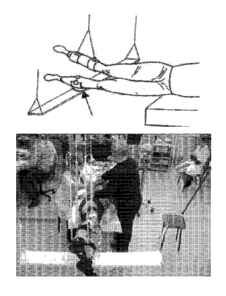
[0160] The subjects were tested while lying on their right side with
the upper leg
supported directly in the area of the shank and the lower leg placed on a
rotating brace
attached to a horizontal board supported by vertical ropes secured to hooks in
the ceiling
(Figure 3). The subjects were instructed not to voluntarily intervene with the
movements
induced by the stimulation. Painless transcutaneous electrical stimulation
(PTES) was
delivered using a 2.5 cm round electrode (Lead_Lok, Sandpoint, United States)
placed
midline on the skin between the spinous processes of C4-05, TII-T12 and L1-L2
as a
cathode and two 5.0 x 10.2 cm2 rectangular plates made of conductive plastic
(Ambu,
Ballerup, Germany) placed symmetrically on the skin over the iliac crests as
anodes. Step-
like movements were evoked by bipolar rectangular stimuli with 0.5 ms duration
filled with
a carrier frequency of 10 kHz and at an intensity ranging from 30 to 200 mA.
The
stimulation frequency was 5 Hz and the duration of exposure ranged from 10 to
30s.
[0161] TES was easily tolerated by subjects and did not cause pain
even when the
strength of current was increased to 200 mA. Lack of pain can be attributed to
the use of
biphasic stimuli with a carrier frequency of 10 kHz that suppresses the
sensitivity of pain
receptors. The threshold intensity of the stimulus that induced involuntary
stepping
movements ranged from 110 to 180 mA (Figure 4).
[0162] MG and kinematics features of locomotor patterns induced by
painless
transcutaneous electrical stimulation at the T11-T12 vertebral level at 5 and
30 Hz of
frequency in non-injured human subjects are shown in Figures 5A and 5B.
Angular
movements of the right (R) knee and left (L) knee joints and representative
EMG activity in
-28-
CA 02906779 2015-09-14
WO 2014/144785
PCMJS2014/029340
the rectus femoris (RF), biceps femoris (BF) tibialis anterior (TA) and medial
gastrocnemius (MG) muscles during involuntary locomotor-like activity induced
by PTES
at the T11 vertebra. Stick diagram decompositions (40 ms between sticks) of
the
movements of the R leg and trajectory of toe movements during one step cycle
during PTES
at T11-T12 are shown in Figure 5B. Arrows in Figure 5B indicate the direction
of
movement.
[0163] EMG and kinematics features of locomotor patterns induced by
PTES at the
C5, T11, and L2 vertebral levels (Figure 6). Angular movements of the right
(R) knee and
left (L) knee joints and representative EMG activity in the biceps femoris
(BF) muscles of
the R and left L legs during involuntary locomotor-like activity induced by
PTES at the
C5+T11+L2 vertebrae simultaneously (left) and sequentially (right).
[0164] Figure 7 shows stick diagram decompositions (40 ms between
sticks) of the
movements of the R leg during one step cycle during PTES at different
vertebral levels in
two subjects are shown. Arrows indicate the direction of movement. Multi-
segmental non-
invasive electrical spinal cord stimulation was used to facilitate
involuntary, coordinated
stepping movements. Simultaneous spinal cord stimulation at multiple sites can
have an
interactive effect on the spinal neural circuitries responsible for generating
locomotion. The
synergistic and interactive effects of multi-site spinal cord stimulation can
be a multi-
segmental convergence of descending and ascending, and most likely
propriospinal,
influences on the spinal circuitries associated with locomotor and postural
activity.
[0165] The various methods and techniques described above provide a
number of
ways to carry out the application. Of course, it is to be understood that not
necessarily all
objectives or advantages described can be achieved in accordance with any
particular
embodiment described herein. Thus, for example, those skilled in the art will
recognize that
the methods can be performed in a manner that achieves or optimizes one
advantage or
group of advantages as taught herein without necessarily achieving other
objectives or
advantages as taught or suggested herein. A variety of alternatives are
mentioned herein. It
is to be understood that some preferred embodiments specifically include one,
another, or
several features, while others specifically exclude one, another, or several
features, while
still others mitigate a particular feature by inclusion of one, another, or
several
advantageous features.
[0166] Furthermore, the skilled artisan will recognize the
applicability of various
features from different embodiments. Similarly, the various elements, features
and steps
-29-
discussed above, as well as other known equivalents for each such element,
feature or step,
can be employed in various combinations by one of ordinary skill in this art
to perform
methods in accordance with the principles described herein. Among the various
elements,
features, and steps some will be specifically included and others specifically
excluded in
diverse embodiments.
[0167] Although the application has been disclosed in the context of
certain
embodiments and examples, it will be understood by those skilled in the art
that the
embodiments of the application extend beyond the specifically disclosed
embodiments to
other alternative embodiments and/or uses and modifications and equivalents
thereof.
[0168] It is understood that the examples and embodiments described herein
are for
illustrative purposes only and that various modifications or changes in light
thereof will be
suggested to persons skilled in the art and are to be included within the
spirit and purview of
this application and scope of the appended claims.
-30-
Date Recue/Date Received 2020-10-21