Note: Descriptions are shown in the official language in which they were submitted.
CA 02907009 2015-09-15
WO 2014/140684 PCT/1B2013/003046
1
IMPLANTABLE ANCHOR
FIELD OF INVENTION
[0001] The present invention relates to a system and method for
implantation and
securement of a small implantable element to monitor and/or treat
physiological conditions of
the body. In addition, the invention describes a novel anchor to position a
small implantable
element in a desired position within a wall of a target tissue of the body.
The invention also
relates to a method for implanting the anchor directly in a target wall of the
body and securing
the anchor in a fixed location without displacement over the life of the
anchor.
BACKGROUND
[0002] Percutaneously-delivered deployment systems are used to embed
implantable
devices within a lumen of the body. Generally, such a deployment system
comprises a catheter,
an implantable device, and an element for releasing the implantable device at
the target location,
for example, described in U.S. Pub. No. 2003/0125790 and U.S. Pub. No.
2008/0071248. The
catheter houses the deployment system and permits the system to be advanced to
the target
location, where the implantable device is released. The implantable device
remains within the
body to perform its intended function after the deployment system is
retracted.
[0003] Importantly, the implantable device must be securely attached to
the target
location before the deployment system releases the device. A device which is
not securely
embedded may become dislodged and pose serious risks to the patient,
especially if the device
migrates from the implantation site. An insufficiently secured device that
circulates in the body
may cause serious injuries, including an acute myocardial infarction, a
stroke, or organ failures.
CA 02907009 2015-09-15
WO 2014/140684 PCT/1B2013/003046
2
Thus, there is a need for an anchor and a deployment system that assures that
the device is
implanted and secured in the body in a fixed location without dislocation over
the life of the
device. Also, there is a need for an anchor that permits the deployment of the
implantable device
with minimal damage to the wall of the organ of the body. Further, there is a
need for an anchor
that permits the desired orientation of the device relative to the lumen of
the body without
relocating or adjusting the implanted device once deployed within the lumen.
[0004] Such an anchoring system is advantageous to the clinician in that
it enables the
implantation and securement of an implantable device at the desired
orientation through a
cannula-based delivery system for ease of deployment while reducing the risk
of displacement of
the device over time. Further, such a system can eliminate the need for a
follow-up procedure to
retrieve the dislodged implantable device, as is the case where the device is
not securely
implanted through a reliable means, or to relocate the implantable device in
order to establish the
desired orientation of the device relative to the lumen. For example, current
procedures for
monitoring hepatic portal pressure and the detection of malignant hypertension
are not
satisfactory and generally involve an indirect measurement of the portal
venous pressure through
the hepatic venous system due to the difficulty in accessing the target site
for the direct
implantation of the monitoring device. Moreover, there are no current
procedures for implanting
a monitor. Thus, a system that is capable of reliably and securely implanting
a detector of portal
pressure, for example, could reduce the complexities of the procedure and the
need for post-
operative treatments, providing favorable outcomes for the patient.
[0005] A need therefore exists for an anchoring system that allows for
simple, safe and
secure implantation of a device in a fixed location, while orienting the
device as desired.
CA 02907009 2015-09-15
WO 2014/140684 PCT/1B2013/003046
3
SUMMARY OF THE INVENTION
[0006] The present invention relates to an anchor and a deployment system
for the
percutaneous delivery and secure implantation of a device in the wall of a
target tissue of the
body to measure or treat various bodily conditions. The anchor comprises a
first stabilizer, a
second stabilizer, a first ring, a second ring, a bridge there between, and a
positioning arm. The
first and second rings are attached to each end of the bridge. The first
stabilizer extends from the
first ring and the second stabilizer extends from the second ring. One of said
stabilizers is
located at the proximal end of the anchor and may be referred to as a proximal
stabilizer, and the
other stabilizer is located at the distal end of the anchor and may be
referred to as a distal
stabilizer. The first and second stabilizers may transition from a crimped
position to a deployed
position upon deployment in a target site wall. The stabilizers are
characterized by their distinct
crimped and deployed configurations. The crimped configuration is
characterized in that the
stabilizers fit inside the delivery system. The deployed configuration is
characterized as having
stabilizers that extend in a direction substantially perpendicular to the
bridge of the anchor to a
diameter sufficient to hold the anchor in position in the wall; that is, the
stabilizers extend in a
direction generally parallel to the target tissue wall. The positioning arm
may house a sensor or
other small implantable element which may be strategically positioned, for
example, to protrude
into the target site after deployment.
[0007] The invention also relates to a system for deploying the anchor
comprising an
introducer cannula, a pushrod, a sheath and an anchor. The deployment system
may deliver the
anchor directly to the target site wall (i.e. extra-luminally) or use a
catheter-based system (i.e.
intra-luminally).
CA 02907009 2015-09-15
WO 2014/140684 PCT/1B2013/003046
4
[0008] Further, the invention relates to a method of deploying the anchor
into a vessel
wall comprising introducing the cannula into the target site wall so that the
tip of the cannula is
in the inner portion of the target site wall; positioning the crimped anchor
with a pushrod
between the inner and outer portion of the target site wall; releasing the
first stabilizer of the
anchor so that the first stabilizer expands from a partially deployed anchor;
releasing the second
stabilizer of the anchor so that the second stabilizer expands forming the
fully deployed anchor;
and retracting the cannula. Further, the invention relates to a method of
manufacturing the
anchor by, for example, use of a mandrel specifically designed for the
manufacturing of an
anchor according to the principles of this invention.
[0009] In another aspect of the invention, the invention includes a
mandrel for
manufacturing the implantable anchor having first and second stabilizers. The
mandrel
comprises a first disc, a second disc and an axel therebetween. Each disc may
have a groove
extending in a shape from the axel in which a material forming the anchor is
placed. Mandrel
coverings may also be used to encase the mandrel during the treatment
processes.
[0010] The present invention provides the advantages of a shortened
procedure time,
lessened procedural discomfort, increased procedural success, and increased
safety. The
invention presents the further advantage of enabling implantation of a
detector without
necessitating x-ray or ultrasound imaging for guidance.
BRIEF DESCRIPTION OF THE DRAWINGS
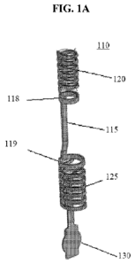
[00 1 1 ] FIG. lA shows an anchor in accordance with this invention in a
fully crimped
state.
CA 02907009 2015-09-15
WO 2014/140684 PCT/1B2013/003046
[0012] FIG. 1B shows the anchor of FIG. lA in a fully deployed state.
[0013] FIG. 2A shows an anchor in accordance with this invention in a
fully crimped
state.
[0014] FIG. 2B shows the anchor of FIG. 2A in a fully deployed state.
[0015] FIG. 3A shows an anchor in accordance with this invention in a
fully crimped
state.
[0016] FIG. 3B shows the anchor of FIG. 3A in a fully deployed state.
[0017] FIG. 4 shows an anchor in accordance with this invention in a
crimped state.
[0018] FIG. 5 shows an anchor in accordance with this invention in a
crimped state.
[0019] FIG. 6 shows an anchor in accordance with this invention in a
crimped state.
[0020] FIG. 7 shows a ring and stabilizers in accordance with this
invention in a crimped
state.
[0021] FIG. 7A shows various perspectives of the ring and stabilizers of
the embodiment
of FIG. 8 in a deployed state.
[0022] FIG. 8 shows various embodiments of rings and stabilizers arranged
on a ring in
the deployed state.
[0023] FIG. 8A shows a top view of various stabilizers and ring
embodiments in
accordance with this invention.
CA 02907009 2015-09-15
WO 2014/140684 PCT/1B2013/003046
6
[0024] FIG. 9A shows one embodiment of an anchor rod, second ring and
first ring in
accordance with this invention.
[0025] FIG. 9B shows the anchor rod, second ring and first ring of FIG.
10A in an
extended state.
[0026] FIG. 9C shows another embodiment of the anchor rod, second ring
and first ring
in accordance with this invention.
[0027] FIG. 9D shows the anchor deployed at a target location having a
thin tissue wall.
[0028] FIG. 9E shows the anchor deployed at another target location
having a thick tissue
wall.
[0029] FIG. 10 shows an introducer cannula loaded with the anchor in
accordance with
the invention, introduced into a wall at the target site.
[0030] FIG. 10A shows the introducer cannula of FIG. 12 wherein the
anchor is in a state
of partial deployment, with the first stabilizer and positioning arm deployed
in the inner portion
of the target site wall.
[0031] FIG. 10B shows the introducer cannula of FIG. 13 wherein the
anchor is in a state
of full deployment, with the second stabilizer deployed outside the vessel
wall and the first
stabilizer and positioning arm deployed in the inner portion of the target
site wall.
[0032] FIG. 11 shows an introducer cannula loaded with the anchor in
accordance with
this invention.
CA 02907009 2015-09-15
WO 2014/140684 PCT/1B2013/003046
7
[0033] FIG. 12A shows a release mechanism in accordance with this
invention in a pre-
deployment state.
[0034] FIG. 12B shows the release mechanism of FIG. 12A in a state of
partial
deployment with the sheath refracted from the pushrod.
[0035] FIG. 13A shows a release mechanism in accordance with this
invention.
[0036] FIG. 13B shows the release mechanism of FIG. 13A in a state of
partial
deployment with the aperture of the pushrod rotated to align with a member of
a stabilizer.
[0037] FIG. 14 shows a flat metal pattern of the anchor of FIG. lA prior
to formation
into an anchor in accordance with this invention.
[0038] FIG. 15 shows a flat metal pattern of the anchor of FIG. 3A prior
to formation
into an anchor in accordance with this invention.
[0039] FIG. 16 shows a flat metal pattern prior to formation into an
anchor in accordance
with the invention.
[0040] FIG. 17 shows one embodiment of an anchor in a deployed state in
accordance
with this invention.
[0041] FIG. 18A shows one embodiment of a mandrel designed to manufacture
an
anchor in accordance with the invention.
[0042] FIG. 18B is the front view of the mandrel of FIG. 18A.
CA 02907009 2015-09-15
WO 2014/140684 PCT/1B2013/003046
8
[0043]
FIGS. 18C and 18D show the mandrel covering(s) for use with the mandrel of
FIG. 18A.
[0044]
The invention is discussed and explained below with reference to the
accompanying drawings. The figures are provided as an exemplary understanding
of the
invention and to schematically illustrate particular embodiments and details
of the invention.
The figures are not necessarily drawn to scale. The skilled artisan will
readily recognize other
similar examples equally within the scope of the invention. The drawings are
not intended to
limit the scope of the invention as defined in the appended claims.
DETAILED DESCRIPTION OF THE INVENTION
[0045]
The invention generally relates to an anchor system, deployment system and
method for percutaneously implanting an anchor in the body carrying a small
implantable
element. The system and method relate to particularly small anchors, e.g.,
between 0.005 to 100
3 i
mm
n volume, which are implanted in the wall of target tissue in the body. The
size parameters
of the anchor will be defined by the thickness of the target tissue wall.
Nonetheless, the anchor
may have an outer diameter in the range of 0.01 to 10 mm, a height that is
preferably no more
than 20 mm, and may preferably be adapted to allow for the integration of a
small implantable
element having a diameter in the range of 0.01 to 10 mm and a height in the
range of 0.01 to 20
mm. It may be desirable that the element is fully integrated into the
positioning arm of the
anchor. The anchor is composed of a non-thrombogenic, non-biodegradable and
non-biofouling
material, preferably, a shape-memory material such as, for example, Nitinol,
stainless steel or
other suitable alloys or polymers. In one embodiment, the anchor has an outer
diameter of 1
mm, a height of less than 0.4 mm and allows for the integration of a small
implantable element
CA 02907009 2015-09-15
WO 2014/140684 PCT/1B2013/003046
9
having a diameter of 0.8 mm and a height of 0.3 mm. One preferred target area
for embedding
the anchor, which may be based on the thickness of the blood vessels at the
target site, may be no
less than 0.5 mm and no greater than 50 mm. Target areas of non-vessel target
structures
include, for example, the septum in the heart or the parenchyma of the liver,
which may have
thicknesses in the range of 0.5 to 10 mm.
[0046] The size and relatively low invasiveness of such anchors make them
particularly
well suited to medical and physiological uses, including, but not limited to,
measuring bodily
fluids, such as for example in the blood vessel/artery/vein. Generally, such
anchors may aid in
measuring chemical or physical parameters of bodily fluids, such as occurs in
the blood, urine or
digestive fluids. Implants in the heart for example may be used for measuring
left atrial pressure
in congestive heart failure applications. Implants in the liver may be used
for intra-abdominal
pressure. Such anchors are also applicable, for example, to aid in monitoring
particular diseases
or conditions or body chemical or physiological parameters, to deliver a
therapeutic agent or
other similar situations.
[0047] The anchor comprises a first stabilizer, a second stabilizer, a
bridge there
between, a first ring, a second ring, and a positioning arm. Each of the first
and second
stabilizers comprise one or members that may be formed in various shapes, such
as, for example,
prongs, coils, helixes, etc. One such stabilizer may be located at the
proximal end of the anchor
(the proximal stabilizer) and the other such stabilizer may be located at the
distal end of the
anchor (the distal stabilizer). Each member of a first or second stabilizer
has a crimped state,
that may be orderly, such as for example in a coil or a straight prong in a
crimped position, or
alternatively may be an irregular shape having bends, loops or twists for
example, or generally
amorphous. Each member of a first or second stabilizer also has a deployed
state, for example, a
CA 02907009 2015-09-15
WO 2014/140684 PCT/1B2013/003046
diameter greater than the diameter of the opening in the target site wall
created by a cannula
upon delivery of the anchor. The target site wall is understood as the tissue
at the site of
deployment through which the anchor protrudes for deployment. Preferably, the
target site tissue
is an organ having bodily fluid transported through it, for example, blood
vessels, heart
chambers, digestive organs, urinary tract organs, the liver and the like. The
members of the first
stabilizer extend from a first ring, and the members of the second stabilizer
extend from a second
ring. The first ring and second ring are connected by the bridge. The
positioning arm may
extend from either the first ring or the second ring. The ring may be of any
shape, including,
circular, and be either hollowed or filled.
[0048] The invention also relates to a deployment system for delivering
an anchor,
comprising an introducer cannula having an inner lumen, which houses a
pushrod, a sheath and
the anchor. In an alternative embodiment, the deployment system may include a
catheter system
for intra-luminal delivery of the anchor. In one embodiment, a sheath
surrounds the crimped
anchor and maintains the first and second stabilizers and positioning arm in a
crimped position in
the introducer cannula. Here, the first stabilizer and positioning arm may be
oriented at the distal
end of the anchor, and the second stabilizer may be oriented at the proximal
end of the anchor
once assembled onto the deployment system. The sheathed anchor is placed in
the desired
position using the cannula and pushrod such that the bridge traverses the
target site wall with the
first stabilizer and positioning arm protruding into the target site. Once in
position, the sheath is
retracted, releasing the first stabilizer and the positioning arm from their
crimped position into
the target site so that the positioning arm is positioned on one side of the
target site wall. Next,
the second stabilizer is released from its crimped position onto the other
side of the target site
wall, thereby securing the anchor in a fixed position traversing the target
site wall.
CA 02907009 2015-09-15
WO 2014/140684 PCT/1B2013/003046
11
[0049] In an alternative embodiment the positioning arm may be located at
the proximal
end of the anchor, thereby co-located with the second stabilizer. Here, the
deployment system
further comprises a catheter and the anchor is deployed intra-lumenally such
that the anchor is
positioned in a vessel wall target site from within a vessel, for example. In
this embodiment, the
cannula and pushrod position the distal end of the anchor at the target site
wall so that the first
stabilizer is on the outer side of the vessel wall. Next the sheath is pulled
back allowing the first
stabilizer to deploy, then the second stabilizer thereby deploying the anchor.
[0050] In yet another embodiment, the deployment system may comprise a
catheter with
the anchor disposed therein. The positioning arm may be located at the distal
end of the anchor,
co-located with the first stabilizer. In this embodiment, the catheter may be
advanced intra-
lumenally in a first vessel in order to access a target site located in a
second, nearby vessel.
Once the target site is reached, an access device, e.g., a needle, may be used
to access the target
site in the second vessel from the first vessel. Thereafter, the anchor may be
deployed in the
second vessel with the positioning arm extended within the second vessel. For
example, the
portal vein may be accessed from the hepatic vein (intrahepatic) by this
method.
[0051] The present invention also comprises a method for using a
deployment system
comprising a cannula, sheath and anchor. The target location may be identified
by fluoroscopy
or ultrasound and accessed by well-known access routes. The method comprises
the steps of (i)
advancing the cannula to the target site wall; (ii) introducing the cannula
into the target site wall
so that the tip of the cannula is in the inner portion of the target site
wall; (iii) advancing the
crimped anchor to said target site through said cannula, thereby positioning
the crimped anchor
between the inner and outer portion of the target site wall; (v) administering
a controlled amount
of force to retract the sheath to deploy the anchor system; (vi) releasing the
first stabilizer of the
CA 02907009 2015-09-15
WO 2014/140684 PCT/1B2013/003046
12
anchor so that the one or more members of the first stabilizer expand; (vii)
releasing the second
stabilizer of the anchor so that the one or more members of the second
stabilizer expand; and
(viii) retracting said cannula. An additional optional step comprises slightly
retracting the
delivery system to ensure the first stabilizer is flush against the vessel
wall and deployed.
[0052] The invention also relates to an optional feature of the
deployment system
comprising an introducer cannula having an interior lumen that houses a
pushrod and a sheath
that covers the anchor, wherein the pushrod may have one or more apertures
through which a
member of a first or second stabilizer protrudes into the space between the
pushrod and the
cannula. The frictional force between the outer wall of the pushrod and the
inner wall of the
sheath holds the member in place until by one or more methods, the frictional
force on the
member is released for deployment. Thus, the method of deployment in
connection with this
embodiment further comprises the steps of (a) withdrawing the sheath holding
the one or more
members of the second stabilizer in place and (b) withdrawing the pushrod to
release the one or
more members of the second stabilizer through the one or more apertures of the
pushrod. In an
alternative embodiment, each of the one or more apertures of the pushrod
consist of an "L" shape
formed by an aperture neck and an aperture arm. In the pre-deployment
position, a member of
the second stabilizer may extend through the aperture arm. Thus, the method of
deployment in
connection with this embodiment further comprises the step of (a) rotating the
pushrod such that
the aperture neck aligns with the member of the second stabilizer, and (b)
withdrawing the sheath
to release the member of the second stabilizer.
[0053] The invention further comprises a method of manufacturing the
anchor
comprising the steps of (i) producing a wire from a suitable material; and
(ii) applying a heat
treatment, as needed, to the wire to conform the wire to a predetermined shape
with the use of a
CA 02907009 2015-09-15
WO 2014/140684 PCT/1B2013/003046
13
mandrel. In one embodiment, the mandrel comprises a first disc having a
groove, a second disc
having a groove and an axel therebetween. Another method of manufacturing
according to this
invention comprises the steps of (i) laser cutting a preselected pattern from
a flat metal sheet,
such as, for example, Nitinol; and (ii) forming the pattern into an anchor
having a first stabilizer,
a second stabilizer and a bridge positioned therebetween through application
of heat treatment,
welding or mechanical force.
[0054] The following sections describe various exemplary illustrations of
the invention.
It is understood that these figures represent examples of the features of the
invention but are not
limiting. The skilled person will readily recognize other embodiments within
the scope of the
invention.
[0055] Figure lA illustrates one embodiment of the anchor of the
invention. Anchor 110
comprises bridge 115, a first ring 119 and a second ring 118. Extending from
the first ring 119 is
a first stabilizer 125, which is coiled when in a crimped state, as well as a
positioning arm 130
positioned within the coil formed by the first stabilizer 125. Extending from
the second ring 118
is a second stabilizer 120 that is coiled when in a crimped state. The
positioning arm 130 may
extend from the first or second ring depending upon how the anchor is
delivered. If delivered
directly into the target site using a cannula only (e.g. extra-luminally),
then the positioning arm is
preferably attached to the first ring; whereas, if the anchor is delivered
using a catheter-based
system (e.g. intra-luminally), then the positioning arm is preferably attached
to the second ring.
The first stabilizer 125 and the second stabilizer 120 are each formed of a
member in Figure 1A.
The member may be in the form of a wire, band, strip or other appropriate
configuration so as to
function as described herein. In the embodiment illustrated in Figure 1, the
member is formed by
a strip. In Figure 1A, the anchor 110 is in a pre-deployment state as in a
delivery system,
CA 02907009 2015-09-15
WO 2014/140684 PCT/1B2013/003046
14
wherein the first stabilizer 125, second stabilizer 120 and positioning arm
130 are in a crimped
state. In one embodiment, the ring associated with the positioning arm may be
larger than the
other ring or the opposite depending on the configuration of the pushrod.
[0056] The bridge 115 may be of any length necessary depending on the
thickness of the
target site wall. The length of the bridge will be determined to ensure that
the first ring 119 and
first stabilizer 125 can extend into the target site lumen, thus allowing the
first stabilizer 125 to
extend (in this embodiment, by partially unspooling from a crimped coil),
while at the same time
the second ring 118 and second stabilizer 120 remain outside the target site
wall and the second
stabilizer 120 is allowed to unspool as well. Figure 1B illustrates anchor 110
of Figure lA in a
fully deployed state. Figure 1C illustrates anchor 110 implanted in vessel
tissue 150. Bridge 115
extends
[0057] Alternatively, the first and second stabilizers 125, 120 may be
designed to adapt
upon deployment to variability in the thickness of the wall by ¨ for example ¨
extending at an
angle relative to parallel to the target site wall on either side, such that
the far end of each
stabilizer is closer to the target site wall than the point at which each said
stabilizer is attached to
the ring, to compensate in the event that the bridge substantially exceeds the
width of the target
site wall.
[0058] Figure 2A illustrates another embodiment of anchor 210 in a
crimped state
wherein the first stabilizer has a first member 225a and a second member 225b,
as well as a
second stabilizer having a first member 220a and a member 220b. The first
member 225a and
second member 225b of the first stabilizer are positioned around the first
ring 219 and are coiled
in parallel with each other. Likewise, the first member 220a and the second
member 220b of the
CA 02907009 2015-09-15
WO 2014/140684 PCT/1B2013/003046
second stabilizer are positioned 180 apart on the second ring 218 and are
coiled in parallel with
each other. Alternatively, stabilizers of this embodiment may form the helical
configuration
illustrated in Figure 2A wherein each of the first and second members of each
stabilizer are
formed from a continuous loop so that the helical coil of each stabilizer does
not include blunt
ends, i.e., each stabilizer forms a double helix. Figure 2B illustrates anchor
210 in a fully
deployed state, with each of the first and second members 225a, 225b of the
first stabilizer and
the first and second members 220a, 220b of the second stabilizer fully
deployed. The deployed
configuration is characterized as having stabilizers that extend in a
direction substantially
perpendicular to the bridge of the anchor so that the anchor is fully engaged,
for example, to a
diameter greater than the diameter of the opening in the target site wall
created by the cannula
upon delivery of the anchor.
[0059] Figure 3A illustrates yet another embodiment of anchor 310 in a
pre-deployment
state, wherein the first stabilizer includes a first member 325a, a second
member 325b and a third
member 325c, and the second stabilizer includes a first member 320a, a second
member 320b
and a third member 320c. In the pre-deployed state, the first, second and
third members 325a,
325b, 325c of the first stabilizer extend approximately straight from the
first ring 318 in the
direction of the second ring 319; and the first, second and third members
320a, 320b, 320c of the
second stabilizer extend approximately straight from the second ring 318 in
the direction of the
first ring 319. In Figure 3A, the members are shown extending from one ring
approximately
straight in the direction of the other ring. Figure 3B illustrates anchor 310
in a fully deployed
state, whereby the first, second and third members 325a, 325b, 325c of the
first stabilizer and the
first, second and third members 320a, 320b, 320c of the second stabilizer bend
away from the
bridge 315.
CA 02907009 2015-09-15
WO 2014/140684 PCT/1B2013/003046
16
[0060] Other forms of first and second stabilizers may be utilized in
connection with the
anchor, as illustrated, for example, in Figure 4, showing a first stabilizer
425 and second
stabilizer 420 in a crimped state wherein the first and second stabilizers
425, 420 coil inward
toward the center of the anchor 410. Figure 5 shows an anchor 510 having a
first member 525a
and a second member 525b of a first stabilizer extending from a first ring 519
that fold inward
when in a crimped state, as well as a first member 520a and a second member
520b of a second
stabilizer, as well as a positioning arm 530 extending from a first ring 519
that fold inward when
in a crimped state. Figure 6 shows a hybrid combination in which the first and
second members
625a, 625b of the first stabilizer on the first ring 619 coil inward while the
first and second
members 620a, 620b of the second stabilizer on the second ring 618 fold inward
when the anchor
610 is in a crimped state.
[0061] Figure 7 illustrates a ring 770 that may be used at the proximal
or distal end of an
anchor, having a plurality of stabilizer members 775a-d, which, in a crimped
state are configured
to bend inward as shown in Figure 7. The stabilizer members of this embodiment
may be
configured to bend outward in a variety of shapes upon deployment of the
anchor, as illustrated
in Figure 7A. Alternatively, the stabilizer extending from ring 870 may have a
plurality of
members each forming a loop ¨ for example, first member 875a and second member
875b may
each form a loop a continuous loop extending outward in a plurality of
directions as illustrated
by Figure 8. Figure 8A illustrates a top perspective of various embodiments of
ring 870 having
coiled stabilizer members in a deployed state. Figure 17 illustrates yet
another embodiment of
the anchor 1710 in a fully deployed state.
[0062] As illustrated in the embodiments of Figures 9A-9C, the anchor
bridge 915 of the
anchor 910 may be formed of an elastic or flexible material capable of
stretching or contracting
CA 02907009 2015-09-15
WO 2014/140684 PCT/1B2013/003046
17
to adjust to the dimensions of the vessel wall in which the anchor is
deployed. Alternatively, the
bridge may be preset at an angle such that the angle can be straightened upon
deployment to
accommodate the thickness of the tissue wall. In this embodiment, the preset
angle or degree of
bend in the bridge will define the thinnest tissue wall for secure
implantation of the anchor,
whereas the fully extended length of the bridge will define the thickest
tissue wall. Upon
crimping into the delivery system, the bridge may be straightened. Upon
deployment, the anchor
bridge will maintain a varying degree of contraction to accommodate the
thickness of the body
tissue wall at the implantation site.
[0063] Figure 9A shows the anchor 910 in a semi-contracted state, Figure
9B shows the
anchor 910 in an extended state, and Figure 9C shows the anchor in a
contracted state. Figures
9D and 9E illustrate the anchor deployed in two different target tissues.
Target tissue 920 of
Figure 9D is thinner than target tissue 930 of Figure 9E. When deployed at
target tissue 920,
bridge 915 of the anchor may be contracted such that first and second
stabilizers 940 and 950 are
both in contact with the target tissue, illustrated in Figure 9D. When
deployed at target tissue
930, ridge 915 may be in a more extended state so that stabilizers 940 and 950
are also both in
contact with the target tissue, illustrated in Figure 9E. The elastic bridge
915 allows the anchor
to be secured embedded in target tissues having different thicknesses.
[0064] The present invention also relates to a deployment system for the
percutaneous
delivery and secure implantation of an anchor in the target site wall. Figure
10 illustrates an
introducer cannula 1040 for percutaneous delivery of an anchor having an
interior lumen 1041
and a tip 1042. The introducer cannula 1040 is adapted to house an anchor
1010, a sheath 1050,
and a pushrod 1060. The cannula 1040 may comprise an outer diameter in the
range between 1
to 50 G, an inner diameter in the range of 0.01 to 20 mm, a length of 1 to 200
cm, and comprises
CA 02907009 2015-09-15
WO 2014/140684 PCT/1B2013/003046
18
a suitable semi-flexible, biocompatible material for use within the body.
Suitable materials
include, for example, silicones, polyvinyl chloride (PVC) or other medical
grade biocompatible
polymers. In one particular embodiment, the introducer cannula 1040 has an
outer diameter of
17 G, an inner diameter of 1.06 mm, a length of 20 cm and is made of a semi-
flexible,
biocompatible material. The anchor 1010 is designed such that, in a crimped
state, the first
stabilizer 1025 and the second stabilizer 1020 have a diameter of sufficient
width to fit within the
interior lumen 1041 of the introducer cannula 1040 without causing bulges.
[0065] The pushrod 1060 is contained within the interior lumen 1041 of
the introducer
cannula 1040 and directly abuts the anchor 1010. The pushrod 1060 may have an
outer diameter
in the range of less than 0.01 to no greater than 20 mm, a length in the range
of 1 to 200 cm, and
an inverted cone in the piston of the pushrod 1060, which is adapted to
protect the area around
the anchor 1010. The pushrod 1060 is adapted to move lengthwise inside the
interior lumen
1041 of the introducer cannula 1040 from the proximal end of the interior
cannula 1040 to the
target implantation site to deploy the anchor 1010. The pushrod 1060 may be
solid or hollow,
and comprises a suitable semi-flexible biocompatible material, such as
Nitinol, silicone, PVC,
titanium or stainless steel. The materials of the introducer cannula 1040 and
the pushrod 1060
may be same or different, provided that the pushrod 1060 is comprised of a
material sufficiently
rigid to resist collapse.
[0066] The anchor 1010 is oriented such that the first stabilizer 1025
and positioning arm
1030 are located at the distal end of the anchor 1010 near the tip 1042 of the
cannula 1040, and
the second stabilizer 1020 is located at the proximal end of the anchor 1010.
In the embodiment
illustrated by Figure 10, wherein the anchor is delivered directly unto the
target site using a
CA 02907009 2015-09-15
WO 2014/140684 PCT/1B2013/003046
19
cannula only (e.g. extra-luminally), the positioning arm 1130 is preferably
oriented at the distal
end of the anchor.
[0067] The sheath 1050 extends over the anchor 1010 and is adapted to be
controllably
retracted to release the anchor 1010 at the deployment site. The sheath 1050
may be retracted to
deploy the anchor 1010 while the pushrod 1060 remains stationary to position
the anchor 1010 at
the target implantation site. The sheath 1050 may be manipulated by the
operator directly or
remotely, so that the anchor 1010 is released from the sheath 1050 at the
discretion of the
operator. For example, the sheath 1050 may extend from the anchor 1010 through
the interior
lumen 1041 of the introducer cannula 1040 to the proximal end, allowing the
operator to
manipulate the sheath 1050 directly through mechanical means. Alternatively,
the stabilizers
may also be releasable using shape-memory materials, for example, Nitinol or
shape-memory
polymers, which may be controllable by well-known means in the art, such as
heat, light,
chemical, pH, magnetic or electrical stimuli, described in, for example, U.S.
Pat. No. 6,720,402
and U.S. Pat. No. 2009/0306767, both of which are incorporated by reference in
their entirety.
For example, the shape-memory material may be in a form of a spring, capable
of contraction
and expansion as an electric current is applied or removed. Electroactive
polymers or magnetic
shape memory alloys may also be employed in a similar fashion. Another
alternative may be a
remote control mechanism, such as for example, a string and loop-mechanism
where the string is
threaded through a loop or similar hoop structure on the sheath 1050, and the
two ends of the
string are located towards the proximal end of the introducer cannula 1040. To
verify the secure
embedding of the anchor, both ends of the string may be pulled to ensure the
sheath 1050 is not
dislodged. Releasing one end of the string unthreads the string from the
loops, and the sheath
1050 may be retracted thereafter. The sheath 1050 may comprise any suitable
size or shape to be
CA 02907009 2015-09-15
WO 2014/140684 PCT/1B2013/003046
arranged within the interior lumen 1041 of the introducer cannula 1040. The
sheath 1050 may
be formed of a braided polymer, such as polyimide, that may be braided
together with a
biocompatible material, such as a silicone, PVC, titanium or stainless steel.
The sheath 1050
may be comparatively less rigid than the pushrod 1060.
[0068] The present invention also relates to a method of implanting an
anchor into a
target site wall. The method comprises protruding the cannula 1040 through the
target site wall
1080, as illustrated by Figure 10. Figure 10A illustrates the anchor 1010 in
an early stage of
deployment wherein an application of force to the pushrod 1060 extends the
anchor 1010 into the
target site 1090, retracting sheath 1050 from the anchor 1010 and deploying
the first stabilizer
1025 and positioning arm 1030. The first stabilizer 1025 and the positioning
arm 1030 revert to
a deployed state within the target site 1090, while the bridge 1015 traverses
the target site wall
1080. The deployed state of the first stabilizer 1025 may be any shape that
expands the area of
the coil in a direction perpendicular to the axis of the bridge, i.e.,
generating a configuration that
prevents the anchor from dislodging from its position in the target site wall.
The semi-deployed
anchor 1010 may then be tugged gently in the counter direction (i.e. pulled
snug against the
target site wall) to ensure that the anchor 1010 is properly embedded in the
vessel prior to full
deployment. Figure 10B illustrates the anchor 1010 in a state of full
deployment following full
retraction of the sheath 1050 and cannula 1040, thereby deploying the second
stabilizer 1020
from a crimped state and permitting the second stabilizer 1020 to unspool
partially. In the
deployment stage, each of the first and second stabilizers form a flattened
coil configuration.
Thus, the first stabilizer 1025 unspools along the outer wall 1081 of the
target site, the anchor
bridge 1015 traverses the target site wall 1080, the second stabilizer 1020
unspools along the
inner wall 1082 of the target site, and the positioning arm 1030 reverts to a
preselected position
CA 02907009 2015-09-15
WO 2014/140684 PCT/1B2013/003046
21
to orient the anchor in a specific orientation relative to the target site,
according to design
specifications. The introducer cannula 1040, sheath 1050 and pushrod 1060 are
fully retracted
from the anchor 1010.
[0069] Preferably, the sheath has a feedback mechanism that assures the
anchor is
securely implanted prior to the retraction of the pushrod. In one embodiment,
feedback is
provided to the operator by resistance of the unsheathed first ring against
the inner wall of the
vessel at the target implantation site. Mechanical resistance to retracting
the anchor signals to
the operator that the anchor is successfully deployed within the inner portion
of the vessel and
that the second ring may be unsheathed to fully deploy the anchor.
[0070] The force feedback mechanism may be adapted to the user-controlled
sheath
described above. In another embodiment, a force meter may be used with the
sheath to ensure
that the anchor is securely deployed at the target site. The force meter may
be used to measure
the degree of force of the first stabilizer in a deployment position against
the interior vessel wall
upon partial deployment of the anchor, thus signal to the operator that the
second stabilizer may
be unsheathed for full deployment. The force meter also may be used to measure
the degree of
pushing force used to pierce the vessel wall, as well as the amount of pulling
strain demonstrated
by the anchor to ensure that the anchor will remain engaged within the body
lumen and not
prematurely dislodge. One example of a force meter that may be incorporated
within the system
of this invention is described in U.S. Pub. No. 2010/0024574, the contents of
which are
incorporated herein by reference.
[0071] In one embodiment, illustrated by Figure 11, the pushrod 1160 may
be hollow and
house the anchor 1110 within the pushrod 1160 up to the first ring 1119. The
pushrod 1160
CA 02907009 2015-09-15
WO 2014/140684 PCT/1B2013/003046
22
comprises an aperture 1161 through which the second stabilizer 1120 of the
anchor 1110
extends. As further illustrated by Figure 12A, the sheath 1250 covers the
outside of the pushrod
1260, thus forming a release mechanism 1280 that compresses the portion of the
second
stabilizer 1220 protruding through the aperture 1261 between the pushrod 1260
and the sheath
1250. The release mechanism is advantageous in preventing premature release of
the stabilizer
prior to deployment at the target site. Figure 12B illustrates the result of
retracting or releasing
the sheath 1250 from the pushrod 1260, whereby the second stabilizer 1220 is
released from the
compression.
[0072] Figures 13A-B illustrate another embodiment of the release
mechanism 1380,
whereby the aperture of the pushrod 1360 comprises an "L"-shape slit formed by
an aperture arm
1361a and an aperture neck 136 lb. In the deployment position, the second
stabilizer 1320
extends through the aperture arm 1361a as illustrated by Figure 13A. Upon
rotation of the
pushrod 1360, the aperture neck 136 lb aligns with the second stabilizer 1320,
as illustrated by
Figure 13B, thus permitting the deployment of the second stabilizer 1320 upon
retraction of the
sheath 1350. Rotation of the pushrod 1360 around the second stabilizer 1320 is
possible due to
the frictional force of the first stabilizer once deployed against the
opposite-facing wall of the
target site.
[0073] The deployment system described above may be used to implant the
anchor in
any accessible tissue wall of the body, such as in the cardiovascular system,
the hepatic-portal
venous system, or in the gastrointestinal tract. In one embodiment, the
invention may be useful
in the hepatic-portal venous systems during portal venous catheterization
procedures to implant
the anchor in the portal vein. In another embodiment, arteries of the cardio-
vascular system can
CA 02907009 2015-09-15
WO 2014/140684 PCT/1B2013/003046
23
be monitored through implantation of a small implantable element in an artery
or in certain
veins.
[0074]
The small implantable element may monitor any bodily characteristic within a
body cavity or lumen. Examples of such elements measure physical or chemical
characteristics
of the body, such as, for example, sensors, monitors, attenuators, or
regulators of luminal or
extraluminal function. Alternatively, the small implantable element may treat
a medical
condition, for example, by releasing a therapeutic agent.
[0075]
In any of the embodiments above, the anchor may comprise a radiopaque marker
attached to a component of the anchor, e.g., the positioning arm.
Alternatively, the anchor,
partly or in whole, may be composed of a radiopaque material. Radiopaque
material may
include gold, boron, tantalum, platinum iridium or similar materials. The use
of radio-opaque
materials allows visualization by x-ray or patterned with ultrasonic grating,
or both.
[0076]
The invention further relates to a method of manufacturing an anchoring
system,
comprising the steps of producing a wire from a suitable material, such as,
for example, Nitinol;
and applying a heat treatment to the wire to conform the wire into the shape
of a flattened coil to
thermomechanically preset the stabilizer's deployed configuration.
Other methods of
manufacturing according to the invention includes, for example, laser cutting,
chemical etching,
electrochemical machining, electrical discharge machining, or other
traditional machine
methods. The invention may be manufactured from a flat metal sheet or a stock
tube, such as,
for example, Nitinol or stainless steel, into a preselected pattern. The
pattern thus can be coiled
through application of heat treatment, welding or mechanical force.
CA 02907009 2015-09-15
WO 2014/140684 PCT/1B2013/003046
24
[0077] Figure 14 illustrates a pattern of the anchor 110 for example as
shown in Figure
1A, having a bridge 115 with a second end 116 and a first end 117. A second
band 111 occurs at
the second end 116 of the bridge 115 with a second stabilizer 120 extending
therefrom in a
direction parallel to the bridge 115. The second band 111 may be formed into a
ring and welded
together, and upon heat treatment or other means, the second stabilizer 120
may be configured
into a pre-deployment state. A first band 112 occurs at the first end 117 of
the bridge 115 with a
first stabilizer 125, as well as positioning arm 130 extending therefrom in a
direction parallel to
the anchor bridge 115.
[0078] The positioning arm 130 may be located in various positions around
the first band
112, for example as illustrated, out of alignment with the bridge 115. The
first band 112 may be
formed into a ring and welded together, and upon heat treatment or other
means, the first
stabilizer 125 may be configured into a pre-deployment state. Figure 15
illustrates a laser-cut
pattern of the anchor for example, as shown in Figure 2A, having a first
member 225a and a
second member 225b of the first stabilizer positioned 180 degrees apart around
the ring, as well
as a first member 220a and a second member 220b of the second stabilizer
similarly positioned.
Alternatively, the members may be positioned variously around the band. In yet
another
alternative, the ends of the members may be connected to form a continuous
loop. Figure 16
illustrates yet another laser-cut pattern further including a third member
220c of the second
stabilizer. Similarly other embodiments having varying numbers of members
variously
positioned along the bands will be readily apparent to the skilled person and
are within the scope
of the invention.
[0079] Figure 18A illustrates a mandrel 1800 having a first disc 1810, a
second disc 1820
and an axel 1830 therebetween. As illustrated by Figure 18B, the first disc
1810 has a first
CA 02907009 2015-09-15
WO 2014/140684 PCT/1B2013/003046
surface 1815 that is convex towards the second disc 1820, and the second disc
1820 likewise has
a second surface 1825 that is convex towards the first disc 1810. Although
Figure 18A shows
convex surfaces on the first and second discs, the invention contemplates
other surface shapes,
including flat or concave surfaces. On each first and second surfaces 1815,
1825, grooves 1816,
1826, respectively, spiral away from axel 1830 toward the edges of the discs
1810, 1820.
[0080] Mandrel coverings are configured to encase mandrel 1800, which may
be of any
shape or size. As illustrated in Figure 18C, one mandrel covering 1850 is
designed to encase
roughly half of the surface area of axel 1830 and first or second surfaces
1825 or 1826. Mandrel
covering comprises axel covering portion 1860. Figure 18D shows two mandrel
coverings that
encase substantially the entirety of axel 1830 and first and second surfaces
1825 and 1826.
While Figures 18C and 18D illustrate the two mandrel coverings 1850 that are
symmetrical, this
invention contemplates that the number, shape and size of the mandrel
coverings are not limited
to the illustrated embodiments. Mandrel coverings 1850 may be secured around
the mandrel by
any securement means in the art, including latches or clasps. Further, a wire
may be spindled
around axel covering portion 1860 to keep mandrel covering 1850 secure on the
mandrel.
[0081] In the manufacturing process, a material, e.g., a wire, forming
the anchor is placed
in the groove of the mandrel. Once placed, the mandrel coverings are secured
onto the mandrel,
thereby restricting the movement of the material. The material-loaded mandrel,
along with the
mandrel coverings are then heat treated as known in the art, such that the
material retains the
shape of the groove of the mandrel. Materials placed in the grooves of the
first and second
surfaces form the first and second stabilizers, while materials in contact
with the axel form the
bridge and first and second rings. Preferably, the material forming the ring
does not join in a
CA 02907009 2015-09-15
WO 2014/140684 PCT/1B2013/003046
26
circular shape when placed onto the mandrel, allowing for the anchor to be
removed after the
heat treatment.
[0082] In manufacturing the embodiment having an extendable bridge, such
as, for
example, the embodiments of the anchor illustrated in Figure 9A-9E, the
mandrel may comprise
a first disc, a second disc and a axel therebetween. The axel of this
embodiment may be
configured with one or a plurality of bends such that when the material is
placed onto the
mandrel and heated, the resulting bridge has bend(s) in the relaxed state in
accordance with the
shape of the axel. Similarly, the shape and size of the mandrel coverings may
be modified to
complement the profile of the axel of this mandrel.
[0083] Following the formation of the anchor as described above, the
anchor is removed
from the mandrel, allowing for further manufacturing processes, including
welding, soldering,
brazing or attachment of additional components, e.g., joining of the first and
second rings,
attaching a positioning arm, or attaching a small implantable element.
[0084] It will be appreciated by persons having ordinary skill in the art
that many
variations, additions, modifications, and other applications may be made to
what has been
particularly shown and described herein by way of embodiments, without
departing from the
spirit or scope of the invention. Although the invention has been particularly
shown and
described herein by way of embodiments, it will be appreciated by persons
having ordinary skill
in the art that also various kinds of combinations of these embodiments or
combinations of
specific features of these embodiments may be made without departing from the
spirit or scope
of the invention. Therefore, it is intended that the scope of the invention,
as defined by the
claims below, includes all foreseeable variations, additions, modifications,
or applications.