Note: Descriptions are shown in the official language in which they were submitted.
CA 02907613 2015-09-18
WO 2013/155354 PCT/US2013/036253
SURGICAL INSTRUMENT FOR TISSUE REMOVAL
BACKGROUND OF THE INVENTION
1. Field of the Invention
[0001] The present invention generally relates to a surgical instrument for
tissue removal. More
specifically, the present invention relates to a surgical instrument for
performing the removal of either
palatine and pharyngeal types of tonsils, and even more particularly, the
latter type of tonsils, which is
commonly referred to as adenoids.
2. Description of Related Technology
[0002] As seen in Figure 1, tonsils (T) and adenoids (A) are a masses of
lymphoid tissue
generally found in the oral and nasal cavities (C., CO respectively. The
tonsils are a set of tissue
located on both sides at the back of the throat. Adenoids, on the other hand,
comprise a single clump
of tissue that is not directly visible from outside the mouth. The adenoids
are located rearward of the
nasal cavity and above the soft palate, generally where the nasal cavity
merges with the throat.
[0003] Both tonsils and adenoids are subject to infection, particularly in
children. When infected,
the enlarged tissue may impair breathing through the nose, cause snoring,
cause retention of fluid (and,
therefore, infection of the ears (caused by the adenoids)), cause accumulation
of nasal secretions (and,
therefore, sinus infections (caused by the adenoids), and cause difficulty in
swallowing and breathing
(caused by the tonsils). Since neither tissue has been observed to serve an
immunological or other
function in adulthood, when infections are common and recurring, one preferred
treatment is the
surgical removal of the tissue, which is called either an adenoidectomy or a
tonsillectomy.
[0004] Common methods for removing the adenoids and tonsils include
utilization of a curette,
forceps or an electrocautery device. A curette is a surgical instrument having
a spoon or otherwise
shaped end that is used to scrape and remove the desired tissue. With an
electrocautery device, radio¨
frequency energy is applied to tissue, heating the water in the local tissues,
thereby weakening the
tissue, allowing mechanical scraping removal of tissue and cauterizing of the
removal site to reduce or
stop bleeding.
[0005] Of the two procedures, some physicians prefer electrocautery since
it minimizes the
bleeding associated with removal of the tissue. However, current instruments
for electrocautery are
not specifically designed for rapid removal of either the tonsils or the
adenoids.
1
CA 02907613 2015-09-18
WO 2013/155354 PCT/US2013/036253
SUMMARY
[0006] In view of the above limitations and drawbacks, in one aspect, an
electrosurgical
instrument for removal of tissue from a patient is provided, the
electrosurgical instrument comprising:
a handle portion; a pair of end effectors configured to remove laryngeal
tissue, the end effectors being
connected to and supported by the handle portion for relative movement
generally toward one another;
one of the end effectors including a cutting portion, the other of the end
effectors including an
opposing portion, the opposing portion being brought into a position generally
opposing the cutting
portion during relative movement of the end effectors toward one another; and
one of the cutting
portion and the opposing portion being configured to receive electrical energy
from an electrical
energy source, wherein when electrical energy is conducted through the one of
the cutting portion and
opposing portion to facilitate removal and cauterization of the tissue from
the patient.
[0007] In another aspect, the cutting portion is configured to receive
electrical energy from the
electrical energy source, the opposing portion being non-conductive, the
electrosurgical instrument
being a unipolar electrosurgical instrument.
[0008] In a further aspect, the end effector includes a curved arm portion
and a tip at a distal end
thereof, the cutting portion being provided in the tip.
[0009] In another aspect, the tip is enlarged relative to the arm portion.
[0010] In an additional aspect, the tip portion is ovoid or ring shape.
[0011] In yet a further aspect, at least part of the handle portion is
formed of metal and the metal
electrically connects the end effector to the electrical energy source.
[0012] In still another aspect, the opposing portion is conductive and
configured to be electrically
coupled to the electrical energy source, the electrosurgical instrument being
a bi-polar electrosurgical
instrument.
[0013] In another aspect, the cutting portion and the opposing portion are
opposite one another so
as to contact against each other when brought fully together.
[0014] In an additional aspect, the cutting portion facilitates cautery of
the tissue in a direction
that is other than the direction of movement of the cutting portion.
[0015] In a further aspect, the end effectors are removably connected to
the handle portion to
allow for disposal or sanitizing of the end effectors apart from the handle
portion.
[0016] In still another aspect, the handle portion is connected to the end
effectors by pivotably
connected lever members providing the electrosurgical instrument with a
scissors construction.
[0017] In yet another aspect, one of the lever members is electrically
conductive and electrically
connected to the cutting portion.
2
CA 02907613 2015-09-18
WO 2013/155354 PCT/US2013/036253
[0018] In still a further aspect, the cutting portion is a blade in the
form of a planar body or a
wire.
[0019] In an additional aspect, the cutting portion further defines an
exposed width portion, the
exposed width portion extending in a direction away from a leading edge of the
cutting portion.
[0020] In another aspect, the end effectors are disposable.
[0021] In a further aspect, the end effectors further include generally
opposing gripping portions
to grip tissue, the gripping portions being located on the tips in a position
toward only one lateral side
of the cutting and opposing portions.
[0022] In an additional aspect, the clamping portions are adjacent to the
cutting portion and the
opposing portion.
[0023] In yet another aspect, the cutting portion is curved with a radius
of curvature to facilitate
removal of adenoid tissue.
[0024] In still a further aspect, the one of the cutting portion and the
opposing portion is directly
or indirectly connected to the electrical energy source.
[0025] In another aspect, the invention provides a method of removing
tissue from a patient
utilizing an electrosurgical instrument, the method comprising: providing an
electrosurgical
instrument having a pair of opposing end effectors, at least one of the end
effectors including a
conductive cutting portion and the other of the end effectors including an
opposing portion;
positioning the conductive cutting portion on one lateral side of the tissue
to be removed and
positioning the opposing portion on an opposing lateral side of the tissue to
be removed; causing
relative movement of the end effectors toward each other; providing
electrosurgical energy to the
conductive cutting portion; passing electrosurgical energy through the tissue
to be removed; dissecting
the tissue through a combination of the electrosurgical energy passing through
the tissue and the
conducting cutting portion; cauterizing the tissue through a combination of
the electrosurgical energy
passing through the tissue and the conducting cutting portion, cauterization
being effectuated in a
direction other than the direction of the end effectors; and being able to
remove the electrosurgical
instrument from the tissue without requiring without causing relative movement
of the end effectors
away from each other.
[0026] In an additional aspect, the step of dissecting the tissue utilizes
a wire or sharpened edge
formed as part of the conducting cutting portion.
[0027] In a further aspect, the step of cauterizing the tissue utilizes a
width of the conductive
cutting portion extending in a direction away from a leading edge.
[0028] In still a further aspect, the step of contacting an electrode with
the patient at a location
remote from the site of the tissue to be removed.
3
CA 02907613 2015-09-18
WO 2013/155354 PCT/US2013/036253
[0029] In yet another aspect, the positioning steps position tonsil tissue
between the end effectors.
[0030] In still a further aspect, the tonsil tissue is pharyngeal tonsil
tissue.
[0031] Further objects, features and advantages of this invention will
become readily apparent to
persons skilled in the art after a review of the following description, with
reference to the drawings
and claims that are appended to and form a part of this specification.
BRIEF DESCRIPTION OF THE DRAWINGS
[0032] Figure 1 is a diagrammatic illustration of the oral and nasal
cavities of a person showing
the relative locations of the tonsils and adenoids therein;
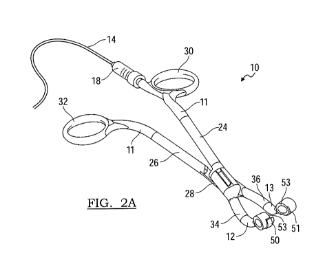
[0033] Figures 2a-2c are perspective views of cautery forceps embodying the
principles of the
present invention;
[0034] Figure 3 is a perspective, enlarged view of the end of the cautery
forceps seen in Figures
2a-2c;
[0035] Figure 4 is an exploded perspective view an end effector as utilized
in with the cautery
forceps seen in Figure 2a-2c;
[0036] Figure 5 is a diagrammatic illustration of the end effectors of the
cautery forceps
positioned at the interface of the oral and nasal cavities and adjacent to the
soft palate and adenoids as
may be positioned during use;
[0037] Figure 6 is a perspective illustration of an alternative embodiment
of cautery forceps
embodying the principles of the present invention;
[0038] Figure 7 is a perspective illustration of a further embodiment of
cautery forceps
embodying the principles of the present invention;
[0039] Figure 8 is an inverted, perspective illustration of an additional
embodiment of cautery
forceps embodying the principles of the present invention;
[0040] Figure 9 is a perspective illustration of the embodiment of Figure 8
showing the
underlying frame members of the construction, also in an inverted position;
[0041] Figure 10 is an enlarged view of the frame members seen in Figure 9;
and
[0042] Figure 11 is an enlarged view of the cautery forceps of Figure 8,
turned over and seen
from the opposing side thereof.
DETAILED DESCRIPTION
[0043] The present invention provides an electrosurgical instrument for
removal of tissue. While
those skilled in the art will appreciate that instruments incorporating the
principles of the present
invention are suitable for use in removing multiple types of tissue, described
herein are instruments for
4
CA 02907613 2015-09-18
WO 2013/155354 PCT/US2013/036253
removal of tonsil tissue, such as the palatine tonsils (commonly referred to
as just the "tonsils") and,
more particularly, pharyngeal tonsils (commonly referred to as the
"adenoids"). For simplicity, the
discussion that follows simply refers to the removal of the adenoids.
[0044] Additionally, various individual features are presented in the
embodiments. Unless
specifically stated otherwise, individual features of one embodiment are
understood to be employable
with another embodiment, either with all the features of that other embodiment
or with less than all the
features of that other embodiment.
Overview
[0045] Referring now to Figures 2a-2c and 3, illustrated therein, and
designated at 10, is an
electrosurgical instrument in accordance with the principles of the present
invention. As seen in these
figures, the electrosurgical instrument 10 may generally be described as being
in the shape of a pair of
forceps. Unlike common forceps, however, the surgical instrument 10,
hereinafter referred to as
cautery forceps 10, includes a pair of handles 11 to which are
mounted/connected a pair of removable
or releasably attached end effectors 12, 13. Additionally, the cautery forceps
10 include a means 14,
such as a power cord, by which at least one of the end effectors 12, 13 can be
connected to a suitable
electrical power source. Additional embodiments of the cautery forceps are
seen in Figures 6-11.
Power Source
[0046] Preferably, the power source is an available source of power located
in the room where the
cautery forceps 10 are to be used. As such, the power source includes the
required componentry
needed to provide the proper voltage, current and frequency for electrocautery
surgery. Generally,
electrocautery requires a frequency in the radio frequency range, above 100
KHz and up to 5 MHz.
This power source itself can be provided as an integrated system such that the
power cable 14 is
merely plugged into an outlet (not show) in the room. Alternately, the power
source for any of the
disclosed embodiments can be provided as a stand-alone power system located in
the operating room
or as a battery incorporated into the electrosurgical instrument.
Unipolar and Bipolar
[0047] The cautery forceps 10 according to the present invention are
preferably unipolar (also
known as monopolar) in their construction. In such a construction, the cautery
forceps 10 themselves
include a single electrode in one of the end effectors 12, which is further
discussed below. During use,
current flows from the electrode, through the patient, and to a return
electrode affixed elsewhere on the
patient's body.
[0048] In an alternative embodiment, the cautery forceps 10 may be of a
bipolar construction. In
a bipolar construction, a second electrode is provided on the other end
effector 13. During use of the
bipolar cautery forceps 10, electrical current passes primarily from one
electrode on one end effector
CA 02907613 2015-09-18
WO 2013/155354 PCT/US2013/036253
to the second electrode on the other end effector. The current thus passes
primarily through a
localized portion of the affected tissue of the patient, which is located
between the electrodes, and in
contrast to flowing through the body of the patient to a remotely located
electrode in the unipolar
construction.
Handles
[0049] The handles 13 of the cautery forceps 10 in the illustrated
embodiment are provided in
conjunction with two lever members 24, 26 that are arranged such that the
cautery forceps 10 operate
in a traditional, scissors-like construction. In such a construction, the two
lever members 24, 26 cross
one another at a central pivot joint 28, which includes pivot axle. The
handles 11 may be provided
with finger rings or grips 30, 32 at one end, while the lever members 24, 26
terminate in mounting tips
34, 36 at the opposing end. By bringing the finger grips 30, 32 together, the
opposing mounting tips
34, 36 are likewise brought toward one another.
[0050] Preferably, the lever members 24, 26 are solidly constructed of a
metal material, such as
surgical grade stainless steel, aluminum or another conductive material.
Constructed in this manner, at
least one of the lever members 24, 26 has the benefit that the lever member
itself operates as a
conductor to transmit current through the cautery forceps 10, as further
discussed below. The pivot
joint 28 between the two lever members 24, 26 is constructed such that the two
members 24, 26 are
electrically isolated from one another by way of an insulating bushing (not
shown) or other feature.
Failure to electrically isolate the two lever members 24, 26 would result in
the shorting of the
electrical circuit and would render the end effectors 12, 13, which are
themselves further discussed
below, as inoperative in certain respects.
[0051] The finger rings 30, 32 of the handles 11 may be formed as a
traditional ringed portion so
as to facilitate the grasping of the cautery forceps 10 and the manipulation
thereof by the surgeon.
Alternatively, they may be provided with other shapes. As such, the handles 11
and lever members 24,
26 may be unitarily formed with each other.
[0052] The handles 11 and lever members 24, 26 are electrically isolated
from the surgeon by
covering the handles 11 and lever members 24, 26 with an insulative material.
Various means by
which the handles 11 and lever members 24, 26 may be covered are envisioned
and include dip
coating or spray coating with rubber or plastisol or overmolding of the
handles 11 and lever members
24, 26 with a polymer material, such as polycarbonate, ABS, HDPE, acrylic, or
other material having
the appropriate insulative characteristics. The material covering the handles
11 and lever members 24,
26 should also facilitate grasping of the handles 11 and minimize potential
slipping of the cautery
forceps 10 when held and in use by the surgeon.
6
CA 02907613 2015-09-18
WO 2013/155354 PCT/US2013/036253
[0053] The previously mentioned power cable 14 is configured at one end
with an electrical
connector 16, suitable for making the required electrical connection with the
electrical power source.
The opposing end of the power cable 14 is also provided with an electrical
connector 18. This latter
electrical connected 18 matingly engages a receptacle terminal 20 associated
with one of the lever
members 24, 26 and/or the handles 11. As such, the connectors 16, 18 on each
end of the power cable
14 are preferably one of a plug or receptacle terminal, such as a banana-plug
or a multiple pronged RF
surgical plug, or other connector suitable for a robust connection and
electrical power supply.
[0054] In the illustrated embodiment, the terminal 20 is formed adjacent to
the hand grip 30
associated with the lever member designated at 24. While the terminal 20 is
illustrated as being
located adjacent to the ring portion of the hand grip 30, generally on the
inward side thereof, it will be
understood that the terminal 20 could be located elsewhere on the hand grip
30, the handle 11 or the
lever member 24. Additionally, the terminal 22 may alternatively be attached
to the lever member 24
via a suitable connection or it may be insert molded during the formation of
the lever member 24.
End Effectors
[0055] The end effectors 12, 13 are seen in Figures 2a-5 and each has the
same general
construction, being principally composed of an arm portion 40 and a tip
portion 42, with the tip
portion 42 being formed at a distal end of the arm portion 40. The arm portion
40 is curved along its
length and the tip portion 42 is formed generally in an ovoid ring shape. The
curvature and length of
the arm portion 40 is such that it facilitates the positioning of the tip
portion 42 in the vicinity of the
adenoids, as seen in Figure 5, so as to enhance the ease with which the
adenoids are accessed and the
ease with which the cautery forceps 10 are manipulated during the surgical
procedure. The length and
curvature of the arm portions 40 may be provided in a variety of
configurations, including having
straight portions, so as to accommodate differing patient anatomies and the
preferences of the
surgeons. Generally, however, the curvature of the arm portions 40 is such
that during use and entry
into the patient's oral cavity, the curvature of the arm portions 40 is
directed upward toward at least
the ends of the arm portions 40.
[0056] In all of the embodiments, the end effectors 12, 13, and
particularly the arm portions 40,
but also the tip portions 42, may be rigid or may be bendable. If bendable,
they should only be
bendable only to such a degree to allow the surgeon some amount of
adjustability to accommodate
variations in patient anatomy. They should not be so bendable that they
undergo bending during use
by the surgeon. This offers the benefit of allowing slight re-configuration of
the end effectors 12, 13
by the surgeon without enabling further bending during the actual surgical
procedure.
[0057] The oval ring shape of the tip portions 42 of the end effectors 12,
13 is enlarged relative
to the arm portions 40 and assists in the removal of the adenoids. On one side
of the ring shape, which
7
CA 02907613 2015-09-18
WO 2013/155354 PCT/US2013/036253
is the inward, lower side seen in Figures 2a-3, the tip portions 42 are
provided with cutting and
opposing portions 50, 51. The other side of the ring shape, the inward upper
side in Figures 2a-3 and
toward only one lateral side of the cutting and opposing portions 50, 51,
gripping portions 53 are
provided, which function as a mechanism by which the end effectors can grasp
the tissue that is being
cut and cauterized. This grasping of the tissue by the end effectors 12, 13 on
only one side of the
cutting portion 50 allows for the removal of the cut tissue from the procedure
site, which would be
prevented if gripped on both sides of the cutting portion 50. While shown with
an ovoid ring shape,
the tip portions 42 of the end effectors 12, 13 may have alternative shapes,
such as a recti-linear, a
curvilinear or other shape.
[0058] Like the arm portions, the length and width of the ovoid ring shapes
of the tip portions 42
may vary. By varying the shape of the tip portions 42, a variety of sized and
shaped end effectors 12,
13 can be provided in kit form, allowing a surgeon to readily select, during
the procedure, the end
effector 12, 13 with a configuration that works best with a particular
patient's anatomy.
[0059] The end effectors 12, 13 do, however, differ in one regard.
Specifically, one end effector
12 includes a conductive insert 44 that extends through the arm and tip
portions 40, 42 and operates as
an electrode for cautery purposes. In one embodiment, the other end effector
13 may be formed with
an insert 45 that is similar, but does not operate as the electrode. In
another embodiment, the other end
effector 13 may be formed with a partial insert in the tip portion 42 or
without any insert in the end
effector 13. The insert 44 operating as the electrode is best seen in the
exploded view of Figure 4.
Hereafter, when referring to characteristics common to both inserts, the
inserts are collectively referred
to as "inserts"; and when referring characteristics relating to only the
insert operating as an electrode,
the insert is referred to as "insert/electrode."
[0060] One end of the insert/electrode 44 defines an electrical contact 52
that is ultimately
coupled to the power source. In one embodiment, the coupling of the electrical
contact 52 to the
power source is achieved during the mounting of the end effector 12 on the
mounting tip 34. In the
instance where the lever member 24 serves as the conductor through the handle
22, the electrical
contact 52 engages the mounting tip 34 directly when the end effector 12 is
attached thereto. In an
alternative construction, the lever member 24 might not serve as the conductor
through the handle 22.
In such an instance, the conductor associated with the handle 22 may be in the
form of a lead (see
Figure 6, for example) embedded within or mounted along the lever member 24,
and terminating in a
terminal. The electrical contact 52 would in that instance electrically engage
the terminal when the
end effector 12 is connected to the mounting tip 34.
[0061] Provided in the tip portions 42 of the end effectors 12, 13 are the
means by which the
adenoids are removed. These means are oriented so that they generally oppose
one another. In a
8
CA 02907613 2015-09-18
WO 2013/155354 PCT/US2013/036253
preferred embodiment, a cutting portion 50 defined by the insert/electrode 44
is conductive and further
defined as a curved blade. An opposing portion 51 in the tip portion 42 of the
other end effector 13 is
non-conductive and is defined by the body of the end effector 13 so as to form
a bearing surface 55,
such as an anvil, a pocket, a flat surface or free space, or as an insert in
the body, shaped so as to
correspond to the shape of the cutting portion 50. In an alternative
embodiment, the opposing portion
51 of the other end effector 13 may also be provided as a blade, defined by an
insert and be oriented
with its edges aligned along a common cutting plane, so that the blades/edges
of both portions 50, 51
abut one another when the handles 22 are brought together. Alternatively, the
blades/edges of the
portions 50, 51 may be slightly off-set relative to one another so as to be
able to by-pass each in close
proximity and shear the tissue during tissue removal; or the blades/edges of
the portions 50, 51 could
abut one another at their adjacent cutting edges, but be out of plane with
each other, so as to prevent
inadvertent contact by the body of the blade with tissue near the surgical
site. In one additional
embodiment, the portions 50, 51 could be provided such that they initially
engage one another, but that
with additional force, they slip or snap passed one another.
[0062] As shown in Figure 3, the blade of the cutting portion 50 of the
insert/electrode 44 is a
curved planar body and may be provided with a sharpened leading edge 54 that
interacts with the
correspondingly or congruently shaped bearing surface 55 of the opposing
portion 51 of the other end
effector 13, or a sharpened edge thereof, as mentioned above. Alternatively,
for this embodiment and
each of the other embodiments, the blade of the cutting portion 50 could be
preferably provided in the
form of a rigid wire instead of a planar body. In the various embodiments of
the present invention, the
conductive area of the blade is beneficially minimized thereby concentrating
electrical energy
(increasing the power density for a given wattage) and producing a high
quality and consistent cut
through the tissue. For example, it has been found that a wire having a
diameter of up to about lmm
(0.039 inches) is preferred, with 0.5mm (0.020 inches) or less being more
preferred. At greater
diameters, and therefore greater exposed surfaces areas in contact with the
tissue (whether the blade is
provided as a wire or planar body), inconsistent cuts have been observed.
Maximum exposed surface
area has been found to be preferably 0.013 sq. in. per watt or less (cutting
power can range from 1-100
watts, but are typically between 20-40 watts) to provide a consistent cut
through the tissue. Minimum
thicknesses for the wire tend to be dictated by the tensile strength of the
wire.
[0063] The curvature of the cutting portion 50 is such that it facilitates
removal of the adenoid
tissue. More specifically, the radius of curvature of the cutting portion 50
is less than the radius of
curvature of the bands of the adenoid tissue. This allows the end sections of
the cutting portion 50 to
be positioned just outside of the tissue while the intermediate portion of the
cutting portion 50
(between the end sections) engages the tissue. As a result, tissue can be cut
and cauterized and the
9
CA 02907613 2015-09-18
WO 2013/155354 PCT/US2013/036253
surgical instrument can be removed from the surgical site, or current surgical
pass, without requiring
that the jaws of the instrument be opened.
[0064] Additionally, it is noted that while the cutting portion 50 and
opposing portion 51 move
toward one another during the surgical procedure, cutting is not principally
effectuated by compressive
pressure on the tissue between these components. Rather, the opposing portion
51 prevents movement
or holds the tissue in position or directs the tissue toward the cutting
portion 50, the latter of which
effectuates cutting as a result of the energy being provided thereto.
[0065] In the present embodiment, cutting and cautery of the adenoids are
performed by the same
component, the cutting portion 50. To this end, adjacent to the blade and
leading edge 54, the cutting
portion 50 may be provided with a cautery portion 57. The cautery portion 57
defines a width
extending in a direction away from the leading edge 54 and is unitarily formed
as part of the blade.
Thus, immediately after tissue is dissected from the surgical site by the
leading edge 54 of the cutting
portion 50, the cautery portion 57 is brought into contact with the dissection
site and the remaining
tissue cauterized. The wire form of the cutting portion 50 discussed above may
be provided and used
either with or without an associated cautery portion 57. If no separate
cautery portion 57 is provided,
the wire form of the cutting portion 50 itself performs the cautery.
[0066] As seen from the above, cautery is performed or effectuated in a
direction other than the
direction of applied pressure for cutting or movement of the cutting portion
50 as the end effectors are
brought together. This is beneficial in that the resulting surgical instrument
does not require clamping
of tissue to perform cautery and allows for removal of tissue and the
instrument without requiring
unclamping or opening of the jaws/end effectors of the surgical instrument.
[0067] The cutting and opposing portions 50, 51 may also optionally be
provided with a non-stick
coating, or other technology, on their surfaces to prevent adhesion and build-
up of tissue on the
surfaces of the cutting portions 50, 51.
[0068] In the above described and all embodiments, cautery is
simultaneously effectuated by the
same component that performs cutting of the adenoid, namely the cutting
portion 50. The cautery and
cutting functions could, however, be performed by separate components. For
example, the cutting
portion of the insert/electrode of the embodiments may be replaced with
separate cutting and cautery
blades (non-cutting) in the tip portions. The cautery blade, in that instance,
would be coupled to the
electrical contact and the power source. To sever the adenoids, the separate
cutting blade would be
provided adjacent to the cautery blade, preferably at a location radially
inward on the ovoid ring
shaped tip portion. Provided in this manner, cautery would be performed as a
separate function, but
concurrently with removal of the adenoids via the cutting blade.
CA 02907613 2015-09-18
WO 2013/155354 PCT/US2013/036253
[0069] The insert/electrode 44 of the end effector 12 is preferably insert
molded within rigid
plastic or another material so that only the cutting portion 50 and the
electrical contact 52 are exposed.
Insert molded in this manner, inadvertent contact is minimized between other
portions of the
insert/electrode 44 and any tissue that is not the subject of the surgical
procedure. As an alternative to
insert molding, the insert/electrode 44 could be inserted into previously
formed arm and tip portions
40, 42 of the end effector 12. This construction may be employed in all
embodiments.
[0070] The insert 45 of the other end effector 13, if provided with an
insert, may similarly be
insert molded within the end effector 13 or subsequently inserted into the end
effector 13 after
molding thereof. If no insert is provided in the end effector 13, the inward
side of the tip portion 42 is
formed in the desired shape of the anvil or blade, depending on the particular
configuration of the end
effector 13 as described above.
[0071] The end effectors 12, 13 are respectively engaged with and mounted
to the mounting tips
34, 36 of the lever members 24, 26. In order to mount the end effectors 12, 13
to the handles 22 and
lever members 22, 24 of the cautery forceps 10, mounting ends 46, 48 of the
end effectors 12, 13, are
formed so as to matingly engage with the mounting tips 34, 36 of the lever
members 22, 24 . This
engagement between the mounting tips 34, 36 of the lever members 22, 24 and
the mounting ends 44,
46 of the end effectors 12, 13 is preferably a detachable or releasable
engagement. As such, the
engagement may be a press-fit or snap-fit engagement where a portion of the
end effector 12, 13 is
displaced as it passed over a corresponding portion of the mounting tips 34,
36, and then resiliently
snaps back into substantially it original position. Also, a
twist/screw/threaded engagement, a keyed
engagement, a magnetic engagement or a positive locking or latching
construction can be provided. In
the illustrated construction of all of the figures, a snap-fit engagement is
provided wherein a portion of
the mounting tips 34, 36 is matingly received within a hollow portion of the
mounting ends 44, 46.
Alternative Basic Constructions
[0072] Two alternative constructions of cautery forceps according to the
principles of the present
invention are shown in Figures 6 and 7. In Figure 6, a monopolar construction
is shown wherein
current is provided to the conductive end effector via an external lead. In
Figure 7, a bipolar
construction is generally illustrated.
[0073] Referring to bipolar construction of Figure 7, the cautery forceps
110 have a construction
is similar to that discussed above in connection with the embodiment of
Figures 2a-3. The bipolar
construction differs from the unipolar construction in that both end effectors
112 are conductive and
include electrodes for cautery purposes. As a result, each end effector 112 is
therefore connected to
the power source through its respective the lever member 124, such as with a
power cable 114 having
terminals 118 for connection to the handles 111 and a plug 116 for connecting
to a power source.
11
CA 02907613 2015-09-18
WO 2013/155354 PCT/US2013/036253
While they may be the same, the cutting and opposing portions 150 of the end
effectors 112 also do
not need to be identical. They may be appropriately varied as discussed above
in connection the end
effectors 12, 13 of the prior embodiments. Attention is therefore directed to
that section of this
description.
[0074] In Figure 6, the means by which the end effectors 212 are connected
to the power source
is via an external, insulated electrical lead 260. The insulated lead 260 is
connected to and supported
along the length of the cautery forceps 210. Specifically, the lead 260 is
directly connected with the
end effectors 212 and does not utilize the lever members 224, 226 of the
cautery forceps 210 as a
means to electrically couple the power source to the end effectors 212. The
lead 260 itself may form a
power cable 214 that is connected to the power source, as illustrated, or may
be joined with such a
cable 214 at a plug connection.
[0075] In supporting the leads 260 on the cautery forceps 260, the leads of
the cable may be
extended through a series of retainers integrally formed on the lever members
224, 226. The retainers
may be of any desired shape, e.g. a rectangular shape, an annular ring shape
or a C-shape. With a side
opening, such as in a C-shaped retainer, the lead 260 may be snapped or
pressed into the retainer as
opposed to being threaded through an opening formed in an enclosed retainer.
The retainers therefore
may be rigidly or resiliently formed and are sized to positively engage the
exterior surface of a lead
and retain it positioned therein. The retainers themselves are not illustrated
in Figure 6.
[0076] When the cautery forceps 210 are constructed in the manner seen in
Figure 6, the retainers
are positioned so as to maintain the lead adjacent to the lever member 224, in
an unobtrusive manner,
and along the length of the lever member 224. The lead may be position on
either side (top and
bottom) of the lever member 224
[0077] Alternatively, in a bipolar embodiment, each of the two leads may
follow the length of its
own lever member 224, 226 until engaging the respective end effector 212
mounted thereto. The two
leads may alternatively follow one lever member 224, 226 and connect to the
end effector 212
associated with the other lever member 224, 226, thereby avoiding the
necessity of having the leads
260 cross over one another in the region of the pivot joint 228. In any of the
alternative constructions,
the retainers 262 should prevent the leads 260 from loosely hanging and should
minimize potential
interfere by the leads 260 with the surgeon during the surgical procedure.
[0078] As an alternative to the scissor-like construction described in the
previous embodiments, it
should be apparent that the handles could be provided with a tweezers-type
construction (a
construction where the handles are generally U-shaped or V-shaped and gripped
in front of the pivot
connection of the lever members and behind the end effectors). The various
features of the above
discussed embodiments, individually or collectively, could accordingly be
applied to the tweezers-type
12
CA 02907613 2015-09-18
WO 2013/155354 PCT/US2013/036253
embodiment, or any of the embodiments discussed herein. As a further
embodiment, the construction
of the cautery forceps could be such that the end effectors are provided as a
set of jaws on one end of a
shaft, tube or hand piece and operated via manipulation of a trigger mechanism
on the other end, as in
an endoscope type construction or a pistol grip type of construction.
[0079] With all of the above described constructions, the cautery forceps
10, 110, 210 incorporate
a reusable handle, after appropriate sterilization. The end effectors
themselves may be of a disposable
nature or maybe constructed so as to allow for sterilization and reuse.
Additionally, the end effectors
may be provided, in a variety of sizes and curvatures so as to facilitate
their use with different patients
and to accommodate a range of different anatomical variations in those
patients. As such, the end
effectors may be offered individually, either with or without the handles, or
maybe offered in a kit
format whereby a set of different end effectors, are provided either with or
without the handles.
[0080] Seen in Figures 8 and 9 is an additional construction of cautery
forceps 310 embodying
the principles of the present invention. The cautery forceps 310 of this
embodiment are unipolar in
their construction, like some of the earlier described embodiments, but do not
include detachable end
effectors. Rather, the end effectors 312, 313 are non-detachably formed with
handles 311 and lever
members 324, 326 of the forceps 310. Constructed in this manner, the entire
cautery forceps 310 may
be of a disposable nature or of a reusable nature, after sterilization,
depending on their specific
construction.
[0081] Like the prior embodiment, the handles 313 of the cautery forceps
310 operate with the
two lever members 324, 326 in a traditional, scissors-like construction. The
two lever members 324,
326 cross one another at a central pivot joint 328 defined by a pivot axle 329
Each handle 311
includes a finger grip 330, 332 in the form of a ring. By bringing the finger
grips 330, 332 together,
the end effectors 312, 313 are likewise brought toward one another enabling
them to affect the desired
tissue of the patient.
[0082] The handles 311, lever members 324, 326 and end effectors 312, 314
are formed by
overmolding underlying, rigid frame members 325, 327, which are seen in Figure
9. The frame
members 325, 327 themselves can be constructed of a variety of materials, so
long as they impart
sufficient strength to the cautery forceps 310 for the surgical procedure to
be performed. As such, the
materials for the frame members 325, 327 can be a metal material, such as
surgical stainless steel,
aluminum or another material formed by stamping, cutting, grinding, casting or
forging. However, at
least one frame member, hereafter the active frame member 325, is constructed
from an electrically
conductive material so as to be able to transmit electric current through the
cautery forceps 310, as
further discussed below.
13
CA 02907613 2015-09-18
WO 2013/155354 PCT/US2013/036253
[0083] The pivot joint 328 between the frame members 325, 327 is
constructed such that it
electrically isolates the frame members 325, 327 from one another,
particularly if both frame members
325, 327 are formed of metal, and the joint may utilize an insulating bushing.
[0084] The overmolding of the frame members 325, 327 electrically isolates
them, and in
particular the active frame member 325, from the surgeon. While various
techniques may be
employed to form the overmold, including those discussed above, it is
preferred that overmold is
formed by insert/injection molding. The exterior surface of the overmold can
be textured or smooth,
but should facilitate grasping of the handles 311 and finger grips 330, 332,
thereby reducing the
potential for the cautery forceps 310 to slip in the grasp of the surgeon.
[0085] In this embodiment, and alternatively in the prior embodiments, the
power cable 314,
which is a single strand standard coated RF surgical cable, and may be
permanently attached to one
end of the active frame member 325 by soldering, prior to overmolding. The
opposing end of the
cable 314 includes an accessory plug 316 or a standard three prong surgical
plug.
[0086] The end effectors 312, 313 have the same general construction and
attributes mentioned
above, the discussion of which is incorporated by reference, other than being
removable, and are
principally composed of an arm portions 340 and a tip portion 342, 343, with
the latter being formed at
the distal end of the arm portions 340. Likewise as previously discussed, the
arm portions 340 may
be bendable to allow adjustment by the surgeon to accommodate variations in
patient anatomy.
[0087] As with the prior embodiments, the tip portions 342, 343 are
illustrated as having an oval,
ring shape to assists in the removal of the adenoids. On the inward and lower
side of one of the tip
portions 342 is a cutting portion 350. The cutting portion 350 is integrally,
and preferably unitarily,
formed as an end part of the active frame member 325 and is not fully over
molded like other portions
of the frame member 325. As such, the cutting portion 350 forms an exposed
portion of the active
frame member 325 and operates as the conductive electrode of the cautery
forceps 310. The cutting
portion 350 may therefore include a blade with a sharpened edge, optionally
provided with a non-stick
coating, and a cautery blade having an exposed width for cautery purposes. The
cutting portion 350
may alternatively include the wire form of the cutting portion discussed
above.
[0088] The opposing tip portion 343 preferably does not include a cutting
portion integrally
formed with the frame member 327. Rather, the cutting portion 351 of this tip
portion 343 opposes the
cutting portion 350 of the active frame member 325 and is preferably formed
and defined by the
material of the overmold. The cutting portion 351 may define a bearing surface
opposing the cutting
portion 350 or an edge/blade interacting with the cutting portion 350, as
discussed in connection with
any of the prior embodiments.
14
CA 02907613 2015-09-18
WO 2013/155354 PCT/US2013/036253
[0089] As with the prior embodiments, the upper side of ring shape of the
tip portions 342, 343
operate as a mechanism by which the end effectors 312, 314 can grasp the
tissue that is being cut,
cauterized and removed from the procedure site. While again shown with an
ovoid ring shape, the tip
portions 342, 343 of the end effectors 312, 313 may have alternative shapes,
lengths and widths.
Method of Use
[0090] When using the unipolar variations of the cautery forceps,
electrical current is delivered to
the end effector having the insert/electrode. The cutting portion on the end
effector is then brought to
bear against the adenoid tissue that is to be excised. As the adenoid tissue
is being contacted, current
flows from the cutting portion of the insert/electrode, through the adenoid
tissue, and out of the
patient's body at another electrode that has been attached in a remote
location apart from the adenoid
tissue. With this passage of current, radio¨frequency energy is applied to
tissue, heating the water in
the local tissues. The heating of the water inherent in the tissue results in
a weakening and/or severing
of the tissue, allowing removal of the tissue and simultaneous cauterizing of
the removal site.
Removal of the tissue is principally effectuated by this release of energy
through the tissue and not as
a result of the compression of tissue between the cutting and opposing
portions.
[0091] In the bipolar constructions, the return path for the electric
current is defined by the
opposing end effector when it is brought near the other end effector. The
electrical current will travel
through the subject tissue from one end effector to the other. Accordingly,
during the cutting and
removal of the adenoid tissue, the cutting portion of the end effector not
only effectuates removal of
the subject tissue, but also cauterizing of the remaining tissue at the
removal site.
[0092] To electrically actuate the end effectors of any of the embodiments,
a switch may be
provided in-line with the power cable. The switch may be in the form of a hand
or foot operated
switch, or it may be provided as part of the power source. In a further
embodiment, the end effectors
might be automatically energized when brought into close proximity of one
another and therefore not
require manipulation of a switch per se. This automatic energizing could be
achieved via proximity
sensors or limit switches provided as part of the cautery forceps or simply by
completing the
conductive circuit through the tissue.
[0093] As a person skilled in the art will readily appreciate, the above
description is meant as an
illustration of implementation of the principles this invention. This
description is not intended to limit
the scope or application of this invention in that the invention is
susceptible to modification, variation
and change, without departing from spirit of this invention, as defined in the
following claims.