Note: Descriptions are shown in the official language in which they were submitted.
CA 02907865 2015-09-23
WO 2014/167323 PCT/GB2014/051105
1
SINGLE NUCLEOTIDE DETECTION METHOD
This invention relates to a method for characterising polynucleotides such as
those
derived from naturally occurring RNA or DNA by capturing and detecting an
ordered sequence of
single nucleotide bases generated therefrom by progressive pyrophosphorolysis.
Next generation sequencing of genetic material is already making a significant
impact on
the biological sciences in general and medicine in particular as the unit cost
of sequencing falls in
line with the coming to market of faster and faster sequencing machines. Thus,
in one such
machine, a double-stranded DNA analyte is first broken down into a plurality
of smaller
polynucleotide fragments each of which is first adenylated on both ends of one
strand so that a
single-stranded first oligonucleotide can be bound to both ends of its
compliment by hybridisation
to the unpaired adenine base. The treated fragments so obtained are then size-
selected and
captured on a surface coated with bound single-stranded second
oligonucleotides which
themselves are the sequence compliment of the first so that in effect a
library of surface-bound
double-stranded fragments can be created by further hybridisation. In a
subsequent clustering
step, these library components are then clonally amplified millions of times
on the surface using
extension and isothermal bridging reactions to utilise unused second
oligonucleotides. This, in
effect, creates a dense concentration of the polynucleotide fragment bound to
the surface
through one of its strands. The unbound complimentary strand of each fragment
is then removed
to leave bound single-stranded fragments ready for sequencing. In the
sequencing stage, each of
these single-stranded fragments is primed and its complimentary strand
recreated by extension
using the polymerase chain reaction and a mixture of the four characteristic
nucleotide bases of
DNA in dideoxynucleotide triphosphate (ddNTP) form. Each ddNTP type is end-
blocked with a
moiety which is labelled with a different fluorophore fluorescing at a
different wavelength. The
extension reaction then takes the form of a cycle of three steps; first the
relevant ddNTP is
incorporated into to the growing strand; secondly the nucleotide base it
contains is identified by
illuminating the sample and detecting the wavelength of the fluorescence and
finally the end
block and its associated fluorophore are removed to allow the next extension
event to occur. By
this means, the sequence of the complimentary strand can be built up base-by-
base. It will be
appreciated that, whilst this approach can be highly automated and can
generate sequence reads
of high accuracy, its speed of operation is limited by the rate of the
extension cycle. Thus, in
practice, use of the technology tends to involve parallel processing of
relatively short
polynucleotide fragments and assembly of the whole sequence from the various
reads obtained
CA 02907865 2015-09-23
WO 2014/167323 PCT/GB2014/051105
2
therefrom. This in itself can lead to computational complexities and the
potential introduction of
errors.
More recently efforts have been made to develop direct sequencing methods. For
example, WO 2009/030953 discloses a new fast sequencer in which inter alio the
sequence of
nucleotide bases or base pairs in a single- or double-stranded polynucleotide
sample (e.g.
naturally occurring RNA or DNA) is read by translocating the same through a
nano-perforated
substrate provided with plasmonic nanostructures juxtaposed within or adjacent
the outlet of the
nanopores. In this device, the plasmonic nanostructures define detection
windows (essentially an
electromagnetic field) within which each nucleotide base (optionally labelled)
is in turn induced to
fluoresce or Raman scatter photons in a characteristic way by interaction with
incident light. The
photons so generated are then detected remotely, multiplexed and converted
into a data stream
whose information content is characteristic of the nucleotide base sequence
associated with the
polynucleotide. This sequence can then be recovered from the data stream using
computational
algorithms embodied in corresponding software programmed into a microprocessor
integral
therewith or in an ancillary computing device attached thereto. Further
background on the use of
plasmonic nanostructures and their associated resonance characteristics can be
found in for
example Adv. Mat. 2004, 16(19) pp. 1685-1706.
Another apparatus for fast sequencing polynucleotides is described, for
example, in US
6627067, US 6267872 and US 6746594. In its simplest form, this device employs
electrodes,
instead of plasmonic nanostructures, to define the detection window across the
substrate or in or
around the outlet of the nanopore. A potential difference is then applied
across the electrodes
and changes in the electrical characteristics of the ionic medium flowing
therebetween, as a
consequence of the electrophoretic translocation of the polynucleotide and
associated electrolyte
through the nanopore, is measured as a function of time. In this device, as
the various individual
nucleotide bases pass through the detection window they continuously block and
unblock it
causing 'events' which give rise to characteristic fluctuations in current
flow or resistivity. These
fluctuations are then used to generate a suitable data stream for analysis as
described above.
The generation of stable droplet streams, especially microdroplet streams, is
another
developing area of technology that already has applications in molecular
biology. For example,
US7708949 discloses a novel microfluidic method for generating stable water
droplets in oil whilst
for example U52011/0250597 describes utilisation of this technology to
generate microdroplets
containing a nucleic acid template (typically a polynucleotide DNA or RNA
fragment) and a
plurality of primer pairs that enable the template to be amplified using the
polymerase chain
CA 02907865 2015-09-23
WO 2014/167323 PCT/GB2014/051105
3
reaction. Other patent applications relating to the field generally include
JP2004/290977,
JP2004/351417, U52012/0122714, U52011/0000560, US2010/01376163, US2010/0022414
and
U52008/0003142.
WO 2004/002627 discloses a method for creating liquid-liquid and gas-liquid
dispersions
using various devices comprising creating a discontinuous section between
upstream and
downstream microfluidic regions. However its application to single nucleotide
DNA sequencing is
not taught.
WO 2010/077859 teaches a droplet actuator comprising a substrate provided with
electrodes, a reactor path and nucleotide base, wash-buffer, sample and enzyme
reservoirs.
Whist the actuator is generically said to be useful for the amplification and
sequencing of nucleic
acids, there is no teaching of the analyte degradation method we describe
below. Rather, it is
concerned with a completely different approach; observing the synthesis of a
complimentary
strand of the analyte using pyrosequencing. US 2009/0280475 is concerned with
similar subject-
matter.
Biological probes, which typically comprise single-stranded oligonucleotides
of known
sequence order less than 1000 nucleotides long, are widely used in analytical
molecular biology.
Such probes typically work by attaching themselves to the target (for example
one derived from
the DNA of a naturally-occurring pathogen) when sufficient sequence
complimentarity exists
between the nucleotide bases of the probe and the target. Typically the
nucleotides of such
probes are labelled with detectable elements such as radioactive or
fluorescent markers so that
when the probe is used to treat an analyte solution or substrate in or on
which the target is
thought to have been captured, the presence or absence of the target is
revealed by searching for
and detecting the detection element's characteristic detection property.
One class of such probes is represented by materials known in the art as
'molecular
beacons' as for example described in U58211644. These probes are comprised of
single-stranded
oligonucleotides which have been in effect folded back onto themselves to
create a residual
single-stranded loop which acts as the probe's sensor and a short stem where
the nucleotides
adjacent the two ends are bound to each other through complimentary nucleotide
base pairing;
thereby creating a double-stranded region. This arrangement, which can be
likened to a hairpin
in which the single-stranded loop is attached to complimentary strands of the
same end of a
notional double-stranded oligonucleotide, is highly strained. To the free 3'
and 5' ends of the
oligonucleotide (now adjacent to one another and at the remote end of the
stem) are respectively
attached a fluorophore and a quencher. Their geometric proximity to each other
then ensures
CA 02907865 2017-01-18
4
that no significant fluorescence occurs. In use, the target binds to the
single-stranded loop causing
additional strain so that when the probe is heated the stem unzips causing
distancing of the
fluorophore and quencher and allowing the former to fluoresce.
We have now developed a new sequencing method which in one embodiment involves
generating a stream of nucleotide bases whose ordering is characteristic of
the sequence in the
analyte by progressive degradation of the analyte; and a subsequent capture of
each nucleotide
base in a way which enables it to be detected.
WO 94/18218 discloses a genome sequencer in which an ordered stream of single
nucleotides is separated from an analyte and thereafter contained in a
fluorescent-enhancing
solid matrix where each nucleotide is excited using a laser and its
characteristic spectroscopic
emission detected. The single nucleotide transfer method used by this
sequencer involves
creating a single dual-sheath of flowing immiscible liquids rather than a
series of droplets.
Furthermore, the sequencer described seeks to detect the single nucleotides
directly rather than
employing a capture system and fluorophore release method of the type we
describe. We believe
that this is a drawback as it will lead to signal-to-noise ratio problems when
the emission come to
be detected. This will compromise the overall sensitivity and therefore
practical applicability of
the sequencer itself.
Stephan et al Journal of Biotechnology 86 (2001) pp. 255-267 teaches a general
method for
counting single nucleotides generated by exonucleolytic degradation of an
immobilised DNA
sample labelled with fluorophores. However no information is provided about
differentiating
between the different single nucleotide types generated.
The use of the progressive exonucleolytic degradation of polynucleotides to
generate a
stream of single nucleotide bases has been disclosed in schematic form
although little information
about the actual methodology employed is provided. Furthermore, WO 03/080861
describes a
sequencing method in which a DNA analyte is sequentially degraded to an
ordered stream of
single nucleotides by means of pyrophosphorolysis carried out in the presence
of a
pyrophosphate anion labelled with an intelligent dye. In one example the
pyrophosphate anion is
labelled with the dye JF-4 which has differing fluorescent lifetimes depending
on the particular
nucleotide type to which it is attached. The stream of labelled single
nucleotides is then excited
by a laser and analysed spectroscopically to determine the nature and
therefore the ordering of
the nucleotides. Once again the single nucleotides are detected directly
rather than by employing
the capture
CA 02907865 2015-09-23
WO 2014/167323 PCT/GB2014/051105
system and fluorophore release method we describe below. It is believed
therefore that this
method will also lead to signal-to-noise ratio and therefore sensitivity
problems.
According to the present invention there is therefore provided a method for
determining
the sequence of nucleotide bases in a polynucleotide analyte characterised by
the steps of (1)
5 generating a stream of single nucleotide bases from the analyte by
pyrophosphorolysis; (2)
producing captured molecules by reacting each single nucleotide base with a
capture system
labelled with detectable elements in an undetectable state; (3) releasing the
detectable elements
from each captured molecule in a detectable state and (4) detecting the
detectable elements so
released and determining the sequence of nucleotide bases therefrom.
Step (1) of the method of the present invention comprises generating a stream
of single
nucleotide bases from the polynucleotide analyte by pyrophosphorolysis. The
analyte employed
in this step is suitably a double-stranded polynucleotide comprised of many
nucleotide bases. In
principal the length of the polynucleotide can be unlimited including up to
the many millions of
nucleotide bases found in a human genome fragment. The analyte itself is
suitably RNA or DNA of
natural origin although the method can also be used to sequence synthetically
produced RNA or
DNA or other nucleic acids made up wholly or in part of nucleotide bases that
are not commonly
encountered in nature; i.e. nucleotide bases other than adenine, thymine,
guanine, cytosine and
uracil. Examples of these include 4-acetylcytidine, 5-
(carboxyhydroxylmethyl)uridine, 2-0-
methylcytidine, 5-carboxymethylaminomethy1-2-thiouridine,
5-carboxymethylam ino-
methyluridine, dihydrouridine, 2-0-methylpseudouridine, 2-0-methylguanosine,
inosine, N6-
isopentyladenosine, 1-methyladenosine, 1-methylpseudouridine, 1-
methylguanosine, 1-
methylinosine, 2,2-dimethylguanosine, 2-methyladenosine, 2-methylguanosine, 3-
methylcytidine,
5-methylcytidine, N6-methyladenosine, 7-methylguanosine, 5-
methylaminomethyluridine, 5-
methoxyaminomethy1-2-thiouridine, 5-methoxyuridine, 5-methoxycarbonylmethy1-2-
thiouridine,
5-methoxycarbonylmethyluridine, 2-methylthio-N6-isopentenyladenosine, uridine-
5-oxyacetic
acid-methylester, uridine-5-oxyacetic acid, wybutoxosine, wybutosine,
pseudouridine, queuosine,
2-thiocytidine, 5-methyl-2-thiouridine, 2-thiouridine, 4-thiouridine, 5-
methyluridine, 2-0-methyl-
5-methyluridine and 2-0-methyluridine.
In one embodiment of the invention, step (1) comprises a first sub-step of
attaching the
polynucleotide analyte to a substrate. Typically, the substrate comprises a
microfluidic surface, a
micro-bead or a permeable membrane made out of glass or a non-degradable
polymer.
Preferably, the substrate further comprises a surface adapted to receive the
analyte. There are
many ways in which the analyte can be attached to such surfaces all of which
can in principle be
CA 02907865 2015-09-23
WO 2014/167323 PCT/GB2014/051105
6
used. For example, one method involves priming a glass surface with a
functionalised silane such
as an epoxysilane, an aminohydrocarbylsilane or a mercaptosilane. The reactive
sites so
generated can then be treated with a derivative of the analyte which has a
terminal amine,
succinyl or thiol group.
It is a preferable feature of step (1) that the analyte is pyrophosphorolysed
to generate a
stream of single nucleotide bases whose ordering corresponds to that of the
analyte. This step is
preferably carried out at a temperature in the range 20 to 90 C in the
presence of a reaction
medium comprising an enzyme. Preferably it is carried out under conditions of
non-equilibrium
flow so that the single nucleotide bases are continually removed from the
reaction zone. Most
preferably, the reaction is carried out by causing an aqueous buffered medium
containing the
enzyme and the other typical additives to continuously flow over the surface
to which the analyte
is bound.
In one preferred embodiment, the enzyme used is one which can cause
progressive 3'-5'
pyrophosphorolytic degradation of the analyte to yield deoxyribonucleotide
triphosphates with
high fidelity and at a reasonable reaction rate. Preferably this degradation
rate is as fast as
possible and in one embodiment lies in the range 1 to 50, preferably 1 to 20
nucleotide bases per
second. Further information about the pyrophosphorolysis reaction as applied
to the degradation
of polynucleotides can be found for example in J. Biol. Chem. 244 (1969) pp.
3019-3028. The
enzyme which is preferably employed in this pyrophosphorolysis reaction is
suitably selected from
the group consisting of those polymerases which show essentially neither exo-
nor endonuclease
activity under the reaction conditions. Examples of polymerases which can be
advantageously
used include, but are not limited to, the prokaryotic pol 1 enzymes or enzyme
derivatives
obtained from bacteria such as Escherichia coli (e.g. Klenow fragment
polymerase), Thermus
aquaticus (e.g. Taq Pol) and Bacillus stearothermophilus, Bacillus caldovelox
and Bacillus
caldotenax. Suitably, the pyrophosphorolytic degradation is carried out in the
presence of a
medium which further comprises pyrophosphate anion and magnesium cations;
preferably in
millimolar concentrations.
In step (2) of the method of the present invention each single nucleotide base
generated in
step (1) is captured by a capture system itself comprising an oligomer of
nucleotide bases.
Preferably, before this step is carried out the aqueous medium containing the
single nucleotide
bases is treated with a pyrophosphatase to hydrolyse any residual
pyrophosphate to phosphate
anion. In a first embodiment, the capture system comprises one of a class of
pairs of first and
second oligonucleotides. The first oligonucleotide in such a pair preferably
comprises (a) a first
CA 02907865 2015-09-23
WO 2014/167323 PCT/GB2014/051105
7
double-stranded region and (b) a second single-stranded region comprised of n
nucleotide bases
wherein n is greater than 1 preferably greater than 5. In one sub-class, the
first oligonucleotide
can be regarded as having a molecular structure derived from a notional or
actual single-stranded
oligonucleotide precursor where the double-stranded region has been created by
partially folding
the 3' end of the precursor back on itself to generate a configuration which
can be termed 'j
shaped'. In another sub-class, the first oligonucleotide is generated by
hybridising a third, shorter
single-stranded oligonucleotide onto the 3' end of a longer fourth single-
stranded oligonucleotide
and then rendering the end of the resulting molecule which is double-stranded
'blunt' by means
of a protecting group which for example bridges the final nucleotides of the
two strands.
Typically, the total length of the first oligonucleotide is up to 150
nucleotide bases, preferably
between 20 and 100 nucleotide bases. At the same time it is preferred that the
integer n is
between 5 and 40, preferably between 10 and 30.
As regards the second oligonucleotide in the pair, this is single-stranded and
suitably has a
nucleotide base sequence which is wholly or partially the compliment of that
of the single-
stranded region of the first oligonucleotide starting one nucleotide base
beyond the end of the
double-stranded region. The length of the second oligonucleotide is not
critical and can be longer
or shorter than the single-stranded region to which it can bind although it is
preferably not n-1
nucleotide bases long. More preferably, the length of the second
oligonucleotide is chosen so that
in the captured molecule a short overhang of unpaired nucleotide bases (e.g. 2
to 10 nucleotide
bases) remains on one or other of the two strands thereof. Preferably, in this
class the detectable
elements are located on the second oligonucleotide. Capture systems of this
class work by
attaching the single nucleotide base to the double-stranded end of the first
oligonucleotide and
hybridising the second oligonucleotide onto the remaining single-stranded
region to generate a
captured molecule which is double-stranded apart from its overhang.
In a second embodiment, the capture system comprises a class of single
oligonucleotides
each consisting of a single-stranded nucleotide region the ends of which are
attached to two
different double-stranded regions. In the capture systems of this class, the
single-stranded
nucleotide region is comprised of one nucleotide base only making the probe
extremely selective
for the detection of the target i.e. the complimentary single nucleotide base
in the droplet
stream.
Turning to the double-stranded oligonucleotide region(s), it is preferred that
they are
derived or derivable from two oligonucleotide precursors, each preferably
closed looped, or from
a common single-stranded oligonucleotide precursor by folding the latter's
ends back on
CA 02907865 2015-09-23
WO 2014/167323 PCT/GB2014/051105
8
themselves to create two closed-loop oligonucleotide base regions with an
intermediate gap
constituting the single-stranded nucleotide region. In all cases the effect is
the same; adjacent to
the ends of the single-stranded nucleotide region will be 3' and 5' free ends
on the other strand of
the oligonucleotide region to which the corresponding 5' and 3' ends of the
target can be
attached. Thus use of the capture system involves a process of attaching the
single-stranded
nucleotide region to the target single nucleotide base by joining up the
available 3' and 5' ends of
the capture system to generate a captured molecule which is double-stranded
along its whole
length.
Suitably, the double-stranded oligonucleotide region(s) are up to 50
nucleotide base pairs
long, preferably up to 45 nucleotide base pairs, more preferably in the range
5 to 40 nucleotide
base pairs and most preferably in the range 10 to 30. Longer regions may be
used but the
potential risk that access to the single-stranded nucleotide region by the
target may become
restricted through entanglement. This makes this embodiment potentially less
attractive.
In this class it is preferred that the detectable elements bound to the double-
stranded
oligonucleotide region(s) are located remote from the single-stranded
nucleotide region. Finally
in one embodiment it is preferred that at least one of the double-stranded
oligonucleotide
regions comprises at least one restriction enzyme recognition site preferably
adjacent the region
where the detectable elements are located or clustered. For these capture
systems, liberation of
the fluorophores comes about by first a restriction enzyme exhibiting
endonucleolytic behaviour
and making a double-stranded cut in the captured molecule at the site
mentioned above. The
short fragments so created may then be degraded further by an exonuclease into
single
nucleotides at least some of which will be labelled with fluorophores. Thus,
when the captured
molecule comprises multiple fluorophores this leads to the release of a
cascade of fluorophores
which, by virtue of them now being separated from each other and/ or their
associated
quenchers, are now free to fluoresce in the normal way. Such a restriction
enzyme recognition
site will typically comprise a specific sequence of from 2 to 8 nucleotide
pairs. In another
preferred embodiment the restriction enzyme recognition site will be one
created by binding of
the single nucleotide to the single-stranded nucleotide region.
For both of the classes mentioned above, it is preferred to employ a mixture
of at least two
different sets of capture molecules each selective for a different
complimentary nucleotide base
and each employing a different detectable element. These may be from the same
or different
classes. In a preferred embodiment, each set of capture molecules will have
different associated
detectable elements so that, when the corresponding detection property is
eventually detected,
CA 02907865 2015-09-23
WO 2014/167323 PCT/GB2014/051105
9
the nucleotide base can be uniquely identified. For example, when the analyte
is DNA or RNA it is
most preferable to employ four different capture systems with each one being
selective for a
different nucleotide base characteristic of these molecules.
It is a further feature of all the capture systems of the present invention,
that they are
labelled with multiple detectable elements which are substantially
undetectable when the
capture system is in an unused state. Suitably these detectable elements are
ones which are
adapted to be detected by an optical event. In one preferred embodiment, the
detectable
elements comprise fluorophores and each unused capture system is essentially
non-fluorescing at
those wavelengths where the fluorophores are designed to be detected. Thus,
although a
fluorophore may exhibit general, low-level background fluorescence across a
wide part of the
electromagnetic spectrum, there will typically be one or a small number of
specific wavelengths
or wavelength envelopes where the intensity of the fluorescence is at a
maximum. It is at one or
more of these maxima where the fluorophore is characteristically detected that
essentially no
fluorescence should occur. In the context of this patent, by the term
'essentially non-fluorescing'
or equivalent wording is meant that the intensity of fluorescence of the total
number of
fluorophores attached to the second oligonucleotide at the relevant
characteristic wavelength or
wavelength envelope is less than 25%; preferably less than 10%; more
preferably less than 1%
and most preferably less than 0.1% of the corresponding intensity of
fluorescence of an
equivalent number of free fluorophores.
In principle, any method can be used to ensure that in the unused state of the
capture
system the fluorophores are essentially non-fluorescing. One approach is to
additionally attach
quenchers in close proximity to them. Another is based on the observation that
when multiple
fluorophores are attached to the capture system in close proximity to each
other they tend to
quench each other sufficiently well that the criterion described in the
previous paragraph can be
achieved without the need for quenchers. In this context of this patent, what
constitutes 'close
proximity' between fluorophores or between fluorophores and quenchers will
depend on the
particular fluorophores and quenchers used and possibly the structural
characteristics of the
single oligonucleotide. Consequently, it is intended that this term be
construed with reference to
the required outcome rather than any particular structural arrangement on the
various elements
of the capture system. However and for the purposes of providing
exemplification only, it is
pointed out that when adjacent fluorophores or adjacent fluorophores and
quenchers are
separated by a distance corresponding to the characteristic Forster distance
(typically less than
5nm) sufficient quenching will generally be achieved.
CA 02907865 2015-09-23
WO 2014/167323 PCT/GB2014/051105
Suitably the capture system is labelled with up to 20, for example up to 10
and most
preferably up to 5 fluorophores. To obtain maximum advantage, it is preferred
that the capture
system is labelled with at least 2 preferably at least 3 fluorophores.
Consequently, ranges
constructed from any permutation of these maxima and minima are specifically
envisaged herein.
5 If quenchers are employed, it is likewise preferred that the capture
system is labelled with up to
20, preferably up to 10 and most preferably up to 5 of the same. Whilst it is
envisaged that more
than one type of fluorophore can be attached to the capture system, for
example to give it a
characteristic fingerprint, it is preferred that all the fluorophores employed
in each capture
system type are the same.
10 As
regards the fluorophores themselves, they can in principle be chosen from any
of
those conventionally used in the art including but not limited to xanthene
moieties e.g.
fluorescein, rhodamine and their derivatives such as fluorescein
isothiocyanate, rhodamine B and
the like; coumarin moieties (e.g. hydroxy-, methyl- and aminocoumarin) and
cyanine moieties
such as Cy2, Cy3, Cy5 and Cy7.
Specific examples include fluorophores derived from the
following commonly used dyes: Alexa dyes, cyanine dyes, Atto Tec dyes, and
rhodamine dyes.
Examples also include: Atto 633 (ATTO-TEC GmbH), Texas Red, Atto 740 (ATTO-TEC
GmbH), Rose
Bengal, Alexa FIuOrTM 750 C5-maleimide (Invitrogen), Alexa FIuOrTM 532 C2-
maleimide (Invitrogen)
and Rhodamine Red C2-maleimide and Rhodamine Green as well as phosphoramadite
dyes such
as Quasar 570. Alternatively a quantum dot or a near infra-red dye such as
those supplied by LI-
COR Biosciences can be employed. The fluorophore is typically attached to the
second
oligonucleotide via a nucleotide base using chemical methods known in the art.
Suitable quenchers are those which work by a Forster resonance energy transfer
(FRET)
mechanism. Examples of commercially available quenchers which can be used in
association with
the above mentioned-fluorophores include but are not limited to DDQ-1, Dabcyl,
Eclipse, Iowa
Black FQ and RQ, IR Dye-QC1, BHQ-0, BHQ-1, -2 and -3 and QSY-7 and -21.
Step (2) is suitably effected by contacting each single nucleotide base in the
stream with the
capture system, most preferably the multi-component capture system mentioned
above, at a
temperature in the range 30 to 80 C in the presence of a two component enzyme
system
comprising a second polymerase and a ligase. In a preferred embodiment, the
second
polymerase is the same as that used in step (1) thereby avoiding the need to
add this in the form
of an extra component.
In step (3) of the method of the present invention, the detectable elements
are released
from the captured molecule in a detectable form by action of an exonuclease or
the exonuclease
CA 02907865 2015-09-23
WO 2014/167323 PCT/GB2014/051105
11
activity of a polymerase. In doing so it is important that the fluorophores
present in any of the
unused sets of capture molecules are not at the same time released. In the
case of the first class
of capture system, this may be achieved for example by using a polymerase
having 3'-5'
exonuclease activity to degrade the captured molecule by virtue of its single-
stranded overhang
region. Alternatively, and especially in the case of the second class of
capture systems, this may
be achieved by incorporating into the capture system or the captured molecule
at least one
restriction enzyme recognition site preferably adjacent the region where the
detectable elements
are located or clustered. Such a restriction enzyme recognition site will
typically comprise a
specific sequence of from 2 to 8 nucleotide pairs. In a preferred embodiment
of this approach,
the restriction enzyme recognition site may be one created by binding of the
single nucleotide
base to the capture system.
Step (3) is also suitably carried out at a temperature in the range 30 to 80
C. Suitable
examples of exonucleases or polymerases which can be used in this step include
Phusion, Phusion
HS, Dnase I (RNase-free), Exonuclease I or III (ex E.coli), Exonuclease T,
Exonuclease V (RecBCD),
Lambda Exonuclease, Micrococcal Nuclease, Mung Bean Nuclease, Nuclease BAL-31,
Recif, T5
Exonuclease and T7 Exonuclease. The net effect of step (3) is that the
constituent nucleotides
bases of the captured molecule will be liberated some of which will be
labelled with the
characteristic detectable element. Thus, when the captured molecule comprises
multiple
quenched fluorophores, this leads to a 'cascade' of liberated fluorophores
which, by virtue of
them becoming separated from each other and/or their associated quenchers, are
now free to
fluoresce in the normal way.
Thereafter, and in step (4), the detectable elements liberated from the
degraded captured
molecule are detected, the particular single nucleotide base identified and
the sequence of
nucleotide bases in the analyte recovered from the data stream associated with
the detection.
Methods of doing this are well-known in the art; for example fluorescence may
be detected using
a photodetector or an equivalent device tuned to the characteristic
fluorescence wavelength(s) or
wavelength envelope(s) of the various fluorophores. This in turn causes the
photodetector to
generate an electrical signal characteristic of a particular nucleotide base
type which can be
processed and thereafter analysed.
In a particularly preferred embodiment, the method of the present invention is
carried out
wholly or partially in microdroplets. Such a method may begin, for example, by
inserting the
single nucleotide bases generated in step (1) one-by-one into a corresponding
stream of aqueous
microdroplets in an immiscible carrier solvent such as a hydrocarbon or
silicone oil to help
CA 02907865 2015-09-23
WO 2014/167323 PCT/GB2014/051105
12
preserve the ordering. Advantageously, this can be effected by directly
creating the microdroplet
downstream of the pyrophosphorolysis reaction zone; for example by causing the
reaction
medium to emerge from a microdroplet head of suitable dimensions into a
flowing stream of the
solvent. Alternatively, small aliquots of the reaction medium can be
sequentially injected into a
stream of pre-existing aqueous microdroplets suspended in the solvent. If this
latter approach is
adopted, each microdroplet may suitably contain the various components of the
capture system
and the enzymes and any other reagents (e.g. buffer) required to effect steps
(2) and (3). Finally,
the microdroplets created in the former embodiment can be caused to coalesce
subsequently
with a stream of such pre-exiting microdroplets to achieve a similar outcome.
In this
embodiment, step (4) then preferably involves interrogating each droplet in
turn to identify the
detectable elements liberated and hence the nature of the nucleotide base it
contains.
To avoid the risk that a given microdroplet contains more than one single
nucleotide base,
it is preferred to release the single nucleotide bases in step (1) at a rate
such that each filled
microdroplet is separated by from 1 to 20 preferably 2 to 10 empty ones.
Thereafter the stream
of filled and unfilled microdroplets in the solvent is caused to flow along a
flow path, suitably a
microfluidic flow path, at a rate and in a manner such that the microdroplets
are maintained in a
discrete state and do not have the opportunity to coalesce with each other.
Suitably the
microdroplets employed have a diameter less than 100 microns, preferably less
than 50 microns,
more preferably less than 20 microns and even more preferably less than 15
microns. Most
preferably of all their diameters are in the range 2 to 20 microns. In one
embodiment, the
microdroplet flow rate through the whole system is in the range 50 to 3000
droplets per second
preferably 100 to 2000.
The method described above can be used to advantage in a sequencing device and
such
devices are envisaged as being within the scope of the invention.
The present invention will now be illustrated with reference to the following
examples.
Preparation and use of a capture system
The following experiment illustrates the capture of a single nucleotide base
and release of
fluorophores using a capture system wherein the first oligonucleotide is j-
shaped and the second
is single-stranded.
A sample of a j-shaped oligonucleotide as described above is prepared by
folding a 75
nucleotide base, single-stranded oligonucleotide having the following
sequence:
gtaggtcctggcacagaaaaaaggagGcagtgatgttccatgactgatttttttttcagtcatggaacatcact*g
CA 02907865 2015-09-23
WO 2014/167323 PCT/GB2014/051105
13
wherein g, t, c, and a represent the conventional notation for the nucleotide
bases of DNA and *
represents the presence of a phosphorothioate linkage. Folding is carried out
by heating an
aqueous solution of this oligonucleotide to 95 C and then cooling it slowly
back to room
temperature at a rate of 10 minutes per C. The j-shaped molecule so obtained
comprises a
residual single-stranded oligonucleotide region (gtaggtcctggcacagaaaaaaggag)
attached to a single
nucleotide base which is the site of capture (capitalised in the above-
mentioned sequence).
A corresponding single-stranded oligonucleotide is also prepared, having the
following
sequence:
ActccTTXTTtctgtgccaga
wherein A represents a 5 phosphate group, a capitalised T represents a thymine
base labelled
with Alexa Fluor 488 dye via an azide linker, and an X represents a thymine
base labelled with a
BHQ-0 quencher.
Separate capture and nucleotide base mixtures are then prepared. The capture
mixture
has a composition corresponding to that derived from the following
formulation:
2.5u1 10x Buffer!!
Sul 10x Taq Ligase buffer (NEB)
2.5u1100nM of the j-shaped molecules mentioned above
Sul 100nM of the single-stranded oligonucleotide mentioned above
2u1Thermostable Inorganic Pyrophosphatase (NEB)
Sul Taq Ligase (NEB)
1uI25mM MnSO4
water to 25u1
whilst the nucleotide base mixture, whose composition is designed to mimic the
material,
obtained from the pyrophosphorolysis step, corresponds to that derived from
the formulation:
2.5u110 Buffer!! (supplied with Amplitaq; magnesium-free)
1.5u1MgC12 25mM
2.5u110nM of deoxycytidine triphosphate (dCTP)
2u1Amplitaq (5U/up
2.5u110mM sodium pyrophosphate
water to 25u1.
Capture of the dCTP is then effected by mixing together equal volumes of these
two mixtures and
incubating the resulting product at 50 C. This is typically complete in 30
minutes. At the end of
this time a sample of the mixture (50u1) is treated with 1u1 HotStart Phusion
DNA polymerase
CA 02907865 2015-09-23
WO 2014/167323 PCT/GB2014/051105
14
(NEB) and activated at 98 C x 20s so that exonucleolytic degradation of the
completed capture
molecules can occur. Degradation is typically complete within 30 minutes, and
the released
fluorophores can be detected by illuminating the sample at or close to the
peak absorption
wavelength (496nm), and detecting the resulting fluorescence at the
characteristic emission
wavelength (519nm).
Figure 2 shows the result of this reaction over time using radio-labelled
nucleotides and
gel electrophoresis. The capture of the radio-labelled nucleotides onto the j-
shaped
oligonucleotide occurs within the first 2 minutes of the reaction, with
ligation of the single
stranded oligonucleotide occurring over the first 30 minutes. In this
experiment the Phusion
polymerase is added at time t = 30 minutes, and it can be seen that the
completed capture
molecules are rapidly digested (in this case digestion occurs within 30
seconds of adding the
polymerase).
Figure 3 shows the fluorescence measured as a function of time for the full
reaction
performed in the presence (broken line) or absence (solid line) of
nucleotides. In this experiment
the polymerase is heat-activated at time t = 20 minutes. A significant
increase in fluorescence is
observed for the reaction performed in the presence of nucleotides, while
little or no
fluorescence increase is observed in their absence.
Droplet microfluidic method using the capture system
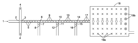
Figure 1 illustrates a microfluidic sequencing device in which a stream of
microdroplets at
least some of which contain a single nucleotide base are made to undergo
reaction with a capture
system of the first class described above.
An aqueous medium 1 comprising a stream of discrete deoxyribonucleotide
triphosphates
obtained by the progressive pyrophosphorolysis of a 100 nucleotide base
polynucleotide analyte
derived from human DNA is caused to flow through a ten micron diameter
microfluidic tube
fabricated from PDMS polymer. The pyrophosphorolysis reaction itself is
carried out by passing a
stream of an aqueous, buffered (pH 8) reaction medium at 72 C, comprising Tao
Pol and a 2
millimoles per litre concentration of each of sodium pyrophosphate and
magnesium chloride,
over a glass micro bead onto which the analyte has been previously attached by
means of a
succinyl bridge. The order of the single nucleotide bases in stream 1, which
is downstream of the
micro bead, corresponds to the sequence of the analyte. 1 emerges from a
droplet head 2 into a
first chamber 3 where it is contacted with one or more streams of immiscible
light silicone oil 4.
The velocities of these streams are chosen to avoid turbulent mixing and to
create in 3 aqueous
spherical droplets 5 suspended in the oil each having a diameter of
approximately eight microns.
CA 02907865 2015-09-23
WO 2014/167323 PCT/GB2014/051105
Typically, the rate of pyrophosphorolysis and/or the rate of flow of 1 are
adjusted so that
between adjacent filled droplets there are 10 empty ones. A stream of 5 is
then carried forward
along a second microfluidic tube of the same diameter at a rate of 1000
droplets per second to a
second chamber 6 into which a second stream of five micron aqueous spherical
droplets 7 is also
5 fed by means of a second droplet head 8. Droplets 5 and 7 are caused to
coalesce in a sequential
fashion to form enlarged aqueous droplets 9 approximately nine microns in
diameter. Each of 7
contains pyrophosphatase to destroy any residual pyrophosphate anion present
in each of 5.
A stream of 9 is then carried forward at the same rate via microfluidic tubing
into a third
chamber 10 where these droplets are contacted with a third stream of five
micron aqueous
10 spherical droplets 11 also fed thereto through a corresponding droplet
head 12. The time taken
for each of 9 to move between chambers 6 and 10 is c.2 minutes.
Droplets 9 and 11 are then caused to coalesce in 10 to produce droplets 13
(approximately
ten microns in diameter). Each of 11 contains a mesophilic ligase and a
capture system comprising
four pairs of j-shaped first oligonucleotides and four corresponding second
single-stranded
15 oligonucleotides. In this example, each j-shaped first oligonucleotide
is 60 nucleotide bases long
and is prepared by folding a 60 nucleotide base single-stranded
oligonucleotide precursor about
the 45th nucleotide base from the 5' end to generate a 3 nucleotide base
single stranded loop, a
12 nucleotide base pair double-stranded region and a 33 nucleotide base single-
stranded region.
Each of these four first oligonucleotides has a different 33rd base (measured
from the single-
stranded end) characteristic of the four characteristic nucleotide base types
of DNA (i.e. A, T, G
and C). The four different second oligonucleotides are each 28 nucleotide
bases long and have
sequences which are complimentary to that part of the single-stranded region
defined by the 4th
and 32nd nucleotide bases of their first oligonucleotide pair. The four
different second
oligonucleotide types are labelled respectively with the fluorophores Quasar
570, Fluorescein,
Texas Red and Cy-5 (five fluorophores moieties per second oligonucleotide). In
each case
fluorescence is quenched by the inclusion of one quencher moiety on each
second
oligonucleotide (BHQ-2 for Quasar 570 and Texas Red, BHQ-0 for Fluorescein and
BHQ-3 for
cyanine-5).
A stream of 13 is next carried forward at the same rate via microfluidic
tubing into a fourth
chamber 14 where it is caused to coalesce with a fourth stream of five micron
aqueous spherical
droplets 15 also fed thereto through a droplet head 16. The time taken for
each of 9 to move
between the two chambers is 30 minutes in which time the single nucleotide
base is captured by
its capture system pair and the captured molecule formed. Each of 15 contains
Phusion
CA 02907865 2015-09-23
WO 2014/167323 PCT/GB2014/051105
16
exonuclease to degrade the capture molecule and release the relevant
fluorophores in detectable
form. A stream of the coalesced microdroplets 17 is then taken forward to a
container 18 in
which their progress is tracked until they reach one of array of sites 19a
where they are held 19b
until such time as they are analysed.
After 2 hours each droplet held in the array is illuminated in turn and in the
correct order
with one or more high intensity light sources, for example one or more lasers
emitting coherent
light at the relevant frequencies of the fluorophores and the fluorescence so
generated detected
by a photodetector operating at those wavelengths characteristic of the four
fluorophore types.
From the information received the single nucleotide base is identified in each
droplet and nil
responses from empty droplets rejected. The results are then processed by a
computer
programmed to recreate the original nucleotide base sequence of the analyte.
If so desired,
multiple cycles of illumination and detection can be performed across the
array of droplets at
various intervals which can be averaged to improve the single to noise ratio
and therefore the
reliability of the results.