Note: Descriptions are shown in the official language in which they were submitted.
CA 02908517 2015-09-30
WO 2014/168987
PCT/US2014/033393
CARDIAC ABLATION CATHETERS AND METHODS OF USE THEREOF
CROSS REFERENCE TO RELATED APPLICATIONS
[0001] This application is a continuation-in-part of U.S. App. No.
13/106,658, filed May 12,
2011, which is a continuation-in-part of U.S. App. No. 12/616,758, filed
November 11, 2009,
now U.S. Pat. No. 8,295,902, both of which are incorporated by reference
herein. U.S. App. No.
13/106,658 also claims the benefit of priority of the following U.S.
Provisional Applications:
Appin. No. 61/334,154, filed May 12, 2010; App. No. 61/113,228, filed November
11, 2008;
App. No. 61/160,204, filed March 13, 2009; App. No. 61/179,654, filed May 19,
2009; App. No.
61/232,756, filed August 10, 2009; and App. No. 61/253,683, filed October 21,
2009. All of the
above-mentioned disclosures are incorporated by reference herein.
[0002] This application also claims the benefit of the following fourteen
U.S. Provisional
Applications, the disclosures of which are incorporated by reference herein:
App. No.
61/809,629, filed April 8, 2013; App. No. 61/809,646, filed April 8, 2013;
App. No. 61/895,880,
filed October 25, 2013; App. No. 61/809,636, filed April 8, 2013; App. No.
61/864,335, filed
August 9, 2013; App. No. 61/829,985, filed May 31, 2013; App. No. 61/820,992,
filed May 8,
2013; App. No. 61/821,001, tiled May 8, 2013; App. No. 61/821,014, filed May
8, 2013; App.
No. 61/934,640, filed January 31, 2014, App. No. 61/939,185, filed February
12, 2014; App. No.
61/934,647, filed January 31, 2014; App. No. 61/945,005, filed February 26,
2014, and App. No.
61/947,950, filed March 4, 2014. All of the above-mentioned disclosures are
incorporated by
reference herein.
INCORPORATION BY REFERENCE
[0003] All publications and patent applications mentioned in this
specification are herein
incorporated by reference to the same extent as if each individual publication
or patent
application was specifically and individually indicated to be incorporated by
reference.
BACKGROUND
[0004] Energy transmission to tissues can be used to treat a variety of
medical conditions.
Electrodes can be used to deliver energy to tissues and cells for the purpose
of sensing, mapping,
ablating, and/or stimulate muscles and/or nerves. Stimulation of muscles
and/or nerves can be
used to trigger signals to the brain or directly to a specified muscle
cell/group. When the
treatment requires removing or destroying a target tissue, thermal ablation
therapy can be used to
heat a target tissue with a surgical instrument such as a needle or probe
electrode coupled to an
- 1 -
CA 02908517 2015-09-30
WO 2014/168987
PCT/US2014/033393
energy source that heats the probe tip, the target tissue, or both. In such
cases the thermal energy
may be delivered directly by heating or cooling the probe or indirectly by
generating energy
fields within the tissue which in turn generate heat, or both. Energy fields
commonly used to
create heat indirectly are RF and acoustic energy fields. The goal for most
ablation procedures is
to achieve cell death quickly, precisely and with minimal to no collateral
damage.
[0005] In the case of thermal ablation therapy for terminating
destructive cardiac conductive
pathways, energy can be delivered to the aberrant cells using minimally-
invasive techniques such
as an electrode-tip catheter. Pulmonary vein isolation via radio frequency
catheter ablation has
been demonstrated to be an effective treatment for some patients experiencing
atrial fibrillation
(AF). The cornerstone of the AF ablation procedures is electrical isolation of
relatively large
pulmonary vein antra. Ablation of large confluent areas or lines of ablation
with older
generation AF ablation devices is accomplished by point to point manipulation
and RF
application with the single electrode tip. The single electrode catheter
technique is extremely
time-consuming, complex and fraught by subjectivity. Furthermore, efficient
and complete
mapping of the electrical activity in target tissues often requires the
placement of multiple
catheters in the left atrium, the use of a 3D-mapping, and/or steering system.
It is often desirable
to create relatively large surface area lesions with relatively shallow depths
of ablation.
[0006] Newer larger electrode arrays for "one shot" ablation have been
used to improve
catheter ablation treatments. These ablation systems have been adopted as a
way to provide full
contact to tissues having a complex 3-D anatomy and an overall larger lesion
area. But known
devices incorporate electrodes that are bulky, stiff and limited in their
ability to be packed
efficiently and effectively into the small space of the treatment catheter.
The stiffness of these
devices limits conformability against the tissue resulting in the need for
additional repositioning
and overlapping patterns to ensure uninterrupted lines of ablation.
SUMMARY OF THE DISCLOSURE
[0007] One aspect of the disclosure is an ablation catheter comprising:
an expandable
membrane and a plurality of ablation electrodes secured to the exterior of the
expandable
membrane; an imaging member disposed within the expandable membrane; a diffuse
reflector
secured to at least a proximal portion of the expandable membrane; and a light
source disposed
within the expandable member and positioned to direct light towards the
diffuse reflector such
that diffuse reflection of the light is directed towards a field of view of
the imaging member.
[0008] In some embodiments the imaging member is generally distally
facing and the light
source is generally proximally facing. The imaging member and the light source
can be secured
to an inner catheter shaft. The imaging member can be a plurality of cameras
oriented to provide
- 2 -
CA 02908517 2015-09-30
WO 2014/168987 PCT/US2014/033393
a 360 degree view around a longitudinal axis of the catheter. The imaging
member can be
disposed distally relative the light source.
[0009] In some embodiments the diffuse reflector does not extend to the
distal end of the
expandable membrane when in an expanded configuration. The diffuse reflector
can extend no
further than about half-way along the distal length of the expandable membrane
when in an
expanded configuration.
[0010] In some embodiments the diffuse reflector comprises first and
second portions
divided by a flex circuit secured to the exterior of the expandable membrane,
the flex circuit
comprising at least one conductive layer in electrical communication with at
least one of the
plurality of electrodes.
[0011] One aspect of the disclosure is an inflatable assembly adapted to
be positioned within
a patient, comprising an expandable membrane; an imaging member disposed
within the
expandable membrane; a diffuse reflector secured to at least a proximal
portion of the
expandable membrane; and a light source disposed within the expandable member
and
positioned to direct light towards the diffuse reflector such that diffuse
reflection of the light is
directed towards a field of view of the imaging member.
[0012] One aspect of the disclosure is an ablation catheter, comprising:
an expandable
membrane and at least one ablation electrode secured to the exterior of the
expandable
membrane; an imaging member disposed within the expandable membrane, the
imaging member
having a field of view; a light source disposed within the expandable member
adapted to deliver
light towards the field of view of the imaging member; and a reflection
adjuster adapted to
reduce specular reflection of light from at least one of the plurality of
ablation electrodes into the
field of view of the imaging member. The reflection adjuster can be a light
absorber. The
reflection adjuster can be adapted to scatter light away from the field of
view of the imaging
member. The reflection adjuster can be an anti-reflective coating on at least
one of an inside of
balloon or the at least one electrode.
[0013] One aspect of the disclosure is a video display process,
comprising receiving a
plurality of images from a camera in motion secured to a catheter; calculating
a mean rotation of
a center of mass of an anatomical feature shown in the images relative to a
feature whose
position is fixed relative to the camera; and communicating as output images
in which the
anatomical feature is fixed and the feature whose position is fixed relative
to the camera is shown
to be moving.
[0014] One aspect of the disclosure is a method of stabilizing an image
of cardiac tissue
while moving a camera positioned within the heart; comprising providing an
ablation catheter
within a left atrium, the ablation catheter including an expandable membrane,
a plurality of
- 3 -
CA 02908517 2015-09-30
WO 2014/168987
PCT/US2014/033393
electrodes secured to an exterior surface of the expandable membrane, at least
one camera
positioned within the expandable membrane with a field of view fixed relative
to the position of
the plurality of electrodes when the expandable membrane is in an expanded
configuration, and a
light source; and in response to movement of the camera within the left
atrium, and, while the
camera is being moved, displaying a video of cardiac tissue in which the
position of the cardiac
tissue is fixed and the plurality of electrodes in the field of view are
moving.
[0015] One aspect of the disclosure is a method of superimposing an image
of cardiac tissue
with additional information, comprising positioning an ablation catheter
within a left atrium, the
ablation catheter including an expandable membrane, a plurality of electrodes
secured to an
exterior surface of the expandable membrane, at least one camera positioned
within the
expandable membrane, and a light source; capturing an image with the at least
one camera,
wherein the image shows at least one of at least one of the plurality of
electrodes and the cardiac
tissue; obtaining additional information indicative of at least one of a
characteristic of the cardiac
tissue and a characteristic of the ablation catheter; displaying the image
that shows the at least
one of at least one of the plurality of electrodes and the cardiac tissue with
the with the additional
information superimposed thereon.
[0016] In some embodiments the additional information comprises an
indicator of cardiac
tissue adjacent one of the plurality of electrodes. The additional information
can comprise
temperature of cardiac tissue adjacent one of the plurality of electrodes.
[0017] In some embodiments the additional information is a qualitative
indicator.
[0018] In some embodiments the additional information is a quantitative
indicator.
[0019] In some embodiments the additional information comprises a state
of at least one of
the plurality of electrodes, such as on or off.
[0020] One aspect of the disclosure is an ablation catheter comprising:
an expandable
membrane and a plurality of ablation electrodes secured to the exterior of the
expandable
membrane; at least one imaging member disposed within the expandable membrane,
the at least
one imaging member having a field of view that include the plurality of
ablation electrodes; and
an electrode identifier associated with each of the plurality of electrodes
and adapted to be
visually identifiable in the field of view so that each of the plurality of
electrodes can be visually
identifiable.
[0021] In some embodiments the electrode identifiers comprise
alphanumeric characters on
or near each of the electrodes.
[0022] In some embodiments the electrode identifiers are colors
associated with each of the
electrodes.
[0023] In some embodiments the electrode identifiers are shapes of the
electrodes.
- 4 -
CA 02908517 2015-09-30
WO 2014/168987
PCT/US2014/033393
[0024] In some embodiments the electrode identifiers are a first type of
identifier for at least
one of the plurality of electrodes, and a second type of identifier for at
least a second of the
plurality of electrodes.
BRIEF DESCRIPTION OF THE DRAWINGS
[0025] Figures 1A-1C illustrate an exemplary ablation device in expanded
configurations.
[0026] Figure 1D illustrates an exemplary ablation device in a collapsed
configuration.
[0027] Figure 2A is a side view of an exemplary distal end of an ablation
catheter.
[0028] Figure 2B is a close up side view of the inside of the catheter
from Figure 2A.
[0029] Figure 3 is a perspective view showing inside the expandable
membrane.
[0030] Figure 4 illustrates a camera assembly.
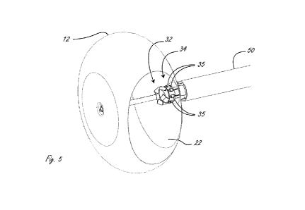
[0031] Figure 5 is a perspective view of a distal end of an ablation
catheter, with a cutaway
of an expandable member.
[0032] Figure 6 is an exemplary flat view of the LED flex circuit.
[0033] Figure 7 illustrates the distal end of a device incorporating a
slideable sheathing tool
comprising a sheathing tube.
[0034] Figure 8 is a flat view showing three individual flex circuits
that are secured to the
exterior of membrane and to electrodes.
[0035] Figure 9A illustrates a portion of one of the flex circuits and
electrodes in Figure 8.
[0036] Figure 9B illustrates the exemplary different layers of the flex
circuit from section S-
S from Figure 9A.
[0037] Figure 10 illustrates each of the three flex circuit tails
terminating in terminations
extending proximally from the distal end of the balloon and extending
proximally within an
outer shaft and secured to the outer surface of the proximal end of the
balloon and irrigation
shaft.
[0038] Figures 11A-Figures 16 illustrate exemplary ablation catheter
adapted with mapping
structures or adapted to be used with mapping structures.
[0039] Figure 17 is a side view of a distal portion of an exemplary
visualization catheter.
[0040] Figures 18A-18D show the orientations of the axes of four cameras
in relationship to
the longitudinal axis of a catheter shaft.
[0041] Figure 19 shows the geometry of one of the four cameras, and all
four have the same
geometry.
[0042] Figure 20 shows a picture of a regular grid pattern target taken
by a representative
camera.
- 5 -
CA 02908517 2015-09-30
WO 2014/168987 PCT/US2014/033393
[0043] Figures 21A-21C show parameterization that can be used to unwrap the
3D surface of
the ellipsoidal balloon into a 2D plane.
[0044] Figure 22 shows a set of four camera images simulated using a known
pattern, in this
case, ablation electrodes painted on the membrane.
[0045] Figure 23 shows the panoramic image generated by projecting the
images from
Figure 22 back onto the unwrapped balloon surface using the methods described
above.
[0046] In Figure 24 the panoramic image is generated by projecting the
component images
back onto the unwrapped balloon surface.
[0047] Figure 25 shows tissue images acquired by four cameras using the
methods described
herein.
[0048] Figures 26A-26C illustrate an electromechanical device providing for
the continuous
or semi-continuous adjustment of the transfer of AC power from a source to a
load by means of
linearly displaceable core.
[0049] Figure 27 shows a graph illustrating movement of the core versus
magnitude of AC
output.
[0050] Figures 28A and 28B represent one embodiment of where a core is
displaced by a
micro-stepper motor and screw mechanism.
[0051] Figure 29 illustrates only one of the four fields of view for one of
the four cameras in
the camera assembly.
[0052] Figure 30 illustrates the four fields of view from the four cameras,
each overlaid with
at least one other field of view, to give the physician a 360 degree view.
[0053] Figures 31A-31C illustrate an exemplary method of ablating cardiac
tissue.
[0054] Figures 32A-32C illustrate an exemplary method of ablating cardiac
tissue.
[0055] Figure 33 is an exemplary schematic of the electrical aspect of an
exemplary
embodiment.
[0056] Figure 34 illustrates mapping signals from a plurality of channels.
[0057] Figures 35 and 36 illustrate aspects of an external console.
[0058] Figure 37 illustrates an exemplary block diagram of a cardiac
ablation system.
[0059] Figure 38 illustrates exemplary information and indicators that can
be superimposed
on the images from the cameras.
[0060] Figure 39 represents an exemplary flexible circuit for application
to the outer surface
of a balloon.
[0061] Figure 40 shows an assembled flexible circuit affixed to a balloon.
[0062] Figures 41A and 41B illustrate a composite view as described herein
from a four
camera array as presented to the user on a display.
- 6 -
CA 02908517 2015-09-30
WO 2014/168987
PCT/US2014/033393
[0063] Figures 42 and 43 illustrate an exemplary embodiment of an
ablation catheter
wherein the balloon is configured for contact (physical) measurements.
[0064] Figure 44 illustrates an ablation balloon in the left atrium and
esophageal temperature
balloon positioned and inflated in the esophagus.
[0065] Figure 45 illustrates an embodiment that includes an endocardial
catheter and an
epicardial catheter.
DETAILED DESCRIPTION
[0066] The disclosure describes methods of, and systems and devices
configured for,
diagnosing, preventing, and/or treating cardiac arrhythmias. The disclosure
includes methods of
and devices configured for ablating cardiac tissue. The disclosure is related
to and incorporates
by reference the devices and methods described in U.S. Pat. No. 8,295,902,
issued 10/23/2012,
and U.S. Pub. No. 2012/0071870, published 3/22/2012, the disclosures of which
are incorporated
by reference herein. Devices herein can incorporate suitable structural
features in embodiments
in the aforementioned applications even if the disclosure fails to expressly
include them.
Additionally, the methods of use herein can include suitable method steps in
embodiments in the
aforementioned applications even if the disclosure fails to expressly include
them.
[0067] Figures 1A-1C illustrate a distal portion of an exemplary cardiac
ablation catheter.
Figures 1A-1C shows expandable member 10 in an expanded configuration. Figure
IA is a
distal view, Figure 1B is a perspective view, and Figure 1C is a side view.
[0068] The cardiac ablation catheter is configured to deliver ablative
energy to tissue such as
cardiac tissue and to ablate the tissue. Expandable member 10 includes
membrane, or balloon,
12 and a plurality of energy delivery elements 14 secured to the exterior of
membrane 12. In this
embodiment energy delivery elements 14 are electrodes configured and
positioned to deliver
ablative RF energy to tissue when expandable member 10 is inflated and to
ablate the tissue, and
are in electrical communication with an RF generator (not shown) configured to
generate RF
energy.
[0069] Figure 1D illustrates expandable member 10 in a collapsed, or
deflated, configuration
prior to full inflation.
[0070] Figure 2A is a side sectional view of the distal portion of the
ablation catheter shown
in Figures 1A-1C. Figure 2B is a highlighted side sectional view of components
within outer
shaft 51. Figure 2A shows membrane 12 expanded at the distal end of outer
lumen 50, which is
the annular space between outer shaft 51 and irrigation shaft 55. The distal
end of membrane 12
is secured, such as by press-fit and/or adhesive, to distal hub assembly 20,
between an inner
member and an outer member of assembly 20 as shown. The proximal end of
membrane 12 is
- 7 -
CA 02908517 2015-09-30
WO 2014/168987
PCT/US2014/033393
secured to the outer surface of irrigation shaft 55. Hub 20 is secured to
guide wire shaft 54,
which in this embodiment defines guidewire lumen 53 so that the ablation
catheter can be
advanced over a guidewire (not shown). Guidewire shaft 54 and irrigation shaft
55 are adapted
to be axially movable relative to one another, which allows the distal end of
membrane 12 to be
moved relative to the proximal end of membrane 12. Relative movement between
the two
components can allow for the shape of the balloon to be changed. The movement
also assists in
transitioning expandable member 10 to a collapsed configuration, as shown in
Figure 1D.
[0071] Visualization system 30 includes a camera assembly 32 and
illumination sources 35
disposed on the guide wire shaft 54. The cameras are configured to enable real-
time imaging of
the procedure from within the expandable member 10 to visualize the membrane
and electrodes,
cardiac tissue when the membrane/electrodes and cardiac tissue interface, as
well as lesion
formation during the ablation procedure, as is described in more detail below.
[0072] Figure 2B shows radially outer shaft 51, irrigation shaft 55 that
defines irrigation
lumen 52, and guide wire shaft 54 that defines guidewire lumen 53.
[0073] The materials of the membranes 12 described herein can vary.
Generally, the
membrane material is thin, readily foldable into a low profile and refoldable
after expansion.
The materials can be elastic, inelastic, stretchy, non-stretchy, compliant,
semi-compliant, or non-
compliant. In an embodiment, membrane 12 has an expandable structure and can
be constructed
of materials such as those materials used in the construction of balloon
catheters known in the
art, including, but not limited to polyvinyl chloride (PVC), polyethylene
(PE), cross-linked
polyethylene, polyolefms, polyolefin copolymer (POC), polyethylene
terephthalate (PET), nylon,
polymer blends, polyester, polyimide, polyamides, polyurethane, silicone,
polydimethylsiloxane
(PDMS) and the like. Membrane 12 can be constructed of relatively inelastic
polymers such as
PE, POC, PET, polyimide or a nylon material. Membrane 12 can be constructed of
relatively
compliant, elastomeric materials including, but not limited to, a silicone,
latex, urethanes, or
Mylar elastomers. Membrane 12 can be embedded with other materials such as for
example,
metal, Kevlar or nylon fibers. Membrane 12 can be constructed of a thin, non-
extensible
polymer film such as polyester or other flexible thermoplastic or
thermosetting polymer film. In
one embodiment flexible membrane 12 can be about 0.001" to about 0.002" in
thickness to
provide sufficient burst strength and allow for foldability. In some
embodiments it is preferable
to have the electrode mechanical properties as close to the membrane
mechanical properties as
possible. One way of providing this is to use an inelastic membrane that will
not stretch as it is
expanded. This helps secure the branches to the membrane. Membrane 12 has a
front, or distal,
face that is generally flat but can have other shapes as well.
- 8 -
CA 02908517 2015-09-30
WO 2014/168987 PCT/US2014/033393
[0074] Expandable member 10 includes what is generally referred to in
U.S. Pat. No.
8,295,902, issued 10/23/2012, and U.S. Pub. No. 2012/0071870, published
3/22/2012, as flex
circuits. A flex circuit as used herein generally refers to a conductive
layer, an insulation layer,
and optionally a substrate layer. A flex circuit is in electrical
communication with at least one
electrode.
[0075] Figure 8 is a flat view showing three individual flex circuits
that are secured to the
exterior of membrane 12. Each of the three flex circuits includes six energy
delivery elements
14, and a tail terminating in termination 41 for the six conductive traces,
one for each of the six
electrodes. The terminations may be in the form of a connector or solder pads
or other such
suitable interface. The terminations 41extend proximally from energy delivery
elements on the
expandable member, one of which can be seen in Figure 1D. Each of the tails
branch off into
three branches 16, each one of which includes two energy delivery elements.
Each of the two
side branches 16 extend away from the longitudinal axis of the connector at
substantially the
same angle and each of two electrodes on a side branch is disposed at the same
axial position (in
the distal/proximal direction) as the other corresponding electrode on the
other side branch. The
central branch, however, initially extends along the same general direction as
the longitudinal
axis of a tail, and the first electrode on the central branch is axially
disposed at the same general
location as the second electrodes on the right and left branch. The central
branch then extends
away from the longitudinal axis of the tail, and the second (distal) electrode
on the central branch
is disposed further distally than the other five electrodes on the flex
circuit, and is disposed
radially (relative the longitudinal axis of tail) at the same general position
as the first (proximal)
electrode on one of the other side branches. In Figure 8, the six electrodes
on one of the flex
circuits are labeled A-F. The two side branches of the flex circuit include
electrodes A-B and E-
F respectively. The central branch includes electrodes C and D. In the flat
view, electrode C
(the distal electrode of the central branch) is axially disposed at the same
general position as
electrodes B and F. Electrode D is disposed further distally than the other
five electrodes, and is
positioned radially in the same general position as electrode A. Electrodes A
and E are disposed
in the same general axial position, as are electrodes B, C, and F. Each of the
three flex circuits is
positioned on the expandable member, and the arrangement and size of
electrodes provides for
eighteen electrodes secured to the expandable member. As can be seen in
Figures lA and 1B,
there are three electrodes closely surrounding hub 20.
[0076] Figure 9A illustrates a portion of one of the flex circuits in
Figure 8 (the flex circuit in
which termination 41 is at the "6 o'clock" position), including six energy
delivery elements 14.
Figure 9A shows as alternative embodiment in which the distal electrode on the
central branch
16 extends to the right on the page rather than the left, as is shown in
Figure 8. This arrangement
- 9 -
CA 02908517 2015-09-30
WO 2014/168987
PCT/US2014/033393
provides the same general arrangement of the eighteen electrodes on the
balloon. In the
embodiment in Figures 1A-1C, there are three of the flex circuits from Figure
9A disposed on
membrane 12, and thus eighteen energy delivery elements secured to membrane
12. Figure 9B
illustrates the exemplary different layers of the flex circuit from section S-
S from Figure 9A.
Electrically non-conductive substrate layer 13 is deposited on membrane 12,
upon which
conductive layers, or traces, 15 are deposited. Insulation layer 17 is
deposited on top of
conductive layers 15 except where the electrodes 14 are located. For example,
to the left in
Figure 9B, an electrode 14 is disposed on electrically conductive element 15,
thus electrically
coupling electrode 14 and conductive layer 15, which is electrically coupled
to an RF generator.
On the right side of Figure 9B, insulation layer 17 prevents conductor15 on
the right side from
being electrically coupled to electrode 14. Instead, the conductor 15 on the
right side will be
electrically coupled to the distal electrode on that branch. Each individual
conductor 15 is
therefore electrically coupled to only one electrode 14. In the figure shown
in 9A, there are six
individual conductive traces 15, each of which is individually coupled to one
electrode. As is
described in detail in U.S. Pat. No. 8,295,902, issued 10/23/2012; U.S. Pub.
No. 2012/0071870,
published 3/22/2012, the electrodes are sized and configured to extend over a
portion of the flex
circuit and a portion of membrane not covered by the flex circuit. In this
manner a large surface
area electrode can be deposited onto and secured to the membrane. Each
electrode is shown with
an irrigation aperture in the middle thereof, as is described herein to
irrigate tissue adjacent the
electrodes and to prevent the irrigation fluid inside the membrane from
becoming too hot and
interfering with the tissue ablation.
[0077]
The conductor or conductive layer 15 can be a material such as, but not
limited to, a
metal or metal foil of copper, gold, silver, tin, nickel, steel, cupronickel
(copper-nickel alloy),
KOVAR (nickel-cobalt ferrous alloy) or other material. In an embodiment, more
than one
conductive material can be used in the conductive layer 15. In an embodiment,
a conductive
layer 15 of copper can be plated with a thin layer of an additional conductive
material at the
conductive pad beneath electrode 14. In an embodiment, the thin layer of
additional conductive
material can be gold. The flex circuit and its components can be manufactured
using techniques
as known in the art.
[0078] The materials used to create the electrodes 14 can vary. The
electrodes 14 can be a
thin film of an electro-conductive or optical ink. The ink can be polymer-
based for better
adhesion to the membrane. The electrode material can be a biocompatible, low
resistance metal
such as silver, silver flake, gold, and platinum which are additionally
radiopaque. Inks may
additionally comprise materials such as carbon and/or graphite in combination
with the more
conductive materials already described. The addition of carbon and/or graphite
can increase the
- 10 -
CA 02908517 2015-09-30
WO 2014/168987 PCT/US2014/033393
conductivity of the polymer matrix. When incorporated as fibers the carbon
and/or graphite add
additional structural integrity to the ink electrode. Other fiber materials
may be substituted to
attain the same end. When the electrode material is not particularly
radiopaque, additives such
as tantalum and tungsten may be blended with the electrode material to enhance
radiopacity. An
example of an electro-conductive ink is provided by Engineered Conductive
Materials, LLC
(ECM) which is a polyurethane-based silver loaded ink. Another example is
Creative Materials
Inc., which manufactures conductive inks, films, as well as radiopaque inks.
As mentioned
above, the electrodes 14 can be applied to the membrane 12 and flex circuit
using an adhesive.
Alternatively, the electrode material can have adhesive properties or be an
adhesive-loaded with
conductive particles such as silver flakes such that electrodes 14 can adhere
the components of
the flex circuit to the membrane 12. If an additional adhesive layer is used
to adhere the
electrode 14 to the membrane 12 and flex circuit, the adhesive layer can
include a conductive or
non-conductive material. The electrodes formed with electro-conductive or
optical ink or thin
metal film can be visualized under fluoroscopy to provide a general sense of
the shape of the
membrane and location of the electrode. To enhance visualization under
fluoroscopy,
radiopaque additives can be included in the electrode material or radiopaque
markers laid out
next to, on top or below the electrodes as will be discussed in more detail
below. Additionally,
the bonding layer or substrate will be optimally comprised of a minimally
reflective material.
[0079] Each of the electrodes is individually addressable, or can be
used with any other
electrode. The electrodes can operate in monopolar mode or bipolar mode, as is
indicated in the
exemplary schematic shown in Figure 34. Electrodes sets can be chosen such
that the lesion is,
for example without limitation, linear, a spot, or a hollow circle.
[0080] Figure 3 illustrates the coupling of the distal end of membrane
12 and hub 20, which
can be press fit, adhesive coupling or a combination of both.
[00811 To prevent or reduce the likelihood of charring of tissue that is in
contact with the
energy delivery elements and coagulation of blood adjacent the electrodes,
each of the flex
circuits at the locations of the electrodes includes an irrigation aperture
therethrough, and as
shown are in the center of the electrodes. The irrigation apertures also
prevent the
inflation/irrigation fluid inside the membrane from becoming too hot, which
would interfere with
the ablation. Irrigation fluid, which is also the fluid that inflates membrane
12 causing it to be
reconfigured toward its expanded configuration, is pumped from a fluid source
through irrigation
lumen 52, into membrane 12, through the irrigation apertures (not labeled),
and towards the
tissue that is in contact with the electrodes to cool the target tissue. One
of the drawbacks of
previous attempts at cardiac ablation is that the ablation procedures cause
blood to coagulate or
tissue to char due to lack of a cooling feature. Additionally, since each
electrode is individually
-11-
CA 02908517 2015-09-30
WO 2014/168987
PCT/US2014/033393
addressable, and the visualization system allows the operator to identify
whether an individual
electrode is in contact with tissue, only electrodes in contact with tissue
may be turned on. Thus
energy is more efficiently coupled to just the sites where ablation is desired
and little to no
energy is dissipated into the blood.
[0082] One of the significant advantages of ablation catheters herein is
that, when in use, the
ablation procedures can be visualized with an imaging, or visualization,
member with a
perspective from within the inflatable membrane. In the embodiment in Figures
1A-1D, imaging
member 30 includes camera assembly 32 that includes a plurality of cameras 33
and a plurality
of illumination, or light, sources, 35 (e.g., LEDs). Expandable member 10 also
includes diffuse
reflector 22 that is secured to the external surface of membrane 12. Reflector
22 is a diffuse
reflector adapted to create diffuse reflection of light incident upon it from
the illumination
sources. Reflector 22 is adapted to reflect light in a diffuse manner, as
opposed to specular
reflection, to better illuminate as much of the camera field of view as
possible. If the reflector
were adapted for specular reflection rather than diffuse reflection, light
from the illumination
sources that is reflected from the reflector would appear in the camera's
field of view as a
localized spot and would not illuminate as much of the field of view as
possible.
[0083] Illumination sources 35 are configured and positioned to provide
illumination
generally radially outward towards reflector 22. Diffuse reflector 22 thus
diffusely reflects light
forward toward the camera's fields of view. The illumination sources thus
provide lighting for
the cameras to visualize the procedure, including the tissue, and the lesion
formation.
[0084] In some embodiments the diffuse reflector is printed on the
exterior of the balloon.
The diffuse reflector can be comprised of silicone or urethane resins filled
with nonconductive
white pigment such as TiO, BaO, BaSo4, styrene or other polymer beads, or of
metal particles.
Optimal materials will be minimally reflective such as a black adhesive.
[0085] In this embodiment the diffuse reflector is secured to the membrane
such that it does
not completely overlap any of the electrodes, and is positioned so that the
illumination sources,
when activated, emit light towards the reflector. In this embodiment the
diffuse reflector, or
reflectors, is secured to the membrane at a location that does not extend all
the way to the distal
end of the membrane. In this embodiment the reflector is secured to the
membrane such that it
does not extend further distally than the proximal-most electrode. In
alternative embodiments,
however, the reflector can extend distally to the proximal-most electrode in
some locations
around the membrane. For example, the distal edge of the reflector can be
curved rather than
straight, and depending on the electrode layout on the membrane, some portions
of the reflector
may extend distally relative to the proximal-most electrode. If the membrane
in its inflated
configuration can be divided in half between the distal most location and
proximal most location
-12-
CA 02908517 2015-09-30
WO 2014/168987
PCT/US2014/033393
defining a distal portion and proximal portion, the reflector is disposed at
least on the proximal
portion. In the embodiment shown in Figures 1A-1C, the reflector is disposed
only on the
proximal portion.
[0086] One aspect of the disclosure is an expandable member that includes
a diffuse reflector
but does not include any ablation element. For example, medical devices that
include an
inflatable member and at least one camera and at least one light source
therein can benefit from a
diffuse reflector even if the device is not used for ablation procedures.
[0087] While the reflector herein is described as being a diffuse
reflector, there may be some
uses in which a reflector that reflects light in a specular manner may be
beneficial. Alternatively,
a reflector can have portions that reflect light in a diffuse manner and
portions that reflect light in
a specular manner.
[0088] Figure 4 shows an exemplary camera assembly 32 that includes four
cameras 33,
which are disposed within camera hub 37 at an angle relative to the
longitudinal axis of the
catheter. Camera hub 37 is secured to guide wire shaft 54, and includes lumen
39 configured to
receive guide wire shaft 54 therein.
[0089] Figure 5 is another perspective view of expandable member 10 with
a cutaway of the
membrane. Figure 6 is an exemplary flat view of the LED flex circuit,
including the LEDs, that
is wrapped around the illumination hub proximal to the cameras.
[0090] As set forth above, light is reflected from the diffuse reflector
to provide illumination
in the field of the view of the at least one camera. The field of view of the
camera can include
the view of an electrode secured to the membrane. As set forth herein, the
electrodes can be
highly reflective, such as if they are comprised of silver. Reflective
electrodes causes light
incident upon the electrodes to reflect into the camera field of view, which
can cause the
electrodes to appear as bright spots on the display, possibly interfering with
viewing the
procedure. It can thus be beneficial to include in the catheter a reflection
adjuster that is adapted
to reduce specular reflection of light from at least one of the plurality of
ablation electrodes into
the field of view of an imaging member.
[0091] In some embodiments the reflection adjuster is a light absorber.
The light absorber
can be positioned between the bottom of the electrodes and the membrane. In
some
embodiments the light absorber is a black adhesive that adheres portions of
the electrode to the
membrane, as well as acts as a light absorber.
[0092] In some embodiments the reflection adjuster is an anti-reflective
coating. Exemplary
anti-reflective coatings include, for example without limitation, a deposited
thin layer of Ti02,
MgF2, and "moth eye" structures comprised of nanoparticles approximately 200
nm in diameter
spaced 300 nm range, random microstructure secured to or created on the
interior surface of the
- 13 -
CA 02908517 2015-09-30
WO 2014/168987
PCT/US2014/033393
membrane that is adapted to reduce reflection. The anti-reflective coating can
be adhered to only
a portion of the membrane, such as the portion where the electrodes are
disposed. For example,
an anti-reflective coating could be applied to only the distal portion of the
inner membrane.
[0093] A reflection adjuster will reduce the amount of reflection from
the bottom of the
electrodes, creating a clearer image of the membrane and electrodes from
within the membrane.
[0094] When the images or video provided by the at least camera are
displayed on the
display, it can be helpful to be able to visually identify the electrodes on
the display. For
example, a user interface can be used to control delivery parameters for any
of the electrodes,
and enabling the physician to easily determine and confirm that a given
electrode on the video is
a particular electrode on the user interface simplifies the procedures and
ensures that the correct
electrodes are being activated and used as intended.
[0095] In some embodiments the catheter includes an electrode identifier
associated with at
least one of the plurality of electrodes, and is some embodiments the catheter
includes an
electrode identifier with each of the plurality of electrodes. The electrode
identifier need not be
unique to each of the electrode, but in some embodiments it is unique to each
electrode. The
electrode identifier is visually identifiable and allows an individual to
visually associate the
identifier with an electrode.
[0096] In some embodiments the electrode identifier is an alphanumeric
characters disposed
on or near each of the electrodes. An example of this type of identifier is
described and shown
below. For example, an alphanumeric character can be printed on the back of an
electrode, or
the back of a portion of the flex circuit that is associated with an
electrode. An alphanumeric
character can also be printed on the membrane near the electrode so that the
identifier can be
easily associated with a particular electrode.
[0097] In some embodiments the electrode identifiers are colors
associated with one or more
of the electrodes. For example, the electrodes can be color-coded so that a
user can visually
identify each of the electrodes. In some embodiments a group of electrodes can
have a particular
color, such as all of the electrodes connected to the same flex circuit are
all one color. An
additional example of an electrode identifier is the shape of the electrode so
that the electrode or
group of electrodes can be visually identified based on their shape. For
example, groups of
electrodes can be circular, oval, hexagonal, rectangular, square, etc. Each
electrode could have a
unique shape to it as well.
[0098] An example of electrode identifiers is described below in the
context of overlaying
field of view images from a plurality of cameras.
[0099] Figure 10 illustrates each of the three flex circuit tails
terminating in terminations 41
(one for each flex circuit) extending proximally from the distal end of the
balloon and extending
- 14 -
CA 02908517 2015-09-30
WO 2014/168987
PCT/US2014/033393
proximally within outer shaft 51 and secured to the outer surface of the
proximal end of the
balloon and irrigation shaft 55. The proximal aspect of the configuration can
also be seen in
Figure 2B. In Figure 10, six conductive wires 18 can be seen extending
proximally from one of
the terminations 41, each one of which is in electrical communication with one
of the six
electrodes in that particular flex circuit. The six wires 18 extend the length
of the catheter and
are in communication with the RF generator. In an alternate embodiment, not
shown, the six
conductive traces 15 extend the length of the catheter and are in
communication with the RF
generator. Camera flex circuit 43 for the visualization system is also shown
in Figure 10,
extending proximally from the visualization system in the catheter.
[0100] Exemplary materials for the membrane and flex circuit materials can
be found in U.S.
Pat. No. 8,295,902, issued 10/23/2012; U.S. Pub. No. 2012/0071870, published
3/22/2012.
Additional examples of membrane material include PET, Polyurethane, etc.
Exemplary
materials for the reflector include metalized paints, silicone or urethane
resin filled with
nonconductive white pigment such as TiO or BaO or BaSo4, preferably non-
conductive.
Exemplary materials for the electrodes include silver filled silicone or
urethane. Exemplary
materials for the conductive traces are conductive metals including copper or
other such
conductive materials. The insulation layers can be known dielectric materials.
Exemplary
materials for the substrate include Kapton.
[0101] As described herein ablation catheters can include ablation and
mapping electrodes
secured to the exterior of the membrane. In such embodiments the area of
tissue mapped is
limited to the area of contact defined by the inflatable structure. The rotors
being mapped can,
however, be larger than the contact area of the inflatable structure, making
it more difficult and
time consuming to properly map the atrial chamber for rotors. In some
embodiments the
ablation catheter includes an inflatable membrane, and is also adapted to
increase the area that
can be mapped to an area that is greater than that defined by the expandable
membrane contact
surface.
[0102] In some of these embodiments mapping arms when appropriately stiff
may provide a
way to limit the accidental entry of the ablation elements into the pulmonary
arteries thereby
minimizing the risk of accidental ablation of the artery wall and consequent
risk of subsequent
stenosis.
[0103] In some embodiments a mapping structure on which at least one
mapping electrode is
disposed is carried outside of the balloon and collapsed between the wall of
the delivery catheter
and the outside of the ablation catheter. The mapping structure can be secured
to the exterior of
the ablation catheter. In some embodiments the one or more mapping structures
can be
deformable splines, the use of which has been described in the cardiac
ablation space. For
- 15 -
CA 02908517 2015-09-30
WO 2014/168987
PCT/US2014/033393
example, the mapping structures can be made of nitinol and are adapted to
deform. The mapping
structure can thus expand on release from the delivery catheter and can be
collapsed to a
collapsed delivery configuration when the delivery catheter is advanced
distally relative the
ablation catheter
[0104] In other embodiments a mapping electrode structure is adapted to be
delivered
through the guide wire lumen of the ablation catheters herein.
[0105] Figures 11A and 11B depict an exemplary ablation catheter 300 that
includes an array
of mapping electrodes 302 (only one is labeled for clarity) carried on the
surface of a plurality of
reconfigurable mapping arms 308. Figure 11A is a side view and Figure 11B is a
distal view.
Arms 308 together have a "basket" configuration and are disposed outside of
the inflated
membrane 306. In Figures 11A and 11B arms 308 are in their expanded
configurations, after
being released from within the delivery catheter. Arms 308 are collapsed into
the space between
the delivery catheter and the ablation catheter 300 during delivery and
retrieval, and are adapted
to self-expand on release by retraction of the delivery catheter or delivery
past the distal end of
the delivery catheter. Six arms 408 are shown, each with a plurality of
electrodes 302, but more
or fewer arms of the basket can be included. The arms can all be secured to
the same mapping
basket hub (or made from a single piece of material), or they can be secured
independently to the
ablation catheter. Figures 11A and 11B show catheter 300 with arms 308 in
retracted positions
in with proximal ends of arms 308 are retracted and positioned between the
delivery catheter and
the ablation catheter. Arms 308 are closer to the surface of expanded membrane
306 than in the
expanded configurations shown in Figures 11A and 11B.
[0106] Figure 13 is a distal view of a distal end of an exemplary
ablation catheter 320. In
this embodiment the ablation catheter includes an alternative spiral structure
328 that carries a
plurality of mapping electrodes 322 (only three are labeled). The spiral
mapping structure can be
adapted to be delivered through the guidewire lumen 323, or it can be adapted
to be expanded
from between the delivery catheter and ablation catheter shaft, similar to the
embodiment in
Figures 11A and 11B. In the embodiment in Figure 13 in which the spiral
structure is adapted to
be delivered via a guidewire lumen, the spiral, in a side view, can be in a
single plane, or the
spiral can have a conical configuration that is adapted to be deformed into a
single plane when
the spiral is pushed distally into contact with tissue. Ablation electrodes
are not labeled on the
ablation balloon for clarity on Figures 13 - 17.
[0107] Figure 14A is a simplified side view illustrating an alternative
ablation catheter 340
with a dedicated mapping structure 348 with a plurality of mapping electrodes
342 (only two are
labeled) thereon. In this embodiment the two mapping arms 348 have expanded
loop
configurations as shown and are adapted to be delivered through guidewire
lumen 347 as shown.
- 16 -
CA 02908517 2015-09-30
WO 2014/168987
PCT/US2014/033393
There may be more or fewer than two arms. Figure 14B is a distal view of an
alternative
embodiment in which the mapping structure 350 includes a plurality of loops in
their expanded
configurations. In this embodiment at least one loop 352 has an expanded
"height" (a distance
measured from the longitudinal axis of the catheter along a line perpendicular
to the axis) greater
than a height of a second loop 354. In particular, there are four arms 352
with a first height
greater than a height of four other arms 354. There can any number of loops of
varying height
dimension.
[0108] Figure 15 illustrates an exemplary configuration of mapping arms
and electrodes 362
in collapsed configurations within guidewire lumen 360, and is merely
illustrative to show how a
plurality of arms can be disposed within a guidewire lumen. More or fewer arms
can be disposed
therein.
[0109] Figure 16 shows a simplified side view of an exemplary ablation
catheter 370 in
which the mapping arms 378 terminate at their respective distal ends 379. That
is, each arm has
a free end. Catheter 370 includes balloon 376, guidewire lumen 377, mapping
electrodes 372 on
arms 378, similar to other embodiments herein. Any of the described mapping
arms may
comprise a stiffening member such as NiTi wire such that on release the
mapping member takes
on a predetermined shape.
[0110] Any of the mapping arms that are delivered through the guidewire
lumen can
alternatively be configured for delivery in the space between the ablation
catheter and the
delivery catheter, and vice versa.
[0111] In yet other embodiments the mapping arms may be woven into a
conical braid or
braid structure which increases in diameter as it extends distally.
[0112] In use, the visualization system allows for real-time
visualization of the procedure
with a view by one or more cameras disposed within the balloon. The
visualization allows for
the entire procedure to be visualized, allowing physicians to assess the
degree of tissue contact,
and see the electrodes, tissue, and lesion formation as it occurs. For
clarity, Figure 29 illustrates
only one of the four field of views for one of the four cameras in the camera
assembly. Figure
illustrates the four field of views from the four cameras, each overlaid with
at least one other
field of view, to give the physician a 360 degree view (with the longitudinal
axis of the catheter
30 as the reference) of the treatment area. While there is a blind spot
shown in the center of the four
images, different lensing systems than those used in the current embodiments
can allow for
elimination of that spot. Since there are electrodes disposed around the
entire catheter, the 360
degree view allows the physician to visualize an entire lesion that utilizes
electrodes disposed
around the catheter. The visualization of the entire procedure including
lesion formation at any
of the electrode locations is immensely helpful to the physician.
- 17 -
CA 02908517 2015-09-30
WO 2014/168987
PCT/US2014/033393
[0113] The description herein of overlaying camera field of views is
related to the disclosure
in U.S. Pub. No. 2012/0071870, in particular Figures 38H-38R, and the textual
descriptions
thereof. One aspect of this disclosure is an exemplary method of generating a
panoramic image
display using images from a plurality of cameras attached to an endoscopic
catheter. In some
embodiments a plurality of images captured from a plurality of cameras are
overlayed with at
least one other image to create the panoramic image around the longitudinal
axis of the ablation
catheter. Two or more cameras can image various sections of the expandable
member (from
within the expandable member) and the anatomy, and the geometric relationships
between the
cameras are either known a priori (by design or measurement), or can be
estimated from the
images themselves using common anatomical features of the balloon as
landmarks.
[0114] In general, for each camera, a mapping function that maps a pixel
into a virtual
unwrapped display screen, e.g. a dome-shaped screen, surrounding the cameras
is computed.
The images are then projected back to this virtual display screen using
inverse projection, i.e.,
using cameras as projectors. Data in overlapping regions are combined using
compositing
including blending or some other means.
[0115] Figure 17 is a side view of a distal portion of an exemplary
visualization catheter.
Figure 17 shows the geometry of the distal portion, which includes four
cameras attached to the
distal end of the central shaft of the catheter, surrounded by a membrane
filled with saline. Each
camera is imaging a section of the closed membrane from within the membrane.
The conical
shape shown in Figure 17 represents the field of view of one of the plurality
of cameras. In this
embodiment, while not shown in Figure 17, a plurality of radio frequency
electrodes are secured
to the exterior of the membrane. When the distal portion is positioned inside
a cardiac chamber
such as the left atrium, the cameras are able to visualize blood or tissue
outside the balloon as
well as the inner surface of the balloon. This provides a way to verify that
the electrodes are in
contact with tissue prior to starting the ablation and the balloon is located
properly relative to
anatomical landmarks such as a pulmonary vein.
[0116] Figures 18A-18D show the orientations of the axes of the four
cameras in relationship
to the longitudinal axis of the catheter shaft. Arrows AP, BQ, CR and DS shown
in Figure 18C
represent the axes of the respective cameras. OM is the longitudinal axis of
the catheter shaft.
The parameter "c" is the shortest distance between the axis of the catheter
shaft OM and an axis
of a camera (see Figure 18A). The camera axis is also at an angle (/) relative
to the axis of the
catheter shaft OM (see Figure 18B). The distal surface of the membrane can be
modeled as an
elliptical solid of revolution, as shown in the side geometrical view of
Figure 18D. Parameters a
and b define the ellipsoid. The equator of the ellipsoid, as labeled in Figure
18D, is at a distance
- 18-
CA 02908517 2015-09-30
WO 2014/168987 PCT/US2014/033393
"d" from the point "0" shown in Figure 18D. The imaging plane of the camera
with the axis CR
is at a distance e from C, as shown in Figure 18D.
[0117] Figure 19 shows the geometry of one of the four cameras field of
view, and all four
have the same geometry. A pixel in the imaging plane,P(u, v), is related to a
point Q (x, y, z) in
space by equations (1) and (2), where f is the focal length of the camera.
f¨ ¨ f ¨ z (1)
and
f f ¨ z
[0118] Furthermore, the image captured by the camera can have lens barrel
aberration.
Figure 20 shows a picture of a regular grid pattern target taken by a
representative camera. As
can be seen, barrel aberration causes the grid points farther away from center
390 to appear
smaller and compressed to each other.
[0119] The mapping function that maps the original pixel coordinates,
P(u, v), to a distorted
pixel coordinate system due to barrel aberration, 13(ü, 13), can be determined
by using the grid
target:
01
[1 = [ F (1.1)1 [G(v)i (3)
[0120] The 3D surface of the ellipsoidal balloon can be unwrapped into a
2D plane using the
parameterization shown in Figures 21A-21C. In Figure 21A, the parameters of a
and b describe
the balloon as an elliptical solid of revolution. The parameter m corresponds
to the arc length
along the balloon surface, starting from the zenith. In Figure 21B the
rotation angle y describes
the azimuthal angle of the solid of revolution. In Figure 21C, the unwrapped
balloon surface is
defined by the parameters (m, y) in polar coordinates or (2, Si) in
rectilinear coordinates.
[0121] A point on the balloon surface can be: (x, y, z). A planar
unwrapped image can be
constructed from the ellipsoidal balloon geometry by unwrapping the balloon
surface as follows:
nz [a sin 0 cos y
y = a sin sin y ...(4)
b cos y
Where:
0 = g(m) (5)
and g (m) is the well-known "Complete Elliptic Integral of the Second Kind."
The unwrapped
2D surface is defined by the polar coordinates: (m, y) or in rectilinear
coordinates, (2, j)), where:
. 11= cos
(6)
Em y
- 19 -
CA 02908517 2015-09-30
WO 2014/168987 PCT/US2014/033393
[0122] In summary, the parameters in Table 1 (below) describe the camera
geometry of this
multi-camera system.
Parameter Description
1 a
Ellipsoidal balloon geometry
2
3
Distance offsets
4
6 f Focal length
7 Camera angulation
8 Barrel aberration mapping function
5 Table 1
[0123] Using the parameters of Table 1, the (5e, 5) coordinates of the
point on the unwrapped
balloon corresponding to each pixel in an image produced by a given camera can
be computed.
Then the intensity of that pixel can be painted on the unwrapped balloon
surface. If more than
one camera projects data on to the same location on the unwrapped balloon
surface, the data can
be combined using any number of exemplary ways, such as blending, maximum
value, adaptive
blending, alpha blending, weighted averaging, etc. These techniques fall into
the general
=
category of "Compositing" as described in Foley et al., "Computer Graphics
Principles and
Practice", 1990, Addison Wesley, 2nd Edition. ISBN 0-201-12110-7. In the
overlapping areas of
images from two or more cameras, the underlying anatomical structure may be
slightly
misaligned even after following the above steps to grossly align the image due
to inaccuracies in
the geometric model. In this case, a given tissue structure may appear twice
in the overlapping
area, similar to double vision. To address this problem, images can be locally
warped by using
feature tracking. See U.S. Pat. 6,659,953, issued 12/9/2003 to Sumanaweera et
al., titled
"morphing diagnostic ultrasound images for perfusion assessment," for a
description of an
exemplary local warping technique.
[0124] Figure 22 shows a set of four camera images simulated using a
known pattern, in this
case, ablation electrodes 601 painted on the membrane. Electrodes 601 can be
in the pattern of
the eighteen electrodes shown in Figures 1A-1D. Electrodes 601 also have an
identifier
associated with them, in this case a unique alphanumeric character.
[0125] Figure 23 shows the panoramic image generated by projecting the
images from figure
22 back onto the unwrapped balloon surface using the methods described above.
Figure 25 also
- 20 -
CA 02908517 2015-09-30
WO 2014/168987
PCT/US2014/033393
illustrates exemplary electrode identifiers in the form of numbers printed on
each electrode to
enable visual identification of each of the electrodes. Figure 25 also
illustrates how the collected
images comprise common regions to images that are positioned adjacent to them,
and that the
common regions are overlapped to create the panoramic image.
[0126] In Figure 24 the panoramic image is generated by projecting the
component images
back onto the unwrapped balloon surface, but the electrodes 370 do not have
electrode identifiers
associated with them. Figure 25 shows tissue images acquired by four cameras
using the
methods described above. Figure 25 shows the panoramic image generated by
projecting these
images back onto the unwrapped balloon using the present invention.
[0127] The exemplary method above acquires an image from each of a
plurality of cameras,
and combines the images to produce a panoramic image. As set forth above, the
images from
each camera can be deformed using a geometric transformation. The deforming
can comprise
information associated with the known geometric relationship between the
cameras. The
deforming procedure can comprise geometric transformations generated using
compositing in
the overlapping areas of the images. The procedure can comprise the use of
weighted averaging.
The procedure comprises alpha blending. The deforming procedure can comprise
geometric
transformations generated using feature tracking in the overlapping areas of
the images. The
characterization of the geometric relationship between the cameras can
comprise the use of
experimentally determined optical targets. The geometric relationship can be
determined
analytically by geometrically modeling the cameras, the fixture containing the
cameras and the
balloon. The geometric transformation can include geometric transformations
that map the
balloon onto a planar surface while maintaining the distance between any
arbitrary set of points
on the 3D surface.
[0128] One aspect of this disclosure is an electromechanical device
providing for the
continuous or semi-continuous adjustment of the transfer of AC power from a
source to a load
by means of linearly displaceable core. The electromechanical device can be
used with any of
the ablation catheters herein. An understanding of the operation of a linear
variable differential
transformer ("LVDT") assists in the discussion of this aspect of the
disclosure. An LVDT is
comprised of a primary center coil winding connected to an AC signal source
and one or two
"secondary" coil windings connected in series to a load. A ferromagnetic core
couples the
magnetic field at the primary coil to the secondary coil(s) thereby creating a
voltage differential
across the coils which changes in magnitude with core displacement.
[0129] This aspect of the disclosure is a derivative of the LVDT sensor
having only a single
primary and single secondary coil with a displaceable core. This derivative,
called a linear
displacement power transformer ("LDPT"), provides a means to transfer power
from a primary
- 21 -
CA 02908517 2015-09-30
WO 2014/168987
PCT/US2014/033393
coil to a secondary coil by means of core position. When the core exists
across both coils,
maximum (power) coupling occurs between primary ("P") and secondary ("S")
coils. As the core
is displaced out of the "P" or alternatively out of "S," the coupling is
reduced along with the
power transfer.
[0130] Figures 26A - 26C provide an illustrated schematic of this aspect.
In Figure 26A
ferromagnetic rod core 101 is aligned with a secondary coil "S" but not a
primary coil "P," a
decoupled state resulting in minimal current output as charted on the graph of
Figure 27. Figure
26B shows the rod core displaced to partially align with coil "P" at a
theoretical halfway point
somewhat coupling fields)? andfS to produce a theoretical current output of
50% percent
maximum. Figure 26C shows the rod core displaced into alignment with coils "P"
and "S" fully
coupling fieldsfP andfS providing maximum current output to the load.
[0131] Figures 28A and 28B represent one embodiment of this aspect where
core 453 is
displaced by a micro-stepper motor and screw mechanism 454. Primary winding
451 and
secondary winding 452 are wound radially along a common axis through which
core 453 may be
displaced. Figure 28A shows the LDPT in a minimal output position and Figure
28B shows the
LDPT in a maximal output position. The power transfer is electrically
noiseless and the use of a
ferrite rod core minimizes eddy current loss.
[0132] Such a variable transformer is of particular use in a treatment
system requiring a
multichannel, low noise, linear RF power distribution system. In such linear
RF power
distribution systems, an LDPT can be comprised in each output channel, a
selection of output
channels, or alternatively as the power source to all of the channels.
[0133] Such treatment systems are of particular use in providing
percutaneous ablation
treatments such as for the treatment of atrial fibrillation as set forth
herein.
[0134] One aspect of the disclosure is an assembly that includes a
primary winding,
secondary winding, a ferromagnetic core, a way to linearly move the
ferromagnetic core, where
the windings are positioned coaxially, a ferromagnetic rod movable along the
coaxial axis,
wherein the ferromagnetic rod is adapted such that it can be positioned
adjacent to both windings
simultaneously, and wherein the ferromagnetic rod is adapted to be positioned
adjacent to only
one winding. The ferromagnetic core can be displaced by a stepper motor and
screw
mechanism.
[0135] One aspect of the disclosure Is a method of adjusting output power
to an RF electrode
by moving a ferromagnetic core within a transformer comprised of two windings.
One aspect of
the disclosure is a method of adjusting power to an RF electrode by moving a
ferromagnetic core
within a transformer. In either method the RF ablation electrode is
percutaneously delivered to a
treatment site within a living being.
- 22 -
CA 02908517 2015-09-30
WO 2014/168987
PCT/US2014/033393
[0136] In an exemplary method of use, the catheter is used to ablate
cardiac tissue in the
treatment of a cardiac arrhythmia. The catheter is advanced into the left
atrium using known
access procedures including guide wire and guide catheter techniques.
Inflation/irrigation fluid
is then pumped from a fluid source down inflation/irrigation lumen 52 to
inflate the balloon to
the configuration shown in Figures 1A-1C within the left atrium. The camera
can be activated at
any time during the procedure, but generally before inflation so the physician
can see if there are
any problems with the inflation. At this point the balloon is surrounded by
blood, which can be
seen. The catheter is advanced distally towards the atrial wall, and as the
balloon contacts tissue
the blood will be displaced, providing a clear view of the tissue. The
physician can then
determine if the balloon needs to be moved depending on the desired treatment
tissue or desired
area to map. An advantage of the visualization system in the devices herein is
that the physician
can easily see, simply by viewing a display showing the camera field of views,
when the balloon
is properly positioned. This also simplifies the system in that an analysis of
reflected energy
need not be performed, as in the case in some previous attempts at cardiac
ablation.
[0137] Once it has been determined, depending on the visualization
information such as
proper placement around a pulmonary vein or mapping electrical information,
that the balloon
has been properly positioned at the treatment site, an external console,
generally shown in
Figures 35 and 36, is used to activate certain electrodes and control the
energy delivery
parameters of the procedure. An RF generator generates the RF energy and it is
delivered to the
electrodes. An exemplary schematic of the electrical aspect of the embodiment
shown herein is
shown in Figure 33. It is understood that eighteen channels are included while
only three are
shown. Alternate embodiments, not shown, may comprise more or less channels.
As shown in
Figure 33, the mapping capabilities of the system are shown to the right of
the electrode. Each
electrode can be used in monopolar or bipolar mode, and impedance and voltage
can be
measured with each electrode.
[0138] The generator is configured such that electrodes can be used to
map tissue, ablate
tissue, and stimulate tissue, as desired. Ablation of cardiac tissue to treat
aberrant signals is
described generally herein and known. The generator is also configured,
however, to generate
and deliver electrical tissue stimulation signals to the electrodes so that
the electrodes stimulate
the cardiac tissue. The schematic in Figure 33 illustrates that each electrode
can be selected for
either ablation or stimulation, while mapping from each electrode occurs
continuously. The
mapping portion includes filters configured to filter out ablation bandwidths,
and other non-
essential bandwidths that may be delivered or otherwise present so that
mapping can occur
continuously. The disclosure herein thus includes a generator configured such
that each
electrode can be used to both map and ablate tissue at the same time, or
stimulate and ablate
- 23 -
CA 02908517 2015-09-30
WO 2014/168987
PCT/US2014/033393
tissue at the same time. The system is also configured such that ablation,
stimulation, and
mapping can all be occurring at the same time, although the stimulation and
ablation would not
be occurring at any given time from the same electrode. These processes in
addition can be
performed sequentially.
[0139] Stimulation of the cardiac tissue can be done for a number of
reasons. In an
exemplary embodiment stimulation of tissue can be performed during a
diagnostics procedure to
make sure the electrodes are working. For example, RF energy can be delivered
to a first
electrode and sensed with another electrode, thereby transferring energy
between pairs of
electrodes to make sure the pair of electrodes is working. In this exemplary
use, the stimulating
energy could be delivered before the balloon makes contact with tissue or
after it makes contact
with tissue, as blood generally has low enough impedance so as not to prevent
the diagnostic
test. In an alternative embodiment cardiac tissue can be stimulated while
tissue is being ablated
with other electrodes. For example without limitation, three electrodes could
be used to deliver
ablation energy to create a lesion between the three electrodes (e.g., a
linear ablation), while an
electrode on one side of the lesion could be used to deliver stimulating
energy to an electrode on
another side of the lesion to determine if the tissue is effectively ablated.
Exemplary tissue
stimulation delivery signal capabilities include currents of 0 to 20ma, pulse
widths of 0 to 100
ms, repetition rates of up to 300 bpm. More preferably 0 to 10 ma, 0 to 10 ms,
and up to 180
bpm. Stimulating cardiac tissue in these ways is different than mapping in
that mapping
measures impedance, while stimulation delivers energy configured to stimulate
the cardiac
tissue. The disclosure herein therefore includes methods of stimulating
cardiac tissue during an
ablation procedure, including before the actual ablation, while ablating, or
after the ablation has
occurred.
[0140] Figures 31A-31C illustrate an exemplary method of ablating atrial
tissue around a
pulmonary vein ostia to isolate the pulmonary vein, and show it from the view
generated by the
four field of views from the camera. Figures 31A-31C are meant to be the view
the physician
would see when using the system. Again, the blind spot in the middle can be
removed
depending on the camera assembly and arrangement of cameras therein. In Figure
31A, the
balloon has been advanced into contact with atrial tissue surrounding ostia
1501 of the
pulmonary vein lumen 1502. None of the electrodes have been activated in
Figure 31A,
although mapping procedures could also take place at this stage to assess the
conduction of the
cardiac tissue. Figure 31B show certain electrodes "A" being activated and
lesion regions 1503
starting to form in the tissue after the electrodes are making contact and
power is applied.
Electrodes designated "B" are not being activated in this example. Figure 31C
shows continued
- 24 -
CA 02908517 2015-09-30
WO 2014/168987
PCT/US2014/033393
ablation of tissue and formation of lesion region 1504 that generally extends
around the
pulmonary vein ostia.
[0141] Figures 32A-32C illustrate an exemplary method of using the
system herein to create
lesion for treatment of a rotor. Figure 32A shows the balloon advanced against
cardiac tissue
other than an ostia region, where none of the electrodes have been activated.
Figure 32B shows
only electrodes "A" being activated, and ablation lesions 1601 starting to
form where the
electrodes are in contact with tissue and activated. In this embodiment,
electrodes A are the
distal most electrodes from each of the three flex circuits. Figure 32C shows
continued ablation
and the formation of lesion region 1604 targeted at a rotor. The blind spot in
the middle hides
that the lesion extends over tissue that can't be seen. In alternative
embodiments of use, more
than three electrodes can be used to perform a rotor ablation, such as four or
electrodes.
[0142] One aspect of the disclosure is a method of superimposing an
image or images
provided by the camera with information or an image that is an indication of
at least one of a
characteristic of the cardiac tissue and a characteristic of the ablation
catheter. The
superimposed images (or superimposed information and image) are presented to
the physician in
a visual display, such as a monitor, and can be part of a remote user
interface. The aspect
includes methods and systems adapted to superimpose images. The methods and
devices herein
are also adapted to obtain the information and superimpose the images.
[0143] The information that is being superimposed can be any suitable
visual indicator of a
characteristic of the cardiac tissue or a characteristic of the ablation
catheter.
[0144] In some embodiments the information that is superimposed onto the
image from the
cameras is the electrical activity on the cardiac tissue contacting the
expandable member.
[0145] In some embodiments the information that is superimposed onto the
image from the
cameras is the localized impedance of the ablation circuit.
[0146] In some embodiments the information that is superimposed onto the
image from the
cameras is the temperature of the cardiac tissue opposed to the balloon.
[0147] In some embodiments the camera comprising CMOS cameras are
adapted to be
responsive to light in the infrared range. The response can be used to
estimate the temperature of
the tissue before, during and or after ablation. The response can be
interpreted by an algorithm
and displayed superimposed to the visual light image from the cameras.
[0148] In some embodiments an accelerometer is placed at a location in,
on or near the
ablation balloon. The accelerometer can be used to detect the orientation of
the balloon in
relation to gravity. The accelerometer can produce acceleration data that is
used to determine the
accelerometer position in relation to an initial position. The position can be
used to construct a
database of locations visited by the balloon and/or information collected by
the electrodes on the
- 25 -
CA 02908517 2015-09-30
WO 2014/168987
PCT/US2014/033393
balloon and/or RF power applied to the balloon electrodes. The collection of
information can be
used to reconstruct a model to provide guidance to the physician in relation
to the locations that
are treated and locations that need to be treated.
[0149] Figure 38 illustrates exemplary information and indicators that
can be superimposed
on the images from the cameras. Indicators 402 and 404 are examples of way to
convey
temperature of the tissue adjacent an electrode. For example, indicator 402 is
a series of lines
indicating qualitatively the temperature, such as "medium." Indicator 404 is a
series of
intersection lines and can indicate "high" temperature. Any type of visual
indicators can thus be
used to indicate the qualitative temperature of one or more tissue regions
adjacent any of the
electrodes.
[0150] Superimposed information 406 provides a qualitative indication of
tissue temperature,
in this example, 99 degrees. Information 406 is next to the image of the
electrode, whereas
information 408 is information that is on the electrode image. Indicator 410
is a red color
superimposed on top of the electrode, providing a qualitative indication of
"hot." Information
414 and 416 are superimposed to indicate that the respective electrodes are
"on" and "off."
[0151] In some embodiments the superimposed information is all the same
type of
information. For example, each electrode can, at the same time, be
superimposed with
information indicating the temperature of tissue. In other embodiments, the
type of
superimposed information can be different for any of the electrodes.
[0152] Additional examples of the type of information that can be
superimposed include
electrical impedance, which can be visualized quantitatively or qualitatively
using any of the
indicators herein (e.g., color, numbers). Additionally, mapping signals can be
superimposed on
the camera images as well.
[0153] Figure 39 represents an exemplary flexible circuit for application
to the outer surface
of a balloon, with a thin polyimide substrate 101 approximately 0.002-0.003"
thick and a total
structural thickness between 0.004-0.006".
[0154] The outline is that of the final ablation pads 102 (only the large
square and the
triangle). Apertures 103 are for saline flow. Circuit traces 104 terminate in
exposed areas on the
ablation pads. Conductive silver paint is used to create the ablation pad
geometry and the
exposed trace provides conductivity.
[0155] Alternately, a black adhesive may be used to darken the areas
under silver painted
ablation pads 102 to prevent reflections inside the balloon, as is described
herein. One method
of employing polyimide substrate 101 can eliminate the black adhesive
providing a thinner and
more compliant mounting surface.
- 26 -
CA 02908517 2015-09-30
WO 2014/168987
PCT/US2014/033393
[0156] A dielectric area 105 is provided to prevent cross talk and
conductivity to the blood or
other medium. The proximal side of the flex circuit has two small solder pads
106 where the
wires are attached.
[0157] An assembled flexible circuit as represented in Figure 39 can be
affixed to balloon
201 as shown in figure 40, such balloon being located around a central stem
202, and such stem
having a system to capture the image of the internal surface of the balloon
(not shown) and
transmit such image to a display outside the patient. An optional long
protrusion 203 distal to
the triangle pad which wraps around the front of the balloon to create a
physical anchor for the
circuit.
[0158] Additionally an accelerometer 204 is placed at a location in, on or
near the ablation
balloon, such accelerometer can be used to detect the orientation of the
balloon in relation to
gravity and to construct treatment relevant data sets as described herein.
[0159] When the physician moves the catheters as described herein, more
specifically, when
the physician rotates the system around the longitudinal axis of the catheter,
the image display
will show the internal surface of the balloon fixed and everything outside the
balloon (e.g.,
cardiac tissue) moving. This is due to the fact that the cameras, in the
embodiments herein, are
fixed in relation to the catheter and balloon system.
[0160] Figures 41A and 41B illustrate a composite view as described
herein from a four
camera array as presented to the user on a display. The images are mapped to a
composite image
representing the arrangement and orientation of cameras carried by the balloon
on the shaft
within the balloon. The mapping registration relies on mapping common features
within each
camera field of view over each other where there are common features within
two or more
images. As illustrated, one electrode, the orientation registration electrode,
is identifiable by a
marking in the shape of an asterisk (as shown) which has been printed on the
balloon prior to the
electrode and is visible to the camera. In other embodiments each electrode
may be marked with
its own unique identifier or some or all electrodes may have different shapes
which help to
identify them. The common fixed features (relative to the cameras) include
traces, electrodes
and other fixed markings. Figure 41A illustrates an initial image taken just
after burns 502 and
504 created by electrodes 514 and 510 respectively. The balloon is centered
around a pulmonary
vein 506. Figure 41B illustrates a second image captured by the camera array
after the balloon is
rotated. Each composite image has been processed such that the fixed features
(relative to the
cameras) are mapped to the user display in a fashion such that the
registration mark (and hence
the entire image) is rotated an amount equal and opposite to the rotation
measured for the center
of mass of one or more of the anatomical features around the center of the
composite image such
- 27 -
CA 02908517 2015-09-30
WO 2014/168987
PCT/US2014/033393
as burns 502 or 504. By so doing the image of the fixed features will rotate
while the portion of
the image behind the fixed features will remain fixed as the balloon is
manipulated.
[0161] Disclosed here therefore is a system to, through image processing,
show the internal
surface of the balloon rotating while maintaining still, or fixed, the image
of everything outside
the balloon (e.g., tissue). In this manner, the image of everything that is
not part of the catheter
will remain fixed, and everything that is part of the catheter will be shown
in the video to rotate.
In this alternate embodiment, the image that the user views shows the fixed
features (e.g.,
electrodes) being rotated while anatomical features remain still. The
anatomical features are the
non-fixed features or non-balloon related features in the tissue such as,
represented in this view,
the pulmonary vein, and the images of burns created by ablation. This is
accomplished even
though the fixed features move as the camera moves. Keeping the tissue fixed
for the user, and
having the device components move allows the physician to better control the
movement of the
device relative to the tissue. To facilitate this procedure the mean rotation
of the center of mass
of one or more of the key anatomical feature are calculated relative to the
location of the fixed
features. The mean or other suitable representation of the rotation(s) is then
used to rotate the
composite image as presented on the user display.
[0162] Figure 37 illustrates an exemplary block diagram of a cardiac
ablation system, details
of which are described herein. Any of the system components in Figure 38 can
be incorporated
and used with any of the individual components described herein.
[0163] The number and arrangement of the electrodes disposed on the
expandable member,
each of which is individually addressable and can be used to deliver energy in
either monopolar
or bipolar mode, provides for a wide variety of lesion formations without
having to remove and
insert a separate RF catheter. The exemplary methods shown in Figures 31 and
32 are merely
exemplary. Linear lesions and arc lesions are additional examples of lesion
shapes that can be
created depending on the desired ablation procedure. In the specific example
provided herein,
there are eighteen individually addressable electrodes disposed on
substantially the distal portion
of expandable member 10. Any of them can be energized while others are not,
allowing for
many different lesion formations to be made in cardiac or other tissue for
treating cardiac
arrhythmias. Any of the electrodes can be used in bipolar mode with any other
electrode as well.
Depth and width of lesions may be controlled by choosing and/or varying what
combination of
electrodes are being used in bipolar and monopolar configurations. Monopolar
configuration
creates deeper, narrower lesions, and bipolar configuration creates shallower,
wider lesions.
[0164] One of the advantages of the devices herein is that the number and
arrangement of
electrodes allow for a wide variety of lesion formations without removing and
inserting a new
catheter. And the visualization system allows for the entire procedure to be
visualized.
-28-
CA 02908517 2015-09-30
WO 2014/168987
PCT/US2014/033393
[0165] Figure 7 illustrates the distal end of the device incorporating a
slideable sheathing
tool 100 comprising sheathing tube 102. In use, balloon 12 is collapsed as
previously described
and then the sheathing tool is slid over the collapsed balloon. The sheathing
tube 102 is then fit
into the delivery catheter, not shown. The sheathing fixture is then removed,
leaving the
collapsed balloon within the deliver catheter ready for advancement to the
delivery site.
[0166] One aspect of the disclosure is a delivery catheter comprising
concentric sheaths as a
steering mechanism with a mapping system built into the distal tip, where a
mapping basket
resides during delivery in the space between the two concentric shafts and on
delivery is pushed
forward out into the heart chamber. Examples of deployable mapping baskets are
described
above. An ablation catheter may then be delivered through the delivery
catheter with the
mapping basket in place. Target locations for ablation can then be identified
using the electrodes
on the mapping basket and target locations are then ablated with the ablation
catheter. The
location of the ablation catheter may in addition be identified and verified
by the mapping
basket.
[0167] One aspect of the disclosure is an ablation catheter that includes
an electrode structure
that is about 1 cm to about 5 cm in diameter and resides on the end of an
inflatable or expandable
structure and may comprise any of the following: an ablation catheter with a
balloon carrying
multiple electrodes. In some embodiments the multiple electrodes are used
alternatively as a
single ablation electrode then as a set of individual impedance sensing
electrodes capable of
monitoring the inter electrode impedance. Such measurements are useful in
characterizing the
efficacy of the burn resulting from the ablation ancUor mapping the ablated
are before or after the
burn. In some embodiments contact pressure sensitive electrodes may be
incorporated as a
means of verifying appropriate contact of the electrode to the cardiac tissue.
In many
embodiments irrigation is provided as described elsewhere herein, wherein the
irrigation system
incorporates a pressure sensor. In such embodiments contact pressure may be
inferred from
changes in pressure within the irrigation system associated with increasing
the outflow resistance
at the irrigation outflow ports press against tissue. In other embodiments a
balloon within a
balloon configuration is used such that irrigation pressure may be isolated
from inflation
pressure. The change in pressure within the inflation system then is directly
correlated to the
contact pressure. In another alternative cooling may be provided by
recirculation within the
balloon as opposed to irrigation.
[0168] In some embodiments the contact pressure of an electrode is
measured by impedance
matching. An alternate means of characterizing the quality of lesions is to
measure changes in
acoustic impedance in the ultrasonic pass band. The acoustic impedance will be
changed from
that of normal tissue both as a function of temperature and denaturation. In
such an embodiment
- 29 -
CA 02908517 2015-09-30
WO 2014/168987 PCT/US2014/033393
a forward looking US transponder can be incorporated in the balloon or on the
surface of the
balloon. Such a sensor may be embodied as an array of one or more
transponders, an array of
one or more transmitters and an array of one or more receivers, or a single
transponder.
[0169] In an alternate embodiment temperature of the lesion may be
monitored by
microwave radiometry.
[0170] Figures 42 and 43 illustrate an exemplary embodiment of an
ablation catheter
wherein the balloon is configured for contact (physical) measurements. Contact
pressure of the
balloon and therefore electrodes as characterized by variations in the
internal balloon pressure
resulting from irrigation holes in the balloon which pass through electrodes
being occluded as
the electrode is pressed against the tissue. Pressure will increase
transiently as the balloon is
pressed against the tissue and then reach a new equilibrium associated with
any decrease in
outflow resistance associated with the occlusion or partial occlusion of
irrigation ports. This
contact pressure can be mapped by previous experiments to an electrode contact
surface area.
[0171] A visual contact monitor comprised of a camera within the
expandable structure
monitors contact as a change in the visual appearance of transparent windows
in the balloon.
The changes in visual appearance result from differences in the appearance of
blood and tissue.
[0172] Contact monitoring may be used control power delivery.
Measurements of electrode
contact obtained by any of the means described herein can be used to mediate
the amount of
power delivered to an electrode. One control algorithm limits power to an
electrode such that the
power per area of contact surface is maintained at a constant level.
[0173] Figure 42 illustrates a prototype balloon configured for contact
measurement.
Balloon 714 is affixed to the end of shaft 711. Strain gages 713 is affixed to
shaft 711 and leads
712 which are interfaced with a strain gage amplifier not shown. There are two
additional strain
gages affixed to the shaft at plus and minus 120 degrees. Figure 43 is a
representation of a
similar device in which all three strain gages are configured in strain gage
assemble 755 on shaft
751 which comprises the leads to the strain gage assembly. Balloon 754
comprises electrodes
756. In alternate embodiments the pressure of enclosed volumes of fluids or
gels arranged in
cells near the proximal attachment of the balloon may be monitored via one or
more pressure
sensors. In yet other embodiments the strain gages may be replaced with
displacement sensors.
As indicated above measurements from such sensing systems can be mapped to an
estimate of
electrode contact surface. The balloon of Figure 42 is 2 cm in diameter and
that of Figure 43 may
be 1 to 3 cm in diameter. The configuration of electrodes on the device of
Figure 43 comprises
eight electrodes. Such a small profile allows small delivery size and precise
maneuverability.
Such a system is compatible with a single RF generator and may comprise an
irrigation system,
not shown, to minimize unwanted injury.
- 30 -
CA 02908517 2015-09-30
WO 2014/168987
PCT/US2014/033393
[0174] The use of RF ablation in the treatment of atrial
fibrillation as described herein poses
the risk of thermal damage to the esophagus. This disclosure includes systems
and methods to
measure temperature of the esophageal wall during RF ablation. In some
embodiments a balloon
is placed in the esophagus and inflated to make contact with the esophageal
wall. A pattern of
temperature sensitive material deposited on the balloon measures the
temperature change
induced by RF ablation. An electronic circuit senses the temperature change to
alert the
operator.
[0175] A thermistor is a type of resistor whose resistance
changes with temperature. A
negative temperature thermistor (NTC) resistance decreases with temperature
due to increased
mobility of electrons and subsequent increased ability to conduct current.
Commercial NTC
thermistors are fabricated from common metal oxides of manganese, nickel,
cobalt, iron, copper
and titanium using basic ceramics technology. In the basic process, a mixture
of a metal oxide
powder and suitable binder are sintered in a suitable atmosphere and
configuration to achieve the
desired temperature coefficient characteristics.
[0176] Initial NTC thermistors were fabricated using silver sulfide (Ag2S)
powder. More
recently, miniaturized, planar silver ion-specific electrodes based on silver
sulfide have been
fabricated entirely by screen-printing using low-temperature curing polymer
pastes and polyester
substrates in the form of flexible foils (Sensors and Actuators B 96, 2003,
482-488). Ostensibly,
in addition to sensing silver ions, such constructions may also be sensitive
to temperature.
[0177] A pattern of temperature-sensitive material is deposited on a
flexible balloon which is
sized to occlude the esophagus. The pattern includes two flexible thermistors
(flextors). The
two flextors are used in a battery-powered Wheatstone bridge electrical
circuit to measure the
r.
differential temperature of the two flextors. When placed in the esophagus,
the differential
temperature induced by RF heating is sensed. If a temperature differential
exceeds a limit, the
circuit alerts the operator to modify the RF ablation treatment.
[0178] An additional way to improve temperature measurement
sensitivity may be possible
by the design of the flextor pattern. If the pattern is a loop and the loops
are diametrically
screened on the balloon, then it may be possible to sense the near field
component of the RF field
generated by the ablation electrode(s). An electronic circuit is connected to
one of the flextors to
measure the RF energy picked up by it. At the beginning of RF ablation, the
operator rotates the
balloon shaft such that the RF signal received by the flextor is maximized.
This implies that the
flextor is closest to the RF source (ablation electrodes) and subsequently to
the tissue being
heated. In this alignment, differential sensing is enhanced as one flextor
will be in the heating
field with the other being on the other side of the balloon and not being
heated. Figure 44
illustrates ablation balloon 500 in the left atrium, esophageal temperature
balloon 502 positioned
-31 -
CA 02908517 2015-09-30
WO 2014/168987 PCT/US2014/033393
and inflated in the esophagus 506, temperature sensor 506 that has a loop
configuration. Aortic
arch 508 and tricuspid valve 510 are also shown for reference.
[0179] Figure 45 illustrates an embodiment that includes an endocardial
catheter and an
epicardial catheter. The catheters have electrodes on their bodies and/or on
their distal ends, such
as described herein. The endocardial electrodes are positioned inside a
chamber of the heart and
the epicardial electrodes are positioned outside such chamber on the
epicardium tissue. The
electrodes are positioned opposite each other across the wall defining the
chamber the heart. The
combination of electrodes is energized in such a way that electrical current
flows from the
epicardial electrode to the endocardium electrode or vice-versa. Figure 45
illustrates a method of
positioning an endocardial catheter and an epicardial catheter.
- 32 -