Note: Descriptions are shown in the official language in which they were submitted.
CA 02908780 2015-10-05
WO 2014/197989
PCT/CA2014/050544
COMPUTER ASSISTED SUBCHONDRAL INJECTION
CROSS-REFERENCE TO RELATED APPLICATION
[0001] The present application claims priority of provisional United
States
Provisional Patent Application Serial No. 61/833,652, filed on June 11, 2013,
incorporated herein by reference.
FIELD OF THE INVENTION
[0002] The present application relates to subchondral injection as
performed in
computer-assisted surgery.
BACKGROUND OF THE ART
[0003] In some patients, some subchondral voids are formed in the bone
structure at joints, which voids may weaken the bone. The voids are typically
in the
trabecular bone structure. In such cases, for some patients, it may not be
necessary
to use implants, as the injection of a compound in such voids may suffice in
solidifying the bone. It would be desirable to use computer assistance to
render
subchondral injection minimally invasive and ensure the adequate injection of
compound or filler material in bone voids.
SUMMARY
[0004] Therefore, there is provided a novel method for assisting
subchondral
injection.
[0005] In accordance with the present disclosure, there is provided
amethod for
assisting subchondral injection comprising: creating a model of bone and soft
tissue
of a patient; modeling at least one void in the bone from the model of bone
and soft
tissue; identifying an injection site from the model of bone and soft tissue
and
modeling of the at least one void; and outputting data for guiding at least in
the
locating of the injection site and drilling of the bone to reach the void.
[0006] Further in accordance with the present disclosure, void filling
parameters
are calculated from the modeling of the at least one void.
[0007] Still further in accordance with the present disclosure,
calculating void
filling parameters comprises calculating a volume of filler material to be
injected.
1
CA 02908780 2015-10-05
WO 2014/197989
PCT/CA2014/050544
[0008] Still further in accordance with the present disclosure,
outputting data
comprises outputting a model of a patient specific jig, the patient specific
jig having
at least one component positioned relative to the injection site to guide
instrument
manipulation at the injection site.
[0009] Still further in accordance with the present disclosure,
identifying an
injection site from the model of bone and soft tissue comprises identifying a
percutaneous abutment location, and wherein outputting the model of the
patient
specific jig comprises outputting the model of the jig with abutments for
percutaneous abutment against the leg at the percutaneous abutment location.
[0010] Still further in accordance with the present disclosure,
outputting the model
of the patient specific jig comprises outputting the model with the component
being a
drill guide for drilling a hole in the bone at the injection site.
[0011] Still further in accordance with the present disclosure,
outputting the model
of the patient specific jig comprises outputting the model with the component
being
an injection guide for positioning an injection device relative to the
injection site.
[0012] In accordance with the present disclosure, there is provided a
patient-
specific jig for subchondral injection, comprising: a structure; abutments on
the
structure, a position of the abutments in the structure based on a patient-
specific
bone and soft tissue model, the abutments each having a contour-matching
surface
fabricated as a function of planned abutment locations of the patient-specific
bone
and soft tissue model; and at least one guiding component in the structure, a
position of the at least one guiding component in the structure based on a
planned
injection site on the patient-specific bone and soft tissue model, the at
least one
guiding component adapted to guide a tool for effecting subchondral injection
surgery.
[0013] Further in accordance with the present disclosure, a patient-
specific file
comprising a 3-D model of a bone and soft tissue of the patient is provided.
[0014] Still further in accordance with the present disclosure, the at
least one
guiding component is a tube adapted to receive therein at least one of the
drill bits
and an injection device.
[0015] Still further in accordance with the present disclosure, the tube
has a
height selected as a function of a depth of the drilling tool.
2
CA 02908780 2015-10-05
WO 2014/197989
PCT/CA2014/050544
[0016] Still further in accordance with the present disclosure, the
abutments are
percutaneous abutments, the abutments having the contour-matching surface
fabricated taking into consideration soft tissue on the bone.
[0017] Still further in accordance with the present disclosure, the
abutments abut
against soft tissue covering at least two bones.
[0018] Still further in accordance with the present disclosure, a second
structure
is connected to a distal location of the planned injection site, an interface
between
the structure and the second structure.
[0019] Still further in accordance with the present disclosure, the
interface
comprises a telescopic joint.
DESCRIPTION OF THE DRAWINGS
[0020] Fig. 1 is a flowchart showing a method for planning and executing
subchondral injection surgery, in accordance with the present disclosure.
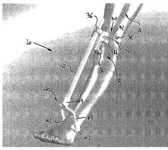
[0021] Fig. 2 is a perspective view of a PSI jig in accordance with the
present
disclosure as positioned on a leg;
[0022] Fig. 3 is an elevation view of the PSI jig of Fig. 2; and
[0023] Fig. 4 is a schematic view of a PSI jig in accordance with the
present
disclosure.
DETAILED DESCRIPTION
[0024] Referring to Fig. 1, there is illustrated a method for planning
and executing
subchondral injection surgery at 10. The method 10 may be performed on any
appropriate bone (e.g., spinal applications), but is typically used for
subchondral
injection in either one of the tibia and femur at the knee joint. The method
10 is
used to inject a compound or filler material through cortical bone structure
into voids
of trabecular bone structure.
[0025] According to step 12, the bone and soft tissue are modelled. The
modeling
is typically a three-dimensional (3D) reconstruction based on the segmentation
of
magnetic resonance imagery (MRI). The segmented structures may potentially
include: the bone, the bone void, cartilage, the skin and various types of
soft tissue.
Other appropriate types of imagery techniques may be used to enable the
modeling
performed in step 12, such as radiography. MRI reconstruction is however well
suited for the method 10 as it allows to see the voids in the bone.
3
CA 02908780 2015-10-05
WO 2014/197989
PCT/CA2014/050544
[0026] According to step 14, the voids are modeled or identified using
the bone
and soft tissue models obtained in step 12. By way of the void modeling of
step 12,
the size (i.e., volume) and location of the voids are determined relative to
the bone
and soft tissue model of step 12. The void modeling 14 may be performed with
the
assistance of an operator looking at the images obtained in step 12 and may
include
various manipulations on the images (segmentation) to delimit the void and
hence
enable the calculation of the void size and location.
[0027] According to step 16, subchondral injection is planned. The
planning may
include various steps. For instance, according to substep 16A, a void filling
calculation is performed to determine the volume of compound that is necessary
for
each void to be filled. The calculation may also include parameters and
simulations
such as the flow rate of compound to be administered by the instrument (e.g.,
syringe), the bone density in the void, and a flow simulation with pressure
profile to
avoid any overflow of compound outside of the bone.
[0028] According to substep 16B, the planning may include injection site
determination. Injection site determination comprises identifying a location
on the
bone that may be drilled or pierced for injection of the compound
therethrough.
Injection site determination as in 16B may include factors as surrounding soft
tissue
from the models of step 12, and bone structure (e.g., thickness of cartilage
and
cortical bone structure) again using the bone models of step 12. Substep 16B
may
include calculating a drilling depth required to reach the void and
determining a
drilling diameter.
[0029] The subchondral injection planning 16 may also include PSI
(Patient
Specific Instrumentation) creating as in substep 16C. In the event that the
method
is used with patient specific instrumentation, PSI creating as in substep 16C
entails identifying locations on the leg upon which a support jig may be
abutted
relative to the injection site identified in substep 16B. In an embodiment,
the jigs are
made for percutaneous abutment. The PSI creating as in substep 16C takes into
consideration the bone and soft tissue modeling of the step 12 to identify
adequate
leg locations for abutment. It is desired that the actual injection site be as
close as
possible to the planned injection site identified in substep 16B, whereby
abutment
locations on the leg are typically where soft tissue is relatively thin and
thus allows
minimized movements of a support jig thereon relative to the bone. For the
knee
joint, examples of locations that could be used as abutments are the malleoli,
such
4
CA 02908780 2015-10-05
WO 2014/197989
PCT/CA2014/050544
as the posterior aspect of the malleoli, the talus, the epicondyles (e.g.,
posterior
aspect of epicondyles), the tibial tuberosity, the anterior aspect of tibial
shaft, the
proximal area of the fibula, and the patella (if it is in the same position as
where it
was on bone models of step 12). These are only provided as examples, but may
be
used for knee joint.
[0030] There is
shown in Figs. 2-4 some jigs fabricated using patient-specific
technology, to abut percutaneously against selected locations of the leg at
the knee
joint for subchondral injection, and will be described hereinafter. It is
also
considered to abut the jig directly against the bone, although it may be
desired to opt
for percutaneous abutment to render the surgery minimally invasive.
[0031] With all
information obtained from the planning of step 16, the operator
may proceed with subchondral injection surgery as in step 18. Depending on
whether patient specific instrumentation or other type of guidance (such as
optical
navigation) is used, various steps may be performed during subchondral
injection
surgery as supported by a computer.
[0032] The PSI
jig is manufactured specifically for the patient as shown in Figs. 2
and 3, so as to support various instruments that will help the operator in
locating the
injection site as planned, in drilling or piercing the cartilage and cortical
bone
structure at the location site. For instance, the PSI jig may have a depth
indicator for
the drilling depth to be monitored as planned. The PSI jig may or may not be
used
to guide the manipulations of the injection tools (e.g., syringe). The PSI jig
may be a
guide channel located opposite the injection site, as planned due to PSI
technology,
whereby the jig abuts against the leg at desired locations.
[0033] Alternatively, optical navigation may be used. In such
a case, a
registration pointer may be used to reference the bones relative to the 3D
models of
step 12, for subsequent optical navigating of the tools relative to the bone.
The
registration pointer is typically used to identify landmark points on the bone
(e.g.,
percutaneous registration may suffice), with the points gathered used to
reference
the actual bone to the models of step 12. This results in navigation being
possible
for tools such as a drill for the piercing of a hole at the planned injection
site and an
injection syringe for the injection of the compound therethrough.
[0034] It is
also considered to use local fluoroscopy imaging prior to or during the
drilling to ensure that the location sites are correctly placed and are
opposite the
CA 02908780 2015-10-05
WO 2014/197989
PCT/CA2014/050544
voids in the bone, or to verify that the void filling procedure adequately
filled the
voids.
[0035] Referring to Figs. 2 and 3, a PSI jig in accordance with an
embodiment of
the present disclosure is generally shown at 20. The PSI jig 20 is of the type
used to
drill a hole in a knee femur A at a knee B for subsequent subchondral
injection
surgery as in item 18 of Fig. 1. The PSI jig 20 may be the result of various
steps of
the method 10 of Fig. 1. The PSI jig 20 has a knee jig portion 30, an ankle
jig portion
40, and a bar 50.
[0036] The knee jig portion 30 is adapted to position itself
percutaneously on the
femur side of the knee B.
[0037] The ankle jig portion 40 positions itself percutaneously on the
ankle and,
more particularly, against the malleoli, and provides additional stability to
the PSI jig
20.
[0038] The bar 50 interfaces the knee jig portion 30 to the ankle jig
portion 40.
[0039] Fig. 4 shows embodiments in which a knee jig portion 30 is used
without
the ankle jig portion 40 and the bar 50, with straps anchoring the knee jig
portion 30
to the knee B. However, the embodiment of Fig. 2 is well suited to provide a
stable
connection of the PSI jig 20 to the leg, with the desired alignment of the PSI
jig 20
relative to the injection site.
[0040] The knee jig portion 30 has a body or structure 31. The body 31
may be,
as in Fig. 2, an arch that has abutment pads 32 to abut percutaneously against
landmarks of the femur A. The abutment pads 32 are patient-specific, in that
their
contact surfaces are machined in contour-matching geometry to be a replica of
the
site against which they will abut. Likewise, an abutment pad 33, projecting
downwardly from the body 31, may be used as an abutment against the kneecap,
again with a contour-matching surface made as per the method 10. Additional
support may be provided by abutment pad 34.
[0041] Guiding tube 35 is one of the possible configurations used to
provide
guidance to tools. The guiding tube 35 may adequately be positioned to orient
a drill
bit and an injection device relative to the planned injection site of 16 (Fig.
1). It is
also observed that the guide tube 35 may have a given height taking into
consideration a drill stop, so as to control the depth of drilling of a drill
bit entering
6
CA 02908780 2015-10-05
WO 2014/197989
PCT/CA2014/050544
the guide tube 35. Bar interface 36 projects upwardly from the body 31, and
will host
the bar 50 in telescopic relation.
[0042] The ankle jig portion 40 also has a body 41 shaped as an arch at
the ends
of which are abutment pads 42. While the abutment pads 42 may be patient-
specific, it is also considered to have generic abutment pads, with a
telescopic
relation between the bar 50 and the bar interface 36, simply to provide
leveraging
support for the knee jig portion 30, which knee jig portion 30 is the
component of the
PSI jig 20 that must be positioned with highest precision. The ankle jig
portion 40
has a bar interface 43 to receive an end of the bar 50. As shown, the bar
interface
36 of the knee jig portion 30 forms a prismatic joint with the bar 50, so as
to enable
the telescopic relation between the knee jig portion 30 and the ankle jig
portion 40.
[0043] The PSI jig 20 is given as an exemplary embodiment of a jig that
may be
used to help in performing the subchondral injection surgery according to
planning.
7
