Note: Descriptions are shown in the official language in which they were submitted.
CA 02909537 2015-10-15
WO 2013/166606
PCT/CA2013/050361
PHANTOM FOR CALIBRATION OF IMAGING SYSTEM
FIELD OF THE INVENTION
[0001] The present invention relates to imaging systems, more particularly to
a
phantom usable for the calibration thereof.
BACKGROUND ART
[0002] Imaging systems such as Magnetic Resonance Imaging (MRI) systems allow
for
the gathering of information on internal structures of the body for a variety
of medical
applications. However the images obtained from such systems are subject to
distortions
which in some cases may jeopardize the safety and/or the accuracy of the
medical
procedure for which the information is gathered.
[0003] Phantoms with interconnected hollow tubes providing control points to
calibrate
MRI imaging systems help improve the accuracy of the patient images obtained,
but the
accuracy may remain insufficient for some applications, for example the custom
manufacturing of some implants and prosthesis.
SUMMARY
[0004] In one aspect, there is provided a phantom for a medical imaging system
using
a coil, the phantom comprising: a casing having at least one open end
sealingly closed
by a first removable cover and defining a sealed enclosure filled with a
fluid, the casing
being adapted to fit within an opening of the coil; and a matrix of spherical
hollow
elements having a fixed position relative to one another and received within
the
enclosure, the matrix being defined by a plurality of interconnected rows of
the elements,
adjacent ones of the elements of a same one of the rows being interconnected
by and in
fluid communication through a hollow tube extending therebetween, each row
defining at
least one open end in fluid communication with the elements thereof, the at
least one
open end being sealingly closed by a second removable closing member, each
element
being filled with a solution having a different contrast than that of the
fluid.
[0005] In another aspect, there is provided a method of correcting
tridimensional
patient data from a medical imaging system, the method comprising: scanning a
body
portion of a patient received within an opening of a coil to create patient
images, the coil
having a given position in the imaging system; inserting a phantom within the
opening of
the coil at the given position before or after scanning the body portion and
with the
- 1 -
CA 02909537 2015-10-15
WO 2013/166606
PCT/CA2013/050361
opening being free of the body portion; scanning the phantom to create phantom
images
of a plurality of elements of the phantom, each element having a center point
with a
respective known position with respect to a reference coordinate system of the
phantom;
determining a position of the center point of each element in the phantom
images with
respect to the reference coordinate system; computing a difference between the
position
of the center point of each element in the phantom images and the
corresponding known
position to create a distortion map; and applying a correction to
tridimensional patient
data based on the distortion map, the tridimensional patient data
corresponding to the
patient images or to subsequent data created from the patient images.
[0006] In a further aspect, there is provided a phantom for a medical imaging
system,
the phantom comprising: a casing having at least one open end sealingly closed
by a
first removable closing member and defining a fluid-filled sealed enclosure,
the casing
having an outer shape adapted to be received in an opening of a coil of the
imaging
system; and a plurality of groups of interconnected hollow elements received
within the
sealed enclosure, the elements of a same one of the groups being in fluid
communication with one another, the elements of different ones of the groups
being
sealed from one another, each group including at least one open end in fluid
communication with the elements of the group and sealingly closed by a second
removable closing member, the groups being interconnected and retained within
the
sealed enclosure in a fixed position relative to one another, each element
having a
geometrical shape from which a center of the element can be determined, each
element
being filled with a fluid, the fluid filling the elements of at least one of
the groups being
more contrasting than the fluid filling the elements of at least another one
of the groups.
BRIEF DESCRIPTION OF THE DRAWINGS
[0007] Reference will now be made to the accompanying drawings, showing by way
of
illustration one of more particular embodiment(s) of the present invention and
in which:
[0008] Figure 1 is a tridimensional view of a phantom according to a
particular
embodiment;
[0009] Figure 2 is a tridimensional view of the phantom of Figure 1 with a
cover
member thereof removed;
[0010] Figure 3 is a tridimensional cross-sectional view of the phantom of
Figure 1 with
only part of the casing thereof being shown;
- 2 -
CA 02909537 2015-10-15
WO 2013/166606
PCT/CA2013/050361
[0011] Figure 4 is a tridimensional cross-sectional view of part of the
phantom of Figure
1, showing hollow elements thereof in cross-section;
[0012] Figure 5 is a tridimensional view of one end of the casing of the
phantom of
Figure 1;
[0013] Figure 6 is a tridimensional view of a spacer received within the
phantom of
Figure 1;
[0014] Figure 7 is a diagram of a process of application of a distortion map
in
accordance with a particular embodiment, using a phantom such as shown in
Figure 1;
[0015] Figures 8a-8b together show a diagram of a segmentation of the phantom
in the
process of Figure 7, in accordance with a particular embodiment;
[0016] Figure 9 is a diagram of a computation of the distortion map in the
process of
Figure 7, in accordance with a particular embodiment;
[0017] Figure 10 is a diagram of the application of the distortion map to
images in the
process of Figure 7, in accordance with a particular embodiment; and
[0018] Figure 11 is a diagram of the application of the distortion map to a
point cloud or
mesh in the process of Figure 7, in accordance with a particular embodiment.
DETAILED DESCRIPTION OF PARTICULAR EMBODIMENTS
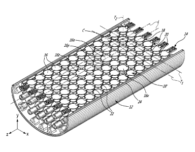
[0019] Referring to Figures 1-2, a phantom 10 generally includes a casing 12
defining
a sealed enclosure 13, and a matrix 14 of hollow elements 16 (Figure 2)
received within
the enclosure 13. The casing 12 is shaped and sized to snugly fit within the
opening of a
particular coil used for example in a MRI scan. In the embodiment shown, the
casing 12
has a substantially tubular shape, with an elliptical cross-section, and is
shaped and
sized to be received within a knee coil. In a particular embodiment, the
casing 12 is
shaped to snuggly fit within the opening of the coil. Other shapes are also
possible
depending on the coil used during calibration; for example the casing 12 may
have a
spherical or substantially spherical shape complementary to the opening of a
particular
head coil.
[0020] Referring to Figures 2-3, the hollow elements 16 are rigidly
interconnected such
as to have a fixed position relative to each other. In the embodiment shown,
the
elements 16 are retained within the enclosure 13 such as to have a fixed
position relative
to the casing 12; alternately, the matrix of elements 16 may be movable within
the
enclosure 13. In the embodiment shown, the elements 16 have a spherical shape;
such
- 3 -
CA 02909537 2015-10-15
WO 2013/166606
PCT/CA2013/050361
a shape facilitates the determination of the center of each element.
Alternately, any other
type of element having a shape (e.g. inner or outer wall surfaces) from which
a center
thereof can be readily determined may be used.
[0021] The elements 16 are fluidly interconnected in groups, with elements 16
of
different groups being sealed from one another. In the embodiment shown and as
can
be more clearly seen in Figure 4, each group corresponds to a longitudinal row
18, 18' of
the elements 16. As such, each row 18, 18' includes regularly spaced apart
elements 16
having a center located along a common axis L, with adjacent ones of the
elements 16
of the row 18, 18' being interconnected through a hollow tube extending
therebetween
and having a central axis corresponding to the common axis L.
[0022] Referring to Figures 3-4, the elements 16 of adjacent rows 18, 18' are
interconnected through pins 22 extending therefrom, which do not allow fluid
communication therethrough. In the embodiment shown, the pins 22 extend along
first
and second transverse axes T1, T2 of each spherical element 16. The transverse
axes
T1, T2 extend through the center C of the spherical element 16 and are
perpendicular to
each other and to the common axis L of the row 18, 18'. As such, the spherical
elements
16 within the matrix 14 are aligned to defined a perpendicular, tridimensional
grid
pattern. Other relative positions are also possible.
[0023] In the embodiment shown, the elements 16 of the rows adjacent the walls
24 of
the casing 12, or outer rows 18', are also connected to or otherwise engage
the walls 24
to prevent relative movement. As shown in Figure 5, in a particular embodiment
the inner
surface 26 of the casing walls 24 includes longitudinally extending rails 28
protruding
therefrom, between which the pins 22 extending from the adjacent elements 16
are
slidingly and snugly received.
[0024] Each row 18, 18' of elements 16 is completely filled with a fluid
containing a
contrasting agent detectable by the scanning machine. In a particular
embodiment, the
fluid is a solution of copper pentasulfate in water; alternate contrasting
agents which may
be used include, but are not limited to, nickel chloride and sodium chloride.
In a
particular embodiment, at least two of the rows 18, 18' have different
concentrations of
the contrasting agent from one another. In another embodiment, all the rows
18, 18'
have the same concentration of the contrasting agent. Once filled, the
elements 16 can
be scanned (CT scan, MRI, etc.) to ensure that no air remains therein.
[0025] Each row 18, 18' thus includes at least one open end 30 for insertion
of the fluid
therein. Referring back to Figure 3, in the embodiment shown, each row 18, 18'
includes
two opposed open ends 30 to facilitate filling, each end 30 being sealed by a
removable
cover member 32, shown here in the form of a threaded cap or screw. Each open
end 30
- 4 -
CA 02909537 2015-10-15
WO 2013/166606
PCT/CA2013/050361
is defined by a hollow end tube 34 extending from the element 16 at the end of
the row
18, 18' and in fluid communication therewith. The hollow end tube 34 is in
alignment and
opposed to the hollow tube 20 extending to the adjacent element of the row 18,
18'.
Although a particular embodiment for the cap 32 is shown, any other adequate
type of
cover member may also be used to seal the open end(s) 30 of each row 18, 18'.
For
example, the cover member 32 may be configured with a curved slot receiving a
pin of
the hollow end tube 34 to form a bayonet lock (not shown). An o-ring (not
shown) may be
provided between the cover member 32 and the hollow end tube 34 to help seal
their
engagement.
[0026] Referring back to Figure 1, the casing 12 also has at least one open
end
sealingly closed by a removable cover member 40 to allow the enclosure 13 to
be fluid-
tight. Although not shown, the opposed end of the casing 12 may also be
openable and
sealingly closed by a removable cover member. In the embodiment shown and as
depicted in Figures 2 and 5, bosses 40 extend from the inner surface 26 of the
casing
wall 24 adjacent the open end 44, with each boss 40 having a longitudinally
extending
threaded hole 46 defined therethrough. Referring back to Figure 1, the cover
member 40
has a rim (not shown) engaging the edge of the wall 24 around the open end 44,
and
has holes 48 defined therethrough in alignment with each of the threaded holes
46 of the
bosses 42. The cover 40 is retained in place by threaded fasteners 50 received
in the
aligned holes 46, 48. A seal such as an o-ring may be provided where the cover
40
engages the edge of the wall 24 around the open end 44. Although a particular
embodiment for the cover member 40 is shown, any other adequate type of cover
member may also be used to seal the open end(s) 44 of the casing 12.
[0027] Referring to Figure 6, in the embodiment shown, the phantom 10 further
includes a spacer 52 which is received within the enclosure 13 to abut the
closed cover
member 40 and the portion of the matrix 14 adjacent thereto, to prevent
movement in the
longitudinal direction. The spacer 52 includes a perimeter wall 54 having oval
portions 56
with a shape complementary to that of the inner surface 26 of the casing wall
24, such
as to be in abutment therewith. Inwardly protruding portions 58 are defined
between the
oval portions 56, located, sized and shaped to be complementary to the bosses
42
extending from the inner surface 26 of the casing wall 24 to surround and abut
them.
Arms 60 extend from the perimeter wall 54, here from the oval portions 56
thereof, and
are sized to contact the matrix 14, i.e. the elements 16 or hollow tubes 20
extending
therebetween, while the opposed edge of the perimeter wall 54 abuts the closed
cover
member 40. Thus, in the embodiment shown, the spacer 52 prevents longitudinal
movement of the matrix 14 of elements 16 within the enclosure 13, while the
abutment
between the pins 22 extending from the elements 16 of the outer rows 18' and
the inner
- 5 -
CA 02909537 2015-10-15
WO 2013/166606
PCT/CA2013/050361
surface 26 of the casing wall 24 prevents radial movement of the matrix 14,
and the
engagement of the pins 22 extending from some of the elements 16 of the outer
rows 18'
between the rails 28 prevents circumferential movement of the matrix 14. Other
types of
engagement/connections between the matrix 14 of elements 16 and the casing 12
may
be provided, as long as movement of the matrix 14 within the sealed enclosure
13 is
prevented.
[0028] The enclosure 13 is filled with a fluid, which in a particular
embodiment has a
different contrast than that of the fluid contained in the elements 16. In a
particular
embodiment, the enclosure 13 is filled with a fluid less contrasting than the
fluid
contained in the elements 16. In another particular embodiment, the enclosure
13 is filled
with a fluid having a similar contrast than the fluid contained in the
elements 16. In a
particular embodiment, the enclosure 13 is filled with distilled water. An
alternate fluid
which may be used includes, but is not limited to, silicon oil.
[0029] The individual elements 16 are sized such that at least three elements
fit within
the field of view of the scanning machine. The individual elements 16 have
each have a
center point having a known position, and the center points are thus located
at known
distances from one another. In the embodiment shown the elements 16 are
regularly
spaced apart and have known similar dimensions, but alternately spacing
between the
elements 16 and/or size of the elements 16 may vary. The interconnected
elements 16
are manufactured using a process having small tolerances and/or measured using
a
process having small tolerances, such that the dimensions of each element 16
and its
relative position within the matrix 14 is known with a precision exceeding
that of the
scanning equipment being calibrated.
[0030] In a particular embodiment, the elements 16 include at least three (3)
reference
elements which are distinguishable from the other elements and from one
another, and
which are disposed such as to define a reference coordinate system of the
phantom 10.
In a particular embodiment, the reference elements have a larger diameter than
that of
the remaining elements 16 such as to be distinguishable therefrom, and are
differently
spaced with respect to one another such as to be distinguishable from one
another. In
other embodiments, the reference elements may be distinguishable by having
smaller
dimensions than the other elements, different dimensions from one another,
different
shapes with respect to the other elements and/or to one another, different
wall
thicknesses with respect to the other elements and/or to one another, a
different contrast
level with respect to the other elements and/or to one another, etc.
[0031] In the particular embodiment shown and with reference to Figures 3 and
4, the
elements 16 include four (4) reference elements, in order to be able to locate
the
- 6 -
CA 02909537 2015-10-15
WO 2013/166606
PCT/CA2013/050361
reference coordinate system even if one of the reference elements is missing
from or
incomplete in the scan. A reference element 160 defines the origin of the
reference
coordinate system; a reference element 16y is located immediately adjacent the
reference element 160 in the direction of the Y-axis; a reference element 16x
is spaced
apart from the reference element 160 in the direction of the X-axis, with one
of the other
elements 16 being located therebetween; and a reference element 16z is spaced
apart
from the reference element 160 in the direction of the Z-axis, with two of the
other
elements 16 being located therebetween. Different arrangements are also
possible. For
example, the reference elements could be located elsewhere than directly on
the X, Y
and Z axes, and the position of the axes may then be calculated from the
relative
position of the reference elements.
[0032] In a particular embodiment, the rows 18, 18' of elements 16 and the
casing 12
are formed using a rapid prototyping method, for example selective laser
sintering from
powder or liquid polymer material. The elements 16 and casing 12 can be
manufactured
separately and then assembled, or alternately be manufactured in a single
piece. Rapid
prototyping methods allow for the elements 16 to be manufactured with small
tolerances
with respect to the original CAD drawing(s), such as to accurately know the
dimensions
of each element 16 and its relative position within the matrix 14. In a
particular
embodiment, the dimensions of each element 16 are known within a tolerance of
100microns or lower, and its relative position within the matrix 14 is known
with a
tolerance of 50 microns or lower.
[0033] In use, and referring to Figure 7, the body portion of the patient
which needs to
be scanned is placed in an appropriate type of coil, and the patient is
scanned with the
scanning system, which in a particular embodiment is a MRI, in order to obtain
patient
images, as set forth in step 106. Once the images are obtained, the patient is
removed
from the MRI, and the phantom 10 is placed in the coil. With the coil in the
same position
within the scanning machine as during the patient scan, the phantom 10 is
scanned to
obtain phantom images, as set forth in step 100. The phantom images are
segmented,
as shown in 102, and the distortion map is computed, as shown in 104.
[0034] In a particular embodiment, a typical processing of the patient images
is
performed as follows: the patient images are segmented to create contour(s) or
point
cloud(s) as per step 108, a mesh is then computed from the contour(s) or point
cloud(s)
as per step 110, and tridimensional surfaces are then created from the mesh,
as per
step 112. The patient images, contour(s), point cloud(s), mesh and
tridimensional
surfaces may be affected by the distortion of the scanning system. In a
particular
embodiment, the scan of the phantom 10 is used to calibrate the tridimensional
patient
- 7 -
CA 02909537 2015-10-15
WO 2013/166606
PCT/CA2013/050361
data for increased precision. As illustrated in Figure 7, a distortion map
compensating for
the distortion of the system can be applied directly to the patient images, to
the
contour(s)/point cloud(s), or to the mesh, so that calibrated tridimensional
surfaces are
obtained at step 112, as will be detailed further below.
[0035] Figures 8a-8b show the details of the segmentation of the phantom
images of
step 102, in accordance with a particular embodiment. For each image, a
thresholding of
the image is performed at 114, where the pixels of the images are separated
into a first
group above a given threshold and a second group below the given threshold,
such as to
create a black and white image. In this image, regions of different
intensities each
representing one of the elements are detected at 116, and the corresponding
slice of
each element, which is in a particular embodiment using spherical elements is
disc-
shaped, is detected in each region at 118. Then, a filtration of the center
points is
performed, to compensate for false detection due to noise. For every center
point, an
evaluation is performed to determine if the center point is located in a
region
corresponding to the intensity of an element, as shown at 120. If there is no
correspondence, the center point is deleted, as shown at 122. If the center
point
corresponds to a region, the center point is kept. The next center point is
evaluated until
all the center points of the image have been filtered. The following image is
then treated.
When all the images have been treated, the segmentation is converted to the
imaging
reference system at 124, as determined with the help of the reference
elements. Groups
of center points are then formed from the neighborhood of each center point at
126.
[0036] A filtration of the groups is then performed, such as to eliminate the
groups of
center points which are not linked to the elements. For every group, the
number of
center points within the group is computed at 128. A constant N is fixed by
the user to
determine the threshold of noise. In a particular embodiment, N is 10; other
values may
also be used. If the number of center points is smaller than N, the group is
kept. If not,
the group is deleted at 130. The next group is then evaluated until all the
groups have
been filtered. The center of each group is then computed at 132, and a point
cloud 134
corresponding to the real grid of points of the phantom is obtained.
[0037] Figure 9 shows the details of the computation of the distortion map of
step 104,
in accordance with a particular embodiment. The distortion map is computed
from the
point cloud 134 corresponding to the grid of points of the scan of the phantom
10 and
from the nominal point cloud 136 of the phantom (for example from a CAD model)
including a known position for the center point of each element 16. For every
center
point, a determination of the associated nominal element from the nominal
point cloud is
performed through a search of its neighborhood, as shown at 138. A point cloud
is
- 8 -
CA 02909537 2015-10-15
WO 2013/166606
PCT/CA2013/050361
generated at 140 in correspondence with the nominal element, and the nominal
element
is aligned with the segmented element from the real grid of points, at 142. In
a particular
embodiment, alignment of the nominal element with the segmented element is
performed using an iterative closest point algorithm or ICP. The position of
the aligned
nominal element is computed at 144, and the vector between the nominal element
and
the segmented element is then computed at 146. When these operations have been
performed for every center point, the distortion map 148 is defined.
[0038] As mentioned above and as shown in Figure 7, the distortion map 148 can
be
applied to any tridimensional data obtained from the patient scan, including
the patient
images, the contour(s)/point cloud(s) generated through the segmentation
thereof, and
the mesh computed therefrom, in order to obtain calibrated tridimensional
surfaces.
[0039] Referring to Figure 10, the application of the distortion map 148 to a
patient
point cloud or mesh 150 (such as obtained after steps 108 or 110 of Figure 7)
is shown,
in accordance with a particular embodiment. The volume of the point cloud or
mesh is
separated following the grid of the phantom, as shown at 152. Then, for all
blocks of the
grid, the distortion map is applied to the points in that block, as shown at
154. When this
operation has been performed for every block of the grid, a calibrated point
cloud or
mesh 156 is obtained.
[0040] Referring to Figure 11, the application of the distortion map 148 to
the patient
images 158 (such as obtained after step 106 of Figure 7) is shown, in
accordance with a
particular embodiment. For each image, the distortion map is interpolated in
the plane of
the image, as shown at 160. Each pixel of the image is then deformed based on
that
interpolated distortion map, as per 162. When these operations have been
performed for
every patient image, calibrated patient images 164 are obtained.
[0041] Calibration of the distortion also allows for calibration of the 3D
magnetic field of
the scanning machine.
[0042] The phantom may 10 may be scanned before or after the body portion of
the
patient. Calibration with the phantom 10 may also be regularly performed
independently
of the number of patients being scanned between the calibration sessions.
[0043] The phantom images can also be used to calibrate the contrast of the
scanning
machine, either by comparison of the scan of elements 16 having different
concentrations of contrasting agent, or by comparison of the scan of the
elements 16
and the surrounding fluid which has a different contrast than the fluid
contained in the
elements 16. A calibration is performed to associate the elements 16 having
different
- 9 -
CA 02909537 2015-10-15
WO 2013/166606
PCT/CA2013/050361
contrasts or the elements 16 and surrounding fluid with an expected intensity
level of the
scanned image.
[0044] The embodiments of the invention described above are intended to be
exemplary. Those skilled in the art will therefore appreciate that the
foregoing description
is illustrative only, and that various alternate configurations and
modifications can be
devised without departing from the spirit of the present invention.
Accordingly, the
present invention is intended to embrace all such alternate configurations,
modifications
and variances which fall within the scope of the appended claims.
-10-