Note: Descriptions are shown in the official language in which they were submitted.
LASER FIDUCIALS FOR AXIS ALIGNMENT IN CATARACT SURGERY
CROSS-REFERENCE
[0001] This application claims priority to U.S. provisional application No.
61/813,172 filed on
April 17,2013, which is related to U.S. Patent Application Serial No.
14/199,087, filed on March 6,
2014, entitled "MICROFEMTOTOMY METHODS AND SYSTEMS," which claims priority to
U.S. Provisional Application No. 61/788,201.
BACKGROUND
[0002] The present disclosure relates generally to the marking of anatomical
features to facilitate the
treatment of the nearby tissue structures, such as a tissue of an eye.
Although specific reference is
made to marking tissue for surgery such as eye surgery, embodiments as
described herein can be
used in many ways with many anatomical structures to facilitate the treatment
of many tissue
structures.
[0003] Cutting of materials can be done mechanically with chisels, knives,
scalpels and other tools
such as surgical tools. However, prior methods and apparatus of cutting can be
less than desirable
and provide less than ideal results in at least some instances. For example,
at least some prior
methods and apparatus for cutting materials such as tissue may provide a
somewhat rougher surface
than would be ideal. Pulsed lasers can be used to cut one or more of many
materials and have been
used for laser surgery to cut tissue.
[0004] Examples of surgically tissue cutting include cutting the cornea and
crystalline lens of the
eye. The lens of the eye can be cut to correct a defect of the lens, for
example to remove a cataract,
and the tissues of the eye can be cut to access the lens. For example the
cornea can be to access the
cataractous lens. The cornea can be cut in order to correct a refractive error
of the eye, for example
with laser assisted in situ keratomileusis (hereinafter "LASIK").
100051 Many patients may have visual errors associated with the refractive
properties of the eye such
as nearsightedness, farsightedness and astigmatism. Astigmatism may occur when
the corneal
curvature is unequal in two or more directions. Nearsightedness can occur when
light focuses before
the retina, and farsightedness can occur with light refracted to a focus
behind the retina. There are
numerous prior surgical approaches for reshaping the cornea, including laser
assisted in situ
keratomilcusis (hereinafter "LAS1K"), all laser LAS1K, femto LAS1K,
comcaplasty, astigmatic
-1-
Date Recue/Date Received 2020-08-28
CA 02909684 2015-10-16
WO 2014/172545 PCMJS2014/034508
keratotomy, corneal relaxing incision (hereinafter "CRI"), and Limbal Relaxing
Incision (hereinafter
"LRI"). Astigmatic Keratotomy, Corneal Relaxing Incision (CRI), and Limbal
Relaxing Incision
(LRI), corneal incisions are made in a well-defined manner and depth to allow
the cornea to change
shape to become more spherical.
100061 Cataract extraction is a frequently performed surgical procedure. A
cataract is formed by
pacification of the crystalline lens of the eye. The cataract scatters light
passing through the lens
and may perceptibly degrade vision. A cataract can vary in degree from slight
to complete opacity.
Early in the development of an age-related cataract the power of the lens may
increase, causing near-
sightedness (myopia). Gradual yellowing and pacification of the lens may
reduce the perception of
blue colors as those shorter wavelengths are more strongly absorbed and
scattered within the
cataractous crystalline lens. Cataract formation may often progresses slowly
resulting in progressive
vision loss.
[0007] A cataract treatment may involve replacing the opaque crystalline lens
with an artificial
intraocular lens (IOL), and an estimated 15 million cataract surgeries per
year are performed
worldwide. Cataract surgery can be performed using a technique termed
phacoemulsification in
which an ultrasonic tip with associated irrigation and aspiration ports is
used to sculpt the relatively
hard nucleus of the lens to facilitate removal through an opening made in the
anterior lens capsule.
The nucleus of the lens is contained within an outer membrane of the lens that
is referred to as the
lens capsule. Access to the lens nucleus can be provided by performing an
anterior capsulotomy in
which a small round hole can be formed in the anterior side of the lens
capsule. Access to the lens
nucleus can also be provided by performing a manual continuous curvilinear
capsulorhexis (CCC)
procedure. After removal of the lens nucleus, a synthetic foldable intraocular
lens (IOL) can be
inserted into the remaining lens capsule of the eye.
1000811 At least some prior laser surgery systems can provide less than ideal
results when used to
place an intraocular lens in the eye to treat aberrations of the eye such as
low order aberrations
comprising astigmatism or higher order aberrations. While accommodating IOLs
can correct
refractive error of the eye and restore accommodation, the prior accommodating
TOLs can provide
less than ideal correction of the astigmatism of the eye.
100091 Thus, improved methods and systems would be helpful for more precisely
marking and
tracking anatomical features in tissue, particularly the eye, to better
position tissue cuts and place
implants such as intraocular lenses (IOLs) in the eye.
-2-
CA 02909684 2015-10-16
WO 2014/172545 PCMJS2014/034508
SUMMARY
[0010] Embodiments as described herein provide improved methods and apparatus
of marking and
tracking the tissue structures such as the eye, in many embodiments to
facilitate surgical procedures
for the eye such as the implantation of an artificial intraocular lens (TOL)
or other lens placed with
the eye. In many embodiments, a fiducial is generated on an anatomical
structure of the eye in order
to position an axis of the IOL with an axis of the eye. In many embodiments,
an implantable lens
device comprises a marker, and the implantable lens device is positioned so
that the marker of the
implantable device is placed in a positional relationship relative to the
fiducial. In many
embodiments, the implantable device comprises an artificial intraocular lens
such as a tonic
intraocular lens which can treat astigmatism of the eye. Positioning the
implantable device so that
the fiducial is in the positional relationship relative to the fiducial can
comprise aligning the axis of
the implantable device with an axis of the eye, such as an astigmatic axis of
the eye.
[0011] In a first aspect, a method of implanting an implantable device in an
eye of a patient is
provided. A fiducial is generated on an anatomical structure of the eye. The
implantable device is
placed so that a marker of the implantable device is in a positional
relationship relative to the
fiducial.
100121 In many embodiments, the eye is retained with a patient interface
coupled to the eye with
suction. The fiducial can be generated when the eye is retained with the
patient interface. In some
cases, the patient interface may distort one or more tissue structures in the
eye which can lead to
inaccurate fiducial generation. Thus, the fiducial can alternatively be
generated prior to retaining the
eye with the patient interface.
[0013] In many embodiments, the implantable device comprises an intraocular
lens. The marker of
the intraocular lens and the fiducial generated on the eye can be visible to a
user with a camera
image or an operating microscope image provided to the user when the
intraocular lens has been
placed.
[0014] A user can input a treatment axis of an astigmatism of the eye. A first
fiducial and a second
fiducial can be generated on an internal anatomical structure of the eye to
define the treatment axis
extending across a pupil of the eye. The marker can comprises a first marker
and a second marker
placed on opposite sides of the implantable device to define a lens axis of an
intraocular lens. The
marker and the fiducial can be visible to a user to determine an alignment of
the treatment axis with
the lens axis. In some embodiments. the first fiducial and the second fiducial
are located on the
cornea away from an entrance pupil of the eye, and the first marker, the
second marker, the first
fiducial and the second fiducial are displayed in an image visible to a user.
-3-
CA 02909684 2015-10-16
WO 2014/172545 PCMJS2014/034508
[0015] A measurement structure of the eye can be measured with a laser system
when the patient has
been placed on a patient support of the laser system. The fiducial can be
generated on the
anantomical structure of the eye in response to the orientation of the
measurement structure. The
measurement structure of the eye can comprise one or more of a cornea of the
eye, an iris of the eye
or a crystalline lens of the eye and wherein the orientation comprises one or
more of an angle of an
astigmatic axis of the cornea, a rotational angle of the iris about a pupil of
the eye or an astigmatic
axis of the lens of the eye.
[0016] The implantable device can comprise an artificial intraocular lens such
as a tonic intraocular
lens. The positional relationship can comprise a pre-determined positional
relationship.
[0017] The implantable device can be positioned so that the fiducial in the
positional relationship
relative to the fiducial to align a vision correcting axis of the implantable
device with an aberration
axis of the eye. The aberration axis of the eye may comprise an astigmatic
axis or an axis of a higher
order aberration. And, the implantable device can corrects a higher order
aberration of the eye
comprising one or more of coma, trefoil or spherical aberration.
[0018] 1 be marker of the implantable device and the fiducial placed on the
internal anatomical
structure of the eye can have many shapes, including one or more of a dot, a
line, a rectangle, an
arrow, a cross, a trapezoid, a rectangle, a square, a chevron, a pentagon, a
hexagon, a circle, an
ellipse, or an arc. The fiducial may have a shape corresponding to a shape of
the marking of the
implantable device. The shape of the fiducial may be similar to the shape of
the marking. The shape
of the fiducial may be complementary to the shape of the marking.
[0019] Typically, the fiducial is generated on the anatomical structure of the
eye by marking the
anatomical structure with a laser. The internal anatomical structure may
comprise an internal
structure of one or more of the limbus, the cornea, the sclera, in the lens
capsule, the iris, the stroma,
or in the crystalline lens nucleus. And, the internal structure can be visible
to a user when
implantable lens is placed. In many embodiments, the fiducial is generated at
least on the periphery
of the cornea or on the limbus.
[00201 At least two fiducials may be generated on the anatomical structure of
the eye, for example, a
first fiducial and a second fiducial can be generated. A shape of the first
fiducial can be different
from a shape of the second fiducial. A shape of the first fiducial can be the
same as a shape of the
second fiducial. The at least two fiducials can form a line corresponding to
an axis of the eye and
the implantable device can comprise at least two marks to determine a
centration of the lens with
respect to a pupil of the eye when the at least two marks are positioned near
the at least two
-4-
fiducials. The axis may comprise an astigmatic axis of the eye. The line
formed from the at least two
fiducials can be aligned with, parallel to, transverse to, or perpendicular to
the axis of the eye.
[0021] In another aspect, an apparatus is provided. The apparatus comprises a
laser to generate a
laser beam, a scanner to scan the laser beam, and a processor operatively
coupled to the laser and the
scanner. The processor comprises a tangible medium configured with
instructions to perform any
variation of the above methods.
[0022] In yet another aspect, an apparatus for implanting an implantable
device in an eye of the
patient is provided. The apparatus comprises a laser to generate a laser beam,
a scanner to scan the
laser beam, and a patient interface. The scanner scans the laser beam onto the
eye of a patient to
generate a fiducial on an anatomical structure of the eye. The patient
interface is coupled to the eye
with suction. The apparatus can further comprise an operating microscope to
provide an image of
the generated fiducial to a user. The apparatus can further comprise a user
input for inputting a
treatment axis of an astigmatism of the eye. The scanner can be configured to
generate a first
fiducial and a second fiducial on an internal anatomical structure of the eye
to define the treatment
axis extending across a pupil of the eye.
[0022A] In one embodiment there is provided an apparatus that includes: a
laser to generate a laser beam;
a scanner to scan the laser beam; an implantable device having a vision
correcting axis and a marker; and
a processor operatively coupled to the laser and the scanner. The processor is
configured to cause the
apparatus to generate a fiducial on an anatomical structure of an eye of a
patient to enable the marker of
the implantable device to be placed in a positional relationship relative to
the fiducial to align the vision
correcting axis of the implantable device with an aberration axis of the eye.
The processor is configured
to cause the apparatus to generate at least two fiducials on the anatomical
structure of the eye; wherein a
first fiducial and a second fiducial are generated on an internal anatomical
structure of the eye to define
the treatment axis extending across a pupil of the eye. The marker comprises a
first marker and a second
marker placed on opposite sides of the implantable device to define the vision
correcting axis of the
implantable device. The marker and the fiducial are visible to a user to
determine an alignment of the
aberration axis with the vision correcting axis.
10022B1 In one embodiment there is provided an apparatus that includes: a
laser to generate a laser beam;
a scanner to scan the laser beam; an implantable device having a marker; and a
processor operatively
coupled to the laser and the scanner to cause the apparatus to generate a
fiducial on an anatomical
structure of an eye of a patient to enable the marker of the implantable
device to be
-5-
Date Recue/Date Received 2020-08-28
placed in a positional relationship relative to the fiducial. The shape of the
fiducial is complementary to
the shape of the marker and the complementary shapes comprise an empty outline
shape and a filled
shape that corresponds to and fits within the empty outline shape.
BRIEF DESCRIPTION OF THE DRAWINGS
[0023] Figure 1 shows a perspective view showing a laser eye surgery system,
in accordance with
many embodiments;
[0024] Figure 2 shows a simplified block diagram showing a top level view of
the configuration of a
laser eye surgery system, in accordance with many embodiments;
[0025] Figure 3A shows a simplified block diagram illustrating the
configuration of an optical
assembly of a laser eye surgery system, in accordance with many embodiments;
[0026] Figure 3B shows a mapped treatment region of the eye comprising the
cornea, the posterior
capsule, and the limbus, in accordance with many embodiments;
[0027] Figure 4 shows a method of treating a patient, in accordance with many
embodiments;
[0028] Figure 5A1 shows a front view of the eye having a fiducial created
thereon, in accordance
with many embodiments;
[0029] Figure 5A2 shows a side view of the front of the eye of Figure 5A1;
[0030] Figure 5B1 shows a front view of the eye having a fiducial created
thereon, in accordance
with many embodiments;
[0031] Figure 5B2 shows a side view of the front of the eye of Figure 5B1;
-5a-
Date Recue/Date Received 2020-08-28
CA 02909684 2015-10-16
WO 2014/172545 PCMJS2014/034508
[0032] Figure 5C1 shows a front view of the eye having a fiducial created
thereon, in accordance
with many embodiments;
[0033] Figure 5C2 shows a side view of the front of the eye of Figure 5C1;
[0034] Figure 6 shows various configuration of fiducials, in accordance with
many embodiments;
and
[0035] Figures 7A to 7D show front views of one or more fiducials created on
the eye for placement
in predetermined positional relationships with an artificial intraocular lens
(TOL);
[0036] Figure 8 shows an TOL placed in an eye, in accordance with many
embodiments; and
[0037] Figure 9 shows haptics of an IOL positioned with corresponding
Fiducials, in accordance
with many embodiments.
DETAILED DESCRIPTION
[0038] Methods and systems related to laser eye surgery are disclosed. In many
embodiments, a
laser is used to form precise incisionsin the limbus, the cornea, in the lens
capsule, the iris, the
stroma, and/or in the crystalline lens nucleus. Although specific reference is
made to tissue marking
and alignment for laser eye surgery, embodiments as described herein can be
used in one or more of
many ways with many surgical procedures and devices, such as orthopedic
surgery, robotic surgery
and microkeratomes.
100391 The embodiments as describe herein are particularly well suit for
treating tissue, such as with
the surgical treatment of tissue. In many embodiments, the tissue comprises an
optically
transmissive tissue, such as tissue of an eye. The embodiments as described
herein can be combined
in many ways with one or more of many known surgical procedures such as
cataract surgery, laser
assisted in situ keratomileusis (hereinafter "LASIK"), laser assisted
subepithelial keratectomy
(hereinafter "LASEK"),
100401 Methods and systems related to laser treatment of materials and which
can be used with eye
surgery such as laser eye surgery are disclosed. A laser may be used to form
precise incisions in the
cornea, in the lens capsule, and/or in the crystalline lens nucleus, for
example. The embodiments as
described herein can be particularly well suited for decreasing the amount of
energy to the eye and
increasing the accuracy of the cutting of the material such as tissue, for
example.
[0041] The present disclosure provides methods and apparatus for providing
adjustment to
compensate for variations in disposable elements and other attachments,
tolerances in hardware and
alignment, and patient anatomy. The methods and apparatus may comprise a
software look up table
(hereinafter "LUT") embodied in a tangible medium. The LUT may comprise a map
of locations of
-6-
CA 02909684 2015-10-16
WO 2014/172545 PCMJS2014/034508
the cutting volume in order to the control of actuators that direct the
ranging (target detection) and
the cutting modalities. A baseline LUT can be generated for a generalized
system using optical
based rules and physics, detailed modeling of components, and anchoring (one
time) to a finite data
set as described herein. The expected variations can be reduced into a set of
finite and manageable
variables that are applied to modify the tables subsequent to the original
generation of the tables.
For a constructed system having constructed components with manufacturing
tolerances, fine tuning
and modification of the LUTs can be achieved thiu simple modifications of the
tables based on
individual system and automated measurements. These individualized
measurements of a
constructed system can be applied to variations due to one or more of: tool-to-
tool variation, tool to
itself variation (for example align variations), output attachment variations
(for example disposable
contact lenses), or patient to patient (for example individual patient
anatomy), and combinations
thereof, for example.
[0042] In many embodiments, one or more of the following steps can be
performed with the
processor and methods as described herein. For example, baseline LUT
generation can be
performed comprising mapping and position detection in order to provide
actuator commands to
evaluate system output performance. A baseline transfer function can be
generated for a patient
coordinate reference system such as XYZ to detect actuators of the system, for
example. Baseline
LUT generation can be performed to map cutting to actuators. A transfer
function can be generated
for XYZ to cutting actuators, for example. Baseline LUTs (transfer functions)
can be generated via
model (ray trace), data, or a combination, for example. The baseline LI Yrs
can be modified given
variations in the system, disposable, eye, application, for example. The
baseline LUT modification
may comprise an adjustment to the baseline LUT, for example. The baseline LUT
modification may
comprise a software (hereinafter "SW") adjustment to compensate for hardware
(hereinafter "HVC)
variations, for example. The LUT modification as described herein can extend
surgical volume, so
as to treat the cornea, the Embus and the posterior capsule, either in lateral
extent, axial extent, and
resolution, for example. The LUT methods and apparatus can enable switching in
tools for
calibration and other optical components to accessorize ¨ output attachments,
for example. The
LUT can be set up so that the system is capable of measuring location of
attachments at two surfaces
and then can accurately place cuts in targeted material volume based on
modifying the baseline LUT
using this the locations of the two surfaces, for example. The LUTS can
provide more cuts ranging
from lens, capsule, corneal incisions for cataract, cornea flaps, for example.
The different sub-
systems as described herein can have separate LUTS, which can be combined with
calibration
process as described herein, for example.
-7-
CA 02909684 2015-10-16
WO 2014/172545 PCMJS2014/034508
[0043] Alternatively, or in combination, the same sub-system can be used for
both ranging and
cutting, for example. The UF system can be used at a low power level to find
surfaces and then used
at high power for cutting, for example. The LU'l s can be used such that the
location mode differs
from the cutting mode. For example, the cut locations can differ based on
changes with power level.
The cut location may not occur at focus, for example when the energy per pulse
substantially
exceeds the threshold amount of energy, for example.
[0044] In many embodiments, the LUTs of the methods and apparatus as described
herein follow
these principles. The baseline LUT can generated by ray tracing and data
anchoring using specific
tooling, for example. In many embodiments, each optically transmissive
structure of the patient
interface, for example a lens, is read by the system to determine its
thickness and location. These
numbers can be used to modify the LUTS to attain <100um accuracy, for example.
[00451 In many embodiments, the LUTs of the methods and apparatus as described
herein are also
modified to account for alignment tilts, contact lens mounting, contact lens
variations so as to
achieve <I 00um accuracy on cuts, for example. In many embodiments, a bubbles
in plastic flatness
test with the calibration apparatus as described herein generates offset and
tilt adjustments of
baseline UF LUT.
[0046] In many embodiments, the baseline component specifications may be less
than ideal for
delivering an appropriate system performance, and the final performance can be
refined using SW
corrections and factors based on the components of the individual system which
can be determined
from optically-grounded data-anchored baseline LUTs further modified for
enhanced performance,
for example.
[0047] A feedback loop can be used to build the enhanced or modified LUTs for
the individual laser
system, for example. The feedback methods and apparatus as described herein
can allow SW
adjustments based on LUTs and other SW factors that may not be corrected with
hardware
alignment, for example.
[0048] The LUTs and the methods an apparatus configured to modify the look up
tables so as to
enhance system performance can provide an improvement within the 3D surgical
volume as
described herein. The methods and apparatus as described herein can provide
improved surgery for
more patients with a level of high performance. The methods and apparatus as
described herein can
provide high performance using off-the-shelf components, such as high volume
low cost
components, such that the surgical procedures as described herein can be
available to many patients.
[0049] As used herein, the terms anterior and posterior refers to known
orientations with respect to
the patient. Depending on the orientation of the patient for surgery, the
terms anterior and posterior
-8-
CA 02909684 2015-10-16
WO 2014/172545 PCMJS2014/034508
may be similar to the terms upper and lower, respectively, such as when the
patient is placed in a
supine position on a bed. The terms distal and anterior may refer to an
orientation of a structure
from the perspective of the user, such that the terms proximal and distal may
be similar to the terms
anterior and posterior when referring to a structure placed on the eye, for
example. A person of
ordinary skill in the art will recognize many variations of the orientation of
the methods and
apparatus as described herein, and the terms anterior, posterior, proximal,
distal, upper, and lower
are used merely by way of example.
100501 As used herein, the terms first and second are used to describe
structures and methods
without limitation as to the order of the structures and methods which can be
in any order, as will be
apparent to a person of ordinary skill in the art based on the teachings
provided herein,
[00511 Figure 1 shows a laser eye surgery system 2, in accordance with many
embodiments,
operable to form precise incisions in the cornea, in the lens capsule, and/or
in the crystalline lens
nucleus. The system 2 includes a main unit 4, a patient chair 6, a dual
function footswitch 8, and a
laser footswitch 10.
[0052] The main unit 4 includes many primary subsystems of the system 2. For
example, externally
visible subsystems include a touch-screen control panel 12, a patient
interface assembly 14. patient
interface vacuum connections 16, a docking control keypad 18, a patient
interface radio frequency
identification (RFID) reader 20, external connections 22 (e.g., network, video
output, footswitch,
USB port, door interlock, and AC power), laser emission indicator 24,
emergency laser stop
button 26, key switch 28, and USB data ports 30.
100531 The patient chair 6 includes a base 32, a patient support bed 34, a
headrest 36, a positioning
mechanism, and a patient chair joystick control 38 disposed on the headrest
36. The positioning
control mechanism is coupled between the base 32 and the patient support bed
34 and headrest 36.
The patient chair 6 is configured to be adjusted and oriented in three axes
(x, y, and z) using the
patient chair joystick control 38. The headrest 36 and a restrain system (not
shown, e.g., a restraint
strap engaging the patient's forehead) stabilize the patient's head during the
procedure. The
headrest 36 includes an adjustable neck support to provide patient comfort and
to reduce patient
head movement. The headrest 36 is configured to be vertically adjustable to
enable adjustment of
the patient head position to provide patient comfort and to accommodate
variation in patient head
size.
100541 The patient chair 6 allows for tilt articulation of the patient's legs,
torso, and head using
manual adjustments. The patient chair 6 accommodates a patient load position,
a suction ring
capture position, and a patient treat position. In the patient load position,
the chair 6 is rotated out
-9-
CA 02909684 2015-10-16
WO 2014/172545 PCMJS2014/034508
from under the main unit 4 with the patient chair back in an upright position
and patient footrest in a
lowered position. In the suction ring capture position, the chair is rotated
out from under the main
unit 4 with the patient chair back in reclined position and patient footrest
in raised position. In the
patient treat position, the chair is rotated under the main unit 4 with the
patient chair back in reclined
position and patient footrest in raised position.
[0055] The patient chair 6 is equipped with a "chair enable" feature to
protect against unintended
chair motion. The patient chair joystick 38 can be enabled in either of two
ways. First, the patient
chair joystick 38 incorporates a "chair enable" button located on the top of
the joystick. Control of
the position of the patient chair 6 via the joystick 38 can be enabled by
continuously pressing the
"chair enable" button. Alternately, the left foot switch 40 of the dual
function footswitch 8 can be
continuously depressed to enable positional control of the patient chair 6 via
the joystick 38.
[0056] In many embodiments, the patient control joystick 38 is a proportional
controller. For
example, moving the joystick a small amount can be used to cause the chair to
move slowly.
Moving the joystick a large amount can be used to cause the chair to move
faster. Holding the
joystick at its maximum travel limit can be used to cause the chair to move at
the maximum chair
speed. The available chair speed can be reduced as the patient approaches the
patient interface
assembly 14.
[0057] The emergency stop button 26 can be pushed to stop emission of all
laser output, release
vacuum that couples the patient to the system 2, and disable the patient chair
6. The stop button 26
is located on the system front panel, next to the key switch 28.
[0058] The key switch 28 can be used to enable the system 2. When in a standby
position, the key
can be removed and the system is disabled. When in a ready position, the key
enables power to the
system 2.
[0059] The dual function footswitch 8 is a dual footswitch assembly that
includes the left foot
switch 40 and a right foot switch 42. The left foot switch 40 is the "chair
enable" footswitch. The
right footswitch 42 is a "vacuum ON" footswitch that enables vacuum to secure
a liquid optics
interface suction ring to the patient's eye. The laser footswitch 10 is a
shrouded footswitch that
activates the treatment laser when depressed while the system is enabled.
[0060] In many embodiments, the system 2 includes external communication
connections. For
example, the system 2 can include a network connection (e.g, an R.:145 network
connection) for
connecting the system 2 to a network. The network connection can be used to
enable network
printing of treatment reports, remote access to view system performance logs,
and remote access to
perform system diagnostics. The system 2 can include a video output port
(e.g., IIDMI) that can be
-10-
CA 02909684 2015-10-16
WO 2014/172545 PCMJS2014/034508
used to output video of treatments performed by the system 2. The output video
can be displayed on
an external monitor for, for example, viewing by family members and/or
training. The output video
can also be recorded for, for example, archival purposes. The system 2 can
include one or more data
output ports (e.g, USB) to, for example, enable export of treatment reports to
a data storage device.
The treatments reports stored on the data storage device can then be accessed
at a later time for any
suitable purpose such as, for example, printing from an external computer in
the case where the user
without access to network based printing.
[0061] Figure 2 shows a simplified block diagram of the system 2 coupled with
a patient eye 43.
The patient eye 43 comprises a cornea 43C, a lens 43L and an iris 431. The
iris 431 defines a pupil
of the eye 43 that may be used for alignment of eye 43 with system 2. The
system 2 includes a
cutting laser subsystem 44, a ranging subsystem 46, an alignment guidance
system 48, shared
optics 50, a patient interface 52, control electronics 54, a control panel/GUI
56, user interface
devices 58, and communication paths 60. The control electronics 54 is
operatively coupled via the
communication paths 60 with the cutting laser subsystem 44, the ranging
subsystem 46, the
alignment guidance subsystem 48, the shared optics 50, the patient interface
52, the control
panel/GUI 56, and the user interface devices 58.
[0062] In many embodiments, the cutting laser subsystem 44 incorporates
femtosecond (FS) laser
technology. By using femtosecond laser technology, a short duration (e.g.,
approximately 10-13
seconds in duration) laser pulse (with energy level in the micro joule range)
can be delivered to a
tightly focused point to disrupt tissue, thereby substantially lowering the
energy level required as
compared to the level required for ultrasound fragmentation of the lens
nucleus and as compared to
laser pulses having longer durations.
[0063] The cutting laser subsystem 44 can produce laser pulses having a
wavelength suitable to the
configuration of the system 2. As a non-limiting example, the system 2 can be
configured to use a
cutting laser subsystem 44 that produces laser pulses having a wavelength from
1020 nm to
1050 nm. For example, the cutting laser subsystem 44 can have a diode-pumped
solid-state
configuration with a 1030 (+/- 5) nm center wavelength.
[0064] The cutting laser subsystem 44 can include control and conditioning
components. For
example, such control components can include components such as a beam
attenuator to control the
energy of the laser pulse and the average power of the pulse train, a fixed
aperture to control the
cross-sectional spatial extent of the beam containing the laser pulses, one or
more power monitors to
monitor the flux and repetition rate of the beam train and therefore the
energy of the laser pulses, and
a shutter to allow/block transmission of the laser pulses. Such conditioning
components can include
-11-
CA 02909684 2015-10-16
WO 2014/172545 PCMJS2014/034508
an adjustable zoom assembly to adapt the beam containing the laser pulses to
the characteristics of
the system 2 and a fixed optical relay to transfer the laser pulses over a
distance while
accommodating laser pulse beam positional and/or directional variability,
thereby providing
increased tolerance for component variation.
[0065] The ranging subsystem 46 is configured to measure the spatial
disposition of eye structures in
three dimensions. The measured eye structures can include the anterior and
posterior surfaces of the
cornea, the anterior and posterior portions of the lens capsule, the iris, and
the limbus. In many
embodiments, the ranging subsystem 46 utilizes optical coherence tomography
(OCT) imaging. As
a non-limiting example, the system 2 can be configured to use an OCT imaging
system employing
wavelengths from 780 nm to 970 urn. For example, the ranging subsystem 46 can
include an OCT
imaging system that employs a broad spectrum of wavelengths from 810 nm to 850
mm. Such an
OCT imaging system can employ a reference path length that is adjustable to
adjust the effective
depth in the eye of the OCT measurement, thereby allowing the measurement of
system components
including features of the patient interface that lie anterior to the cornea of
the eye and structures of
the eye that range in depth from the anterior surface of the cornea to the
posterior portion of the lens
capsule and beyond.
[0066] The alignment guidance subsystem 48 can include a laser diode or gas
laser that produces a
laser beam used to align optical components of the system 2. The alignment
guidance subsystem 48
can include LEDs or lasers that produce a fixation light to assist in aligning
and stabilizing the
patient's eye during docking and treatment. The alignment guidance subsystem
48 can include a
laser or LED light source and a detector to monitor the alignment and
stability of the actuators used
to position the beam in X, Y, and Z. The alignment guidance subsystem 48 can
include a video
system that can be used to provide imaging of the patient's eye to facilitate
docking of the patient's
eye 43 to the patient interface 52. The imaging system provided by the video
system can also be
used to direct via the GUI the location of cuts. The imaging provided by the
video system can
additionally be used during the laser eye surgery procedure to monitor the
progress of the procedure,
to track movements of the patient's eye 43 during the procedure, and to
measure the location and
size of structures of the eye such as the pupil and/or limbus.
[0067] The shared optics 50 provides a common propagation path that is
disposed between the
patient interface 52 and each of the cutting laser subsystem 44, the ranging
subsystem 46, and the
alignment guidance subsystem 48. In many embodiments, the shared optics 50
includes beam
combiners to receive the emission from the respective subsystem (e.g, the
cutting laser
subsystem 44, and the alignment guidance subsystem 48) and redirect the
emission along the
-12-
CA 02909684 2015-10-16
WO 2014/172545 PCMJS2014/034508
common propagation path to the patient interface. In many embodiments, the
shared optics 50
includes an objective lens assembly that focuses each laser pulse into a focal
point. In many
embodiments, the shared optics SO includes scanning mechanisms operable to
scan the respective
emission in three dimensions. For example, the shared optics can include an XY-
scan mechanism(s)
and a Z-scan mechanism. The XY-scan mechanism(s) can be used to scan the
respective emission in
two dimensions transverse to the propagation direction of the respective
emission, The Z-scan
mechanism can be used to vary the depth of the focal point within the eye 41
In many
embodiments, the scanning mechanisms are disposed between the laser diode and
the objective lens
such that the scanning mechanisms are used to scan the alignment laser beam
produced by the laser
diode. In contrast, in many embodiments, the video system is disposed between
the scanning
mechanisms and the objective lens such that the seaming mechanisms do not
affect the image
obtained by the video system.
100681 The patient interface 52 is used to restrain the position of the
patient's eye 43 relative to the
system 2. In many embodiments, the patient interface 52 employs a suction ring
that is vacuum
attached to the patient's eye 43. The suction ring is then coupled with the
patient interface 52, for
example, using vacuum to secure the suction ring to the patient interface 52.
In many embodiments,
the patient interface 52 includes an optically transmissive structure having a
posterior surface that is
displaced vertically from the anterior surface of the patient's cornea and a
region of a suitable liquid
(e.g., a sterile buffered saline solution (BSS) such as Alcon BSS (Alcon Part
Number 351-55005-1)
or equivalent) is disposed between and in contact with the patient interface
lens posterior surface and
the patient's cornea and forms part of a transmission path between the shared
optics 50 and the
patient's eye 43. The optically transmissive structure may comprise a lens 96
having one or more
curved surfaces. Alternatively, the patient interface 22 may comprise an
optically transmissive
structure having one or more substantially flat surfaces such as a parallel
plate or wedge. In many
embodiments, the patient interface lens is disposable and can be replaced at
any suitable interval,
such as before each eye treatment.
100691 The control electronics 54 controls the operation of and can receive
input from the cutting
laser subsystem 44, the ranging subsystem 46, the alignment guidance subsystem
48, the patient
interface 52, the control panel/GUI 56, and the user interface devices 58 via
the communication
paths 60. The communication paths 60 can be implemented in any suitable
configuration, including
any suitable shared or dedicated communication paths between the control
electronics 54 and the
respective system components. The control electronics 54 can include any
suitable components,
such as one or more processor, one or more field-programmable gate array
(FPGA), and one or more
-13-
CA 02909684 2015-10-16
WO 2014/172545 PCMJS2014/034508
memory storage devices. In many embodiments, the control electronics 54
controls the control
panel/GUI 56 to provide for pre-procedure planning according to user specified
treatment parameters
as well as to provide user control over the laser eye surgery procedure.
[0070] The user interface devices 58 can include any suitable user input
device suitable to provide
user input to the control electronics 54. For example, the user interface
devices 58 can include
devices such as, for example, the dual function footswitch 8, the laser
footswitch 10, the docking
control keypad 18, the patient interface radio frequency identification (RFID)
reader 20, the
emergency laser stop button 26, the key switch 28, and the patient chair
joystick control 38.
[0071] Figure 3A is a simplified block diagram illustrating an assembly 62, in
accordance with
many embodiments, that can be included in the system 2. The assembly 62 is a
non-limiting
example of suitable configurations and integration of the cutting laser
subsystem 44, the ranging
subsystem 46, the alignment guidance subsystem 48, the shared optics 50, and
the patient
interface 52. Other configurations and integration of the cutting laser
subsystem 44, the ranging
subsystem 46, the alignment guidance subsystem 48, the shared optics 50, and
the patient
interface 52 may be possible and may be apparent to a person of skill in the
art.
100721 The assembly 62 is operable to project and scan optical beams into the
patient's eye 43. The
cutting laser subsystem 44 includes an ultrafast (UF) laser 64 (e.g., a
femtosecond laser). Using the
assembly 62, optical beams can be scanned in the patient's eye 43 in three
dimensions: X, Y, Z. For
example, short-pulsed laser light generated by the UF laser 64 can be focused
into eye tissue to
produce dielectric breakdown to cause photodisraption around the focal point
(the focal zone),
thereby rupturing the tissue in the vicinity of the photo-induced plasma. In
the assembly 62, the
wavelength of the laser light can vary between 800nm to 1200nm and the pulse
width of the laser
light can vary from 10fs to 10000fs. The pulse repetition frequency can also
vary from 10 kHz to
500 kHz. Safety limits with regard to unintended damage to non-targeted tissue
bound the upper
limit with regard to repetition rate and pulse energy. Threshold energy, time
to complete the
procedure, and stability can bound the lower limit for pulse energy and
repetition rate. The peak
power of the focused spot in the eye 43 and specifically within the
crystalline lens and the lens
capsule of the eye is sufficient to produce optical breakdown and initiate a
plasma-mediated ablation
process. Near-infrared wavelengths for the laser light are preferred because
linear optical absorption
and scattering in biological tissue is reduced for near-infrared wavelengths.
As an example, the
laser 64 can be a repetitively pulsed 1031 rim device that produces pulses
with less than 600 fs
duration at a repetition rate of 120 kHz (+/- 5%) and individual pulse energy
in the 1 to 20 micro
joule range.
-14-
CA 02909684 2015-10-16
WO 2014/172545 PCMJS2014/034508
[0073] The cutting laser subsystem 44 is controlled by the control electronics
54 and the user, via
the control panel/GUI 56 and the user interface devices 58, to create a laser
pulse beam 66. The
control panel/GUI 56 is used to set system operating parameters, process user
input, display gathered
information such as images of ocular structures, and display representations
of incisions to be
fainted in the patient's eye 43.
[0074] The generated laser pulse beam 66 proceeds through a zoom assembly 68.
The laser pulse
beam 66 may vary from unit to unit, particularly when the UF laser 64 may be
obtained from
different laser manufacturers. For example, the beam diameter of the laser
pulse beam 66 may vary
from unit to unit (e. g. , by +/- 20%). The beam may also vary with regard to
beam quality, beam
divergence, beam spatial circularity, and astigmatism. In many embodiments,
the zoom assembly 68
is adjustable such that the laser pulse beam 66 exiting the zoom assembly 68
has consistent beam
diameter and divergence unit to unit.
[0075] After exiting the zoom assembly 68, the laser pulse beam 66 proceeds
through an
attenuator 70. The attenuator 70 is used to adjust the transmission of the
laser beam and thereby the
energy level of the laser pulses in the laser pulse beam 66. The attenuator 70
is controlled via the
control electronics 54.
100761 After exiting the attenuator 70, the laser pulse beam 66 proceeds
through an aperture 72. The
aperture 72 sets the outer useful diameter of the laser pulse beam 66. In turn
the zoom determines
the size of the beam at the aperture location and therefore the amount of
light that is transmitted.
The amount of transmitted light is bounded both high and low. The upper is
bounded by the
requirement to achieve the highest numerical aperture achievable in the eye.
High NA promotes low
threshold energies and greater safety margin for untargeted tissue. The lower
is bound by the
requirement for high optical throughput. Too much transmission loss in the
system shortens the
lifetime of the system as the laser output and system degrades over time.
Additionally, consistency
in the transmission through this aperture promotes stability in deteunining
optimum settings (and
sharing of) for each procedure. Typically to achieve optimal performance the
transmission through
this aperture as set to be between 88% to 92%.
[0077] After exiting the aperture 72, the laser pulse beam 66 proceeds through
two output
pickoffs 74. Each output pickoff 74 can include a partially reflecting mirror
to divert a portion of
each laser pulse to a respective output monitor 76. Two output pickoffs 74 (e,
g. , a primary and a
secondary) and respective primary and secondary output monitors 76 are used to
provide redundancy
in case of malfunction of the primary output monitor 76.
-15-
CA 02909684 2015-10-16
WO 2014/172545 PCMJS2014/034508
[0078] After exiting the output piekoffs 74, the laser pulse beam 66 proceeds
through a system-
controlled shutter 78. The system-controlled shutter 78 ensures on/off control
of the laser pulse
beam 66 for procedural and safety reasons. The two output pickoffs precede the
shutter allowing for
monitoring of the beam power, energy, and repetition rate as a pre-requisite
for opening the shutter.
[0079[ After exiting the system-controlled shutter 78, the optical beam
proceeds through an optics
relay telescope 80. The optics relay telescope 80 propagates the laser pulse
beam 66 over a distance
while accommodating positional and/or directional variability of the laser
pulse beam 66, thereby
providing increased tolerance for component variation. As an example, the
optical relay can be a
keplerian afocal telescope that relays an image of the aperture position to a
conjugate position near
to the xy galvo mirror positions. In this way, the position of the beam at the
XY galvo location is
invariant to changes in the beams angle at the aperture position. Similarly
the shutter does not have
to precede the relay and may follow after or be included within the relay.
[0080] After exiting the optics relay telescope 80, the laser pulse beam 66 is
transmitted to the
shared optics 50, which propagates the laser pulse beam 66 to the patient
interface 52. The laser
pulse beam 66 is incident upon a beam combiner 82, which reflects the laser
pulse beam 66 while
transmitting optical beams from the ranging subsystem 46 and the alignment
guidance
subsystem: AIM 48,
[0081] Following the beam combiner 82, the laser pulse beam 66 continues
through a
Z-telescope 84, which is operable to scan focus position of the laser pulse
beam 66 in the patient's
eye 43 along the Z axis. For example, the Z-telescope 84 can include a
Galilean telescope with two
lens groups (each lens group includes one or more lenses). One of the lens
groups moves along the
Z axis about the collimation position of the Z-telescope 84. In this way, the
focus position of the
spot in the patient's eye 43 moves along the Z axis. In general, there is a
relationship between the
motion of lens group and the motion of the focus point. For example, the Z-
tele scope can have an
approximate 2x beam expansion ratio and close to a 1:1 relationship of the
movement of the lens
group to the movement of the focus point. The exact relationship between the
motion of the lens and
the motion of the focus in the z axis of the eye coordinate system does not
have to be a fixed linear
relationship. The motion can be nonlinear and directed via a model or a
calibration from
measurement or a combination of both. Alternatively, the other lens group can
be moved along the
Z axis to adjust the position of the focus point along the Z axis. The Z-
telescope 84 functions as z-
scan device for scanning the focus point of the laser-pulse beam 66 in the
patient's eye 43. The Z-
telescope 84 can be controlled automatically and dynamically by the control
electronics 54 and
selected to be independent or to interplay with the X and Y scan devices
described next.
-16-
CA 02909684 2015-10-16
WO 2014/172545 PCMJS2014/034508
[0082] After passing through the Z-telescope 84, the laser pulse beam 66 is
incident upon an X-scan
device 86, which is operable to scan the laser pulse beam 66 in the X
direction, which is dominantly
transverse to the Z axis and transverse to the direction of propagation of the
laser pulse beam 66.
The X-scan device 86 is controlled by the control electronics 54, and can
include suitable
components, such as a motor, galvanometer, or any other well known optic
moving device. The
relationship of the motion of the beam as a function of the motion of the X
actuator does not have to
be fixed or linear. Modeling or calibrated measurement of the relationship or
a combination of both
can be determined and used to direct the location of the beam.
[0083] After being directed by the X-scan device 86, the laser pulse beam 66
is incident upon a
Y-scan device 88, which is operable to scan the laser pulse beam 66 in the Y
direction, which is
dominantly transverse to the X and Z axes. The Y-scan device 88 is controlled
by the control
electronics 54, and can include suitable components, such as a motor,
galvanometer, or any other
well known optic moving device. The relationship of the motion of the beam as
a function of the
motion of the Y actuator does not have to be fixed or linear. Modeling or
calibrated measurement of
the relationship or a combination of both can be determined and used to direct
the location of the
beam. Alternatively, the functionality of the X-Scan device 86 and the Y-Scan
device 88 can be
provided by an XY-scan device configured to scan the laser pulse beam 66 in
two dimensions
transverse to the Z axis and the propagation direction of the laser pulse beam
66. The X-scan and
Y-scan devices 86, 88 change the resulting direction of the laser pulse beam
66, causing lateral
displacements of UP focus point located in the patient's eye 43.
[0084] After being directed by the Y-scan device 88, the laser pulse beam 66
passes through a beam
combiner 90. The beam combiner 90 is configured to transmit the laser pulse
beam 66 while
reflecting optical beams to and from a video subsystem 92 of the alignment
guidance subsystem 48.
[0085] After passing through the beam combiner 90, the laser pulse beam 66
passes through an
objective lens assembly 94. The objective lens assembly 94 can include one or
more lenses. In
many embodiments, the objective lens assembly 94 includes multiple lenses. The
complexity of the
objective lens assembly 94 may be driven by the scan field size, the focused
spot size, the degree of
teleeentricity, the available working distance on both the proximal and distal
sides of objective lens
assembly 94, as well as the amount of aberration control.
[0086] After passing through the objective lens assembly 94, the laser pulse
beam 66 passes through
the patient interface 52. As described above, in many embodiments, the patient
interface 52 includes
a patient interface lens 96 having a posterior surface that is displaced
vertically from the anterior
surface of the patient's cornea and a region of a suitable liquid (e.g., a
sterile buffered saline solution
-17-
CA 02909684 2015-10-16
WO 2014/172545 PCMJS2014/034508
(BSS) such as Alcon BSS (Alcon Part Number 351-55005-1) or equivalent) is
disposed between and
in contact with the posterior surface of the patient interface lens 96 and the
patient's cornea and
forms part of an optical transmission path between the shared optics 50 and
the patient's eye 43.
[00871 The shared optics 50 under the control of the control electronics 54
can automatically
generate aiming, ranging, and treatment scan patterns. Such patterns can be
comprised of a single
spot of light, multiple spots of light, a continuous pattern of light,
multiple continuous patterns of
light, and/or any combination of these. In addition, the aiming pattern (using
the aim beam 108
described below) need not be identical to the treatment pattern (using the
laser pulse beam 66), but
can optionally be used to designate the boundaries of the treatment pattern to
provide verification
that the laser pulse beam 66 will be delivered only within the desired target
area for patient safety.
This can be done, for example, by having the aiming pattern provide an outline
of the intended
treatment pattern. This way the spatial extent of the treatment pattern can be
made known to the
user, if not the exact locations of the individual spots themselves, and the
scanning thus optimized
for speed, efficiency, and/or accuracy. The aiming pattern can also be made to
be perceived as
blinking in order to further enhance its visibility to the user. Likewise, the
ranging beam 102 need
not be identical to the treatment beam or pattern. The ranging beam needs only
to be sufficient
enough to identify targeted surfaces. These surfaces can include the cornea
and the anterior and
posterior surfaces of the lens and may be considered spheres with a single
radius of curvature. Also
the optics shared by the alignment guidance: video subsystem does not have to
be identical to those
shared by the treatment beam. The positioning and character of the laser pulse
beam 66 and/or the
scan pattern the laser pulse beam 66 forms on the eye 43 may be further
controlled by use of an input
device such as a joystick, or any other appropriate user input device (e.g.,
control panel/GUI 56) to
position the patient and/or the optical system.
100881 The control electronics 54 can be configured to target the targeted
structures in the eye 43
and ensure that the laser pulse beam 66 will be focused where appropriate and
not unintentionally
damage non-targeted tissue. Imaging modalities and techniques described
herein, such as those
mentioned above, or ultrasound may be used to determine the location and
measure the thickness of
the lens and lens capsule to provide greater precision to the laser focusing
methods, including 2D
and 3D patterning. Laser focusing may also be accomplished by using one or
more methods
including direct observation of an aiming beam, or other known ophthalmic or
medical imaging
modalities, such as those mentioned above, and/or combinations thereof.
Additionally the ranging
subsystem such as an OCT can be used to detect features or aspects involved
with the patient
-18-
CA 02909684 2015-10-16
WO 2014/172545 PCMJS2014/034508
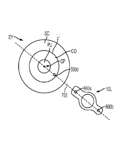
interface. Features can include fiducials places on the docking structures and
optical structures of
the disposable lens such as the location of the anterior and posterior
surfaces.
[0089] In the embodiment of Figure 3A, the ranging subsystem 46 includes an
OCT imaging device.
Additionally or alternatively, imaging modalities other than OCT imaging can
be used. An OCT
scan of the eye can be used to measure the spatial disposition (e. g. , three
dimensional coordinates
such as X, Y, and Z of points on boundaries) of structures of interest in the
patient's eye 43. Such
structure of interest can include, for example, the anterior surface of the
cornea, the posterior surface
of the cornea, the anterior portion of the lens capsule, the posterior portion
of the lens capsule, the
anterior surface of the crystalline lens, the posterior surface of the
crystalline lens, the iris, the pupil,
and/or the limbus. The spatial disposition of the structures of interest
and/or of suitable matching
geometric modeling such as surfaces and curves can be generated and/or used by
the control
electronics 54 to program and control the subsequent laser-assisted surgical
procedure. The spatial
disposition of the structures of interest and/or of suitable matching
geometric modeling can also be
used to determine a wide variety of parameters related to the procedure such
as, for example, the
upper and lower axial limits of the focal planes used for cutting the lens
capsule and segmentation of
the lens cortex and nucleus, and the thickness of the lens capsule among
others.
[0090] The ranging subsystem 46 in Figure 3A includes an OCT light source and
detection device
98. The OCT light source and detection device 98 includes a light source that
generates and emits
light with a suitable broad spectrum. For example, in many embodiments, the
OCT light source and
detection device 98 generates and emits light with a broad spectrum from 810
nm to 850 nm
wavelength. The generated and emitted light is coupled to the device 98 by a
single mode fiber optic
connection.
[0091] The light emitted from the OCT light source and detection device 98 is
passed through a
beam combiner 100, which divides the light into a sample portion 102 and a
reference portion 104.
A significant portion of the sample portion 102 is transmitted through the
shared optics 50. A
relative small portion of the sample portion is reflected from the patient
interface 52 and/or the
patient's eye 43 and travels back through the shared optics 50, back through
the beam combiner 100
and into the OCT light source and detection device 98. The reference portion
104 is transmitted
along a reference path 106 having an adjustable path length. The reference
path 106 is configured to
receive the reference portion 104 from the beam combiner 100, propagate the
reference portion 104
over an adjustable path length, and then return the reference portion 106 back
to the beam combiner
100, which then directs the returned reference portion 104 back to the OCT
light source and
detection device 98. The OCT light source and detection device 98 then directs
the returning small
-19-
CA 02909684 2015-10-16
WO 2014/172545 PCMJS2014/034508
portion of the sample portion 102 and the returning reference portion 104 into
a detection assembly,
which employs a time domain detection technique, a frequency detection
technique, or a single point
detection technique. For example, a frequency-domain technique can be used
with an OCT
wavelength of 830 run and bandwidth of 10 am.
100921 Once combined with the UF laser pulse beam 66 subsequent to the beam
combiner 82, the
OCT sample portion beam 102 follows a shared path with the UF laser pulse beam
66 through the
shared optics 50 and the patient interface 52. In this way, the OCT sample
portion beam 102 is
generally indicative of the location of the UF laser pulse beam 66. Similar to
the UF laser beam, the
OCT sample portion beam 102 passes through the Z-telescope 84, is redirected
by the X-scan device
86 and by the Y-scan device 88, passes through the objective lens assembly 94
and the patient
interface 52, and on into the eye 43. Reflections and scatter off of
structures within the eye provide
return beams that retrace back through the patient interface 52, back through
the shared optics 50,
back through the beam combiner 100, and back into the OCT light source and
detection device 98.
The returning back reflections of the sample portion 102 are combined with the
returning reference
portion 104 and directed into the detector portion of the OCT light source and
detection device 98,
which generates OCT signals in response to the combined returning beams. The
generated OCT
signals that are in turn interpreted by the control electronics to determine
the spatial disposition of
the structures of interest in the patient's eye 43. The generated OCT signals
can also be interpreted
by the control electronics to measure the position and orientation of the
patient interface 52, as well
as to determine whether there is liquid disposed between the posterior surface
of the patient interface
lens 96 and the patient's eye 43.
100931 The OCT light source and detection device 98 works on the principle of
measuring
differences in optical path length between the reference path 106 and the
sample path. Therefore,
different settings of the Z-tele scope 84 to change the focus of the UF laser
beam do not impact the
length of the sample path for a axially stationary surface in the eye of
patient interface volume
because the optical path length does not change as a function of different
settings of the Z-
telescope 84. The ranging subsystem 46 has an inherent Z range that is related
to light source and
the detection scheme, and in the case of frequency domain detection the Z
range is specifically
related to the spectrometer, the wavelength, the bandwidth, and the length of
the reference path 106.
In the case of ranging subsystem 46 used in Figure 3A, the Z range is
approximately 4-5 mm in an
aqueous environment. Extending this range to at least 20-25 ram involves the
adjustment of the path
length of the reference path 106 via a stage ZED within ranging subsystem 46.
Passing the OCT
sample portion beam 102 through the Z-telescope 84, while not impacting the
sample path length,
-20-
CA 02909684 2015-10-16
WO 2014/172545 PCMJS2014/034508
allows for optimization of the OCT signal strength. This is accomplished by
focusing the OCT
sample portion beam 102 onto the targeted structure. The focused beam both
increases the return
reflected or scattered signal that can be transmitted through the single mode
fiber and increases the
spatial resolution due to the reduced extent of the focused beam. The changing
of the focus of the
sample OCT beam can be accomplished independently of changing the path length
of the reference
path 106.
100941 Because of the fundamental differences in how the sample portion 102
(e.g, 810 tun to 850
nm wavelengths) and the UF laser pulse beam 66 (e.g., 1020 nin to 1050 tun
wavelengths) propagate
through the shared optics 50 and the patient interface 52 due to influences
such as immersion index.
refraction, and aberration, both chromatic and monochromatic, care must be
taken in analyzing the
OCT signal with respect to the UF laser pulse beam 66 focal location. A
calibration or registration
procedure as a function of X, Y, and Z can be conducted in order to match the
OCT signal
information to the UF laser pulse beam focus location and also to the relative
to absolute
dimensional quantities.
100951 There are many suitable possibilities for the configuration of the
OC'll interferometer. For
example, alternative suitable configurations include time and frequency domain
approaches, single
and dual beam methods, swept source, etc, are described in U.S. Pat. Nos.
5,748,898; 5,748.352;
5,459,570; 6,111,645; and 6,053,613.
100961 The system 2 can be set to locate the anterior and posterior surfaces
of the lens capsule and
cornea and ensure that the IT laser pulse beam 66 will he focused on the lens
capsule and cornea at
all points of the desired opening. Imaging modalities and techniques described
herein, such as for
example, Optical Coherence Tomography (OCT), and such as Purkinje imaging,
Scheimpflug
imaging, confocal or nonlinear optical microscopy, fluorescence imaging,
ultrasound, structured
light, stereo imaging, or other known ophthalmic or medical imaging modalities
and/or combinations
thereof may be used to determine the shape, geometry, perimeter, boundaries,
andlor 3-dimensional
location of the lens and lens capsule and cornea to provide greater precision
to the laser focusing
methods, including 2D and 3D patterning. Laser focusing may also be
accomplished using one or
more methods including direct observation of an aiming beam, or other known
ophthalmic or
medical imaging modalities and combinations thereof, such as but not limited
to those defined
above.
100971 Optical imaging of the cornea, anterior chamber and lens can be
performed using the same
laser and/or the same scanner used to produce the patterns for cutting.
Optical imaging can be used
to provide information about the axial location and shape (and even thickness)
of the anterior and
-21-
CA 02909684 2015-10-16
WO 2014/172545 PCMJS2014/034508
posterior lens capsule, the boundaries of the cataract nucleus, as well as the
depth of the anterior
chamber and features of the cornea. 'Ibis information may then be loaded into
the laser 3-D
scanning system or used to generate a three dimensional
modelfrepresentationiimaae of the cornea,
anterior chamber, and lens of the eye, and used to define the cutting patterns
used in the surgical
procedure.
[0098] Observation of an aim beam can also be used to assist in positioning
the focus point of the
UF laser pulse beam 66. Additionally, an aim beam visible to the unaided eye
in lieu or the infrared
OCT sample portion beam 102 and the UF laser pulse beam 66 can be helpful with
alignment
provided the aim beam accurately represents the infrared beam parameters. The
alignment guidance
subsystem 48 is included in the assembly 62 shown in Figure 3A. An aim beam
108 is generated by
an aim beam light source 110, such as a laser diode in the 630-650nm range.
[0099] Once the aim beam light source 110 generates the aim beam 108, the aim
beam 108 is
transmitted along an aim path 112 to the shared optics 50, where it is
redirected by a beam
combiner 114. After being redirected by the beam combiner 114, the aim beam
108 follows a shared
path with the UF laser pulse beam 66 through the shared optics 50 and the
patient interface 52. In
this way, the aim beam 108 is indicative of the location of the UF laser pulse
beam 66. The aim
beam 108 passes through the Z-telescope 84, is redirected by the X-scan device
86 and by the Y-
scan device 88, passes through the beam combiner 90, passes through the
objective lens assembly 94
and the patient interface 52, and on into the patient's eye 43.
[00100] The video subsystem 92 is operable to obtain images of the patient
interface and the
patient's eye. The video subsystem 92 includes a camera 116, an illumination
light source 118, and
a beam combiner 120. The video subsystem 92 gathers images that can be used by
the control
electronics 54 for providing pattern centering about or within a predefined
structure. The
illumination light source 118 can be generally broadband and incoherent. For
example, the light
source 118 can include multiple LEDs. The wavelength of the illumination light
source 118 is
preferably in the range of 700nm to 750run, but can be anything that is
accommodated by the beam
combiner 90, which combines the light from the illumination light source 118
with the beam path for
the UF laser pulse beam 66, the OCT sample beam 102, and the aim beam 108
(beam combiner 90
reflects the video wavelengths while transmitting the OCT and UF wavelengths).
The beam
combiner 90 may partially transmit the aim beam 108 wavelength so that the aim
beam 108 can be
visible to the camera 116. An optional polarization element can be disposed in
front of the
illumination light source 118 and used to optimize signal. The optional
polarization element can be,
for example, a linear polarizer, a quarter wave plate, a half-wave plate or
any combination. An
-22-
CA 02909684 2015-10-16
WO 2014/172545 PCMJS2014/034508
additional optional analyzer can be placed in front of the camera. The
polarizer analyzer
combination can be crossed linear polarizers thereby eliminating specular
reflections from unwanted
surfaces such as the objective lens surfaces while allowing passage of
scattered light from targeted
surfaces such as the intended structures of the eye. The illumination may also
be in a dark-filed
configuration such that the illumination sources are directed to the
independent surfaces outside the
capture numerical aperture of the image portion of the video system.
Alternatively the illumination
may also be in a bright field configuration. In both the dark and bright field
configurations, the
illumination light source can be used as a fixation beam for the patient. The
illumination may also
be used to illuminate the patient's pupil to enhance the pupil iris boundary
to facilitate iris detection
and eye tracking. A false color image generated by the near infrared
wavelength or a bandwidth
thereof may be acceptable.
1001011 The illumination light from the illumination light source 118 is
transmitted through
the beam combiner 120 to the beam combiner 90. From the beam combiner 90, the
illumination
light is directed towards the patient's eye 43 through the objective lens
assembly 94 and through the
patient interface 94. The illumination light reflected and scattered off of
various structures of the
eye 43 and patient interface travel back through the patient interface 94,
back through the objective
lens assembly 94, and back to the beam combiner 90. At the beam combiner 90,
the returning light
is directed back to the beam combiner 120 where the returning light is
redirected toward the
camera 116. The beam combiner can be a cube, plate or pellicle element. It may
also be in the form
of a spider mirror whereby the illumination transmits past the outer extent of
the mirror while the
image path reflects off the inner reflecting surface of the mirror.
Alternatively, the beam combiner
could bc in the form of a scraper mirror where the illumination is transmitted
through a hole while
the image path reflects off of the mirrors reflecting surface that lies
outside the hole. The
camera 116 can be a suitable imaging device, for example but not limited to,
any silicon based
detector array of the appropriately sized format. A video lens forms an image
onto the camera's
detector array while optical elements provide polarization control and
wavelength filtering
respectively. An aperture or iris provides control of imaging NA and therefore
depth of focus and
depth of field and resolution. A small aperture provides the advantage of
large depth of field that
aids in the patient docking procedure. Alternatively, the illumination and
camera paths can be
switched. Rztherniore, the aim light source 110 can be made to emit infrared
light that would not be
directly visible, but could be captured and displayed using the video
subsystem 92.
1001021 Figure 3B shows a mapped treatment region of the eye comprising the
cornea, the
posterior capsule, and the limbus. The treatment region can be mapped with
computer modeling, for
-23-
CA 02909684 2015-10-16
WO 2014/172545 PCMJS2014/034508
example ray tracing and phased based optical modeling. The treatment volume is
shown extending
along the Z-axis from the posterior surface of the optically transmissive
structure of the patient
interface a distance of over 15 mm, such that the treatment volume includes
the cornea, and the lens
in which the treatment volume of the lens includes the anterior capsule, the
posterior capsule, the
nucleus and the cortex. The treatment volume extends laterally from the cornea
to the limbus. The
lateral dimensions of the volume are defined by a Y contour anterior to the
limbus and by an X
contour posterior to the limbus. The treatment volume shown can be determined
by a person of
ordinary skill in the art based on the teachings described herein. The lateral
positions of optical
breakdown for ZL fixed to 30 mm and ZL fixed to 20 mm are shown. These
surfaces that extend
transverse to the axis 99 along the Z-dimension correspond to locations of
optical scanning of the X
and Y galvos to provide optical breakdown at lateral locations away from the
axis 99. The curved
non-planner shape of the scan path of optical breakdown for ZL- 30 mm and ZL-
20 mm can be
corrected with the mapping and look up tables as described herein. The curved
shape of the focus
can be referred to as a warping of the optical breakdown depth and the look up
tables can be warped
oppositely or otherwise adjusted so as to compensate for the warping of the
treatment depth, for
example.
[00103] The treatment region is shown for setting the laser beam energy
about four times the
threshold amount for optical breakdown near the center of the system. The
increased energy allows
the beam system to treat the patient with less than ideal beam focus.
[00104] The placement of the posterior surface of the optically
transmissive structure of the
patient interface away from the surface of the cornea can provide the extended
treatment range as
shown, and in many embodiments the optically transmissive structure comprises
the lens. In
alternative embodiments, the posterior surface of the optically transmissive
structure can be placed
on the cornea, for example, and the mapping and look up tables as described
herein can be used to
provide the patient treatment with improved accuracy.
[00105] The optically transmissive structure of the patient interface may
comprise one or
more of many known optically transmissive materials used to manufactures
lenses, plates and
wedges, for example one or more of glass, BK-7, plastic, acrylic, silica or
fused silica for example.
[00106] Figure 4 shows a method 400 of treating a patient, for example with
the laser eye
surgery system 2 described herein, in accordance with many embodiments.
[00107] Examples of tissue treatment methods and apparatus suitable for
combination in
accordance with embodiments as described herein are described in U.S. Pat.
App. Ser. Nos.
12/510,148, filed Jul. 27, 2009, and 11/328,970, filed on Jan. 9, 2006, both
entitled "METHOD OF
-24-
PATTERNED PLASMA-MEDIATED LASER TREPHINATION OF THE LENS CAPSULE AND
THREE DIMENSIONAL PHACO-SEGMENTATION," in the name of Blumenkranz et al.
1001081 At a step 410, the geometry of one or more fiducials is selected.
The fiducials may
have a shape in the form of one or more dots, lines, rectangles, arrows,
crosses, trapezoids,
rectangles, squares, chevrons, pentagons, hexagons, circles, ellipses, arcs,
and combinations thereof
100109j At a step 420, an appropriate anatomical site is located on the
eye. This anatomical
site may be located using the ranging subsystem 46 of the laser eye surgery
system 2 described
herein. Appropriate anatomical sites include, but are not limited to, the
limbus, the cornea, the
sclera, the lens capsule, the iris, the stToma, or the crystalline lens
nucleus. In many embodiments,
the appropriate anatomical site on the eye is the periphery of the cornea.
Typically, the anatomical
site will be in a predetermined position relative to an axis of the eye such
as the astigmatic axis.
1001101 At a step 430, the anatomical site is marked and may be cut, for
example, with the
laser subsystem 44 of the laser eye surgery system 2, to generate the fiducial
on the eye at the
anatomical site located by step 420. One or more fiducials may be generated,
for example. two
fiducials to define a line in a known relation to an astigmatic axis of the
eye or two or more fiducials
to define more than one anatomical location of the eye.
1001111 At a step 440, a patient interface, for example, the patient
interface 58 of the laser eye
surgery system 2 described herein, is coupled with the eye often by suction.
Any number of laser
eye surgery procedures can now be performed. The fiducial generated on the eye
can be used in
such procedures to facilitate the precise positioning of treatment regimens,
implantations, etc.
1001121 In many embodiments, cataract surgery is performed. For example,
at a step 450, eye
tissue is cut. At a step 460, a cataract is removed or destroyed, for example,
using the procedures
described in U.S. Pat. App. Ser. Nos. 12/510,148 and 11/328,970, both entitled
"METHOD OF
PATTERNED PLASMA-MEDIATED LASER TREPHINATION OF THE LENS CAPSULE AND
THREE DIMENSIONAL PHACO-SEGMENTATION," in the name of Blumenkranz et al. At a
step 470, a marker on an artificial intraocular lens is positioned in a
predetermined relation to the
fiducial on the eye. The marker will typically have a geometry corresponding
to that of the fiducial.
The marker may have the same or complementary geometry as the fiducial. The
predetermined
relation may be one of linear alignment, for example, with a line formed by
the fiducial and the
center of the pupil or a line formed by two or more fiducials, or one where
the marker is offset from
such lines at a predetermined angle such as 30, 45, 60, or 90 degrees. At a
step 480, the positioned
intraocular lens is implanted. At a step 490, the remainder of the eye surgery
is completed.
-25-
Date Recue/Date Received 2020-08-28
CA 02909684 2015-10-16
WO 2014/172545 PCMJS2014/034508
[00113] The placement of the IOL can proceed in accordance with known IOLs
and methods
modified in accordance with the teachings provided herein, and the IOL may
comprise an
accommodating or non-accommodating IOL, for example.
[00114] Although the above steps show method 400 of treating a patient in
accordance with
embodiments, a person of ordinary skill in the art will recognize many
variations based on the
teaching described herein. The steps may be completed in a different order.
Steps may be added or
deleted. Some of the steps may comprise sub-steps. Many of the steps may be
repeated as often as
beneficial to the treatment.
1001151 One or more of the steps of the method 400 may be performed with
the circuitry as
described herein, for example, one or more of the processor or logic circuitry
such as the
programmable array logic for field programmable gate array. The circuitry may
be programmed to
provide one or more of the steps of the method 400, and the program may
comprise program
instructions stored on a computer readable memory or programmed steps of the
logic circuitry such
as the programmable array logic or the field programmable gate array, for
example.
[00116] As discussed above, one or more fiducials can be generated in
various locations on
the eye including various internal anatomical structures. For example, Figure
5A1 shows a front
view of the eye EY having a fiducial 500a generated thereon. As shown in
Figures 5A1 and 5A2, a
fiducial 500a having an X-shape is generated on the periphery of the cornea
CO.
[00117] Figures 5B1 and 5B2 show a front view and a side view,
respectively, of an eye EY
having an X-shaped fiducial 500a generated on the limbus LI.
[00118] Figure 5C1 sand 5B2 show a front view and a side view,
respectively, of an eye EY
having an X-shaped fiducial 500a generated on the sclera SC.
[00119] Figures 5A1 to 5C2 also show other anatomical features of the eye
EY at or near the
generated fiducial 500a, including the pupil PU and lens LE.
[00120] Figure 6 shows various examples of shapes of fiducials in
accordance with many
embodiments. These fiducials can be cut onto the eye EY using a laser
subsystem 44 of the laser eye
surgery system 2 described herein. A fiducial 500a can be X-shaped. A fiducial
500b can be in the
shape of a cross. A fiducial 500c can be in the form of a circular dot. A
fiducial 500d can be in the
shape of a circle. A fiducial 500e can be in the shape of a line segment. A
fiducial 500f can be in
the shape of a filled triangle. A fiducial 500g can be in the shape of an
empty triangle. A fiducial
500h can be in the shape of a filled square. A fiducial 500i can be in the
shape of an empty square.
A fiducial 500j can be in the shape of a filled chevron. A fiducial 500k can
be in the shape of an
empty chevron. A fiducial 5001 can be in the shape of a filled trapezoid. A
fiducial 500m can be in
-26-
CA 02909684 2015-10-16
WO 2014/172545 PCMJS2014/034508
the shape of an empty trapezoid. A fiducial 500n can be in the shape of a
filled rectangle. A filled
fiducial 500o can be in the shape of an empty rectangle. A fiducial 500p can
be in the shape of a
filled diamond. A fiducial 500q can be in the shape of an empty diamond. A
fiducial 500r can be in
the shape of a filled pentagon. A fiducial 500s can be in the shape of an
empty pentagon. A fiducial
500t can be in the shape of a filled 5-pointed star. A fiducial 500u can be in
the shape of an empty 5
pointed star. A fiducial 500v can be in the shape of a filled oval. A fiducial
500w can be in the
shape of an empty oval. A fiducial 500x can be in the shape of a filled 6-
pointed star. A fiducial
500y can be in the shape of an empty 6-pointed star. A fiducial 500z can be T-
shaped.
[00121] Figures 7A to 7D show front views of one or more fiducials created
on the eye BY
positioned in pre-determined positional relationships with an artificial
intraocular lens IOL.
[00122] A person of ordinary skill in the art will recognize that the JUL
can be placed in the
eye in accordance with known method and apparatus, and that the aberration
correcting axis of the
JUL and lens of the JUL will extend across pupil PU when placed, and that
Figs. 7A-7D show the
IOL configured for placement positioning and alignment with the fiducials. The
aberration corrected
may comprise a lower order aberration such as astigmatism, or higher order
aberration such as trefoil
can coma. Further, the marker of the JUL may be used to define an axis of the
lens to be aligned with
the eye, for example an X, Y, or Z reference of the eye to be aligned with an
X,Y or Z axis of a
wavefront correcting JUL.
[00123] As shown in Figure 7A, two circular fiducials 500d1, 500d2 can be
generated on the
periphery of the cornea CO of an eye EY. These two fiducials 500d1, 500d2
define a line 701 which
may be aligned with or parallel to the astigmatic axis of the eye EY. The
artificial intraocular lens
JUL can be positioned so that markers 600a, 600b on the lens JUL can be
aligned with the fiducials
500d1, 500d2 by being on the same line 701. The shape of the markers 600a,
600b can correspond
to the shape of the fiducials 500d1, 500d2. For example in Figures 7A, the
markers 600a, 600b can
be in the form of circular dots which may fit within the circles of the
fiducials 500d1, 500d2 when
the artificial intraocular lens JUL is properly positioned and aligned within
the eye BY. Other
complementary shapes may also be used to facilitate the positioning and
alignment of the artificial
intraocular lens JUL within the eye EY.
[00124] In some embodiments, the two fiducials 500d1, 500d2 can define a
line 702 which
may be perpendicular or otherwise transverse to the astigmatic axis of the eye
EY. As shown in
Figure 7B, the artificial intraocular lens JUL can be positioned so that the
markers 600a, 600b on the
lens JUL form a line perpendicular to the line 702 foimed by the fiducials
500d1, 500d2. Thus, the
lens JUL can be properly positioned in alignment with the astigmatic axis of
the eye EY. In other
-27-
CA 02909684 2015-10-16
WO 2014/172545 PCMJS2014/034508
embodiments, the artificial intraocular lens IOL can be positioned so that the
markers 600a, 600b on
the lens IOL form a line transverse to the line 702 formed by the fiducials
500d1, 500d2, for
example, at predetermined angles of 30 degrees, 45 degrees, or 60 degrees.
[00125] As shown in Figure 7C, a single circular fiducials 500d can be
generated on the
periphery of the cornea CO of an eye EY. The fiducials 500d and the center of
the pupil CP can
define a line 703 which may be aligned with or parallel to the astigmatic axis
of the eye EY. The
artificial intraocular lens IOL can be positioned so that markers 600a, 600b
on the lens IOL can be
aligned with the fiducial 500d and pupil center CP by being on the same line
703.
[00126] In some embodiments, the fiducial 500d and the pupil center CP can
define a line 704
which may be perpendicular or otherwise transverse to the astigmatic axis of
the eye EY. As shown
in Figure 7D, the artificial intraocular lens TOL can be positioned so that
the markers 600a, 600b on
the lens IOL form a line 703 perpendicular to the line 704 formed by the
fiducials 500d1, 500d2.
Thus, the lens JUL can be properly positioned in alignment with the astigmatic
axis of the eye EY.
In other embodiments, the artificial intraocular lens IOL can be positioned so
that the markers 600a,
600b on the lens IOL form a line transverse to the line 703 formed by the
fiducial 500d and the pupil
center CP, for example, at predetermined angles of 30 degrees, 45 degrees, or
60 degrees.
[001271 Figure 8 shows an JUL placed in an eye, in accordance with many
embodiments. The
axis 701 is shown positioned relative to axis 702 to determine an alignment of
the TOT,. The pupil
center PC is shown in relation to a center of the IOL that may or may not be
marked. The two
fiducials 500d1, 500d2 define a line 701 which may be aligned with or parallel
to the astigmatic axis
of the eye EY or other axis as described herein. The artificial intraocular
lens JUL can be positioned
so that markers 600a, 600b on the lens JUL can be aligned with the fiducials
500d1, 500d2 and can
be on substantially the same line 701. The shape of the markers 600a, 600b can
correspond to the
shape of the fiducials 500d1, 500d2 as described herein.
[00128j In many embodiments, the Fiducials are located on the eye for
benefit of the patient.
After surgery, the lens markers 500D1 and 500D1 may not be visible under
normal viewing
conditions and the Fiducials 600A, 600B are placed away from the pupil of the
eye to inhibit visual
artifacts seen by the patient. The Fiducials 500d1, 500d2, can be placed on
the cornea outside of a
large natural pupil PUN of the eye that corresponds to a maximum natural pupil
size such as a pupil
of a dark adapted eye. Alternatively or in combination, the Fiducials may be
placed on the lens
capsule outside the large natural pupil PUN and within the surgically dilated
pupil PUD. The large
natural pupil can be, for example about 8 or 9 mm for younger patients
receiving accommodating
IOLs, and about 4-5 mm for older patients having cataract surgery for example.
The pupil PU may
-28-
CA 02909684 2015-10-16
WO 2014/172545 PCMJS2014/034508
be dilated during with a eycloplegic so as to comprise a dilated pupil PUD
having a diameter larger
than the naturally dilated pupil PUD, for example so as to allow visualization
of the markers and
Fiducials when the IOL is placed. The markers 600A, 600B of the JUL can be
separated by a
distance larger than the optical zone of the IOL, or may comprise small marks
within the optical
zone of the IOL.
[00129] Fig. 9 shows haptics of an IOL positioned with corresponding
Fiducials, in
accordance with many embodiments. The marks placed on the JUL can be located
on one or more of
an optic of the IOL or a haptic of an IOL, for example. In some embodiments,
the Fiducials 500D1,
500D2, 500D3 can be placed on the eye as described herein at locations
corresponding to target
locations of haptics RA, for example, such that the Fiducials can be used to
align the haptics for
placement in the eye.
[00130] In many embodiments, an operating microscope as described herein
has a
magnification providing a depth of field capable of simultaneously imaging the
Fiducials 500D1,
500D2 and markers 600A, 600B, and the fidieials that mark the cornea are sized
and shaped to as to
be visible with the markers. In many embodiments, the marks on the cornea
comprise marks near
the limbus, and may comprise marks formed in the limbus, conjunctiva or
sclera. Alternatively or in
combination, a dye can be applied to the exterior of the eye that is absorbed
by the marks to improve
visibility of the laser placed marks.
[00131] The methods and apparatus as described herein are suitable for
combination with one or
more components of laser eye surgery systems that are under development or
commercially
available such as:
[00132] an adaptive patient interface is described in Patent Cooperation
Treaty Patent Application
(hereinafter "PCT") PCT/US2011/041676, published as WO 2011/163507, entitled
"ADAPTIVE
PATIENT INTERFACE";
[00133] a device and method for aligning an eye with a surgical laser are
described in
PCT/IB2006/000002, published as WO 2006/09021, entitled "DEVICE AND METHOD FOR
ALIGNING AN EYE WITH A SURGICAL LASER";
[00134] a device and method for aligning an eye with a surgical laser are
described in
PCT/IB2006/000002, published as WO 2006/09021, entitled "DEVICE AND MEHTOD FOR
ALIGNING AN EYE WITH A SURGICAL LASER";
[00135] an apparatus for coupling an element to the eye is described in U.S.
Application Serial No.
12/531,217, published as U.S. Pub. No. 2010/0274228. entitled "APPARATUS FOR
COUPLING
AN ELEMENT TO THE EYE"; and
-29-
CA 02909684 2015-10-16
WO 2014/172545 PCMJS2014/034508
[00136] a servo controlled docking force device for use in ophthalmic
applications is described in
U.S. Application Serial No. 13/016,593, published as U.S. Pub. No. US
2011/0190739, entitled
"SERVO CONTROLLED DOCKING FORCE DEVICE FOR USE IN OPHTHALMIC
APPLICATIONS".
[00137] With the teachings described herein, a person of ordinary skill in
the art can modify
the above referenced devices to practice many of the embodiments described
herein.
[00138] While preferred embodiments of the present disclosure have been
shown and
described herein, it will be obvious to those skilled in the art that such
embodiments are provided by
way of example only. Numerous variations, changes, and substitutions will be
apparent to those
skilled in the art without departing from the scope of the present disclosure.
It should be understood
that various alternatives to the embodiments of the present disclosure
described herein may be
employed without departing from the scope of the present invention. Therefore,
the scope of the
present invention shall be defined solely by the scope of the appended claims
and the equivalents
thereof.
-30-