Note: Descriptions are shown in the official language in which they were submitted.
81792170
Purification of recombinantly produced polypeptides
1. INTRODUCTION
Claim of Priority
This application claims the benefit of prior U.S. Provisional Application No.
61/823,520, filed on
May 15, 2013.
1.1. Reference to a Sequence Listing
Sequence Listing submitted with this application as text file PURIF300W01 SL
created on May 13, 2014 and having a size of 8,521 bytes.
1.2. Field of the Invention
The present invention relates to the purification of recombinantly produced
polypeptides.
In a more particular embodiment, the invention relates to methods of
separating recombinantly
produced polypeptides from host cell proteins (HCPs).
1.3. Background of the Invention
Recombinantly produced polypeptides, such as antibodies and other proteins,
are used
in a wide array of diagnostic and therapeutic applications. The process of
manufacturing
recombinant polypeptides generally involves expression of the polypeptide in a
host cell, and
purification of the polypeptide.
Expression generally involves culturing a prokaryotic or eukaryotic host cell
under
appropriate conditions for the host cells to produce the recombinant
polypeptide. The
recombinant polypeptide can be expressed in different locations within the
host cell, which can
impact the methods used for isolation and purification of the product.
Once a recombinant polypeptide is expressed, intact host cells and cell debris
are
separated from the cell culture media in a process referred to as "cell
harvesting." For example,
host cells can be separated from the cell culture media by centrifugation or
filtration to provide a
clarified fluid (which can be referred to as the "cell culture supernatant")
that includes the
recombinant polypeptide and other impurities. Examples of impurities that may
be found in the
clarified cell culture supernatant include, but are not limited to, host cell
proteins (HCP), nucleic
acids, endotoxins, viruses, protein variants and protein aggregates.
1
Date Recue/Date Received 2020-11-17
CA 02910065 2015-10-21
WO 2014/186350 PCT/US2014/037821
Purification refers to the removal of impurities from the clarified cell
culture supernatant
and typically involves one or more chromatography steps. Typical processes
include capture,
intermediate purification or polishing, and final polishing steps. Affinity
chromatography, for
example, Protein A chromatography or ion exchange chromatography, is often
used as a
capture step. Often, capture is followed by at least two intermediate
purification or polishing
steps to increase purity and remove of viral contaminants. Intermediate
purification or polishing
steps are often accomplished by affinity chromatography, ion exchange
chromatography, or
hydrophobic interaction chromatography (H IC). In many processes, the final
polishing step is
accomplished using ion exchange chromatography, hydrophobic interaction
chromatography, or
gel filtration.
Preferably, biopharmaceutical products have a very high purity, with the
concentration of
impurities, such as host cell proteins, reduced to the range of parts per
million relative to the
desired product, or lower. Consequently, there remains a need for purification
processes that
optimize removal of impurities, in particular, host cell proteins.
2. SUMMARY OF THE INVENTION
Described herein is a method for separating a recombinantly produced
polypeptide from
host cell protein (HCP). In one embodiment, the method includes steps of:
providing a clarified
cell culture supernatant that includes the recombinantly produced polypeptide
and the HCP;
loading the clarified cell culture supernatant onto a Protein A chromatography
column; washing
the Protein A chromatography column with a wash buffer including a fatty acid
having a chain
length of at least about 6 carbon atoms, or a fatty acid salt thereof to
remove HCP; and
recovering the recombinantly produced polypeptide. In another embodiment, the
method
includes steps of equilibrating a Protein A chromatography column with an
equilibration buffer;
loading the clarified cell culture supernatant onto the Protein A
chromatography column; re-
equilibrating the loaded Protein A chromatography column with the
equilibration buffer; washing
the loaded Protein A chromatography column with a first wash buffer including
a fatty acid
having a chain length of at least about 6 carbon atoms, or a fatty acid salt
thereof to remove
HCP; washing the loaded Protein A chromatography column with a second wash
buffer; and
eluting the recombinantly produced polypeptide with an elution buffer. In
another embodiment,
a method of reducing protease contamination in a formulation including a
recombinantly
produced polypeptide is described. In another embodiment, a method of
increasing stability of a
recombinantly produced polypeptide is described. In another embodiment, a
method of reducing
2
CA 02910065 2015-10-21
WO 2014/186350
PCT/US2014/037821
HCP levels, including protease levels in a formulation is provided. Examples
of proteases that
may be removed include, but are not limited to, serine proteases, aspartyl
proteases, such as
cathepsin-D, cysteine proteases, metalloproteases and aminopeptidases.
In one embodiment, the chain length of the fatty acid or fatty acid salt is
between 6 and
12 carbon atoms. In another embodiment, the chain length of the fatty acid or
fatty acid salt is
between 8 and 12 carbon atoms. In another embodiment, the chain length of the
fatty acid or
fatty acid salt is between 8 and 10 carbon atoms. In one embodiment, the fatty
acid or fatty acid
salt is selected from enanthic acid, caprylic acid, pelargonic acid, capric
acid, undecyclic acid,
lauric acid, and combinations thereof. In a more particular embodiment, the
wash buffer
includes caprylic acid, or a caprylic acid salt. In one embodiment, the wash
buffer includes
between about 25 mM to about 200 mM fatty acid. In another embodiment, the
wash buffer
includes between about 50 mM to about 100 mM fatty acid. In one embodiment,
the wash
buffer includes about 100 mM fatty acid.
In one embodiment, the wash buffer includes sodium chloride. In a more
particular
embodiment, the wash buffer includes the sodium chloride at a concentration
between about
1.0M to about 2.5M. In one embodiment, the wash buffer includes sodium
chloride at a
concentration between about 2M to about 2.5M. In one embodiment, the wash
buffer includes
sodium chloride at a concentration of about 2.5M.
In one embodiment, the wash buffer has a pH between about 7 to about 9. In
another
embodiment, the wash buffer has a pH between about 8 to about 9. In another
embodiment,
the wash buffer has a pH between about 8.5 to about 9. In one embodiment, the
wash buffer
has a pH of about 9.
In a more particular embodiment, the wash buffer includes between about 50 mM
and
about 100 mM sodium caprylate at a pH between about 8 to about 9. In another
embodiment,
the wash buffer includes between about 50 mM and about 100 mM sodium caprylate
at a pH
between about 8 to about 9 and between about 2.0M to about 2.5 M sodium
chloride. In
another embodiment, the wash buffer includes about 100mM sodium caprylate in
100mM Tris at
a pH of about 9Ø In one embodiment, the wash buffer includes about 100mM
sodium
caprylate in 100mM Tris at a pH of about 9.0 and about 2.5M sodium chloride.
In one embodiment, the cell culture harvest is clarified to obtain a clarified
cell culture
harvest, which is loaded onto the Protein A chromatography column. In one
embodiment, the
loaded Protein A column is re-equilibrated with an equilibration buffer prior
to washing the
column with the wash buffer. In one embodiment, the equilibration buffer
includes sodium
phosphate. In one embodiment, the equilibration buffer includes between about
10 mM and
3
CA 02910065 2015-10-21
WO 2014/186350 PCT/US2014/037821
about 100 mM sodium phosphate. In one embodiment, the equilibration buffer
includes
between about 20 mM and about 50 mM sodium phosphate at a pH between about 6
and about
8. In another embodiment, the equilibration buffer includes about 20 mM sodium
phosphate at
a pH of about 7.
In one embodiment, the method includes a second wash step after the column is
washed with the fatty acid wash buffer. In one embodiment, the second wash
buffer includes
sodium phosphate. In one embodiment, the second wash buffer includes between
about 10
mM and about 100 mM sodium phosphate. In one embodiment, the second wash
buffer
includes between about 20 mM and about 50 mM sodium phosphate at a pH between
about 6
and about 8. In one embodiment, the second wash buffer includes about 20 mM
sodium
phosphate at a pH of about 7.
In one embodiment, the recombinant protein is recovered by eluting the
recombinant
protein from the Protein A column with an elution buffer. In one embodiment,
the elution buffer
includes sodium citrate. In one embodiment, the elution buffer includes
between about 25 mM
and about 200 mM sodium citrate. In one embodiment, the elution buffer
includes between
about 50 mM and about 100 mM sodium citrate. In one embodiment, the elution
buffer has a
pH between about 2.0 and about 5Ø In one embodiment, the elution buffer has
a pH of
between about 3.0 and about 4Ø In one embodiment, the elution buffer
includes about 100
mM sodium citrate at a pH of about 3.5.
In one embodiment, the recombinantly produced polypeptide purified by the
method
above is an antibody, or a binding fragment thereof. In one embodiment, the
recombinantly
produced polypeptide includes a fully human monoclonal antibody selected from
antibody 1 (a
human anti-interleukin (IL)-6 antibody) or antibody 2 (a monoclonal antibody
to human IL-18)
(NCIMB accession number 41786).
In another embodiment, the recombinantly produced polypeptide includes an
antibody
having a light chain acid variable sequence of antibody 1 (SEQ ID NO:8). In
another
embodiment, the recombinantly produced polypeptide includes an antibody having
a heavy
chain variable sequence of antibody 1 (SEQ ID NO:7). In another embodiment,
the
recombinantly produced polypeptide includes an antibody having a light chain
variable
.. sequence of antibody 1 (SEQ ID NO: 8) and a heavy chain variable sequence
of antibody 1
(SEQ ID NO:7).
In another embodiment, the recombinantly produced polypeptide includes an
antibody
having a light chain acid variable sequence of antibody 2 (SEQ ID NO:18). In
another
embodiment, the recombinantly produced polypeptide includes an antibody having
a heavy
4
CA 02910065 2015-10-21
WO 2014/186350 PCT/US2014/037821
chain variable sequence of antibody 2 (SEQ ID NO.16). In another embodiment,
the
recombinantly produced polypeptide includes an antibody having a light chain
variable
sequence of antibody 2 (SEQ ID NO: 18) and a heavy chain variable sequence of
antibody 2
(SEQ ID No.:16).
In one embodiment, the antibody includes a heavy chain amino acid sequence
having
one or more complementarity determining regions (CDRs) of antibody 1 or
antibody 2. The
terms CDR region or CDR, refer to the hypervariable regions of the heavy and
light chains of
the immunoglobulin as defined by Kabat et al. (Kabat, E. A. et al. (1991)
Sequences of Proteins
of Immunological Interest, 5th Edition. US Department of Health and Human
Services, Public
Service, NIH, Washington or later editions) or Chothia and Lesk (J. Mol.
Biol., 196:901-917
(1987)). An antibody typically contains 3 heavy chain CDRs and 3 light chain
CDRs. The term
CDR or CDRs is used here in order to indicate, according to the case, one of
these regions or
several, or even the whole, of these regions which contain the majority of the
amino acid
residues responsible for the binding by affinity of the antibody for the
antigen or the epitope
which it recognizes. Among the six short CDR sequences, the third CDR of the
heavy chain
(HCDR3) has a greater size variability (greater diversity essentially due to
the mechanisms of
arrangement of the genes which give rise to it). It may be as short as 2 amino
acids although
the longest size known is 26. CDR length may also vary according to the length
that can be
accommodated by the particular underlying framework. Functionally, HCDR3 plays
a role in part
.. in the determination of the specificity of the antibody. One of skill in
the art is able to determine
CDR regions of an antibody. In general, HCDR1 is about 5 amino acids long,
consisting of
Kabat residues 31-35; HCDR2 is about 17 amino acids long, consisting of Kabat
residues 50-
65; HCDR3 is about 11 or 12 amino acids long, consisting of Kabat residues 95-
102, optionally
including Kabat residue 100D; LCDR1 is about 11 amino acids long, consisting
of Kabat
.. residues 24-34; LCDR2 is about 7 amino acids long, consisting of Kabat
residues 50-56; and
LCDR3 is about 8 or 9 amino acids long, consisting of Kabat residues 89-97,
optionally
including Kabat residue 95.
In one embodiment, the recombinantly produced polypeptide is an antibody or
binding
fragment thereof that includes a light chain amino acid sequence that includes
one or more light
.. chain CDR sequences for antibody 1 selected from LCDR1 (SEQ ID NO:4); LCDR2
(SEQ ID
NO: 5), LCDR3 (SEQ ID NO:6), and combinations thereof. In one embodiment, the
recombinantly produced polypeptide is an antibody that includes a heavy chain
amino acid
sequence that includes one or more of the heavy chain CDR sequences for
antibody 1 selected
from HCDR1 (SEQ ID NO: 1); HCDR2 (SEQ ID NO: 2), HCDR3 (SEQ ID NO:3), and
5
CA 02910065 2015-10-21
WO 2014/186350 PCT/US2014/037821
combinations thereof. In one embodiment, the recombinantly produced
polypeptide is an
antibody or binding fragment thereof that includes a light chain amino acid
sequence that
includes LCDR1 (SEQ ID NO:4); LCDR2 (SEQ ID NO: 5), and LCDR3 (SEQ ID NO:6)
from
antibody 1 and a heavy chain amino acid sequence that includes HCDR1 (SEQ ID
NO: 1);
HCDR2 (SEQ ID NO: 2) and HCDR3 (SEQ ID NO:3) of antibody 1.
In one embodiment, the recombinantly produced polypeptide is an antibody or
binding
fragment thereof that includes a light chain amino acid sequence that includes
one or more light
chain CDR sequences for antibody 2 selected from LCDR1 (SEQ ID NO:12); LCDR2
(SEQ ID
NO: 13), LCDR3 (SEQ ID NO:14), and combinations thereof. In one embodiment,
the
recombinantly produced polypeptide is an antibody that includes a heavy chain
amino acid
sequence having one or more of the heavy chain CDR sequences for antibody 2
selected from
HCDR1 (SEQ ID NO: 9); HCDR2 (SEQ ID NO: 10), HCDR3 (SEQ ID NO: 11), and
combinations thereof. In one embodiment, the recombinantly produced
polypeptide is an
antibody or binding fragment thereof that includes a light chain amino acid
sequence that
includes LCDR1 (SEQ ID NO: 12); LCDR2 (SEQ ID NO: 13), and LCDR3 (SEQ ID NO:
14) from
antibody 2 and a heavy chain amino acid sequence that includes HCDR1 (SEQ ID
NO: 9);
HCDR2 (SEQ ID NO: 10) and HCDR3 (SEQ ID NO: 11) of antibody 2.
In another embodiment, a wash buffer for separating a recombinantly produced
polypeptide from host cell protein (HCP) in a Protein A chromatography column
is provided. In
one embodiment, the wash buffer includes a fatty acid or a fatty acid salt
having a chain length
of at least 6 carbon atoms. In one embodiment, the chain length of the fatty
acid or fatty acid
salt is between 6 and 12 carbon atoms. In one embodiment, the chain length of
the fatty acid or
fatty acid salt is between 8 and 12 carbon atoms. In one embodiment, the chain
length of the
fatty acid or fatty acid salt is between 8 and 10 carbon atoms. In one
embodiment, the fatty acid
.. or fatty acid salt is selected from enanthic acid, caprylic acid,
pelargonic acid, capric acid,
undecyclic acid, lauric acid, and combinations thereof. In one embodiment, the
wash buffer
includes caprylic acid, or a caprylic acid salt. In one embodiment, the wash
buffer includes
between about 25 mM to about 200 mM fatty acid. In one embodiment, the wash
buffer
includes between about 50 mM to about 100 mM fatty acid. In one embodiment,
the wash
buffer includes about 100 mM fatty acid. In one embodiment, the wash buffer
includes sodium
chloride. In one embodiment, the wash buffer includes the sodium chloride at a
concentration of
between about 1.0M to about 2.5M. In one embodiment, the wash buffer includes
sodium
chloride at a concentration of about 2M to about 2.5M. In one embodiment, the
wash buffer
includes sodium chloride at a concentration of about 2.5M. In one embodiment,
the wash buffer
6
81792170
has a pH between about 7 to about 9. In one embodiment, the wash buffer has a
pH
between about 8 to about 9. In one embodiment, the wash buffer has a pH
between
about 8.5 to about 9. In one embodiment, the wash buffer has a pH of about 9.
In one
embodiment, the wash buffer includes between about 50 mM and about 100 mM
sodium caprylate at a pH between about 8 to about 9. In one embodiment, the
wash
buffer includes between about 50 mM and about 100 mM sodium caprylate at a pH
between about 8 to about 9 and between about 2.0M to about 2.5 M sodium
chloride.
In one embodiment, the wash buffer includes about 100mM sodium caprylate in
100mM Tris at a pH of about 9Ø In one embodiment, the wash buffer includes
about
100mM sodium caprylate in 100mM Tris at a pH of about 9.0 and about 2.5M
sodium
chloride.
In an embodiment, there is provided a method of reducing host cell protein
(HCP) level in a composition comprising a recombinantly produced polypeptide,
the
method comprising: providing a clarified cell culture supernatant comprising
the
recombinantly produced polypeptide and one or more HCP; loading the clarified
cell
culture supernatant onto a Protein A chromatography column; washing the
Protein A
chromatography column with a wash buffer comprising between 50mM and 100 mM
sodium caprylate at a pH between 8 and 9 to remove HCP.
3. BRIEF DESCRIPTION OF THE FIGURES
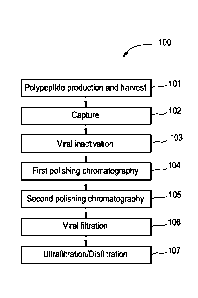
Figure 1 is a flow chart of a sample purification process.
Figure 2 shows the effects of wash pH, sodium chloride wash and sodium
caprylate on eluate purity.
Figure 3 shows the effect of the interaction between sodium caprylate and
sodium chloride on eluate purity.
Figure 4 shows the impact of the wash using sodium chloride and sodium
caprylate on recovery.
Figure 5 shows the effect of the interaction between sodium chloride and
sodium caprylate on recovery.
7
Date Recue/Date Received 2020-11-17
81792170
Figure 6 shows the interactions between wash pH, wash sodium chloride and
sodium caprylate on HCP levels (pre-filtration).
Figure 7 shows the quadratic interactions for wash pH and sodium chloride
on HCP levels (pre-filtration).
Figure 8 shows the interaction between wash sodium chloride and sodium
caprylate on HCP levels (post-filtration).
Figure 9 shows the combined effects of wash pH, sodium chloride and
sodium caprylate on purity, recovery and HCP levels pre-filtration.
Figures 10A-C are graphs showing the effect of wash pH, sodium chloride
and sodium caprylate, in combination, on HCP levels pre-filtration.
Figure 11 shows the effect of wash pH, sodium chloride and sodium
caprylate on HCP levels.
7a
Date Recue/Date Received 2020-11-17
CA 02910065 2015-10-21
WO 2014/186350 PCT/US2014/037821
Figure 12 shows the effect of wash pH, sodium chloride and sodium caprylate on
eluate
purity.
Figure 13 shows the combined effects of wash pH, sodium chloride and sodium
caprylate on purity:
Figure 14 shows the effect of wash pH on recovery.
Figure 15 shows the effect of sodium caprylate on HCP eluate levels.
Figure 16 shows the effects of wash pH, sodium chloride and sodium caprylate
on HCP
eluate levels.
Figure 17 shows the interaction between sodium chloride and sodium caprylate
and the
effect of the interaction on HCP eluate levels.
Figure 18 shows the effect of wash pH, sodium chloride and sodium caprylate on
protease activity.
Figure 19 shows the interaction of wash pH, sodium chloride and sodium
caprylate and
the effect of the interaction on protease activity.
Figures 20A and B show the effect of wash pH and sodium chloride on protease
activity
without (A) and with (B) sodium caprylate.
Figure 21 shows the combined effects of pH, sodium chloride and sodium
caprylate on
protease activity.
Figure 22 shows the effect of pH, sodium chloride and sodium caprylate on
protease
activity.
Figure 23 is a graph showing the HCP levels in anti-IL-18 eluate for the eight
fatty acid
wash buffer runs in Example 3.
Figure 24 is a mass spec showing undetectable levels of HCP in pelleted
particles
containing anti-1L6 antibodies.
Figure 25 is a graph showing Rates of Purity Loss, Aggregation, and
Fragmentation
(measured by SEC) of several lots of anti-1L6 antibodies after storage at 40 C
containing
different Host Cell Protein (HOP) levels.
Figure 26 is a graph showing the effect of HCP on fragmentation rate at 40 C
(by RP-
HPLC).
Figure 27 is a graph showing the effect of caprylate wash on protease
activity.
Figure 28 is a graph showing aspartyl and serine protease activity in a
particle forming
lot.
8
CA 02910065 2015-10-21
WO 2014/186350 PCT/US2014/037821
4. DETAILED DESCRIPTION
4.1. Introduction
A common problem encountered during Protein A purification is non-specific
binding of
impurities such as host cell protein (HOP), DNA and other cell culture-derived
impurities to the
column resin and to the protein of interest. For example, eluate from a
Protein A column having
large amounts of host cell proteins, for example, up to about 500 ng/mg, 600
ng/mg, 700 ng/mg,
800 ng/mg, 900 ng/mg, 1000 ng/mg, 1500 ng/mg, 2000 ng/mg or more HOP has been
observed. The presence of HOP can be problematic, not only because of health
regulations
relating to acceptable levels of contaminants in recombinant antibody
products, but also
because the presence of HOP can adversely impact product stability and/or
efficacy, including,
for example, protease activity and formation of visible particulates,
fragments or aggregates
over time.
Applicants have found that including a fatty acid in at least one Protein A
wash buffer
can substantially decrease the level of host cell proteins in the Protein A
eluate. In one
example, inclusion of a fatty acid in at least one Protein A wash buffer can
substantially
decrease protease activity in the eluate. Examples of proteases include serine
proteases,
aspartyl proteases, such as cathepsin-D, cysteine proteases, metalloproteases,
aminopeptidases, and combinations thereof. Additionally, including a fatty
acid in a Protein A
wash buffer can reduce protease activity in a formulation containing a
recombinantly produced
polypeptide. Furthermore, including a fatty acid in a Protein A wash buffer
can increase stability
of a recombinantly produced polypeptide, for example, by reducing particle
formulation and/or
fragmentation.
Described herein is a purification process for recombinantly produced
polypeptides. In a
.. more particular embodiment, a purification process for recombinantly
produced antibodies is
described.
4.2. Terminology
Unless otherwise defined, scientific and technical terms used herein shall
have the
meanings that are commonly understood by those of ordinary skill in the art.
Further, unless
otherwise required by context, singular terms shall include pluralities and
plural terms shall
include the singular. Generally, nomenclatures used in connection with, and
techniques of, cell
and tissue culture, molecular biology, and protein and oligo- or
polynucleotide chemistry and
hybridization described herein are those well-known and commonly used in the
art. Amino
9
CA 02910065 2015-10-21
WO 2014/186350 PCT/US2014/037821
acids may be referred to herein by either their commonly known three letter
symbols or by the
one-letter symbols recommended by the IUPAC-IUB Biochemical Nomenclature
Commission.
Nucleotides, likewise, may be referred to by their commonly accepted single-
letter codes.
As used in accordance with the present disclosure, the following terms, unless
otherwise
indicated, shall be understood to have the following meanings:
As used herein, the term "about" is used to modify, for example, the quantity
of an
ingredient in a composition, concentration, volume, process temperature,
process time, yield,
flow rate, pressure, and ranges thereof, employed in describing the invention.
The term "about"
refers to variation in the numerical quantity that can occur, for example,
through typical
measuring and handling procedures used for making compounds, compositions,
concentrates
or formulations; through inadvertent error in these procedures; through
differences in the
manufacture, source, or purity of starting materials or ingredients used to
carry out the methods,
and other similar considerations. The term "about" also encompasses amounts
that differ due to
aging of a formulation with a particular initial concentration or mixture, and
amounts that differ
due to mixing or processing a formulation with a particular initial
concentration or mixture.
Where modified by the term "about" the claims appended hereto include such
equivalents.
As used herein, the term "antibody" refers to a polypeptide or group of
polypeptides that
include at least one binding domain that is formed from the folding of
polypeptide chains having
three-dimensional binding spaces with internal surface shapes and charge
distributions
complementary to the features of an antigenic determinant of an antigen. An
antibody typically
has a tetrameric form, with two pairs of polypeptide chains, each pair having
one "light" and one
"heavy" chain. The variable regions of each light/heavy chain pair form an
antibody binding site.
Each light chain is linked to a heavy chain by one covalent disulfide bond,
while the number of
disulfide linkages varies between the heavy chains of different immunoglobulin
isotypes. Each
heavy and light chain also has regularly spaced intrachain disulfide bridges.
Each heavy chain
has at one end a variable domain (VH) followed by a number of constant domains
(CH). Each
light chain has a variable domain at one end (VL) and a constant domain (CL)
at its other end;
the constant domain of the light chain is aligned with the first constant
domain of the heavy
chain, and the light chain variable domain is aligned with the variable domain
of the heavy
chain. Light chains are classified as either lambda chains or kappa chains
based on the amino
acid sequence of the light chain constant region. The variable domain of a
kappa light chain
may also be denoted herein as VK.
The terms "antibody," "antibodies" and "immunoglobulins" as used herein
encompass
monoclonal antibodies (including full-length monoclonal antibodies),
polyclonal antibodies,
CA 02910065 2015-10-21
WO 2014/186350 PCT/US2014/037821
multispecific antibodies formed from at least two different epitope binding
fragments (e.g.,
bispecific antibodies), CDR-grafted, human antibodies, humanized antibodies,
camelised
antibodies, chimeric antibodies, single-chain Fvs (scFv), single-chain
antibodies, single domain
antibodies, Fab fragments, Fab' fragments, F(ab)2 fragments, antibody
fragments that exhibit a
desired biological activity (e.g. the antigen binding portion), disulfide-
linked Fvs (dsFv), and anti-
idiotypic (anti-Id) antibodies, intrabodies, and epitope-binding fragments or
derivatives of any of
the above. In particular, antibodies include immunoglobulin molecules and
immunologically
active fragments of immunoglobulin molecules, i.e., molecules that contain at
least one antigen-
binding site. Immunoglobulin molecules can be of any isotype (e.g., IgG, IgE,
IgM, IgD, IgA and
IgY), subisotype (e.g., IgG1, IgG2, IgG3, IgG4, IgA1 and IgA2) or allotype
(e.g., Gm, e.g.,
G1m(f, z, a or x), G2m(n), G3m(g, b, or c), Am, Em, and Km(1, 2 or 3)).
Antibodies may be
derived from any mammalian species, including, but not limited to, humans,
monkeys, pigs,
horses, rabbits, dogs, cats, mice, etc., or other animals such as birds (e.g.
chickens).
Antibodies may be fused to a heterologous polypeptide sequence, for example, a
tag to
facilitate purification.
The term "bind" or "binding" when discussing the interaction between a
molecule and a
column material means exposing the molecule to the column material under
conditions such
that the molecule is reversibly immobilized in or on the column material.
The term "cell culture supernatant" refers to a solution that is obtained by
culturing host
cells that produce a recombinant polypeptide of interest. In addition to the
recombinant
polypeptide, the cell culture supernatant may also include components of cell
culture medium,
metabolic byproducts secreted by the host cells as well as other components of
the cultured
cells. As used herein, the term "clarified cell culture supernatant" refers to
a composition from
which the host cells have been removed or harvested, such that the cell
culture supernatant is
generally free of cellular debris and/or intact cells.
The term "excipient" as used herein refers to an inert substance which is
commonly
used as a diluent, vehicle, preservative, binder or stabilizing agent for
drugs which imparts a
beneficial physical property to a formulation, such as increased protein
stability, increased
protein solubility, and/or decreased viscosity. Examples of excipients
include, but are not limited
to, proteins (for example, but not limited to, serum albumin), amino acids
(for example, but not
limited to, aspartic acid, glutamic acid, lysine, arginine, glycine),
surfactants (for example, but
not limited to, SDS, Tween 20, Tween 80, polysorbate and nonionic
surfactants), saccharides
(for example, but not limited to, glucose, sucrose, maltose and trehalose),
polyols (for example,
11
CA 02910065 2015-10-21
WO 2014/186350 PCT/US2014/037821
but not limited to, mannitol and sorbitol), fatty acids and phospholipids (for
example, but not
limited to, alkyl sulfonates and caprylate).
The phrase "host cell" or "host cells" refers to cells which express a
recombinant
polypeptide. In particular, the term "host cell" refers to a cell that can or
has taken up a nucleic
acid, such as a vector, and supports replication of the nucleic acid and
production of one or
more encoded products. The term "host cell" can refer to a variety of cell
types including
prokaryotic cells, such as Escherichia coil, Lactococcus lactis and Bacillus
species; yeast cells,
such as Pichia pastoris, Pichia methanolica, and Saccharomyces cerevisiae;
insect cell, such as
bacculovirus and eukaryotic cells. Examples of eukaryotic host cells include
mammalian cells,
for example, Chinese hamster ovary (CHO) cells, human embryonic kidney (HEK
293) cells,
Vero cells, baby hamster kidney (BHK) cells, HeLa cells, CV1 monkey kidney
cells, Madin-
Darby Canine Kidney (MOCK) cells, 3T3 cells, myeloma cell lines, COS cells
(e.g., COSI and
COS7) P012, WI38 cells. The term host cell also encompasses combinations or
mixtures of
cells including, e.g., mixed cultures of different cell types or cell lines.
The term "impurity" refers to any foreign material, particularly a biological
macromolecule
such as DNA, RNA, or a protein, other than the recombinantly produced
polypeptide that is
present in a sample. Contaminants can include host cell proteins other than
the recombinant
polypeptide of interest.
The term "purify" or "purifying" a recombinant polypeptide from a composition
or solution
that includes the recombinant polypeptide and one or more contaminants means
increasing the
degree of purity of the desired protein in the composition or solution by
removing (completely or
partially) at least one contaminant from the composition or solution.
The term "mAb" refers to a monoclonal antibody.
The phrase "pharmaceutically acceptable" as used herein means approved by a
regulatory agency of a Federal or state government, or listed in the U.S.
Pharmacopeia,
European Pharmacopia or other generally recognized pharmacopeia for use in
animals, and
more particularly in humans.
The terms "polypeptide" or "protein" can be used interchangeably to refer to a
molecule
having two or more amino acid residues joined to each other by peptide bonds.
The term
"polypeptide" can refer to antibodies and other non-antibody proteins. Non-
antibody proteins
include, but are not limited to, proteins such as enzymes, receptors, ligands
of a cell surface
protein, secreted proteins and fusion proteins or fragments thereof. The
polypeptide may or
may not be fused to another polypeptide. Polypeptides can also include
modifications such as,
but not limited to, glycosylation, lipid attachment, sulfation, gamma-
carboxylation of glutamic
12
CA 02910065 2015-10-21
WO 2014/186350 PCT/US2014/037821
acid residues, hydroxylation and ADP-ribosylation. Polypeptides can be of
scientific or
cornmercial interest, including protein-based therapeutics.
The term "recombinant" refers to a biological material, for example, a nucleic
acid or
protein, that has been artificially or synthetically (i.e., non-naturally)
altered by human
intervention.
The term "remove," when used in context of removal of host cell proteins,
refers to
reduction in the amount of host cell protein in the purified product. Removal
may or may not
result in the absence of host cell protein from the purified product. In
general, removal refers to
at least a 2 fold, 3 fold, 4 fold, 5 fold, 10 fold, 15 fold, 20 fold, 25 fold
and up to 30 fold, 35 fold,
40 fold, 45 fold or 50 fold reduction in host cell protein in the purified
product when compared to
the level of host cell proteins in the original composition.
The terms "stability" and "stable" as used herein in the context of a
formulation of a
recombinantly produced polypeptide, for example, a pharmaceutical formulation
that includes a
recombinantly produced antibody or antibody fragment, refer to the resistance
of the
polypeptide in the formulation to particle formation, aggregation, degradation
or fragmentation
under manufacture, preparation, transportation and storage conditions. A
"stable" formulation
retains biological activity under manufacture, preparation, transportation and
storage conditions.
Stability can be assessed by degrees of particle formation, aggregation,
degradation or
fragmentation, as measured by HPSEC, static light scattering (SLS), Fourier
Transform Infrared
Spectroscopy (FTIR), circular dichroism (CD), urea unfolding techniques,
intrinsic tryptophan
fluorescence, differential scanning calorimetry, and/or ANS binding
techniques, as compared to
a reference formulation.
As used herein, "substantially pure" refers to a biological material that is
the predominant
species present (e.g., on a molar basis it is more abundant than any other
individual species in
the composition). In one embodiment, a substantially purified fraction is a
composition wherein
the biological material includes at least about 50% (on a molar basis) of all
macromolecular
species present. Generally, a substantially pure composition will include more
than about 80%
of all macromolecular species present in the composition, or more than about
85%, more than
about 90%, more than about 95%, or more than about 99%. In one embodiment, the
biological
material is purified to essential homogeneity (contaminant species cannot be
detected in the
composition by conventional detection methods) and the composition includes
essentially a
single macromolecular species.
13
CA 02910065 2015-10-21
WO 2014/186350 PCT/US2014/037821
4.3. Recombinant Polypeptide Production
In one embodiment, a recombinant polypeptide is produced using host cells that
have
been transfected, either stably or transiently, with a vector capable of
expressing one or more
polypeptides of interest. As used herein, the term "vector" refers to
composition of matter which
can be used to deliver a nucleic acid of interest to the interior of a cell.
Numerous vectors are
known including, but not limited to, linear polynucleotides, polynucleotides
associated with ionic
or amphiphilic compounds, plasmids, and viruses. The term "vector" can include
an
autonomously replicating plasmid or a virus or a vector or plasmid that is not
autonomously
replicating. The term "transfection" refers to the introduction of exogenous
genetic material into
cells to produce genetically modified cells. Vectors can be introduced into a
host cell using
methods known in the art. For example, a vector can be transferred into a host
cell by physical,
chemical or biological means. Physical methods for introducing a
polynucleotide into a host cell
include, but are not limited to, calcium phosphate precipitation, lipofection
(including positively
charged liposome mediated transfection), particle bombardment, microinjection,
DEAE-dextran
mediated transfection and electroporation. Biological methods for introducing
a vector into a
host cell include the use of DNA and RNA vectors, including, for example,
viral vectors,
Chemical means for introducing a polynucleotide into a host cell include
colloidal dispersion
systems, such as macromolecule complexes, nanocapsules, microspheres, beads,
and lipid-
based systems including oil-in-water emulsions, micelles, mixed micelles, and
liposomes. The
host cells can be genetically engineered to express a recombinant polypeptide,
for example, a
polypeptide of commercial or scientific interest.
The term "cell culture" refers to the growth and propagation of cells outside
of a
multicellular organism or tissue. Cell culture conditions such as pH,
temperature, humidity,
atmosphere and agitation can be varied to improve growth and/or productivity
characteristics of
the cell culture. Host cells may be cultured in suspension or while attached
to a solid substrate.
Host cells can be cultured in small scale cultures, for example, in a
laboratory setting at volumes
as low as 25 ml and up to about 50 ml, up to about 100 ml, up to about 150 ml
or up to about
200 ml. Alternatively, the cultures can be large scale, for example, at
volumes from about 300
ml, 500 ml or 1000 ml and up to about 5000 ml, up to about 10,000 ml and up to
about 15,000
ml. Commercial scale bioreactors can also be used, for example, at volumes of
up to about
1,000L, up to about 5,000L or up to about 10,000L of media. Large scale
production of
recombinant polypeptides by mammalian cells can include continuous, batch and
fed-batch
culture systems. Host cells may be cultured, for example, in fluidized bed
bioreactors, hollow
fiber bioreactors, roller bottles, shake flasks, or stirred tank bioreactors,
with or without
14
81792170
microcarriers, and operated in a batch, fed batch, continuous, semi-
continuous, or perfusion
mode. Large scale cell cultures are typically maintained for days, or even
weeks, while the cells
produce the desired protein product(s).
Suitable host cells for production of recombinant polypeptides include both
prokaryotic
and eukaryotic cells. Examples of eukaryotic cells include mammalian cells.
Examples of
mammalian cells suitable for production of recombinant polypeptides include,
but are not limited
to, Chinese hamster ovary (CHO) cells, mouse myeloma (NSO), human embryonic
kidney (HEK
293), baby hamster kidney (BHK) cells, Vero cells, HeLa cells, Madin-Darby
Canine Kidney
(MDCK) cells, CV1 monkey kidney cells, 313 cells, myeloma cell lines such as
NSO and NS1,
P012, WI38 cells, COS cells (including COS-1 and COS-7), and 0127. In general,
mammalian
cell cultures are maintained at a pH between about 6.5 and about 7.5 and at a
temperature
between about 36 C and about 38 C, typically at about 37 C and a relative
humidity between
about 80% and about 95%. Mammalian cell culture media typically contain
buffering systems
that require a carbon dioxide (CO2) atmosphere between about 1% and about 10%,
or between
about 5% and about 6%.
The host cells can be maintained in a variety of cell culture media. The term
"cell culture
medium" refers to a nutrient solution in which the host cells are grown. Cell
culture media
formulations are well known in the art. Typically, cell culture media include
buffers, salts,
carbohydrates, amino acids, vitamins and trace essential elements. The cell
culture medium
may or may not contain serum, peptone, and/or proteins. Cell culture media may
be
supplemented with additional or increased concentrations of components such as
amino acids,
salts, sugars, vitamins, hormones, growth factors, buffers, antibiotics,
lipids, trace elements and
the like, depending on the requirements of the cells to be cultured and/or the
desired cell culture
parameters. Various culture media, including serum-free and defined culture
media, are
commercially available, and include, but are not limited to, Minimal Essential
Medium (MEM,
Sigma, St. Louis, Mo.); Ham's F10 Medium (Sigma); Dulbecco's Modified Eagles
Medium
(DMEM, Sigma); Minimal Essential Medium (MEM); Basal Medium Eagle (BME); RPMI-
1640
Medium (Sigma); HyClonemcell culture medium (HyClone Logan, Utah); and
chemically-defined
(CD) media, which are formulated for particular cell types, e.g., CD-CHO
Medium (lnvitrogen,
Carlsbad, Calif.). Supplementary components or ingredients can be added to
commercially
available media, if desired.
The term "recombinant polypeptide" as used herein refers to a genetically
engineered
polypeptide or protein produced by a cultured host cell. As used herein, the
term "heterologous"
refers to a recombinant polypeptide that is produced by a host cell that does
not normally
Date Recue/Date Received 2020-11-17
CA 02910065 2015-10-21
WO 2014/186350 PCT/US2014/037821
express that polypeptide. However, a heterologous polypeptide can include
polypeptides that
are native to an organism, but that have been intentionally altered in some
manner. For
example, a heterologous polypeptide can include a polypeptide that is
expressed by a host cell
that has been transfected with a vector that expresses the polypeptide. The
recombinant
.. polypeptides expressed by the cell culture may be produced intracellularly
or be secreted into
the culture medium from which they can be recovered and/or collected.
In one embodiment, the recombinant polypeptide is an antibody or binding
fragment
thereof. An antibody may be oligoclonal, polyclonal, monoclonal, chimeric,
camelised, CDR-
grafted, multi-specific, bi-specific, catalytic, humanized, fully human, anti-
idiotypic and
.. antibodies that can be labeled in soluble or bound form as well as
fragments, including epitope-
binding fragments, variants or derivatives thereof, either alone or in
combination with other
amino acid sequences. An antibody may be from any species. The term antibody
also includes
binding fragments, including, but not limited to Fv, Fab, Fab', F(ab)2 single
stranded antibody
(svFC), dimeric variable region (Diabody) and disulphide-linked variable
region (dsFv).
Immunoglobulin molecules can be of any type (e.g., IgG, IgE, IgM, IgD, IgA and
IgY), class
(e.g., IgG1, IgG2, IgG3, IgG4, IgA1 and IgA2) or subclass. In one embodiment,
the antibody or
antigen binding fragment thereof may be fused to a heterologous polypeptide
sequence, such
as an affinity tag, to facilitate purification. Examples of affinity tags
include, but are not limited
to, polyhistidine tags, GFP tags, FLAG tags, GST tags, V5 tags and Myc tags.
In one embodiment, the antibody is an anti-IL-18 antibody or an anti-1L6
antibody, or a
fragment thereof. In other embodiments, the antibody can be any antibody that
co-purifies with
a host cell protein.
In another embodiment, the recombinantly produced polypeptide includes an
antibody
having a light chain acid variable sequence of antibody 1 (SEQ ID NO:8). In
another
.. embodiment, the recombinantly produced polypeptide includes an antibody
having a heavy
chain variable sequence of antibody 1 (SEQ ID NO:7). In another embodiment,
the
recombinantly produced polypeptide includes an antibody having a light chain
variable
sequence of antibody 1 (SEQ ID NO: 8) and a heavy chain variable sequence of
antibody 1
(SEQ ID NO:7).
In another embodiment, the recombinantly produced polypeptide includes an
antibody
having a light chain acid variable sequence of antibody 2 (SEQ ID NO:18). In
another
embodiment, the recombinantly produced polypeptide includes an antibody having
a heavy
chain variable sequence of antibody 2 (SEQ ID NO.16). In another embodiment,
the
recombinantly produced polypeptide includes an antibody having a light chain
variable
16
CA 02910065 2015-10-21
WO 2014/186350 PCT/US2014/037821
sequence of antibody 2 (SEQ ID NO: 18) and a heavy chain variable sequence of
antibody 2
(SEQ ID No.:16).
In one embodiment, the antibody includes a heavy chain amino acid sequence
having
one or more complementarity determining regions (CDRs) of antibody 1 or
antibody 2. The
terms CDR region or CDR, refer to the hypervariable regions of the heavy and
light chains of
the immunoglobulin as defined by Kabat et al. (Kabat, E. A. et al. (1991)
Sequences of Proteins
of Immunological Interest, 5th Edition. US Department of Health and Human
Services, Public
Service, NIH, Washington or later editions) or Chothia and Lesk (J. Mol.
Biol., 196:901-917
(1987)). An antibody typically contains 3 heavy chain CDRs and 3 light chain
CDRs. The term
CDR or CDRs is used here in order to indicate, according to the case, one of
these regions or
several, or even the whole, of these regions which contain the majority of the
amino acid
residues responsible for the binding by affinity of the antibody for the
antigen or the epitope
which it recognizes. Among the six short CDR sequences, the third CDR of the
heavy chain
(HCDR3) has a greater size variability (greater diversity essentially due to
the mechanisms of
arrangement of the genes which give rise to it). It may be as short as 2 amino
acids although
the longest size known is 26. CDR length may also vary according to the length
that can be
accommodated by the particular underlying framework. Functionally, HCDR3 plays
a role in part
in the determination of the specificity of the antibody. One of skill in the
art is able to determine
CDR regions of an antibody. In general, HCDR1 is about 5 amino acids long,
consisting of
Kabat residues 31-35; HCDR2 is about 17 amino acids long, consisting of Kabat
residues 50-
65; HCDR3 is about 11 or 12 amino acids long, consisting of Kabat residues 95-
102, optionally
including Kabat residue 100D; LCDR1 is about 11 amino acids long, consisting
of Kabat
residues 24-34; LCDR2 is about 7 amino acids long, consisting of Kabat
residues 50-56; and
LCDR3 is about 8 or 9 amino acids long, consisting of Kabat residues 89-97,
optionally
including Kabat residue 95.
In one embodiment, the recombinantly produced polypeptide is an antibody or
binding
fragment thereof that includes a light chain amino acid sequence that includes
one or more light
chain CDR sequences for antibody 1 selected from LCDR1 (SEQ ID NO:4); LCDR2
(SEQ ID
NO: 5), LCDR3 (SEQ ID NO:6), and combinations thereof. In one embodiment, the
recombinantly produced polypeptide is an antibody that includes a heavy chain
amino acid
sequence that includes one or more of the heavy chain CDR sequences for
antibody 1 selected
from HCDR1 (SEQ ID NO: 1); HCDR2 (SEQ ID NO: 2), HCDR3 (SEQ ID NO:3), and
combinations thereof. In one embodiment, the recombinantly produced
polypeptide is an
antibody or binding fragment thereof that includes a light chain amino acid
sequence that
17
81792170
includes LCDR1 (SEQ ID NO:4); LCDR2 (SEQ ID NO: 5), and LCDR3 (SEQ ID NO:6)
from
antibody 1 and a heavy chain amino acid sequence that includes HCDR1 (SEQ ID
NO: 1);
HCDR2 (SEQ ID NO: 2) and HCDR3 (SEQ ID NO:3) of antibody 1.
In one embodiment, the recombinantly produced polypeptide is an antibody or
binding
fragment thereof that includes a light chain amino acid sequence that includes
one or more light
chain CDR sequences for antibody 2 selected from LCDR1 (SEQ ID NO:12); LCDR2
(SEQ ID
NO: 13), LCDR3 (SEQ ID NO:14), and combinations thereof. In one embodiment,
the
recombinantly produced polypeptide is an antibody that includes a heavy chain
amino acid
sequence having one or more of the heavy chain CDR sequences for antibody 2
selected from
HCDR1 (SEQ ID NO: 9); HCDR2 (SEQ ID NO: 10), HCDR3 (SEQ ID NO: 11), and
combinations thereof. In one embodiment, the recombinantly produced
polypeptide is an
antibody or binding fragment thereof that includes a light chain amino acid
sequence that
includes LCDR1 (SEQ ID NO: 12); LCDR2 (SEQ ID NO: 13), and LCDR3 (SEQ ID NO:
14) from
antibody 2 and a heavy chain amino acid sequence that includes HCDR1 (SEQ ID
NO: 9);
HCDR2 (SEQ ID NO: 10) and HCDR3 (SEQ ID NO: 11) of antibody 2.
4.4 Purification
The first step in the recovery of a recombinantly produced polypeptide (also
referred to
herein as a "target polypeptide" or "target") from a cell culture is the
removal of intact host cells
and host cell debris from the culture media, referred to as "harvesting," to
yield a clarified cell
culture supernatant that contains the recombinantly produced polypeptide along
with other
remaining impurities. Harvesting is generally accomplished by centrifugation,
flocculation/precipitation, depth filtration and sterile filtration, although
other approaches can be
used.
Recombinantly produced antibodies can be produced intracellularly, in the
periplasmic
space, or directly secreted into the medium. If the antibody is produced
intracellularly, as a first
step, the particulate debris, either host cells or lysed fragments, is
removed, for example, by
centrifugation or ultrafiltration. When the antibody is secreted into the
medium, supernatants
from the expression system can be concentrated, for example, using a
commercially available
protein concentration filter, such as an AmiconmorTM ultrafiltration unit.
A
protease inhibitor or protease inhibitor cocktail that includes one or more
protease inhibitor such
as bestatin, aprotinin, pepstatin, leupeptin, 4-(2-Aminoethyl) benzenesulfonyl
fluoride
hydrochloride (AEBSF), pr phenylmethanesulfonylfluoride (PMSF) may be included
to inhibit
proteolysis. In other embodiments, one or more antibiotics may be included to
prevent the
18
Date Recue/Date Received 2020-11-17
CA 02910065 2015-10-21
WO 2014/186350 PCT/US2014/037821
growth of adventitious contaminants. Examples of suitable antibiotics include,
but are not
limited to, actinomycin D, ampicillin, carbenicillin, cefotaxime,
fosmidomycin, gentamicin,
kanamycin, neomycin, penicillin, polymyxin B, and streptomycin.
After the clarified cell culture supernatant has been obtained, the target
polypeptide can
be further purified by removal of other impurities in the cell culture
supernatant that may include,
but are not limited to, host cell proteins (HCP), DNA, adventitious and
endogenous viruses,
endotoxin, aggregates and other species. Most purification methods involve
some form of
chromatography in which target molecules in solution (mobile phase) are
separated based on a
difference in chemical or physical interaction with a stationary material
(solid phase). General
chromatographic methods and their use are known to persons skilled in the art.
See for
example, Sambrook, J., et al. (eds.), Molecular Cloning: A Laboratory Manual,
Second Edition,
Cold Spring Harbor Laboratory Press, Cold Spring Harbor, N.Y., 1989. Many
different methods
for recombinant polypeptide purification are known, and include, but are not
limited to affinity
chromatography, ion exchange chromatography, hydrophobic interaction
chromatography,
hydroxylapatite chromatography, size exclusion chromatography, gel
electrophoresis, dialysis
and combinations thereof. Other techniques for protein purification such as
fractionation on an
ion-exchange column, ethanol precipitation, isoeletric focusing, Reverse Phase
HPLC,
chromatography on silica, chromatography on heparin, SEPHAROSE chromatography
on an
anion or cation exchange resin (such as a polyaspartic acid column),
chromatofocusing, SDS-
PAGE, and ammonium sulfate precipitation can also be included in the
purification process.
Often, a combination of different purification processes are used, such that
the different
processes separate the polypeptide based on different principles, such as
affinity, charge,
degree of hydrophobicity, and/or size. Many different chromatography resins
are available for
each technique, such that a purification scheme can be tailored to the
particular recombinant
polypeptide. Column chromatography can be performed with automated systems,
such as the
GE Healthcare AKTA AVANT system, which use a pump to force solvent over a
packed column
at a set flow rate, or can be run by gravity flow. Both automated and gravity
flow systems can
be coupled to automatic fraction collecting systems.
In one embodiment, a combination of purification processes is employed as a
purification scheme. One example of purification scheme 100 is shown as a flow
chart in Figure
1. The sample purification scheme 100 includes a first step in which the
recombinant
polypeptide is produced, for example, by expression in a host cell and
harvested 101. Methods
for recombinant polypeptide production and harvesting are discussed above. The
recombinant
polypeptide is then captured 102, for example, using affinity chromatography.
In one
19
CA 02910065 2015-10-21
WO 2014/186350 PCT/US2014/037821
embodiment, the recombinant polypeptide is an antibody and Protein A affinity
chromatography
is used for capture 102. The purification process can also include one or more
polishing
chromatography steps 104, 105. To improve viral clearance, a viral
inactivation step 103 and/or
a viral filtration step 106 may also be included in the purification scheme
100. Lastly, the
purified product can be concentrated and diafiltered into a final formulation
buffer 107. It is
noted that the scheme provided in Figure 1 is merely an example, and
variations, for example,
in the order of steps, number of steps, and purification methods used for each
step, are well
within the abilities of one of skill in the art.
In one embodiment, capture 102 is accomplished by affinity chromatography.
Affinity
chromatography refers to a chromatographic method in which a biomolecule such
as a
recombinantly produced polypeptide is separated based on a specific reversible
interaction
between the polypeptide and a binding partner covalently coupled to the solid
phase. Examples
of affinity interactions include, but are not limited to the reversible
interaction between an
antigen and antibody, enzyme and substrate, or receptor and ligand. In one
embodiment,
affinity chromatography involves the use of microbial proteins, such as
Protein A or Protein G.
Protein A is a bacterial cell wall protein that binds to mammalian IgGs
primarily through their Fc
regions. Protein A resin is useful for affinity purification and isolation of
a variety antibody
isotypes, particularly IgGi, IgG2, and Igat. There are many Protein A resins
available that are
suitable for use in the purification process described herein. The resins are
generally classified
based on their backbone composition and include, for example, glass or silica-
based resins;
agarose-based resins; and organic polymer based resins.
In one embodiment, Protein A affinity chromatography is used to capture a
recombinantly produced antibody. The flow rate through an affinity
chromatography support is
an important parameter for optimizing separation. Although a reduced
separation time may be
desirable, a flow rate that is too fast a flow may cause the mobile phase to
move past the solid
phase faster than the diffusion time necessary to reach the internal bead
volume. Generally, a
flow rate of at least about 50 cm/h, 100 cm/h, 150 cm/h, 200 cm/hour or 250
cm/hour and up to
about 300 cm/hour, 350 cm/hour, 400 cm/hour, 450 cm/hour or 500 cm/hr is used.
The column
dimensions can also be varied. While laboratory bench scale columns generally
have a column
diameter of less than 1 cm, or less than 5 cm, large scale or commercial
production scales can
use columns having diameters of up to 1 meter or even up to 2 meters. For
large scale or
commercial production, the column bed height is generally at least about 10
cm, 15 cm or 20
cm, and up to about 25 cm or 30 cm.
CA 02910065 2015-10-21
WO 2014/186350 PCT/US2014/037821
The composition of the buffer solutions and the volume of buffer solutions
used in
connection with Protein A purification can be varied. The term "buffer" or
"buffered solution"
refers to a solution that is able to resist changes in pH. Often a buffer is
made of a weak
conjugate acid-base pair, for example, a weak acid and its conjugate base or a
weak base and
its conjugate acid. In some buffers, the buffering agent is supplied as a
crystalline acid or base,
for example, Tris is supplied as a crystalline base, which is dissolved in
water to form a buffering
solution. The pH of the buffering solution can be adjusted using an
appropriate acid or base.
For example, hydrochloric acid (NCI) can be used to adjust the pH of a Tris
buffering solution.
Other buffers are prepared by mixing two components, such as a free acid or
base and a
corresponding salt, in ratios that achieve the desired pH. For example, a
sodium citrate buffer
solution can be made and adjusted to the desired pH by combining citric acid
and trisodium
citrate to form a solution with the desired pH. Other buffers are made by
mixing a buffer
component and its conjugate acid or base. For example, a phosphate buffer can
be made by
mixing monobasic and dibasic sodium phosphate solutions in a ratio to achieve
a desired pH.
In another embodiment, a sodium bicarbonate buffer system can be prepared by
combining
solutions of sodium carbonate and sodium bicarbonate to form a buffer solution
having a
desired pH.
In one embodiment, the column is equilibrated with an "equilibration buffer"
prior to
loading. The term "equilibration buffer" refers to a buffer that can be used
to remove undesired
residual from the column matrix and to prepare the solid phase of the column
matrix for loading
the target protein, for example, by adjusting the pH of the column. When used
for antibody
purification, the pH of the equilibration buffer is at least about 6.0, 6.1,
6.2, 6.3, 6.4, 6.5, 6.6, 6.7,
6.8, 6.9, 7.0, 7.1, 7.2, 7.3, 7.4, 7.5, 7.6, 7.7, 7.8, or 7.9, and up to about
8.0, 8.1, 8.2, 8.3, 8.4,
8.5, 8.6, 8.7, 8.8, 8.9, or 9Ø In one embodiment, the equilibration buffer
includes a buffering
agent such as tris(hydroxymethyl)aminomethane (often referred to as "Tris")
(pH range 5.8-8.0),
4-(2-hydroxyethyl)-1-piperazineethanesulfonic acid (HEPES) (pH range 6.8-8.2),
3-(N-
morpholino)propanesulfonic acid (MOPS) (pH range 6.5-7.9) or other phosphate
buffering
agents (pH 5.8-8.0) at a concentration of at least about 10 mM, 25 mM, 50 mM
or 75 mM and
up to about 100 mM, 125 mM or 150 mM. In one embodiment, the pH of the
buffering solution
can be adjusted using an appropriate acid or base, such as hydrochloric acid
(HCI) or sodium
hydroxide/potassium hydroxide (Na0H/KOH). In one embodiment, the equilibration
buffer
includes at least about 10 mM, 15 mM, or 20 mM and up to about 25 mM, 30 mM,
50 mM or
100 mM sodium phosphate at a pH of at least about 6.0, 6.1, 6.2, 6.3, 6.4,
6.5, 6.6, 6.7, 6.8, 6.9,
7.0, 7.1, 7.2, 7.3, 7.4, 7.5, 7.6, 7.7, 7.8, or 7.9, and up to about 8.0, 8.1,
8.2, 8.3, 8.4, 8.5, 8.6,
21
CA 02910065 2015-10-21
WO 2014/186350 PCT/US2014/037821
8.7, 8.8, 8.9, or 9Ø Additionally, the buffer may include one or more
additives to increase
protein purity, stability, and function, including, but not limited to
reducing agents such as 2-
mercaptoethanol (BME), dithiothreiotol (DDT) or Tris(2-carboxyethyl)phosphine
(TCEP) to
protect against oxidative damage, protease inhibitors, including but not
limited to leupeptin,
pepstatin A and phenylmethanesulfonylfluoride (PMSF) to inhibit endogenous
proteases from
degrading the target polypeptide, metal chelators, including but not limited
to
ethylenediaminetetraacetic acid (EDTA) and ethylene glycol tetraacetic acid
(EGTA), to
inactivate metalloproteases, osmolytes, including but not limited to glycerol,
detergents and
sugars to stabilize protein structure or ionic stabilizers, including but not
limited to salts such as
NaCI, KCI and (NH4)2SO4 to enhance solubility. In one embodiment, the column
is equilibrated
using at least about 5, and up to about 10 or 20 column volumes of the
equilibration buffer prior
to loading the recombinantly produced polypeptide onto the column.
In one embodiment, a clarified cell culture supernatant is loaded onto the
column. In
one embodiment, the clarified cell culture supernatant is loaded onto the
column after the
column has been equilibrated with an equilibration buffer. In a further
embodiment, the clarified
cell culture supernatant is loaded onto the column in combination with a
loading buffer. The
term "loading buffer" refers to a buffer that is combined with a composition
that includes the
target polypeptide prior to loading the target onto a column. In general, the
target polypeptide is
loaded at a concentration of at least about lmg/ml, 5 mg/ml, 10 mg/ml, 15
mg/ml, 20 mg/ml or
25 mg/ml and up to about 30 mg/ml, 35 mg/ml, 40 mg/ml, 45 mg/ml, 50 mg/ml, 75
mg/ml or 100
mg/ml. In one embodiment, clarified cell culture supernatant is diluted with a
loading buffer at a
ratio of about 1:1, 1:2 or 1:3, for example, to achieve a desired
concentration for the target
polypeptide and/or to adjust the pH of the solution. In other embodiments, the
clarified cell
culture supernatant is loaded directly onto the column (i.e., the supernatant
is not diluted with a
loading buffer). In one embodiment, the column is re-equilibrated with an
equilibration buffer
after the clarified cell culture supernatant has been loaded. In a more
particular embodiment,
the column is re-equilibrated with at least about 5 and up to about 10 or 20
column volumes of
the equilibration buffer or loading buffer after the target polypeptide is
loaded onto the column.
In general, the target polypeptide is loaded onto the Protein A column at a pH
of at least about
6.0, 6.1, 6.2, 6.3, 6.4, 6.5, 6.6, 6.7, 6.8, 6.9, 7.0, 7.1, 7.2, 7.3, 7.4,
7.5, 7.6, 7.7, 7.8, or 7.9, and
up to about 8.0, 8.1, 8.2, 8.3, 8.4, 8.5, 8.6, 8.7, 8.8, 8.9, or 9Ø In some
embodiments, the
loading buffer is the same as the equilibration buffer. In other embodiments,
the loading buffer
and the equilibration buffer are not the same. In other embodiments, the
loading buffer is also
used as a wash buffer to wash the column after loading.
22
CA 02910065 2015-10-21
WO 2014/186350 PCT/US2014/037821
In one embodiment, the loading buffer includes a buffering agent such as
tris(hydroxymethyl)aminomethane (often referred to as "Tris") (pH range 5.8-
8.0), 4-(2-
hydroxyethyl)-1-piperazineethanesulfonic acid (HEPES) (pH range 6.8-8.2), 30-
morpholino)propanesulfonic acid (MOPS) (pH range 6.5-7.9) or other phosphate
buffering
agents, such as sodium phosphate or phosphate-citrate buffers (pH 5.8-8.0) at
a concentration
of at least about 10 mM, 20 mM, 30 mM, 40 mM or 50 mM and up to about 60 mM,
70 mM, 80
mM, 90 mM, or 100 mM. In one embodiment, the pH of the buffering solution can
be adjusted
using an appropriate acid or base, such as hydrochloric acid (HCI) or sodium
hydroxide/potassium hydroxide (Na0H/KOH). When used for antibody purification,
the pH of
the loading buffer is generally adjusted to at least about 6.0, 6.1, 6.2, 6.3,
6.4, 6.5, 6.6, 6.7, 6.8,
6.9, 7.0, 7.1, 7.2, 7.3, 7.4, 7.5, 7.6, 7.7, 7.8, or 7.9, and up to about 8.0,
8.1, 8.2, 8.3, 8.4, 8.5,
8.6, 8.7, 8.8, 8.9, or 9Ø In a more particular embodiment, the loading
buffer includes at least
about 10 mM, 15 mM or 20 mM and up to about 25 mM, 30 mM, 50 mM or 100 mM
sodium
phosphate at has a pH of at least about 6.0, 6.1, 6.2, 6.3, 6.4, 6.5, 6.6,
6.7, 6.8, 6.9, 7.0, 7.1,
7.2, 7.3, 7.4, 7.5, 7.6, 7.7, 7.8, or 7.9, and up to about 8.0, 8.1, 8.2, 8.3,
8.4, 8.5, 8.6, 8.7, 8.8,
8.9, or 9Ø In one embodiment, the column is re-equilibrated after loading
using at least about
5, and up to about 10 or 20 column volumes of the equilibration or loading
buffer. Additionally,
the equilibration buffer may include one or more additives to increase protein
purity, stability,
and function, including, but not limited to reducing agents such as 2-
mercaptoethanol (BME),
dithiothreiotol (DDT) or Tris(2-carboxyethyl)phosphine (TCEP) to protect
against oxidative
damage, protease inhibitors, including but not limited to leupeptin, pepstatin
A and
phenylmethanesulfonylfluoride (PMSF) to inhibit endogenous proteases from
degrading the
target polypeptide, metal chelators, including but not limited to
Ethylenediaminetetraacetic acid
(EDTA) and ethylene glycol tetraacetic acid (EGTA), to inactivate
metalloproteases, osmolytes,
including but not limited to glycerol, detergents and sugars to stabilize
protein structure or ionic
stabilizers, including but not limited to salts such as NaCI, KCI and
(NH4)2SO4 to enhance
solubility.
The term "wash buffer" refers to a buffer that is passed over the column
material after
the target composition has been loaded onto the column and prior to elution of
the
recombinantly produced target polypeptide. The wash buffer may serve to remove
one or more
contaminants, for example, host cell protein, from the column material,
without substantial
elution of the target. In general, the wash buffer has a pH of at least about
6.0, 6.1, 6.2, 6.3,
6.4, 6.5, 6.6, 6.7, 6.8, 6.9, 7.0, 7.1, 7.2, 7.3, 7.4, 7.5, 7.6, 7.7, 7.8, or
7.9, and up to about 8.0,
8.1, 8.2, 8.3, 8.4, 8.5, 8.6, 8.7, 8.8, 8.9, or 9Ø In one embodiment, the
process includes one
23
CA 02910065 2015-10-21
WO 2014/186350
PCT/US2014/037821
wash buffer, wherein the column is washed using at least about 5, or up to
about 10 or 20
column volumes of a single wash buffer. In other embodiments, the process may
include more
than one wash buffer, for example, the process may include two different wash
buffers. For
example, the process may include a first wash step in which the column is
washed using at
least about 5, or up to about 10 or 20 column volumes of a first wash buffer
and a second wash
step in which the column is washed using at least about 5, or up to about 10
or 20 column
volumes of a second wash buffer. In one embodiment, at least one wash buffer
is the same as
the equilibrating buffer. In another embodiment, at least one wash buffer is
different from the
equilibration buffer.
In a further embodiment a purification process is described in which residual
levels of
host cell protein (HCP) in eluate from a Protein A purification are reduced.
In a more particular
embodiment, residual HOP levels are reduced by including a fatty acid in a
wash buffer used
with the Protein A column. In one embodiment, a method of separating a
recombinantly
produced polypeptide from host cell protein is described. In another
embodiment, a method for
enhancing stability of a recombinantly produced polypeptide is described. In
another
embodiment, a method for reducing protease contamination in a formulation
containing a
recombinantly produced polypeptide is provided.
In general, short chain fatty acids, for example, fatty acids having a chain
length of less
than about 6 carbon atoms, do not significantly alter the level of HOP in the
eluate. However, as
fatty acid chain length is increased, a reduction in HOP is observed. In
particular, inclusion of
fatty acids with a medium chain length (i.e., between about 6 carbon atoms and
about 12
carbon atoms) in the wash buffer significantly reduces the amount of HOP
observed in the
eluate. Examples of suitable fatty acids or fatty acid salts for inclusion in
a Protein A wash
buffer include, but are not limited to, fatty acids having a chain length of
at least about 6, 7, 8 or
9 carbon atoms and up to about 10, 11 or 12 carbon atoms, or fatty acid salts
thereof including,
but not limited to, enanthic acid, caprylic acid, pelargonic acid, capric
acid, undecyclic acid,
lauric acid or combinations and salts thereof. In one embodiment, the fatty
acid is included in a
wash buffer at a concentration of at least about 25 mM or 50 mM, and up to
about 75 mM, 100
mM, 125 mM, 150 mM or 200 mM. In one embodiment, the wash buffer includes a
fatty acid
solution prepared using a buffering agent such as
tris(hydroxymethyl)aminomethane (often
referred to as "Tris") (pH range 5.8-8.0), 4-(2-hydroxyethyl)-1-
piperazineethanesulfonic acid
(HEPES) (pH range 6.8-8.2), 3-(N-morpholino)propanesulfonic acid (MOPS) (pH
range 6.5-7.9),
wherein the fatty acid has a concentration of at least about 25 mM or 50 mM,
and up to about
75 mM, 100 mM, 125 mM, 150 mM or 200 mM and the buffering agent has a
concentration of at
24
CA 02910065 2015-10-21
WO 2014/186350 PCT/US2014/037821
least about 10 mM, 25 mM, 50 mM or 75 mM and up to about 100 mM, 125 mM or 150
mM and
the solution has a pH of at least about 6.0, 6.1, 6.2, 6.3, 6.4, 6.5, 6.6,
6.7, 6.8, 6.9, 7.0, 7.1, 7.2,
7.3, 7.4, 7.5, 7.6, 7.7, 7.8, or 7.9, and up to about 8.0, 8.1, 8.2, 8.3, 8.4,
8.5, 8.6, 8.7, 8.8, 8.9, or
9Ø In one embodiment, the pH of the buffering solution can be adjusted using
an appropriate
acid or base, such as hydrochloric acid (NCI) or sodium hydroxide/potassium
hydroxide
(Na0H/KOH).
While not wishing to be bound by theory, it is believed that the fatty acid
removes HCPs
by out-competing with the HCPs for binding sites on the antibody. For example,
the level of
host cell proteins can be reduced to 75%, 50%, 25%, 10% or even as low as 5%
of the level of
host cell proteins in eluate obtained from a column that was washed with a
control buffer that
does not include a fatty acid. In another embodiment, the level of host cell
proteins can be
reduced at least 2 fold, 3 fold, 4 fold, 5 fold, 10 fold, 15 fold, 20 fold, 25
fold and up to 30 fold, 35
fold, 40 fold, 45 fold or 50 fold when compared to the level of host cell
proteins obtained using a
wash that does not include a fatty acid.
In another embodiment, inclusion of a fatty acid in a Protein A wash can
increase
stability of a recombinantly produced polypeptide, such as an antibody,
obtained from the
Protein A purification. In one embodiment, inclusion of a fatty acid in a
Protein A wash reduces
protease activity, which can improve stability of the recombinantly produced
polypeptide, for
example, recombinantly produced antibody. In one embodiment, inclusion of a
fatty acid in a
Protein A wash reduces activity of proteases such as serine proteases,
aspartyl proteases such
as cathepsin-D, cysteine proteases, metalloproteases and aminopeptidases in
the eluate or in a
downstream formulation containing the recombinantly produced polypeptide
obtained from the
Protein A purification step. The reduction of protease activity can be
measured using any
suitably sensitive assay, including, but not limited to EnzChek Protease
Assay Kit E6638 from
Molecular Probes, Eugene, OR. Any assay that can reliably detect or quantify
levels >10 ng/mL
for control preparations of proteases such as the aspartyl protease, Cathepsin-
D, or the serine
protease, Trypsin may also be used.
In another embodiment, inclusion of a fatty acid in a Protein A wash results
in a
reduction in particle formation, which is related to product stability, in the
eluate or in a
downstream formulation containing the recombinantly produced polypeptide
obtained from the
Protein A purification step. In another embodiment inclusion of a fatty acid
in a Protein A wash
results in a reduction in delayed-onset particle formation, which is related
to product stability, in
the eluate or in a downstream formulation containing the recombinantly
produced polypeptide
obtained from the Protein A purification step. Particle formation can be
determined by visual
CA 02910065 2015-10-21
WO 2014/186350 PCT/US2014/037821
inspection. For example, visual inspection for particles, clarity/opalescence,
and color can be
performed based on procedures adapted from the European Pharmacopeia (PhEur)
sections
2.9.20, 2.2.1 and 2.2.2, respectively. Particles levels in samples can be
compared against a
series of barium sulfate visible particle standards. As an example, the
standards that can be
used include: 'free from visible particles' (0); four gradations of
'practically free from visible
particles' (1 through 4); and three gradations of 'contains visible particles'
(5, 6, and 7). In one
embodiment, any sample designated as 'contains visible particles' is
dispositioned as a 'fail.' In
one embodiment, opalescence can be assessed by comparison with dilutions of a
stabilized
formazin standard (2660642) obtained from Hache-Lange (Loveland, CO). Color
can be
assessed by comparison with standards (83952) from Sigma-Fluke (St. Louis,
MO). As used
herein, the term "delayed-onset" refers to particle formation that, while not
observed in the
formulation initially, form after a period of time, for example, after 1
month, 2 months, 3 months,
6 months or up to 12 months, 18 months or 24 months. In one embodiment,
particle formation
is reduced when a formulation containing the recombinantly produced
polypeptide is stored at a
temperature at or at least about 0 C, 5 C or 10 land at or up to about 25 C
or 40 C.
In another embodiment, inclusion of a fatty acid in a Protein A wash results
in reduction
of polypeptide fragmentation, which is also related to product stability, in
the eluate or in a
downstream formulation. In one embodiment, the recombinantly produced
polypeptide in a
formulation downstream of a purification process in which a fatty acid was
included in at least
one Protein A wash has a fragmentation rate of less than 5%, 4.5%, 4%, 3.5%,
3%, 2.5%, 2%,
1.5%, 1%, 0.95%, 0.90%, 0.80% or 0.75% per month when stored at 40 C. Methods
for
detecting polypeptide fragmentation are known and include, for example,
reverse phase high
performance liquid chromatography (RP-HPLC).
In addition to the fatty acid, other elements of the wash buffer can impact
the level of
HOP detected in the eluate. In particular, as the pH of the wash buffer is
increased, the HOP
level may decrease. Additionally, the concentration of salt, such as sodium
chloride, in the
wash buffer can also impact the level of HOP detected in the eluate. In
general, as the
concentration of salt or sodium chloride increases, the HOP level in the
eluate decreases. In
one embodiment, the wash buffer has a pH of at least about 6.0, 6.1, 6.2, 6.3,
6.4, 6.5, 6.6, 6.7,
6.8, 6.9, 7.0, 7.1, 7.2, 7.3, 7.4, 7.5, 7.6, 7.7, 7.8, or 7.9, and up to about
8.0, 8.1, 8.2, 8.3, 8.4,
8.5, 8.6, 8.7, 8.8, 8.9, or 9.0 and includes at least about 0.25 M, 0.5 M, 1
M, 1.25 M, 1.5M, 1.75
M or 2M salt, such as sodium chloride and up to about 2.0 M, 2.25 M, 2.5 M,
2.75 M or 3.0 M
salt, such as sodium chloride and a fatty acid at a concentration of at least
about 25 mM or 50
mM, and up to about 75 mM, 100 mM, 125 mM, 150 mM or 200 mM. In one
embodiment, the
26
CA 02910065 2015-10-21
WO 2014/186350 PCT/US2014/037821
wash buffer includes between about 50 mM and about 100 mM sodium caprylate at
a pH
between about 8 to about 9. In another embodiment, the wash buffer includes
between about
50 mM and about 100 mM sodium caprylate at a pH between about 8 to about 9 and
between
about 2.0 M to about 2.5 M sodium chloride. In a more particular embodiment,
the wash buffer
includes between about 50 mM to about 100mM sodium caprylate in 100mM Tris at
a pH
between about 8.0 and about 9Ø In a more particular embodiment, the wash
buffer includes
about 50 mM to about 100mM sodium caprylate in 100mM Tris at a pH between
about 8.0 and
about 9.0 and between about 2.0 M and about 2.5 M sodium chloride.
Additionally, the wash buffer may include one or more additives to increase
protein
purity, stability, and function, including, but not limited to reducing agents
such as 2-
mercaptoethanol (BME), dithiothreiotol (DDT) or Tris(2-carboxyethyl)phosphine
(TCEP) to
protect against oxidative damage, protease inhibitors, including but not
limited to leupeptin,
pepstatin A and phenylmethanesulfonylfluoride (PMSF) to inhibit endogenous
proteases from
degrading the target polypeptide, metal chelators, including but not limited
to
Ethylenediarninetetraacetic acid (EDTA) and ethylene glycol tetraacetic acid
(EGTA), to
inactivate metalloproteases, osmolytes, including but not limited to glycerol,
detergents and
sugars to stabilize protein structure or ionic stabilizers, including but not
limited to salts such as
NaCI, KCI and (NH4)2SO4 to enhance solubility.
The term "elution buffer" refers to a buffer used to elute (i.e., remove) the
target
polypeptide from the column. The elution pH can vary depending upon the
binding affinity of the
polypeptide to the column. Some antibodies demonstrate a higher binding
affinity and may
require a lower elution pH. In general, the pH of the elution buffer is lower
than the pH of the
loading buffer. Typically, the elution buffer has a pH of at least about 2.0,
2.1, 2.2, 2.3, 2.4, 2.5,
2.6, 2.7, 2.8, 2.9, 3.0, 3.1, 3.2, 3.3, 3.4, or 3.5 and up to about 3.6, 3.7,
3.8, 3.9, 4.0, 4.1, 4.2,
4.3, 4.4, 4.5, 4.5, 4.7, 4.8, 4.9 or 5Ø Examples of elution buffers include
buffers including
sodium citrate, citric acid or acetic acid at a concentration of at least
about 25 mM, 50 mM and
up to about 100 mM, 150 mM or 200 mM. In one embodiment, the elution buffer
includes at
least about 25 mM, 50 mM and up to about 100 mM, 150 mM or 200 mM sodium
citrate at has a
pH of at least about 2.0, 2.1, 2.2, 2.3, 2.4, 2.5, 2.6, 2.7, 2.8, 2.9, 3.0,
3.1, 3.2, 3.3, 3.4, or 3.5
and up to about 3.6, 3.7, 3.8, 3.9, 4.0, 4.1, 4.2, 4.3, 4.4, 4.5, 4.5, 4.7,
4.8, 4.9 or 5Ø
Additionally, the elution buffer may include one or more additives to increase
protein purity,
stability, and function, including, but not limited to reducing agents such as
2-mercaptoethanol
(BME), dithiothreiotol (DDT) or Tris(2-carboxyethyl)phosphine (TCEP) to
protect against
oxidative damage, protease inhibitors, including but not limited to leupeptin,
pepstatin A and
27
CA 02910065 2015-10-21
WO 2014/186350 PCT/US2014/037821
phenylmethanesulfonylfluoride (PMSF) to inhibit endogenous proteases from
degrading the
target polypeptide, metal chelators, including but not limited to
Ethylenediaminetetraacetic acid
(EDTA) and ethylene glycol tetraacetic acid (EGTA), to inactivate
metalloproteases, osmolytes,
including but not limited to glycerol, detergents and sugars to stabilize
protein structure or ionic
stabilizers, including but not limited to salts such as NaCI, KCI and
(NH4)2SO4 to enhance
solubility. In one embodiment, the target molecule is eluted using at least 5
and up to 10 or up
to 20 column volumes of elution buffer. The eluate can be monitored using
techniques well
known to those skilled in the art, for example by monitoring the absorbance
using a
spectrophotometer set at 0D280nm. In one embodiment, the Protein A
purification step has a
recovery rate of at least about 70%, 75%, 80%, 81%, 82%, 83%, 84% or 85% and
up to about
86%, 87%, 88%, 89%, 90% or 95%. Recovery can be determined, for example, by
calculating
the percentage of protein in the Eluate relative to the amount that was loaded
onto the column.
Polishing chromatography steps 104, 105 provide additional viral, host cell
protein
(HOP), endotoxin and DNA clearance, as well as assist in the removal of
aggregates, unwanted
product variants and other minor contaminants. Polishing steps 104, 105
generally include one
or more chromatographic steps such as ion exchange chromatography, mixed mode
chromatography, hydrophobic interaction chromatography, and combinations
thereof.
In one embodiment, the purification process includes at least one ion exchange
chromatography step. The term "ion exchange chromatography" refers to a
chromatographic
process using an immobile matrix that carries covalently bound charged
substituents. The "ion
exchange material" has the ability to exchange its counter ions, which are not
covalently bound,
for similarly charged binding partners or ions in the surrounding solution.
Polypeptides have
numerous functional groups that can have either positive or negative charges.
Ion exchange
chromatography separates polypeptides based on net charge, which is dependent
on the pH
and/or ionic concentration of the mobile phase. Polypeptides can thus be
separated by
adjusting the pH and/or ionic concentration of the mobile phase. In some
embodiments, the
target polypeptide is captured by the column and then eluted (also referred to
as "bind and
elute" mode). In other embodiments, the target polypeptide flows through the
column and
contaminants are bound (also referred to as a "flow through mode"). Elution
from an ion
exchange material is generally achieved by increasing the ionic strength of
the buffer to
compete with the recombinant polypeptide for charged sites of the ion exchange
matrix. The
elution process be gradual (gradient elution) or stepwise (step elution) and
the eluate can be
monitored using a UV spectrophotometer set at 0D280 nm.
28
CA 02910065 2015-10-21
WO 2014/186350 PCT/US2014/037821
Depending on the charge of the counter ions, "ion exchange chromatography" can
be
referred to as "cation exchange," "anion exchange," or "mixed-mode ion
exchange." The term
"cation exchange" refers to a chromatographic method having a solid phase that
is negatively
charged with free cations available for exchange with cations in an aqueous
solution passed
over or through the column. Cation exchange chromatography can be used to
purify a
recombinant polypeptide if the target is maintained under conditions in which
the target
polypeptide is positively charged. For example, the solution can be titrated
so that the solution
pH is lower than the isoelectric point of the polypeptide. Other positively
charged impurities may
also be bound to the cationic column resin in addition to the target
polypeptide. As such, the
target polypeptide can be recovered by elution from the column under
conditions (e.g., pH and
salt concentration) in which the target polypeptide elutes while impurities
remain bound to the
resin. Cation exchange resins can include strong acidic ligands such as
sulphopropyl,
sulfoethyl and sulfoisobutyl groups or weak acidic ligand such as carboxyl
groups. Examples of
commonly used cation exchange resins include carboxymethyl (CM),
sulfoethyl(SE),
.. sulfopropyl(SP), phosphate(P) and sulfonate(S) resins.
The term "anion exchange" refers to a chromatographic method having a solid
phase
that is positively charged with free anions available for exchange with anions
in an aqueous
solution passed over or through the solid phase. The anion exchange columns
are typically
operated in a flow through mode, such that negatively charged impurities are
bound to the resin
while the positively charged target polypeptide is recovered in the flow
through stream.
However, anion exchange columns may also be used in a bind and elute mode,
depending
upon the pl of the target polypeptide and the impurities to be removed.
Examples of positively
charged groups that are used in anion exchange include weakly basic groups
such as
diethylamino ethyl (DEAE) or dimethylamino ethyl (DMAE) and strongly basic
groups such as
.. quaternary amine(Q) groups, trimethylammonium ethyl (TMAE) or quaternary
aminoethyl
(QAE).
In one embodiment, the eluate obtained from the Protein A capture step 102 is
subjected
to one, more than one, or two ion exchange separation steps in which the
second ion exchange
separation involves a separation based on the opposite charge than the first
ion exchange
separation. For example, if an anion exchange step 104 is employed after
capture 102, the
second ion exchange chromatographic step 105 may be a cation exchange step.
Conversely, if
capture 102 was followed by a cation exchange step 104, that step would be
followed by an
anion exchange step 105. Alternately, in other embodiments the purification
scheme 100 may
include only a cationic exchange step or only an anionic exchange step.
29
CA 02910065 2015-10-21
WO 2014/186350 PCT/US2014/037821
In one embodiment, the purification scheme 100 includes at least one a
hydrophobic
interaction separation as a polishing step 104 or 105. "Hydrophobic
interaction
chromatography" (HIC) refers to chromatographic separation based on the
reversible interaction
between a polypeptide and a hydrophobic ligand bound to the solid phase of the
chromatography resin. Hydrophobic interaction chromatography is often used to
remove protein
aggregates, such as antibody aggregates, and process-related impurities.
During HIC, the
target polypeptide binds to the column at a high salt concentration and is
eluted by decreasing
the salt concentration. Since the interaction between the target polypeptide
and the
hydrophobic ligand are enhanced by the use of buffers with high ionic
strength, HIC can be a
suitable purification step for use after ion exchange chromatography. Various
ions can be
arranged in a so-called soluphobic series depending on whether they promote
hydrophobic
interactions (salting-out effects) or disrupt the structure of water
(chaotropic effect) and lead to
the weakening of the hydrophobic interaction.
In other embodiments, the purification scheme can include "hydrophobic charge
induction chromatography" (HCIC) as a polishing step 104 or 105. HCIC is based
on the pH-
dependent behavior of ligands that ionize at low pH. HCIC employs heterocyclic
ligands at high
densities such that hydrophobic interaction between the target polypeptide and
the column
material are possible without the need for a high salt concentration. Elution
in HCIC can be
accomplished by lowering the pH to produce charge repulsion between the
ionizable ligand and
the bound protein.
In one embodiment, the purification scheme 100 includes one or more viral
inactivation
103 and/or viral clearance 106 steps, for example, to remove endogenous
retroviruses and
adventitious viruses. In one embodiment, a viral inactivation step 103 can be
included after the
target molecule is captured 102. Viral inactivation techniques are known and
include, for
example, heat inactivation (pasteurization), pH inactivation, disruption of
the lipid envelope
using solvent/detergent, UV and y-ray irradiation and the use of chemical
inactivating agents. In
one embodiment, viral inactivation includes a step of low pH viral
inactivation, which includes
incubating the mixture for a period of time at low pH, neutralizing the pH and
removing
particulates by filtration. In one embodiment, the low pH viral inactivation
includes titrating the
recombinant polypeptide to a pH between about 2 and about 5, or between about
3 and about
4, or between about 3.3 and about 3.8. The pH of the sample mixture may be
lowered by any
suitable acid including, but not limited to, citric acid, acetic acid,
caprylic acid, or other suitable
acids. The choice of pH level depends on the stability profile of the
recombinantly produced
polypeptide and other buffer components. Typically, the titrated solution is
incubated for at least
CA 02910065 2015-10-21
WO 2014/186350 PCT/US2014/037821
about 30 or 45 minutes and up to about 1, 1.5 hours, or 2 hours, typically at
room temperature.
After viral inactivation, the pH of the recombinant polypeptide solution can
be adjusted to a more
neutral pH, for example, between about 4.5 to about 8.5, or between about 4.5
and about 5.5
prior to continuing the purification process.
In another embodiment, a viral clearance step 105, such as a viral filtration,
can be
included in the purification scheme. Virus-retentive filters are commercially
available and
include ultrafilters or microfilters such as hydrophilic polyethersulfone
(PES), hydrophilic
polyvinylidene (PVDF) and regenerated cellulose filters. Based on the size of
viruses that are
removed, virus filters can be categorized into retrovirus filters and
parvovirus filters.
In one embodiment, the purification scheme includes an ultrafiltration (UF)
and/or
diafiltration (DF) step 107 to further purify and concentrate the antibody
sample. UF/DFcan
increase the concentration of the target polypeptide as well as replace
buffering salts with a
particular formulation buffer. Ultrafiltration (UF) refers to a type of
membrane filtration in which
hydrostatic pressure forces a liquid against a semipermeable membrane.
Suspended solids
and solutes of high molecular weight, such as the target polypeptide, are
retained in the
retentate, while water and low molecular weight solutes pass through the
membrane in the
filtrate. In this manner, the target antibodies are concentrated as liquid and
salt are removed.
Generally, the low molecular weight composition in the concentrate remains
constant so the
ionic strength of the concentrated solution remains relatively constant.
"Diafiltration" refers to a
method that uses ultrafiltration membranes to remove, replace, or lower the
concentrations of
salts or buffering components from solutions containing proteins, such as
antibodies, peptides,
nucleic acids, and other biomolecules. Continuous diafiltration (also referred
to as constant
volume diafiltration) involves washing out the original buffer salts (or other
low molecular weight
species) in the retentate by adding water or a new buffer, such as a
formulation buffer, to the
.. retentate to form a formulation containing the recombinantly produced
polypeptide. Typically,
the new buffer is added at the same rate as filtrate is being generated such
that the retentate
volume and product concentration does not change appreciably during
diafiltration.
4.5. Formulations
In one embodiment, the purified recombinantly produced polypeptide is prepared
in a
liquid formulation. In a more particular embodiment, the recombinantly
produced polypeptide in
the liquid formulation is an antibody or antibody fragment. In one embodiment,
the liquid
formulation includes an aqueous carrier, such as water. In one embodiment, the
liquid
formulation is sterile. In another embodiment, the liquid formulation is
homogeneous. In
31
CA 02910065 2015-10-21
WO 2014/186350 PCT/US2014/037821
another embodiment, the formulation is isotonic. In one embodiment, the liquid
formulation
includes at least about 1 mg/ml, 5 mg/ml, 10 mg/ml, 20 mg/ml, 25 mg/ml, 50
mg/ml, 75 mg/ml,
100 mg/ml, 125 mg/ml, 150 mg/ml, 175 mg/ml, 200 mg/ml, 250 mg/ml, or 300 mg/ml
of the
purified recombinantly produced polypeptide. In one embodiment, the
formulation has a pH of
at least about 3.0, 3.5, 4.0, 4.5, 5.0, 5.1, 5.2, 5.3, 5.4, 5.5, 5.6, 5.7,
5.8, 5.9, 6.0, 6.1, 6.2, 6.3,
6.4 or 6.5 and up to about 6.6, 6.7, 6.8, 6.9, 7.0, 7.5, 8.0, 8.5, or 9Ø The
formulation may also
include common excipients and/or additives, including, but not limited to
buffering agents,
sugars, saccharides, salts, surfactants, solubilizers, diluents, binders,
stabilizers, lipophilic
solvents, amino acids, chelators, preservatives or combinations thereof.
In another embodiment, the purified recombinantly produced polypeptide is
prepared as
a lyophilized formulation. In a more particular embodiment, the recombinantly
produced
polypeptide in the lyophilized formulation is an antibody or antibody
fragment. As used herein,
the term "lyophilized" refers to a formulation that has been subjected to a
drying procedure,
such as lyophilization, where at least 50% of moisture has been removed. In
one embodiment,
the lyophilized formulation includes a lyoprotectant. The term "lyoprotectant"
refers to a
molecule which, when combined with a recombinantly produced polypeptide of
interest,
significantly prevents or reduced chemical and/or physical instability of the
polypeptide upon
lyophilization and subsequent storage. Lyoprotectants include, but are not
limited to, sugars
and their corresponding sugar alcohols; an amino acid such as monosodium
glutamate, arginine
or histidine; a methylamine such as betaine; a lyotropic salt such as
magnesium sulfate; a polyol
such as trihydric or higher molecular weight sugar alcohols, e.g. glycerin,
dextran, erythritol,
glycerol, arabitol, xylitol, sorbitol, and mannitol; propylene glycol;
polyethylene glycol;
Pluronics ; and combinations thereof. Additional examples of lyoprotectants
include, but are not
limited to, glycerin and gelatin, and the sugars mellibiose, melezitose,
raffinose, man notriose
and stachyose. Examples of reducing sugars include, but are not limited to,
glucose, maltose,
lactose, maltulose, iso-maltulose and lactulose. Examples of non-reducing
sugars include, but
are not limited to, non-reducing glycosides of polyhydroxy compounds selected
from sugar
alcohols and other straight chain polyalcohols. Examples of sugar alcohols
include, but are not
limited to, monoglycosides, compounds obtained by reduction of disaccharides
such as lactose,
maltose, lactulose and maltulose. The glycosidic side group can be either
glucosidic or
galactosidic. Additional examples of sugar alcohols include, but are not
limited to, glucitol,
maltitol, lactitol and iso-maltulose. In specific embodiments, trehalose or
sucrose is used as a
lyoprotectant. In one embodiment, the lyoprotectant is added to the
formulation in a
"Iyoprotecting amount" which means that, following lyophilization of the
protein in the presence
32
CA 02910065 2015-10-21
WO 2014/186350 PCT/US2014/037821
of the lyoprotecting amount of the lyoprotectant, the protein essentially
retains its physical and
chemical stability and integrity upon lyophilization and storage. A
"reconstituted" formulation is
one which has been prepared by dissolving a lyophilized formulation in a
diluent such that the
recombinantly produced polypeptide is dispersed in the reconstituted
formulation. The
reconstituted formulation is suitable for administration to a patient. The
"diluents" includes
pharmaceutically acceptable diluents, including, but not limited to, sterile
water, bacteriostatic
water for injection (BWFI), a pH buffered solution (e.g. phosphate-buffered
saline), sterile saline
solution, Ringer's solution or dextrose solution. In an alternative
embodiment, diluents can
include aqueous solutions of salts and/or buffers. In one embodiment, the
recombinant
polypeptide is included in a lyophilized formulation at a concentration of at
least about 10 mg/ml,
mg/ml, 30 mg/ml, 40 mg/ml or 50 mg/ml and up to about 60 mg/ml, 70 mg/ml, 80
mg/ml, 90
mg/ml or 100 mg/ml. In a more particular embodiment, the lyophilized
formulation includes an
amino acid, such as histdine, arginine or glutamic acid as a buffer at
concentration of at least
about 10 mM, 15 mM, 20 mM or 25 mM and up to about 30 mM, 40 mM or 50 mM. In
one
15 embodiment, the lyophilized formulation includes a sugar such as
trehalose or sucrose at a
concentration of at least about 50 mM, 100 mM, 150 mM, 175 mM, 200 mM or 225
mM and up
to about 250 mM or 300 mM. In one embodiment, the lyophilized formulation
includes at least
about 0.01%, 0.02% 0.03%, 0.04% or 0.05% (w/v) and up to about 0.06%, 0.07%,
0.08%,
0.09% or 0.1% (w/v) of a surfactant, such a polysorbate 80. In one embodiment,
the lyophilized
20 formulation has a pH of at least about 6.0, 6.1, 6.2, 6.3, 6.4, 6.5,
6.6, 6.7, 6.8, 6.9, 7.0, 7.1, 7.2,
7.3, 7.4, 7.5, 7.6, 7.7, 7.8, or 7.9, and up to about 8.0, 8.1, 8.2, 8.3, 8.4,
8.5, 8.6, 8.7, 8.8, 8.9, or
9Ø
In one embodiment, the recombinantly produced polypeptide is formulated for
parenteral
administration. In one embodiment, the formulation is injectable. In one
embodiment, the
recombinantly produced polypeptide is formulated for intravenous,
subcutaneous, or
intramuscular administration.
The formulations prepared with a recombinantly produced polypeptide purified
as
described herein exhibit stability, low to undetectable levels of antibody
fragmentation, low to
undetectable levels of particle formation (i.e. remain free or practically
free from visible particles
and clear to slightly opalescent), low to undetectable levels of protease
activity and very little to
no loss of the biological activities of the recombinantly produced polypeptide
during
manufacture, preparation, transportation, and storage.
The term "low to undetectable levels of fragmentation" as used herein refers
to samples
containing less than about 80%, 85%, 90%, 95%, 98% or 99% of the full length
polypeptide, for
33
CA 02910065 2015-10-21
WO 2014/186350 PCT/US2014/037821
example, in a single peak as determined by high performance size exclusion
chromatography
(HPSEC), representing the non-degraded polypeptide, and containing no other
single peaks
having more than about 5%, 4%, 3%, ^0,0,
z/ 1%, or 0.5% of the polypeptide in each. In another
embodiment, the formulation exhibits reduced fragmentation of the
recombinantly produced
polypeptide. In one embodiment, a formulation that includes recombinantly
produced
polypeptide in which a fatty acid has been included in a Protein A wash buffer
includes less than
5% fragmented polypeptides as determined by RP-HPLC upon storage at about 40 C
for at
least about 1 day, 2 days, 3 days, 4 days, 5 days, 6 days or 7 days; or at
least about 1 week, 2
weeks, 3 weeks, 4 weeks, 5 weeks or 6 weeks. In another embodiment, a
formulation that
includes recombinantly produced polypeptide in which a fatty acid has been
included in a
Protein A wash buffer includes less than 5% fragmented polypeptides as
determined by RP-
HPLA upon storage at about 4 C or about 5 C for atleast about 1 week, 2 weeks,
3 weeks, 4
weeks, 5 weeks, 6 weeks, 7 weeks, 8 weeks, 9 weeks, 10 weeks, 11 weeks or 12
weeks; or at
least about 1 month, 2 months, 3 months, 4 months, 5 months, 6 months, 7
months, 8 months,
9 months, 10 months, 11 months or 12 months.
The phrase "low to undetectable levels of particle formation" as used herein
refers to
samples which "remain free or practically free from visible particles and
clear to slightly
opalescent" upon visual inspection for particles and clarity/opalescence
following procedures
adapted from a suitable compendial method such as the European Pharmacopeia
(PhEur)
sections 2.9.20, 2.2.1 and 2.2.2 or samples in which no particles are detected
(i.e., the
formulation remains clear and colorless) upon visual inspection. In one
embodiment, a
formulation prepared with a recombinantly produced polypeptide purified as
described herein is
clear and colorless, as determined by visual inspection, upon storage at about
40 C for at least
about 1 day, 2 days, 3 days, 4 days, 5 days, 6 days, or 7 days; or at least
about 1 week, 2
weeks, 3 weeks, 4 weeks, 5 weeks, or 6 weeks. In another embodiment, the
formulation is
clear and colorless, as determined by visual inspection, upon storage at about
4 C or about 5 C
for at least about 1 week, 2 weeks, 3 weeks, 4 weeks, 5 weeks, 6 weeks, 7
weeks, 8 weeks, 9
weeks, 10 weeks, 11 weeks, or 12 weeks; or at least about 1 month, 2 months, 3
months, 4
months, 5 months, 6 months, 7 months, 8 months, 9 months, 10 months, 11
months, or 12
months.
The term "low to undetectable protease activity" as used herein refers to
samples in
which protease activity is below the detection limit (for example, less than
about 10 ng/mL), for
example, using a fluorescent protease assay such as EnzChek Protease Assay
Kit (E6638,
Molecular Probes, Eugene, OR). In one embodiment, a formulation prepared with
a
34
CA 02910065 2015-10-21
WO 2014/186350 PCT/US2014/037821
recombinantly produced polypeptide purified as described herein shows low to
undetectable
protease activity upon storage at about 40 C for atleast about 1 day, 2 days,
3 days, 4 days, 5
days, 6 days, or 7 days; or at least about 1 week, 2 weeks, 3 weeks, 4 weeks,
5 weeks, or 6
weeks. In another embodiment, the formulation shows low to undetectable
protease activity
upon storage at about 4 C or about 5 C for at leastabout 1 week, 2 weeks, 3
weeks, 4 weeks,
5 weeks, 6 weeks, 7 weeks, 8 weeks, 9 weeks, 10 weeks, 11 weeks, or 12 weeks;
or at least
about 1 month, 2 months, 3 months, 4 months, 5 months, 6 months, 7 months, 8
months, 9
months, 10 months, 11 months, or 12 months.
In one embodiment, the formulation exhibits improved stability of the
recombinantly
.. produced polypeptide. In particular, inclusion of a fatty acid in the
Protein A wash buffer results
in an antibody formulation having improved stability when compared to a
formulation prepared
using a Protein A wash buffer that does not include a fatty acid. In one
embodiment, a
formulation prepared with a recombinantly produced polypeptide purified as
described herein is
stable upon storage at about 40 C for at least aboLt 1 day, 2 days, 3 days, 4
days, 5 days, 6
days, or 7 days; or at least about 1 week, 2 weeks, 3 weeks, 4 weeks, 5 weeks,
or 6 weeks; or
at least about 1 month, 2 months, 3 months, 4 months, 5 months, or 6 months.
In another
embodiment, a formulation prepared with a recombinantly produced polypeptide
purified as
described herein is stable upon storage at about 4 C or about 5 C for at least
about 1 week, 2
weeks, 3 weeks, 4 weeks, 5 weeks, 6 weeks, 7 weeks, 8 weeks, 9 weeks, 10
weeks, 11 weeks,
.. or 12 weeks; or at least about 1 month, 2 months, 3 months, 4 months, 5
months, 6 months, 7
months, 8 months, 9 months, 10 months, 11 months, or 12 months; or at least
about 1 year, 2
years, 3 years, 4 years, 5 years, 6 years, 7 years, 8 years, 9 years, 10
years, 11 years, or 12
years.
.. 4.6. Host Cell Protein detection
Methods for determining the residual levels of host cell protein (HCP)
concentration are
known and include, for example detecting residual HCP levels using an
immunoassay, such as
an enzyme-linked immunosorbant assay (ELISA), for example, in which the
primary antibody is
specific to the HCPs produced from a particular host cell, e.g., CHO cells or
E. coli cells, used to
.. generate the recombinant polypeptide. The primary antibody may be produced
according to
conventional methods known to those of skill in the art. For example, the
primary antibody may
be generated against a cell lysate of the host cells used for antibody
production. One HCP
immunoassay platform is commercially available from Gyros (Warren, NJ).
CA 02910065 2015-10-21
WO 2014/186350 PCT/US2014/037821
4.5. Protease detection
The EnzCheke Protease Assay Kit (E6638, Molecular Probes, Eugene, OR) can be
used to assess protease activity in samples. Samples can be read at a single
pH or at multiple
pHs to generate a profile of protease activity vs pH. Different proteases have
different activity
vs. pH profiles and measurements at different pH levels therefore increases
the ability to detect
proteases of many different types in a sample.
In another embodiment, the BODIPY fluorescence assay can be used over a pH
range
4-9 with less than a ca. 10% error. The detection substrate in the kit is
BODIPY -casein
conjugate. When dye-labeled fragments of digested casein substrate are
released by active
proteases in the samples there can be a fluorescence intensity increase due to
decreased self-
quenching of the dye. Protein samples can be diluted 10-fold into 20 mM
citrate-phosphate
buffer at pHs typically ranging from 4 to 8. Matching placebos can be
prepared. The citrate-
phosphate buffer can be prepared by mixing 20 mM citric acid and 20 mM dibasic
sodium
phosphate to achieve the desired pHs. The BODIPYe-casein conjugate detection
substrate
can be diluted in the appropriate buffer to generate a working reagent at each
pH from 4 to 8 at
about 10 pg/mL. An equal (typically 100 pL) volume of sample and working
reagent can be
added to wells in a white microplate to generate triplicate assay samples and
matching
placebos over the pH range 4 to 8. The samples can be sealed and incubated at
40 C for a
typical duration of 3 to 5 hours. The dye fluorescence can be excited at 485
nm and the
emission intensity at 530 nm can be recorded using a 495 nm cutoff filter on a
Molecular
Devices SpectraMax fluorescent plate reader (Sunnyvale, CA). The intensity
vs. pH for the
samples can then be recorded. Intensity increases of the sample compared to
the blank of less
than about 20% can be dispositioned as negative for protease activity per the
manufacturer
guidelines based on variability in the method. Results for samples can be
buffer-subtracted and
plotted vs. pH and can be used to determine the presence of protease activity
in samples.
Multiple replicates can be run to assess the variability for low readings. For
relative
quantification control preparations of known proteases such as Cathepsin-D or
Trypsin as
comparators can be used. To confirm the presence of proteases, protease
inhibitors can be
added to samples resulting in a relative decrease, or elimination of the
detected protease
activity as a confirmation of the results. Examples of protease inhibitors
used to diagnose the
presence of proteases in samples can include the broad protease inhibitor
cocktail (Catalog No.
P2714, Sigma-Aldrich, St. Loius, MO) containing AEBSF, Aprotinin, Bestatin-
HCI, E-64, EDTA,
Leupeptin used at lx suggested concentration. Each individual inhibitor can
also be used for
further confirmation of protease activity. Protease inhibitors are
commercially available and can
36
CA 02910065 2015-10-21
WO 2014/186350 PCT/US2014/037821
be sourced from a suitable supplier such as ThermoScientific, Rockford, IL:
AEBSF-HCI
(inhibits serine proteases), Catalog No. 78431; Aprotinin (inhibits serine
proteases), Catalog No.
78432; Bestatin (inhibits Amino-peptidases), Catalog No. 78433; E-64 (inhibits
cysteine
proteases), Catalog No. 78434; Leupeptin (inhibits serine and cysteine
proteases), Catalog No.
78435; Pepstatin-A (inhibits aspartyl proteases), Catalog No. 78436; EDTA
(ethylenediaminetetraacetic acid, inhibits metalloproteases).
37
CA 02910065 2015-10-21
WO 2014/186350 PCT/US2014/037821
5. EXAMPLES
The examples below are given so as to illustrate the practice of this
invention. They are
not intended to limit or define the entire scope of this invention.
Sequence Information:
SEQ ID NO05Ape#CiipkirtRM
1 AB1 VH CDR1 PRT
2 AB1 VH CDR2 PRT
3 AB1 VH CDR3 PRT
4 AB1 VL CDR1 PRT
AB1 VL CDR2 PRT
6 AB1 VL CDR3 PRT
7 AB1 VH PRT
8 AB1 VL PRT
9 AB2 VH CDR1 PRT
AB2 VH CDR2 PRT
11 AB2 VH CDR3 PRT
12 AB2 VL CDR1 PRT
13 AB2 VL CDR2 PRT
14 AB2 VL CDR3 PRT
AB2 VH DNA
16 AB2 VH PRT
17 AB2 VL DNA
18 AB2 VL PRT
5
The reagents employed in the examples are commercially available or can be
prepared
using commercially available instrumentation, methods, or reagents known in
the art. The
examples illustrate various aspects of the invention and practice of the
methods of the invention.
The examples are not intended to provide an exhaustive description of the many
different
10 embodiments of the invention. Thus, although the invention has been
described in some detail
by way of illustration and example for purposes of clarity of understanding,
those of ordinary skill
in the art will realize readily that many changes and modifications can be
made thereto without
departing from the spirit or scope of the appended claims.
38
81792170
Example 1: The effect of pH, sodium chloride and sodium owl/late on HCP levels
Large amounts of HOP (i.e., 630 ng/mg) were observed in a standard
purification
process for a human IgG1 monoclonal antibody that specifically binds human IL-
18 (NCIMB
Accession Number 41786), which was not cleared by Protein A purification or
other process
steps. Additional wash steps were evaluated for their ability to reduce HOP
levels. In particular,
the effects of wash pH, wash sodium chloride, sodium caprylate and
combinations thereof on
HOP clearance, recovery and product were examined. The experiment included 16
experimental runs plus an additional run using a standard wash buffer (run
17). The
compositions of the experimental washes are shown in Table 1. All experimental
wash buffers
.. in Table 1 were made in 100mM Tris and pH adjusted using concentrated HCI.
Protein A
chromatography buffers were prepared as shown in Table 2. All runs were
performed using a
MabSelect SuRecolumn (011-032 TRICORNT,MGE Healthcare) having a bed height of
20 cm
and a column volume of 3.93 mL and were performed overnight at 350 cm/hr (1.17
mL/min)
using the AKTA AVANT system (GE Healthcare) according to the process outlined
in Table 3.
39
Date Recue/Date Received 2020-11-17
81792170
Table 1: Experimental Washes
ilgoci!1!1!1!11!1!1!1!11!1!*t4olti!1!1!1!!1!iwo:;:otimi!1!1!1!1!!!$.6....dit.00
,
pH.mi:i:i:i::i:iPodiiomilgilit4P4..61.iiteiMi
iiiommomommouiufi.)i6ii.a:001iii$iiiikAiiiiiOiMill
.......:......:.:.:.;::::::::::::::::=;:::0::::::i:,.:0:::.,.:,
(f.Y.:.4MRNRImEn:.4.aiRii
1 7 1.25 50
2 8 1.25 100
3 8 1.25 50
4 8 1.25 50
7.5 2.5 100
6 9 0 0
7 9 2.5 100
8 7 0 0
9 8 1.25 0
9 2.5 0
11 9 1.25 50
12 8 0 50
13 8 2.5 50
14 7 0 100
9 0 100
16 7 2.5 0
17 9 2.5 50
Table 2: Protein A Chromatography Buffers
..,lattfMcm:ge!m!!E!MMEEMMR !pci.00.6.66119iiminiMilmille.tsiMati
::::õ.:::.,:::::::::::::::::;=;m2::::w::,]:]:::::::,]mi:,
20mM Sodium Phosphate, Equilibration and re-
pH 7.0 equilibration
100mM sodium citrate,
pH 3.5 Elution
100mM citric acid Strip
0.5M sodium Hydroxide Sanitisation
0.1M Tris Neutralisation
Date Recue/Date Received 2020-11-17
81792170
Table 3: Process conditions
ParaPribtetaaiiiiiii]MaiffdrAuMENEMMENEMNotes Counin volumes
Equilibration 20mM sodium phosphate, 5
pH 7.0
Load Clarified Cell Culture Harvest Loaded to NA
40mg/mL
capacity
Re equilibration 20mM sodium phosphate, 5
pH 7.0
Wash 1 Experimental Wash Buffer 5
Wash 2 20mM sodium phosphate, 5
pH 7.0
Elution Eluate collected 5
100mM sodium citrate,
from 0.50D to
pH 3.5
0.50D
Strip 100mM citric acid 2
Sanitisation/clean 0.5M sodium hydroxide 2
Upon elution from the Protein A column, the eluates were neutralised to pH 5.0
with
0.1M Tris and a 1mL sample was taken for HCP analysis. The eluate was filtered
using a
MilliporTAterifliPm(Prod No. SCGP00525) to remove a precipitate associated
with high HCP
levels. Removal of this precipitate by filtration can help reduce HCP levels.
Following filtration
the neutralised eluate was further sampled for concentration determination,
HCP assay (post
filtration) and HPSEC. The concentration of each sample was determined by the
adsorption at
280nm. Since all samples were diluted 1:20, the value was multiplied by 20 (to
account for
dilution) and divided by the extinction coefficient of 1.55 (experimentally
determined). Size
TM
Exclusion Chromatography (SEC) analysis was performed on an Agilent HPLC
system with a
TM
TSK-Gel G3000. 250 pg was injected by diluting the formulation to about 10
mg/mL in PBS and
injecting 25 pL. Samples were analyzed for Host Cell Protein using an open
immunoassay
41
Date Recue/Date Received 2020-11-17
CA 02910065 2015-10-21
WO 2014/186350 PCT/US2014/037821
platform (Gyros) with affinity purified sheep antisera to detect HOP from CHO
cells. A
Fluorescent protease detection kit (23266 from Pierce Biotechnology) was used
to measure
proteolytic activity of samples using FTC-Casein as the substrate. The
procedures included in
the kit instructions were followed and the results were reported as units
calculated as the
equivalent ng/mL value for a Trypsin standard curve. The results are shown in
Table 4.
Table 4: Recovery, purity and HCP
Purity (%) Pre Filtration Post Filtration
Recovery HOP HOP HOP HOP
Run (%) Agg Monomer Frag (mg/m L) (ng/mg)
(mg/mL) (ng/mg)
1 84.80 1.85 98.15 0 6022.62 772.39 2757.66 382.32
2 83.19 2.4 97.6 0 3557.52 420.41 1785.85 226.15
3 83.64 2.8 97.2 0 4020.47 428.71 2615.26 306.17
4 87.58 2.6 97.4 0 5703.68 606.86 2418.31
279.31
5 82.44 2.2 97.8 0 2117.23 221.83 924.47 105.83
6 88.18 2.6 97.4 0 5926.16 577.56 2799.62 296.41
7 80.85 2.1 97.93 0 1020.41 105.70 743.01
83.45
8 88.89 2.7 97.3 0 17568.70 1618.22
3617.73 372.84
9 87.72 2.1 97.9 0 4854.61 556.89 3049.51
374.54
89.94 3.7 96.1 0.14 5288.24 599.00 3080.29 365.58
11 90.28 3.3 96.7 0 2784.78 327.75 2219.73 278.36
12 89.07 2.1 97.9 0 7030.49 769.58 2450.02
286.39
13 87.86 2.7 97.3 0 3465.77 359.95 2408.91
268.23
14 92.22 2.7 97.3 0 6080.68 601.70 3228.27 345.09
15 92.18 3.7 96.3 0 2490.16 241.45 1971.22 206.45
16 90.55 3 96.9 0.1 8430.85 838.00 4666.13 492.01
17 85.65 2.9 97.1 0 2677.29 293.06 2031.21
238.88
As can be seen in Table 4, the purity was significantly affected by each
factor (pH,
sodium chloride and sodium caprylate) individually. Additionally, the results
indicate an
10 interaction between sodium caprylate and sodium chloride.
Table 5 shows the significance of the effect (Prob>F values greater than 0.05
would be
determined to be not significant).
42
CA 02910065 2015-10-21
WO 2014/186350 PCT/US2014/037821
Table 5: Purity
Intercept 1
Wash pH(7,9) 0.03032
Wash Sodium chloride
(M)(0,2.5) 0.01887
Sodium caprylate
(mM)(0,100) 0.01383
Wash pH*Wash Sodium
chloride (M) 0.97956
Wash pH*Sodium
caprylate (mM) 0.66562
Wash Sodium chloride
(M)*Sodium caprylate
(mM) 0.00573
Wash pH*Wash pH 0.22777
Wash Sodium chloride
(M)*Wash Sodium
chloride (M) 0.28559
Sodium caprylate
(mM)*Sodium caprylate
(mM) 0.43541
The effects of the three factors (wash pH, sodium chloride wash, and sodium
caprylate)
on eluate purity is shown in Figure 2. Increasing the pH had the biggest
effect on eluate purity,
resulting in a drop of 0.6% as the pH was increased from 7.0 to 9.0 when both
wash sodium
chloride and sodium caprylate were at midpoint values (displayed in the top
left corner). Figure
3 shows a reduction in the eluate purity as wash sodium chloride was increased
in the absence
of sodium caprylate (grey line). However, in the presence of sodium caprylate,
this effect was
reversed and the purity increased as the wash sodium chloride was increased
(black line).
Additionally, the results indicate that the individual factors (wash sodium
chloride and sodium
caprylate) had a significant effect on recovery, again showing as an
interaction between sodium
caprylate and wash sodium chloride as can be seen by the quadratic interaction
between wash
sodium chloride and wash sodium chloride. The last column in Table 6 shows the
significance of
the effect.
43
CA 02910065 2015-10-21
WO 2014/186350 PCT/US2014/037821
Table 6: Recovery
Intercept 1
Wash pH(7,9) 0.68717
Wash Sodium 0.0003
chloride (M)(0,2.5)
Sodium caprylate 0.00043
(mM)(0,100)
Wash pH*Wash 0.88387
Sodium chloride
(M)
Wash pH*Sodium 0.92432
caprylate (mM)
Wash Sodium 0.00032
chloride
(M)*Sodium
caprylate (mM)
Wash pH*Wash pH 0.85434
Wash Sodium 0.04666
chloride (M)*Wash
Sodium chloride
(M)
Sodium caprylate 0.65567
(mM)*Sodium
caprylate (mM)
The effects of wash sodium chloride and sodium caprylate on recovery are shown
in
Figure 4. Wash sodium chloride is represented by a curved line - a
representation of the
quadratic interaction of wash sodium chloride and sodium caprylate. The
results in Figure 4
show that increasing wash sodium chloride and sodium caprylate reduced the
overall recovery
by 2-4% each. Figure 5 shows that, as wash sodium chloride was increased in
the absence of
sodium caprylate (bottom left), there was a slight increase in recovery (grey
line). However, in
the presence of sodium caprylate, there was a reduction in recovery (black
line). The effect of
sodium caprylate and wash sodium chloride is demonstrated in the top right
corner - with no
wash sodium chloride present there was an increase in recovery as the sodium
caprylate was
increased (grey line). However there was a decrease in recovery in the
presence of high wash
sodium chloride (black line).
The analysis for HOP pre-filtration identified a significant effect for all
the individual
factors (wash pH, wash sodium chloride and sodium caprylate) as well as an
interaction
44
CA 02910065 2015-10-21
WO 2014/186350 PCT/US2014/037821
between sodium caprylate and wash pH and a wash pH with wash sodium chloride.
The last
column in Table 7 shows the significance of the effect.
Table 7: HCP Pre-Filtration
lip.iii.giiii6t6pggawwaaomvk5bwmi
Intercept 1
Wash pH(7,9) 0.0004
Wash Sodium chloride 0.00172
(M)(0,2.5)
Sodium caprylate 0.00014
(mM)(0,100)
Wash pH*Wash Sodium 0.01272
chloride (M)
Wash pH*Sodium 0.04634
caprylate (mM)
Wash Sodium chloride 0.55415
(M)*Sodium caprylate
(mM)
Wash pH*Wash pH 0.54008
Wash Sodium chloride 0.31138
(M)*Wash Sodium
chloride (M)
Sodium caprylate 0.62944
(mM)*Sodium caprylate
(mM)
Increasing all three factors from low to high values resulted in a decrease in
HCP levels
in the eluate, suggesting that a wash buffer at pH 9.0, with 2.5M Sodium
chloride and 100mM
sodium caprylate improved HCP clearance. This was confirmed experimentally.
The lowest
HCP levels achieved during the experiment was for run 7 (105.7mg/mg). In
contrast, the
highest HCP level was observed with low values for the three factors in run 8
(1618ng/mg).
Additionally, two interactions were identified, which can be seen in Figure 6.
The
interaction between wash pH and wash sodium chloride can be seen in the left
box, which
shows that there was only a little effect on HCP levels pre-filtration when
wash sodium chloride
was increased at pH 9 (black line) but shows a larger effect when the pH was
reduced to 7 (grey
line). The effect was the same for sodium caprylate - while HCP levels pre-
filtration were lower
at pH 9 with and without sodium caprylate (black line), the level of reduction
was greater as
sodium caprylate levels were increased at pH 7 (grey line).
The analysis identified a significant effect for all the individual factors
(wash pH, wash
sodium chloride and sodium caprylate) for HCP levels post-filtration, as well
as interactions
CA 02910065 2015-10-21
WO 2014/186350 PCT/US2014/037821
between sodium caprylate and wash pH, sodium caprylate and wash sodium
chloride and two
quadratic interactions, one with wash pH the other with wash sodium chloride.
The last column
in Table 8 shows the significance of the effects.
Table 8: HCP Post Filtration
Intercept 1
Wash pH(7,9) 0.00002
Wash Sodium 0.00001
chloride (M)(0,2.5)
Sodium caprylate 3.20E-08
(mM)(0,100)
Wash pH*Wash 0.78926
Sodium chloride (M)
Wash pH*Sodium 0.70475
caprylate (mM)
Wash Sodium 2.68E-06
chloride (M)*Sodium
caprylate (mM)
Wash pH*Wash pH 0.02018
Wash Sodium 0.00916
chloride (M)*Wash
Sodium chloride (M)
Sodium caprylate 0.38244
(mM)*Sodium
caprylate (mM)
Figure 7 shows HCP levels post-filtration with the two quadratic interactions
represented
by curved lines for wash pH and wash sodium chloride. The result for wash
sodium chloride is
interesting because the lowest HCP levels were achieved at high and low wash
sodium chloride
levels. This could be due to the samples with low wash sodium chloride having
high HCP
levels, which may have resulted in more turbidity and precipitation which was
then removed by
0.2pm filtration, thereby reducing the HCP level post-filtration.
Figure 8 shows the interaction between wash sodium chloride and sodium
caprylate. In
the absence of wash sodium chloride (grey line), less reduction in HCP post-
filtration was
observed than in the presence of 2.5M sodium chloride (black line).
The combined effect of each factor (wash pH, wash sodium chloride and sodium
caprylate) on each response (purity, recovery, HCP pre-filtration and HCP post
filtration) is
shown in Figure 9. The columns on the right hand side show that, as the wash
pH was
increased the HCP levels decreased indicating that a higher pH is better for
HCP clearance.
46
CA 02910065 2015-10-21
WO 2014/186350 PCT/US2014/037821
However, purity decreased as pH increased - a trait that is generally not
desirable. Figure 9
shows pH having no effect on recovery, because pH was not included in the
model for recovery.
The left hand column shows that increased wash sodium chloride reduced HCP.
However, it
also shows that increasing wash sodium chloride reduced recovery slightly and
did not affect
purity. The middle column shows that increasing sodium caprylate reduced HCP
and increased
purity. However, increasing sodium caprylate also reduced recovery. The
predicted values
using these conditions can be seen on the left hand axis.
Figures 10A-C show the combined effects of the three factors on HCP levels pre-
filtration. All three graphs show that the lowest HCP levels were achieved
when the factors
were at their highest as indicated by the contour grid. This indicates that,
for HCP removal,
increasing the levels further may result in additional clearance of HCP.
However, it may not be
desirable to remove all HCPs at the expense of 50% product recovery. To
optimise the
conditions for all responses, a desirability function was added to the model
and ranges were set
for each response and an importance assigned. The importance for HCP removal
was given
the highest importance, since this was the purpose of the wash step. Recovery
was assigned a
higher importance than purity, because this is a capture step and aggregates
could be removed
by the additional steps in the process.
In Figure 11, the optimal condition is displayed on the X axis in which both
wash sodium
chloride and sodium caprylate are still at the maximum values (i.e., 2.5 M and
100 mM,
respectively), with a wash pH set at pH 8.45. Increasing the pH any further
did not increase
recovery or HCP clearance but did have a negative impact on purity. It is also
important to note
that a reasonably low recovery (81%) was observed under these conditions. For
Protein A, a
recovery of >90% would be expected. However, the wash sodium chloride was
decreased to
increase recovery, which in turn increased HCP levels. This suggests that the
high levels of
sodium chloride in the wash may have washed away product.
The experiment demonstrated that sodium caprylate and pH can affect eluate
purity.
Although increasing pH resulted in lower purities (which is undesirable), this
is the wash step on
a capture column and there are additional chromatography steps in the process
for the removal
of aggregates. Additionally, the results indicate that addition of sodium
caprylate to the wash
.. buffer increased purity. While not wishing to be bound by theory, it is
believed that sodium
caprylate removed HCPs by out-competing for binding sites on the antibody. If
this is the case,
then it would also explain why purity increased. Although the effects of
sodium caprylate and
wash sodium chloride on recovery were not entirely desirable, this may be
outweighed by the
reduction in HCP levels.
47
CA 02910065 2015-10-21
WO 2014/186350 PCT/US2014/037821
The results for HCP pre-filtration show that all three factors had the ability
to reduce
HCP levels when used in a Protein A wash buffer. Additionally, the results
showed a synergistic
interaction between wash pH and sodium caprylate that increased HCP clearance.
Although
the HCP levels post-filtration somewhat contradicted the HCP pre-filtration
conclusions, the
result for HCP pre-filtration were given more weight that the post-filtration
levels because, even
though the HCPs can be removed by filtration, the precipitate could block the
filter making this
less desirable in manufacturing.
Example 2: Effect of pH, sodium chloride and sodium caprvlate on HCP removal
The purpose of this experiment was to evaluate the effectiveness of three
factors (pH,
sodium chloride and sodium caprylate) and combinations thereof on HCP removal
when
included in a Protein A wash buffer for a second monoclonal antibody that
specifically binds to
human interleukin-6 (IL-6). The procedure was the same as described for
Example 1, above.
The results are shown in Table 9.
48
CA 02910065 2015-10-21
WO 2014/186350 PCT/US2014/037821
Table 9: Recovery, purity and HCP
Relative protease
Recovery HCP HCP activity
(Intensity)
Run Agg Monomer
(%) (mg/mL) (ng/mg) at pH 7
after 5 hrs
at 40C (ave of 3)*
1 89.6 1 99 5291.06 453.7 25
2 88.4 1 99 3487.82 270.4 28
3 91.1 0.9 99.1 18116.3 1306.0 23
4 92.8 0.8 99.2 13144.2 921.9 25
81.1 1.4 98.6 1879.88 142.6 0
6 97.2 0.9 99.1 26385.3 1773.5 44
7 96.4 2.2 97.8 1319.62 91.5 o
8 80.6 0.9 99.1 12386.9 969.9 61
9 93.4 1 99 35504.4 2637.4 51
91.2 1 99 8742.6 635.1
11 90.8 0.9 99.1 11230.2 803.5 29
12 94.3 1 99 13366.9 930.7 50
13 92.2 1 99 13428.6 938.0 33
14 83.0 0.9 99.1 16169.1 1274.2
85.0 0.8 99.2 10385.1 821.4 46
16 88.9 0.9 99.1 29621.7 2241.3 57
17 86.5 1.9 98.1 5972.23 474.6 o
The results in Table 10 show that purity was significantly affected by each
factor (pH,
sodium chloride and sodium caprylate) individually. Additionally, the results
indicate an
interaction between sodium caprylate and wash sodium chloride, and wash sodium
chloride with
5 wash pH. The effects of wash pH, sodium chloride and sodium caprylate on
eluate purity are
also shown in Figure 12. From this is can be seen that increasing the levels
of all three
components resulted in a reduction in eluate purity.
Figure 13 shows that, in the absence of sodium chloride, changing the levels
of either
the pH or sodium caprylate levels (black lines on left and right) had little
effect on purity.
10 However, in the presence of sodium chloride, there was a negative effect
on the eluate purity
when either sodium caprylate or pH increased (grey line on left and right).
The recovery
analysis only identified wash pH and its quadratic interaction as significant
for recovery (Table
11). The results shown in Figure 14 indicate that increasing the pH resulted
in higher recovery
values. Interestingly, the analysis for HCP in eluate only identified sodium
caprylate as a
15 significant factor (Table 12). The impact of sodium caprylate on HCP
levels in eluate is shown
in Figure 15.
49
CA 02910065 2015-10-21
WO 2014/186350 PCT/US2014/037821
Due to the large levels of noise in the model, an alternate analysis was
performed. The
alternate analysis was a standard least squares analysis of the log HOP in
eluate HOP values.
The results from the alternate analysis show that wash sodium chloride had a
significant effect
and that there was an interaction between sodium caprylate and wash sodium
chloride, as well
as the effect of sodium caprylate individually (Table 13).
Table 10: Purity
Parameter Prob>P
Intercept 1
Wash pH(7,9) 0.07146
Wash NaCl (M)(0,2.5) 0.00519
Sodium caprylate 0.02021
(mM)(0,100)
Wash pH*Wash NaCl (M) 0.06449
Wash pH*Sodium 0.40489
caprylate (mM)
Wash NaCl (M)*Sodium 0.02306
caprylate (mM)
Wash pH*Wash pH 0.17991
Wash NaCl (M)*Wash 0.11551
NaCl (M)
Sodium caprylate 0.41818
(mM)*Sodium caprylate
(mM)
CA 02910065 2015-10-21
WO 2014/186350
PCT/US2014/037821
Table 11: Recovery
,
Intercept 1
Wash pH(79) 0.01709
Wash NaCI 0.7095
(M)(0,2.5)
Sodium caprylate 0.17451
(mM)(0,100)
Wash pH*Wash 0.06164
pH
Wash pH*Wash 0.83189
NaCI (M)
Wash pH*Sodium 0.40947
caprylate (mM)
Wash NaCI 0.88938
(M)*Wash NaCI
(M)
Wash NaCI 0.52097
(M)*Sodium
caprylate (mM)
Sodium caprylate 0.39495
(mM)*Sodium
caprylate (mM)
Table 12: HCP In Eluate
Para
Intercept 1
Wash pH(7,9) ,0.44677
Wash NaCI (M)(0,2.5) 0.23983
Sodium caprylate (mM)(0,100) 0.00589
Wash pH*Wash pH 0.4861
Wash pH*Wash NaCI (M) 0.31068
Wash pH*Sodium caprylate 0.74476
(mM)
Wash NaCI (M)*Wash NaCI 0.47748
(M)
Wash NaCI (M)*Sodium 0.22755
caprylate (mM)
Sodium caprylate 0.37547
(mM)*Sodium caprylate (mM)
51
CA 02910065 2015-10-21
WO 2014/186350
PCT/US2014/037821
Table 13: Log HCP in Eluate
Intercept <.0001*
Wash pH(7,9) 0.6705
Wash NaCI 0.0323*
(M)(0,2.5)
Sodium 0.0029*
caprylate
(mM)(0,100)
Wash pH*Wash 0.2971
NaCI (M)
Wash 0.8843
pH*Sodium
caprylate (mM)
Wash NaCI 0.0262*
(M)*Sodium
caprylate (mM)
Wash pH*Wash 0.3116
pH
Wash NaCI 0.7846
(M)*Wash NaCI
(M)
The data in Figure 16 shows the effects of the three factors on HCP levels.
Increasing
sodium caprylate levels showed the greatest effect on HCP levels. The
interaction between
wash sodium chloride and sodium caprylate can be seen in Figure 17. In the
absence of
sodium chloride, there was a small decrease in the levels of HCP as sodium
caprylate was
increased (grey line). However, in the presence of sodium chloride, there was
a further
reduction in clearance as sodium caprylate was increased (black line).
The backward analysis for protease activity identified a significant effect
for all the
individual factors (wash pH, Wash Sodium chloride and sodium caprylate) as
well as an
interaction between sodium caprylate and wash pH, sodium caprylate and wash
sodium
chloride and wash pH and wash sodium chloride. The last column in Table 14
shows the
significance of the effects.
52
CA 02910065 2015-10-21
WO 2014/186350 PCT/US2014/037821
Table 14: Protease Activity
Intercept 1
Wash pH(7,9) 0.01128
Wash NaCI 0.00097
(M)(0,2.5)
Sodium caprylate 0.00125
(mM)(0,100)
Wash pH*Wash NaCI 0.05577
(M)
Wash pH*Sodium 0.00985
caprylate (mM)
Wash NaCI 0.09843
(M)*Sodium caprylate
(mM)
Wash pH*Wash pH 0.26659
Wash NaCI 0.5699
(M)*Wash NaCI (M)
Sodium caprylate 0.44294
(mM)*Sodium
caprylate (mM)
Figure 18 shows a clear decrease in protease activity for HCP levels post-
filtration as
each factor (wash pH, sodium chloride and sodium caprylate) was increased.
Figure 19 shows
the interaction of wash pH and sodium caprylate. In particular, in the
presence of sodium
caprylate, there was little difference in the activity. However, in the
absence of sodium caprylate
increasing the wash pH reduced protease activity.
Figures 20A and B show the benefits of including sodium caprylate in the wash.
Figure
20 A shows protease activity in the absence of sodium caprylate. Figure 20B
shows protease
activity in the presence of sodium caprylate. The lowest activity was observed
in the absence of
.. sodium caprylate (A), with a high pH and sodium chloride concentration. In
contrast, the
highest activity was observed with low pH and sodium chloride levels. However,
in the
presence of sodium caprylate the contours changed (B). The highest value was
observed with
high pH and no sodium chloride. As wash sodium chloride was increased, the
contours
flattened and the effect of pH was removed, since both pH 7 and 9 showed no
activity. Figure
21 displays all the effects in a single graph.
To improve HOP removal, wash sodium caprylate was set at the maximum value
(i.e.,
100 mM). (Figure 22) Wash pH was set at pH 8.1, since increasing the pH any
further resulted
in a decrease in recovery and wash sodium chloride was set at 1.9 M, since
increasing sodium
chloride levels any further reduced purity. The results confirm that sodium
caprylate and pH
53
CA 02910065 2015-10-21
WO 2014/186350
PCT/US2014/037821
impact eluate purity. However, in this example, increasing all three factors
resulted in a
decrease in purity whereas for the IL-18 antibody in Example 1, there was an
increase in purity
in the presence of sodium caprylate. The three factors had little impact on
recovery, except that
a reduction in recovery was observed when wash pH was decreased. However, this
could be
attributed to noise from the other factors. Only sodium caprylate
significantly affected HOP in
the eluate. When a log transformation was performed to transform the data into
a linear model,
the results indicated that increasing wash sodium chloride and sodium
caprylate reduced HPC
values. The same was true for pH, although the effect was not as significant.
As seen in the IL-
18 antibody in Example 1, the optimum conditions include all three factors at
their high value.
The effect on protease activity mirrored the overall effect on HOP, which was
expected. The
combination of the results indicates that combining all three factors provides
an improved wash
for HOP removal.
Example 3: Evaluation of other fatty acids for HCP reduction
The purpose of this experiment was to evaluate the effectiveness of fatty
acids other
than sodium caprylate as constituents of a Protein A wash buffer on HOP
removal for the
monoclonal antibody used in Example 1 [an anti-IL-18 antibody]. The
experiments were
performed using the wash pH of 9.0 and 2.5M sodium chloride, the optimal
conditions
determined for the anti-IL-18 antibody in Example 1. All experimental wash
buffers were made
.. in 100mM Tris and pH adjusted using HCI or Tris to achieve a final pH of
9Ø Based on
solubility, some of the buffers included 2.5 M sodium chloride, while others
were made without
the 2.5 M sodium chloride, see Table 16. The Protein A chromatography buffers
were prepared
as described in Example 1 and the procedure was the same as described in
Example 1. Table
15 provides a list of the unsaturated fatty acids and Table 16 provides the
details for the
experimental wash buffers.
54
CA 02910065 2015-10-21
WO 2014/186350 PCT/US2014/037821
Table 15: unsaturated fatty acids
Otiniendii:Natne2:::sya6rtiatid:Ndlii62:.::::::stfudtti.:rArFormulaCI:Cdebbit::
:U.:CI:d.H:::EgAii
p.,,:...;..,,.....:v,m.m,:,,.',:.:,,nn,:,=:..-
ss
Atofs.....:::m;.;;;
Propionic acid Propanoic acid CH3CH2000H 3 Short
Butyric acid Butanoic acid CH3(CH2)2COOH 4 Short
Valeric acid Pentanoic acid CH3(CH2)3000H 5 Short
Caproic acid Hexanoic acid CH3(CH2)4000H 6 Short/Medium
Enanthic acid Heptanoic acid CH3(CH2)6)COOH 7 Medium
Caprylic acid Octanoic acid CH3(CH2)6COOH 8 Medium
Pelargonic acid Nonanoic acid CH3(CH2)7000H 9 Medium
Capric acid Decanoic acid CH3(CH2)8COOH 10 Medium
Undecylic acid Undecanoic acid CH3(CH2)9000H 11 Medium
Lauric acid Dodecanoic acid CH3(CH2)10000H 12 Medium/Long
Tridecylic acid Tridecanoic acid CH3(CH2)11C00H 13
Long
Myristic acid Tetradecanoic acid CH3(CH2)12C00H 14
Long
Pentadecylic acid Pentadecanoic acid CH3(CH2)13000H 15
Long
Palmitic acid Hexadecanoic acid CH3(CH2)14C00H 16 Long
Margaric acid Heptadecanoic acid CH3(CH2)16000H 17
Long
Stearic acid Octadecanoic acid CH3(CH2)16000H 18 Long
Nonadecylic acid Nonadecanoic acid CH3(CH2)17C00H 19 Long
Arachidic acid Eicosanoic acid CH3(CH2)18C00H 20 Long
Heneicosylic acid Heneicosanoic acid CH3(CH2)19000H 21
Long
Behenic acid Docosanoic acid CH3(CH2)20C00H 22 Long
Tricosylic acid Tricosanoic acid CH3(CH2)21000H 23
Very Long
Lignoceric acid Tetracosanoic acid CH3(CH2)22000H 24
Very Long
Pentacosylic acid Pentacosanoic acid CH3(CH2)23C00H 25
Very Long
Cerotic acid Hexacosanoic acid CH3(CH2)24C00H 26 Very Long
Heptacosylic acid Heptacosanoic acid CH3(CH2)26000H 27
Very Long
Montanic acid Octacosanoic acid CH3(C1-12)26C00H 28 Very Long
Nonacosylic acid Nonacosanoic acid CH3(CH2)27000H 29 Very Long
Melissic acid Triacontanoic acid CH3(CH2)28000H 30
Very Long
Henatriacontylic
acid _ Henatriacontanoic acid CH3(CH2)29C00H , 31
, Very Long ,
Lacceroic acid Dotriacontanoic acid CH3(CH2)30000H 32
Very Long
Psyllic acid Tritriacontanoic acid CH3(CH2)31C00H 33
Very Long
Geddic acid Tetratriacontanoic acid CH3(CF12)32C00H 34 Very Long
Ceroplastic acid Pentatriacontanoic
acid CH3(CH2)33000H 35 Very Long
Hexatriacontylic
acid Hexatriacontanoic acid CH3(CH2)34C00H 36 Very Long
CA 02910065 2015-10-21
WO 2014/186350 PCT/US2014/037821
Table 16: experimental wash buffers
Amount
Buffer Buffer Fatty acid... .Molecular Added Final pH
used (100mM) weight
ioomm 100 Sodium Butyrate 110.09 1.1009 9.02
Tris, 100 Butyric acid 88.11 0.8811 9.04
2.5M 100 Sodiumate 138.14 1.3814 8.98
sodium Hexano
chloride
' 100 Hexanoic acid 116.16 1.1616 9.08
pH 9.0
Sodium
100 ate 194.25 1.9425 9
decano
100mM
100 Denoic acid 172.26 1.7226 8.96
Tris, pH
9.0 100 Sodium 222.3 2.223 8.91
dodecanoate
100 Lauric acid 200.32 2.0032 9.01
The results are shown in Table 17. The purity for all runs was lower than
expected.
However, the reduction in purity could be due to the age of the anti-IL-18
antibody clarified
harvest. Consequently, only HCP clearance and recovery were evaluated and
compared to the
control runs from the previous Examples.
56
CA 02910065 2015-10-21
WO 2014/186350 PCT/US2014/037821
Table 17: Results from fatty acid runs
ppm _________________________________________________ gmomognomomigmmg
Run Fatty
. .. .. .
.
Fl1t?,14tiOtg::!!:MElitedtiOn:M N:11
Sodium
1 82.7621 844.0165 458.1015
Butyrate
2 Butyric acid 84.77823 599.8767 418.207
Sodium
3 89.86984 502.5348 418.1989
Hexanoate
4 Hexanoic acid 85.96452 408.2152
Sodium
noate 79.20161 85.71799 140.7224
deca
6 Denoic acid 75.82016 88.02499 138.5411
Sodium
7 noate 41.01694 106.9883
dodeca
8 Lauric acid 46.96774 99.34895 118.2844
Table 18: control wash buffers with no fatty acids
(from example 1)
HCPPre simmimmimminnommEmowiRomigmgiimagimm
Run Wash pH Ftration Fthraton
. . .. . .. . .
6 9 88.18 577.56 296.41
9 89.94 599.00 365.58
The recoveries from runs 1-6 were comparable to those from the two control
wash
buffers. In runs 7 and 8, a significant reduction in recovery was observed
(almost half that of
5 the other runs). For these runs, a large peak was observed during the
chromatography runs in
the wash. Therefore, it appears that sodium dodecanoate and lauric acid result
in a loss of
recovery when added to a Protein A wash buffer.
Figure 23 shows a large range in the levels of HCP levels pre-filtration: as
high as
850ng/mg for run 1 (using the short chain fatty acid sodium butyrate) and down
to 85ng/mg
10 (using sodium dodecanoate). To determine whether these values
demonstrate a decrease in
eluate HCP, they were compared to the control buffer. Runs 1-4 were run in
100mM tris, 2.5M
sodium chloride, pH 9.0 (plus the fatty acid at 100mM) and were compared to
the second
control run that also contained 2.5M sodium chloride (Table 18), which
achieved a pre-filtration
Protein A eluate HCP concentration of 599ng/mg. Therefore, it can be seen that
both sodium
butyrate and butric acid (runs 1 and 2) did not demonstrate increased
clearance of HCPs at the
57
CA 02910065 2015-10-21
WO 2014/186350
PCT/US2014/037821
level tested. The values for sodium hexanoate (run 3) are slightly lower than
the control
(500mg/mg compared with 599ng/mg) demonstrating a small reduction over the
control.
Sodium decanoate and denoic acid did not dissolve in the buffer containing
2.5M sodium
chloride. Therefore, they should be compared to the buffer with no sodium
chloride in (i.e.,
.. 100mM tris, pH 9.0). The reduction from 600ng/mg to 85 ng/ml and 97ng/mg
for sodium
decanoate and denoic acid, respectively,
resent a significant reduction in HCP levels. This
was lower than the result obtained for
rep sodium d aprylate (206ng/mg) obtained under the same
conditions.
For sodium dodecanoate and lauric acid, the results were 88 and 99ng/mg,
respectively.
.. However, the recovery was low (-45%). It is possible that the low recovery
resulted in the low
HCP levels, since it is possible that both pr acceptable
and HCP were washed from the column.
However, further optimization could provide
conditions for both HCP removal and
recovery.
The post-filtration results for runs 1 to 4 are comparable to those for pre-
filtration. HCP
.. levels were reduced post filtration, likely due to precipitate removal
during filtration.
The sodium decanoate and denoic acid runs (runs 5 and 6) demonstrated improved
HCP removal as compared to the control wash conditions. Interestingly, the HCP
levels
were140 ng/mg and 139ng/mg, respectively, which were higher than the values
pre-filtration.
This could be due to an error in the
Regardless of this variation, both the average
assay.
20 .. and highest result represent a significant an dt arepd ryuction in HCP
levels compared to both the control
buffer and the buffer containing so
late (Table 19). A summary of HCP levels in he
eluate for the different fatty acid wash buffers is provided in Figure 23.
decanoate and denoic acid compare,
Table1 9 :..Res to control buffersPre Filtration
.. ..
B!:!!!!!g!!!1!1!i!i!=6dig Pre Reduction over
Filtration 19041I Reduction over
TIJA111145404==0#11
sodium
rate 85.72 120.72 491.84
deca
decanoic acid 88.02 118.42 489.54
sodium
206.44 k\kµV\ \\\. = .x\\\\\\.
\=== õ:====
caprylate
None 577.56 kµµN.
58
81792170
It is clear that there was a wide variation in the removal of HCP for the
fatty acids
assessed. The short chain fatty acids and their sodium salts; including sodium
butyrate, butric
acid, did not result in lower HCP levels than the buffer controls. However, as
the fatty acid
chain length increased, from 4 to 6 for sodium hexanoate and hexanoic acid a
reduction in HCP
was observed, from 844 ng/mg to 502ng/mg for the sodium salts and 844 ng/mg to
599ng/mg
for the fatty acid.
Sodium caprylate contains 8 carbon atoms and is classified as a medium chain
fatty acid
(7-12 carbon atoms). The results from the previous Examples demonstrated that
sodium
caprylate was able to reduce HCP levels to 206 mg/mg in a buffer containing
100mM sodium
caprylate in 100mM Tris, pH 9. Similarly, the results for sodium decanoate,
decanoic acid,
sodium dodecanoate and lauric acid demonstrate that these fatty acids were
also able to
reduced HCP levels (to 15Ong/mg), indicating that medium chain fatty acids
have the ability to
reduce HCP when included in a Protein A wash buffer. Because it is important
to maintain a
high recovery during antibody purification, for example, to reduce the cost of
goods, the reduced
recovery observed with sodium dodecanoate and lauric acid was not entirely
desirable.
However, when balanced against the reduction in HCP levels, the reduction in
recovery may be
acceptable.
The results demonstrate that many fatty acids can be included in a Protein A
wash buffer
to reduce HCP levels. In general, the HCP levels were decreased as the chain
length increased.
In particular, the results suggest that medium chain length fatty acids may be
the most desirable
candidates due to the potentially undesirable effect on recovery observed with
fatty acids having
a chain length of 12 and the reduction of HCP removal observed with fatty
acids having a chain
length of 6. However, it may be possible to optimize the conditions to make
these additional
fatty acids viable components in Protein A wash buffers.
Example 4: The effect of caprvlate wash buffers on HCP removal durina Protein
A
chromatoaraphv
The purpose of this experiment was to test the effectiveness of caprylate wash
buffers
for the removal of host cell protein (HCPs) for anti-IL-18 antibody (NCIMB
Accession No. 41786)
during Protein A chromatography. Two forms of caprylate; sodium caprylate and
caprylic acid
were tested at a concentration of 50mM in a 100mM Tris buffer containing 2.5M
sodium
chloride, pH 9Ø Four chromatography runs were performed using a MabSelect
SurRe matrix
(GE Healthcare) in a TricorTMn 0.5 mm column with a bed height of 20 cm and a
column volume of
3.98 mL at a linear flow rate of 350cm/hr. Prior to use, the column was
sanitized with 2 column
59
Date Recue/Date Received 2020-11-17
CA 02910065 2015-10-21
WO 2014/186350 PCT/US2014/037821
volumes of 0.5M sodium hydroxide followed by a 15 minute hold with. The
process buffers and
column volumes are shown in Table 20. After use, the column was sanitized with
2 column
volumes of 0.5M sodium hydroxide followed by a 15 minute hold. For eluate peak
collection,
OD was started at 100mAU and ended at 100mAU (0.50D). All fractions were
assayed at A280
.. for protein concentration and HOP was quantified using the Gyros HOP assay
described in
Example 1.
Table 20: Process buffers and column volumes
6146
!St-OVIEMEIM !ii.O.ffOta.IMEMEN !1!.:130.WIME!!1!= !.:RtaZ==!!i!!Run 3 Run
4
Equilibration 20mM sodium 5 5 5 5
phosphate, pH
7.0
Load Anti-IL-18 To 30mg To 30mg To 30mg To 30mg
antibody antibody /mL antibody /mL antibody /mL antibody
/mL
Clarified Harvest Matrix Matrix Matrix Matrix
Re 20mM sodium 10 5 5 5
equilibration phosphate, pH
7.0
Run 2 was a control run with one sodium caprylate wash and no second wash.
However, the absence of a second wash resulted in a high sodium chloride
concentration in the
leading edge of the elution peak. The anti-IL-18 antibody used in this
experiment (NCIMB
Accession No. 41786) is known to phase separate at high salt concentrations
and did so in this
eluate. Run 2 was therefore discarded and not analysed. The results from the
runs 1, 3 and 4
.. are also shown in Table 21.
Table 21: Results
Run # Recovery (%) HOP
(ng/mg)
1 97.25 680.2
3 89.79 18.5
4 100 152.6
The results indicate that caprylic acid was capable of clearing HOP, but not
to the same
extent as sodium caprylate at the levels tested. In both cases, HOP levels
were significantly
reduced when compared to the control. In the control eluate, the level of HOP
was 680ng/mL,
which was reduced 4 fold to 152ng/mg in the caprylic acid experiment, and 35
fold in the sodium
caprylate experiment. It is possible that increasing the concentration of the
caprylic acid wash
81792170
may further reduce the HCP in the eluate. The results demonstrate that both
caprylic acid and
sodium caprylate are capable of reducing HCP levels in Protein A eluates.
Example 5: Reduction of Host Cell Protease Induced Particle Formation
The purpose of this experiment was to investigate delayed-onset particle
formation for
an anti-1L6 antibody. Delayed onset particle formation was observed for in a
formulation
containing 50 mg/mL anti-1L6 antibody after 6 months at 5 C. The bioburden was
0 cfu/mL,
indicating no bacterial contamination. A Mass Spec analysis of the particles
revealed that the
particles contained anti-1L6 antibody and elevated levels of fragments. No
heavy elements were
detected by SEM. No HCP was detected by 2D SDS PAGE (Figure 24).
HCP levels in the eluate from Protein A purification was greater than 100
ng/mg when
using a wash buffer that included 20 mM Iris, 1 M NaCI at pH 7.5 ("standard
wash buffer").
Inclusion of a caprylate wash containing 100 mM Tris; 50 mM sodium caprylate,
2.5 M NaCI, pH
9.0 during Protein A purification reduced HCP levels from more than 100 ng/ml
to less than 10
ng/ml. The purity of anti-1L6 antibody was measured by Size Exclusion
Chromatography (SEC)
using a TSK-GET_M
I G3000SWXL column (Tosoh Bioscience LLC, Mongomeryville, PA, USA) with
UV detection at 280 nm. About 250 jag of protein was injected onto the assay
column. Elution
of soluble aggregates, monomer, and fragments occurred at approximately 6 to 8
min, 8.5 min,
and 9 toll .5 min respectively. A flow rate of 1.0 mL/min for 20 minutes using
a pH 6.8 mobile
phase containing 0.1 M sodium phosphate, 0.1 M sodium sulfate, and 0.05% (w/v)
sodium azide
was used to assay the samples.
SEC detected a higher fragmentation rate at 40 C fa anti-1L6 antibody lots
with highest
HCP levels (i.e., greater than about 100 ng/mg) (Figures 25 and 26). Anti-1L6
antibody samples
were diluted to about 1 mg/mL in 1 mM pH 7.0 phosphate buffer and a 104
injection volumes
were analyzed via reversed phase chromatography (RP-HPLC) for fragmentation.
To perform
the analyses a Michrom BioresourceTsm(Auburn, CA, USA) PLRP-S 0M810092/00
column with a
gradient elution at 75 C using 0.1% TFA in water (mobile phase A, mp A) and
0.1% TFA (mp B)
in acetonitrile was used. Species were eluted using a gradient of mp B (3 min
hold at 5%, 5-
34% over 3 min, 34-44% over 16 min, 44-75% over 2.5 min, followed by column
conditioning at
95% for 8 min).
When analyzed using RP-HPLC at 40 C the fragmentatbn rates were above about 3%
per month for lots with highest HCP levels (i.e., greater than about 100
ng/mg) whereas lots that
were purified with the caprylate wash step, with much lower HCP levels and
without detectable
protease activity, had fragmentation rates that were much lower at about 2 to
2.5% per month.
61
Date Recue/Date Received 2020-11-17
CA 02910065 2015-10-21
WO 2014/186350
PCT/US2014/037821
All lots with HOP levels greater than 100 ng/mg formed delayed-onset visible
particles
based on visual inspection (Table 24).
Table 24: HCP Level
Will!li!114,111!1!11!ticp.q,(09007ifil!IRIttioil!Ilifi;iiiiiiiiit oitogg2
iotwo'!1!1!Ili!!ifot.iornAlllito ooli!l!i!!!1!!ollooligifriiti
iwwwia]]]]]gaimmotidtitiocamiwnwi
hisisiliaoiiigmiognigeiliiogidgmowwwih=o*ii]o
A 428 <1 <2 Not <2 5 7 7
done
B 120 <1 <1 3 3 <6 7 7
C 263 <1 <2 <2 4 4 4 4
D 157 <1 <1 6 6 6 7 7
E (w/wash) 6 <1 <1 <1 1 1 1 1
F (w/wash) 0.9 <1 <1 <1 1 1 1 1
G (w/wash) <10 <1 <1 <1 1 1 1 1
A fluorescent protease assay (Invitrogen EnzCheke Protease Assay Kit) was
implemented to diagnose stability related to HOP proteases. All the lots with
protease activity
formed delayed onset particles. In contrast, the lots purified with caprylate
wash (open
symbols) did not demonstrate protease activity (See Figure 27) and did not
form delayed onset
particles (Table 25).
Table 25: Protease Activity
it Lot Scale
i!iiiiii!iiiiigpootomiiigiiiii!i!iiiiilii$iiiigHowiiiiiii!gaptotogocliigiiiii!i
ogiiiii$!iii!FiRmjpgGi!ii gigolo:1mA
ootifloototiiiiiiiiiiiiiiiiiiiiiiiiiiiiiiksiduiiiiiiiiiiiiiiiiiiiiiioolioitvaii
i i!iiiiii!i!iitgj. ogoi!iiii iiiiiiiiiiiiii!Fiwi!iiiiiiiiiii
iiiiiiiiiiiiiiohooniiiiiii
iii M yiiiiiiiiiiiiiiiiiiiiiiiiiiiiiiiiiiiiiiiIiiliiiFiiilliliiWOlattOiiilii
iiliiii;iiSa*Mni ilii#41010,0i
at40 C
iiiiiiiiiiiiii4000iiiiiiiiiiiii
SIMErtiEii1:2EiEii!ii
ilBgi!iilililElEiEREMililMiEi!iilil:REiliiIMEE:EiEi!i!i!E2.!iiiiNtiii6IfiE ilf
Kiiiii6iiji!i!ig MMEggidi
A 35L Standard Wash 428 Yes 6.6 4.3
Yes
B 100L Standard Wash 120 Yes 1.9 2.9
Yes
C 2x20 L Standard Wash 263 Yes 3.5
3.7 Yes
D 36 L Standard Wash 157 Yes 1.6 2.6
Yes
E 36 L Caprylate Wash 6 No 2.4 2.0 No
F 100 L Caprylate Wash 0.9 No 3.5 2.7 No
G 500 L Caprylate Wash <10 No 3.7 1.9 No
The results in Table 25 clearly demonstrate that the most stable lots (lots E,
F and G)
were the lots with the caprylate wash during Protein A purification. As shown
in Table 26, lot D,
which underwent a standard wash during Protein A purification and showed high
HOP levels
and detectable protease activity demonstrated significant particle formation
at 3-12 months. In
contrast, lot E, which was purified from the same cell culture media as lot D
and had a
significantly lower HOP level and no protease activity, showed very little
particle formation over
62
CA 02910065 2015-10-21
WO 2014/186350 PCT/US2014/037821
time. Thus, it appears that reducing HCP/protease levels with a caprylate wash
during Protein
A purification mitigates HCP protease-induced delayed-onset particle
formation. It is believed
that the best predictor of delayed onset particle formation is elevated HOP
levels and protease
activity.
Apartyl and serine protease activity were examined in particle forming Lot B
using
lnvitrogen EnzChek Protease Assay Kit for microplate HTS format. The results
are shown in
Figure 28. Pepstatin (inhibitor of aspartyl proteases) dramatically reduced
protease activity.
AEBSF (serine protease inhibitor) had a smaller impact at higher pH (other
serine inhibitors also
had an impact). Other classes of inhibitors did not reduce protease activity
(not shown)
Table 26: Stability
Lot Purification HCP Protease
SEC RP Visual Inspection results for
Process level activity purity Frag.
particles at 5 C (months)
(rig/mg) loss rate Rate at
D Standard 157 Yes 1.6 2.6 <1 <1 6 6 7 7
wash
E Caprylate 6 No 2.4 2.0 <1 <1 <1 1 1 1
wash
Example 6: Identification of host cell proteins at various points in the
purification
process
In this example, the Host Cell Proteins (HOP) and high-throughput diagnosis of
protease
activity were examined and identified at several steps in the anti-1L6
purification process.
Samples were analyzed from the cell culture media and then in the Protein A in-
process product
for both the standard and caprylate wash and in the wash fractions from the
protein A step for
both the standard and caprylate process.
A fluorescent protease assay (Invitrogen EnzChek Protease Assay Kit) was used
along
with various commercially available protease inhibitors to detect and diagnose
the presence and
class/types of proteases present in the final drug substance and any
intermediate purification
samples. The EnzChek Protease Assay Kit (E6638, Molecular Probes, Eugene, OR)
was used
to assess protease activity in samples. The detection substrate in the kit is
BODIPY -casein
conjugate. Samples were diluted 10-fold into 20 mM citrate-phosphate buffer at
pHs typically
ranging from 4 to 8. Matching placebos were prepared. The citrate-phosphate
buffer was
prepared by mixing 20 mM citric acid and 20 mM dibasic sodium phosphate to
achieve the
desired pHs. The preparation of reagents was adapted from the supplied
procedure. The
63
CA 02910065 2015-10-21
WO 2014/186350 PCT/US2014/037821
BODIPY -casein conjugate detection substrate was diluted in the appropriate
buffer to generate
a working reagent at each pH from 4 to 8 at about 10 pg/mL. An equal
(typically 100 pL)
volume of sample and working reagent were added to wells in a white microplate
to generate
triplicate assay samples and matching placebos over the pH range 4 to 8. The
samples were
sealed and incubated at 40 C for a typical duration of 3 to 5 hours. The dye
fluorescence was
excited at 485 nm and the emission intensity at 530 nm recorded using a 495 nm
cutoff filter on
a Molecular Devices SpectraMax fluorescent plate reader (Sunnyvale, CA). The
intensity vs.
pH for the samples was recorded. Intensity increases of the sample compared to
the blank of
less than about 20% were dispositioned as negative for protease activity per
the manufacturer
guidelines based on variability in the method. Results for samples were buffer-
subtracted and
plotted vs. pH and used to determine the presence of protease activity in
samples. Multiple
replicates were run to assess the variability for low readings. For relative
quantification, control
preparations of known proteases such as Cathepsin-D or Trypsin as comparators
were used.
To confirm the presence of proteases, protease inhibitors were added to
samples resulting in a
relative decrease, or elimination, of the detected protease activity as a
confirmation of the
results.
Two dimensional mass spectrometry (20-MS) was used to identify serine protease
and
aspartyl protease (cathepsin-D) in the cell culture media and in the various
protein A product
and wash steps. Two hundred and sixty-four HCPs were identified in the cell
culture media,
including proteases. Using a standard wash, 24 HCPs were observed in the
protein A product
In contrast, using a caprylate wash, only 8 HCPs were noted in the protein A
product. The
results clearly demonstrate that HCP reduction was achieved by caprylate
treatment.
Because of the very low levels of proteases in the final drug substance,
affinity
purification and enrichment and concentration of the aspartyl protease (using
Immobilized
pepstatin, 786-789, G-Biosciences, St. Louis, MO) was needed to generate a
sample with high
enough protease levels to enable positive identification of the presence of
cathepsin-D using
mass spectrometry. Affinity purification was used to identify the proteases in
the final drug
substance. Immobilized pepstatin (786-789, G-Biosciences, St. Louis, MO) was
used to
capture, enrich and elute aspartyl proteases from the drug substance. The
enriched sample was
analyzed by 2D mass spectrometry to identify the protease. This affinity resin
material contains
the ligand that binds aspartyl proteases. The manufacturer's procedures were
followed to bind
the aspartyl proteases to the affinity resin, wash away the anti-IL6 antibody
and finally elute the
enriched/concentrated captured aspartyl proteases from the column for further
analysis and
64
CA 02910065 2015-10-21
WO 2014/186350 PCT/US2014/037821
identification. A large volume of drug substance was used to
concentrate/enrich these samples
to improve detectability
65