Note: Descriptions are shown in the official language in which they were submitted.
CA 02910076 2017-02-08
ANTIBODY-CONJUGATED DOUBLE-EMULSION NANOCAPSULE AND
PREPARATION METHODS THEREOF
[0001]
BACKGROUND
Technical Field
[0002] The disclosure relates to an antibody-conjugated nanostructure.
More particularly, the disclosure relates to an antibody-conjugated
double-emulsion nanocapsule.
Description of Related Art
[0003] At present, some nanocapsules, having nanocapsules, are
prepared from organic material to be a drug carrier for carrying drug. These
nanocapsules include liposomes composed of lipid bilayer and micelles
composed of amphoteric polymer. However, the
structure of these
nanocapsules is unstable, and the preparation of these nanocapsules is
complex and thus is difficult to be controlled.
CA 02910076 2015-10-21
WO 2014/194150
PCT/US2014/040107
SUMMARY
[0004] In one aspect, an antibody-conjugated double-emulsion
nanocapsule is provided. The
antibody-conjugated double-emulsion
nanocapsule, having a diamtere of about 50 nm to about 400 nm, comprises an
aqueous core, an oily shell enclosing the aqueous core, and at least an
antibody. A composition of the oily shell comprises a polymer and a plurality
of
hydrophobic magnetic nanoparticles but does not comprise other surfactants.
The polymer is a linking polyvinyl alcohol or a combination of polyvinyl
alcohol
(PVA) and a linking polymer, and the linking polyvinyl alcohol and the linking
polymer above have a linking group. The antibody is chemically bonded to the
linking group via a coupling agent.
[0005] According to some embodiments, the linking group may be a
carboxylic group, a thiol group, an aldehyde group, an amine group, or a
hydroxyl group.
[0006] According to some other embodiments, the linking polyvinyl
alcohol is carboxymethylated polyvinyl alcohol (CMPVA), thiolated polyvinyl
alcohol (TPVA), or a copolymer of PVA-TPMAA,
[0007] According to some other embodiments, the linking polymer is
polyacrylic acid (PAA), polymethacrylic acid (PMAA), carboxymethylated
polyvinyl alcohol (CMPVA), thiolated polyvinyl alcohol (TPVA), thiolated
polymethacrylic acid (TPMAA), or a copolymer of PVA-TPMAA.
2
CA 02910076 2015-10-21
WO 2014/194150
PCT/US2014/040107
[0008] According to some other embodiments, the hydrophobic
magnetic nanoparticles are nanoparticles having a hydrophobic functional
groups-modified surface and made from Fe203, Fe304, CoFe204, or MnFe204.
[0009] According to some other embodiments, the antibody comprises
breast cancer antibody of trastuzumab, colorectal cancer antibody of
cetuximab,
epidermal growth factor receptor antibody of panitumumab, or angiogenesis
inhibitor antibody of bevacizumab.
[0010] According to some other embodiments, the coupling agent is
4-(N-maleimidomethyl) cyclohexane carboxylic acid N-hydroxysuccinimide ester
(SMCC), N-(3-dimethylaminopropyI)-N-ethyl carbodiimide hydrochloride (EDC),
N-hydroxysulfosuccinimide sodium salt (Sulfo-N HS), or
3-(2-pyridyldithio)propionic acid N-hydroxysuccinimide ester (SPDP)
[0011] According to some other embodiments, the oily shell further
comprises a hydrophobic drug.
[0012] According to some other embodiments, the aqueous core further
comprises a hydrophilic drug,
[0013] In another aspect, a single-emulsion method of preparing the
antibody-conjugated double-emulsion nanocapsules above is provided. First,
an aqueous solution comprising the linking polyvinyl alcohol having the
linking
group but not comprising other polymers or other surfactants is prepared. An
organic solution comprising the hydrophobic magnetic nanoparticles is also
prepared. The aqueous solution and the organic solution are mixed to form an
emulsion solution comprising double-emulsion nanocapsules. The organic
solvent used by the organic solution is subsequently removed to obtain the
double-emulsion nanocapsules. A first dispersion solution comprising the
3
CA 02910076 2017-02-08
double-emulsion nanocapsules and a second dispersion solution comprising the
antibody bonded with the coupling agent are respectively prepared. The first
dispersion solution and the second dispersion solution are mixed to chemically
react the linking group with the coupling agent to obtain the antibody-
conjugated
double-emulsion nanocapsules.
[0014] According to some embodiments, wherein the hydrophilic drug
may be added into the aqueous solution.
[0015] According to some other embodiments, wherein the hydrophobic
drug may be added into the organic solution.
[0016] In another aspect, a double emulsifying method of preparing the
antibody-conjugated double-emulsion nanocapsules above is provided. A first
aqueous solution comprising polyvinyl alcohol but not comprising other
polymers or other surfactants and an organic solution comprising the
hydrophobic magnetic nanoparticles are respectively prepared. The first
aqueous solution and the organic solution are mixed to form a first emulsion
solution, and the first emulsion solution is a water-in-oil emulsion solution.
A
second aqueous solution comprising a linking polymer having a linking group
but not comprising other polymers or other surfactants is then prepared. The
first emulsion solution and the second aqueous solution are mixed to form a
second emulsion solution comprising double-emulsion nanocapsules. The
organic solvent used by the organic solution is then removed to obtain the
double-emulsion nanocapsules. A first dispersion solution comprising the
double-emulsion nanocapsules and a second dispersion solution comprising the
antibody bonded to the coupling agent are respectively prepared. The first
dispersion solution and the second dispersion solution are mixed to chemically
4
CA 02910076 2017-02-08
react the linking group with the coupling agent to obtain the antibody-
conjugated
double-emulsion nanocapsules.
[0017] According to some embodiments, wherein the hydrophilic drug
may be added into the first aqueous solution.
[0018] According to some other embodiments, wherein the hydrophobic
drug may be added into the organic solution.
[0019] It is to be understood that both the foregoing general description
and the following detailed description are by examples, and are intended to
provide further explanation of the invention as claimed.
[0020] The foregoing presents a simplified summary of the disclosure in
order to provide a basic understanding to the reader. This summary is not an
extensive overview of the disclosure and it does not identify key/critical
elements of the present invention or delineate the scope of the present
invention. Its sole purpose is to present some concepts disclosed herein in a
simplified form as a prelude to the more detailed description that is
presented
later. Many of the attendant features will be more readily appreciated as the
same becomes better understood by reference to the following detailed
description considered in connection with the accompanying drawings,
BRIEF DESCRIPTION OF THE DRAWINGS
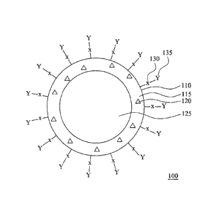
[0021] Fig, 1A is a cross-sectional diagram of an antibody-conjugated
double-emulsion nanocapsule according to some embodiments of this
disclosure.
CA 02910076 2015-10-21
WO 2014/194150
PCT/US2014/040107
[0022] Fig. 1B is a cross-sectional diagram of a double-emulsion
nanocapsule in Fig. IA used as a drug carrier according to some other
embodiments of this disclosure.
[0023] Fig, 2A is a flow chart of the single emulsifying method for
preparing double-emulsion nanocapsules having linking groups thereon
according to some embodiments of this disclosure.
[0024] Fig, 28 is a flow chart of the double emulsifying method for
preparing double-emulsion nanocapsules having linking groups according to
some other embodiments of this disclosure.
[0025] Fig. 2C is a flow chart of a method for reacting an
antibody-coupling agent conjugate and the double-emulsion nanocapsules
having linking groups according to some embodiments of this disclosure.
[0026] Fig. 3 is scanning electron microscopic (SEM) images of the
vacant DENCs using various PVAs having various average molecular weights,
[0027] Figs. 4A and 48 are SEM images of vacant DENCs before and
after linking the breast cancer antibody of trastuzumab.
[0028] Fig. 5A shows drug release profiles of hydrophobic PTX
encapsulated in DENCs containing various amounts of TPMAA at pH 4.
[0029] Fig. 58 shows drug release profiles of hydrophilic Dox
encapsulated in DENCs containing various amounts of TPMAA at pH 4.
[0030] Fig. SC shows drug release profiles of PTX and Dox both
encapsulated in DENCs at pH 4 and pH 7.
[0031] Figs. 6A and 68 are transmission electron microscopic (TEM)
images of trastuzurnab-DENCs containing PVAiTPMAA mixture respectively at
pH 7 and pH 4.
6
CA 02910076 2015-10-21
WO 2014/194150
PCT/US2014/040107
[0032] Fig. 7A shows flow cytometry analysis results of SkBr3 cells
added with various amounts of trastuzumab-DENC encapsulating Dox.
[0033] Fig. 76 shows flow cytometry analysis results of SkBr3 cells
added with various amounts of IgG-DENC encapsulating Dox.
[0034] Fig. 8 shows confocal microscopic images of SkBr3 cells,
unencapsulated Dox, IgG-DENC encapsulating Dox, and trastuzumab-DENC
encapsulating Dox,
[0035] Fig. 9 shows the cell viability of the SkBr3 cells after the SkBr3
cells incubated with various samples,
[0036] Figs, 10A and 106 were IVIS images of the nude mice
experiments on the first day and the third day.
[0037] Fig, 11 shows volumes of the solid tumor varied at different
times.
[0038] Fig. 12 was a SEM image of trastuzumab-DENCs containing a
mixture of PVA and PAA.
[0039] Figs. 13A-13C are SEM images of double-emulsion
nanocapsules containing TPVA having a molecular weight of 25000, 47000,
and 78000, respectively.
DETAILED DESCRIPTION
[0040] The detailed description provided below in connection with the
appended drawings is intended as a description of the present examples and is
not intended to represent the only forms in which the present example may be
constructed or utilized. The description sets forth the functions of the
example
and the sequence of steps for constructing and operating the example.
7
CA 02910076 2017-02-08
However, the same or equivalent functions and sequences may be
accomplished by different examples.
Antibody-Conjugated Double-Emulsion Nanocaps Wes
[0041] Fig. IA is a cross-sectional diagram of an antibody-conjugated
double-emulsion nanocapsule according to some embodiments of this
disclosure, in Fig, 1A, the antibody-conjugated double-emulsion nanocapsule
100 is formed from an oily shell 110 enclosing an aqueous core 125. The
composition of the oily shell 110 includes a polymer 115 and hydrophobic
magnetic nanoparticles 120. The surface of the oily shell 110 has linking
groups 130 and antibody 135 bonded to the linking groups 130 via a coupling
agent (not shown in Fig. IA), The diameter of the antibody-conjugated
double-emulsion nanocapsule 100 is about 50 nrn to about 400 nm.
10042] The polymer 115 includes at least a linking polyvinyl alcohol,
which is modified from polyvinyl alcohol (PVA) to have the linking groups 130,
or
a combination of polyvinyl alcohol and a linking polymer having the linking
groups 130. Furthermore, it is emphasized that the composition of the oily
shell 110 does not need to include any other surfactants or other polymers.
[0043) The polyvinyl alcohol or the linking polyvinyl alcohol itself can
turn the hydrophilic group toward the aqueous core 125 inside the oily shell
110
and the aqueous solution outside the oily shell 110. Therefore, the inner
water-oil interface and the outer oil-water interface of the oily shell 110
can be
simultaneously stabilized without using any other surfactants or any other
polymers.
8
CA 02910076 2015-10-21
WO 2014/194150
PCT/US2014/040107
[0044] The linking group 130 above may be a carboxylic group, a thiol
group, an aldehyde group, an amine group, or a hydroxyl group. For example,
the linking polyvinyl alcohol above may be carboxymethylated polyvinyl alcohol
(CMPVA), thiolated polyvinyl alcohol (TPVA), or a copolymer of PVA-TPMAA.
The linking polymer above may be polyacrylic acid (PAA), polymethacrylic acid
(PMAA), carboxymethylated polyvinyl alcohol (CMPVA), thiolated polyvinyl
alcohol (TPVA), thiolated polymethacrylic acid (TPMAA), or a copolymer of
PVA-TPMAA.
[0045] The chemical structures of the PAA, PMAA, CMPVA, TPVA, and
TPMAA are listed in the table 1 below.
Table Exemplified linking polymers, including linking PVAs
1H2 H
I n PAA
COOH
CH;
H2 I
C -C PMAA
n
COOH
H HI 1H2 H
I x I
OH 0 CMPVA
CH2COOH
1-12"---- H CH2 -
X I
OH 0 TPVA
COCH2SH
9
CA 02910076 2015-10-21
WO 2014/194150
PCT/US2014/040107
CH3 CH3
H, II H2
c
-c ¨ c ¨c
I x - Y TPMAA
COOH CO
NHCH2CH2SH
[0046] The antibody 135 may be any needed antibody. The selection
of the antibody depends on the antigen needed to be bound. For example, the
coupling agent-antibody conjugate 135 may be breast cancer antibody
trastuzumab (commercial name is Herclon or Herceptin), colorectal cancer
antibody cetuximab, epidermal growth factor receptor antibody panitumumab, or
angiogenesis inhibitor antibody bevacizumab.
[0047] The hydrophobic magnetic nanoparticles 120 may be
nanoparticles having a hydrophobic functional groups-modified surface and
made from Fe203, Fe304, CoFe204, or MnFe204. The hydrophobic functional
group may be a long-chained alkyl group or a long-chained alkenyl group, such
as oleic acid or oleylamine. The hydrophobic paramagnetic nanoparticles 120
can stabilize the oily shell 110 to prevent the oily shell 110 from
collapsing. In
addition to being a contrast agent of magnetic resonance imaging (MRI), the
hydrophobic paramagnetic nanoparlicles 120 also can be used to locally heat
and then break the oily shell 110 by magnetic fluid hyperthermia (MFH) under a
high frequency magnetic field (HFMF).
[0048] Since the double-emulsion nanocapsule 100 has the oily shell
110 and the aqueous core 125 to respectively accommodate a hydrophobic
drug and a hydrophilic drug therein, the double-emulsion nanocapsule 100 can
be used as a drug carrier of the hydrophobic drug, the hydrophilic drug, or a
CA 02910076 2015-10-21
WO 2014/194150
PCT/US2014/040107
combination thereof. Furthermore, the release rate of a drug can be controlled
by the strength and on/off state of an applied external alternative magnetic
field,
[0049] Fig. 1B is a cross-sectional diagram of a double-emulsion
nanocapsule in Fig. 1A used as a drug carrier according to some other
embodiments of this disclosure. In Fig.
1B, a hydrophilic drug 145 is
accommodated in the aqueous core 125 of the double-emulsion nanocapsule
100. A hydrophobic drug 140 is accommodated in the oily shell 110 of the
double-emulsion nanocapsule 100 For example, the hydrophilic drug 145
may be doxorubicinl (DOXO) or cisplatin, and the hydrophobic drug 140 may be
paclitaxel (PTX), docetaxel (Dtxl), camptothecin (CPT), or cururmine.
Preparation Method of Antibody-Conjugated Double-Emulsion
Nanocapsule
[0050] The preparation method of antibody-conjugated double-emulsion
nanocapsules includes two stages. At the
first stage, double-emulsion
nanocapsules having linking groups are prepared by a single emulsifying
method or a double emulsifying method. At the second stage, the obtained
double-emulsion nanocapsules are reacted with antibody to form
antibody-conjugated double-emulsion nanocapsules.
[0051] Fig. 2A is a flow chart of the single emulsifying method for
preparing double-emulsion nanocapsules having linking groups thereon
according to some embodiments of this disclosure. In Fig. 2A, an aqueous
solution containing linking PVA (step 202a) and an organic solution containing
hydrophobic magnetic nanoparticles (step 202b) are respectively prepared.
The aqueous solution and the organic solution are then mixed (step 212) to
11
CA 02910076 2015-10-21
WO 2014/194150
PCT/US2014/040107
form an emulsion solution (step 222). The organic solvent in the emulsion
solution is then removed (step 230) to obtain double-emulsion nanocapsules
having linking groups (step 235).
[0052] In the step 202a above, a hydrophilic drug may be further added
into the aqueous solution. In the step 202b above, a hydrophobic drug may be
further added into the organic solution.
[0053] When the organic solution contains only the hydrophobic
magnetic nanoparticles; the organic solvent is better to have the properties
of
effectively dissolving or dispersing the hydrophobic magnetic nanoparticles,
immiscible with water, and lower boiling point. When the organic solution
further contains a hydrophobic drug, the organic solvent is better to further
have
the property of effectively dissolving or dispersing the hydrophobic drug.
[0054] The reason for choosing an organic solvent with a lower boiling
point is that the organic solvent can be easily removed without over-heating
to
prevent the outer shape of the double-emulsion nanocapsules from being
influenced by non-controllable adverse effects. The boiling point of the
organic
solvent can be lower than 90 C. The organic solvent can be chloroform,
dichloromethane, trichloroethane, or acetonitrile, for example.
[0055] In the step 212 above, the method of mixing may be ultrasound
sonication, for example. In step 230 above, the method of removing the
organic solvent may be volatilization at room temperature or reduced pressure
distillation.
[0056] Fig. 2B is a flow chart of the double emulsifying method for
preparing double-emulsion nanocapsules having linking groups according to
some other embodiments of this disclosure. In Fig. 2B, a first aqueous
solution
12
CA 02910076 2015-10-21
WO 2014/194150
PCT/US2014/040107
containing PVA (step 205a) and an organic solution containing hydrophobic
magnetic nanoparticles (step 205b) are respectively prepared. A small amount
of the first aqueous solution and a large amount of the organic solution are
mixed (step 210) to form a first emulsion solution (step 215a), which is a
water-in-oil emulsion solution. This is the first emulsifying stage.
[0057] In step 205a above, a hydrophilic drug may be further added in
to the first aqueous solution, In step 205b above, a hydrophobic drug may be
further added into the organic solution. The selection of the organic solvent
for
the organic solution in step 205b is the same as the step 202b in Fig. 2A, and
hence omitted here.
[0058] A second aqueous solution containing a linking polymer is then
prepared (step 215b), The first emulsion solution and the second aqueous
solution are mixed (step 220) to form a second emulsion solution (step 225).
This is the second emulsifying stage.
[0059] The mixing method of step 210 and step 220 above may be
ultrasound sonication, for example. In step
230 above, the method of
removing the organic solvent may be volatilization at room temperature or
reduced pressure distillation.
[0060] Fig. 2C is a flow chart of a method for reacting an
antibody-coupling agent conjugate and the double-emulsion nanocapsules
having linking groups according to some embodiments of this disclosure. In
Fig. 2C, a first dispersion solution containing double-emulsion nanocapsules
(step 240a) and a second dispersion solution containing antibody-coupling
agent conjugates (step 240b) are respectively prepared. The first and the
second dispersion solutions are mixed (step 245) to react the antibody-
coupling
13
CA 02910076 2015-10-21
WO 2014/194150
PCT/US2014/040107
agent conjugates with the linking groups on the double-emulsion nanocapsules.
Next, the unbound antibody is removed by centrifugation (step 250) to obtain
antibody-conjugated double-emulsion nanocapsules (step 255).
[0061] Usually, the antibody uses a free primary amine group to
connect with a coupling agent to form an antibody-coupling agent conjugate.
According to some embodiments, some suitable coupling agents for forming the
antibody-coupling agent conjugates above are listed in Table 2 below, For
example, when the linking group above is a thiol group, the coupling agent may
be SMCC or SPDP. When the linking group of the linking polymer used in the
double emulsifying method above is a carboxylic group, the coupling agent may
be a combination of EDC and sulfo-NHS,
Table 2: some common coupling agents
0
SMCC
4-(N-maleimidomethyl) cyclohexane
carboxylic acid
N-hydroxysuccinimide ester
0
0
SPDP
aiN-0 3-(2-Pyridyldithio)propionic acid
0 oN-hydroxysuccinimide ester
14
CA 02910076 2015-10-21
WO 2014/194150
PCT/US2014/040107
CH3
- HCI
H3C EDC
11,
H3C. = N-(3-Dimethylaminopropyl)- N-ethyl
H3C-Ni Cl- carbodiimide hydrochloride
HaC N N
0
S¨ONa Suifo-NHS
o 0
o N-Hydroxysulfosuccinimide sodium
N
salt
OH
[0062] The selection of the solvents for the first dispersion solution (step
240a) and the second dispersion solution (step 240b) depends on the coupling
agent used. For example, if SMCC is used as the coupling agent, phosphate
buffered saline (PBS) solution, which contains 0.1 M Na3PO4 and 0.15 M NaCl
and pH value is 7.4, may be used. If EDC and sulfo-NHS are used as the
coupling agent, MES buffer solution containing 0.1 M
2-(N-morpholino)ethanesulfonic acid (MES) and 0.5 M NaCI and pH value is 6.0,
may be used.
[0063] In the embodiments described below, "double-emulsion
nanocapsule" is abbreviated as "DENG," and "antibody-conjugated
double-emulsion nanocapsule" is abbreviated as 'antibody-DENC" to simplify
the writing.
Embodiment 1: Preparing Fe304 Nanoparticles Covered With Oleic Acid
CA 02910076 2015-10-21
WO 2014/194150
PCT/US2014/040107
[0064] In this embodiment, Fe304 nanoparticles covered with oleic acid
(abbreviated as 10-OA nanoparticles) with a diameter of about 5 nm was
prepared. The exemplified preparation method of 10-0A nanoparticles is
described below. Furthermore, the preparation method of 10-0A nanoparticles
may refer to Sun, S. H.; Zeng, FL; Robinson, D. B.; Raoux, S.; Rice, P. M.;
Wang, S. X.; Li, G. X. Journal of the American Chemical Society, 2004, 126,
(1),
273-279, which is incorporated herein by reference.
[0065] 0.708 g of Fe(acac)3, 2.58 g of 1 ,2-Hexadecanediol, O565 g of
oleic acid, 0.535 g of oleylamine, 20 mL of benzyl ether were added into a
three-necked flask. The mixture above was heated, under a condition of
nitrogen atmosphere and cycled cooling water, respectively at 100T for 30
minutes, 200T for 60 minutes, and 285 C for 30 minutes to form 10-0A
nanoparticles. Next, the obtained 10-0A nanoparticles were dispersed in
ethanol and then centrifuged at 6000 rpm to remove the upper solution. After
repeating for several times, the obtained 10-0A nanoparticles were stored in
ethanol.
Embodiment 2: Preparing Thiolated Poiymethacrylic Acid
[0066] In this embodiment, thiolated polymethacrylic add (TPMAA) was
prepared. The exemplified preparation method is described below, and the
synthesis scheme I is also referred at the same time.
16
CA 02910076 2015-10-21
WO 2014/194150
PCT/US2014/040107
CH3
H2 1 H 2N
C C _________ n
SH
COON CH3 CH3
EDC
_______________________________ )1104 CH2CH2 Ci _____________
Sulfo I
-NHS x 1I 1Y
COON CO
NHCH2CH2SH
Synthesis Scheme I
[0067] 250 mg of aqueous solution containing 30 wt% of PMAA was
sequentially added with 5 mt.. of pH 8 PBS solution, 75 mg of catalyst EDC,
and
40 mg of catalyst sulfo-NHS. After mixing and stirring for 15 minutes, 5 mg of
cysteamine was then added. The mixture was stirred until the next day to
react the primary amine group of the cysteamine with the carboxylic group of
the PMAA to form amide bond and obtain TPMAA. Dialysis was used to
remove catalyst EDC and sulfo-NHS, and water was then removed by freeze
dry to obtain TPMAA crystals.
Embodiment 3: Preparing PVA-TPMAA Copolymer
[0068] In this embodiment, PVA-TPMAA copolymer was prepared.
The exemplified preparation method is described below.
[0069] PVA and TPMAA obtained above were mixed. Concentrated
sulfuric acid was then added to form PVA-TPMAA copolymer and side product
of water. Next,
saturated sodium carbonate was used to separate
17
CA 02910076 2015-10-21
WO 2014/194150
PCT/US2014/040107
PVA-TPMAA copolymer and reactants of TPMAA and PVA to obtain the
product of PVA-TPMAA copolymer.
Embodiment 4: Preparing Thiolated Polyvinyl Alcohol
[0070] In this embodiment, thiolated polyvinyl alcohol (TPVA) was
prepared. The exemplified preparation method was described below, and the
synthesis scheme II below was referred at the same time. The reference for
the preparation of TPVA is Gupta B, Anjum S and lkram S. Preparation of
thiolated polyvinyl alcohol hydrogels. Journal of Applied Polymer Science.
2013;
129: 815-21.
H2 C001-1-0-12-Sti H2 H 1 H2 .. H ..
c-cl 1
________________________________ )101- C --C ____ IC --C1 ___
H1SO4 x Y
OH OH 0
COCH2SH
Synthesis Scheme II
[0071] PVA was dissolved in deioinized water to form 2 wt% of PVA
aqueous solution. 20-99% (v/v) of thioglycolic acid and 0.1-i wt% of sulfuric
acid aqueous solution were slowly added into the PVA aqueous solution. The
mixture was heated in an oil bath to perform an esterification reaction. Next,
methanol was slowly poured into the PVA esterification solution to form
precipitate. The precipitate was collected and purified for several times by
methanol to obtain powder. The powder was then freeze dried to obtain TPVA
white crystal powder.
18
CA 02910076 2015-10-21
WO 2014/194150
PCT/US2014/040107
Embodiment 5: Preparing Carboxymethylated Polyvinyl Alcohol
[0072] In this embodiment, carboxymethylated polyvinyl alcohol
(CMPVA) was prepared, and the exemplified method was described below.
Synthesis scheme III is referred at the same time. The reference for the
preparation of CMPVA is Yu C. and Li B. Preparation and characterization of
carboxymethyl polyvinyl alcohol¨graphite nanosheet composites. Polymer
Composites. 2008; 29: 998-1005.
[NaOH CH2-4-1¨ _________________ )8" ______________ _
I n I I
OH OH 0-NaC10E+
-)COON a Fci cH ,l_tcH2 161
________________________________ 3rio
I x I Y
OH 0
CH2COONa
FIC1
cH2 ........................................... H __ icH2¨h8i
lxr I Y
OH 0
ri¨x+y CH2COOH
Synthesis Scheme III
[0073] First, NaOH was added into 2 wt% of PVA aqueous solution to
activate the ¨OH group of PVA. Chloroacetic add (CICH2COOH) was
dissolved in ethanol and neutralized by NaOH to form an ethanol solution of
sodium chloroacetate (CICH2000Na). The two solutions above were mixed to
form a sodium salt of Ctv1PVA. After 5 hours, appropriate amount of HC 1 was
added to adjust the pH value to 6. Subsequently, excess amount of alcohol
19
CA 02910076 2015-10-21
WO 2014/194150
PCT/US2014/040107
was added to purify CMPVA. The ethanol purification step was repeated for
several times.
Embodiment 6: Preparation of Antibody-DENC Containing PVA/TPMAA
Mixture
[0074] In this embodiment, DENC containing PVA/TPMAA mixture was
prepared by using the double emulsifying method in Fig. 2B. The drug used
included hydrophilic doxorubicin (Dox) and cisplatin, and hydrophobic
paclitaxel
(PTX) and camptothecin (CPT). The antibody was then bound to the DENG
containing PVATTPMAA mixture by using the method of Fig. 2C.
[0075] A first aqueous solution of a hydrophilic drug and PVA, a CHCI3
solution of a hydrophobic drug and 10-0A nanoparticles, and a second aqueous
solution of PVA and TPMAA were respectively prepared. In the first aqueous
solution of the hydrophilic drug and PVA, the concentration of the PVA was 20
mg/mL, the concentration of the hydrophilic drug (doxorubicin or cisplatin)
was 8
mg/mL. In the CHCI3 solution of the hydrophobic drug and the 10-0A
nanoparticles, the concentration of the 10-OA nanoparticles was 20 mg/m1_, as
well as the concentration of paclitaxel was 30 mg/mL when the hydrophobic
drug was paclitaxel, and the concentration of camptothecin was 5 mg/mL when
the hydrophobic drug was camptothecin. In the second aqueous solution of
PVA and TPMAA, the concentration of PVA was 20 mg/mL, and the
concentration of TPMAA was 2 mg/m1... The average molecular weight of the
PVA used was respectively 16,000, 25,000, 31,000, and 47,000. In TPMAA,
about 37% of the carboxylic group was modified to have a thiol group.
CA 02910076 2015-10-21
WO 2014/194150
PCT/US2014/040107
[0076] 0.2 ml._ of the first aqueous solution containing the hydrophilic
drug and PVA, as well as 0.5 mL of the CHCI3 solution containing the 10-0A
nanoparticles and the hydrophobic drug were mixed and emulsified by
ultrasound sonication at a frequency of 20 kHz. After the emulsifying, 1.5 mL
of the second aqueous solution containing PVA and TPMAA was further added,
and the mixture was emulsified again by ultrasound sonication at 20 kHz to
obtain DENCs containing PVA/TPMAA mixture. The volatile CHCI3 was
removed by placing the final obtained emulsion solution at an open space to
evaporate the CHCI3. The temperature of evaporating the CHCI3 may change
the morphology of the DENCs. Next, the DENCs containing PVA/TPMAA
mixture were dispersed in 3 mL of PBS solution containing 0.1 M of sodium
phosphate and 0.15 M of NaCI.
[0077] When only one drug was encapsulated by the DENCs above,
the encapsulation efficiency and the diameter of the DENCs are listed in table
3
below. From table 3, it can be known that the encapsulation efficiency of the
hydrophobic drugs was usually greater than the encapsulation efficiency of the
hydrophilic drug. Therefore, the diameter of the DENCs encapsulating the
hydrophobic drugs was usually larger. Besides, the encapsulation efficiency of
the hydrophilic drugs was more than 75%, which is quite good for using the
double emulsifying method to prepare the DENCs encapsulating the hydrophilic
drugs.
Table 3: Encapsulating efficiency of single hydrophilic drug or single
hydrophobic drug
Encapsulated drug Encapsulating Carrier
21
CA 02910076 2015-10-21
WO 2014/194150
PCT/US2014/040107
efficiency (%) diameter (nm)
paclitaxel 95 138
Hydrophobic drug
camptothecin 91 133
cisplatin 76 131
Hydrophilic drug
doxorubicin 83 130
[0078] Fig. 3 is scanning electron microscopic (SEM) images of the
vacant DENCs using various PVAs having various average molecular weights.
In Fig 3, the average molecular weights of PVA contained in the vacant DENCs
were 16000, 25000, 31000, and 47000, respectively. The vacant DENCs did
not encapsulate drugs. From Fig. 3, the greater the average molecular weight
of the PVA was, the smaller the diameter of the vacant DENCs was.
[0079] Next, 1 mg of breast cancer antibody trastuzumab and 4.8 mg of
coupling agent SMCC were respectively dissolved in 2 mL and 5 mL of PBS
solutions containing 0.1 M of sodium phosphate and 0.15 M of NaCI and then
mixed together. The mixture was reacted at 4 C for 2 hours to obtain
trastuzumab-SMCC conjugate, and then centrifuged at 8000 rpm to remove
unreacted SMCC. The trastuzumab-SMCC conjugate was then re-dispersed
in 1 mL of PBS solution containing 0.1 M of sodium phosphate and 0.15 M of
NaCI.
[0080] The dispersion solutions of the DENCs and the
trastuzumab-SMCC conjugates were mixed and reacted at 4 C for 2 hours.
After centrifugation, the unreacted trastuzumab-SMCC conjugate was removed,
and the product of trastuzumab-DENCs was re-dispersed in 4 mL of deionized
water.
22
CA 02910076 2015-10-21
WO 2014/194150
PCT/US2014/040107
[0081] The coupling agent SMCC became a bridge to link the ¨SH
linking group of TPMAA with the ¨NH2 group of the breast cancer antibody of
trastuzumab. Hence, trastuzumab was bound on the outer surface of the oily
shell of the DENCs through SMCC and the thiol group of TPMAA.
[0082] Figs, 4A and 46 are SEM images of vacant DENCs before and
after linking the breast cancer antibody of trastuzumab. The PVA used had an
average molecular weight of 16000. In Fig. 4A, the vacant DENCs containing
TPMAA/PVA mixture had been washed by deionized water, re-dispersed in
deionized water, and then freeze dried. The vacant DENCs still maintain the
hollow spherical structure. In Fig. 46, the vacant DENCs in Fig. 4A were
bound to trastuzumab-SMCC conjugate. Since the surface of the vacant
DENCs was modified by trastuzumab-SMCC conjugate, the morphology of the
trastuzumab-DENCs was changed.
Embodiment 7: Effect of pH Values of Solutions on Release of Drugs
Encapsulated in DENCs Containing PVNTPMAA Mixture
[0083] In this embodiment, the effect of pH values of solutions on the
release of drugs encapsulated in antibody-DENCs containing PVAITPMAA
mixture was tested. The oily shell was composed of PVA having a molecular
weight of 16000 and TPMAA. The drug used included hydrophilic doxorubicin
(Dox) and hydrophobic paclitaxel (PTX).
[0084] TPMAA is a modified PMAA polymer having thiol functional
groups, and PMAA is a pH-sensitive polymer. The carboxylic acid groups and
methyl groups on side chains of PMAA are the main factors affecting PMAA to
show different appearance in various environments having various pH values.
23
CA 02910076 2015-10-21
WO 2014/194150
PCT/US2014/040107
In a neutral environment, PMAA is randomly coiled and hydrophilic. In an acid
environment, PMAA is transformed and shrunk to a globule-like structure and
becomes hydrophobic. It was hoped that the pH-sensitive property of PMAA
can be preserved after being modified by thiol groups for linking breast
cancer
antibody of trastuzumab. Therefore, DENCs encapsulating dual drugs were
respectively placed in a neutral environment (pH 7) and an acidic environment
(pH 4) to observe the release amount of drugs.
[0085] Fig. 5A shows drug release profiles of hydrophobic PTX
encapsulated in DENCs containing various amounts of TPMAA at pH 4. Fig.
5B shows drug release profiles of hydrophilic Dox encapsulated in DENCs
containing various amounts of TPMAA at pH 4. In Figs, 5A and 56, the
modification percentage of TPMAA was 37%. From Figs. 5A and 58, it can be
known that the addition amount of TPMAA was increased during the
preparation process, the release amounts of PTX and Dox both were increased
at the acidic environment at pH 4. This result shows that TPMAA may
increase the release rate of drugs from the DENCs in an acidic environment.
[0086] Fig. 5C shows drug release profiles of PTX and Dox both
encapsulated in DENCs at pH 4 and pH 7, In Fig. Sc, the modification
percentage of TPMAA was 37%. The addition amount of TPMAA was 1 wt%,
From Fig. 5C, it can be known that the release amounts of drugs at pH 4 are
far
greater than the release amounts of drugs at pH 7. In the acidic environment
of pH 4, the release amount of the hydrophobic PTX was greater than the
release amount of the hydrophc Dox.
[0087] Figs. 6A and 6B are transmission electron microscopic (TEM)
images of trastuzumab-DENCs containing PVA/TPMAA mixture respectively at
24
CA 02910076 2015-10-21
WO 2014/194150
PCT/US2014/040107
pH 7 and pH 4. The addition amount of TPMAA was 1 wt%, and the
modification percentage of TPMAA was 37%. Fig. 6A shows that the
trastuzumab-DENC had a spherical shell in the neutral environment. Fig. 6B
shows that the shell of the trastuzumab-DENC was shrunk and deformed in the
acidic environment.
[0088] The drug release behaviors above were consistent with that the
TPMAA shrunk in an acidic environment and was transformed to be
hydrophobic. When a lot of hydrogen ions are present in the environment, the
TPMAA in the shell begins to shrink and the shell is thus deformed and
extruded. Therefore, the hydrophobic drug located in the oily shell could be
released more than the hydrophilic drug located in the aqueous core.
Embodiment 8: Recognition of Trastuzumab-Conjugated Carrier to HER-2
Overexpressing Cells
[0089] In this embodiment, whether the trastuzumab-DENC can target
HER-2 overexpressing cells or not was verified. The
selected HER-2
averexpressing cell clone was SkBr3 (human breast adenocarcinoma) cells.
The shell of the tested trastuzumab-DENC was composed of a mixture of PVA
having a molecular weight of 16000 and TPMAA. The addition amount of
TPMAA was 1 wt%, and the modification percentage of TPMAA was 37%.
[0090] First, the DENCs encapsulating hydrophilic Dox were
respectively conjugated with the breast cancer antibody trastuzumab and an
antibody IgG, which is not specific to SkBr3 cells. Then, the antibody-DENCs
encapsulating hydrophilic doxorubicin (Dox) and the SkBr3 cells were incubated
together at 37 0C for 30 minutes. Since Dox can emit fluorescence (excited at
CA 02910076 2015-10-21
WO 2014/194150
PCT/US2014/040107
a wavelength of 488 nm and emitting at a wavelength of 580 nm), flow
cytometer can be used to detect the fluorescence intensity of Dox bound onto
the cell surface of the SkBr3 cells via the interaction of antibody-antigen.
[0091] Fig. 7A shows flow cytometry analysis results of SkBr3 cells
added with various amounts of trastuzumab-DENC encapsulating Dox. In Fig.
7A, the trastuzumab-DENC encapsulating Dox is denoted as T, and the addition
amount of the trastuzumab-DENC encapsulating Dox for the curve denoted as
control was zero. It can be seen that more SkBr3 cells have more intense
fluorescence when the addition amount of trastuzumab-DENC encapsulating
Dox was more. This result showed that the trastuzumab-DENC encapsulating
Dox can recognize the SkBr3 cells and bind onto the surface of SkBr3 cells,
[0092] Fig, 7B shows flow cytometry analysis results of SkBr3 cells
added with various amounts of IgG-DENC encapsulating Dox, In Fig. 7B, the
IgG-DENC encapsulating Dox is denoted as IgG, and the addition amount of
the IgG-DENC encapsulating Dox for the curve denoted as control was zero. It
can be seen that no matter the addition amount of the IgG-DENC encapsulating
Dox was, the fluorescence intensity distribution were almost the same. This
result showed that the IgG-DENC encapsulating Dox does not show any
specificity to the SkBr3 cells.
[0093] In order to further confirm the results above, after respectively
incubating the trastuzumab-DENC encapsulating Dox and SkBr3 cells as well
as IgG-DENC encapsulating Dox and SkBr3 cells, the nuclei of the SkBr3 cells
were stained by a dye of 4',6-diamidino-2-phenylindole (DAPI). The
distribution of the fluorescence Dox and nuclei was observed by confocal
26
CA 02910076 2015-10-21
WO 2014/194150
PCT/US2014/040107
microscopy. In addition, pure SkBr3 cells and free Dox were also observed by
the confocal microscopy. The obtained results were shown in Fig. 8.
[0094] Fig. 8 shows confocal microscopic images of SkBr3 cells, free
Dox, IgG-DENC encapsulating Dox, and trastuzumab-DENC encapsulating Dox.
In Fig. 8, the columns each denoted by "cell", "Dox", "IgG-Dox", and "T-Dox"
respectively represent the samples of SkBr3 cells, free Dox, IgG-DENC
encapsulating Dox, and trastuzumab-DENC encapsulating Dox. The lines
each denoted by "nucleus", "Dox" and "image merge" respectively represent the
positions of nuclei, Dox, as well as an overlapping image of the positions of
nuclei and Dox.
[0095] Fig. 8 shows that the positions of trastuzumab-DENC
encapsulating Dox and the nuclei of SkBr3 cells had many overlaps. This
means that trastuzumab-DENC encapsulating Dox really could recognize SkBr3
cells. However, in other samples, almost only nuclei could be observed, and
almost no Dox images were observed to overlap the nuclei images. This
means that other samples were almost not attached on the SkBr3 cells, i.e.
other samples could not recognize SkBr3 cells.
Embodiment 9: Cytotoxicity Effect of Various DENCs on HER-2
Overexpressing Cells
[0096] In this embodiment, the cytotoxicity of various DENCs to HER-2
overexpressing cells was studied. The shell of the DENC was a mixture of
PVA having a molecular weight of 16000 and TPMAA. The addition amount of
TPMAA was 1 wt%, and the modification percentage of TPMAA was 37%.
27
CA 02910076 2015-10-21
WO 2014/194150
PCT/US2014/040107
[0097] The unconjugated vacant DENC (the control group), DENC
encapsulating PTX (PTX group), DENC encapsulating Dox (Dox group),
trastuzumab-DENC (T group), trastuzumab-DENC encapsulating PTX (T-PTX
group), DENC encapsulating PTX and Dox (PTX-Dox), trastuzumab-DENC
encapsulating PTX and Dox (T-PTX-Dox) were respectively added into SkBr3
cell cultures and then respectively co-cultured at 37 0C for 24 hours. Next,
MIT assay was used to assess cell viability of each sample. The obtained
results are listed in table 4 below and shown in Fig. 9
Table 4: Cytotoxicity effect of various DENCs on SkBr3 cells
Cell
Cell viability percentage of
Carriers viability
trastuzumab-DENC*
(To)
Unconjugated vacant DENC 100.99
(control) 1.01
74.62%
trastuzumab-conjugated vacant 75.36
DENC (T) 3,86
56.40
DENC encapsulating PTX (PTX)
4.40
47.73%
trastuzumab-DENC encapsulating 26.92
PTX (T-PTX) 3.50
34.80
DENC encapsulating Dox (Dox)
4.33
83.93%
trastuzumab-DENC encapsulating 29.21
Dox (T-Dox) 3.34
DENC encapsulating PTX and Dox 23,60
(PTX-Dox) 3.25
59.11%
trastuzumab-DENC encapsulating 13.95
PTX and Dox (T-PTX-DOX) 2.89
28
CA 02910076 2015-10-21
WO 2014/194150
PCT/US2014/040107
* calculated by (the cell viability of trastuzumab-DENC/ the cell viability of
DENC)
x 100%
[0098] From the results shown in table 4 and Fig. 9, it can be known
that the vacant DENC did not have cytotoxicity effect on the SkBr3 cells and
thus was nontoxic to the SkBr3 cells. However, after the vacant DENC
conjugated with trastuzumab, the cell viability was decreased to about 75%.
Comparing the cell viability of the DENC encapsulating PTX before and after
conjugated with trastuzumab, the cell viability was decreased from about 56%
to about 27%, which is only about 48% of the DENC encapsulating PTX before
conjugated with trastuzumab. Comparing the cell viability of the DENC
encapsulating Dox before and after conjugated with trastuzumab, the cell
viability was decreased from about 35% to about 29%, which is about 84% of
the DENC encapsulating Dox before conjugated with trastuzumab. Comparing
the cell viability of the DENC encapsulating both PTX and Dox before and after
conjugated with trastuzumab, the cell viability was decreased from about 24%
to about 14%, which is about 59% of the DENC encapsulating both PTX and
Dox before conjugated with trastuzumab.
[0099] From the comparisons above, it can be known that after
conjugated with trastuzumab, all kinds of DENCs have a better cytotoxicity
effect on the SkBr3 cells.
Embodiment 10; in vivo Animal Experiments
[0100] In this embodiment; nude mice bearing SkBr3 solid tumors were
used to perform in vivo animal experiments. The shell of the DENCs
29
CA 02910076 2015-10-21
WO 2014/194150
PCT/US2014/040107
composed of a mixture of PVA having a molecular weight of 16000 and TPMAA.
The addition amount of TPMAA was 1 wt%, and the modification percentage of
TPMAA was 37%. The shell had been linked to a dye Cyanine 5.5 (Cy5,5).
[01011 First, the distribution of the DENCs in nude mice was observed
by using a non-invasion in vivo imaging system (MS). A 3700 G magnet was
attached to the tumor on the left side of the nude mice, and no magnets were
attached to the tumor on the right side of the nude mice. The observed results
of the first day and the third day after injecting the DENCs into the nude
mice
under IVIS were shown in Figs. 10A and 10B.
[0102] In the image of the first day shown in Fig. 10A, a large amount of
DENCs were accumulated on the both sites of the tumor 110a on the left side
and the tumor 120a on the right side. However, in the image of the third day
shown in Fig. 10B, the accumulative amount of the DENCs on the left tumor
110b was much greater than the accumulative amount of the DENCs on the
right tumor 120b, and the accumulative amount of the left tumor 110b was
about two times of the accumulative amount of the right tumor 120b.
Therefore, an external applied magnetic field indeed can affect the
distribution
of the magnetic-sensitive DENCs in the nude mice,
[0103] Next, nude mice xenograft tumor model was used to analyze the
therapeutic effect of various DENCs. In the experiment, the tested various
DENCs were injected into the nail veins of nude mice respectively at the
first,
fifth, ninth, and thirteenth days to treat the tumor. Then, the IVIS was used
to
observe the tumor size during the 1-30 days. The obtained results were
shown in Fig. 11.
CA 02910076 2015-10-21
WO 2014/194150
PCT/US2014/040107
[0104] The experimental groups shown in Fig. 11 include saline, vacant
DENC, DENC encapsulating PTX (PTX), DENC encapsulating Dox (Dox),
trastuzumab-DENC encapsulating PTX (T-PTX), DENC encapsulating PTX and
Dox (PTX-Dox), trastuzumab-DENC encapsulating PTX and Dox but no applied
magnetic field (T-PTX-DOX No MT), and trastuzumab-DENC encapsulating
PTX and Dox (T-PTX-DOX). Each experiment group had a 2000 G magnet
attached on the tumor site, only the group of T-PTX-DOX No MT did not has a
magnet attached on the tumor site.
[0105] From the results shown in Fig 11, it can be known that the
therapy effect of the T-PTX-DOX group was the best, and the tumor size was
increased to only 1.96 times of the original tumor size after 30 days.
However,
for the saline group, the tumor size was increased to 17.6 times of the
original
tumor size after 30 days.
Embodiment 11: Antibody-DENC Containing Mixture of PVA and
PVA-TPMAA Copolymer
[0106] In this embodiment, DENCs containing a mixture of PVA and
PVA-TPMAA copolymer were prepared by the double emulsifying method in Fig.
2B, The
encapsulated drug had hydrophilic doxorubicin (Dox) and
hydrophobic paclitaxel (PTX) as examples. These two drugs are common
chemotherapy drugs for cancer therapy. The DENCs containing mixture of
PVA and PVA-TPMAA copolymer were then conjugated with an antibody on the
shells.
[0107] The preparation method of the DENCs containing mixture of
PVA and PVA-TPMAA copolymer was similar to the DENCs containing a
31
CA 02910076 2015-10-21
WO 2014/194150 PCT/US2014/040107
mixture of PVA and TPMAA in embodiment 6. The only difference was that
the second aqueous solution of PVA and TPMAA was replaced by 2 wt% of
PVA-TPMAA copolymer aqueous solution, and the modification percentage of
the TPMAA in PVA-TPMAA copolymer was 37%. Finally, the obtained
trastuzumab-DENCs containing mixture of PVA and PVA-TPMAA copolymer
was dispersed in deionized water.
[0108] First, the content of TPMAA in PVA-TPMAA copolymer was
investigated to see the effect on the antibody conjugation percentage, DENC
diameter, and encapsulating efficiency. The obtained results are listed in
table
below. From table 5, it can be known that the antibody conjugation
percentage was greater when the TPMAA content was more, since the
antibodies needed thiol groups of TPMAA to link with the DENCs. In addition,
the higher the antibody conjugation percentage was, the larger the DENC's
diameter was. The encapsulating efficiency of drugs was not affected much by
the TPMAA content, and thus not by the antibody conjugation percentage. It
may be that the drugs had been encapsulated in the DENCs before the
antibodies were conjugated with the DENCs.
Table 5: Effect of TPMAA content in PVA-TPMAA copolymer on the
antibody conjugation percentage, DENC's diameter, and drug's encapsulating
efficiency
Antibody
Molar ratio of DENC's Encapsulating Encapsulating
conjugation
PVA: TPMAA diameter efficiency of efficiency of
percentage
in copolymer (nm) PTX (%) Dox (%)
(%)
6:1 50.92 156.43 96.20 82A2
32
CA 02910076 2015-10-21
WO 2014/194150
PCT/US2014/040107
5:1 64.29 167.21 99.10 80.67
4:1 77.47 175.23 96.61 81.30
3:1 84.26 198.20 98.45 78,21
[0109] Next, the volatile temperature and volatile time of the organic
solvent chloroform in the preparation was investigated to see the effect on
the
DENC's diameter and drug's encapsulating efficiency. The molar ratio of PVA
to TPMAA of the PVA-TPMAA copolymer was 4: 1. The obtained results are
listed in table 6 below. In table 6, the volatile temperature and volatile
time of
chloroform did not have obvious effect on the DENCs diameter and drug's
encapsulating efficiency below 55 C.
Table 6. Effect of volatile temperature and volatile time of organic solvent
chloroform on DENC's diameter and drug's encapsulating efficiency
,. .,
Volatile Encapsulating Encapsulating
Volatile diameter
temperature
efficiency of PTX efficiency of Dox
time (hi) (nm)
( C) ( ,10) (%)
4 ' 164.3 95.50 79.32
25 C
162.6 94.64 77.34
3 159.7 97.12 82.43
35 C 4 158.1 ' 96.67 81.30
..
5 159.3 96.10 80.33
2.5 159.1 98.20 81,70
3 ' 157.5 98.00 81.46
45 C .
4 ' 158.6 96.56 81.23
5 ' 155.3 94.40 80.67
55 C ' 1 ' 164.9 98.87 83.43
2 157.2 98.30 82.23
33
CA 02910076 2015-10-21
WO 2014/194150
PCT/US2014/040107
3 160.5 97.62 81.78
4 154.7 97.20 81.21
[0110] The effect of the emulsifying time on the DENC's diameter and
drug's encapsulating efficiency was subsequently investigated. The molar
ratio of PVA to TPMAA in PVA-TPMAA copolymer was 4: 1. The obtained
result was listed in table 7 below. In table 7, the length of the first and
the
second emulsifying time did not have obvious effect on the DENC's diameter
and drug's encapsulating efficiency.
Table 7: Effect of emulsifying time on DENC's diameter and drug's
encapsulating efficiency
Emulsifying
Diameter Encapsulating Encapsulating
time (s)
(nm) efficiency of PTX (%) efficiency of Dox (%)
first second
35 161.1 93.12 77.65
15 45 158.5 95.14 78.54
55 162.1 96.50 78.91
35 157.8 94.34 79.76
20 45 158.1 96.05 80.45
55 162.4 96.43 80.73
[0111] The effect of PVA molecular weight and TPMAA content of
PVA-TPMAA copolymer on the product morphology was investigated. The
obtained result was listed in table 8 below. In table 8, when the molecular
weight of PVA was from 25000 to 61000, the diameter of the DENC was
increased as the TPMAA content was increased. When PVA has a molecular
34
CA 02910076 2015-10-21
WO 2014/194150
PCT/US2014/040107
weight of 47000, about half number of the nanostructures had the core-shell
structure. When the PVA had a molecular weight of 61000, only a few number
of the nanostructures had the core-shell structure. When the PVA had a
molecular weight of 78000, no nanocapsules were formed. This result shows
that no nanocapsules will be formed when the molecular weight of PVA was too
large.
Table 8: Effect of PVA molecular weight and TPMAA content of
PVA-TPMAA copolymer on the product morphology
PVA TPMAA's content Core-shell
Diameter (nm)
MW (mol%) structure
Yes 135.6
25000 20 Yes 143.4
30 Yes 154.6
10 Yes 131.6
31000 20 Yes 136.8
30 Yes 142.3
10 Half 141.5
47000 20 Half 145.3
30 Half 149.8
10 Few 124.3
61000 20 Few 129.1
30 Few 133.2
10 No 116.5
78000 20 No 125.6
30 No 134.4
Embodiment 12: Antibody-Conjugated Carrier Containing PVA/TPVA
Mixture
CA 02910076 2015-10-21
WO 2014/194150
PCT/US2014/040107
[0112] In this embodiment, double-emulsion nanocapsules (i.e. DENCs)
containing a mixture of PVA and TPVA were prepared by the double
emulsifying method in Fig. 28. The exemplary drugs used were hydrophilic
doxorubicin (Dox) and hydrophobic paclitaxel (PTX). These two drugs are
common chemotherapy drugs for cancer therapy. The DENCs containing a
mixture of PVA and TPVA were then conjugated with antibody on the shell's
surface by the method of Fig. 2C.
[0113] The preparation method of the DENCs containing mixture of
PVA and TPVA was similar to the DENCs containing a mixture of PVA and
TPMAA in embodiment 6. The only difference was that the second aqueous
solution of PVA and TPMAA was replaced by 2 wt% of TPVA aqueous solution,
and the modification percentage of TPVA was 30%. Finally, the obtained
trastuzumab-DENCs containing mixture of PVA and TPVA was dispersed in
deionized water.
[0114] First, the TPVA content was investigated to see the effect on the
antibody conjugation percentage, DENC's diameter, and encapsulating
efficiency_ The obtained results were listed in table 9 below. In table 9, the
antibody conjugation percentage was greater when the TPVA content was more,
since the antibodies needed thiol groups of TPVA to link with the DENCs. In
addition, the higher the antibody conjugation percentage was, the larger the
DENCs diameter was. The encapsulating efficiency of drugs was not affected
much by the TPVA content, and thus not by the antibody conjugation
percentage. It may be that the drugs had been encapsulated in the DENCs
before the antibodies were conjugated with the DENCs.
36
CA 02910076 2015-10-21
WO 2014/194150
PCT/US2014/040107
Table 9: Effect of TPVA content on the antibody conjugation percentage,
DENas diameter, and drug's encapsulating efficiency
antibody
TPVA DENC's Encapsulating Encapsulating
conjugation
content diameter efficiency of efficiency of
percentage
(mol%) (nm) PTX (%) Dox (%)
(%)
11,85 135.3 94.11 79,32
23.50 14T5 92.31 77.31
40.65 165.3 92,81 78.35
54.53 173.4 95.25 79.89
[0115] The effect of the emulsifying time on the DENC's diameter and
drug's encapsulating efficiency was subsequently investigated. The TVPA
content was 30 mol%, and the molecular weight of PVA was 16000. The
obtained result was listed in table 10 below. In table 10, the length of the
first
and the second emulsifying time does not obvious effect on the DENC's
diameter and drug's encapsulating efficiency.
Table 10: Effect of emulsifying time on DENC's diameter and drug's
encapsulating efficiency
Emulsifying
Diameter Encapsulating
Encapsulating
time (s)
(nm) efficiency of PTX (%) efficiency of Dox (%)
first second
35 131.4 92.1 77,2
15 45 137.6 95.1 79.2
55 134.1 92.6 78.4
20 35 133,4 90,5 80,3
131.1 927 75.9
37
CA 02910076 2015-10-21
WO 2014/194150
PCT/US2014/040107
55 136.6 93.1 78.3
[0116] The effect of PVA molecular weight and TPVA content on the
product morphology was investigated. The obtained results were listed in table
11 below. In table 11, when the molecular weight of PVA was from 25000 to
61000, the diameter of the DENG was increased as the TPVA content was
increased. When PVA has a molecular weight of 47000, about half number of
the nanostructures had the core-shell structure. When the PVA had a
molecular weight of 61000, only a few number of the nanostructures had the
core-shell structure. When the PVA had a molecular weight of 78000, no
nanocapsules were formed. This result shows that no nanocapsules will be
formed when the molecular weight of PVA was too large.
Table 11: Effect of PVA molecular weight and TPVA content of
PVA-TPMAA copolymer on the product morphology
PVA MW TPVA content (mol%) Core-shell structure Carrier's diameter (nm)
Yes 126.4
25000 20 Yes 131.5
30 Yes 137.6
10 Yes 121.6
31000 20 Yes 135.8
30 Yes 138.3
10 Half 137.5
47000 20 Half
146.3
30 Half 151.8
108,4(solid core)/
61000 10 Few
141.6(core-shell)
38
CA 02910076 2015-10-21
WO 2014/194150
PCT/US2014/040107
112.5(solid core)/
20 Few
148 1(core-shell)
119.4(solid core)/
30 Few
153.4(core-shell)
No 116.5
78000 20 No 125.6
30 No 134.4
Embodiment 13: Antibody-DENCs Containing PVA/PAA Mixture
[0117] In this embodiment, double-emulsion nanocapsules (i.e. DENCs)
containing a mixture of PVA and PM were prepared by the double emulsifying
method in Fig. 2B The exemplary drugs used were hydrophilic doxorubicin
(Dox) and hydrophobic paclitaxel (PTX). These two drugs are common
chemotherapy drugs for cancer therapy. The DENCs containing a mixture of
PVA and PAA were then conjugated with antibody on the shell's surface by the
method of Fig. 2C
[0118] The preparation method of the DENCs containing mixture of
PVA and PM was similar to the DENCs containing a mixture of PVA and
TPMAA in embodiment 6. The first difference was that the second aqueous
solution of PVA and TPMM was replaced by an aqueous solution of PVA and
PAA. In the aqueous solution of PVA and PAA, the concentration of PVA was
mg/mL, and the concentration of PM was 2 mg/m1.... The second difference
was that the coupling agent of SMCC was replaced by a combination of EDC
and sulfo-NHS to link the primary amine group of the breast cancer antibody
trastuzumab to the carboxylic group of PM. The molecular weight of the PVA
was 16000.
39
CA 02910076 2015-10-21
WO 2014/194150
PCT/US2014/040107
[0119] In the reaction of the breast cancer antibody trastuzumab and
the coupling agent, 0.1 M of MES buffer solution containing 0.1 M of MES and
0.5 M of NaCI and having a pH value of 6.0 was first prepared. Then, DENCs,
50 pg of EDC, and 60 pg of SuIfo-NHS were sequentially added into the 3 mL of
MES buffer solution. The mixture was stirred and reacted for 15 minutes, 1
pL of 2-mercaptoethanol was then added into the above MES buffer solution to
stop the activation reaction of EDC. Next, high concentration of PBS solution
was added into the MES buffer solution to increase the pH value to more than
7.
Subsequently, 500 pg of breast cancer antibody trastuzumab was added and
reacted at room temperature for 2 hours to obtain trastuzumab-DENCs,
[0120] The obtained trastuzumab-DENCs were dispersed in deionized
water, and unreacted agents were removed after the dispersion solution was
centrifuged at 7000 rpm. The steps of dispersion and centrifugation were
repeated for several times to purify the trastuzumab-DENCs. The purified
trastuzumab-DENCs were dispersed in a solvent, such as saline.
[0121] Fig. 12 was a SEM image of trastuzumab-DENCs containing a
mixture of PVA and PAA. In Fig. 12, it can be observed that the spherical
shape of the unconjugated DENC was changed to the irregular shape of
trastuzumab-DENCs. This may be caused by conjugating the DENCs with the
breast cancer antibody trastuzumab. However, it still can be seen that the
trastuzumab-DENCs had a hollow structure from the black and white contrast of
the SEM image. In addition, the obtained trastuzumab-DENCs containing
PVA/PAA mixture can be uniformly dispersed in solution without forming
precipitation Therefore, the trastuzumab-DENCs containing PVA/PAA mixture
CA 02910076 2015-10-21
WO 2014/194150
PCT/US2014/040107
were similar to the trastuzumab-DENCs containing PVA/TPMAA mixture in
Embodiment 6.
Embodiment 14: Antibody-DENCs Containing PVA/PMAA mixture
[0122] In this embodiment, double-emulsion nanocapsules (i.e.
DENCs) containing a mixture of PVA and PMAA were prepared by the double
emulsifying method in Fig. 2B. The exemplary drugs used were hydrophilic
doxorubicin (Dox) and hydrophobic paclitaxel (PTX). These two drugs are
common chemotherapy drugs for cancer therapy. The DENCs containing a
mixture of PVA and PMAA were then conjugated with antibody on the shell's
surface by the method of Fig. 2C.
[0123] The preparation method of the DENCs containing mixture of
PVA and PMAA was similar to the DENCs containing a mixture of PVA and
TPMAA in embodiment 6. The only difference was that the second aqueous
solution of PVA and TPMAA was replaced by an aqueous solution containing a
mixture of PVA and PMAA, and the molecular weight of the PVA was 16000.
[0124] The obtained trastuzumab-DENCs containing mixture of PVA
and PMAA also had an irregular morphology observed under SEM, but still
maintain a hollow structure. In addition, the trastuzumab-DENCs containing
mixture of PVA and PMAA also could be uniformly dispersed in solution without
forming precipitation. Therefore, the trastuzumab-DENCs containing
PVA/PMAA mixture were similar to the trastuzumab-DENCs containing
PVA/TPMAA mixture in Embodiment 6.
Embodiment 15: Antibody-DENCs Containing PVA/CMPVA Mixture
41
CA 02910076 2015-10-21
WO 2014/194150
PCT/US2014/040107
[0125] In this embodiment, double-emulsion nanocapsules (i.e. DENCs)
containing a mixture of PVA and CMPVA were prepared by the double
emulsifying method in Fig. 28. The exemplary drugs used were hydrophilic
doxorubicin (Dox) and hydrophobic paclitaxel (PTX). These two drugs are
common chemotherapy drugs for cancer therapy, The DENCs containing a
mixture of PVA and CMPVA were then conjugated with antibody on the shell's
surface by the method of Fig. 2C
[0126] The preparation method of the DENCs containing mixture of
PVA and CMPVA was similar to the DENCs containing a mixture of PVA and
PAA in embodiment 13. The only difference was that the second aqueous
solution of PVA and PAA was replaced by an aqueous solution containing a
mixture of PVA and CMPVA. The molecular weight of the PVA was 16000,
and the modification percentage of the CMPVA was 30%.
[0127] First, the CMPVA content was investigated to see the effect on
the antibody conjugation percentage, DENC's diameter, and encapsulating
efficiency. The obtained results were listed in table 12 below. In table 12,
the
antibody conjugation percentage was greater when the CMPVA content was
more, since the antibodies needed carboxylic groups of CMPVA to link with the
DENCs. In addition, the higher the antibody conjugation percentage was, the
larger the DENCs diameter was.
[0128] The encapsulating efficiency of drugs was not affected much by
the CMPVA content, and thus not by the antibody conjugation percentage. It
may be that the drugs had been encapsulated in the DENCs before the
antibodies were conjugated with the DENCs However, comparing the DENCs
containing the mixture of PVA and CMPVA (table 12) and the DENCs
42
CA 02910076 2015-10-21
WO 2014/194150
PCT/US2014/040107
containing the mixture of PVA and TPVA (table 9), since protons are easily
dissociated from the carboxylic groups of CMPVA, the encapsulating efficiency
of Dox by DENC$ containing the mixture of PVA and CMPVA was increased by
5-10%.
Table 12: Effect of CMPVA content on the antibody conjugation
percentage, DENC's diameter, and drug's encapsulating efficiency
antibody
DENC's Encapsulating Encapsulating
CMPVA content conjugation
diameter efficiency of efficiency of
(mol%) percentage
(nm) PTX (%) Dox (%)
(%)
17.82 143.6 96.31 84.34
29.10 149.1 97.10 83.65
43.25 166,5 95.71 87.65
61.32 169,6 96.75 88.53
85,64 178.7 95,67 89.46
[0129] Next, the effect of the emulsifying time on the DENC's diameter
and drug's encapsulating efficiency was subsequently investigated. The
CMPVA content was 30 mol%, and the molecular weight of PVA was 16000.
The obtained results were listed in table 13 below. In table 13, the length of
the first and the second emulsifying time did not have obvious effect on the
DENC's diameter and drug's encapsulating efficiency.
Table 13: Effect of emulsifying time on DENC's diameter and drug's
encapsulating efficiency
43
CA 02910076 2015-10-21
WO 2014/194150
PCT/US2014/040107
Emulsifying
Diameter Encapsulating Encapsulating
time (s)
(nm) efficiency of PTX (%) efficiency of Dox (To)
first second
35 153.4 95.21 79.32
15 45 148.2 96,70 77.31
55 156.2 96.10 78.35
35 154.3 97.25 79.89
20 45 156.3 94.67 79.32
55 149.4 95.31 77.31
[0130] Next, the effect of PVA molecular weight and CMPVA content on
the product morphology was investigated. The obtained result was listed in
table 14 below. In table 14, when the molecular weight of PVA was from
25000 to 61000, the diameter of the DENCs was increased as the CMPVA
content was increased. When the PVA had a molecular weight of 61000, only
a few number of the nanostructures had the core-shell structure. When the
PVA had a molecular weight of 78000, no nanocapsules were formed. This
result shows that no nanocapsules will be formed when the molecular weight of
PVA was too large.
Table 14: Effect of PVA molecular weight and CMPVA content of
PVA-TPMAA copolymer on the product morphology
PVA MW TPVA content (mol%) Core-shell structure Carrier's diameter (nm)
25000 10 Yes 124.6
30 Yes
131.4
44
CA 02910076 2015-10-21
WO 2014/194150
PCT/US2014/040107
50 Yes
133.6
Yes
121.5
31000 30 Yes
127.6
50 Yes
131.5
10 Yes
134.5
47000 30 Yes
139.5
50 Yes
142.4
10 Few 116.5
61000 30 Few 127.1
50 Few 133.2
10 No 91.5
78000 30 No 96_7
50 No 109.3
Embodiment 16: Antibody-Conjugated Carrier Containing Mixture of
PVA-TPMAA Copolymer
[0131] In this embodiment, double-emulsion nanocapsules (Le. DENCs)
containing PVA-TPMAA copolymer were prepared by the single emulsifying
method in Fig. 2A. The exemplary drugs used were hydrophilic doxorubicin
(Dox) and hydrophobic paclitaxel (PTX). These two drugs are common
chemotherapy drugs for cancer therapy. The DENCs containing PVA-TPMAA
copolymer were then conjugated with antibody on the shell's surface by the
method of Fig 2C.
CA 02910076 2015-10-21
WO 2014/194150
PCT/US2014/040107
[0132] An aqueous solution of PVA-TPMAA copolymer and Dox, as well
as a chloroform solution of 10-0A nanoparticles and PTX were respectively
prepared. In the aqueous solution of PVA-TPMAA copolymer and Dox, the
concentration of PVA-TPMAA copolymer was 20 mg/mL, and the concentration
of Dox was 8 mg/mt... In the chloroform solution of 10-OA nanoparticles and
PTX, the concentration of 10-0A nanoparticles was 20 mg/mL, and the
concentration of Dox was 30 mgimL.
[0133] 2.5 mL of the first aqueous solution containing PVA-TPMAA
copolymer and Dox, as well as 1 mL of the CHCI3 solution containing I0-0A
nanoparticles and PTX were mixed and emulsified by ultrasound sonication at a
frequency of 20 kHz to obtain DENCs containing PVA-TPMAA copolymer. The
modification percentage of the TPMAA copolymerized with PVA was 37%.
The volatile 0HCI3 of the emulsion solution was then removed by placing the
final obtained emulsion solution at an open space to evaporate the CHCI3.
The temperature of evaporating the CHCI3 may change the morphology of the
DENCs. Next, the DENCs containing PVA-TPMAA copolymer were dispersed
in 3 mL of PBS solution containing 0.1 M of sodium phosphate and 0.15 M of
NaCl.
[0134] Next, the breast cancer antibody trastuzumab was conjugated
with the obtained DENCs containing PVA-TPMAA copolymer, The details of
the conjugation method was the same as the conjugation method of
Embodiment 6, and hence omitted here,
[0135] First, the TPMAA content in the PVA-TPMAA copolymer was
investigated to see the effect on the antibody conjugation percentage, DENCs
diameter, and encapsulating efficiency. The obtained results were listed in
46
CA 02910076 2015-10-21
WO 2014/194150 PCT/US2014/040107
table 15 below. In table 15, the antibody conjugation percentage was greater
when the TPMAA content was more, since the antibodies needed thiol groups
of TPMAA to link with the DENCs. In addition, the higher the antibody
conjugation percentage was, the larger the DENC's diameter was. The
encapsulating efficiency of Dox and PTX was not affected much by the TPMAA
content, and thus not by the antibody conjugation percentage. It may be that
the drugs had been encapsulated in the DENCs before the antibodies were
conjugated with the DENCs.
Table 15: Effect of TPMAA content on the antibody conjugation
percentage, DENC's diameter, and drug's encapsulating efficiency
antibody
DENC's Encapsulating Encapsulating
Molar ratio of PVA conjugation
diameter efficiency of efficiency of
percentage
to TPMAA (nm) PTX (%) Dox (%)
(To)
6:1 22.41 142.3 97.0 84.7
5:1 39.30 151.6 98.2 83.5
4:1 61.23 166.2 97.3 84.3
3:1 87.12 178.3 98.5 87.6
Embodiment 17: Antibody-Conjugated Carrier Containing TPVA
[0136] In this embodiment, double-emulsion nanocapsules (i.e. DENCs)
containing TPVA were prepared by the single emulsifying method in Fig. 2A.
The exemplary drugs used were hydrophilic doxorubicin (Dox) and hydrophobic
paclitaxel (PTX). These two drugs are common chemotherapy drugs for
47
CA 02910076 2015-10-21
WO 2014/194150
PCT/US2014/040107
cancer therapy. The DENCs containing TPVA were then conjugated with
antibody on the shell's surface by the method of Fig. 2C.
[0137] The preparation method of the DENCs containing TPVA was
similar to the DENCs containing PVA-TPMAA copolymer in embodiment 16.
The only difference was that the aqueous solution of PVA-TPMAA copolymer
and Dox was replaced by an aqueous solution containing TPVA and Dox. In
the aqueous solution of TPVA and Dox, the concentration of TPVA was 20
mg/mL, and the concentration of Dox was 8 mg/mL. The used PVA had a
molecular weight of 16000.
[0138] Figs, 13A-13C are SEM images of double-emulsion
nanocapsules containing TPVA having a molecular weight of 25000, 47000,
and 78000, respectively. Figs 13A-13C show that the aggregation of the
DENCs was increased as the molecular weight of TPVA was increased. The
reason may be that the hydrophobicity of the DENCs was increased as the
molecular weight of TPVA was increased. Especially, when the molecular
weight of TPVA was 78000, which was too great to form nanostructures with
core-shell structure_
[0139] First, since TPVA was obtained by modifying PVA with
thioglycolic acid, the modification percentage of TPVA was investigated to see
the effect on the antibody conjugation percentage, DENC's diameter, and
encapsulating efficiency. The TPVA was obtained from PVA with a molecular
weight of 16000, and the obtained results were listed in table 16 below, In
table 16, the antibody conjugation percentage was greater when the
modification percentage of TPVA was more, since the antibodies needed thiol
groups of TPVA to link with the DENCs. In addition, the higher the antibody
48
CA 02910076 2015-10-21
WO 2014/194150 PCT/US2014/040107
conjugation percentage was, the larger the DENC's diameter was. The
encapsulating efficiency of drugs was not affected much by the modification
percentage of PVA, and thus not by the antibody conjugation percentage. It
may be that the drugs had been encapsulated in the DENCs before the
antibodies were conjugated with the DENCs.
Table 16: Effect of TPVA modification percentage on the antibody
conjugation percentage, DENC's diameter, and drug's encapsulating efficiency
antibody
Modification DENC's Encapsulating Encapsulating
conjugation
percentage of diameter efficiency of efficiency of Dox
percentage
TPVA (mol%) (nm) PTX (%) (%)
(%)
8.54 121.3 95.65 74.58
13.55 133.4 98.25 76.80
29.97 141.6 94.60 78.80
50.88 155.3 98.50 77.41
Embodiment 18: Antibody-Conjugated Carrier Containing CMPVA
[0140] In this embodiment, double-emulsion nanocapsules (i.e. DENCs)
containing CMPVA were prepared by the single emulsifying method in Fig. 2A.
The exemplary drugs used were hydrophilic doxorubicin (Dox) and hydrophobic
paclitaxel (PTX). These two drugs are common chemotherapy drugs for
cancer therapy. The DENCs containing CMPVA were then conjugated with
antibody on the shell's surface by the method of Fig. 2C.
[0141] The preparation method of the DENCs containing CMPVA was
similar to the DENCs containing PVA-TPMAA copolymer in embodiment 16.
The only difference was that the aqueous solution of PVA-TPMAA copolymer
49
CA 02910076 2015-10-21
WO 2014/194150
PCT/US2014/040107
and Dox was replaced by an aqueous solution containing CMPVA and Dox. In
the aqueous solution of CMPVA and Dox, the concentration of CMPVA was 20
mg/mL, and the concentration of Dox was 8 mg/mL. The used PVA had a
molecular weight of 16000.
[0142] The method of conjugating antibody was similar to the DENCs
containing mixture of PVA and PAR in embodiment 13. The coupling agent
was a combination of EDC and sulfo-NHS to linking the carboxylic group of
CMOVA with the primary amine group of the breast cancer antibody
trastuzumab.
[0143] Since CMPVA was obtained by modifying PVA, the modification
percentage of CMPVA was investigated to see the effect on the antibody
conjugation percentage, DENCs diameter, and encapsulating efficiency. The
obtained CMPVA was obtained from PVA with a molecular weight of 16000,
and the obtained results were listed in table 17 below. In table 17, the
antibody conjugation percentage was greater when the modification percentage
of PVA was more, since the antibodies need thiol groups of PVA to link with
the
DENCs. In addition, the higher the antibody conjugation percentage was, the
larger the DENC's diameter was
[0144] The encapsulating efficiency of drugs was not affected much by
the modification percentage of PVA, and thus not by the antibody conjugation
percentage. It may be that the drugs had been encapsulated in the DENCs
before the antibodies were conjugated with the DENCs. However, comparing
the DENCs containing the CMPVA (table 17) and the DENCs containing the
mixture of PVA and CMPVA (table 12), since only CMPVA was used in the
single emulsifying method, the carboxylic groups could distribute on the inner
CA 02910076 2015-10-21
WO 2014/194150
PCT/US2014/040107
surface and the outer surface of the DENCs shells, In addition, since protons
are easily dissociated from the carboxylic groups of CMPVA, the encapsulating
efficiency of Lox by DENCs containing the mixture of PVA and CMPVA was
increased by 1-5%
Table 17: Effect of PVA modification percentage on the antibody
conjugation percentage, DENC's diameter, and drug's encapsulating efficiency
Modification antibody
DENC's Encapsulating Encapsulating
percentage of conjugation
diameter efficiency of
efficiency of Dox
CMPVA percentage
(nm) PTX (%) ( % )
(mol%) (%)
14,50 133.5 94.58 87.41
26.12 141.2 96.40 86.73
38.40 149,5 93.25 89.43
55.68 156,3 94.87 91.44
71.58 164.8 96.55 93.20
[0145] In light of the foregoing, the single emulsifying method may be
used to let the linking PVA form double-emulsion nanocapsules to present
linking groups on both the inner surface and the outer surface of the
double-emulsion nanocapsules. The double emulsifying method may also be
used to let a mixture of PVA and a linking polymer form double-emulsion
nanocapsules to present linking groups on outer surface of the double-emulsion
nanocapsules. The linking groups can be used to bind the needed antibody,
and thus the antibody can be bound on the outer surface of the
double-emulsion nanocapsules. Therefore, the double-emulsion nanocapsules
encapsulating drugs can target to some certain cells to conduct targeted
51
CA 02910076 2015-10-21
WO 2014/194150
PCT/US2014/040107
therapy. Moreover, an external applied magnetic field can be further used to
increase the accumulative amount of drugs, and the therapy effect can be
further increased,
[0146] All the features disclosed in this specification (including any
accompanying claims, abstract, and drawings) may be replaced by alternative
features serving the same, equivalent or similar purpose, unless expressly
stated otherwise. Thus, each feature disclosed is one example only of a
generic
series of equivalent or similar features.
52