Note: Descriptions are shown in the official language in which they were submitted.
CA 02910327 2015-10-23
WO 2014/179959
PCT/CN2013/075385
1
METHOD AND SYSTEM FOR ASSESSING HEALTH CONDITION
FIELD OF THE INVENTION
The present invention relates to a method of assessing whether a subject
mammal has a
target condition. The present invention also relates to a computer-aided
system for assessing
whether a subject mammal has a target condition. The present invention further
relates to a
computer-readable medium for assessing whether a subject mammal has a target
condition.
BACKGROUND OF THE INVENTION
Health condition of a subject is customarily evaluated on the basis of a
variety of symptoms.
However, many of the symptoms used for health evaluation today, because of
their subjective
description and uncertain relationship to the disease state, are misleading.
Typically, an undesirable condition starts as an asymptomatic disorder which,
if left
untreated, progresses to a more serious condition. It can be difficult to
detect such disorder in its
early stage. Whilst doctors or other medical professionals are trained in
disease detection, a
proper examination is time consuming. Furthermore, even for a trained medical
professional,
quantification of the severity of the disorder is often difficult, and
subjectivity in the assessment
can lead to incorrect diagnosis. It is particularly difficult to assess the
progression or remission
of the disorder within an individual over time. As a consequence, when
evaluating products or
methods for treating such disorders, reliable clinical trials typically
require large base sizes and
may need to be run for several months in order to be able to detect
differences between treatment
products or methods, even though such differences may be clinically important.
Other factors
affecting such evaluations include high variability between test subjects,
relative scarcity of
suitable test subjects; and whilst the trial is being run, deviation from the
desired protocol by
individual test subjects, such as omission to use, or incorrect use of a
treatment product or
method. All of these make clinical trials very expensive to run, which in turn
acts as a barrier in
the development of effective treatment products or methods.
Much effort has been put into improving methods for assessing health
conditions. A simple
and well know method of assessing oral health condition is the use of a plaque
disclosing
product, which reveals the amount of bacterial plaque build-up on the teeth.
Whilst the test is
simple to perform, it focuses on those bacteria which are more harmful than
others.
The oral cavity is a major site for microbial colonization. Oral microbial
community varies
among different individuals, different locations within the same oral cavity,
or same location at
CA 02910327 2015-10-23
WO 2014/179959
PCT/CN2013/075385
2
different points in time. The differences in microbial community determine the
oral microbial
ecosystem, which is directly associated with oral health status and
potentially overall systemic
health status. Maintaining oral health is a key concern. Since many oral
diseases are generally
preventable and treatable, it is a modifiable risk factor for more serious
systemic diseases. Early
detection of warning signs that oral disease is or may be present is important
to the prevention
and treatment of diseases and maintenance of overall health.
Gingivitis, which involves inflammation of the soft tissues surrounding the
teeth, is one of
the most prevalent infections and the most common oral disease in humans. As a
worldwide
health concern, it affects most children and adolescents. The disease is
believed to be a result
from build-up of bacterial plaque and ensuing interactions between the plaque
microbiota and
host tissues. Although no apical migration of the junctional epithelium
occurs, these tissues
become erythematous and bleed upon probing. Moreover, chronic gingivitis can
progress to
periodontitis, which is an irreversible periodontal infection characterized by
alveolar bone loss,
attachment loss, formation of periodontal pockets, and eventually tooth loss.
Therefore,
preventive measures against gingivitis, and improved tools for prognosis and
early diagnosis
thereof, are of particular clinical importance.
Several factors have hindered investigation of the etiology of gingivitis,
which remains
poorly understood. In natural human populations, gingivitis symptoms can be
reversible and
volatile, as numerous internal or external factors, including oral hygiene
practices (personal or
professional), impairment of immune system, injury, diet and oral state, may
all potentially affect
disease development, thereby confounding disease monitoring. Moreover,
clinical diagnosis of
gingivitis is based on individual observations and judgment by human
examiners. Consequently,
the results can be difficult to compare between different patients and
different examiners.
Furthermore, despite the complexity of oral microbial communities and the
suspected
polymicrobial nature of chronic oral infections, population-wide surveys of
gingivitis-associated
microbiota have been limited to only a few culturable bacteria (e.g. the "red
complex" including
Porphyromonas gin givalis, Tannerella forsythia, and Treponema denticola),
which provide
insufficient data points for a thorough analysis of various microbes that may
potentially cause
gingivitis.
Accordingly, there continues to be a need for improved diagnostic capabilities
for assessing
the health condition of a subject. There continues to be a need for an
objective, reproducible and
sensitive measure of a subject's health condition. There continues to be a
need for early
detection of disease well before symptoms appear so that early intervention
and preventive
CA 02910327 2015-10-23
WO 2014/179959
PCT/CN2013/075385
3
measures can be taken. There continues to be a need for accurate determination
of a subject's
susceptibility to a disease so as to better prevent and control development of
undesirable
conditions and diseases.
SUMMARY OF THE INVENTION
To address these challenges and/or needs, the present invention takes a
properly balanced
oral environment into consideration for assessing the health condition,
specifically in terms of a
balance in the oral microbial community.
In one aspect, the present invention relates to a method of assessing whether
a subject
mammal has a target condition, comprising the steps:
a) defining the target condition;
b) defining a first group of biomarkers each having a higher abundance in oral
cavities of a
set of test mammals with said target condition compared to oral cavities of a
set of test
mammals without said target condition;
c) defining a second group of biomarkers each having a lower abundance in the
oral
cavities of the set of test mammals with said target condition compared to the
oral
cavities of the set of test mammals without said target condition;
d) formulating a function of the abundances of the first group of biomarkers
and the
abundances of the second group of biomarkers that is useful for assessing
whether the
subject mammal has the target condition;
e) obtaining a sample from an oral cavity of the subject mammal, wherein the
obtained
sample is capable of containing the first group of biomarkers and the second
group of
biomarkers;
f) measuring abundances of the first group of biomarkers in the obtained
sample from the
subject mammal;
g) measuring abundances of the second group of biomarkers in the obtained
sample from
the subject mammal; and
h) inputting the measured abundances of the first group and the second group
of
biomarkers into the formulated function to assess whether the subject mammal
has the
target condition.
In another aspect, the present invention relates to a computer-aided system
for assessing
whether a subject mammal has a target condition, comprising:
CA 02910327 2015-10-23
WO 2014/179959
PCT/CN2013/075385
4
a) a sampling section configured for sampling an oral cavity sample of the
subject mammal,
wherein the sampled oral cavity sample is capable of containing:
i) a first group of biomarkers each having a higher abundance in oral cavities
of a set of
test mammals with said target condition compared to oral cavities of a set of
test mammals
without said target condition; and
ii) a second group of biomarkers each having a lower abundance in the oral
cavities of the
set of test mammals with said target condition compared to the oral cavities
of the set of test
mammals without said target condition;
b) a measuring section in communication with the sampling section, wherein
said measuring
section is configured for measuring the sampled oral cavity sample to obtain
abundances of the
first group and the second group of biomarkers in the sampled oral cavity
sample; and
c) a computing section in communication with the measuring section, wherein
said computing
section stores a function of abundances of the first group of biomarkers and
abundances of the
second group of biomarkers that is useful for assessing whether the subject
mammal has the
target condition, and wherein the computing section is configured for applying
the function to
the obtained abundances of the first group and the second group of biomarkers
in the sampled
oral cavity sample to assess whether the subject mammal has the target
condition.
In a further aspect, the present invention relates to a computer-readable
medium for
assessing whether a subject mammal has a target condition, comprising:
a) a memory storing a function of abundances of a first group of biomarkers
and abundances
of a second group of biomarkers that is useful for assessing whether the
subject mammal has the
target condition, wherein
each of the first group of biomarkers has a higher abundance in oral cavities
of a set of test
mammals with said target condition compared to oral cavities of a set of test
mammals without
said target condition, and
each of the second group of biomarkers has a lower abundance in the oral
cavities of the set
of test mammals with said target condition compared to the oral cavities of
the set of test
mammals without said target condition; and
b) a computer code comprising instructions for applying the function to a data
set obtained
from the subject mammal, wherein the data set comprises abundances of the
first group and the
second group of biomarkers in an oral cavity sample of the subject mammal,
assessing whether
the subject mammal has the target condition.
CA 02910327 2015-10-23
WO 2014/179959
PCT/CN2013/075385
By the method and system described herein, the present invention provides an
objective,
reproducible and sensitive measure of a health condition, especially prior to
or immediately upon
appearance of symptoms of a disease development. Further, the present
invention provides a
relatively convenient means of assessing health condition and/or evaluating
treatment products
5 and interventions (e.g., compared to clinical trials).
These and other features, aspects, and advantages of the present invention
will become
evident to those skilled in the art from the detailed description which
follows.
BRIEF DESCRIPTION OF THE DRAWINGS
While the specification concludes with claims particularly defining and
distinctly
claiming the invention, it is believed that the invention will be better
understood from the
following description of the accompanying figures. In the accompanying
figures,
Fig. lA illustrates a design of longitudinal study simulating gingivitis
development in
human population according to a specific embodiment of the present invention.
Fig. 1B shows
values of certain clinical parameters for 50 subjects throughout the study at
different time points.
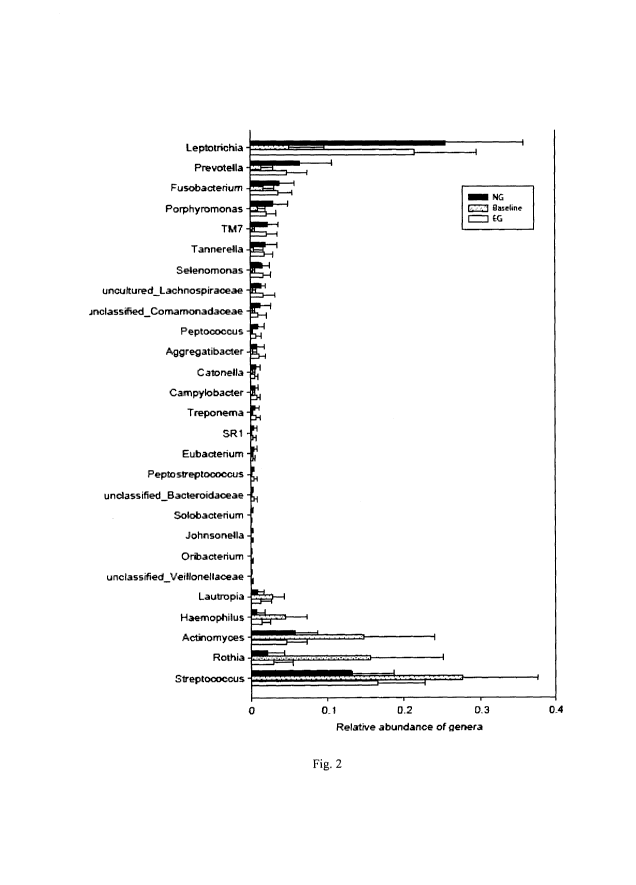
Fig. 2 shows the abundances of 27 genus-level bacterial biomarkers that
distinguish
between a healthy state and gingivital state(s) (including both naturally
occurring gingivitis state
and experimentally induced gingivitis state) in a set of test subjects,
according to a specific
embodiment of the present invention.
Figs. 3A and 3B show plots of principal components 1 and 2 (PC1 and PC2) from
a
principal component analysis (PCA) of genus-level bacteria data measured for
150 oral cavity
samples collected from 50 subjects at three different stages, i.e., a
naturally occurring gingivital
stage ("NG"), a baseline stage ("Baseline"), and an experimentally induced
gingivital stage
("EG"), according to a specific embodiment of the present invention.
Figs. 4A, 4B, 4C, and 4D show the identification of two types of hosts with
distinct
sensitivity to gingivitis according to a specific embodiment of the present
invention. Fig. 4A
shows patterns of microbiota structural (i.e. PC1-values) change and Mazza
Gingival Index
change along RPM. Fig. 4B shows distribution of the 50 subjects along
principal components 1
and 2 (PC1 and PC2) of the PCA, wherein the vertical dash line divides the 50
subjects into
Type-I and Type-II hosts. Fig. 4C shows difference in gingivitis sensitivity
between Type-I and
Type-II hosts.
Fig. 4D shows the abundances of 8 genus-level bacterial biomarkers that
distinguish between Type-I and Type-II hosts.
CA 02910327 2015-10-23
WO 2014/179959
PCT/CN2013/075385
6
Fig. 5 shows a trial classification based on the presence of gingivitis using
a microbial
index of gingivitis, MiG27, which is calculated from a function based on
abundances of 27
biomarkers defined according to a specific embodiment of the present
invention.
Fig. 6 shows a trial classification based on the severity of gingivitis using
a microbial
index of gingivitis, MiG15, which is calculated from a function based on
abundances of 15
biomarkers defined according to another specific embodiment of the present
invention.
Fig. 7 shows a trial classification based on the sensitivity to gingivitis
using a microbial
index of gingivitis, MiG-S, which is calculated from a function based on
abundances of 8
biomarkers defined according to a further specific embodiment of the present
invention. The
accuracy of MiG-S is measured by the area under the ROC (receiver operating
characteristic)
curve of plaque-microbiota-based (i.e. MiG-S-based) gingivitis-sensitive host-
type classification
as shown in the left diagram.
DETAILED DESCRIPTION OF THE INVENTION
Definitions
As used herein, the term "mammal" refers to any of various warm-blooded
vertebrate
animals of the class Mammalia, including humansIn the context herein, the
mammal can also be
called "subject" or "host".
As used herein, the term "a set of mammals" means a number of mammals gathered
together into a group for the purpose of study. The number in the set can be
any countable
number no less than 1. Depending on the purpose accuracy requirement of a
specific study, the
number of mammals in the set can be up to 1000, 10000 or even larger.
As used herein, the terms "microbial community", "microbiota", "microflora",
"microbial flora"
and "flora" are used interchangeably herein and refer to a population of
diverse microorganisms that
typically inhabits the mammal, specifically an organ (e.g., skin, digestive
tract) or an orifice (e.g,
mouth) of the mammal. The term "microorganism" means an organism of
microscopic or
submicroscopic size, especially a bacterium or protozoan, more preferably
bacterium.
As used herein, the term "microbe-related disease" includes an illness in the
mammal
caused or influenced or associated by a microorganism.
As used herein, the term "biomarker" includes indicators or markers present or
absent in the
biological system, site or sample that indicate occurrence of a biological
process or event.
As used herein, the terms "sample" or "biological sample" is a biological
material isolated
from a subject for analysis according to the present methods, such as saliva,
gingival crevicular
CA 02910327 2015-10-23
WO 2014/179959
PCT/CN2013/075385
7
fluid (GCF), supragingival plaque, subgingival plaque, breath or exhaled air,
oral lavage, tongue
scrapings, swabs or biopsies from oral tissue and serum. A sample is ideally
capable of
containing a microbial community.
As used herein, the term "statistical significance" is a mathematical tool
that is used to
determine whether the outcome of an experiment is the result of a relationship
between specific
factor(s) or merely the result of chance. Statistical significance is used to
reject or accept what is
called the null hypothesis. A hypothesis is an explanation that a researcher
is trying to prove.
The null hypothesis typically holds that the factor(s) at which a researcher
is looking have no
effect on differences in the data or that there is no connection between the
factors. Statistical
significance is usually written, for example, as t=0.02, p<0.05. Here, "t"
stands for the test score
and "p<0.05" means that the probability of an event occurring by chance is
less than 5 percent.
These numbers would cause the null hypothesis to be rejected.
As used herein, with reference to a disease or condition, the term
"sensitivity" and its
adjective form "sensitive" can be used interchangeably with "susceptibility"
and its adjective
form "susceptible" to mean the likelihood of suffering from a disease or
condition when exposed
to a noxious stimulus or pathogen.
As used herein, the articles including "a" and "an" when used in a claim, are
understood to
mean one or more of what is claimed or described.
As used herein, the terms "comprise", "comprises", "comprising", "include",
"includes",
"including", "contain", "contains", and "containing" are meant to be non-
limiting, i.e., other
steps and other sections which do not affect the end of result can be added.
The above terms
encompass the terms "consisting of' and "consisting essentially of'.
Target Condition
The present invention provides a method of assessing whether a subject mammal
has a
target condition. The target condition can be any condition which is used to
describe a
mammal's health status, including but not limited to presence of a disease,
severity of a disease,
sensitivity to a disease, and combinations thereof.
According to a specific embodiment, the disease is a micro-related disease,
preferably
selected from the group consisting of gingivitis, periodontitis, dental
caries, halitosis, oral ulcer,
premature birth, low birth weight, diabetes, respiratory disease, heart
disease, stroke, bacteremia,
whole body health, and combinations thereof.
CA 02910327 2015-10-23
WO 2014/179959
PCT/CN2013/075385
8
Sample Collection & Storage
Depending on the specific condition, the sample from an oral cavity,
preferably in the form
of a biofilm on the surfaces of the teeth, prostheses (when present), gums,
and tongue, can be
selected from the group consisting of a salivary sample, a plaque sample, a
tongue dorsum
sample, a tongue coating sample, a mucous membrane sample, and combinations
thereof. The
plaque sample can be from various locations. For example, the plaque sample
can be selected
from the group consisting of a supragingival plaque sample, a subgingival
plaque sample, a tooth
plaque sample, and combinations thereof. The selection of the sample may be
critical to the
accuracy of assessing the target condition. For example, plaque microbiota is
believed to be
more sensitive to gingivitis than salivary microbiota. Therefore, in the case
of gingivitis, the
sample is preferably a plaque sample.
The samples, once collected, can be used in subsequent steps immediately.
Alternatively,
the samples can be put in a freezer for later use. In some cases, the newly
collected samples are
immediately deep frozen, typically below -20 C, preferably below -50 C, more
preferably below
-70 C, and most preferably below -90 C. The samples remain frozen until
preparation for
analysis.
Biomarkers
The potential of microbiota for tracking and diagnosing a mammal's condition
(diseases,
diets, etc.) is dependent on, and limited by, the degree of heterogeneity in
the link between the
microbiota and the condition at the population level. In the gut, the
variation of microbiota
structure between subjects appears to dominate variation among conditions
(e.g. lean or obese, or
on a normal or high-fat diet). However, the present inventors surprisingly
found that the
opposite appears to be true for oral microbiota. That is, it is surprisingly
found that the
differences between healthy and diseased oral microbiota within a subject are
larger than inter-
subject differences. This suggests that the oral microbiota might offer
certain advantages as
biomarkers for oral, and perhaps even systemic, diseases.
Oral microbial community comprises an extremely diverse microflora, some of
which are
potentially harmful or "bad"; and some of which are not harmful or even
beneficial, as to
beconsidered as "good" bacteria, in part because they serve to prevent
proliferation of other more
harmful organisms. Thus, achieving a healthy oral status does not necessarily
require eradicating
all bacteria, but it is important to maintain a certain balance between the
"good" bacteria and the
"bad" bacteria. For example, "good" bacteria typically include the genus
Lactobacillus. The
CA 02910327 2015-10-23
WO 2014/179959
PCT/CN2013/075385
9
most prevalent strains in healthy persons include Lactobacillus gasseri and
Lactobacillus
fermentum and the strongest antimicrobial activity is associated with strains
including L.
paracasei, L. plantarum, L. rhamnosus, and L. salivarius. "Bad" or harmful
bacteria include, for
example, Streptococcus mutans, Tannerella forsythia, Porphyromonas gin
givalis, and F.
nucleatum.
Many studies have demonstrated a shift in the microbial community from
prevalent "good"
biomarkers to "bad" biomarkers when a disease occurs. For example, a shift is
reported in the
microbial community from a predominately gram-positive facultative aerobic to
a predominately
gram-negative anaerobic flora correlated with the formation of foul odors from
incubated saliva
(T.F. McNamara, et al., Oral Surg Oral Med. Oral Pathol. (1972), 34(1):41-8;
J. Tonzetich, J.
Periodontol. (1977), 48(1):13-20). In the oral cavity, the most common
consequences of
imbalance of microbiota are dental caries, halitosis and
gingivitis/periodontitis. The status of
gingivitis/periodontitis can be predicted by a characteristic microbial shift
from the early
prevalence of Gram-positive facultative microorganisms (e.g., Streptococcus
spp., Streptococcus
saginus, Actinomyces spp., and A. naeslundii) to the later prevalence of Gram-
negative anaerobic
microorganisms (e.g., Porphyromonas gin givalis and P. endodontalis,
Tannerella forsythia,
Aggregatibacter actinomycetemcomitans, Treponema denticola and T socranskii,
Prevotella
intermedia, Fusobacterium nucleatum, Eikenella corrodens, Campylobacter rectus
and C.
gracilis, and Veillonella parvula); and the status of dental caries can be
predicted by a shift from
non-aciduric bacteria (e.g., Streptococcus saginus and Actinomyces spp.) to
aciduric bacteria
(e.g., Streptococcus mutans, Streptococcus sobrinus, Lactobacillus spp., and
Bifidobacterium
spp.).
According to the present invention, a first group of biomarkers and a second
group of
biomarkers are defined to include "bad" biomarkers and "good" biomarkers,
respectively.
Including but not limited to those disclosed as "bad" and "good" biomarkers in
the prior art, it is
believed that, the first group of biomarkers each has a higher abundance in
oral cavities of a set
of test mammals with the target condition compared to oral cavities of a set
of test mammals
without the target condition, and the second group of biomarkers each has a
lower abundance in
the oral cavities of the set of test mammals with said target condition
compared to the oral
cavities of the set of test mammals without said target condition. Preferably,
the set of test
mammals without the target condition is a control set of test mammals.
According to a specific embodiment, the biomarkers are each independently
selected from
the group consisting of taxonomic categories of a bacterium, functional
categories of a microbe,
CA 02910327 2015-10-23
WO 2014/179959
PCT/CN2013/075385
and combinations thereof. More specifically and preferably, the biomarkers are
each
independently selected from the group consisting of a bacterial phylum, a
bacterial class, a
bacterial family, a bacterial order, a bacterial genus, a bacterial species, a
functional gene of a
microbe, a gene ortholog group of a microbe, a motif of peptide or protein of
a microbe, a
5 conserved peptide or protein domain of a microbe, a none-coding
nucleotide sequence of a
microbe, and combinations thereof, preferably a bacterial genus.
Many techniques can be used to measure abundance of a bio marker in the
sample. On one
hand, by selecting a particular population of microorganisms, culture-based
methods can be used
to investigate the microbial ecology of natural and anthropogenically impacted
environments.
10 Standard culture techniques to characterize microbial ecology involve
isolation and
characterization of microorganisms using commercial growth media such as
Luria¨Bertani
medium, Nutrient Agar, and Tryptic Soy Agar. The major limitation of culture-
based techniques
is that >99% of the microorganisms in any environment observed through a
microscope are not
cultivable by standard culturing techniques. On the other hand, with recent
advances in
genomics and sequencing technologies, a variety of culture-independent
molecular methods
based on direct isolation and analysis of nucleic acids, proteins, and lipids
from samples have
been discovered and revealed structural and functional information about
microbial communities.
Molecular approaches such as genetic fingerprinting, metagenomics,
metaproteomics,
metatranscriptomics, and proteogenomics are vital for discovering and
characterizing the vast
microbial diversity and understanding their interactions with biotic and
abiotic environmental
factors.
According to a specific embodiment, the abundance of a biomarker in the sample
is
measured by one or more methods selected from the group consisting of 16S
rRNA(RiboNucleic
Acid) analysis, genetic fingerprinting, clone library method, denaturing- or
temperature-gradient
gel electrophoresis, random amplified polymorphic DNA(DeoxyriboNucleic Acid),
DNA
amplification fingerprinting, amplified ribosomal DNA restriction analysis,
DNA microarrays,
fluorescence in situ hybridization, DNA¨DNA hybridization, metagenomics,
metaproteomics,
metatranscriptomics, proteogenomics, Luria¨Bertani medium isolation technique,
Nutrient Agar
isolation technique, Tryptic Soy Agar isolation technique, and any combination
thereof.
Molecular analyses of microbial communities have revealed that the cultivable
fraction
represents <1% of the total number of prokaryotic species present in any given
sample.
Preferably, a method selecting from the group consisting of 16S rRNA analysis,
metagenomics,
and combination thereof is used in the present invention to measure the
sample, obtaining
CA 02910327 2015-10-23
WO 2014/179959
PCT/CN2013/075385
11
abundances of one or more biomarkers. Most preferably, 16S rRNA analysis is
used to study the
microbial communities of the samples.
Method of Assessing a Condition
One aspect of the invention provides for a method of assessing whether a
subject mammal
has a target condition comprises the steps:
a) defining the target condition;
b) defining a first group of biomarkers each having a higher abundance in oral
cavities of a
set of test mammals with said target condition compared to oral cavities of a
set of test
mammals without said target condition;
c) defining a second group of biomarkers each having a lower abundance in the
oral
cavities of the set of test mammals with said target condition compared to the
oral
cavities of the set of test mammals without said target condition;
d) formulating a function of the abundances of the first group of biomarkers
and the
abundances of the second group of biomarkers that is useful for assessing
whether the
subject mammal has the target condition;
e) obtaining a sample from an oral cavity of the subject mammal, wherein the
obtained
sample is capable of containing the first group of biomarkers and the second
group of
biomarkers;
f) measuring abundances of the first group of biomarkers in the obtained
sample from the
subject mammal;
g) measuring abundances of the second group of biomarkers in the obtained
sample from
the subject mammal; and
h) inputting the measured abundances of the first group and the second group
of
biomarkers into the formulated function to assess whether the subject mammal
has the
target condition.
According to a specific embodiment, the function of the abundances of the
first group of
biomarkers and the abundances of the second group of biomarkers is selected
from the group
consisting of a linear function, a quadratic function, a cubic function, a
quartic function, a quintic
function, a sextic function, a rational function, and combinations thereof.
According to a further specific embodiment, the function of the abundances of
the first
group of biomarkers and the abundances of the second group of biomarkers is a
linear function,
preferably comprising a formula:
CA 02910327 2015-10-23
WO 2014/179959
PCT/CN2013/075385
12
LEN Eiem Al
f (Ai, Aj) = b( Ai
where N is a total number of the biomarkers in the first group, M is a total
number of the
biomarkers in the second group, At is an abundance of each biomarker i in the
first group, Aj is
an abundance of each biomarker j in the second group, EiEN At is a sum of At
over all
biomarkers i in the first group, EJEõ, Aj is a sum of Aj over all biomarkers j
in the second group,
and b is a constant which, in a particular embodiment, is selected from the
range from 1 to 10000,
preferably from 5 to 1000, more preferably from 6 to 100, and most preferably
from 10 to 50.
According to a specific embodiment, the target condition is selected from the
group
consisting of gingivitis, severity of gingivitis, sensitivity to gingivitis,
and combinations thereof.
According to a specific embodiment, the first group of biomarkers is bacterial
genera
selected from the group consisting of Leptotrichia, Prevotella, Fusobacterium,
TM7,
Porphyromonas, Tannerella, Selenomonas, Lachnospiraceae, Comamonadaceae,
Peptococcus,
Aggregatibacter, Catonella, Treponema, SR1, Campylobacter, Eubacterium,
Peptostreptococcus,
Bacteroidaceae, Solobacterium, Johnsonella, Oribacterium, Veillonellaceae, and
combinations
thereof; and the second group of biomarkers are bacterial genera selected from
the group
consisting of Streptococcus, Rothia, Actinomyces, Haemophilus, Lautropia, and
combinations
thereof. These biomarkers are especially useful for assessing whether a
subject mammal has
gingivitis, when the function of the abundances of the first group of
biomarkers and the
abundances of the second group of biomarkers is:
EtiE22 Ei Es Al
f (Ai, Aj) = b( Ai
22 5
where At is an abundance of each biomarker i in the first group, Aj is an
abundance of each
biomarker j in the second group, jE22 At is a sum of At over all biomarkers i
in the first group,
EJE5 Aj is a sum of Aj over all biomarkers j in the second group, and b is a
constant, preferably
selected from the range from 1, 3, 5, 8, or 10 to 20, 50, 100, 500, or 10000,
alternatively selected
from the range from 1 to 9, 15 to 200, 30 to 600, 800 to 1500, or combinations
thereof .
According to a specific embodiment, the first group of biomarkers is bacterial
genera
selected from the group consisting of Prevotella, Leptotrichia, Fusobacterium,
Selenomonas,
Lachnospiraceae, TM7, Tannerella, Peptococcus, Peptostreptococcus, Catonella,
Treponema,
Solobacterium, Bacteroidaceae, and combinations thereof; and the second group
of biomarkers
are bacterial genera selected from the group consisting of Rothia,
Haemophilus, and combination
thereof. These biomarkers are especially useful for assessing whether a
subject mammal has
CA 02910327 2015-10-23
WO 2014/179959
PCT/CN2013/075385
13
severe or non-severe gingivitis, when the function of the abundances of the
first group of
biomarkers and the abundances of the second group of biomarkers is:
Eici3 Ai E e2
f (Ai, Aj) = b( _______________________________________ jAj
13 2
where At is an abundance of each biomarker i in the first group, Aj is an
abundance of each
biomarker j in the second group, Eic13 At is a sum of At over all biomarkers i
in the first group,
EJE2 Aj is a sum of Aj over all biomarkers j in the second group, and b is a
constant, preferably
selected from the range from 1, 3, 5, 8, or 10 to 20, 50, 100, 500, or 10000,
alternatively selected
from the range from 1 to 9, 15 to 200, 30 to 600, 800 to 1500, or combinations
thereof.
According to a specific embodiment, the first group of biomarkers is bacterial
genera
selected from the group consisting of Selenomonas, Lachnospiraceae,
Peptococcus,
Bacteroidaceae, Peptostreptococcus, Oribacterium, Veillonellaceae and
combinations thereof;
and the second group of biomarkers is a bacterial genus of Abiotrophia. These
biomarkers are
especially useful for assessing whether a subject mammal is sensitive or non-
sensitive to
gingivitis, when the function of the abundances of the first group of
biomarkers and the
abundances of the second group of biomarkers is:
EiE7
f (Ai, Aj) = b( ________________________________________ Ai Ejei Aj
7 1
where At is an abundance of each biomarker i in the first group, Aj is an
abundance of each
biomarker j in the second group, EiE7 At is a sum of At over all biomarkers i
in the first group,
JE, Aj is a sum of Aj over all biomarkers j in the second group, and b is a
constant, preferably
selected from the range from 1, 3, 5, 8, or 10 to 20, 50, 100, 500, or 10000,
alternatively selected
from the range from 1 to 9, 15 to 200, 30 to 600, 800 to 1500, or combinations
thereof .
Computer-aided System and computer readable medium of Identifying a Biomarker
According to the present invention, a computer-aided system helpful in
practicing the
method of the present invention is provided. The present computer-aided system
for assessing
whether a subject mammal has a target condition comprises:
a) a sampling section configured for sampling an oral cavity sample of the
subject mammal,
wherein the sampled oral cavity sample is capable of containing:
i) a first group of biomarkers each having a higher abundance in oral cavities
of a set of
test mammals with said target condition compared to oral cavities of a set of
test mammals
without said target condition; and
CA 02910327 2015-10-23
WO 2014/179959
PCT/CN2013/075385
14
ii) a second group of biomarkers each having a lower abundance in the oral
cavities of the
set of test mammals with said target condition compared to the oral cavities
of the set of test
mammals without said target condition;
b) a measuring section in communication with the sampling section, wherein
said measuring
section is configured for measuring the sampled oral cavity sample to obtain
abundances of the
first group and the second group of biomarkers in the sampled oral cavity
sample; and
c) a computing section in communication with the measuring section, wherein
said computing
section stores a function of abundances of the first group of biomarkers and
abundances of the
second group of biomarkers that is useful for assessing whether the subject
mammal has the
target condition, and wherein the computing section is configured for applying
the function to
the obtained abundances of the first group and the second group of biomarkers
in the sampled
oral cavity sample to assess whether the subject mammal has the target
condition.
The sampling section may comprise one or more devices in the form selected
from the
group consisting of a spoon, a cotton swab, a blade, a brush, a probe, and any
combination
thereof. In a specific embodiment, the sampling section comprises a sterile
cotton swab, and the
sampling is accomplished by gently rubbing exposed tooth surfaces with the
sterile cotton swab.
In a specific embodiment, the present system can comprise a sample storage
section for
storing samples. If the samples collected from the sampling section are not to
be used
immediately, it is recommended to store them in the sample storage section. In
a further specific
embodiment, the sample storage has a temperature adjustment unit which can
provide the sample
storage section with a wide range of storing temperature, preferably below 30
C and more
preferably below 0 C. In a preferred embodiment, the sample storage section
provides a storing
temperature of below -20 C, preferably below -50 C, more preferably below -70
C, and most
preferably below -90 C.
The measuring section may comprise a sub-section performing one or more
methods
selected from the group consisting of 16S rRNA analysis, genetic
fingerprinting, clone library
method, denaturing- or temperature-gradient gel electrophoresis, random
amplified polymorphic
DNA, DNA amplification fingerprinting, amplified ribosomal DNA restriction
analysis, DNA
microarrays, fluorescence in situ hybridization, DNA¨DNA hybridization,
metagenomics,
metaproteomics, metatranscriptomics, proteogenomics, Luria¨Bertani medium
isolation
technique, Nutrient Agar isolation technique, Tryptic Soy Agar isolation
technique, and any
combination thereof.
CA 02910327 2015-10-23
WO 2014/179959
PCT/CN2013/075385
The computing section can be in any form. For example, it can be a personal
computer or a
portable device which comprises a computing program. According to a specific
embodiment,
the computing section comprises:
i) a memory module for storing the function;
5
ii) an input module in communication with the measuring section, wherein the
input module is
for inputting the obtained abundances of the first group and the second group
of biomarkers in
the sampled oral cavity sample;
iii) a data processing module in communication with the memory module and the
input
module, wherein the data processing module is configured for applying the
function to the
10 inputted abundances of the first group and the second group of
biomarkers in the sampled oral
cavity sample; and
iv) an output module in communication with the data processing module, wherein
the output
module is for outputting whether the subject mammal has the target condition.
In a specific embodiment, the sampling section, the measuring section, and the
computing
15
section, alone or in any combination, can be implemented as a computer program
product
comprising computer executable instructions embodied in a computer readable
medium.
In a further specific embodiment, the present invention provides a computer-
readable
medium for assessing whether a subject mammal has a target condition,
comprising:
a) a memory storing a function of abundances of a first group of biomarkers
and abundances
of a second group of biomarkers that is useful for assessing whether the
subject mammal has the
target condition, wherein
each of the first group of biomarkers has a higher abundance in oral cavities
of a set of test
mammals with said target condition compared to oral cavities of a set of test
mammals without
said target condition, and
each of the second group of biomarkers has a lower abundance in the oral
cavities of the set
of test mammals with said target condition compared to the oral cavities of
the set of test
mammals without said target condition; and
b) a computer code comprising instructions for applying the function to a data
set obtained
from the subject mammal, wherein the data set comprises abundances of the
first group and the
second group of biomarkers in an oral cavity sample of the subject mammal,
assessing whether
the subject mammal has the target condition.
Exemplary computer readable media include chip memory devices, disk memory
devices,
flash memory devices, programmable logic devices, application specific
integrated circuits,
CA 02910327 2015-10-23
WO 2014/179959
PCT/CN2013/075385
16
downloadable electrical signals, and the like. In addition, a computer program
product suitable
for the present invention may be located on a single device or computing
platform or may be
distributed across multiple devices or computing platforms.
As necessary, one or more of the sections as stated above can be compacted
into a large-size
apparatus or a small-size portable device.
EXAMPLES
The examples herein are meant to exemplify the present invention but are not
used to limit
or otherwise define the scope of the present invention.
List of Acronyms:
NG: naturally occurring gingivitis
EG: experimental gingivitis
MGI: Modified/Mazza Gingival Index
BOP: Bleeding on Probing
MiGs: Microbial indices of Gingivitis
RPM: retrogression-progression model
PAM: Partitioning Around Medoids (clustering algorithm)
PCA: Principal Component Analysis
PCoA: Principal Coordinates Analysis
FDR: False Discovery Rate
COG: Clustering of Orthologous Groups
MiG: microbial index of gingivitis
MiG-S: microbial index of gingivitis sensitivity
ROC: receiver operating characteristic
CI: confidence interval
KO: KEGG Ortholog
Target Conditions and Biomarkers
A retrogression-progression model (RPM) is designed to simulate the
development of
gingivitis in human population to find biomarkers for gingivitis-related
target conditions. Fifty
human adults undergo a controlled temporal transition from naturally-occurring
gingivitis ("NG")
at Day -21 to healthy gingivae at Day 0 ("Baseline", as control status), then
back to a state of
CA 02910327 2015-10-23
WO 2014/179959
PCT/CN2013/075385
17
experimental gingivitis ("EG") at Day 21. For each host, the structure of the
plaque microbiota
is measured at the three time points along the RPM: NG, Baseline and EG, thus
allowing insight
into dynamics. Taxonomic structures of the plaque microbiota are determined by
pyrosequencing
of 16S rRNA genes.
Fig. lA illustrates a design of longitudinal study simulating gingivitis
development in
human population. Experiments are conducted at Procter & Gamble (Beijing)
Technology Co.,
Ltd. Oral Care Department, with approval from the P&G Beijing Technical Center
(China)
Institutional Review Board and in accordance with the World Medical
Association Declaration
of Helsinki (1996 amendment). ICH Guidelines for Good Clinical Practice (GCP)
are followed.
Fifty subjects are recruited from the Beijing area. Voluntary informed consent
is obtained.
Individuals meeting the following criteria are included: be at least 18 years
of age; possess
a minimum of 12 natural anterior teeth; have at least 5 bleeding sites as
measured by Mazza
Gingival Index (MGI) at initial visit (Day -21); have gingivitis but not
periodontitis; be in good
general health as determined by the Investigator/designee based on a review of
the medical
history/update for participation in the study. Exclusion criteria for
individuals includes: severe
periodontal disease, as characterized by purulent exudates, generalized
mobility, and/or severe
recession; any condition which requires antibiotic premedication for the
administration of a
dental prophylaxis; self-reported pregnancy or intent to become pregnant
during the course of the
study and nursing females; atypical discoloration or pigmentation in the
gingival tissue; fixed
facial orthodontic appliances; atypical discoloration or pigmentation in the
gingival tissue; use of
antibiotics any time during the study; any diseases or conditions that could
be expected to
interfere with the subject safely completing the study. Clinical parameters
for each subject are
measured per week across the whole study. Individuals that fell into the
exclusion criteria at any
time point are excluded from study participation.
The RPM includes three phases.
Phase I, Oral Hygiene Phase (Day -21 to Day 0): Gingivitis examinations using
Mazza
gingival index are conducted at -21, -14, -7 and 0 days. After receiving a
dental prophylaxis
(super and sub gingival prophylaxis) and tooth polishing, each subject is
instructed to return to
the site twice daily at which time they brush under supervision using Mei Li
Liang Jie manual
toothbrush (Crest, Made in China) for three minutes with a currently marketed
anti-cavity
dentifrice without any marked anti-microbial actives and then use the floss to
clean the dental
interproximal area. This brushing regimen is followed for the next 21 days
while recording MGI
CA 02910327 2015-10-23
WO 2014/179959
PCT/CN2013/075385
18
for each subject each visit. During the Oral Hygiene Phase, subjects receive
up to three dental
prophylaxes if the subjects bleeding sites are more than 1.
Phase II, Experimental Gingivitis Phase (Day 0 to Day 21): During this phase,
subjects do
not have any oral hygiene practice including brushing, mouth rinsing with any
products, flossing
and dental prophylaxis. Subjects also receive a gingivitis exam at days 7, 14
and 21 of the
Experimental Gingivitis Phase.
Phase III, Recovery Phase: Subjects are instructed to return to the site twice
daily at which
time they brush under supervision using products and techniques in Phase I.
Subjects receive a
dental prophylaxis during the Recovery Phase and the subjects also received
gingivitis exam,
inclusive of measured bleeding sites, to document and confirm that they have
been returned to
equivalent or preferably better health than when they enter the study. If
needed, subjects
receivean additional prophylaxis and are monitored until deemed healthy.
BOP frequency and mean MGI, as clinical parameters, are recorded for each
subject. MGI
measures both the signs of inflammation and the degree of the severity of
bleeding. Specifically,
probing is performed by a dentist on the mesiobuccal and the distolingual of
each tooth, for a
maximum of 56 sites. Scores range from 0 to 5, with 0 assigned for normal
appearing and
healthy gingival up to a score of 5 for spontaneous bleeding (without
provocation). MGI of all
subjects are measured by the same well-trained dentist to reduce technical
variation.
Fig. 1B shows values of the above clinical parameters for 50 subjects cross
the study. In
Fig. 1B, boxes represent the interquartile range (IQR) and the lines inside
represent the median.
Whiskers denote the lowest and highest values within 1.5x IQR. At -21 day, all
subjects exhibit
a certain level of gingival inflammation that represents the state of
naturally occurring gingivitis
("NG") with BOP ranging from 5 to 27 and average MGI from 1.18 to 2.24. These
subjects then
undergo rigorous oral hygiene practice for three weeks, which results in a
greatly reduced BOP
and MGI (Median BOP and MGI are 1.00 and 1.02 respectively) at 0 day
("Baseline") that
represents a healthy gum state. Then the hosts further undergo an oral hygiene
program for
gingivitis induction for three weeks that results in significantly increased
BOP (median 23) and
MGI (median 2.11) representing the state of experimental gingivitis ("EG").
Supragingival plaque sampling Supragingival plaque samples from each subject
are
collected at Day -21, Day 0 and Day 21 following the procedures below.
Subjects do not have
oral hygiene practice including tooth brushing, flossing, mouth rinsing before
sampling.
Samples are collected after 2 hours food or drink (except water) intake. After
MGI examination,
each subject rinses their mouth with 50m1 sterilized water. After MGI
examination 15 minutes,
CA 02910327 2015-10-23
WO 2014/179959
PCT/CN2013/075385
19
plaque along the gumline within 2 mm depth are collected with Gracey curette
by qualified
dentists. For each subject, plaque samples are collected for all teeth in two
different quadrants (1
and 3 or 2 and 4) and pooled together in one tube. Plaques on the Gracey
curette are collected
via swabbing with a sterilized cotton swab. The tips of swab are put into 0.6
ml TE20 buffer (20
mM Tris-HC1 PH 8.0, 2 mM EDTA (ethylenediaminetetraacetic acid)). Before
isolating DNA,
all samples are stored under -70 C.
Plaque DNA extraction protocol Total DNA is extracted from Human Dental plaque
following Dr. Larry Fernery's protocol with minor modifications (Ravel J, et
at. (2011) Vaginal
microbiome of reproductive-age women. Proc. Natl. Acad. Sci. U. S. A. 108:4680-
4687). In
general, frozen samples are thawed on ice before DNA isolation experiment. The
original
sample (250 pi) is transferred into a clean Bead-Beating-Tube (2m1 Eppendorf
tube). Sample
suspensions are kept on ice while a Lytic-Enzyme Cocktail is prepared. Freshly
prepared Lytic-
Enzyme-Cocktail Master-Mix (100u1; containing 50 1 Lysozyme-500KU=10mg/ml, 6
1
Mutanolysin, 25 KU/ml, 3 1 Lysostaphin, 4000 U/ml in 20 mM sodium acetate and
41 1 TE
buffer) is added to all samples and incubated at 37 C for 45 min. To the
lysate mix 750 mg
cleaned and dry 0.1mm diameter Zirconia-Silica-Beads is added. Samples are
subjected to bead
beating for 2 minutes at room temperature in a Qiagen TissueLyser LT (36
oscillations/ second).
One hundred and eighty 1 of the crude lysate are transferred into a new tube
and DNA isolated
by Qiacube using DNeasy0 Blood & Tissue Mini Kits.
Bacterial 16S rRNA gene amplicon sequencing 150 plaque samples are obtained
and
analyzed from 50 individuals each of whom provides samples at the three
timepoints of NG (Day
-21), Baseline (Day 0) and EG (Day +21). Barcoded 16S rDNA amplicon sequencing
using 454
Titanium yields a total of 3,181,659 raw reads, resulting in totally 1,093,922
processed reads (i.e.,
reads after quality assessment and control measures). The number of processed
reads per sample
ranges from 437 to 28, 456, with an average 7293 reads per sample. All
sequences are deposited
at Sequence Read Archive under Accession ID 5RA058763.
Comparing the phylogenetic structures of plaque microbiota PCA analysis is
first
performed in R using the ade4 package (Dray S & Dufour AB (2007) The ade4
package:
Implementing the duality diagram for ecologists. Journal of Statistical
Software 22(4):1-20) to
visualize the difference of microbial community structure among different time
points.
Procrustes analysis attempts to stretch and rotate the points in one matrix,
such as points
obtained by PCA, to be as close as possible to points in the other matrix,
thus preserving the
relative distances between points within each matrix. Simple Procruste
rotation in R using the
CA 02910327 2015-10-23
WO 2014/179959
PCT/CN2013/075385
ade4 package between two subsets of transformed data (i.e. data matrix of
first-four principal
components of NG-baseline, EG-baseline and NG-EG) is performed to test the
degree of
difference among different time points for the microbiota of the cohort.
Principal coordinates analysis (PCoA) is also performed to confirm the
difference of
5 microbiota structure between populations of gingivitis and health. In
each sample, representative
sequences from each OTU (operational taxonomic unit) are chosen by selecting
the longest
sequence based on UCLUST (Edgar RC (2010). Search and clustering orders of
magnitude
faster than BLAST. Bioinformatics 26(19):2460-2461). Each sequence is assigned
to its closest
relative in the phylogeny in CORE (Griffen AL, et at. (2011) CORE: a
phylogenetically-curated
10 16S rDNA database of the core oral microbiome. PLoS One 6(4):e19051)
using BLAST 's
megablast. The resulted sample ID (identification) mapping file and category
mapping file are
used as inputs to FastUniFrac (Hamady M, Lozupone C, & Knight R (2010) Fast
UniFrac:
facilitating high-throughput phylogenetic analyses of microbial communities
including analysis
of pyrosequencing and PhyloChip data. ISME J4(1):17-27), which allows pairwise
comparisons
15 of inter-community distances based on the fraction of evolutionary
history that separates the
organisms. These distances are then clustered to reduce dimensionality using
PCoA, where the
principal coordinates (PC) describe in descending order the degree of
variation that each of the
axes in the new space explains. In addition, ThetaYC-based community structure
comparisons
are performed using MOTHUR (Schloss PD, Gevers D, & Westcott SL (2011)
Reducing the
20 effects of PCR (Polymerase Chain Reaction) amplification and sequencing
artifacts on 16S
rRNA-based studies. PLoS One 6(12):e27310). ThetaYC measures the structural
dissimilarity
between two communities. A matrix of pairwise thetaYC-based distances among
all samples is
created for clustering and PCoA analysis.
Statistical analysis To test the structural heterogeneity of microbiota,
clustering among the
plaque microbiota is performed by partitioning around medoids (PAM) using
Jensen-Shannon
divergence (JSD) of the normalized genus (or OTU) abundance. The optimal
number of clusters
is chosen based on the maximum of the silhouette index.
PCA analysis is then performed in R using the ade4 package to visualize the
clustering
based on PAM. Prior to the analysis, the data are sample-size normalized and
very low abundant
genera are removed (if their average abundance across all samples is below
0.1%) to decrease
noise. Bacterial genera that exhibited the highest correlation to PC1 are
identified and
highlighted.
CA 02910327 2015-10-23
WO 2014/179959
PCT/CN2013/075385
21
Results For each of the 150 plaque microbiota, bacterial phyla, genera and
species are
identified and their relative abundance quantified via taxonomic assignment
against reference
databases (CORE (Griffen AL, et at. (2011) CORE: a phylogenetically-curated
16S rDNA
database of the core oral microbiome. PLoS One 6(4):e19051)).
At the phylum level, nearly all sequences are from 13 bacterial phyla,
including six
predominant bacterial phyla commonly encountered in the oral cavity:
Firmicutes,
Proteobacteria, Bacteroidetes, Actinobacteria, Fusobacteria and TM7 (each with
average
relative abundance > 1% at least one timepoint). Between the gingivitis states
(NG and EG) and
the healthy gingival state (Baseline), significant difference (p < 0.01;
paired t-test) are found in
five predominate phyla: Actinobacteria, Firmicutes, TM7, Bacteroidetes and
Fusobacteria. A
temporal shift of community-structure along the NG-Baseline-EG progression is
apparent,
characterized by the elevated relative abundance of Actinobacteria and
Firmicutes at Baseline,
and that of TM7, Bacteroidetes and Fusobacteria at NG and EG.
At the genus level, 27 bacterial genera (each with average relative abundance
>0.1% at
least one time point) are differentially distributed (p<0.05, paired t-test;
FDR (false discovery
rate) q<0.2) between Baseline and gingivitis (both NG and EG). Among them,
five
(Streptococcus, Rothia, Actinomyces, Haemophilus and Lautropia) show elevated
abundance at
Baseline, while 22 (Leptotrichia, Prevotella, Fusobacterium, TM7,
Porphyromonas, Tannerella,
Selenomonas, Uncultured Lachnospiraceae, unclassified Comamonadaceae,
Peptococcus,
Aggregatibacter, Catonella, Treponema, SR1, Campylobacter, Eubacterium,
Peptostreptococcus,
unclassified Bacteroidaceae, Solobacterium, Johnsonella,
Oribacterium, and
unclassified Veillonellaceae) are enriched in both NG and EG. Fig. 2 shows
these 27 genus-
level bacterial biomarkers that are believed to denote gum health and
Gingivitis (for both
naturally occurring gingivitis and experimental gingivitis). Relative
abundance of identified oral
bacteria in microbial community at different stages is also displayed. These
bacterial taxa can
potentially serve as disease markers.
Along the RPM, different bacterial species within the same genus usually
exhibited
identical patterns of relative-abundance change, except for several species of
Capnocytophaga,
Actinomyces and Streptococcus.
The projected coordinate of a given microbiota on the PC1 appears to capture
the gradient-
like heterogeneity and development of microbiota structure along disease
retrogression and
progression, as changes in PC1 within subjects and across cohorts are largely
consistent with the
structural segregation between healthy and diseased microbiota (see Figs. 3A
and 3B).
CA 02910327 2015-10-23
WO 2014/179959
PCT/CN2013/075385
22
Moreover, the relative order of microbiota along PC1 defined using all 150
samples is similar to
those defined using healthy-only, NG-only or EG-only microbiota alone
(Spearman correlation;
All vs Healthy-only: rho=0 .95, p<0.001; All vs NG: rho= 0.97, p<0.001; All vs
EG: rho = 0.97,
p<0.001). Therefore PC1 appears to be the primary descriptor and thus a good
proxy for
quantitatively measuring the development of the microbiota in both RPM-
segments (e.g. NG-to-
Baseline and Baseline-to-EG).
For the 50 hosts along RPM, 15 bacterial genera are found to be the drivers of
microbiota
heterogeneity along PC1, as their gradients in abundance are significantly
correlated with the
coordinates of their corresponding samples on PC1 (Spearman rho>0 .7 , FDR
q<0.2), as shown
in Table 1 below. These drivers include Rothia, Haemophilus, Prevotella,
Leptotrichia,
Fusobacterium, Selenomonas, uncultured Lachnospiraceae, TM7, Tannerella,
Peptococcus,
Peptostreptococcus, Catonella, Treponema, Solobacterium and unclassified
Bacteroidaceae.
Table 1. Oral bacterial that shows significant correlation with PC1
Genus Rho value
Rothia -0.76
Haemophilus -0.7
Prevotella 0.85
Leptotrichia 0.81
Fusobacterium 0.71
Selenomonas 0.85
uncultured Lachnospiraceae 0.83
TM7 0.81
Tannerella 0.74
Peptococcus 0.82
Peptostreptococcus 0.73
Catonella 0.73
Treponema 0.82
Solobacterium 0.72
unclassified Bacteroidaceae 0.72
Two of the 15 genera, Rothia and Haemophilus, decrease in relative abundance
along PC1
("negative drivers"), while the other 13 increase along PC1 ("positive
drivers").
Among the 50 subjects, most hosts exhibit a largely consistent microbiota
structure during
the disease progression from NG to EG (see Fig. 4A). Although NG-Baseline (or
Baseline-EG)
PC1-changes vary considerably among the 50-host cohort, the rate of
microbiota change NG-
Baseline and that of microbiota change Baseline-EG are largely similar within
each subject. The
CA 02910327 2015-10-23
WO 2014/179959
PCT/CN2013/075385
23
persistence of disease outcome as well as microbiota structure for majority of
the hosts in EG (as
compared to NG) suggest the presence of host-dependent (and likely personal)
factors in
determining the susceptibility to gingivitis reoccurrence in natural human
populations.
PCA based on within-subject changes of both microbiota (APC1 at NG-to-Baseline
and
APC1 at Baseline-to-EG) and clinical symptom (AMGI at NG-to-Baseline and AMGI
at
Baseline-to-EG) along RPM reveal the divergence of disease susceptibility
among the 50 hosts.
As shown in Fig. 4B, all hosts in the 50-member cohort are plotted on the
first two principle
components of the PCA based on the change profiles of microbiota and MGI. The
histogram
and the kernel density plot (solid line) describing distribution of the 50
hosts along the principle
component of the PCA are shown. The vertical dash line divides the 50 hosts
into Type-I (dots)
and Type-II (triangles). The four variables as main contributors to these
clusters are determined
and plotted by their loadings in these two principle components. "a" denotes
APC1 (NG-
Baseline); "b" denotes AMGI (NG-Baseline); "c" denotes APC1(Baseline-EG); and
"d" denotes
AMGI(Baseline-EG).
The distribution pattern of the 50 hosts suggests a bimodal distribution (p =
0.74 for the
hypothesis of non-bimodal distribution based on Hartigans' dip test for
unimodality), where a
discriminating line can be drawn to divide the hosts into two types which we
designated as less
gingivitis-sensitive Type-I (17 individuals) and gingivitis-sensitive Type-II
(33 individuals).
Type-II hosts are characterized by more acute changes in both microbiota
structure than Type-I
hosts (see Fig. 4A and Fig. 4C). For an average Type-II host, the PC1-change
rate along RPM is
0.33 per day, which are 2.21 fold of an average Type-I host (see Fig. 4C).
At both NG and EG, there are significant relationship between gingivitis-
sensitive types and
the relative abundance of certain taxa (p<0.05, Wilcoxon rank-sum test), which
include
Abiotrophia, Selenomonas, uncultured Lachnospiraceae, Peptococcus,
unclassified
Bacteroidaceae, Peptostreptococcus, Oribacterium and Veillonellaceae; these
taxa are all
enriched in Type-II hosts as compared to Type I hosts, except Abiotrophia
which is enriched in
Type-I (see Fig. 4D). Most (five) of these Type-II-hosts associated genera are
among the 15
P Cl-drivers.
Function Formulation and Assessment
The 50-host cohort is used as a training set for function formulation, while
an additional 41
human subjects with naturally occurring gingivitis are recruited and then each
sampled at both
NG and Baseline (thus 82 additional microbiota samples are sequenced) for
assessment.
CA 02910327 2015-10-23
WO 2014/179959
PCT/CN2013/075385
24
(1) MiG27: The inventors formulate a function as a "microbial index of
gingivitis" (MiG)
based on the relative abundance of the 27 bacterial markers that are
distinguished between the
Baseline stage and the gingivitis stages (NG and EG) in the 50-host cohort
(MiG27), via the
following equation:
E
abundance(gGjflg
enriched)i E abundance( rHealth-enriched)
MiG27 = (1=22 .1=5 )x10
22 5
In the 50-host cohort, this index is highly correlated with MGI during both NG-
to-Baseline
(p<0.001, Student's t-test) and Baseline-to-EG (p<0.001, Student's t-test):
the area under the
receiver operating characteristic (ROC) curve is 99.52% (95% confidence
interval:
98.77%-99.52%) at NG-to-Baseline and 99.84% (95% confidence interval: 99.53%-
99.84%) at
baseline-to -EG.
With MiG27, the inventors predict gingivitis status of the 41 hosts in the new
cohort using
their NG microbiota. Fig. 5 shows the MiG27 indices of the additional cohort
of 41 hosts.
Boxes represent the IQR and the lines inside represent the median. Whiskers
denote the lowest
and highest values within 1.5x IQR. The MiG27 between NG (MGI>1.18) and
Baseline
(MGI<1.12) is significantly different (p<0.001, paired t-test, t statistic=
22.3), e.g. the top 27
samples with the highest MiG27 are all correctly classified as gingivitis. The
overall accuracy of
prediction (based on Linear Discriminant Analysis) for diseased state versus
healthy state is 94%
(i.e., an error rate of 6.1%) (see Table 2 below).
(2) MiG15: To assess diseased severity of gingivitis, MiG15, which is based on
the relative
abundance of 15 bacterial genera that drive the structural heterogeneity of
microbiota along PC1,
is derived. The MiG15 of a given microbiota is calculated via the following
equation:
abundance( Er
High PC1¨enriched)i abundance(
g0 PC1¨enriched)
j
MiG15 = (-13 __________________________________ j=2
)X10
13 2
The inventors then regress the relative PC 1-values (Y: the development of
gingivitis) on
MiG15 (X) using linear regression. The formula for prediction is: Y=-0.97-
4.62X. This revised
function is able to account for 60% of variance in PC1 location in the 50-host
cohort. This
function on disease severity is used to assess the NG microbiota in the 41-
host cohort. Fig. 6
shows the MiG15 indices of the additional cohort of 41 hosts. Boxes represent
the interquartile
range (IQR) and the lines inside represent the median. Whiskers denote the
lowest and highest
values within 1.5x IQR. The heatmap indicates the ability of MiG15 to
discriminate healthy and
CA 02910327 2015-10-23
WO 2014/179959 PCT/CN2013/075385
gingivitis status of hosts. MiG15 shows significant correlation with mean MGI
for each subjects
at NG (p <0.05, spearman correlation). Categorization of both predicted values
and test values
(of PC1) into three quantiles reveals an error rate of prediction at 24.4%
(see Table 2 below).
Therefore, the MiG15-based function is able to predict the gingivitis severity
in human hosts as
5 defined by PC1 at approximately 75% accuracy.
(3) MiG-Sensitivity (MiG-S): The inventors further derive a "microbial index
of gingivitis
sensitivity" (MiG-S) based on the relative abundance of the eight bacterial
markers that
distinguish between the Type-I and Type-II in the 50-host cohort at NG (MiG-
S), via the
following equation:
E abundance(z
Typal -enrtched )i E abundance(gTypel-enrtched)
10 MiG ¨ S = (1=7 _________________ 1=1 )x10
7 1
In the 50-host cohort, this index is highly correlated with types (p<0.05,
Wilcoxon rank-
sum test): the area under the ROC curve is 74.0% (95% confidence interval:
60.2%-74.0%) (see
Fig. 7), suggesting an up to 74.0% accuracy of predicting gingivitis-
sensitivity host-types. Table
2 below shows the same result.
Table 2. Predictive Functions of Human Gingivitis based on Plaque Microbiota
Error rate
MiG27 MiG15 MiG-S
Clinical status Health vs. Gingivitis 6.1% 6.1%
Categorized status of Based on MGI 41.5%
41.5%
gingivitis Based on PC1 24.4%
24.4%
Gingivitis sensitivity of
26.0 /0
the host Based on change-pattern of PC1 and MGI
Discussion
The identified microbial drivers of gingivitis development and susceptibility
and the
formulated functions based on the same provide novel opportunities to improve
clinical practice.
In gingivitis, the gingival tissue exhibits color change, contour alteration,
increased sulcular
exudates and bleeding upon provocation. Based on one or more of such host
symptoms, current
gingival indices proposed or practiced are subjective, prone to human bias and
error and difficult
to reproduce, as such indices are heavily dependent upon the human examiner's
visual
observation and individual judgment. For example, despite its prevalent use in
clinical practice,
CA 02910327 2015-10-23
WO 2014/179959
PCT/CN2013/075385
26
MGI in being a subjective measure of gingivitis severity can be of poor
reproductivity among
different examiners. Moreover, as symptom of gingivitis can vary greatly among
different teeth
(and even probing points), testing two probing sites for each of the 28 teeth
for each patient can
be time- and labor-intensive. These drawbacks have collectively confounded
cross-examiner and
cross-patients analysis of gingivitis. According to the present invention, the
inventors develop
and validate an alternative and likely complementary measure for gingivitis
that is based on
quantitative analysis of plaque microbiota. The proposed MiG-based predictive
functions are
able to predict diseased microbiota at 95% accuracy, distinguish different
disease-stages with 75%
accuracy, and potentially predict disease sensitivity. MiGs can thus serve as
more sensitive,
reliable and objective measures of gum health and gingivitis susceptibility
and thus contribute to
the diagnosis, prognosis and intervention of gum diseases.
Unless otherwise indicated, all percentages, ratios, and proportions are
calculated based on
weight of the total composition. All temperatures are in degrees Celsius ( C)
unless otherwise
indicated. All measurements made are at 25 C, unless otherwise designated. All
component or
composition levels are in reference to the active level of that component or
composition, and are
exclusive of impurities, for example, residual solvents or by-products, which
may be present in
commercially available sources.
It should be understood that every maximum numerical limitation given
throughout this
specification includes every lower numerical limitation, as if such lower
numerical limitations
are expressly written herein. Every minimum numerical limitation given
throughout this
specification will include every higher numerical limitation, as if such
higher numerical
limitations are expressly written herein. Every numerical range given
throughout this
specification will include every narrower numerical range that falls within
such broader
numerical range, as if such narrower numerical ranges are all expressly
written herein.
The dimensions and values disclosed herein are not to be understood as being
strictly
limited to the exact numerical values recited. Instead, unless otherwise
specified, each such
dimension is intended to mean both the recited value and a functionally
equivalent range
surrounding that value. For example, a dimension disclosed as "40 mm" is
intended to mean
"about 40 mm".
Every document cited herein, including any cross referenced or related patent
or application
is hereby incorporated herein by reference in its entirety unless expressly
excluded or otherwise
limited. The citation of any document is not an admission that it is prior art
with respect to any
CA 02910327 2015-10-23
WO 2014/179959
PCT/CN2013/075385
27
invention disclosed or claimed herein or that it alone, or in any combination
with any other
reference or references, teaches, suggests or discloses any such invention.
Further, to the extent
that any meaning or definition of a term in this document conflicts with any
meaning or
definition of the same term in a document incorporated by reference, the
meaning or definition
assigned to that term in this document shall govern.
While particular embodiments of the present invention have been illustrated
and described,
it would be obvious to those skilled in the art that various other changes and
modifications can
be made without departing from the spirit and scope of the invention. It is
therefore intended to
cover in the appended claims all such changes and modifications that are
within the scope of this
invention.