Note: Descriptions are shown in the official language in which they were submitted.
CA 02910512 2015-10-26
WO 2010/010051 PCT/EP2009/059269
- 1 -
TNFSF Single Chain Molecules
Description
The present invention refers to single-chain fusion proteins comprising three
soluble TNF superfamily
(TNFSF) cytokine domains and nucleic acid molecules encoding the fusion
proteins. The fusion
proteins are substantially non-aggregating and suitable for therapeutic,
diagnostic and/or research
applications.
State of the Art
It is known that trimerisation of TNFSF cytokines, e.g., the CD95 ligand
(CD95L), is required for efficient
receptor binding and activation. Trimeric complexes of TNF superfamily
cytokines, however, are difficult
to prepare from recombinant monomeric units.
WO 01/49866 and WO 02/09055 disclose recombinant fusion proteins comprising a
TNF cytokine and
a multimerisation component, particularly a protein from the Cl q protein
family or a collectin. A
disadvantage of these fusion proteins is, however, that the trimerisation
domain usually has a large
molecular weight and/or that the trimerisation is rather inefficient.
Schneider et al. (J Exp Med 187 (1989), 1205-1213) describe that trimers of
TNF cytokines are
stabilised by N-terminally positioned stabilisation motifs. In CD95L, the
stabilisation of the receptor
binding domain trimer is presumably caused by N-terminal amino acid domains
which are located near
the cytoplasmic membrane.
Shiraishi et al. (Biochem Biophys Res Commun 322 (2004), 197-202) describe
that the receptor binding
domain of CD95L may be stabilised by N-terminally positioned artificial a-
helical coiled-coil (leucine
zipper) motifs. It was found, however, that the orientation of the polypeptide
chains to each other, e.g.
parallel or antiparallel orientation, can hardly be predicted. Further, the
optimal number of heptad-
repeats in the coiled-coil zipper motif are difficult to determine. In
addition, coiled-coil structures have
the tendency to form macromolecular aggregates after alteration of pH and/or
ionic strength.
WO 01/25277 relates to single-chain oligomeric polypeptides which bind to an
extracellular ligand
binding domain of a cellular receptor, wherein the polypeptide comprises at
least three receptor binding
sites of which at least one is capable of binding to a ligand binding domain
of the cellular receptor and at
least one is incapable of effectively binding to a ligand binding domain of
the cellular receptor, whereby
the single-chain oligomeric polypeptides are capable of binding to the
receptor, but incapable of
activating the receptor. For example, the monomers are derived from cytokine
ligands of the TNF
family, particularly from TNF-a.
CA 02910512 2015-10-26
WO 2010/010051 PCT/EP2009/059269
- 2 -
WO 2005/103077 discloses single-chain fusion polypeptides comprising at least
three monomers of a
TNF family ligand member and at least two peptide linkers that link the
monomers of the TNF ligand
family members to one another. Recent experiments, however, have shown that
these single-chain
fusion polypeptides show undesired aggregation.
It was an object of the present invention to provide single-chain fusion
proteins comprising at least three
TNF cytokine domains which allow efficient recombinant manufacturing combined
with good stability
concerning aggregation.
Summary of the Invention
The present invention relates to a single-chain fusion polypeptide comprising:
(i) a first soluble TNF-family cytokine domain,
(ii) a first peptide linker,
(iii) a second soluble TNF-family cytokine domain,
(iv) a second peptide linker, and
(v) a third soluble TNF-family cytokine domain,
which is substantially non-aggregating.
The invention further relates to a nucleic acid molecule encoding a fusion
protein as described herein
and to a cell or a non-human organism transformed or transfected with a
nucleic acid molecule as
described herein.
The invention also relates to a pharmaceutical or diagnostic composition
comprising as an active agent
a fusion protein, a nucleic acid molecule, or a cell as described herein.
The invention also relates to a fusion protein, a nucleic acid molecule, or a
cell as described herein for
use in therapy, e.g., the use of a fusion protein, a nucleic acid molecule, or
a cell as described herein for
the preparation of a pharmaceutical composition in the prophylaxis and/or
treatment of disorders
caused by, associated with and/or accompanied by dysfunction of TNFSF
cytokines, particularly
proliferative disorders, such as tumours, e.g. solid or lymphatic tumours;
infectious diseases;
inflammatory diseases; metabolic diseases; autoimmune disorders, e.g.
rheumatoid and/or arthritic
diseases; degenerative diseases, e.g. neurodegenerative diseases such as
multiple sclerosis;
apoptosis-associated diseases or transplant rejections.
Description of the Figures
Figure 1 Domain structure of the inventive single-chain fusion
polypeptide. I., II., Ill. soluble TNF-
CA 02910512 2015-10-26
WO 2010/010051
PCT/EP2009/059269
- 3 -
family cytokine domains.
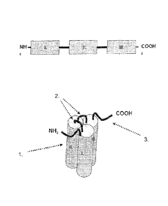
Figure 2 Schematic picture representing the general structure of TNF-SF
proteins. = = = cell membrane, N-terminus located within the cell, 1. anti-
parallel 13-fold of
receptor-binding domain (RBD), 2. interface of RBD and cell membrane, 3.
protease
cleavage site.
Figure 3 Schematic picture representing the structure of the native TNF-
SF trimer. Cylindric
structures represent RBDs, N-termini connect RBD with the cell membrane.
Figure 4 Schematic picture representing the structure of three soluble
domains comprising the
receptor-binding domain of a TNF cytokine. I., 11., III. soluble TNF-family
cytokine
domains.
Figure 5 Trimerisation of the soluble domains comprising the RBD of a TNF
cytokine,
characterised in that the N- and C-termini of the three soluble domains form a
surface.
Figure 6 Schematic picture representing the structure of the single-
chain TNF-SF comprising all or
a part of the stalk-region illustrating the requirement of longer linkers to
compensate for
the distance to the N-terminus of the next soluble domain.
Figure 7 scFv-TNF-SF fusion protein known from the art.
Figure 8 Fc-TNF-SF fusion protein known from the art.
Figure 9 9A Single-chain fusion polypeptide comprising an additional Fab
antibody fragment.
96 Single-chain fusion polypeptide comprisjng an additional scFv antibody
fragment.
Figure 10 Dimerisation of two N-terminally fused scFc fusion polypeptides
via disulfide bridges.
Figure 11 Dimerisation of two C-terminally fused scFc fusion polypeptides
via disulfide bridges.
Figure 12 Dimerisation of single-chain fusion polypeptides via a linker.
CA 02910512 2015-10-26
WO 2010/010051 PCT/EP2009/059269
- 4 -
Figure 13 Single-chain fusion polypeptide comprising an additional Fab
antibody fragment further
fused to a second fusion polypeptide or to a scFy fusion polypeptide.
Figure 14 Dimerisation of two scFab fusion polypeptides via disulfide
bridges.
Figure 15 N-terminally fused scFc fusion polypeptides further comprising
a Fv and/or Fab antibody
fragment.
Figure 16 C-terminally fused scFc fusion polypeptides further comprising a
Fv and/or Fab antibody
fragment.
Figure 17 SEC analysis of recombinantly expressed, purified TNF-SF
members under native
conditions. Exemplarily shown are two SEC analyses of purified TNF-SF members
on a
Superdex200 column under native condition (e.g.: PBS, pH 7.4). The diagrams
show the
absorption at 280nm (mAU) plotted against the elution volume (ml). The filled
arrow
indicates the elution peak for the fraction containing defined, soluble
trimeric TNF-SF
protein. The triangle indicates the elution peak for the oligomerised TNF -SF
. The open
arrow indicates the void volume of the SEC-column that contains protein-
aggregates,
which are too big to be separated (>800kDa).
Figure 17A: TNF-SF protein Aggregation Diagram A exemplarily shows an analysis
of a TNF -SF
protein preparation that contains a high amount of oligomerised/aggregated
protein(
indicated by the high amount of protein eluting in the void volume and the
high amount of
oligomeric protein).
Figure 17 B: TNF-SF protein defined soluble protein Diagram B exemplarily
shows an analysis for a
TNF-SF protein preparation that contains almost exclusively defined soluble
protein
(indicated by the absence of protein eluting in the void volume and by the
very limited
amount of protein eluting as oligomer).
Figure 18 SEC analysis of recombinantly expressed, affinity purified Fab-
scTRAILR2-SSSS.
SEC analysis of Fab-scTRAILR2-SSSS on a Superdex200 column using PBS, pH 7.4.
The diagram shows the absorption at 280nm (mAU) plotted against the elution
volume
(ml). The protein elutes as a distinct peak with an elution volume of 14.56m1,
CA 02910512 2015-10-26
WO 2010/010051
PCT/EP2009/059269
- 5 -
corresponding to an apparent MW of 68 kDa. No additional protein peaks with
lower
retention volume, indicating oligomerised/aggregated protein, could be
observed.
Figure 19 SEC analysis of recombinantly expressed, affinity purified Fab-
scTRAILR2-SNSN.
SEC analysis of Fab-scTRAILR2-SNSN on a Superdex200 column using PBS, pH 7.4.
The diagram shows the absorption at 280nm (mAU) plotted against the elution
volume
(m1). The protein elutes as a distinct peak with an elution volume of 14.12m1,
corresponding to an apparent MW of 87 kDa. No additional protein peaks with
lower
retention volume, indicating oligomerised/aggregated protein, could be
observed.
Figure 20 SEC analysis of recombinantly expressed, affinity purified Fab-
scTRAILwt-SNSN.
SEC analysis of Fab-scTRAILwt-SNSN on a Superdex200 column using PBS, pH 7.4.
The diagram shows the absorption at 280nm (mAU) plotted against the elution
volume
(ml). The protein elutes as a distinct peak with an elution volume of 13.99m1,
corresponding to an apparent MW of 94 kDa. A small additional protein peak at
12.00m1
could be observed. The apparent Mw of this peak corresponds to about 270 kDa,
indicating a defined trimerisation of Fab-scTRAILwt-SNSN. The total protein
amount of
the peak at 12.00m1 accounts for <3% of the total protein. More than 97% of
the
analysed Fab-scTRAILwt-SNSN has a defined soluble state (correct assembly of
the
three receptor binding modules). The peak at 16.12m1 corresponding to a MW of
28 kDa
contains Fab-light-chain polypeptide and was not included for the analysis of
peak areas.
Figure 21 Human scTRAIL Linker glycosylation
Figure 21 A Amino acid sequence of the linker(s) used to combine the receptor
binding modules of
single chain TRAIL constructs. G1y281 encodes the last amino acid of a
respective
receptor binding module, the sequence GSGN/SGN/SGS encodes the linker
sequence,
Arg121 encodes the first amino acid of the following TRAIL receptor binding
domain. The
designed linker sequences contains two putative N-linked glycosylation sites
at position
1 or 2 as indicated. These positions were permutated as indicated (version I,
II, Ill).
Figure 21 B Combination of linker positions: The scTRAIL molecules contain
three homologue
modules (grey barrels) that are connected with linker 1 and linker 2 as
indicated. Each of
the two linkers, can be designed for N-linked glycosylation as described in
"A". A
complete set of 9 different proteins containing all possible combinations of
linkers can be
CA 02910512 2015-10-26
WO 2010/010051 PCT/EP2009/059269
- 6 -
designed based on the sequences shown in B for linker 1 and 2. (Six of these
proteins
were expressed - see "C").
Figure 21 C Nomenclature of scTRAIL constructs expressed to test the influence
of different linker
sequences on glycosylation
Figure 22 Western Blot analysis of recombinant scTRAIL constructs
Single chain TRAIL proteins with different linker sequences were recombinantly
expressed, separated by SDS-PAGE and transferred to a PVDF-membrane. Bound
proteins were detected with a mouse monoclonal antibody recognising the Strep-
Tag
followed by a Peroxidase-conjugated secondary anti-mouse antibody. Different
TRAIL
variants were loaded as indicated. Note the MW-shift indicating differential
glycosylation
of scTRAIL-linker variants.
Figure 23 Cell culture supernatant of HEK293 cells, transiently
expressing scCD95L (SEQ-ID
NO:27) was collected and used to stimulate Jurkat cells at varying
concentrations. The
supernatant was used either directly without further modifications or an anti-
Streptag
antibody (2 microgram/m1) was added to cross-link the scCD95L protein. Jurkat
cells
were incubated with HEK293 cell culture supernatant for three hours at 37 ,
lysed and
analysed for caspase activity. Only cell supernatant that contained cross-
linked
scCD95L-St increased caspase activity in Jurkat cells, indicating that scCD95L
alone
does not form higher order aggregates able to be pro-apoptotic.
Figure 24 The protein scCD95L (SEQ ID NO:27) can be produced by transient
transfection of
HEK293 cells, stable transfection of other eukaryotic cells or by expression
using
prokaryotic cells. The recombinant protein can be affinity purified by using
StrepTactin
Sepharose matrix. Bound protein can be eluted with a buffer containing desthio-
biotin.
Figure 2 shows a silver stained SDS-PAGE of the elution fractions (lanes 1 to
5; fraction
2 is positive) of the affinity purification. The elution fraction containing
scCD95L could be
applied to size exclusion chromatography (SEC). It is expected, that the
protein shows
only a low aggregate content.
Figure 25 Cell culture supernatants of HEK293 cells, transiently
expressing single chain TRAIL
proteins with different linkers (derived from SEQ ID 28) were collected and
used to
CA 02910512 2015-10-26
WO 2010/010051
PCT/EP2009/059269
- 7 -
stimulate Jurkat cells at varying dilutions (exemplarily, a dilution of 1:8 is
shown in this
figure). The supernatants were used either directly without further
modifications or an
anti-Streptag antibody (2 microgram/m1 Strep MAB Immo) was added to cross-link
the
scTRAIL proteins. Jurkat cells were incubated with HEK293 cell culture
supernatant for
three hours at 37 , lysed and analysed for caspase activity. Cell culture
supernatant that
contained cross-linked scTRAILwt proteins induced an increased caspase
activity in
Jurkat cells, indicating that scTRAILwt proteins alone do form only a low
amount of
higher order aggregates able to be pro-apoptotic.
Figure 26 Influence of the module succession of scTRAIL-construct
components on their
expression rate of Fab-scTRAIL fusion proteins. Western blot of HEK293T cell
culture
supernatants from transient expression experiments. The polypeptide chains
necessary
for the formation of the Fab-scTRAIL proteins were either expressed separately
(lanes 1
to 10) or alternatively co-expression experiments were performed (lanes 11-
13). After
reducing SDS-PAGE, proteins were transferred to a nitrocellulose membrane and
proteins containing a Streptag werde detected, using an anti-Streptag specific
mAB as
primary AB . The light-chain-scTRAIL(R2-specific) proteins were secreted even
in the
absence of the accessory heavy chain (lanes 1-4). In contrast, the heavy-chain-
scTRAIL(R2-specific) fusion proteins were not secreted in the absence of the
acessory
light chain (lanes 5-8). As exemplified in lane 13. , the heavy-chain-
scTRAIL(R2-specific)
fusion proteins were only secreted in the presence of the light chain.
Figure 27 Cell culture supernatants of HEK293T cells, transiently
expressing scTRAILwt-Fc fusion
proteins with different linkers were collected and used to stimulate Jurkat
cells at varying
dilutions. The supernatants were used directly without further modifications
(Figure XX-
A). Jurkat cells were incubated with HEK293T cell culture supernatant for
three hours at
37 , lysed and analysed for caspase activity. There was already a pronounced
proapoptotic capacity present in the scTRAILwt-Fc containing supernatants,
indicating
that scTRAILwt-Fc fusion proteins alone do form dimeric assemblies able to be
pro-
apoptotic.
Figure 28 It is well known that the use of artificially cross-linked or
a membrane-bound ligand of the
TNF superfamily has superior bioactivity as compared to soluble, homotrimeric
ligand.
CA 02910512 2015-10-26
WO 2010/010051 PCT/EP2009/059269
- 8 -
Thus the local enrichment of single chain TRAIL (scTRAIL) constructs on cells
that
express the antigen Her2 via the Her2-selective Fab-fragment ("Pertuzumab")
fused to
these scTRAIL proteins should increase their cytotoxic bioactivity. Likewise,
the blocking
of the Her2 binding sites on cells by pre-incubation with the Her2-specific
Fab-fragment
(Pertuzumab-Fab) only should decrease the cytotoxic bioactivity of Fab-scTRAIL
fusion
proteins. As shown in Figure 28A, scTRAIL constructs induce the death of
HT1080 cells,
as the viability decreases with incresing protein concentration. In
accordance, the pre-
incubation of HT1080 cells with the Fab-fragment (Pertuzumab-Fab), followed by
co-
incubation with the Fab-scTRAIL constructs (Fab-scTRAILR2-SNSN or Fab-
scTRAILwt-
1 0 SNSN) over night, reduced the cytotoxic activity of the Fab-scTRAIL
constructs (figure
28B), whereas the Fab only induced no cell death (Pertuzumab -Fab). This means
that
the Fab-scTRAIL constructs bind to HT1080 cells via the Fab fragment thus
increasing
the cytotoxic bioactivity of scTRAIL.
Detailed Description of the Invention
According to the present invention a substantially non-aggregating fusion
polypeptide comprising at
least three soluble TNF family ligand domains connected by two peptide linkers
is provided.
The term "non-aggregating" refers to a monomer content of the preparation of >
50%, preferably > 70%
and more preferably > 90%. The ratio of monomer content to aggregate content
may be determined by
examining the amount of aggregate formation using size-exclusion
chromatography (SEC). The stability
concerning aggregation may be determined by SEC after defined time periods,
e.g. from a few to
several days, to weeks and months under different storage conditions, e.g. at
4 C or 25 C. For the
fusion protein, in order to be classified as substantially non-aggregating, it
is preferred that the
monomer content is as defined above after a time period of several days, e.g.
10 days, more preferably
after several weeks, e.g. 2, 3 or 4 weeks, and most preferably after several
months, e.g. 2 or 3 months
of storage at 4 C, or 25 C.
As an increase of e.g. the apoptosis inducing potential in the case of scCD95L
on human Jurkat cells
correlates with its aggregation state, the stability of the fusion polypeptide
concerning aggregation may
also be determined by examining the biological activity of the fusion
polypeptide.
The single-chain fusion polypeptide may comprise additional domains which may
be located at the N-
and/or C-termini thereof. Examples for additional fusion domains are e.g.
single-chain antibodies or
antibody fragments or other targeting molecules or a further cytokine domain,
e.g. an interleukin.
CA 02910512 2015-10-26
WO 2010/010051
PCT/EP2009/059269
- 9 -
The single-chain fusion protein comprises three soluble domains derived from a
cytokine of the TNF
superfamily. Preferably, those soluble domains are derived from a mammalian,
particularly human
cytokine including allelic variants and/or derivatives thereof. The soluble
domains comprise the
extracellular portion of a TNFSF cytokine including the receptor binding
domain without membrane
located domains. Proteins of the TNF superfamiliy are anchored to the membrane
via an N-terminal
portion of 15-30 amino acids, the so-called stalk-region. The stalk region
contributes to trimerisation and
provides a certain distance to the cell membrane. However, the stalk region is
not part of the receptor
binding domain (RBD).
Importantly, the RBD is characterised by a particular localisation of its N-
and C-terminal amino acids.
Said amino acids are immediately adjacent and are located centrally to the
axis of the trimer. The first
N-terminal amino acids of the RBD form an anti-parallel beta-strand with the C-
terminal amino acids of
the RBD (Fig. 2 and 3).
Thus, the anti-parallel beta-strand of the RBD forms an interface with the
cell membrane, which is
connected to and anchored within the cell membrane via the amino acids of the
stalk region. It is highly
preferred that the soluble domains of the single-chain fusion protein
comprises a receptor binding
domain of the TNF-SF cytokine lacking any amino acids from the stalk region
(Figs. 4 and 5).
Otherwise, a long linker connecting the C-terminus of one of the soluble
domains with the N-terminus of
the next soluble domain would be required to compensate for the N-terminal
stalk-region of the next
soluble domain (Figure 6), which might result in instability and/or formation
of aggregates.
A further advantage of such soluble domains is that the N- and C-terminal
amino acids of the RBD are
not accessible for any anti-drug antibodies.
Preferably, the single-chain fusion polypeptide is capable of forming an
ordered trimeric structure
comprising at least one functional binding site for the respective cytokine
receptor.
The fusion polypeptide may comprise one, two or three functional cytokine
receptor binding sites, i.e.
amino acid sequences capable of forming a complex with a cytokine receptor.
Thus, at least one of the
soluble domains is capable of binding to the corresponding cytokine receptor.
In one embodiment, at
least one of the soluble domains is capable of receptor activation, whereby
apoptotic and/or proliferative
activity may be effected. In a further embodiment, one or more of the soluble
domains are selected as
not being capable of receptor activation.
The soluble domain may be derived from TNF superfamily members, e.g. human
TNFSF-1 to -18 and
EDA-A1 to -A2 as indicated in Table 1, preferably from LTA (SEQ ID N0:1), TNFa
(SEQ ID NO:2), LTB
(SEQ ID N0:3), OX4OL (SEQ ID N0:4), CD4OL (SEQ ID N0:5), CD951_ (SEQ ID NO:6),
CD27L (SEQ
ID NO:7), CD3OL (SEQ ID NO:8), CD137L (SEQ ID NO:9), TRAIL (SEQ ID NO:10),
RANKL (SEQ ID
CA 02910512 2015-10-26
WO 2010/010051
PCT/EP2009/059269
- 10 -
N0:11), TWEAK (SEQ ID NO:12), APRIL 1 (SEQ ID NO:13), APRIL 2 (SEQ ID NO:14),
BAFF (SEQ ID
NO:15), LIGHT (SEQ ID NO:16), TL1A (SEQ ID NO:17), GITRL (SEQ ID NO:18), EDA-
A1 (SEQ ID
NO:19) and EDA-A2 (SEQ ID NO:20). Preferred soluble domains of the respective
proteins are
indicated in Table 1 (NH2-aa to COOH-aa) and, e.g., comprise amino acids 59-
205, 60-205 or 64-205 of
LTA (SEQ ID NO:1), 86-233 of TNFa (SEQ ID NO:2), 82-244 or 86-244 of LTB (SEQ
ID NO:3), 52-183
or 55-183 of OX4OL (SEQ ID NO:4), 112-261, 117-261 or 121-261 of CD4OL (SEQ ID
NO:5), 51-193 or
56-193 of CD27L (SEQ ID NO:7), 97-234, 98-234 or 102-234 of CD3OL (SEQ ID
NO:8), 86-254 of
CD137L (SEQ ID NO:9), 161-317 of RANKL (SEQ ID NO:11), 103-249, 104-249, 105-
249 or 106-249 of
TWEAK (SEQ ID NO:12), 112-247 of APRIL 1 (SEQ ID NO:13), 112-250 of APRIL 2
(SEQ ID NO:14),
140-285 of BAFF (SEQ ID NO:15), 91-251, 93-251 or 97-251 of TL1A (SEQ ID
NO:17), 52-177 of
GITRL (SEQ ID NO:18), 245-391 of EDA-A1 (SEQ ID NO:19), 245-389 of EDA-A2 (SEQ
ID NO:20).
More preferably, the soluble domains are derived from CD95L, TRAIL or LIGHT.
In an especially
preferred embodiment, the soluble domains are selected from human CD95L,
particularly starting from
amino acids 144, 145 or 146 and comprise particularly amino acids 144-281 or
145-281 or 146-281 of
SEQ ID NO:6 or human TRAIL, particularly starting from amino acids 120-122 and
comprise particularly
amino acids 120-281, 121-281 or 122-281 of SEQ ID NO:10. Optionally, amino
acid Lys145 of SEQ ID
NO:6 may be replaced by a non-charged amino acid, e.g. Ser or Gly. Optionally,
amino acid Arg121 of
SEQ ID NO:10 may be replaced by a non-charged amino acid, e.g. Ser or Gly. In
a further preferred
embodiment, the soluble domains are selected from human LIGHT, particularly
starting from amino
acids 93, 94 or 95 of SEQ ID NO:16 and particularly comprise amino acids 93-
240, 94-240 or 95-240 of
SEQ ID NO:16.
As indicated above, the soluble domains may comprise the wild-type sequences
as indicated in SEQ ID
NO: 1-20. It should be noted, however, that it is possible to introduce
mutations in one or more of these
soluble domains, e.g. mutations which alter (e.g. increase or decrease) the
binding properties of the
soluble domains. In one embodiment, soluble domains may be selected which
cannnot bind to the
corresponding cytokine receptor. An example of such a mutation is a
replacement of amino acid Y218
in human CD95L (SEQ ID NO:6) by another amino acid, e.g. R, K, S or D.
Further, a mutation may be
introduced which alters the binding to other cellular and/or extracellular
components, e.g. the
extracellular matrix. An example of such a mutation is a replacement of amino
acid K177 in CD95L
(SEQ ID NO: 6) by another amino acid, e.g. E, D or S.
In a further preferred embodiment of the invention, the soluble cytokine
domain (i) comprises a mutant
of the cytokine of the TNF superfamily or a receptor binding domain thereof
which binds and/or
activates TRAIL-receptor 1 (TRAILR1) and/or TRAIL-receptor 2 (TRAILR2). The
binding and/or activity
CA 02910512 2015-10-26
WO 2010/010051
PCT/EP2009/059269
- 1 1 -
of the mutant may be, e.g., determined by the assays as described in van der
Sloot et al. (PNAS, 2006,
103:8634-8639), Kelley et al. (J. Biol. Chem., 2005, 280:2205-2215), or
MacFarlane et al. (Cancer Res.,
2005, 65: 11265-11270).
The mutant may be generated by any technique and is known by the skilled
person, e.g., the
techniques described in van der Sloot et al. (PNAS, 2006, 103:8634-8639),
Kelley et al. (J. Biol. Chem.,
2005, 280:2205-2215), or MacFarlane et al. (Cancer Res., 2005, 65: 11265-
11270) and may comprise
any type of structural mutations, e.g., substitution, deletion, duplication
and/or insertion of an amino
acid. A preferred embodiment is the generation of substitutions. The
substitution may affect at least one
amino acid of the cytokine of the TNF superfamily or a receptor binding domain
thereof as described
herein. In a preferred embodiment, the substitution may affect at least one of
the amino acids of TRAIL,
e.g., human TRAIL (e.g., SEQ ID NO: 10). Preferred substitutions in this
regard affect at least one of the
following amino acids of human TRAIL of SEQ ID NO:10: R130, G160, Y189, R191,
0193, E195, N199,
K201, Y213, T214, S215, H264,1266, D267, D269. Preferred amino acid
substitutions of human TRAIL
of SEQ ID NO:10 are at least one of the following substitutions: R130E, G160M,
Y189A, Y189Q,
R191K, Q1935, Q193R, E195R, N199V, N199R, K201R, Y213W, T214R, 5215D, H264R,
I266L,
D267Q, D269H, D269R, or D269K.
The amino acid substitution(s) may affect the binding and/or activity of
TRAIL, e.g., human TRAIL, to or
on either the TRAILR1 or the TRAILR2. Alternatively, the amino acid
substitution(s) may affect the
binding and/or activity of TRAIL, e.g., human TRAIL, to or on both, the
TRAILR1 and the TRAILR2. The
binding and/or activity of the TRAILR1 and/or TRAILR2 may be affected
positively, i.e., stronger, more
selective or more specific binding and/or more activation of the receptor.
Alternatively, the binding
and/or activity of the TRAILR1 and/or TRAILR2 may be affected negatively,
i.e., weaker, less selective
or less specific binding and/or less or no activation of the receptor.
Examples of mutants of TRAIL with amino acid substitution(s) of the invention
that affect binding and/or
activation of both TRAILR1 and TRAILR2 may be found, e.g., in Table 1 of
MacFarlane et al. (cf. above)
and may comprise a human TRAIL mutant with the following two amino acid
substitutions of SEQ ID
NO: 10 Y213W and 5215D or with the following single amino acid substitution:
Y189A.
Examples of mutants of TRAIL with amino acid substitution(s) of the invention
that affect binding and/or
activation of TRAILR1 may be found, e.g., in Table 1 of MacFarlane et al. (cf.
above) and may comprise
a human TRAIL mutant with the following four amino acid substitutions of SEQ
ID NO: 10 N199V,
K201R, Y213W and 5215D or with the following five amino acid substitutions:
Q1935, N199V, K201R,
Y213W and 5215D, or may be found in Table 2 of Kelley et al. (cf. above) and
may comprise a human
TRAIL mutant with the following six amino acid substitutions: Y213W, 5215D,
Y189A, Q1935, N199V,
CA 02910512 2015-10-26
WO 2010/010051
PCT/EP2009/059269
- 12 -
and K201R, or with Y213W, S215D, Y189A, Q193S, N199R, and K201R.
Examples of mutants of TRAIL with amino acid substitution(s) of the invention
that affect binding and/or
activation of TRAILR2 may be found, e.g., in Table 1 of MacFarlane et al. (cf.
above) or in Table 2 of
Kelley et al. (cf. above) and may comprise a human TRAIL mutant with the
following six amino acid
substitutions of SEQ ID NO: 10: Y189Q, R191K, Q193R, H264R, I266L, and D267Q,
or may be found in
Table 2 of van der Sloot et al. (cf. above) and may comprise a human TRAIL
mutant with the following
single amino acid substitution: D269H, or with the following two amino acid
substitutions: D269H and
E195R or D269H and T214R.
Thus one preferred embodiment is a fusion protein as described herein wherein
at least one of the
soluble domains comprises a mutant of TRAIL or of a receptor binding domain
thereof which binds
and/or activates TRAILR1 and/or TRAILR2.
Further examples of mutants of TRAIL, which show reduced TRAIL induced
receptor aggregation are
H168 (S, T, Q), R170 (E, S, T, Q) and H177 (S, T).
One preferred embodiment of a fusion protein comprising a mutant of TRAIL or
of a receptor binding
domain as described herein is a fusion protein wherein component (i) comprises
at least one amino
acid substitution, particularly as indicated below.
Such an amino acid substitution affects at least one of the following amino
acid positions of human
TRAIL (SEQ ID NO: 10): R130, G160, H168, R170, H177, Y189, R191, 0193, E195,
N199, K201, Y213,
T214, S215, H264,1266, D267, D269.
Such an amino acid substitution is at least one of the following: R130E,
G160M, H168 (S, T, Q), R170
(E, S, T, Q), H177 (S,T), Y189A, Y189Q, R191K, Q193S, Q193R, E195R, N199V,
N199R, K201R,
Y213W, T214R, S215D, H264R, 1266L, D267Q, D269H, D269R, or D269K.
A preferred TRAIL-R2 selective domain comprises amino acid substitutions
Y189Q, R191K, Q193R,
H264R, I266L and D267Q.
A preferred TRAIL-R1 selective domain comprises amino acid substitutions
Y189A, Q193S, N199V,
K201R, Y213W and S215D.
The single-chain fusion molecule of the present invention comprises
additionally three soluble cytokine
domains, namely components (i), (iii) and (v). According to the present
invention, it was surprisingly
found that the stability of a single-chain TNF family cytokine fusion
polypeptide against aggregation is
enhanced, if the second and/or third soluble TNF family cytokine domain is an
N-terminally shortened
domain which optionally comprises amino acid sequence mutations. Thus,
preferably, both the second
and the third soluble TNF family cytokine domain are N-terminally shortened
domains which optionally
comprise amino acid sequence mutations in the N-terminal regions, preferably
within the first five amino
CA 02910512 2015-10-26
WO 2010/010051
PCT/EP2009/059269
- 13 -
acids of the N-terminus of the soluble cytokine domain. These mutations may
comprise replacement of
charged, e.g. acidic or basic amino acids, by neutral amino acids,
particularly serine or glycine.
In contrast thereto, the selection of the first soluble TNF family cytokine
domain is not as critical. Here, a
soluble domain having a full-length N-terminal sequence may be used. It should
be noted, however, that
also the first soluble cytokine domain may have an N-terminally shortened and
optionally mutated
sequence.
In a preferred embodiment of the present invention, the soluble TNF family
cytokine domains (i), (iii) and
(v) are soluble CD95L domains, particularly soluble human CD95L domains. The
first soluble CD95L
domain (i) may be selected from native, shortened and/or mutated sequences.
The N-terminal
sequence of the first domain (i) may e.g. start between amino acid G1u142 and
Va1146 of human
CD95L, wherein Arg144 and/or Lys145 may be replaced by a neutral amino acid,
e.g. by Ser or Gly.
The second and third soluble CD95L domains (iii) and (v), however, are
selected from shortened and/or
mutated sequences. Preferably, at least one of the soluble CD95L domains,
(iii) and (v), has an N-
terminal sequence which starts between amino acid Arg144 and Va1146 of human
CD95L, and wherein
Arg144 and/or Lys145 may be replaced by a neutral amino acid, e.g. by Ser
and/or Gly. In an especially
preferred embodiment, the second and third soluble CD95L domain start with an
N -terminal sequence
selected from:
(a) Arg144 - (Gly/Ser) 145 - Val (146)
(b) (Gly/Ser) 144 - Lys145 - Val (146) and
(c) (Gly/Ser) 144 - (Gly/Ser) 145 -Val (146).
Further, it is preferred that the CD95L domain ends with amino acid Leu 281
of human CD95L.
The soluble CD95L domain may comprise a mammalian, e.g. a human wild-type
sequence. In certain
embodiments, however, the CD95L sequence may comprise a mutation which results
in a reduction or
complete inhibition of the binding to the extracellular matrix, e.g. a
mutation at position Lys177, e.g.
Lys177-4 Glu, Asp or Ser and/or a mutation which reduces and/or inhibits
binding to the CD95L
receptor, e.g. a mutation at position Tyr218, e.g. Tyr218
Arg, Lys, Ser, Asp. In certain embodiments
of the present invention, one of the three soluble CD95L modules is a sequence
variant with a reduced
receptor binding. In other embodiments, two of the modules contain mutations
resulting in reduced
receptor binding.
In a further preferred embodiment of the present invention, the soluble TNF
family cytokine domains (i),
(iii) and (v) are soluble TRAIL domains, particularly soluble human TRAIL
domains. The first soluble
TRAIL domain (i) may be selected from native, shortened and/or mutated
sequences. Thus, the first
CA 02910512 2015-10-26
WO 2010/010051 PCT/EP2009/059269
- 14 -
soluble TRAIL domain (i) has an N-terminal sequence which may start between
amino acid Glu116 and
Va1122 of human TRAIL, and wherein Arg121 may be replaced by a neutral amino
acid, e.g. by Ser or
Gly. The second and third soluble TRAIL domains (iii) and (v) have a shortened
N -terminal sequence
which preferably starts between amino acid G1y120 and Va1122 of human TRAIL
and wherein Arg121
may be replaced by another amino acid, e.g. Ser or Gly.
Preferably, the N-terminal sequence of the soluble TRAIL domains (iii) and (v)
is selected from:
(a) Arg121 - Va1122 - Ala123 and
(b) (Gly/Ser)121.
The soluble TRAIL domain preferably ends with amino acid G1y281 of human
TRAIL. In certain
embodiments, the TRAIL domain may comprise internal mutations as described
above.
In a further preferred embodiment of the present invention, the soluble TNF
family cytokine domains (i),
(iii) and (v) are soluble LIGHT domains, particularly soluble human LIGHT
domains. The first soluble
LIGHT domain (i) may be selected from native, shortened and/or mutated
sequences. Thus, the first
soluble LIGHT domain (i) has an N -terminal sequence which may start between
amino acid G1u91 and
A1a95 of human LIGHT. The second and third soluble LIGHT domains (iii) and (v)
have a shortened N -
terminal sequence which preferably starts between amino acid Pro94 and A1a95
of human LIGHT. The
soluble LIGHT domain preferably ends with amino acid Va1240.
Components (ii) and (iv) of the single-chain fusion polypeptide are peptide
linker elements located
between components (i) and (iii) or (iii) and (v), respectively. The flexible
linker elements have a length
of 3-8 amino acids, particularly a length of 3, 4, 5, 6, 7, or 8 amino acids.
The linker elements are
preferably glycine/serine linkers, i.e. peptide linkers substantially
consisting of the amino acids glycine
and serine. In cases in which the soluble cytokine domain terminates with S or
G (C-terminus), e.g.
human TRAIL, the linker starts after S or G. In cases in which the soluble
cytokine domain starts with S
or G (N-terminus), the linker ends before this S or G.
It should be noted that linker (ii) and linker (iv) do not need to be of the
same length. In order to
decrease potential immunogenicity, it may be preferred to use shorter linkers.
In addition it turned out
that shorter linkers lead to single chain molecules with reduced tendency to
form aggregates. Whereas
linkers that are substantially longer than the ones disclosed here may exhibit
unfavourable
aggregations properties.
If desired, the linker may comprise an asparagine residue which may form a
glycosylation site Asn-Xaa-
Ser. In certain embodiments, one of the linkers, e.g. linker (ii) or linker
(iv) comprises a glycosylati on
site. In other embodiments, both linkers (iv) comprise glycosylation sites. In
order to increase the
solubility of the scTNF-SF proteins and/or in order to reduce the potential
immunogenicity, it may be
CA 02910512 2015-10-26
WO 2010/010051
PCT/EP2009/059269
- 15 -
preferred that linker (ii) or linker (iv) or both comprise a glycosylation
site.
Preferred linker sequences are selected from GSGSGSGS (SEQ ID NO:52), GSGSGNGS
(SEQ ID
NO:53), GGSGSGSG (SEQ ID NO:21), GGSGSG (SEQ ID NO:22), GGSG (SEQ ID NO:23),
GGSGNGSG (SEQ ID NO:24), GGNGSGSG (SEQ ID NO:25) and GGNGSG (SEQ ID NO:26)
The fusion protein may additionally comprise an N-terminal signal peptide
domain, which allows
processing, e.g. extracellular secretion, in a suitable host cell. Preferably,
the N -terminal signal peptide
domain comprises a protease cleavage site, e.g. a signal peptidase cleavage
site and thus may be
removed after or during expression to obtain the mature protein. Further, the
fusion protein may
additionally comprise a C-terminal element, having a length of e.g. 1-50,
preferably 10-30 amino acids
which may include or connect to a recognition/purification domain, e.g. a FLAG
domain, a Strep -tag or
Strep-tag II domain and/or a poly-His domain.
Further, the fusion polypeptide may additionally comprise N-terminally and/or
C-terminally a further
domain, e.g. a targeting domain such as a single-chain antibody or an antibody
fragment domain.
Specific examples of suitable antibodies are anti-tumour antibodies, such as
antibodies against EGFR-
familiy members. Suitable examples of other targeting molecules are cytokines,
such as interleukins.
Examples of specific fusion proteins of the invention are SEQ ID NOs: 27, 28,
29, 43, 45, 47, 49 and 51.
A further aspect of the present invention relates to a nucleic acid molecule
encoding a fusion protein as
described herein. The nucleic acid molecule may be a DNA molecule, e.g. a
double-stranded or single-
stranded DNA molecule, or an RNA molecule. The nucleic acid molecule may
encode the fusion protein
or a precursor thereof, e.g. a pro- or pre-proform of the fusion protein which
may comprise a signal
sequence or other heterologous amino acid portions for secretion or
purification which are preferably
located at the N- and/or C-terminus of the fusion protein. The heterologous
amino acid portions may be
linked to the first and/or second domain via a protease cleavage site, e.g. a
Factor Xa, thrombin or IgA
protease cleavage site.
Examples of specific nucleic acid sequences of the invention are SEQ ID NOs:
30, 31 32, 44, 46, 48
and 50.
The nucleic acid molecule may be operatively linked to an expression control
sequence, e.g. an
expression control sequence which allows expression of the nucleic acid
molecule in a desired host
cell. The nucleic acid molecule may be located on a vector, e.g. a plasmid, a
bacteriophage, a viral
vector, a chromosal integration vector, etc. Examples of suitable expression
control sequences and
vectors are described for example by Sambrook et al. (1989) Molecular Cloning,
A Laboratory Manual,
Cold Spring Harbor Press, and Ausubel et al. (1989), Current Protocols in
Molecular Biology, John
Wiley & Sons or more recent editions thereof.
CA 02910512 2015-10-26
WO 2010/010051 PCT/EP2009/059269
- 16 -
Various expression vector/host cell systems may be used to express the nucleic
acid sequences
encoding the fusion proteins of the present invention. Suitable host cells
include, but are not limited to,
prokaryotic cells such as bacteria, e.g. E.coli, eukaryotic host cells such as
yeast cells, insect cells,
plant cells or animal cells, preferably mammalian cells and, more preferably,
human cells.
Further, the invention relates to a non-human organism transformed or
transfected with a nucleic acid
molecule as described above. Such transgenic organisms may be generated by
known methods of
genetic transfer including homologous recombination.
A further aspect of the present invention relates to a pharmaceutical or
diagnostic composition
comprising as the active agent at least one fusion protein, a respective
nucleic acid encoding therefore,
or a transformed or transfected cell, all as described herein.
At least one fusion protein, respective nucleic acid encoding therefore, or
transformed or transfected
cell, all as described herein may be used in therapy, e.g., in the prophylaxis
and/or treatment of
disorders caused by, associated with andlor accompanied by dysfunction of TNF-
SF cytokines,
particularly proliferative disorders, such as tumours, e.g. solid or lymphatic
tumours; infectious
diseases; inflammatory diseases; metabolic diseases; autoimmune disorders,
e.g. rheumatoid and/or
arthritic diseases; degenerative diseases, e.g. neurodegenerative diseases
such as multiple sclerosis;
apoptosis-associated diseases or transplant rejections.
The term "dysfunction of TNF-SF cytokines" as used herein is to be understood
as any function or
expression of a TNF-SF cytokine that deviates from the normal function or
expression of a TNF-SF
cytokine, e.g., overexpression of the TNF-SF gene or protein, reduced or
abolished expression of the
TNF-SF cytokine gene or protein compared to the normal physiological
expression level of said TNF-SF
cytokine, increased activity of the TNF-SF cytokine, reduced or abolished
activity of the TNF -SF
cytokine, increased binding of the TNF-SF cytokine to any binding partners,
e.g., to a receptor,
particularly a CD95 or TRAIL receptor or another cytokine molecule, reduced or
abolished binding to
any binding partner, e.g. to a receptor, particularly a CD95 or TRAIL receptor
or another cytokine
molecule, compared to the normal physiological activity or binding of said TNF-
SF cytokine.
The composition may be administered as monotherapy or as combination therapy
with further
medications, e.g. cytostatic or chemotherapeutic agents, corticosteroids
and/or antibiotics.
The fusion protein is administered to a subject in need thereof, particularly
a human patient, in a
sufficient dose for the treatment of the specific conditions by suitable
means. For example, the fusion
protein may be formulated as a pharmaceutical composition together with
pharmaceutically acceptable
carriers, diluents and/or adjuvants. Therapeutic efficacy and toxicity may be
determined according to
standard protocols. The pharmaceutical composition may be administered
systemically, e.g.
CA 02910512 2015-10-26
WO 2010/010051 PCT/EP2009/059269
- 17 -
intraperitoneally, intramuscularly or intravenously or locally, e.g.
intranasally, subcutaneously or
intrathecally. Preferred is intravenous administration.
The dose of the fusion protein administered will of course be dependent on the
subject to be treated, on
the subject's weight, the type and severity of the disease, the manner of
administration and the
judgement of the prescribing physician. For the administration of fusion
proteins, a daily dose of 0.001
to 100 mg/kg is suitable.
Examples
1. Manufacture of a single-chain CD95L fusion protein (scCD95L)
In the following, the general structure of the recombinant proteins of the
invention (Figure 1) is shown
exemplified for the receptor binding domain of the human CD95 ligand.
1.1 Polypeptide structure
A) Amino acids Met1-Ser21
IgKappa-signal peptide, assumed signal peptidase cleavage site after amino
acid G1y20
B) Amino acids G1u22-Leu161
First soluble cytokine domain of the human CD95 ligand (CD95L, amino acids 142-
281 of SEQ
ID NO: 6 including a K145S mutation).
C) Amino acids Gly162-Gly169
First peptide linker element.
D) Amino acids Arg170-Leu307
Second soluble cytokine domain of the human 0095 ligand (CD95L; amino acids
144-182 of
SEQ ID NO: 6 including a K145S mutation).
E) Amino acids Gly308-315
Second peptide linker element.
CA 02910512 2015-10-26
WO 2010/010051 PCT/EP2009/059269
- 18 -
F) Amino acids Arg316-Leu453
Third soluble cytokine domain of the human CD95 ligand (CD95L; amino acids 144-
281 of
SEQ ID NO: 6 including a K145S mutation).
G) Amino acid G1y457-Lys472
Peptide linker with a Strep-tag II motif.
The amino acid sequence of sc CD95L is shown in SEQ ID NO. 27. The fusion
polypeptide comprises
first and second peptide linkers having the sequence GGSGSGSG (SEQ ID NO: 21).
Further preferred
linker sequences are SEQ ID NOs: 22-26 as described above. It should be noted
that the first and
second peptide linker sequences need not to be identical.
The signal peptide sequence (A) may be replaced by any other suitable, e.g.
mammalian signal peptide
sequence. The Strep-tag II motif (G) may be replaced by other motifs, if
desired, or deleted.
As shown in Figure 23, cell culture supernatant of HEK293 cells, transiently
expressing scCD95L (SEQ
ID NO:27) was collected and used to stimulate Jurkat cells at varying
concentrations. The supernatant
was used either directly without further modifications or an anti-Streptag
antibody (2 microgram/ml) was
added to cross-link the scCD95L protein. Only cell supernatant that contained
cross-linked scCD95L-St
increased caspase activity in Jurkat cells, indicating that scCD95L alone does
not form higher order
aggregates able to be pro-apoptotic.
1.2 Gene cassette encoding the polypeptide
The synthetic gene may be optimised in view of its codon-usage for the
expression in suitable host
cells, e.g. insect cells or mammalian cells. A preferred nucleic acid sequence
is shown in SEQ ID NO:
30.
1.3 Cloning strategy
The synthetic gene may be cloned, e.g. by means of a restriction enzyme
hydrolysis into a suitable
expression vector.
2. Manufacture of a single-chain TRAIL fusion protein (sc TRAIL wt)
CA 02910512 2015-10-26
WO 2010/010051 PCT/EP2009/059269
- 19 -
2.1 Polypeptide structure
A) Amino acids Met1-Gly20
lg-Kappa-signal peptide, assumed signal peptidase cleavage site after
amino acid Gly 20.
B) Amino acids GIn21 - G1y182
First soluble cytokine domain of the human TRAIL ligand (TRAIL, amino
acid 120 - 281 of SEQ ID NO:10)
C) Amino acids G1y183 - Ser 190
First peptide linker element, wherein the two amino acids designated X
are both S or one is S and the other one is N.
D) Amino acids Arg191 - G1y351
Second soluble cytokine domain of the human TRAIL ligand (TRAIL,
amino acids 121 -281 of SEQ ID NO:10)
E) Amino acids Gly 352 - Ser359
Second peptide linker element wherein the two amino acids designated X are
both S or one is S and
the other one is N.
F) Amino acids Arg360 - G1y520
Third soluble cytokine domain of the human TRAIL ligand (TRAIL, amino
acids 121 - G1y281 of SEQ ID NO:10).
G) Amino acids G1y521 - Lys538
Peptide linker element with a Streptag II motif.
The amino acid sequence of sc TRAIL wt is shown in SEQ ID NO: 28.
The indicated linkers may be replaced by other preferred linkers, e.g. as
shown in SEQ ID NOs: 21.26.
It should be noted that the first and second peptide linkers do not need to be
identical.
The signal peptide sequence (A) may be replaced by any other suitable, e.g.
mammalian signal peptide
CA 02910512 2015-10-26
WO 2010/010051
PCT/EP2009/059269
- 20 -
sequence. The Strep-tag II motif (G) may be replaced by other motifs, if
desired, or deleted.
Cell culture supernatants of HEK293 cells, transiently expressing single chain
TRAIL proteins with
different linkers (derived from SEQ ID 28, in total nine different linker
combinations) were collected and
used to stimulate Jurkat cells at varying dilutions (exemplarily, a dilution
of 1:8 is shown in Figure 25).
The supernatants were used either directly without further modifications or an
anti-Streptag antibody (2
microgram/ml Strep MAB Immo) was added to cross-link the scTRAILwt proteins.
Jurkat cells were
incubated with HEK293 cell culture supernatant for three hours at 37 , lysed
and analysed for caspase
activity. Cell culture supernatant that contained cross-linked scTRAILwt
proteins induced an increased
caspase activity in Jurkat cells (results shown on the right hand side of the
graph), indicating that
scTRAILwt proteins alone do form only a low amount of higher order aggregates
able to be pro-
apoptotic.
2.2 Gene cassette encoding the polypeptide
1 5 The synthetic gene may be optimised in view of its codon usage for the
expression in suitable host
cells, e.g. insect cells or mammalian cells. A preferred nucleic acid sequence
is shown in SEQ ID NO:
31.
3. Manufacture of a single-chain mutated TRAIL fusion protein (scTRAIL (R2-
specific))
In the following, the structure of a single-chain TRAIL polypeptide comprising
a mutation for selective
binding to TRAIL receptor R2 is shown.
3.1 Polypeptide structure
A) Amino acids Met1 - Ser29
lg-Kappa signal peptide, assumed signal peptidase cleavage site after
amino acid G1y20 and peptide linker
B) Amino acids Arg29 - Gly190
First soluble cytokine domain of the human TRAIL ligand (TRAIL, amino
acids 1 21 -281 of SEQ ID NO: 10 including the mutations Y189Q, R191K,
Q193R, H264R, I266L and D267Q )
CA 02910512 2015-10-26
WO 2010/010051
PCT/EP2009/059269
- 21 -
C) Amino acid G1y191 - Ser198
First peptide linker element, wherein the amino acids designated X are
as indicated in Example 2
D) Amino acids Arg199 - G1y359
Second soluble cytokine domain of the human TRAIL ligand (TRAIL
amino acids 121-281 of SEQ ID NO: 10 including the mutations as
indicated in B)
E) Amino acids G1y360 - Ser367
Second peptide linker element, wherein the amino acids X are as
indicated in Example 2
F) Amino acids Arg368 - G1y528
Third soluble cytokine domain of the human TRAIL ligand (TRAIL, amino
acids 121-281 of SEQ ID NO: 10 including the mutations as indicated in
B)
G) Amino acids G1y529 - Lys546
Peptide linker with a Strep-tag 11 motif
The amino acid sequence of scTRAIL(R2-specific) is shown in SEQ ID NO: 29.
The indicated linkers may be replaced by other preferred linkers, e.g. as
shown in SEQ ID NOs: 21-26.
It should be noted that the first and second peptide linkers do not need to be
identical.
The signal peptide sequence (A) may be replaced by any other suitable, e.g.
mammalian signal peptide
sequence. The Streptag 11 motif (G) may be replaced by other motifs, if
desired, or deleted.
3.2 Gene cassette encoding the polypeptide
The synthetic gene may be optimised in view of its codon usage for the
expression in suitable host
CA 02910512 2015-10-26
WO 2010/010051
PCT/EP2009/059269
- 22 -
cells, e.g. insect cells or mammalian cells. A preferred nucleic acid sequence
is shown in SEQ ID NO:
32.
4. Expression and Purification
a) Cloning, expression and purification of fusion polypeptides
Hek293T cells grown in DMEM + GlutaMAX (GibCo) supplemented with 10% FBS, 100
units/ml
Penicillin and 100 pg/ml Streptomycin were transiently transfected with a
plasmid containing an
expression cassette for a fusion polypeptide. In those cases, where a
plurality of polypeptide chains is
necessary to achieve the final product, e.g. for the Fab-scTNF-SF fusion
proteins (Figure 9A), the
expression cassettes were either combined on one plasmid or positioned on
different plasmids during
the transfection. Cell culture supernatant containing recombinant fusion
polypeptide was harvested
three days post transfection and clarified by centrifugation at 300 x g
followed by filtration through a
0.22 pm sterile filter. For affinity purification Streptactin Sepharose was
packed to a column (gel bed 1
ml), equilibrated with 15 ml buffer W (100 mM Tris-HCI, 150 mM NaCI, pH 8.0)
or PBS pH 7.4 and the
cell culture supernatant was applied to the column with a flow rate of 4
ml/min. Subsequently, the
column was washed with 15 ml buffer W and bound polypeptide was eluted
stepwise by addition of 7 x
1 ml buffer E (100 mM Tris HCI, 150 mM NaCI, 2.5 mM Desthiobiotin , pH 8.0).
Alternately, PBS pH 7.4
containing 2.5 mM Desthiobiotin can be used for this step. The protein amount
of the eluate fractions
was quantitated and peak fractions were concentrated by ultrafiltration and
further purified by size
exclusion chromatography (SEC).
SEC was performed on a Superdex 200 column using an Akta chromatography system
(GE-
Healthcare). The column was equilibrated with phosphate buffered saline and
the concentrated,
Streptactin-purified polypeptide was loaded onto the SEC column at a flow rate
of 0.5 ml/min. The
elution profile of the polypeptide was monitored by absorbance at 280 nm.
For determination of the apparent molecular weight of purified fusion
polypeptide under native
conditions a Superdex 200 column was loaded with standard proteins of known
molecular weight.
Based on the elution volume of the standard proteins a calibration curve was
plotted and the apparent
molecular weight of purified fusion polypeptide was determined.
5. Apoptosis Assay
A cellular assay with a Jurkat A3 permanent T-cell line was used to determine
the apoptosis inducing
activity of different CD95-ligand (CD95L) and TRAIL fusion polypeptide
constructs. Jurkat cells were
CA 02910512 2015-10-26
WO 2010/010051
PCT/EP2009/059269
- 23 -
grown in flasks with RPM' 1640-medium + GlutaMAX (GibCo) supplemented with 10%
FBS, 100
units/ml Penicillin and 100 pg/ml Streptomycin. Prior to the assay, 100,000
cells were seeded per well
into a 96-well microtiterplate. The addition of different concentrations of
fusion peptides to the wells was
followed by a 3 hour incubation at 37 C. Cells were lysed by adding lysis
buffer (250 mM HEPES, 50
mM MgC12, 10 mM EGTA, 5% Triton-X-100, 100 mM DTT, 10 mM AEBSF, pH 7.5) and
plates were put
on ice for 30 minutes to 2 hours. Apoptosis is paralleled by an increased
activity of caspases, e.g.
Caspase-3. Hence, cleavage of the specific caspase substrate Ac-DEVD-AFC
(Biomol) was used to
determine the extent of apoptosis. In fact, Caspase activity correlates with
the percentage of apoptotic
cells determined morphologically after staining the cells with propidium
iodide and Hoechst-33342. For
the caspase activity assay, 20 pl cell lysate was transferred to a black 96-
well microtiterplate. After the
addition of 80 pl buffer containing 50 mM HEPES, 1% Sucrose, 0.1% CHAPS, 50 pM
Ac-DEVD-AFC,
and 25 mM DTT, pH 7.5, the plate was transferred to a Tecan Infinite 500
microtiterplate reader and the
increase in fluorescence intensity was monitored (excitation wavelength 400
nm, emission wavelength
505 nm).
5.1 Cell death assay
For the determination of cell death in HT1080 fibrosarcoma cells 15,000 cells
were plated in 96-well
plates over night in RPMI 1640-medium + GlutaMAX (GibCo) supplemented with 10
% FBS (Biochrom).
Cells were coincubated with cycloheximide (Sigma) at a final concentration of
2.5 g/ml. Cell death was
quantified by staining with buffer IN (0.5% crystal violet, 20% methanol).
After staining, the wells were
washed with water and air-dried. The dye was eluted with methanol and optical
density at 595 nm was
measured with an ELISA reader.
6. Stability/Aggregation Test
6.1. Principle of the aggregation analysis (Definition for soluble protein)
The content of monomers (defined trimeric assembly of TNF-SF receptor binding
modules) and
aggregates is determined by analytical SEC as described in Example 4. For this
particular purpose the
analysis is performed in buffers containing physiological salt concentrations
at physiological pH (e.g.
0.9% NaCI, pH 7.4; PBS pH 7.4). A typical aggregation analysis is done on a
Superdex200 column (GE
Healthcare). This column separates proteins in the range between 10 to 800
kDa.
For determination of the apparent molecular weight of purified fusion
polypeptide under native
CA 02910512 2015-10-26
WO 2010/010051
PCT/EP2009/059269
- 24 -
conditions a Superdex 200 column is loaded with standard proteins of known
molecular weight. Based
on the elution volume of the standard proteins a calibration curve is plotted
and the apparent molecular
weight of purified fusion polypeptide is calculated based on the elution
volume.
SEC analysis of soluble, non aggregated protein s, - e.g. trimeric TNF-SF,
typically shows a distinct
single protein peak at a defined elution volume. This elution volume
corresponds to the apparent native
molecular weight of the particular protein and approximately complies to the
theoretical molecular
weight calculated on the basis of the primary amino acid sequence.
If protein aggregation occurs the SEC analysis shows additional protein peaks
with lower retention
volumes. For TNF-SF family members the aggregation of soluble proteins occurs
in a characteristic
manner. The proteins tend to form oligomers of the "trimers", forming nonamers
(3 x 3) and 27mers (3 x
9). These oligomers serve as aggregation seeds and a high content of oligomers
potentially leads to
aggregation of the protein. Oligomers of large molecular weight and aggregates
elute in the void volume
of the Superdex200 column and cannot be analysed by SEC with respect to their
native molecular
weight. Examples for SEC analysis of a defined soluble trimeric and a
oligomerised/aggregated
preparation of TNF-SF proteins are shown in Figure 17.
Due to the induction of (complete) aggregation, purified preparations of TNF-
SF fusion proteins should
preferably contain only defined trimeric proteins and only a very low amount
of oligomerised protein.
The degree of aggregation/oligomerisation of a particular TNF-SF protein
preparation is determined on
basis of the SEC analysis by calculating the peak areas of the 0D280 diagram
for the defined trimeric
and the oligomer/aggregate fraction, respectively. Based on the total peak
area the percentage of
defined trimeric protein is calculated as follows:
(%Trimer content = [Peak area trimer]/ [Total peak area] x 100)
The definition for soluble protein as used in this text, describes a protein
preparation of purified TNF-SF
protein in a buffer of physiological salt concentrations at physiological pH
that contains a defined
soluble protein (trimeric assembly of TNF-SF domains) content of >90% within a
typical protein
concentration range from 0.2 to 10.0 mg/ml.
6.2 SEC aggregation analysis for purified sc-TRAIL variants
Three different sc-TRAIL variants were transfected and affinity purified as
described. The purified
proteins were subsequently analysed for their content of defined soluble
protein using SEC analysis as
described in 6.1. In the particular case of single chain fusion proteins a
trimer describes a trimeric
assembly of three encoded TNF-SF domains encoded by a single polypeptide
chain. (Formally single
chain TNF-SF proteins are monomers, since single chain assemblies do only form
intramolecular
CA 02910512 2015-10-26
WO 2010/010051 PCT/EP2009/059269
- 25 -
interactions [all protein domains are encoded by a single polypeptide chain]
and do not form
intermolecular interactions between distinct individual polypeptide chains.)
The proteins analysed by SEC were:
1.) Fab-sc-TRAIL(R2-specific)-SNSN (Figure 19):
Fusion protein comprising an Fab domain fused N-terminal to a single chain
fusion protein of TRAIL
specific for TRAIL-receptor 2 interaction, glycosylated
2.) Fab-sc-TRAIL(R2-specific)-SSSS (Figure 18)
Fusion protein comprising an Fab domain fused N-terminal to a single chain
fusion protein of TRAIL
specific for TRAIL-receptor 2 interaction, non glycosylated
3.) Fab-sc-TRAIL-wt-SNSN (Figure 20):
Fusion protein comprising an Fab domain fused N-terminal to a single chain
TRAIL, glycosylated
The SEC analysis for the three purified Fab-sc-constructs of TRAIL revealed a
single protein peak for
all proteins indicating defined soluble protein fractions (>95% trimer). The
calculated apparent MW for
the proteins (based on calibration of the column) strongly indicate a trimeric
association of the TNF-SF-
domains for the purified proteins. None of the analysed proteins showed
indications for aggregation
(Figures 18, 19, 20).
Comparing the potentially glycosylated "Fab-sc-TRAIL-R2-SNSN" with the non
glycolsylated "Fab-sc-
TRAIL-R2-SSSS" indicates a significant difference of the apparent native MW
that is due to
glycosylation of Fab-sc-TRAIL(R2-specific)-SNSN.
Expression of sc-TNF-SF members as fusion protein with an antibody fv-fragment
is known to facilitate
aggregation of the protein. The construction principle of the Fab-sc-TRAIL
variants revealed no
aggregation of the expressed TRAIL variants and is therefore beneficial with
respect to solubility of the
protein.
6.3 Differential glycolsylation of sc-TRAIL-linker variants
Glycosylation of proteins can be beneficial for recombinant sc-TNF-SF
constructs with regard to
potential immunogenicity and stability. In order to get glycosylation of the
sc-TRAIL construct, specific
linker sequences were designed that contained putative N-linked glycosylation
sites at defined positions
CA 02910512 2015-10-26
WO 2010/010051
PCT/EP2009/059269
- 26 -
(see Figure 21-A). Recombinant expression and subsequent Western-Blot analysis
revealed that the
respective position of the Asparagine (N) within the linker sequence is
important for the subsequent
glycosylation of the protein. Surprisingly, the preferential linker position
of the glycosylated asparagine
was identified to be at position "2" as described in Figure 21-A, (G SGSGNG
S). If the asparagine is
localised at other positions (e.g. position "1" [G SG NGS G S] see Figure 21-
A), glycosylation of the
respective asparagines(s) is abolished. This aspect could be confirmed by
Western-Blot analysis of
different sc-TRAIL variants. If both asparagines of linker 1 and linker 2 were
localised at position"2" a
significant glycosylation dependant MW-shift could be observed for the
respective sc-TRAIL variant
(Figure 22). A MW-shift of the glycosylated sc-TRAIL linker variant could also
be confirmed by
calculating the apparent MW after SEC analysis (Figure 18, 19). The non
glycosylated Fab-sc-
TRAIL(R2-specific)SSSS has a clearly lower MW (68 k Da) compared to
glycosylated Fab-sc-
TRAIL(R2-specific)SNSN (87 kDa).
Based on this analysis we claim differential glycosylation of the sc-TRAIL
constructs by modifying the
position of the asparagines within the linker sequence(s). Glycosylation
protects the linker sequence
towards proteolytic degradation and might stabilise the protein. In addition
glycosylation of the linker
sequence potentially prevents recognition of the linker sequence by the immune
system and potentially
reduces the immunogenicity of the protein. Therefore glycosylation of the
linker sequence is beneficial
with regard to immunogenicity and proteolytic stability of the sc-TRAIL
constructs and has potential
influence on the half life of the protein. The linker specific differential
glycosylation can be used to
modify the immunogenicity and stability of recombinant TNF-SF members.
6.3. Expression and analysis of a sc-TRAIL with prolonged linker sequence and
N-terminal stalk
residues (sc-TRAIL-(95-281)-long)
In W0/2005/103077 a single chain TRAIL-fusion polypeptide, herein named sc-
TRAIL-(95-281)-long, is
described, wherein each TRAIL module comprise residues 95 to 281 of SEQ ID
N0:10. The TRAIL
modules are linked by Glycin Serin linker comprising of at least 12 amino
acids (GGGSGGGSGGGS).
Compared to the TRAIL modules of the present invention (comprising residues
121-281 of SEQ ID
NO:10), additional 25 amino acids including the stalk region are present in
each of the adjacent TRAIL
modules.
In order to analyse the influence of the linker sequence on sc-TRIAL
constructs, sc-TRAIL-(95-281)-
long is analysed. Expression, purification and subsequent SEC analysis reveals
that sc-TRAIL-(95-
281)-long with the 12 aa linker and the additional stalk sequence is expressed
and secreted to the cell
culture supernatant of HEK293T cells. However, SEC analysis of the purified
protein indicates that sc-
CA 02910512 2015-10-26
WO 2010/010051 PCT/EP2009/059269
- 27 -
TRAIL-(95-281)-long shows multiple peaks comprising a large amount of protein
in an oligomerised or
aggregated from. Aggregation of sc-TRAIL-(95-281)-long is a direct effect of
the prolonged linker
sequences in combination with the additional residues of the N-terminal stalk.
The results indicate that
the longer linker used in this construct leads to increased aggregation
properties of the construct.
7. Construction of single-chain fusion polypeptides comprising one or more
additional domains
7.1. Assembly of soluble TNF-SF and antibody fragments known from the art
It is known from the art that soluble TNF-SF cytokine domains may be fused to
antibody fragments in
order to obtain trimerisation and/or dimerisation of trimers. Single-chain
scFv-TNF-SF fusion proteins
have been constructed consisting of a single-chain antibody and a soluble
domain comprising a TNF-
RBD and the stalk-region. The corresponding trimers consist of three single-
chain antibodies and three
soluble domains (Fig. 7).
In addition, Fc-TNF-SF fusion proteins, wherein each fusion protein comprises
an N-terminal
intramolecular Fc-domain and a C-terminal soluble domain have been constructed
(Figure 8). The
dimerisation of soluble domains is accomplished by assembly of two Fc-domains
via disulfide bridges.
Trimers are subsequently obtained by a combination of two soluble domains from
one Fc-TNF-SF
fusion protein and one soluble domain from another Fc-TNF-SF fusion protein.
As can be deduced from
Fig. 4, dimerisation of trimers is also mediated by the N-terminal Fc-TNF-SF
fusion. In conclusion, three
Fc-antibody fragments are present per dimer of the trimer. However, such
fusion proteins are likely to
form higher molecular weight aggregates, which represents a major
disadvantage.
7.2 Fusion proteins of the invention comprising one or more additional domains
The inventive fusion proteins comprising one or more additional domains can be
constructed in several
ways. In the following, the construction of fusion proteins with additional
domains is exemplified with the
antibody pertuzumab directed against the cell surface antigen ErbB2.
The amino acid sequence of the heavy chain is shown in SEQ ID NO: 33:
1 EVQLVESGGG LVQPGGSLRL SCAASGFTFT DYTMDWVRQA
PGKGLEWVAD VNPNSGGSIY
61 NQRFKGRFTL SVDRSKNTLY LQMNSLRAED TAVYYCARNL
GPSFYFDYWG QGTLVTVSSA
121 STKGPSVFPL APSSKSTSGG TAALGCLVKD YFPEPVTVSW
NSGALTSGVH TFPAVLQSSG
181 LYSLSSVVTV PSSSLGTQTY ICNVNHKPSN TKVDKKVEPK SC
CA 02910512 2015-10-26
WO 2010/010051
PCT/EP2009/059269
- 28 -
The amino acid sequence of the light chain is shown in SEQ ID NO: 34
1 DIQMTQSPSS LSASVGDRVT ITCKASQDVS IGVAWYQQKP
GKAPKLLIYS ASYRYTGVPS
61 RFSGSGSGTD FTLTISSLQP EDFATYYCQQ YYIYPYTFGQ
GTKVEIKRTV AAPSVFIFPP
121 SDEQLKSGTA SVVCLLNNFY PREAKVQWKV DNALQSGNSQ
ESVTEQDSKD STYSLSSTLT
181 LSKADYEKHK VYACEVTHQG LSSPVTKSFN RGEC
7.2.1
In one embodiment, the fusion polypeptide of the invention further comprises
an N- or C-terminal Fab-
antibody fragment (Fig. 9A).
The fusion of an antibody Fab-fragment to the N-terminus of scTNF-SF fusion
polypeptide may be
accomplished by the following two strategies:
(i) The heavy chain sequence is extended by further amino acids from the IgG1
hinge region and fused
to the single-chain TNF-SF fusion protein.
The IgG1 hinge region comprises the amino acid sequence SEQ ID NO: 35:
....KS CID KTHTC2PP C3PAPE...
In a preferred embodiment, the Fab-domain is chosen such that the C-terminal
cysteine of the heavy
chain (C1 of the hinge region) terminates the CHI domain. This cysteine is
required for forming a
disulfide linkage to the light chain.
The subsequent linker comprises portions of the IgG hinge region (e.g. DKTHT
or DKT), however
without further cysteines of the hinge region. Alternatively, a glycine/serine
inker is used. Due to the
absence of further cysteines, a monomeric fusion protein comprising two
polypeptide chains is
obtained. The linker preferably has a length of 3-15 amino acids. More
preferably, the linker is selected
from the linker 1-7 as shown below.
1. DKTHTG(S)a(G)b; (a=0-5; b=0 oder 1)
2. DKTHTGS(S)a(GS)bG(S)c (a, b =0,1-6; c=0 oder 1)
3. DKTG(S)a(G)b; (a=0-5; b=0 oder 1)
CA 02910512 2015-10-26
WO 2010/010051
PCT/EP2009/059269
- 29 -
4. DKTG(S)a(GS)bG(S)c (a, b =0,1-6; c=0 oder 1)
5. SSG(S)a(GS)bG(S)c (a, b =0,1-6; c=0 oder 1)
6. SS(GGGS)aG(S)b (a=0, 1-4; b=0 oder 1)
7. GSPGSSSSSS(G)a (a=0 oder
Preferred amino acid sequences with the heavy chain module positioned N-
terminal to the scTNF-SF
module are shown in SEQ ID NO: 45, SEQ ID NO: 47 and SEQ ID NO: 49. For
production purposes,
these polypeptide chains are coexpressed with the Fab light chain polypeptide
(SEQ ID NO: 40) to
finally achieve the Fab-scTRAIL fusion polypeptides.
(ii) The light-chain sequence is fused to the single chain TNF-SF fusion
protein.
The constant region of the light chain (e.g. SEQ ID NO: 34) ends with a C-
terminal cysteine residue.
This residue may be covalently bridged with the C1 hinge cysteine of the heavy
chain. Preferably, the
linkers 1-7 as shown below are used for the connection between the light chain
sequence and the TNF-
SF fusion protein. Linkers 5-7 are preferred (see above).
Preferably, the last amino acid in the linker adjacant to the cytokine module
is either Gly or Ser. In the
following, preferred linker sequences are shown:
Further, the linker may comprise N-glycosylation motifs (NXS/T, wherein X may
be any amino acid).
One embodiment of the amino acid sequences with the light chain module
positioned N-terminal to the
scTNF-SF module is shown in SEQ ID NO: 51.
In the case of the Fab-scTNF-SF fusion proteins, the co-expression of two
polypeptide chains is
necessary to achieve the correct assembly of the Fab module in addition to the
scTNF-SF module (see
figure 9A). The Pertuzumab heavy and light chain modules (SEQ ID NO: 33 and
SEQ ID NO: 34) were
equipped with a signal peptide, backtranslated and the resulting synthetic
genes (SEQ ID NO: 41 and
SEQ ID NO: 42) genetically fused upstream of the scTRAILwt- or scTRAILR2-
specific gene modules
(SEQ ID NO: 31 and SEQ ID NO: 32). Examples for the resulting gene cassettes
are shown in SEQ ID
NO: 46, 48 and 50. After subcloning into appropriate expression vectors, a
selection of the resulting
plasmids was used for transient protein expression in HEK293T cells. The heavy
chain TRAIL or light
chain TRAIL expression plasmids were transfected either alone or in
combination with the necessary
light or heavy chain encoding vectors of the Fab-Fragment (F igure 26).
Surprisingly, the module
combination within the fusion proteins influenced the relative stability of
the scTRAIL-protein during
secretory based expression. If the light-chain module of the Fab-domain is
fused N -terminal to the
scTRAIL-domain (exemplified in SEQ ID NO: 51), the expression product is
stable itsself and secreted,
when expressed separatly (Lanes 1-4, figure 26). It can be therefore expected,
when such a fusion
polypeptid is coexpressed with a heavy-chain module, that two major protein
species will be formed
CA 02910512 2015-10-26
WO 2010/010051 PCT/EP2009/059269
- 30 -
during a potential production process: (1) the Fab-scTRAIL fusion protein
consisting of two polypeptide
chains and (2) as contamination a light-chain-scTRAIL fusion protein without a
functional Fab domain.
Therefore, fusing the heavy-chain module N-terminal to the scTNF-SF-module for
the expression is
preferred to avoid this technical disadvantage.
A functional analysis of recombinant inventive Fab comprising-scTRAIL fusion
proteins with the heavy-
chain module fused N-terminal to the scTRAIL-module (Fab-scTRAILR2-SNSN or Fab-
scTRAILwt-
SNSN) is shown in figure 28. As final purification step, size exclusion
chromatography was employed as
exemplified in figures 19 and 20.
Superior bioactivity compared to soluble, homotrimeric ligands can easily be
achieved by the use of
artificially cross-linked or a membrane-bound ligand of the TNF superfamily.
Thus the local enrichment
of single chain TRAIL (scTRAIL) constructs on cells that express the antigen
Her2 via the Her2-
selective Fab-fragment ("Pertuzumab") fused to these scTRAIL proteins should
increase their cytotoxic
bioactivity. Likewise, the blocking of the Her2 binding sites on cells by pre-
incubation with the Her2-
specific Fab-fragment (Pertuzumab-Fab) only should decrease the cytotoxic
bioactivity of Fab-scTRAIL
fusion proteins. As shown in Figure 28A, scTRAIL constructs induce the death
of HT1080 cells, as the
viability decreases with increasing protein concentration. In accordance, the
pre-incubation of HT1080
cells with the Fab-fragment (Pertuzumab-Fab), followed by co-incubation with
the Fab-scTRAIL
constructs (Fab-scTRAILR2-SNSN or Fab-scTRAILwt-SNSN) over night, reduced the
cytotoxic activity
of the Fab-scTRAIL constructs (figure 28B), whereas the Fab only induced no
cell death.
An increased technical effect may be achieved by use of artificially cross-
linked or a membrane-bound
ligands of the TNF superfamily resulting especially in superior bioactivity as
compared to soluble,
homotrimeric ligand. Thus the local enrichment of ligands or single chain
ligands such as examplifed by
single chain TRAIL (scTRAIL) on cells or on neighbouring cells should increase
the bioactivity of these
fusion proteins. The local enrichment (or targeting) of these single chain
ligands can be specifically
induced for instance by fusing the single chain ligands with amino acid
sequences that bind to any
antigen present on cells such as for instance tumor cells. Examples for
antigen binding sequences may
be derived from antibodies such as scFv or Fab fragments. Examples for
antigens expressed on target
cells may be receptors such as from the EGFR family or any other antigen to
which a binding antibody
can be generated. Of special interest in this context are cell surface
antigens specific for tumor or
cancer cells.
7.2.2
In another embodiment, the fusion polypeptide of the invention further
comprises an additional N - or C-
CA 02910512 2015-10-26
WO 2010/010051
PCT/EP2009/059269
- 31 -
terminal scFv-antibody fragment (Fig. 9B).
In this embodiment linkers 5-7 as described above may be used. Further, the
linkers may comprise N-
glycosylation motifs.
A preferred single chain Fv-pertuzumab fragment for fusing to the single-chain
cytokine fusion protein
may comprise amino acids Glu1-Ser119 of SEQ ID NO: 33 and Asp-Lys107 or Thr109
of SEQ ID NO:
34. The VH and VL fragments may be connected by a linker.
One embodiment of a scFv-domain of pertuzumab is shown in the following SEQ ID
NO: 36:
1 METDTLLLVVV LLLWVPAGNG EVQLVESGGG LVQPGGSLRL
SCAASGFTFT DYTMDVVVRQA
61 PGKGLEWVAD VNPNSGGSIY NQRFKGRFTL SVDRSKNTLY
LQMNSLRAED TAVYYCARNL
121 GPSFYFDYWG QGTLVTVSSG GGGSGGGGSG GGGSDIQMTQ
SPSSLSASVG DRVTITCKAS
181 QDVSIGVAVVY QQKPGKAPKL LIYSASYRYT GVPSRFSGSG
SGTDFTLTIS SLQPEDFATY
241 YCQQYYIYPY TFGQGTKVEI KRT
Amino acids 1-20 (underlined) constitute an N-terminal secretory signal
peptide.
7.2.3
In a further embodiment, the fusion polypeptide of the invention comprises an
additional N- or C-
terminal Fc-antibody fragment (Figs. 10 and 11).
Preferably, the Fc-antibody fragment domain is derived from a human
immunoglobulin G heavy chain,
particularly from a human immunoglobulin IgG1 heavy chain. In an especially
preferred embodiment,
the amino acid sequence of the Fc-domain is shown in SEQ ID NO: 37.
1 KSCDKTHTCP PCPAPELLGG PSVFLFPPKP KDTLMISRTP
EVTCVWDVS HEDPEVKFNW
61 YVDGVEVHNA KTKPREEQYN STYRVVSVLT VLHQDWLNGK
EYKCKVSNKA LPAPIEKTIS
121 KAKGQPREPQ VYTLPPSREE MTKNQVSLTC LVKGFYPSDI
AVEWESNGQP ENNYKTTPPV
181 LDSDGSFFLY SKLTVDKSRW QQGNVFSCSV MHEALHNHYT
QKSLSLSPGK
Amino acids Lys1-G1u16 define the hinge region.
CA 02910512 2015-10-26
WO 2010/010051
PCT/EP2009/059269
- 32 -
For a C-terminal fusion (Fig. 11) the Fc-domain preferably comprises the
complete constant domain
(amino acids 17-230 of SEQ ID NO: 37) and a part or the complete hinge region,
e.g. the complete
hinge region or the hinge region starting from amino acid Asp4.
Preferred linkers for connecting a C-terminal Fc-antibody fragment (e.g. Fig.
11) are shown in the
following:
Linker 8
scCD95L/scTRAIL....GG(P/S)a(GS)b(GIS)c KSCDKTHTCPPCPAPE...
(a=0 oder 1; b=0-8; c=0-8)
Linker 9
scCD95L/scTRAIL....GG(P/S),(GSSGS)bGS(G/S)c DKTHTCPPCPAPE...
(a=0 oder 1; b=0-8; c=0-8)
Linker 10
scCD95L/scTRAIL....GG(P/S)a(S)b(GS),(G/S)d DKTHTCPPCPAPE...
(a=0 oder 1; b=0-8; c=0-8; d=0-8)
All linkers start with GlyGly taking in account, however, that the C-terminal
amino acid of TRAIL is a
Gly. At position 3 of the linker, alternatively Pro or Ser are present. Linker
8 comprises the Cys1
cysteine of the heavy chain.
It should be noted that linkers 8-10 are also suitable for the C-terminal
fusion of other polypeptides, e.g.
a further scTNF-SF fusion protein.
In detail, the scTRAILwt module (SEQ ID NO: 28), the scTRAIL(R2-specific)-
module (SEQ ID NO: 29)
and the scCD95L-module (SEQ ID NO: 27) were fused N-terminally to the Fc-
domain of human IgG1,
starting with Asp4 of SEQ ID NO: 37 employing four linker elements as shown in
table 2.
CA 02910512 2015-10-26
WO 2010/010051 PCT/EP2009/059269
- 33 -
Fc-Fusion Amino-acid sequence of the linker element
FC01 ...(G)GSPGSSSSSSGSDKTH...
FCO2 ...(G)GSPGSSSSGSDKTH...
FC03 ...(G)GSPGSSGSDKTH...
FC04 ...(G)GSSDKTH...
Table 2: Sequences linking the Fc-domain C-terminally to scTNF-SF module. The
N-terminal amino-
acid of the IgG1 CH2-domain is underlined. The N-terminal Glycine of the
linking sequence is shown in
brackets. For TNF-SF proteins with a glycine as the C-terminal amino acid
(e.g. TRAIL), the N-terminal
glycine of the linking sequence formally belongs to the scTNF-SF module.
For purification and characterisation, a Strep-tag II (amino acid sequence
WSHPQFEK) was placed C-
terminally to the Fc-domain. This affinity tag was linked to the CH3-domain by
a flexible linker element
(amino acid sequence SSSSSSA), replacing the C-terminal lysine residue of the
CH3-sequence. The
amino acid sequences of the scTNF-SF fusion proteins as well as for the
described protein modules
were backtranslated and their codon usage was optimised for mammalian cell-
based expression. Gene
synthesis was done by ENTELECHON GmbH (Regensburg, Germany). The expression
cassettes for
larger fusion proteins were assembled by common cloning procedures starting
with DNA-modules of
suitable size and suitable restriction enzyme pattern. Exemplarily, the
resulting gene cassette for the
single chain TRAILwt FC01 fusion protein (scTRAILwt-FC01) is shown in SEQ ID
NO: 44 and the
encoded protein sequence is shown in SEQ ID NO: 43. The gene cassettes
encoding the shortened
linker variants (table 1) were generated by PCR based subcloning strategies,
starting from SEQ ID NO:
44. The final expression cassettes were released from intermediate cloning
vectors and subcloned into
to pCDNA4-HisMax-backbone, using unique Hind-I11-, Not-I- or Xba-I sites of
the plasmid. For the
assembly of the Fab- and Fc-fusions proteins, a unique SgS-1 site was
introduced into the vector
backbone, replacing the Not-l-site. All expression cassettes were routinely
verified by DNA sequencing.
The proteins were transiently expressed in HEK293T cells and the cell culture
supernatants were
monitored regarding their pro-apoptotic activity. As shown in figure 27, the
scTRAIL-Fc fusion proteins
of the invention, were able to induce a pronounced increase in caspase
activity, confirming the potency
of the Fc-based dimerisation of two scTRA1Lwt-modules. Similar results were
obtained for scTRAIL(R2-
specific)-Fc fusion proteins (data not shown).
If an Fc-antibody fragment is fused to the N-terminus of an scTNF-SF fusion
protein (cf. Fig. 10), the
amino acid squence of the Fc-module is preferably as shown in SEQ ID NO: 38:
CA 02910512 2015-10-26
WO 2010/010051 PCT/EP2009/059269
- 34 -
1 METDTLLLWV LLLWVPAGNG DKTHTCPPCP APELLGGPSV
FLFPPKPKDT LMISRTPEVT
61 CVVVDVSHED PEVKFNWYVD GVEVHNAKTK PREEQYNSTY
RVVSVLTVLH QDWLNGKEYK
121 CKVSNKALPA PIEKTISKAK GQPREPQVYT LPPSREEMTK
NQVSLTCLVK GFYPSDIAVE
181 WESNGQPENN YKTTPPVLDS DGSFFLYSKL TVDKSRWQQG
NVFSCSVMHE ALHNHYTQKS
241 LSLSPG
Amino acids 1-20 (underlined) constitute an N-terminal secretory signal
peptide.
For connecting the Fc-module to the ScTNF-SF fusion protein, preferably
Gly/Ser linkers are used. All
linkers preferably start with a serine and preferably end with glycine or
serine. Preferred linker
sequences 11-12 are shown in the following:
11. (S)a(GS)bG(S), (a, b =0,1-6; c=0 oder 1)
12. S(GGGS)aGb(S), (a, b =0,1-6; c=0 oder 1)
7.3 Dimerisation of the single-chain fusion proteins of the invention
7.3.1 Single-chain fusion polypeptides comprising one additional domain
The trimeric fusion proteins of the invention can further be dimerised.
In one embodiment, dimerisation will be obtained if the C-terminus of a first
fusion protein is directly
connected to the N-terminus of a second fusion protein via a linker structure
as defined herein (Fig. 12).
In another embodiment, a fusion protein of the invention comprising an Fab-
antibody fragment as an
additional domain, may be connected via a linker as defined herein directly
with a further fusion protein
of the invention or indirectly via an scFv-antibody fragment fused to a
further fusion protein of the
invention (Fig. 13). Thereby, dimerisation of the trimeric fusion proteins of
the invention is
accomplished.
In another embodiment, dimerisation of trimers may be obtained via the
assembly of two fusion proteins
of the invention comprising a Fab-antibody fragment as an additional domain
(Fig. 14). In this case,
intermolecular disulfide bridges are formed.
For the construction of dimerising Fab fragments N-terminal to the scTNF-SF
domain (e.g. Fig. 14),
CA 02910512 2015-10-26
WO 2010/010051
PCT/EP2009/059269
- 35 -
preferably the natural cysteine residues of the IgG hinge region (SEQ ID NO:
35) are used.
Preferably the C-terminal cysteine of the Fab-sequence corresponds to the 01-
residue of the hinge
region, which forms a disulfide bond with the light chain. The second cysteine
C2 may be used for the
covalent linkage of two Fab-modules. A third cysteine residue C3 may be open
or linked with the C3 of
the neighbouring chain. Preferred linkers between the Fab heavy chain sequence
and the N-terminus of
the scTNF-SF domain are linkers 13-22 as shown below.
13. DKTHTCPGSS(GS)aG(S)b
14. DKTHTCPGSSaG(S)b
15. DKTHT C(GSSGS)aGSG(S)b
16. DKTHTCGSS(GS)aG(S)b
17. DKTHTCGSSaG(S)b
18. DKTHTC(GSSGS)aGS(G)b
19. DKTHTCPPCPGSSGSGSGS(G)b
20. DKTHTCPPCP(GSSGS)aGS(G)b
21. DKTHTCPPCPGSS(GS)aGS(G)b
22. DKTHTCPPCPGSSaGS(G)b
Further, the linkers may be modified by incorporation of N-glycosylation
motifs as described above.
In a further embodiment, dimerisation of the fusion proteins of the invention
comprising an Fc-antibody
fragment as an additional N- and/or C-terminal domain, may be obtained by the
formation of
intermolecular disulfide bridges between two of said fusion proteins. In that
case, only one Fc-antibody
fragment is present per dimer of a trimeric fusion protein. Thereby, in
contrast to the Fc-antibody
fragment fusion proteins of the art, formation of higher molecular weight
aggregates is not very likely.
7.3.2 Single-chain fusion polypeptides comprising a plurality of additional
domains
The single-chain fusion polypeptide may comprise one or more additional
domains, e.g. a further
antibody fragment and/or a further targeting domain and/or a further cytokine
domain.
A fusion protein of the invention comprising an Fc-antibody fragment as one
additional domain may be
connected to a further Fab- or scFv-antibody fragment via the N-terminus of an
N-terminal fused Fc-
antibody fragment (Fig. 15) or directly via its N-terminus through a further
linker structure (Fig. 16), if the
Fc-antibody fragment is connected to the fusion protein of the invention via
its C -terminus.
In addition to a further antibody fragment or instead of the further antibody
fragment, a further cytokine,
preferably an interleukin, may be connected to the fusion protein. Thereby, it
is possible to obtain a
combination of an agonistic scCD95L and an antagonistic 5cCD95L molecule or
alternatively
combinations of scTRAIL (R1-specific) and scTRA1L (R2-specific).
Said fusion proteins are especially useful for the induction of apoptosis.