Note: Descriptions are shown in the official language in which they were submitted.
CA 02910561 2015-10-28
WO 2014/179681
PCMJS2014/036567
SYSTEMS AND METHODS FOR SUPER-RESOLUTION ULTRASOUND IMAGING
CROSS-REFERENCE TO RELATED APPLICATIONS
[0001] This
application claims the benefit of U.S. Provisional Patent Application
Serial No. 61/819,346, filed on May 3, 2013, and entitled "SYSTEMS AND METHODS
FOR
SUPER-RESOLUTION ULTRASOUND IMAGING."
STATEMENT REGARDING FEDERALLY SPONSORED RESEARCH
[0002] This
invention was made with government support under EB003268 and
EB009032 awarded by the National Institutes of Health. The government has
certain
rights in the invention.
BACKGROUND OF THE INVENTION
[0003] The field of
the invention is systems and methods for ultrasound imaging.
More particularly, the invention relates to systems and methods for high
resolution
ultrasound imaging capable of sub-millimeter resolutions.
[0004] There is a
need for vascular imaging in the brain that is not met with
available clinical imaging modalities. Using computed tomography ("CT"),
vessels with
a diameter below 400 [im are not consistently detected.
[0005] With
magnetic resonance imaging ("MRI") at a field strength of 1.5 T, the
limit for vessel detection is approximately 300 p.m. With increasing field
strength,
vessels with smaller diameters can be detected, leading to a greater number of
vessels
detected at higher field strengths such as 3 T or 7 T. At a field strength of
8 T, vessels
estimated to be smaller than 100 i.tm have been imaged in the human brain.
Despite the
advances in spatial resolution, MRI remains a costly imaging modality with
limited
availability, and ultra-high field MRI scanners (e.g., those with field
strengths greater
than 7 T) that can detect smaller vessels are not found in routine clinical
practice. Even
at these high field strengths, the ability of MRI to image the smaller vessels
that play a
key role in many diseases and functions of the brain is limited.
[0006] Ultrasound
is an imaging modality that does not use ionizing radiation,
and that has additional advantages in both its relative low cost and
portability. The use
of ultrasound in the brain, however, has been severely limited by the
attenuating and
aberrating effects of the skull bone, which increase with increasing
ultrasound
frequency. Ultrasound imaging through the skull is thus typically performed at
lower
frequencies (e.g., 2-4 MHz) through thin acoustic windows in the skull.
Because the
-1-
CA 02910561 2015-10-28
WO 2014/179681
PCT/US2014/036567
spatial resolution achievable with ultrasound operating in traditional pulse-
echo mode
is dependent on frequency, imaging vessels in the brain with this approach
sacrifices
resolution, which has limited the use of ultrasound to the imaging of major
vessels.
SUMMARY OF THE INVENTION
[0007] The present invention overcomes the aforementioned drawbacks by
providing a method for ultrasound imaging, in which high resolution images are
generated. For instance, the high resolution images are capable of resolving
objects
smaller than 3001.tm. Ultrasound is transmitted to a focal region in a volume-
of-interest
that contains at least one microbubble, and this transmit focus can be steered
over the
volume-of-interest. Signal data is acquired in response to the transmitted
ultrasound,
and a plurality of initial images are reconstructed by beamforming the
acquired signal
data. A position of the at least one microbubble is estimated in each of the
initial
images, and phase and amplitude correction factors are computed using these
position
estimates and the initial images. A plurality of target images are then
reconstructed by
beamforming the acquired signal data using the computed phase and amplitude
correction factors. An image having a higher spatial resolution than the
target images is
then produced by, for example, estimating the position of each bubble in each
of the
target images and fitting a function to the data based on the position
estimates and
uncertainty.
[0008] The foregoing and other aspects and advantages of the invention will
appear from the following description. In the description, reference is made
to the
accompanying drawings which form a part hereof, and in which there is shown by
way
of illustration a preferred embodiment of the invention. Such embodiment does
not
necessarily represent the full scope of the invention, however, and reference
is made
therefore to the claims and herein for interpreting the scope of the
invention.
BRIEF DESCRIPTION OF THE DRAWINGS
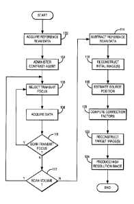
[0009] FIG. 1 is a flowchart setting forth the steps of an example of a
method for
reconstructing a high resolution image using an ultrasound system;
[0010] FIG. 2 is a block diagram of an example of an ultrasound system;
[0011] FIG. 3A is an example of a hemispherical transducer array that may
form a
part of the ultrasound system of FIG. 2; and
[0012] FIG. 3B is an example of a transmit-receive transducer element that
may
-2-
CA 02910561 2015-10-28
WO 2014/179681
PCT/US2014/036567
form a part of the transducer array of FIG. 3A.
DETAILED DESCRIPTION OF THE INVENTION
[0013] Described
here are systems and methods for super-resolution ultrasound
imaging. Instead of traditional pulse-echo imaging, a passive beamforming
technique is
used. With the passive beamforming technique, both the phase and amplitude
information of the received signals are considered and axial resolution no
longer
depends on pulse length, but on frequency and array aperture. Additionally,
the
intensity of the scatter response can be integrated over time to significantly
improve the
signal-to-noise ratio ("SNR"). To overcome the skull attenuation, a low
frequency
transmit array can be used. To increase spatial resolution, a full
hemispherical sparse
receiver array can be used. Large aperture transmit arrays have been used for
transcranial ultrasound therapy research, but have not been used for brain
imaging to
date.
[0014] Micrometer
sized gas bubbles are exceptional scatters of ultrasound and
have been used as intravascular contrast agents for over two decades. Using a
hemispherical array, such as the one described below, and using passive
beamforming,
the imaging resolution for a given frequency can be optimized and sufficient
SNR to
image single bubbles through a human skullcap can be achieved. Here, single
microbubbles are transcranially excited and three-dimensional passive maps of
the
bubbles are generated using a bubble-based phase correction technique. The
result is a
high resolution, transcranial, diffraction limited image of the vessels in
which the
microbubbles are located.
[0015] Additional
techniques can be used to further improve the imaging
resolution. For instance, the position of a distinct source within an image
can be
determined to a much greater level of accuracy than the point spread function
("PSF").
This has been applied to optics, combined with techniques to isolate distinct
sources
within the initial normal resolution images, to allow imaging well beyond the
diffraction
limit. These ideas, and the fact that microbubbles move with blood flow, are
utilized to
provide a method to enhance the three dimensional resolution of transcranial
imaging
beyond the diffraction limit.
[0016] Referring
now to FIG. 1, a flowchart setting forth the steps of an example
of a method for producing super high resolution images with an ultrasound
system is
-3-
CA 02910561 2015-10-28
WO 2014/179681
PCT/US2014/036567
illustrated. The method begins with the acquisition of reference scan data, as
indicated
at step 102. After the reference scan data has been acquired, a microbubble
contrast
agent is administered to the subject, as indicated at step 104. By way of
example, the
microbubble contrast agent is provided with a lower concentration that is
conventionally used in other imaging applications. For instance, the contrast
agent can
be provided with a concentration of approximately 1600 microbubbles per
milliliter.
This low concentration of microbubbles ensures that a single microbubble can
be
selectively excited. An example of a contrast agent suitable for the purposes
described
here is the DefinityTM microbubble contrast agent (Lantheus Medical Imaging,
North
Billerica, MA, USA), which includes microbubbles with a mean diameter of 1-3
p.m.
[0017] While the
microbubble contrast agent is present in the volume-of-interest
to be imaged, such as a blood vessel in a subject, data is obtained by
exciting
microbubbles, such as one microbubble at a time, and recording signals
received in
response to that excitation. Thus, as indicated at step 106, the method
proceeds by
selecting an ultrasound transmit focal point. Signals are then acquired by
exciting the
microbubble located in the focal spot and recording the signals received in
response to
that excitation, as indicated at step 108. This process is repeated for a
plurality of
different focal points in the volume-of-interest, as indicated by decision
block 110. For
example, the transmit focus can be electronically steered through the volume-
of-
interest through a plurality of different focal points in incremental steps,
such as steps
of 2 mm, in which the received waveforms are then recorded at each of these
locations.
[0018] By scanning
the transmit focus through the volume-of-interest, it is
possible to excite different bubbles in different portions of the volume to
create an
image of the larger structure (e.g., the blood vessel or vessels) present in
the volume-of-
interest. Because the contrast agent is very dilute, after a single scan
through the
volume it is possible that only a partial image of the volume will have been
obtained.
However, because the bubbles move with the blood flowing through the volume-of-
interest, multiple scans of the same volume can be performed to produce a
complete
image of the volume-of-interest, as indicated at decision block 112.
[0019] After the
desired amount of data has been obtained, images of the
volume-of-interest are reconstructed as follows. First, the reference scan
data that was
obtained earlier is subtracted from the data acquired in the presence of the
contrast
agent, as indicated at step 114. In doing this subtraction, strong reflections
from the
-4-
CA 02910561 2015-10-28
WO 2014/179681
PCT/US2014/036567
skull bone are suppressed. For instance, the reference scan data can be
subtracted line-
by-line from the data acquired after microbubbles have been introduced to the
volume-
of-interest in order to suppress reflections from the skull.
[0020] Next,
initial uncorrected images are reconstructed, as indicated at step
116. These images are reconstructed using geometric delays to beamform the
images
over a reconstruction grid, with or without the inclusion of additional delay
and
amplitude compensation terms to account for the effects of the skull. The
intensity
value assigned to a voxel in the reconstructed image can be mathematically
expressed
as the summation of the magnitude of the power spectrum over a frequency band
having a bandwidth of M discrete points centered about a center frequency
having
discrete indices, tric:
Al
2
2
i(r)= E Eor;fm) (1);
[0021] where / (r )
is the image intensity at a point, r = (x,y,z) , in the
reconstruction grid, and Qi(r; fm) is the value at the rnth frequency band of
the
discrete Fourier transform of the time-delayed waveform, q,(r;t), for the ith
receiver
element and point, r, over a window of N points:
no-h(N-i)
(r; = q, (r; ) = e-i2jrnmiN (2);
n=no
[0022] The time-delayed waveform, q, (r;tii), can be expressed as:
q,(r;tõ,)= a, = pi t, __________ ¨Si (3);
[0023] where a, is
an amplitude correction term, p1(t) is the pressure value
recorded by the th receiver element at time, t; r is a vector of the
coordinates of the
ith receiver element; c is the speed of sound in the medium; s, is a delay
term to
compensate for the effect of the skull on the waveform received by the ith
receive
element; and HI represents the Euclidean norm. For a given skull geometry and
orientation, the skull delay parameters, si, will be a function of the source
location and
-5-
CA 02910561 2015-10-28
WO 2014/179681
PCT/US2014/036567
receiver element location, but over a small reconstruction grid it is
acceptable to use a
single correction per receive element for all the grid points since the sound
is incident
on the same skull regions. For the same reason, and since variations due to
spherical
spreading will be small over a small volume, the amplitude correction terms,
a., can
also be approximated by a single correction per element over a small
reconstruction
grid. The amplitude and phase correction terms, ai and s7, may also be
functions of
frequency.
[0024] By way of
example, the initial images can be reconstructed by summing
the power spectrum of a small time-window (e.g., 40 vs) at the point of the
expected
bubble response over a narrow range of frequencies (e.g., 100 kHz) about the
center
frequency of the receivers. For excitations resulting in a strong microbubble
response,
an initial distorted image and an initial estimate of the source location can
be achieved.
Thus, using the initial images, the location of the source (e.g., the excited
microbubble)
can be estimated, as indicated at step 118. By way of example, the source
location can
be estimated by fitting a three-dimensional Gaussian to the image. In this
example, the
three-dimensional Gaussian is selected because it is an approximation of the
expected
shape of the main lobe of the hemispherical transducer array described above.
The
Gaussian can be given a fixed standard deviation in each of the three
dimensions based
on an experimentally determined point spread function ("PSF") of the
transducer array
near the geometric focus. However, translation and rotation can be allowed in
the fit.
[0025] The skull
delay parameters, si , and amplitude correction terms, a, are
then computed from the acoustic emissions from a single microbubble, as
indicated at
step 120. As noted above, the source position is estimated from the initial
images
reconstructed as described above. The geometric delays associated with this
source
location are determined. A matched-filter is then used to determine the total
time
delays between the receive elements. By way of example, the individual
channels can be
digitally filtered with a narrowband fourth-order Butterworth band-pass filter
(400-
800 kHz) prior to applying the matched filter. The skull delays are then
determined as
the difference between the total time delays with respect to one channel, and
the
geometric delays with respect to that reference channel. The amplitude
correction
terms can be determined as the reciprocal of the maximum value in each channel
over a
15 is time window over the bubble response, as identified by the matched
filter. As
-6-
CA 02910561 2015-10-28
WO 2014/179681
PCT/US2014/036567
noted above, the skull corrections calculated from a single bubble may be
applied to
correct all sources within a small imaging volume since the regions of the
skull
penetrated by the sound do not substantially change, and spherical spreading
effects
will be small over a small volume.
[0026] Using the
computed correction factors, target phase and amplitude
corrected images are reconstructed using the beamforming described above, as
indicated at step 122. As an example, the images can be produced using a time
window
of 40 [is and a frequency interval of 100 kHz centered about 600 kHz.
[0027] A single,
high resolution image of the volume-of-interest is then produced
from the target images, as indicated at step 124. Target image frames that did
not
contain one clear source are preferably discarded and not used to produce the
high
resolution image. By way of example, target image frames can be selected for
exclusion
if they contain a local maximum with intensity greater than or equal to fifty
percent of
the global maximum in the frame. The single, high resolution image of the
volume is
then produced by normalizing the remaining target image frames to themselves
and
then combining the images using a maximum pixel projection technique. The
response
from the microbubbles is expected to vary, and strong responses would bias the
high
resolution image; hence, the frames without a clear main lobe are removed and
the
remaining frames are normalized to their respective maxima before taking the
maximum pixel projection.
[0028] By way of
example, to obtain the high resolution images, a three-
dimensional Gaussian was fit to the target images in the same manner as
described
above. This fit is performed for each of the target image frames containing a
clear
source. High resolution frames can be plotted as a Gaussian centered at the
estimated
source location and having standard deviations in the three dimensions equal
to the
uncertainties on the position estimate. The complete high resolution image may
be
obtained by combining the normalized frames and taking the maximum pixel
projection. As an example, a final high resolution image can be composed from
hundreds of individually excited bubbles, such as four-hundred or more
individually
excited bubbles. It is noted that target image frames can also be excluded
from this
combination if the uncertainties on their positional estimates are deemed to
be outliers.
As an example, values greater than 1.5 times the interquartile range beyond
the third
quartile can be considered outliers.
-7-
CA 02910561 2015-10-28
WO 2014/179681
PCT/US2014/036567
[0029] Another
method can utilize the time-varying nature of the bubble
emissions to generate the high resolution images. For example, multiple quasi-
static
frames of the same bubble might be used to form the super-resolution image.
[0030] By way of
example, the method of the present invention can be carried
out using an ultrasound system such as the one illustrated in FIG. 2. The
ultrasound
system 200 generally includes a transducer array 202 that is capable of
delivering
ultrasound to a subject 204 and receiving responsive signals therefrom. For
brain
imaging application, the transducer array 202 is preferably configured to
surround an
extent of the subject's head. For example, the transducer array 202 may be an
approximately hemispherical array of transducer elements.
[0031] The
ultrasound system 200 generally includes a processor 206 that is in
communication with a multi-channel transmitter 208 and a multi-channel
receiver 210.
The multi-channel transmitter 208 receives driving signals from the processor
206 and,
in turn, directs the transducer elements of the transducer array 202 to
generate
ultrasound energy. The multi-channel receiver 210 receives acoustic signals
during
and/or after sonications and relays these signals to the processor 206 for
processing in
accordance with embodiments of the present invention. The processor 206 may
also be
configured to adjust the driving signals in response to the acoustic signals
received by
the multi-channel receiver 210. For example, the phase and/or amplitude of the
driving
signals may be adjusted so that ultrasound energy is more efficiently
transmitted
through the skull of the subject 204 and into the target volume-of-interest
212.
Furthermore, the acoustic signals may also be analyzed to determine whether
and how
the extent of the focal region should be adjusted.
[0032] By way of
example, the transducer array 202 may be an approximately
hemispherical phased array with multiple transmit-receive ultrasound elements
sparsely distributed in such a manner that the variation in the distance
between
elements is maximized. The diameter of the array 202 may be, for example, 30
centimeters. The array 202 may contain, for example, 128, 256, or more
elements that
are mounted on a hemispherical surface. As one example, these elements may be
concentric cylindrical elements. Alternatively, the elements can be non-
concentric
cylindrical elements, or other shaped elements that may or may not be
concentric. As
another alternative, each transducer element can operate independently as
transmit or
receive elements that are individually distributed rather than combined in a
single
-8-
location.
[0033] In one example configuration illustrated in FIGS. 3A and 3B, the
transmit-
receive elements 214 in the transducer array 202 are composed of concentric
cylindrical
elements, 214a, 214b, 214c, 214d, that connect to a transmit/receive circuit
("TRC") 216
via a switch 218. The outermost element 214a can be, for example, a 250 kHz
piezoelectric
cylindrical annulus. As an example, the outermost element 214a can have a
diameter of
2.54 mm. The next concentric element 214b is a cylindrical annulus with
dimensions
approximately half of the dimensions of the outermost element 214a. This
sizing results
in the maximum transmit signal of element 214b to be roughly double the
frequency of
the outermost element 214a (i.e., approximately 0.5 MHz). The next inner
element 214c
is approximately half of the dimensions of the next outermost element 214b,
resulting in
a frequency of approximately 1 MHz. The innermost element 214d a cylinder or a
planar
disk with dimensions such that its frequency is approximately 2 MHz.
Optionally, there
could be an additional membrane receiver in front to the whole assembly with
wideband
receiving capability. For all of the elements 214, their diameter is small
enough such that
they produce an adequate transmit/receive beam pattern to cover the area to be
imaged.
[0034] The transducer array 202 can be configured such that the receiver
elements are sparsely distributed in a pseudo-random configuration over a
whole
hemisphere to optimize the imaging resolution. In an example of such a
configuration, the
transmit elements can be selected as a subset of all of the elements in the
array 202. For
instance, the array may contain 1372 transducer elements, of which only 128
are transmit
elements. The center frequency of the transmit array can be selected to be
sufficiently low
so as to undergo minimal distortion and attenuation through the skill] bone.
As an example,
the center frequency can be selected as 300 kHz.
[0035] The transducer array 202 may be operated to generate ultrasound bursts
that are five or more cycles in length, with these bursts being repeated at a
rate of 10 Hz or
higher.
[0036] Additional operational considerations are described below. Phase
correction, if
ultrasound is propagated through an aberrating medium such as the skill], can
be performed
as described in U.S. Patent Application Serial No. 61/771,992.
[0037] Recording of the signals from the microbubbles throughout the imaging
-9-
Date Recue/Date Received 2020-09-21
volume can be performed as described in U.S. Patent Application Serial No.
61/771,992.
[0038] The focal spot size of the hemispherical array depends on the operating
frequency, and the half
maximum beam width is approximately half of the wavelength. The same parameter
for the length
dimension is one wavelength. These dimensions are approximately 0.75 and 1.5
mm, respectively for a 1
MHz array. These dimensions can be made smaller to further increase the
resolution, as described below.
[0039] First, there is evidence that at least some microbubbles show a
threshold behavior as a
function of the transmit pressure amplitude for the generation of second and
half harmonic
frequencies. This behavior can be exploited for imaging by utilizing multiple
sequential
transmissions at different pressure levels. For example, the pressure
amplitude can be increased
gradually until the desired harmonic or sub-harmonic signal is detected. This
approach means that
only the microbubbles at the highest pressure amplitude location are
transmitting the signal and
thus the source size is smaller than the actual focus. In this instance, the
detected signal intensity
can be assigned to a smaller image voxel, analogous to optical imaging, by
repeating the sonication
at a grid spacing corresponding not to the focal spot size but to the smaller
volume that is above
the bubble emission threshold.
[0040] In another approach, two different frequencies for the transmission
sonications can be
focused to the same location. Due to the microbubble nonlinearity, the
microbubbles will scatter
each of the transmit frequencies, as well as their difference and sum
frequencies. This again is
dependent on the nonlinearity of the microbubble, and thus similar methods as
above could be
exploited to increase the image resolution.
100411 In another approach, multiple transmit frequencies can be used to
make the transmit focus
sharper.
[0042] In another approach, the phase of the transmit elements could be
varied such that it rotates
along the center axis of the array by 360 degrees. Thus, the elements on the
opposite side of the center
line would have phases that are 180 degrees out of phase. This results in a
transmit beam that does not
have any pressure wave travelling along the center axis, but has a circular
wave with rotating phase
propagating around the center axis. This configuration will result in bubble
emissions from a cylindrical
focal zone with a rotating phase. The locations of the echoes can then be
located based
-10-
Date Recue/Date Received 2020-09-21
CA 02910561 2015-10-28
WO 2014/179681
PCT/US2014/036567
on their phase.
[0043] With the
systems and methods of the present invention, the imaging
capabilities of clinical CT and MRI to image structures less than 300 p.m in
diameter can
be surpassed. Because the detected microbubbles are on the order of 1-3 tim in
diameter, it is contemplated that images with resolution sufficient to depict
vasculature
at the capillary level can be achieved with proper optimization of the
transmit and
receive arrays and frequencies.
[0044] The systems
and methods of the present invention are unique in their
ability to produce high resolution images transcranially and at depth, making
them
highly relevant to clinical brain imaging. The processing described above can
be
performed off-line, or in real-time or near real-time with the appropriate
hardware.
[0045] In general,
the systems of the present invention would be low cost and
capable of complete vascular imaging in the brain, which would be highly
advantageous
to diagnostic and functional brain imaging, as well as to gaining a better
understanding
of brain disorders.
[0046] The systems
and methods of the present invention are capable of
resolutions that are superior other deep contrast ultrasound imaging
techniques, and
thus can also enhance ultrasound imaging in other parts of the body with
suitable array
geometry and operation frequency modifications for the given anatomical site.
[0047] The present
invention has been described in terms of one or more
preferred embodiments, and it should be appreciated that many equivalents,
alternatives, variations, and modifications, aside from those expressly
stated, are
possible and within the scope of the invention.
-11-