Note: Descriptions are shown in the official language in which they were submitted.
CUSTOM DIE FOR SUPPORTING A MACHINED OBJECT
TECHNICAL FIELD
The present invention relates to the field of object machining, and, more
particularly, to methods for holding an object in place during machining.
BACKGROUND OF THE ART
Prostheses may be used to replace missing body parts or repair damaged
articular joints. Each patient's anatomy being different, it may be desirable
to
design patient-specific prostheses, which are adapted to fit each patient's
unique anatomical features. Using such prostheses may indeed improve the
outcome of the surgical procedure.
Prosthetic components are usually machined to have a surface adapted to mate
with a resected bone surface. Attachment pins may further be provided on the
surface of the prosthesis component for securing the latter to the bone.
During
the machining process, such pins may also be used to secure the prosthesis
component being machined to a support, such as a vise. However, when
dealing with patient-specific prosthesis components, the prosthesis surface is
typically customized to fit the patient's anatomy. As such, the mating of the
prosthesis with the resected bone surface is achieved by the unique form of
the
prosthesis surface and no attachment pins may be used. It may therefore prove
difficult to use conventional supports to secure patient-specific prosthesis
components during machining thereof.
There is therefore a need for an improved device and method for holding a
machined object, such as a prosthesis, in place during machining thereof.
SUMMARY
In accordance with a first broad aspect, there is provided a device for
supporting an object during machining thereof, the object having a first
object
surface having a patient-specific configuration and a second object surface
opposite the first object surface. The device comprises a support member
adapted to support the object, the support member having a support surface
- 1 -
CA 2910704 2017-10-17
shaped using patient-specific modeling and configured to matingly engage at
least a portion of the first object surface for exposing the second object
surface
for machining thereof.
In accordance with a second broad aspect, there is provided a method of
3 machining a workpiece into a desired patient-specific object having a
first
patient-specific surface and a second patient-specific surface opposite the
first
patient-specific surface. The method comprises receiving a digital object
representation representative of the desired patient-specific object,
machining,
in accordance with the received digital object representation, the workpiece
into
a partially machined object, the partially machined object having a first
object
surface replicating the first patient-specific surface and a second object
surface
opposite the first surface, securing the partially machined object to a
support
member, the support member having a support surface shaped using patient-
specific modeling and configured to matingly engage at least a portion of the
first object surface for exposing the second object surface, and machining, in
accordance with the received digital object representation, the exposed second
object surface to replicate the second patient-specific surface.
In accordance with a third broad aspect, there is provided an apparatus for
machining an object having a first object surface and a second object surface
opposite the first object surface, the first object surface having a patient-
specific
configuration. The apparatus comprises a machine frame, a cutting tool
mounted to the machine frame, and a support member for supporting the
object, the support member having a support surface shaped using patient-
specific modeling and configured to matingly engage at least a portion of the
first object surface for exposing the second object surface to the cutting
tool.
BRIEF DESCRIPTION OF THE DRAWINGS
Further features and advantages of the present invention will become apparent
from the following detailed description, taken in combination with the
appended
drawings, in which:
- 2 -
CA 2910704 2017-10-17
,
Figure la is a flowchart of a method for manufacturing a patient-specific
object,
in accordance with an illustrative embodiment of the present invention;
Figure lb is a flowchart of the step of Figure la of manufacturing a custom
die;
Figure lc is a flowchart of the step of Figure la of partially machining a
prosthesis;
Figure ld is a flowchart of the step of Figure la of completing the machining
of
a prosthesis on a custom die;
Figure 2 is a schematic diagram of a workpiece positioned on a milling
machine,
in accordance with an illustrative embodiment of the present invention;
Figure 3a is a side perspective view of a preliminary prosthesis, in
accordance
with an illustrative embodiment of the present invention;
Figure 3b is a side detailed view of the inner surface of the prosthesis of
Figure
3a;
Figure 3c is a front detailed view of the inner and outer surface of the
prosthesis
of Figure 3a;
Figure 4 is a schematic diagram of a custom die, with the preliminary
prosthesis
of Figure 3a coupled thereto, positioned on a milling machine;
Figure 5 is a perspective view of the custom die of Figure 4 without the
preliminary prosthesis supported thereon;
Figure 6a is a perspective view of the custom die of Figure 4 with the
preliminary prosthesis supported thereon; and
Figure 6b is a perspective view of the custom die of Figure 6a with a finished
prosthesis supported thereon.
It will be noted that throughout the appended drawings, like features are
identified by like reference numerals.
- 3 -
CA 2910704 2017-10-17
DETAILED DESCRIPTION
Referring to Figure la, a computer-aided method 100 for manufacturing a
patient-specific object will now be described. It should be understood that,
although the description below refers to the manufacturing of a patient-
specific
prosthesis, other patient-specific objects, such as cutting blocks, surgical
tools,
or the like, which may interact or be mated with anatomical structures of an
individual, e.g. a patient, during a surgical procedure or the like, may
apply.
The method 100 comprises obtaining at step 102 images of anatomical
structures, which refers to acquiring image data of the anatomical region of
the
individual's body where the prosthesis is to be implanted. Such anatomical
region may for example comprise the hip, knee, and ankle regions when total
knee replacement surgery is concerned. Although the method 100 is described
herein with reference to a knee, it should be understood that the method 100
may apply to other articular joints, such as an elbow, shoulder, wrist, or
hip. It
should also be understood that the method may apply to prostheses other than
articular joint repair prostheses. For instance, facial or dental prostheses
may
apply.
The images may be obtained from scans generated using Magnetic Resonance
Imaging (MRI), Computed Tomography (CT), ultrasound, x-ray technology,
optical coherence tomography, or the like. Such images may be provided by a
user, such as a medical technician, a surgeon, or a treating physician, via a
suitable communication means to a computer system (not shown) adapted to
process the method 100. For this purpose, the user may electronically provide
the scans of the individual's anatomy to the computer system via electronic
mail, a Picture Archiving and Communication System (PACS) server, a website,
or the like. The captured images may further be provided in various known
formats, such as Digital Imaging and Communications in Medicine (DICOM), for
handling, storing, printing, and transmitting information via PACS. Other
exemplary formats are GE SIGNA Horizon LX, Siemens Magnatom Vision,
SMIS MRD/SUR, and GE MR SIGNA 3/5 formats.
- 4 -
CA 2910704 2017-10-17
Once the images of the individual's anatomy have been obtained at step 102,
they may be processed and segmented at step 104. Indeed, as images may be
acquired along one or more planes throughout the body part, such as sagittal,
coronal, and transverse, as well as multiple orientations, the data may be
combined or merged during processing. Image segmentation may further be
performed in order to extract from the images information related to the
individual's damaged knee joint, such as the mechanical leg axis or the size
of
the tibial plateau and femoral head. A virtual three-dimensional (3D)
representation of the damaged knee joint may then be created from the
segmented images. It should be understood that a virtual two dimensional (2D)
bone model of the individual's damaged knee joint may also be provided. The
selection of the type of bone model to be generated, namely 2D or 3D, is
illustratively made according to user preferences, such as technical
capabilities
associated with a device the user employs to interact with the computer
system.
Using such a virtual 3D bone model as well as additional design parameters
and patient-related information, which may be provided by the user, a patient-
specific prosthesis adapted to fit the patient's unique anatomy may be
virtually
designed at step 106 using patient-specific modeling. Using such modeling, the
patient-specific prosthesis (or other suitable patient-specific object) can be
created so as to comprise one or more surfaces adapted to interact or be
precisely mated with one or more surfaces of the patient's unique anatomical
structures. The patient-specific modeling can be used to manufacture objects,
e.g. the patient-specific prosthesis or the custom die, having surfaces that
conform to one or more surfaces having a patient-specific configuration that
corresponds to unique anatomical structures of an individual.
A custom die may then be manufactured at step 108 using patient-specific
modeling. Such a die may be used for supporting the prosthesis during
machining thereof, as will be described further below. The prosthesis may
indeed first be partially machined at step 110 using a suitable device, such
as a
milling machine, or the like, from a blank workpiece. The machining may then
be completed at step 112 with the prosthesis being supported on the
manufactured custom die. It should be understood that steps 108 and 110 may
- 5 -
CA 2910704 2017-10-17
be interchanged as the prosthesis may be partially machined prior to
manufacturing the custom die. Also, the step 106 of virtually designing the
prosthesis may be done after the custom die is manufactured 108.
Computer-aided machining (CAM) may be used for performing free-form
machining of the prosthesis at steps 110 and 112. For this purpose, machining
parameters related for example to the prosthesis material, cutting tools, and
cutting operations, may be defined. A machining trajectory used for producing
the prosthesis may then be generated. A computer numerical control (CNC)
code specifying the tool paths as well as any additional information useful
for
avoiding machine collisions may also be generated and sent to machining tools
over a suitable communication link.
Referring to Figure 1 b, the step 108 of manufacturing the custom die may
comprise at least one of manufacturing the die according to the virtual 3D
bone
model at step 114 and manufacturing the die according to the virtual
prosthesis
design (e.g. the digital representation of the prosthesis) at step 116. The
step
108 may further comprise receiving at step 118 images of a three-dimensional
object, such as a second patient-specific prosthesis that the desired
prosthesis
(to be machined at steps 110 and 112 of Figure la) will be mated with when
implanted in the patient's body. The die may then be manufactured at step 120
according to the received object images. The die may be manufactured at any
one of steps 114, 116, and 120 by casting, milling, molding, rapid
prototyping,
or any other suitable method. The die may be manufactured by adding material
to or removing material from a workpiece.
It should be understood that, at step 114, the die may be manufactured on the
basis of part or the whole of the virtual 3D bone model. For instance, when
the
prosthesis to be machined is a femoral component that is to be positioned on a
femur, the die may be manufactured according to only the femoral part of the
virtual 3D bone model. Similarly, if a tibial prosthesis component is being
machined for placement on a tibia, the die may be manufactured according to
only the tibial part of the virtual 3D bone model. Also, when the machined
prosthesis is to be spaced from the bone when implanted, the die may be
- 6 -
CA 2910704 2017-10-17
i
,
manufactured taking this spacing into account. In particular, the die may be
manufactured so that the outer surface thereof is offset from the actual bone
surface by the desired spacing. In this case, the thus manufactured die is not
an
exact replica of the individual's bone but an offset representation thereof.
This
can ensure that, although the prosthesis is to be spaced from the bone surface
when implanted, the prosthesis is supported on the die without any spacing
between the supported prosthesis surface and the die's supporting surface. As
such, the manufactured die illustratively conforms to the bone model by either
completely corresponding thereto (e.g. being a replica of the bone surface) or
being somewhat offset therefrom.
It should also be understood that either an internal or an external surface of
the
prosthesis partially machined at step 106 may need to be subsequently
reworked. As such, either the internal or the external surface of the
partially
machined prosthesis will be supported on the custom die during machining. For
instance, in cases where the internal surface of the prosthesis partially
machined at step 106 is to be subsequently reworked, the external surface of
the partially machined prosthesis may be supported on the custom die.
Accordingly and as will be discussed further below, depending on the
prosthesis
surface to be supported on the die, the latter may be machined to have a
support surface that conforms to (e.g. corresponds to or otherwise cooperates
with) the shape of at least a portion of the prosthesis surface that will be
supported on the die rather than conforming to a shape of a bone surface the
prosthesis surface is to be mated with. This may then enable to provide better
support of the prosthesis component on the die. Consequently, in such cases,
the die may not be manufactured according to the virtual 3D bone model at step
114. Instead, the die may be manufactured at step 116 on the basis of the
virtual design of the prosthesis and using patient-specific modeling. It
should be
understood that the die may also be manufactured on the basis of both the
virtual prosthesis design and the 3D bone model.
Moreover, depending on the type of prosthesis to be manufactured, the
reworked internal or external surface of the prosthesis, once implanted in the
patient's body, may not mate with any of the individual's bones but rather
with a
- 7 -
CA 2910704 2017-10-17
,
second machined object, e.g. a patient-specific prosthesis. This is the case
for
example of a femoral component whose external surface may mate with a
mating surface of a tibial prosthetic component rather than with the tibia. As
such, the custom die may be manufactured at step 120 as an object having a
supporting surface, which corresponds to a mating surface of a machined object
the prosthesis will be mating with when implanted. As such, the supporting
surface may still be adapted to matingly engage the surface of the partially
machined prosthesis that is being supported, e.g. the external surface, while
not
being manufactured to be a representation of a bone of the individual. In this
case, the images obtained at step 118 may comprise images of the second
machined object, e.g. the patient-specific tibial component, the currently
machined object, e.g. the femoral component, is to be mated with. The images
illustratively include images of the mating surface of the second machined
object. Such images may be obtained using techniques for three-dimensional
scanning of objects, e.g. white light, laser dot or line projection, time-of-
flight
techniques, or the like.
Referring to Figure 1c, the step 110 of partially machining the prosthesis
(according to the virtual design obtained at step 106 of Figure la)
illustratively
comprises a step 122 of machining the general shape of the prosthesis, i.e. a
rough or preliminary prosthesis, from a blank workpiece. A first surface, e.g.
the
internal surface, of the obtained preliminary prosthesis may then be fully
machined at step 124 while a second surface opposite the first surface, e.g.
the
external surface, may be partially machined at step 126. It may indeed be
desirable for the internal surface of the preliminary prosthesis to be fully
machined as this internal surface will illustratively be in contact with an
outer
surface of the die the preliminary prosthesis will be positioned on. The
preliminary prosthesis obtained after step 126 illustratively conforms to the
shape of the desired prosthesis and has a fully machined first, e.g. internal,
surface but has a second, e.g. external, surface, which is not yet fully
machined
and on which work remains to be done. As discussed above, it should be
understood that, if the outer surface of the die is adapted to mate with the
external surface of the preliminary prosthesis, the external prosthesis
surface
- 8 -
CA 2910704 2017-10-17
may alternatively be fully machined while the internal surface is partially
machined.
Referring to Figure id, the step 112 of completing the machining of the
prosthesis on the custom die thus comprises fully machining at step 128 a
surface of the preliminary prosthesis, e.g. the external surface, which is
opposed to the surface for which machining has already been completed. This
may be effected with the preliminary prosthesis supported on a custom die
having an outer surface adapted to precisely fit the already machined surface,
as will be discussed further below.
Referring to Figure 2, during machining, a blank workpiece 10 may first be
positioned on a support frame 12 of a milling machine 14 and retained thereon
using clamps or other supports (not shown). A cutting tool 16 coupled to a
frame (not shown) of the machine 14 may then remove material from the
workpiece 10 to aim at achieving a desired prosthesis 18. The material of the
workpiece 10 may comprise any material suitable for biocompatibility, such as
a
metal alloy, titanium, medical grade stainless steel, tantalum, and ceramics.
Although the workpiece 10 has been illustrated as having the shape of a
parallelepiped, it should be understood that any other shape, such as a
cylinder,
may apply. The desired prosthesis 18 is illustratively designed on the basis
of
images of the individual's anatomical structures obtained at step 102 of the
method 100 of Figure la. The thus designed prosthesis 18 may therefore be
precisely fitted to the individual's unique anatomical region, thus increasing
the
outcome of a surgical procedure. The desired prosthesis 18 is illustrated as a
femoral component but may be any other prosthesis component, such as a
tibial component, as known to those skilled in the art.
Referring to Figure 3a in addition to Figure 2, as the cutting tool 16 may
only be
provided limited access to all faces of the workpiece 10, a preliminary
prosthesis 20 rather than the desired prosthesis 18 may be obtained on a first
pass or trajectory of the cutting tool 16. The preliminary prosthesis 20 may
indeed be a finished product, which is as close as possible to the shape and
size of the desired prosthesis 18 but may require additional machining to
arrive
- 9 -
CA 2910704 2017-10-17
at the final shape of the desired prosthesis 18. In particular, the cutting
tool 16
may not be able to fully machine one or more surfaces of the preliminary
prosthesis 20 and this surface or surfaces may then be reworked with more
precision using a custom die (not shown) as a support for the preliminary
prosthesis 20.
Referring to Figure 3b and Figure 3c in addition to Figure 3a, the preliminary
prosthesis 20 may comprise an internal surface 22, which may have been fully
machined by the cutting tool 16 so that the internal surface 22 precisely
conforms to, i.e. replicates, the internal face (not shown) of the desired
prosthesis 18. The preliminary prosthesis 20 may further comprise an external
surface 24, which may be partially machined and outlined by the cutting tool
16
with as much precision as possible on the initial trajectory of the cutting
tool 16.
However, the external surface 24 may need to be reworked by the cutting tool
16 on a supplementary path in order to arrive at the desired final result,
i.e. the
outer face (not shown) of the desired prosthesis 18. The preliminary
prosthesis
may indeed be substantially thicker than the desired prosthesis 18 with the
external surface 24 of the preliminary prosthesis 20 being illustratively
thicker
than the outer surface of the desired prosthesis 18 by a distance d. The
distance d may be in the range between a few tenth of a millimeter and ten
(10)
20 millimeters. It should be understood that the distance d may not be
uniform and
may vary throughout the external surface 24. It should also be understood that
the preliminary prosthesis 20 may have a shape, which is closer to the shape
of
the workpiece 10 rather than resembling the shape of the desired prosthesis 18
(as illustrated in Figure 3b and Figure 3c).
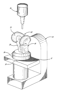
Referring to Figure 4, a custom die 26 may be used to support the preliminary
prosthesis 20 during reworking thereof. For this purpose, the die 26 and
preliminary prosthesis 20 may be positioned on the milling machine 14 enable
the cutting tool 16 to remove excess material from the external and exposed
surface 24 of the preliminary prosthesis 20. The die 26 illustratively
comprises a
support member 28 adapted to receive thereon the preliminary prosthesis 20.
The support member 28 illustratively extends away from a base member 30 and
towards the cutting tool 16 when the die 26 is in position on the milling
machine
- 10 -
CA 2910704 2017-10-17
14. The support member 28 may be attached to the base member 30 using
suitable fastening means, such as screws, bolts, rivets, pins, and the like.
It
should be understood that the support member 28 and the base member 30
may also be machined as a single element, thereby alleviating the need to
attach the members 28, 30 to one another. In this manner, with the preliminary
prosthesis 20 in place on the support member 28, the cutting tool 16 may be
provided access to the external surface 24 for which machining is to be
completed. For this purpose, the base member 30 may comprise pegs 32 for
securing the base member 30 to the support frame 12 of the milling machine
14. The pegs 32 may be mated with corresponding holes (not shown) provided
in the support frame 12, thereby holding the die 26 in place and ensuring
stability thereof during the milling process. A support, such as a vise (not
shown), may further be coupled to the base member 30 during machining of the
preliminary prosthesis 20. It should be understood that other attachment means
for securing the base member 30 to the milling machine 14 may apply.
Referring to Figure 5, the die 26 is illustratively made of plastic, plaster,
metal,
or any other suitable material known to those skilled in the art. The support
member 28 may be manufactured according to the virtual bone model obtained
at step 104 of the method 100 described above. As such, the support member
28 may precisely (or closely when a spacing is to be taken into account, as
discussed above) conform to the shape of the individual's anatomical
structures, and more particularly to the bone(s), to which the desired
prosthesis
18 is to be secured for repairing the individual's damaged joint. For example,
if
the desired prosthesis 18 is a femoral component, as illustrated in Figure 2,
the
die 26 illustratively conforms to the shape of the distal end of the patient's
femur
to which the machined femoral component, and more particularly the internal
surface 22 thereof, is to be engaged with. The die 26 may therefore be
machined to have an outer surface 34, which conforms to the articular joint to
be repaired. In particular, the die 10 may be machined to comprise
representations of anatomical structures of the patient's femur, such as
machined femoral condyles 36a, 36b and a machined patellofemoral groove 38.
In this manner, the outer surface 34 of the die 26 may conform to the
patient's
-11 -
CA 2910704 2017-10-17
actual articular joint surface and may therefore be adapted to precisely mate
with the internal surface 22 of the preliminary prosthesis 20 to be coupled
thereto.
Still, depending on the type of the desired prosthesis 18, the support member
28 may not conform to the shape of the individual's bones, as discussed above.
Instead, the support member 28 may conform to the shape of another machined
object (not shown), such as a tibial prosthetic component, the desired
prosthesis 18 is to be mated with. Also, machining of the support member 28
may depend on the surface of the preliminary prosthesis 20 that is to be
supported on the support member 28. Indeed, as discussed above, the external
surface 24 may be fully machined while the internal surface 22 is partially
machined. As such, the external surface 24 may be supported on the die 26 for
reworking the internal surface 22. In the case where a femoral prosthesis
component is being machined, as illustrated, the preliminary surface 24 may
not
be properly supported on the die 26 if the latter is manufactured such that
the
outer surface 34 conforms to the surface of the individual's tibia that the
preliminary prosthesis 20, once turned into the desired prosthesis 18, will be
mated with. Indeed, due to the arcuate shape of the femoral prosthesis
component, the substantially planar shape of the tibial surface may not prove
suitable for preventing movement of the preliminary prosthesis 20 relative to
the
die 26. As such, when supported on the die 26, the preliminary prosthesis 20
may not be held in place during machining. In order to avoid such an issue,
the
die 26 may be manufactured on the basis of the virtual prosthesis design such
that the outer surface 34 corresponds to at least a portion of the external
surface 24 that is to be supported on the die 26. This may then ensure
adequate mating of the external surface 24 with the outer surface 34, and
accordingly adequate support of the preliminary prosthesis 20 on the die.
In one embodiment, the outer surface 34 may be shaped and sized to precisely
conform to the shape and size of the external surface 24. In other
embodiments, the outer surface 34 may be shaped and sized to conform to the
shape and size of a portion of the external surface 24. It is desirable for
such a
portion of the external surface 24 to be sufficient to securely hold the
- 12 -
CA 2910704 2017-10-17
preliminary prosthesis 20 in place relative to the die 26 when the outer
surface
34 is mated with the portion of the external surface 24. It should be
understood
that the portion of the external surface 24 may vary according to the desired
prosthesis 18 to be machined. 36. It should also be understood that this may
also apply when the internal surface 22 of the preliminary prosthesis 20 is to
be
supported on the die 26. Indeed, in this case, the outer surface 34 may be
manufactured so as to conform to at least a portion of the internal surface 22
rather than being manufactured to conform to the articular joint (e.g. bone
surface) or mating surface the preliminary prosthesis 20 is to be mated with,
as
discussed above.
Referring to Figure 6a and Figure 6b, machining of the external surface 24 of
the preliminary prosthesis 20 may be performed with the latter held in place
on
the die 26. In particular, the preliminary prosthesis 20 may be positioned on
the
die 26 with the internal surface 22 (or alternatively the external surface 24)
of
the preliminary prosthesis 20 matingly engaged with the outer surface 34 of
the
die 26. By machining the external surface 24 (or alternatively the internal
surface 22) using the cutting tool 16, material may be removed, thereby
reducing the thickness d of the external surface 24 (or alternatively the
internal
surface 22) in order to arrive at the desired prosthesis 18. Once the
preliminary
prosthesis 20 has been machined to achieve the desired prosthesis 18, the
latter may then be removed from the die 26 for shipping to a desired location.
Using the approach described herein, precise machining of a prosthesis
component or any other object known to those skilled in the art, may be
achieved. In particular, reworking of the object may be facilitated and the
finished product may therefore be closer to the designed product. Better
results
may in turn be achieved.
The embodiments of the invention described above are intended to be
exemplary only. The scope of the invention is therefore intended to be limited
solely by the scope of the appended claims.
- 13 -
CA 2910704 2017-10-17