Note: Descriptions are shown in the official language in which they were submitted.
DEVICE FOR PREPARATION AND ANALYSIS OF NUCLEIC ACIDS
BACKGROUND
Technical Field
The present invention generally relates to microfluidic devices and
methods for processing samples for molecular diagnostic applications, for
example
detection of target nucleic acid sequences.
Description of the Related Art
The role of molecular diagnostics is critical in today's global health care
environment. In the developing world, 95% of deaths are due to a lack of
proper
diagnostics and the associated follow-on treatment of infectious diseases;
i.e., acute
respiratory infections (ARIs), malaria, HIV, and tuberculosis (TB) (Yager, P
et al, Annu
Rev Biomed Eng 10:107-144, 2008). Recent pandemics like the 2009 H1N1
Influenza
A pandemic, have accentuated the need for tools to effectively detect and
control
infectious diseases. Factors like "rapid pathogen mutation rates,
transformation of
nonhuman pathogens into human pathogens, and recombination of non human
pathogen
with human pathogens" have added to the challenge of managing novel infectious
diseases (Kiechle, FL et al., Clin Lab Med 29(3):555-560, 2009). Increased
global
mobility has aided the rapid spread of infectious diseases from region of
origin to other
parts of the world as seen during the 2009 H1N1 pandemic. This mobility has
highlighted the need for rapid, portable diagnostic (point-of-care [FOC])
devices at
ports of entry to prevent global spread of infections. Current laboratory
culture
methods for pathogens take a day or more to provide results.
For certain other types of infections, in both the developed and
developing worlds, the diagnostic tests need to be repeated periodically to
measure
response to therapy and monitor the disease condition. One such case is
monitoring the
viral load (number of viral particles per milliliter of blood) for infections
like HIV
(Human immunodeficiency virus) and hepatitis C. Sub-Saharan Africa is a region
heavily affected by the AIDS pandemic. The lack of standard laboratory
facilities and
1
Date Recue/Date Received 2020-05-12
CA 02911308 2015-10-30
WO 2014/182847 PCT/US2014/037197
trained laboratory technicians in these regions is a serious bottleneck.
Similar problems
exist in medically underserved areas of the USA. Rapid, low-cost diagnostic
tools that
can be dispersed throughout a community for easy access, possibly even in the
home,
would provide substantial benefit by allowing more rapid diagnosis and
monitoring of
.. disease and infection.
Nucleic acid biomarkers are the target analytes for several infectious
diseases of high global health importance, including HIV, HCV, HBV, pandemic
influenza, and dengue. A major challenge in developing a simple diagnostic
device to
test multiple viral agents is that the genome of some viruses are comprised of
DNA,
while those of other viruses are comprised of RNA. A further challenge for RNA-
based
analytes is specimen handling that protects the integrity of these labile
molecules.
There are several commercially available products that address this latter
problem.
Most of these products are expensive, technically demanding, and/or require
some form
of refrigeration. These requirements cannot be easily met by miniaturized
microfluidic
devices with on-cartridge reagent reservoirs designed for rapid, on-site
diagnostic
analyses. Moreover, these requirements cannot be easily met in low-resource or
remote
settings, as is the case in the majority of the developing world. Thus, there
is a need for
a low-cost, non-instrumented, and simple-to-use diagnostic device that can be
used to
prepare stable samples of nucleic acids and analysis of both DNA and RNA
biomarkers
at the point of care (POC).
Blood is the human tissue routinely used for nucleic acid expression
studies and blood-based biomarker analysis because it can be easily collected.
However, whole blood often contains many factors, such as heme and heparin,
which
interfere with and/or inhibit, many downstream analytic procedures. Moreover,
blood
plasma is extremely high in ribonuclease (RNase) activity, and minimizing this
activity
is critical to any RNA isolation procedure. Although DNA can be prepared from
clinical samples under harsh conditions and stabilized, for example, simply by
spotting
on filter paper and allowing to dry at room temperature, RNA preparation has
typically
required the use of stabilizing agents and refrigeration and/or freezing. The
steps
required to stabilize RNA in clinical samples are cumbersome and not amenable
to
microfluidic, "sample to answer" diagnostic devices.
Variations of two methods have historically been used to prepare RNA
from biological samples: chemical extraction and immobilization on glass,
often
referred to as "solid-phase extraction." Chemical extraction methods usually
use highly
concentrated chaotropic salts in conjunction with acidic phenol or phenol-
chloroform
solutions to inactivate RNases and purify RNA from other biomolecules. These
2
CA 02911308 2015-10-30
WO 2014/182847
PCT/US2014/037197
methods provide very pure preparations of RNA; however, the RNA must typically
be
desalted and concentrated with an alcohol precipitation step. The solid-phase
extraction
method, described in U.S. Patent No. 5,234,809 to Boom et al., relies on the
lysing and
nuclease-inactivating properties of the chaotropic agent guanidinium
thiocyanate
together with the nucleic acid-binding properties of solid-state silica
particles or
diatoms in the presence of this agent. After silica-bound RNA is washed with a
high-
salt buffer containing ethanol, the RNA is eluted in a low-ionic-strength
buffer.
It will be readily appreciated that sample preparation methods requiring
aqueous extraction with organic solvents or chaotropic agents are tedious,
hazardous,
labor-intensive, and slow. Moreover, if great care is not taken in performing
the
procedures, residual contamination with nucleases can occur, and the sample
nucleic
acids will be degraded or lost. Diagnostic tests performed with such samples
can give
false negative results due to such degradation. False negative results can
also be
obtained due to chemical interference, for example from residual anionic
detergents,
.. chaotropic salts, or ethanol remaining in the sample and inhibiting target
amplification
procedures. If anionic detergents and proteases have been used, residual
proteolytic
activity can also degrade the enzymes used in target amplification and/or
hybridization
detection reactions and produce false negative results. Sample preparation
methods
based on the "Boom lysis" protocol disclosed in the '809 patent are commonly
viewed
as adequately addressing these problems. However, the present inventors have
unexpectedly found that such extraction methods, utilizing chaotropic salts
combined
with solid-phase extraction, are not reliably effective in the preparation of
blood or
plasma samples for PCR-based detection of the HBV genome. Thus, none of the
above-cited protocols is suitable for the preparation of a common sample for
detection
of both DNA and RNA targets from complex biological starting materials, e.g.,
whole
blood and blood serum. This is particularly true for infectious disease
diagnosis in
clinical laboratory settings, where time demands are very high, and in low-
resource
areas where cost-effectiveness, reduction of toxic waste streams and
simplicity are also
of prime importance.
While progress has been made in the field, there continues to be a need
in the art for point of care diagnostic devices, such as microfluidic devices,
capable of
isolating and analysis of nucleic acids from test samples. The present
invention fulfills
these needs and provides further related advantages.
3
CA 02911308 2015-10-30
WO 2014/182847
PCT/US2014/037197
BRIEF SUMMARY
Embodiments of the present invention address the above noted global
health needs by providing microfluidic devices for the preparation,
stabilization, and
molecular analysis of nucleic acids from a test sample, such as a blood
product. The
present inventors have surprisingly found that a simple sample preparation
protocol
based on treatment with a clay mineral and alkaline buffer yields samples
containing
DNA and/or RNA that are suitable as immediate reagents in amplification
reactions.
Without being bound by theory, it is believed that the clay mineral functions
to both
protect nucleic acids from enzymatic degradation (due to nuclease activity)
and
hydrolytic degradation (due to alkaline extraction reagents). The nucleic
acids samples
prepared by the devices of the present invention are essentially free of
nuclease activity
and are superior substrates for modifying enzymes. Embodiments of the
microfluidic
devices of the present invention are particularly advantageous in the
simultaneous
detection of RNA and DNA targets from minute samples of human blood or other
test
samples.
In related embodiments, the present invention provides an improved
integrated microfluidic device for integrating nucleic acid sample preparation
with
downstream molecular analysis. Notably, embodiments of the device are suitable
for
the preparation and analysis of both DNA and RNA from a common test sample. In
certain embodiments, the devices of the invention are characterized in that
the reagents
for preparation of nucleic acids suitable for immediate amplification are pre-
loaded into
the device. These reagents include, but are not limited to, a clay mineral and
an
alkaline buffer.
Accordingly, embodiments of the present invention provide a
microfluidic device for preparing and analyzing nucleic acids in a test
sample,
comprising a microfluidic channel having a first end and a second end; a
sample inlet
fluidly connected to the first end of the microfluidic channel for receiving a
test sample;
a clay treatment chamber fluidly connected to said microfluidic channel,
wherein said
clay treatment chamber contains a clay mineral reagent; a sample lysis chamber
fluidly
connected to said clay treatment chamber, wherein said sample lysis chamber
contains
an alkaline solution; one or more sample nucleic acid amplification and
detection wells
fluidly connected to said sample lysis chamber; and one or more sample
outlets. In
another embodiment, the present invention provides a microfluidic device for
preparing
and analyzing nucleic acids in a test sample wherein the clay mineral is
selected from
the kaolinite, smectite, or illite groups. In yet another embodiment, the clay
mineral of
the invention is one of talc, hallosite, bentonite, a synthetic clay mineral,
or laponite. In
4
CA 02911308 2015-10-30
WO 2014/182847 PCT/US2014/037197
another embodiment, the present invention provides a microfluidic device for
preparing
and analyzing nucleic acids in a test sample wherein the alkaline solution is
KOH,
NaOH, or Li0H. In another embodiment, the present invention provides a
microfluidic
device for preparing and analyzing nucleic acids in a test sample that further
comprises
a neutralization chamber downstream of the lysis chamber that contains an acid
reagent.
In other embodiments, the acidic solution is HC1 , C2H402,or H2SO4. In another
embodiment, the present invention provides a microfluidic device for preparing
and
analyzing nucleic acids in a test sample, wherein the test sample comprises
one or more
infectious agents. In another embodiment, the infectious agents are viral
agents. In yet
another embodiment, the infectious agents are at least two viral agents. In
yet another
embodiment, the infectious agents are a DNA virus and a RNA virus. In yet
another
embodiment, the infectious agents are HBV and HCV or HIV. In another
embodiment,
the present invention provides a microfluidic device for preparing and
analyzing nucleic
acids in a test sample wherein the test sample comprises blood, plasma, serum,
urine,
saliva, sputum, respiratory lavage, tears, or tissue swabs. In another
embodiment, the
present invention provides a microfluidic device for preparing and analyzing
nucleic
acids in a test sample wherein the device further comprises an on-device pump
fluidly
connected to the second end of the microfluidic channel. In another
embodiment, the
present invention provides a microfluidic device for preparing and analyzing
nucleic
acids in a test sample wherein the device further comprises a composite
membrane
interposed between the sample inlet and the first end of the microfluidic
channel,
wherein the composite membrane is capable of removing selected particles from
the
blood. In other embodiments, the composite membrane may be comprised of a
material
that activates blood coagulation. In another embodiment, that composite
membrane
may be comprised of a glass filter.
Methods for using the microfluidic devices for preparation and or
analysis of nucleic acid containing samples are also provided. For example, in
one
embodiment the methods comprise:
a) introducing a sample suspected of containing the nucleic acid of
interest into any of the disclosed microfluidic devices;
b) contacting the sample with a clay mineral in the microfluidic
device; and
c) lysing the sample in the microfluidic device.
In some embodiments, the methods further comprise amplifying the
lysed sample in the microfluidic device to obtain an amplified sample and
optionally
detecting the nucleic acid of interest in the amplified sample.
5
CA 02911308 2015-10-30
WO 2014/182847 PCT/US2014/037197
Use of the microfluidic devices for isolating a nucleic acid of interest is
also provided. In some embodiments, the use further comprises amplifying the
nucleic
acid of interest and optionally detecting the nucleic acid of interest.
BRIEF DESCRIPTION OF THE DRAWINGS
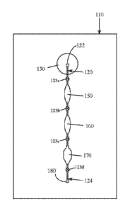
FIG. 1 is a schematic view illustrating the operation of a first
embodiment of a microfluidic device in accordance with aspects of the present
invention.
FIG. 2 is a schematic view illustrating the operation of a second
embodiment of a microfluidic device in accordance with aspects of the present
invention.
FIG. 3 is a schematic view illustrating the operation of a third
embodiment of a microfluidic device in accordance with aspects of the present
invention.
FIG. 4 is a schematic view illustrating the operation of a fourth
.. embodiment of a microfluidic device in accordance with aspects of the
present
invention.
FIGs. 5 A-B are cross-sectional views illustrating the operation of a first
and second embodiment of a composite membrane in accordance with aspects of
the
present invention.
FIG. 6 is a step-by-step guide of examples of processes that may be
undertaken in the device of the present invention.
DETAILED DESCRIPTION
The present inventors have surprisingly found that the combination of a
clay mineral and an alkaline buffer can be used to prepare nucleic acids from
complex
biological test samples for molecular analytic procedures, such as PCR.
Advantageously, these reagents can be used to prepare a single test sample for
the
detection of both DNA and a RNA target molecules without the need for further
purification or isolation of the nucleic acids, offering a vast improvement
over the state-
of-the-art. Without being bound by theory, it is believed that the clay
mineral provides
several beneficial effects, including, but not limited to: protection of
nucleic acids from
hydrolysis under alkaline conditions; protection of nucleic acids from
nuclease-
mediated degradation; protection of downstream assay reagents, such as DNA
polymerases, from inhibitors and other contaminants present in the test
sample; and
general buffering properties.
6
CA 02911308 2015-10-30
WO 2014/182847 PCT/US2014/037197
The present invention relates to microfluidic devices comprising on-
board clay mineral and alkaline buffers reagents for the preparation and
analysis of
nucleic acids samples. In some embodiments, the devices further comprise a
plurality
of microfluidic channels, inlets, valves, membranes, pumps, and other elements
arranged in various configurations manipulate the flow of the fluid sample in
order to
extract nucleic acids from the sample and to perform optional subsequent
molecular
analysis. The devices of the invention may further comprise a composite
membrane for
the separation of a serum sample from a whole blood sample. In the following
description, certain specific embodiments of the present devices and methods
are set
forth, however, persons skilled in the art will understand that the various
embodiments
and elements described below may be combined or modified without deviating
from the
spirit and scope of the invention.
1. Definitions
Test samples: Test samples include biological samples or "biosamples,"
which may be clinical specimens. Representative biosamples include, for
example:
blood, serum, plasma, buffy coat, saliva, wound exudates, pus, lung and other
respiratory aspirates, nasal aspirates and washes, sinus drainage, bronchial
lavage
fluids, sputum, medial and inner ear aspirates, cyst aspirates, cerebral
spinal fluid, stool,
diarrhoea! fluid, urine, tears, mammary secretions, ovarian contents, ascites
fluid,
mucous, gastric fluid, gastrointestinal contents, urethral discharge, synovial
fluid,
peritoneal fluid, meconium, vaginal fluid or discharge, amniotic fluid, semen,
penile
discharge, or the like may be tested. Assay from swabs or lavages
representative of
mucosal secretions and epithelia are acceptable, for example mucosal swabs of
the
throat, tonsils, gingival, nasal passages, vagina, urethra, rectum, lower
colon, and eyes,
as are homogenates, lysates and digests of tissue specimens of all sorts.
Mammalian
cells are acceptable samples. Besides physiological or biological fluids,
samples of
water, industrial discharges, food products, milk, air filtrates, and so forth
are also test
specimens. In some embodiments, test samples are placed directly in the
device; in
other embodiments, pre-analytical processing is contemplated.
Bioassay Target Molecule: or "nucleic acid of interest," or "target
molecule," includes a nucleic acid or nucleic acids. Target nucleic acids
include genes,
portions of genes, regulatory sequences of genes, mRNAs, rRNAs, tRNAs, siRNAs,
cDNA and may be single stranded, double stranded or triple stranded. Some
nucleic
acid targets have polymorphisms, deletions and alternate splice sequences.
7
CA 02911308 2015-10-30
WO 2014/182847 PCT/US2014/037197
Clay mineral: or "clay" refers to any of a group of hydrous aluminum or
magnesium silicates (including phyllosilicates) with a layer (sheet like)
structure and
very small particle size (customarily less than two micrometers). Clay
minerals may
contain significant amounts of iron, alkali metals, or alkaline earths. Clay
minerals
form the main mineral stock of naturally occurring clays and clay stones and
are
produced from such geologic deposits. Clay minerals may also be derived from
other
natural sources, such as silt stones, clay slates and some sands and
sandstones. Clay
minerals may also be produced synthetically.
Phyllosilicate: includes a broad class of minerals described as sheet
silicates, which form parallel sheets of silicate tetrahedra with a
composition of Si205 or
a 2:5 ratio of silicon to oxygen. Phyllosilicates include the following
groups: the
serpentine group of antigorite and chrysotile, the apophyllite group, the
prehnite group,
and the clay mineral groups described below. Any of these phyllosilicates,
including
the mineral known as talc, is suitable for use in the present invention.
Pathogen: an organism associated with an infection or infectious disease.
Pathogenic condition: a condition of a mammalian host characterized by
the absence of health, i.e., a disease, infirmity, morbidity, or a genetic
trait associated
with potential morbidity.
Various embodiments include microfluidic devices capable of analysis
of test samples comprising one or more target infectious agents. Exemplary
target
infectious agents include microorganisms and/or viruses with either a DNA-
based
genome or an RNA-based genome. In some embodiments, suitable viruses include,
but
are not limited to, Hepatitis B virus (HBV), Hepatitis C virus (HCV), human
immunodeficiency viruses (HIV) I and II, influenza A virus, influenza B virus,
respiratory syncytial viruses (RSV) A and B, human metapneumovirus (MPV),
and/or
herpes simplex viruses (HSV) I and/or II.
In other embodiments, viral infectious agents present in a test sample
include, but are not limited to, influenza A, influenza B, RSV (respiratory
syncytial
virus) A and B, human immunodeficiency virus (HIV), human T-cell
lymphocytotrophic virus, hepatitis viruses (e.g., Hepatitis B Virus and
Hepatitis C
Virus), Epstein-Barr Virus, cytomegalovirus, human papillomaviruses, orthomyxo
viruses, paramyxo viruses, adenoviruses, corona viruses, rhabdo viruses, polio
viruses,
toga viruses, bunya viruses, arena viruses, rubella viruses, reo viruses,
Norovirus,
human metapneumovirus (MPV), Herpes simplex virus 1 and 2 (HSV-1 and HSV-2),
West Nile virus, Yellow fever virus, Varicella zoster virus (VZV), Rabies
virus,
Rhinovirus, Rift Valley fever virus, Marburg virus, mumps virus, measles
virus,
8
CA 02911308 2015-10-30
WO 2014/182847 PCT/US2014/037197
Epstein-Barr Virus (EBV), human papilloma virus (HPV), Ebola virus, Colorado
tick
fever virus (CTFV), and/or rhinoviruses.
In different embodiments, bacterial infectious agents in a test sample
include, but are not limited to, Escherichia coli, Salmonella, Shigella,
Campylobacter,
Klebsiella, Pseudomonas, Listeria monocytogenes, Mycobacterium tuberculosis,
Mycobacterium avium-intracellulare, Yersinia, Francisella, Pasteurella,
Brucella,
Clostridia, Bordetella pertussis, Bacteroides, Staphylococcus aureus,
Streptococcus
pneumonia, B-Hemolytic strep., Corynebacteria, Legionella, Mycoplasma,
Ureaplasma,
Chlamydia, Clostridium difficile, Gardnerella, Trichomonas vaginalis,
Neisseria
gonorrhea, Neisseria meningitides, Hemophilus influenza, Enterococcus
faecalis,
Proteus vulgaris, Proteus mirabilis, Helicobacter pylori, Treponema palladium,
Borrelia
burgdorferi, Borrelia recurrentis, Rickettsial pathogens, Nocardia,
Acitnomycetes
and/or Acinetobacter.
In still other embodiments, fungal infectious agents in a test sample
include, but are not limited to, Cryptococcus neoformans, Blastomyces
dermatitidis,
Histoplasma capsulatum, Coccidioides immitis, Paracoccicioides brasiliensis,
Candida
albicans, Aspergillus fumigautus, Phycomycetes (Rhizopus), Sporothrix
schenckii,
Chromomycosis, and/or Maduromycosis.
In more embodiments, parasitic agents present in a test sample include,
but are not limited to, Plasmodium falciparum, Plasmodium malaria, Plasmodium
vivax, Plasmodium ovale, Onchoverva volvulus, Leishmania, Trypanosoma spp.,
Schistosoma spp., Entamoeba histolytica, Cryptosporidum, Giardia spp.,
Trichimonas
spp., Balatidium coli, Wuchereria bancrofti, Toxoplasma spp., Enterobius
vermicularis,
Ascaris lumbricoides, Trichuris trichiura, Dracunculus medinesis, trematodes,
Diphyllobothrium latum, Taenia spp., Pneumocystis carinii, and/or Necator
americanis.
Nucleic acid: The terms "nucleic acid," "polynucleotide," and
"oligonucleotide" are used herein to include a polymeric form of nucleotides
of any
length, including, but not limited to, ribonucleotides and
deoxyribonucleotides.
Relatively short nucleic acid polymers are often used as "primers" or
"probes." The
definition encompasses nucleic acids from natural sources which can be
methylated or
capped, and also synthetic forms, which can contain substitute or derivatized
nucleobases and may be based on a peptide backbone. Nucleic acids are
generally
polymers of adenosine, guanine, thymine, and cytosine and their "deoxy-"
forms, but
may also contain other pyrimidines such as uracil and xanthine, or spacers and
universal
bases such as deoxyinosine. Deoxynucleic acids may be single-stranded or
double-
stranded depending on the presence or absence of complementary sequences, and
on
9
CA 02911308 2015-10-30
WO 2014/182847 PCT/US2014/037197
conditions of pH, salt concentration, temperature, and the presence or absence
of certain
organic solvents such as formamide, n,n-dimethylformamide, dimethylsulfoxide,
and n-
methylpyrrolidinone.
"Target nucleic acid sequence" or "template": As used herein, the term
"target" refers to a nucleic acid sequence in a biosample that is to be
amplified in the
assay by a polymerase and detected. The "target" molecule can be present as a
"spike"
or as an uncharacterized analyte in a sample, and may consist of DNA, cDNA,
gDNA,
RNA, mRNA, rRNA, or miRNA, either synthetic or native to an organism. The
"organism" is not limited to a mammal. The target nucleic acid sequence is a
template
for synthesis of a complementary sequence during amplification. Genomic target
sequences are denoted by a listing of the order of the bases, listed by
convention from 5'
end to 3' end.
Reporter, "Label" or "Tag": refers to a biomolecule or modification of a
biomolecule that can be detected by physical, chemical, electromagnetic and
other
related analytical techniques. Examples of detectable reporters include, but
are not
limited to, radioisotopes, fluorophores, chromophores, mass labels, electron
dense
particles, magnetic particles, dyed particles, QDots, spin labels, molecules
that emit
chemiluminescence, electrochemically active molecules, enzymes, cofactors,
enzymes
linked to nucleic acid probes, and enzyme substrates. Reporters are used in
bioassays
as reagents, and are often covalently attached to another molecule, adsorbed
on a solid
phase, or bound by specific affinity binding.
Probe: A "probe" is a nucleic acid capable of binding to a target nucleic
acid by complementary base pairing with sufficient complementarity to form a
stable
double helix at room temperature. Probes may be labeled with reporter groups.
Suitable labels that can be attached to probes include, but are not limited
to,
radioisotopes, fluorophores, chromophores, mass labels, electron dense
particles,
magnetic particles, spin labels, molecules that emit chemiluminescence,
electrochemically active molecules, enzymes, cofactors, and enzyme substrates.
Tools
for selection of a probe sequence, length, and hybridization conditions are
generally
familiar to those skilled in the art.
Amplification: As used here, the term "amplification" refers to a
"template-dependent process" that results in an increase in the concentration
of a
nucleic acid sequence relative to its initial concentration. A "template-
dependent
process" is a process that involves "template-dependent extension" of a
"primer"
molecule. A "primer" molecule refers to a sequence of a nucleic acid that is
complementary to a known portion of the target sequence. A "template dependent
CA 02911308 2015-10-30
WO 2014/182847 PCT/US2014/037197
extension" refers to nucleic acid synthesis of RNA or DNA wherein the sequence
of the
newly synthesized strand of nucleic acid is dictated by the rules of
complementary base
pairing of the target nucleic acid and the primers.
Amplicon refers to a double stranded DNA product of a prior art
amplification means, and includes double stranded DNA products formed from DNA
and RNA templates.
Two-tailed Amplicon refers to a double stranded DNA product of an
amplification means in which tagged primer pairs are covalently incorporated,
a first
primer conjugated with a peptide hapten or epitope, a second primer conjugated
with an
affinity reporter, tag or "ligand." As used herein, the two-tailed amplicon
functions as a
"hetero-bifunctional" tether, and forms a molecular detection complex on a
solid
substrate.
Primer: as used herein, is a single-stranded polynucleotide or
polynucleotide conjugate capable of acting as a point of initiation for
template-directed
DNA synthesis in the presence of a suitable polymerase and cofactors. Primers
are
generally at least 7 nucleotides long and, more typically range from 10 to 30
nucleotides in length, or longer. The term "primer pair" refers to a set of
primers
including a 5' "forward" or "upstream" primer that hybridizes with the
complement of
the 5' end of the DNA template to be amplified and a 3' "reverse" or
"downstream"
primer that hybridizes with the 3' end of the sequence to be amplified. Note
that both
primers have 5' and 3' ends and that primer extension always occurs in the
direction of
5' to 3'. Therefore, chemical conjugation at or near the 5' end does not block
primer
extension by a suitable polymerase. Primers may be referred to as "first
primer" and
"second primer," indicating a primer pair in which the identity of the
"forward" and
"reverse" primers is interchangeable. Additional primers may be used in nested
amplification.
Polymerases are enzymes defined by their function of incorporating
nucleoside triphosphates, or deoxynucleoside triphosphates, to extend a 3'
hydroxyl
terminus of a primer molecule. For a general discussion concerning
polymerases, see
Watson, J. D. et al, (1987) Molecular Biology of the Gene, 4th Ed., W. A.
Benjamin,
Inc., Menlo Park, Calif. Examples of polymerases include, but are not limited
to, E.
coli DNA polymerasc I, "Klenow" fragment, Taq-polymerase, T7 polymerase, T4
polymerase, T5 polymerase and reverse transcriptase. Examples of reverse
transcriptases include HIV-1 reverse transcriptase from the human
immunodeficiency
virus type 1, telomerase, M-MuLV reverse transcriptase from the Moloney murine
leukemia virus, and AMV reverse transcriptase from the avian myeloblastosis
virus.
11
It should be noted that reverse transcriptase is commonly used in
research to apply the polymerase chain reaction technique to RNA targets. The
classical PCR technique can only be app I led directly to DNA, but by using
reverse
transcriptase to synthesize cDNA from RNA, PCR analysis of RNA targets is
possible.
The technique is collectively called Reverse Transcription-Polymerase Chain
Reaction
(RT-PCR).
Complementary (with respect to nucleic acids) refers to two single-
stranded nucleic acid sequences that can hybridize to form a double helix. The
matching of base pairs in the double helix of two complementary strands is not
necessarily absolute. Selectivity of hybridization is a function of
temperature of
annealing, salt concentration, and solvent, and will generally occur under low
stringency when there is as little as 55% identity over a stretch of at least
14-25
nucleotides. Stringency can be increased by methods well known in the art. See
M.
Kanehisa, Nucleic Acids Res. 12:203 (1984). Regarding hybridization of
primers, a
primer that is "perfectly complementary" has a sequence fully complementary
across
the entire length of the primer and has no mismatches. A "mismatch" refers to
a site at
which the base in the primer and the base in the target nucleic acid with
which it is
aligned are not complementary.
Pre-loading is a term that means that reagents are added to the device
prior to its end use, for example, during the device's manufacture. As such,
solid
reagents may be deposited on the device, for example, by drying a solution of
the
reagent by allowing the solvent in the reagent to evaporate. Alternatively,
reagents may
be pre-loaded in dehydrated form as disclosed in U.S. Patent Application Pub.
No.
2012/0156750 to Battrell et al.
Reagent refers broadly to any chemical or biochemical agent used in a
reaction, including enzymes. A reagent can include a single agent which itself
can be
monitored (e.g., a substance that is monitored as it is heated) or a mixture
of two or
more agents. A reagent may be living (e.g., a cell) or non-living. Exemplary
reagents
for a nucleic acid amplification reaction include, but are not limited to,
buffer, metal ion
(for example magnesium salt), chelator, polymerase, primer, template,
nucleotide
triphosphate, label, dye, nuclease inhibitor, and the like. Reagents for
enzyme reactions
include, for example, substrates, chromogens, cofactors, coupling enzymes,
buffer,
metal ions, inhibitors and activators. Not all reagents are reactants.
12
Date Recue/Date Received 2020-05-12
Specificity: Refers to the ability of an assay to reliably differentiate a
true positive signal of the target biomarker from any background, erroneous or
interfering signals.
Sensitivity: Refers to the lower limit of detection of an assay where a
negative can no longer be reliably distinguished from a positive.
Stability: during storage, any compositional change measured in a
parameter, for example activity, concentration, degradation, viscosity, pH, or
particle
composition, that is greater than 10% over time, denotes instability. Changes
less than
or equal to 10% connote stability. The time period over which stability is
measured is
relative depending on the intended utility of the composition. Accelerated
stability at
higher temperature is sometimes taken as a more speedy way of extrapolating
stability
over longer periods of time than are actually measured.
Endpoint: "Endpoint" or "datapoint" is used here as shorthand for a
"result" from either qualitative or quantitative assays, and may refer to both
stable
endpoints where a constant plateau or level of reactant is attained, and to
rate reactions,
where the rate of appearance or disappearance of a reactant or product as a
function of
time (i.e., the slope) is the datapoint.
Microfluidic cartridge: a "device," "card," or "chip" with fluidic
structures and internal channels having microfluidic dimensions. These fluidic
structures may include chambers, valves, vents, vias, pumps, inlets, nipples,
and
detection means, for example. Generally, microfluidic channels are fluid
passages
having at least one internal cross-sectional dimension that is less than about
500 gm and
typically between about 0.1 gm and about 500 gm Microfluidic channels are
fluid
passages having at least one internal cross-sectional dimension that is less
than 600 gm.
The microfluidic flow regime is characterized by Poiseui I le. or "laminar"
flow. The
particle volume fraction and ratio of channel diameter to particle diameter
(Did) has a
measurable effect on flow characteristics. (See for example, Staben M E et al.
2005.
Particle transport in Poiseuille flow in narrow channels. Intl J Multiphase
Flow 31:529-
47).
Microfluidic cartridges may be fabricated from various materials using
techniques such as laser stenciling, embossing, stamping, injection molding,
masking,
etching, and three-dimensional soft lithography. Laminated microfluidic
cartridges are
further fabricated with adhesive interlayers or by thermal adhesiveless
bonding
techniques, such by pressure treatment of oriented polypropylene. The
microarchitecture of laminated and molded microfluidic cartridges can differ.
13
Date Recue/Date Received 2020-05-12
CA 02911308 2015-10-30
WO 2014/182847
PCT/US2014/037197
Microfluidic channel: also termed "microchannel," is a fluid channel
having variable length, but one dimension in cross-section less than 500 gm.
Microfluidic fluid flow behavior in a microfluidic channel is highly non-ideal
and
laminar and may be more dependent on wall wetting properties, roughness,
liquid
viscosity, adhesion, and cohesion than on pressure drop from end to end or
cross-
sectional area. The microfluidic flow regime is often associated with the
presence of
"virtual liquid walls" in the channel. However, in larger channels, head
pressures of 10
psi or more can generate transitional flow regimes bordering on turbulent, as
can be
important in rinse steps of assays.
Micro channels constructed of layers formed by extrusion molding may
have more rounded channel profiles and a radius on each "via." The internal
channel
surfaces of injection molded parts are also somewhat smoother. The flow
characteristics of the channels are significant because of the profound
surface effects in
the microflow regime. Surface tension and viscosity compound surface roughness
effects. The most narrow dimension of a channel has the most profound effect
on flow.
it follows that flow in channels that are based on rectangular or circular
cross-sectional
profiles is controlled by the diagonal width or diameter, and design is
typically varied to
take advantage of this behavior. Reduction of taper in the direction of flow
leads to a
wicking effect for diameters below 200 microns. Conversely, flow can be
stopped by
opening up a channel to form a bulb unless pressure is applied. Vias in a
channel can
be designed to promote directional flow, a sort of solid state check valve.
As used herein, the term "downstream" means that, in use, a sample
passes sequentially through the different parts of the device. While the term
"downstream" includes within its scope two parts of the device being in direct
fluid
communication, it also includes within its scope when the two parts are
separated by,
for example, a valve or another part of the device. The term "integrated"
means that
two different parts of the device are combined into a single unit, so that,
for example,
the same part of the device can serve to filter the sample and act as a lysis
unit. When
the term "integrated" is applied to the device of the first and second aspects
of the
present invention combined with a nucleic acid amplification unit, it means
that the two
parts of the system are connected to one another so that, in use, they are in
fluid
communication with one another. in another aspect, the term "integrated" means
that
the different parts of the device are preferably formed on a common substrate.
The
term "connected" when applied to two parts of the device means that the two
parts may
be in direct fluid communication with one another (e.g., through either being
joined
directly together or joined through a channel) or may be separated from one
another by,
14
for example, a valve or another part of a device. Preferably, the term
"connected to"
means that two parts of the device are directly joined to one another.
Microfluidic pumps: include for example, bulbs, bellows, diaphragms, or
bubbles intended to force movement of fluids, where the substructures of the
pump
have a thicknesses or other dimension of less than 1 millimeter. Such pumps
include
the mechanically actuated recirculating pumps described in U.S. Pat. No.
6,743,399 to
Weigl and U.S. 2005/0106066 to Saltsman, commonly assigned to the applicant.
Such pumps may be robotically
operated or operated by hand. Electroosmotic pumps are also provided. Such
pumps
can be used in place of external drives to propulse the flow of solubilized
reagents and
sample in microfluidic device-based assays.
Bellows ("Finger") Pump: is a device formed as a cavity, often
cylindrical in shape, bisected in coronal section by an elastomeric diaphragm
to form a
first and a second half-chamber which are not fluidically connected. The
diaphragm is
controlled by a pneumatic pulse generator connected to the first half-chamber.
Positive
pressure above the diaphragm distends it, displacing the contents of the
second half-
chamber, negative gauge pressure (suction) retracts it, expanding the second
half
chamber and drawing fluid in. By half-chamber, it should be understood that
the
effective area of the diaphragm is the lesser of the volume displacement under
positive
pressure and the volume displacement under suction pressure, and it thus
optimal when
the first and second half chambers are roughly symmetrical or equal in volume
above
and below the diaphragm. The second half-chamber is connected to a fluid in-
port and
out-port. The fluid in-port and out-port may be separate ports or a single
port, but in
either case, are under valve control. As described above, a pneumatic pulse
generator is
pneumatically connected to the first half-chamber, generally by a
microchannel, which
is also valved. In the complete apparatus, pneumatic actuation is
programmable. Thus,
programmable pneumatic pressure logic used by the pulse generator has two
functions,
to actuate the diaphragm on signal, and to open and close valves on signal.
When the
pulse generator is off-cartridge, nipples or inlets, a pneumatic manifold and
solenoid
valves are provided.
In use, fluid enters the second half-chamber of a bellows pump through
the inlet valve when negative pressure is applied to the diaphragm (or
passively, when
fluid is pushed in by a second bellows pump). Then, when positive pressure is
applied
to the diaphragm, the fluid contents of the chamber are displaced out through
the outlet
valve. Similarly, positive and negative pressure signals control valve opening
and
closing. By supplying a train of positive and negative pressure pulses to a
diaphragm,
Date Recue/Date Received 2020-05-12
fluid can be moved in and out of a bellows pump chamber. This fluid motion
becomes
directional by the application of synchronized valve logic, thus the pumping
action.
Microfluidic valves: include a genus of hydraulic, mechanic, pneumatic,
magnetic, and electrostatic actuator flow controllers with at least one
dimension smaller
than 500 um. A representative flap valve of the genus is described in U.S.
Pat. No.
6,431,212. Also included are check
valves. One class of valves refers to a configuration in which a flow channel
and a
control channel intersect and are separated by an elastomeric membrane that
can be
deflected into or retracted from the flow channel in response to an actuation
force in the
control channel. Patents describing species of microfluidic valves include
U.S. Pat.
Nos. 5,971,355, 6,418,968, 6,518,99, 6,620,273, 6,748,975, 6,767,194,
6,901,949, and
U.S. Patent Application 2002/0195152 and 2005/02005816, several of which are
commonly assigned to the applicant.
Check valve: is a one way valve. Microscale versions of ball-spring,
flap, and flip-flop valves are check valves.
Passive shut-off valves: are wettable inserts or coatings in microfluidic
channels that swell when immersed, closing the microchannel off to further
flow in
either direction. Analogously, "surface tension valves" consisting of a ring
of
hydrophobic material on the walls of a microchannel have been disclosed to
delay or
stop the flow of a reagent. Stop flow can also be achieved by widening the
taper of a
microfluidic channel diameter.
Self-priming: connotes a microfluidic channel that is fabricated from a
material or is treated so that the channel is wettable and capillary flow
begins generally
without the need to prime the channel.
Via: A step in a microfluidic channel that provides a fluid pathway from
one substrate layer to another substrate layer above or below, characteristic
of
laminated devices built from layers.
Pillow: an on-board reagent pack formed from a deformable sacculus,
.. for example a mylar microbag, optionally enclosed in a pneumatically
actuated device
for puncturing to bag to release its contents at a controlled time. Co-
laminates of a
metal and a plastic are preferred for stability considerations.
Blister pack: an on-board reagent pack under a deformable (or elastic)
diaphragm. Used to deliver reagents by pressurizing the diaphragm and may
appose a
"sharp," such as a metal chevron, so that pressure on the diaphragm ruptures
the
"pillow" (see pillow). These may be used with check valves or closable vents
to
16
Date Recue/Date Received 2020-05-12
CA 02911308 2015-10-30
WO 2014/182847 PCT/US2014/037197
produce directional fluid flow and reagent delivery. Elastic diaphragms are
readily
obtained from polyurethane, polysilicone and polybutadiene, and nitrile for
example
(see elastomer). Deformable, inelastic diaphragms are made with polyethylene
terephthalate (PET), mylar, polypropylene, polycarbonate, or nylon, for
example. Other
suitable materials for the deformable film include parafilm, latex, foil, and
polyethylene
terephthalate. Key factors in selecting a deformable film include the yield
point and the
deformation relaxation coefficient (elastic modulus).
Isolation or "isolated": "Forward isolation" refers here to protection of
the user from exposure to clinical materials potentially contaminated with an
infectious
agent or toxin. "Reverse isolation" refers to protection of the assay device
from
spurious exogenous contamination, such as with a nucleic acid, that may cause
false
positives.
Waste chamber or "pack": is a cavity or chamber that serves as a
receptacle for sequestering discharged sample, rinse solution, and waste
reagents.
Typically also includes a wicking material (see wick). Waste packs may also be
sealed
under an elastic isolation membrane sealingly attached to the body of the
microfluidic
device. This inner membrane expands as the bibulous material expands, thus
enclosing
the waste material. The cavity outside the isolation membrane is vented to
atmosphere
so that the waste material is contained and isolated. Waste packs may
optionally
contain dried or liquid sterilants.
Wick: is a bibulous material used to propulse fluid flow by capillary
wetting in place of, or in concert with, microfluidic pumps. The bibulous core
typically
includes a fibrous web of natural or synthetic fibers, and also often includes
certain
absorbent gelling materials usually referred to as "hydrogels,"
"superabsorbent" or
"hydrocolloid" materials. Materials include papers, sponges, diaper materials,
Contec-
Wipe, and others. Dessicants may also be used, such as calcium sulfate,
calcium
sulfate, silica gel, alone or in combination with bibulous materials.
Trap: a fluid trap with dam that further isolates a waste reservoir from a
vent.
Vent: a pore intercommunicating between an internal cavity and the
atmosphere. A "sanitary" or "isolation vent" also contains a filter element
that is
permeable to gas, but is hydrophobic and resists wetting. Optionally these
filter
elements have pore diameters of 0.45 microns or less. These filters function
both in
forward and reverse isolation. Filter elements of this type and construction
may also be
placed internally, for example to isolate a valve or bellows pump from the
pneumatic
manifold controlling it.
17
Test field: refers to the site in the microfluidic device-based assay where
the assay endpoint is observed or measured, such as an optical window, and is
optionally a detection chamber containing test pads.
"Conventional" is a term designating that which is known in the prior art
to which this invention relates.
"About" and "generally" are broadening expressions of inexactitude,
describing a condition of being "more or less," "approximately," or "almost"
in the
sense of "just about," where variation would be insignificant, obvious, or of
equivalent
utility or function, and further indicating the existence of obvious minor
exceptions to a
norm, rule or limit. For example, in various embodiments the foregoing terms
refer to a
quantity within 20%, 10%, 5%, 1% or 0.1% of the value which follows the term.
Herein, where a "means for a function" is described, it should be
understood that the scope of the invention is not limited to the mode or modes
illustrated in the drawings alone, but also encompasses all means for
performing the
function that are described in this specification, and all other means
commonly known
in the art at the time of filing. A "prior art means" encompasses all means
for
performing the function as are known to one skilled in the art at the time of
filing,
including the cumulative knowledge in the art cited herein by reference to a
few
examples.
A means for polymerizing, for example, may refer to various species of
molecular machinery described as polymerases and their cofactors and
substrates, for
example reverse transcriptases and TAQ polymerase, and includes the cumulative
knowledge of enzymology cited herein by reference to a few examples.
Means for Amplifying include thermocycling and isothermal means.
The first thermocycling technique was the polymerase chain reaction (referred
to as
PCR) which is described in detail in U.S. Pat. Nos. 4,683,195, 4,683,202 and
4,800,159,
Ausubel et al. Current Protocols in Molecular Biology, John Wiley and Sons,
Baltimore, Md. (1989), and in Innis et al., ("PCR Protocols," Academic Press,
Inc., San
Diego Calif., 1990).
Polymerase chain reaction methodologies are well known in the art.
Briefly, in PCR, two primer sequences are prepared that are complementary to
regions
on opposite complementary strands of a target sequence. An excess of
deoxynucleoside
triphosphates are added to a reaction mixture along with a DNA polymerase,
e.g., Taq
polymerase. If the target sequence is present in a sample, the primers will
bind to the
target and the polymerase will cause the primers to be extended along the
marker
sequence by adding on nucleotides. By raising and lowering the temperature of
the
18
Date Recue/Date Received 2020-05-12
reaction mixture, the extended primers will dissociate from the template to
form
reaction products, excess primers will bind to the template and to the
reaction products
and the process is repeated. By adding fluorescent intercalating agents, PCR
products
can be detected in real time.
One isothermal technique is LAMP (loop-mediated isothermal
amplification of DNA) and is described in Notomi, T. et al. Nucl Acid Res 2000
28.
Strand Displacement Amplification (SDA) is another method of carrying
out isothermal amplification of nucleic acids which involves multiple rounds
of strand
displacement and synthesis, i.e., nick translation (Walker et al. Nucleic
Acids Research,
1992: 1691-1696). A similar method, called Repair
Chain Reaction (RCR), involves annealing several probes throughout a region
targeted
for amplification, followed by a repair reaction in which only two of the four
bases are
present. The other two bases can be added as biotinylated derivatives for easy
detection. A similar approach is used in SDA. Target specific sequences can
also be
detected using a cyclic probe reaction (CPR). In CPR, a probe having 3' and 5'
sequences of non-specific DNA and a middle sequence of specific RNA is
hybridized
to DNA that is present in a sample. Upon hybridization, the reaction is
treated with
RNase H, and the products of the probe identified as distinctive products that
are
released after digestion. The original template is annealed to another cycling
probe and
the reaction is repeated.
Another nucleic acid amplification technique is reverse transcription
polymerase chain reaction (RT-PCR). First, complementary DNA (cDNA) is made
from an RNA template, using a reverse transcriptase enzyme, and then PCR is
performed on the resultant cDNA.
Another method for amplification is the ligase chain reaction ("LCR"),
disclosed in EPO No. 320 308. In LCR,
two complementary probe pairs are prepared, and in the presence of the target
sequence, each pair will bind to opposite complementary strands of the target
such that
they abut. In the presence of a ligase, the two probe pairs will link to form
a single unit.
By temperature cycling, as in PCR, bound ligated units dissociate from the
target and
then serve as "target sequences" for ligation of excess probe pairs. U.S. Pat.
No.
4,883,750, describes a method similar to LCR for binding probe pairs to a
target sequence.
QBReplicase, may also be used as still another amplification method in
the present invention. In this method, a rep licative sequence of RNA that has
a region
complementary to that of a target is added to a sample in the presence of an
RNA
19
Date Recue/Date Received 2020-05-12
polymerase. The polymerase will copy the replicative sequence that can then be
detected.
Still further amplification methods, described in GB Application No. 2
202 328, and in PCT Application No. PCT/US89/01025 may be used in accordance
with the present invention.
In the former application, "modified" primers are used in a PCR-
like, template- and enzyme-dependent synthesis. The primers may be modified by
labeling with a capture moiety (e.g., biotin) and/or a detector moiety (e.g.,
enzyme). In
the latter application, an excess of labeled probes are added to a sample. In
the
presence of the target sequence, the probe binds and is cleaved catalytically.
After
cleavage, the target sequence is released intact to be bound by excess probe.
Cleavage
of the labeled probe signals the presence of the target sequence.
Other nucleic acid amplification procedures include transcription-based
amplification systems (TAS), including nucleic acid sequence based
amplification
(NA SBA) and 3SR (Kwoh et al., 1989, Proc. Natl. Acad. Sci. U.S.A., 86: 1173;
Gingeras et al., PCT Application WO 88/10315).
In NASBA, the nucleic acids can be
prepared for amplification by standard phenol/chloroform extraction, heat
denaturation
of a clinical sample, treatment with lysis buffer and minispin columns for
isolation of
DNA and RNA or guanidinium chloride extraction of RNA. These amplification
techniques involve annealing a primer which has target specific sequences.
Following
polymerization, DNA/RNA hybrids are digested with RNase H while double
stranded
DNA molecules are heat denatured again. In either case the single stranded DNA
is
made fully double stranded by addition of second target specific primer,
followed by
polymerization. The double-stranded DNA molecules are then multiply
transcribed by
an RNA polymerase such as T7 or SP6. In an isothermal cyclic reaction, the
RNAs are
reverse transcribed into single stranded DNA, which is then converted to
double
stranded DNA, and then transcribed once again with an RNA polymerase such as
T7 or
5P6. The resulting products, whether truncated or complete, indicate target
specific
sequences.
Davey et al., EPO No. 329 822, disclose a nucleic acid amplification
process involving cyclically synthesizing
single-stranded RNA ("ssRNA"), ssDNA, and double-stranded DNA (dsDNA), which
may be used in accordance with the present invention. The ssRNA is a template
for a
first primer oligonucleotide, which is elongated by reverse transcriptase (RNA-
dependent DNA polymerase). The RNA is then removed from the resulting DNA:RNA
Date Recue/Date Received 2020-05-12
duplex by the action of ribonuclease H(RNase H, an RNase specific for RNA in
duplex
with either DNA or RNA). The resultant ssDNA is a template for a second
primer,
which also includes the sequences of an RNA polymerase promoter (exemplified
by T7
RNA polymerase) 5' to its homology to the template. This primer is then
extended by
DNA polymerase (exemplified by the large "Klenow" fragment of E. coli DNA
polymerase D, resulting in a double-stranded DNA ("dsDNA") molecule, having a
sequence identical to that of the original RNA between the primers and having
additionally, at one end, a promoter sequence. This promoter sequence can be
used by
the appropriate RNA polymerase to make many RNA copies of the DNA. These
copies
can then re-enter the cycle leading to very swift amplification. With proper
choice of
enzymes, this amplification can be done isothermally without addition of
enzymes at
each cycle. Because of the cyclical nature of this process, the starting
sequence can be
chosen to be in the form of either DNA or RNA.
Miller et al. in PCT Application WO 89/06700,
disclose a nucleic acid sequence amplification scheme based on
the hybridization of a promoter/primer sequence to a target single-stranded
DNA
("ssDNA") followed by transcription of many RNA copies of the sequence. This
scheme is not cyclic, i.e., new templates are not produced from the resultant
RNA
transcripts. Other amplification methods include "RACE" and "one-sided PCR"
(Frohman, M. A., In: "PCR Protocols: A Guide to Methods and Applications,"
Academic Press, N.Y., 1990; Ohara et al., 1989, Proc. Natl. Acad. Sci. U.S.A.,
86:
5673-567).
Methods based on ligation of two (or more) oligonucleotides in the
presence of nucleic acid having the sequence of the resulting "di-
oligonucleotide,"
thereby amplifying the di-oligonucleotide, may also be used in the
amplification step of
the present invention. Wu et al., (1989, Genomics 4: 560).
Means for detecting: as used herein, refers to an apparatus for displaying
an endpoint, i.e., the result of an assay, and may include a detection channel
and test
pads, and a means for evaluation of a detection endpoint. Detection endpoints
are
evaluated by an observer visually in a test field, or by a machine equipped
with a
spectrophotometer, fluorometer, luminometer, photomultiplier tube, photodiode,
nephlometer, photon counter, voltmeter, ammeter, pH meter, capacitative
sensor, radio-
frequency transmitter, magnetoresistometer, or Hall-effect device. Magnetic
particles,
beads and microspheres having or impregnated color or having a higher
diffraction
21
Date Recue/Date Received 2020-05-12
CA 02911308 2015-10-30
WO 2014/182847 PCT/US2014/037197
index may be used to facilitate visual or machine-enhanced detection of an
assay
endpoint. Magnifying lenses in the cover plate, optical filters, colored
fluids and
labeling may be used to improve detection and interpretation of assay results.
Means
for detection of magnetic particles, beads and microspheres may also include
embedded
.. or coated "labels" or "tags" such as, but not limited to, dyes such as
chromophores and
fluorophores; radio frequency tags, plasmon resonance, spintronic, radiolabel,
Raman
scattering, chemoluminescence, or inductive moment as are known in the prior
art.
Colloidal particles with unique chromogenic signatures depending on their self-
association are also anticipated to provide detectable endpoints. QDots, such
as CdSe
coated with ZnS, decorated on magnetic beads, or amalgamations of QDots and
paramagnetic Fe304 microparticles, optionally in a sol gel microparticulate
matrix or
prepared in a reverse emulsion, are a convenient method of improving the
sensitivity of
an assay of the present invention, thereby permitting smaller test pads and
larger arrays.
Fluorescence quenching detection endpoints are also anticipated. A variety of
substrate
and product chromophores associated with enzyme-linked immunoassays are also
well
known in the art and provide a means for amplifying a detection signal so as
to improve
the sensitivity of the assay. Detection systems are optionally qualitative,
quantitative or
semi-quantitative. Visual detection is preferred for its simplicity, however
detection
means can involve visual detection, machine detection, manual detection or
automated
detection.
Means for heating and cooling: A number of means for thermocycling a
liquid filled chamber have been described in the prior art. These prior art
means
include convective and conductive heating elements such as electroresistors,
hot air,
lasers, infrared radiation, Joule heating, TEC or Peltier devices, heat pumps,
endothermic reactants, and the like, generally in conjunction with a heat sink
for
dissipating heat during chill-down parts of the cycle. Heating means may also
include
heating by the motion of magnetic beads driven by a high frequency magnetic
field.
Heating and cooling devices for thermocycling fall into two categories:
ramped and fixed temperature. Fixed temperature devices maintain a relatively
constant temperature in a reaction, and at least two reaction chambers are
needed for
thermocycling. Ramped heating devices will vary the temperature between at
least two
set points, and therefore only one reaction chamber is required for
thermocycling.
Combinations of heating elements are possible. Peltier devices may be used for
both
fixed temperature and ramped applications. Water baths are not well adapted to
ramped
temperature control for thermocycling.
22
CA 02911308 2015-10-30
WO 2014/182847
PCT/US2014/037197
Generally, heating and cooling means interface with a fluidics member
so as to effect heat exchange with the liquid contents. For PCR, the relevant
elements
forming the microfluidic channels or chambers where heat exchange takes place
are
termed as part of the "PCR fluidics and thermal interface" assembly.
Unless the context requires otherwise, throughout the specification and
claims which follow, the word "comprise" and variations thereof, such as,
"comprises"
and "comprising" are to be construed in an open, inclusive sense, that is as
"including,
but not limited to."
Reference throughout this specification to "one embodiment" or "an
embodiment" means that a particular feature, structure or characteristic
described in
connection with the embodiment is included in at least one embodiment of the
present
invention. Thus, the appearances of the phrases "in one embodiment" or "in an
embodiment" in various places throughout this specification are not
necessarily all
referring to the same embodiment. Furthermore, the particular features,
structures, or
characteristics may be combined in any suitable manner in one or more
embodiments.
2. Preparation of nucleic acid-containing samples
The present inventors have surprisingly found that the combined use of a
clay mineral and an alkaline solution can be used to prepare complex
biological
samples for nucleic acid analysis. In some embodiments, these reagents can be
advantageously used to prepare a common sample for the detection of both DNA
and
RNA target molecules in a microfluidic device. The method of the invention
offers
improvements over known sample preparation methods in that the present method
does
not require further purification or isolation of the nucleic acids prior to
detection by
amplification, for example. Although not required, embodiments which include
optional purification and/or isolation steps prior to detection by
amplification are also
contemplated. The nucleic acid samples prepared under the present invention
are
essentially free of nuclease activity and are superior substrates for
modifying enzymes.
The sample preparation methods performed by the microfluidic devices disclosed
herein are particularly advantageous in the preparation of blood or serum
samples for
the detection of both DNA and RNA viruses.
In one embodiment, the present invention relates to a microfluidic device
for preparing a nucleic acid-containing sample for diagnostic analysis of
target nucleic
acids. Accordingly, in various embodiments, a test sample loaded into the
device
undergoes several steps, as shown in FIG. 6. The sample preparation method
comprises
contacting the biological sample solution with a clay mineral, mixing the
biological
23
CA 02911308 2015-10-30
WO 2014/182847 PCT/US2014/037197
sample solution and the clay mineral until the clay mineral is evenly
dispersed in the
biological sample solution, filtering the mixed sample to substantially remove
the clay
mineral from the test sample, and contacting the test sample with an alkaline
solution at
a pH suitable for lysis of cell and viral particles to form a nucleic acid
solution. In
further embodiments the methods include performing a molecular assay based on,
for
example, nucleic acid amplification of the nucleic acid solution. The method
may
comprise an additional, optional step of contacting the nucleic acid solution
with an
acidic solution suitable for neutralizing the pH of the nucleic acid solution
after sample
lysis and prior to amplification. In some exemplary embodiments, all of the
reagents
necessary for performing this means of nucleic acid sample preparation are pre-
loaded
onto the microfluidic devices of the present invention. It should be noted
that Figure 6
is provided for purpose of illustration of one embodiment of the present
invention and
all steps illustrated in Figure 6 are not required in all embodiments and
further non-
illustrated steps may also be included.
The clay mineral within the meaning of the invention may be any single
clay mineral or a mixture of different clay minerals. Suitable clay minerals
for use in
the embodiments disclosed herein include, but are not limited to clays of the
following
groups: the kaolinite group or (e.g., kaolinite, dickite, nacrite, halloysite,
hisingerite);
the montmorillonite/smectite group (e.g., beidellite, pyrophyllitevermiculite,
sauconite,
saponite, nontronite and montmorillonite); talc is often, but not always,
placed in this
group); the illite (or the clay-mica) group (e.g., muscovite, illite); and the
chlorite group
(e.g., amesite, baileychlore, chamosite, clinochlore, kaemmererite, cookeite,
corundophilite, daphnite, delessite, gonyerite, nimite, odinite,
orthochamosite,
penninite, pannantite, rhipidolite, prochlore, sudoite, thuringite). Other
clay minerals
suitable in the present invention include, but are not limited to, albites,
phillipsites,
analcites, and gibbsites.
Clay minerals are also defined in the art by their atomic structures. Clay
minerals formed of a series of 1 tetrahedron and 1 octahedron layer each are
referred to
as two-layer clay minerals, 1:1 minerals, or as 7A clay minerals after the
spacing
.. (referred to in the specialist terminology as base spacing), of the
tetrahedron layers.
This group includes, for example, kaolinite, halloysite, dickite and nakrite.
Clay
minerals from formations of 1 octahedron and 2 tetrahedron layers arc referred
to as
three-layer, 10A minerals, or 2:1 minerals. This group includes, for example,
illite and
the smectites, glauconite and vermiculite. Montmorillonite is the main
representative of
.. the smectite group and the main component of bentonite. In practice
bentonite,
smectite and montmorillonite are commonly used as synonyms for multi-layer
silicates.
24
CA 02911308 2015-10-30
WO 2014/182847 PCT/US2014/037197
If a further independent octahedron layer is incorporated between the three-
layer
formations, four-layer, or 14A minerals, are produced. A representative of
this group is
the chlorites. A special clay mineral group is represented by interbedded
minerals.
Between the layer packages, ions and water molecules can, for example, become
embedded. This may lead to an expansion of the layer spacings (swelling),
which is
commonly observed in the smectites. Any of the clay minerals and clay mineral
structures described herein is suitable for the practice of the present
invention.
Various types of clay minerals as described herein are available
commercially from companies such as Thiele Kaolin Co. (Sandersville, Ga.),
Imerys
(Roswell, Ga.), Dry Branch Kaolin Co. (Dry Branch, Ga.), Millennium Inorganic
Chemicals (Baltimore, Md.), and Minerals Technology Inc. (Specialty Minerals,
Bethlehem, Pa.) BYK-Chemie GmbH (Wesel, Germany), Sigma-Aldritch (St. Louis,
Mo.), American Colloid Company (Arlington Heights, Ii.).
According to a particular embodiment of the invention, montmorillonite
or bentonite is used. Montmorillonite is available under the tradename, MK10.
In
practice, bentonite, montmorillonite, and smectite are commonly used as
synonyms for
multi-layer silicates. Montmorillonite is the pure clay mineral. Bentonite is
an impure
mixture of mostly montmorillonite that may also contain illite and kaolinite.
The main
types of bentonite are defined by the dominant cation between the sheets of
clay:
potassium, aluminum, sodium, or calcium. As used here, bentonite contains
sodium,
but all types of bentonite clays are suitable for the practice of the present
invention.
According to another embodiment, halloysite is used as a clay mineral.
According to
yet another embodiment of the invention, Fuller's earth is used as a clay
mineral.
Fuller's Earth is known in the art as a complex mixture that includes
montmorillonites,
kaolinites and attapulgites, as well as other minerals like calcite and
quartz. According
to another embodiment of the invention, the synthetic clay laponite (BYK-
Chemie
GmbH (Wesel, Germany), is used as a clay mineral. Whenever mention is made of
"a
clay mineral" herein, this term is also intended to include mixtures of the
aforementioned clays.
According to embodiments of the present invention, the test sample is in
the form of a suspension solution. The method used to suspend a given
biological
sample in solution will depend upon its nature. Some liquid samples require no
further
suspension, for example, blood products or urine. In some cases, a liquid
solution will
require dilution with phosphate-buffered saline (PBS) or similar diluent. Many
forms
of animal tissue will require more vigorous treatment before being suspended,
such as
freezing and/or pulverizing, or by homogenization with a blender or other
mechanical
CA 02911308 2015-10-30
WO 2014/182847 PCT/US2014/037197
mixing device. In some embodiments, a suspension solution is an aqueous
solution, for
example an aqueous solution comprising a buffer. In one embodiment, the test
sample
comprises an acetate buffer at around pH 6.0
The clay mineral may be pre-loaded into the microfluidic device of the
invention in dry form and become hydrated by suspension in the test sample.
Alternatively, the clay mineral may be pre-loaded into the microfluidic device
in a
hydrated form. In one embodiment of the invention, the clay mineral is pre-
hydrated in
an acetate buffer at around pH 6Ø
In one embodiment of the invention, the clay mineral is pre-loaded into
the microfluidic device such that upon addition of the test sample, the clay
is suspended
at a concentration of around 20 mg/mL. Other suitable concentrations are
contemplated, such as from around 1 mg/mL, 5 mg/mL, 10 mg/mL, 15 mg/mL, 25
mg/mL, 30 mg/mL, 40 mg/mL, 50 mg/mL, 60 mg/mL, 75 mg/mL, 90 mg/mL, 100
mg/mL, 125 mg/mL, 150 mg/mL and up to about 160 mg/mL. It will be appreciated
that the amount of clay mineral added to the biological sample solution will
be an
amount sufficient to prevent degradation of target nucleic acids and
interference with
downstream molecular analyses.
As used herein, alkaline solution, alkaline buffer, and alkaline lysis
solution are used interchangably. The alkaline lysis solution of the present
invention
comprises a base. Preferably the base is sufficiently strong to raise the pH
of the test
sample to a level wherein the structures of the cell membranes and/or viral
particles are
disrupted (i.e. "lysed") and the nucleic acids of interest are released in
undamaged form,
(i.e. "intact"). In one embodiment, the base is potassium hydroxide (KOH). In
other
embodiments, the base is sodium hydroxide (NaOH) or lithium hydroxide (Li0H).
Alkaline solutions or buffers are prepared by mixing the alkaline base in a
suitable
solvent, such as water at a concentration of around 1M. In one embodiment, the
alkaline solution or buffer is added at a final concentration of around 0.1M.
It will be
appreciated by one of skill in the art that other suitable concentrations may
be used in
the present invention to achieve effective treatment of the test sample.
The microfluidic devices of the present invention may also include an
optional pre-loaded neutralization buffer. Several suitable acids may be used
as the
optional acidic solution or buffer of the invention. Exemplary acids include
hydrochloric acid (HO) and acetic acid (C2H402). Acidic solutions or buffers
are
prepared by mixing the acid with water at a concentration of around 1M. For
the
optional neutralization step of the present invention, the acidic solution or
buffer is
added at a concentration sufficient to neutralize the alkaline lysis buffer to
around a
26
CA 02911308 2015-10-30
WO 2014/182847 PCT/US2014/037197
physiologic pH, such as around pH 7.2. In one embodiment, the acidic buffer or
solution is added at a final concentration of around 0.1M.
In certain embodiments, nucleic acid-containing test samples prepared in
accordance to the methods of the present invention are used directly in
downstream
amplification procedures without any further purification or isolation steps.
A single
nucleic acid-containing test sample prepared as disclosed herein may be used
to detect
both DNA and RNA target molecules.
3. Microfluidic devices for preparation and analysis of nucleic
acid¨containing test
samples.
Embodiments of the present invention are generally directed to
microfluidic devices for preparation and optional analysis of samples
containing, or
suspected of containing, a nucleic acid of interest ("a sample" or "test
sample"). In one
embodiment, the invention provides a microfluidic device comprising a
microfluidic
channel having a first end and a second end and a sample inlet fluidly
connected to the
first end of the microfluidic channel. Connected to the second end of the
microfluidic
channel is a clay treatment chamber that may optionally be preloaded with a
clay
mineral or mixture of clay minerals as described herein. Microfluidic device
of this
embodiment are useful for treatment a sample to prepare it for optional
nucleic acid
analyses. These nucleic acid analyses may be either performed on the
microfluidic
device or the sample may be removed after treatment in the clay treatment
chamber and
the subsequent operations performed "off card."
In further embodiments of the foregoing, the sample inlet is designed to
allow a liquid test sample to be loaded into the device. It may be suitable,
for example,
for injection of a sample through a syringe or a micropipette. The device may
also
comprise an optional composite membrane interposed between the sample inlet
and
first end of the microfluidic channel. In one embodiment of the invention, a
composite
membrane may be used when the test sample is a whole blood sample. As used
herein,
the term "membrane" refers to any planar material with a Z-dimension,
including
filters, which are porous membranes. Composite membranes of the invention are
further described in FIG. 5 below.
In other further embodiments, the microfluidic devices are configured to
lyse a clay-treated sample. Accordingly, in some embodiments the device
further
comprises a sample lysis chamber fluidly connected to, and downstream of, the
clay
treatment chamber. The lysis chamber may optionally be preloaded with an
alkaline
buffer or solution suitable for lysing cells or viral particles present in the
sample to
27
CA 02911308 2015-10-30
WO 2014/182847 PCT/US2014/037197
release target nucleic acids. The alkaline buffer or solution may be any
suitable
alkaline buffer, for example KOH, NaOH, or LiOH or other appropriate alkaline
buffer.
The alkaline buffer may be provided as a liquid, stored in a blister pack, and
released
during operation, or provided in dry form, each alternative as further
described herein.
If desired, the device may further comprise a separate sample
neutralization chamber downstream of a sample lysis chamber that contains the
buffers
or reagents necessary to neutralize the extracted sample, as described herein.
In certain
embodiments, the neutralization buffer or reagent is selected from HC1 or
acetic acid.
Similarly, the neutralization buffer may be provided as a liquid, stored in a
blister pack,
and released during operation or provided in dry form, each alternative as
further
described herein.
Optionally, the device also comprises a nucleic acid amplification well
downstream of a nucleic acid lysing chamber. When present, such nucleic acid
amplification wells may contain all of the necessary reagents for such, as
further
described here. In various embodiments, the nucleic acid amplification chamber
also
serves as a detection chamber (i.e., amplification and detection are performed
in the
same chamber). Alternatively, the devices may include one or more separate
detection
chambers where the amplified product from the amplification chamber is
detected.
Accordingly, in one embodiment the invention provides a microfluidic
device comprising:
a microfluidic channel having a first end and a second end;
a sample inlet fluidly connected to the first end of the microfluidic
channel and configured for receiving a test sample;
a clay treatment chamber fluidly connected to said microfluidic channel,
wherein said clay treatment chamber contains a clay mineral reagent;
a sample lysis chamber fluidly connected to said clay treatment chamber,
wherein said sample lysis chamber contains an alkaline solution;
one or more sample nucleic acid amplification or detection wells, or
combinations thereof, fluidly connected to said sample lysis chamber; and
and one or more sample outlets.
The devices find utility in any number of applications, including
preparing and/or analyzing nucleic acids in a test sample.
In certain embodiments, the sample is a biological sample (e.g., blood,
tissue or other sample containing cells). The sample may be provided in
various forms,
for example as a solution, as a suspension or combinations thereof. In various
embodiments the sample is a biological sample solution.
28
CA 02911308 2015-10-30
WO 2014/182847 PCT/US2014/037197
The exact type of clay used in the devices is not particularly limited and
can be selected from clays known to one of skill in the art, for example any
of the
specific clay minerals described herein. In some embodiments, the clay mineral
comprises a kaolinite, smectite, or illite clay mineral. In different
embodiments, the
clay mineral comprises talc. In other embodiments, the clay mineral comprises
halloysite. In more embodiments, the clay mineral comprises bentonite. In yet
other
embodiments, the clay mineral comprises a synthetic clay mineral, for example
a
laponite.
The alkaline solution is also not particularly limited provided the pH is
greater than 7. In some embodiments, the alkaline solution comprises KOH,
NaOH, or
Li0H, or combinations thereof. In some embodiments, the alkaline solution
comprises
KOH. In other embodiments, the alkaline solution comprises NaOH. In different
embodiments, the alkaline solution comprises Li0H. In various embodiments of
the
foregoing, the alkaline solution is an aqueous solution of any of the
foregoing bases.
Alkaline solutions or buffers are prepared by mixing the alkaline base in
a suitable solvent, such as water at a concentration of around 1M. In one
embodiment,
the alkaline solution or buffer is added at a final concentration of around
0.1M. It will
be appreciated by one of skill in the art that other suitable concentrations
may be used
in the present invention to achieve effective treatment of the test sample.
Although not required, certain embodiments include an optional
neutralization chamber downstream of the lysis chamber. Such optional
neutralization
chambers comprise solutions for neutralizing the alkaline lysis solution. The
neutralizing solution will typically be acidic (i.e., pH less than 7). For
example, in
some embodiments, the optional acidic solution comprises HC1 , C2H402,or
H2504. In
some embodiments, the optional acidic solution comprises HC1. In other
embodiments,
the optional acidic solution comprises C2H402. In still more embodiments, the
optional
acidic solution comprises H2504. The optional acidic solution may be provided
in the
form of an aqueous solution of any suitable acid, for example any of the
foregoing
acids.
For the optional neutralization chamber of the present invention, the
acidic solution or buffer is present at a concentration sufficient to
neutralize the alkaline
lysis buffer to around a physiologic pH, such as around pH 7.2. In one
embodiment, the
acidic buffer or solution is present at a final concentration of around 0.1M.
Any sample which contains a nucleic acid of interest may be employed
in the presently disclosed devices. In certain embodiments, the sample
comprises one
or more infectious agents. In certain of these embodiments, the one or more
infectious
29
CA 02911308 2015-10-30
WO 2014/182847 PCT/US2014/037197
agents are viral agents. In some embodiments, the sample comprises at least
two viral
agents. For example, in various embodiments, the sample comprises a DNA virus
and
an RNA virus. In some embodiments, the DNA virus is HBV, and in other
embodiments the RNA virus is HCV or HIV.
In some different embodiments, the device is configured for analysis of a
sample selected from blood, plasma, serum, urine, saliva, sputum, respiratory
lavage,
tears, and tissue swabs. In more specific embodiments, the sample is selected
from
blood, plasma, and serum.
In some other embodiments, the device further comprises an on-device
pump fluidly connected to the second end of the microfluidic channel.
In yet other embodiments, the device further comprises a composite
membrane interposed between the sample inlet and the first end of the
microfluidic
channel, wherein the composite membrane is capable of removing selected
particles
from blood. In some embodiments, the composite membrane comprises a material
that
activates blood coagulation. In other embodiments, the composite membrane
comprises
a glass filter.
In various different embodiments, the device is configured for
performing a nucleic acid amplification step, for example a nucleic acid
amplification
step selected from PCR, RT-PCR, qPCR, and qRT-PCR.
Embodiments of the present invention are better understood in reference
to the following description of the figures. It should be noted that although
the figures
depict embodiments of the microfluidic device which include a clay treatment
chamber,
lysis chamber and amplification chamber, the invention is not so limited, and
embodiments are provided which include a clay treatment chamber with the lysis
or
amplification chamber or a clay treatment chamber and lysis chamber without
the
amplification chamber.
FIG. 1 is a schematic view of device 110 illustrating a first embodiment
of the invention. As shown in FIG. 1, a microfluidic device 110 comprises a
microfluidic channel 120 having a first end 122 and a second end 124. As
illustrated,
device 110 is in the form of a cartridge, however, the form of device 110 is
not essential
to the present invention and persons of ordinary skill in the art can readily
select a
suitable form for a given application. The microfluidic devices of the present
invention,
such as device 110, may be constructed from a material, such as plastic, mylar
or latex,
using a method such as injection molding or lamination as described herein.
As further shown in FIG. 1, device 110 comprises a sample inlet 130
fluidly connected to first end 122 of microfluidic channel 120 for receiving a
test
CA 02911308 2015-10-30
WO 2014/182847 PCT/US2014/037197
sample. The sample inlet is designed to allow a liquid test sample to be
loaded into the
device. It may be suitable, for example, for injection of a sample through a
syringe or a
micropipette. Device 110 may also comprise an optional composite membrane
interposed between sample inlet 130 and first end 122 of microfluidic channel
120. In
one embodiment of the invention, a composite membrane may be used when the
test
sample is a whole blood sample. As used herein, the term "membrane" refers to
any
planar material with a Z-dimension, including filters, which are porous
membranes.
Composite membranes of the invention are further described in FIG. 5 below.
For nucleic acid sample preparation, device 110 comprises a clay
treatment chamber 150 that is preloaded with a clay mineral or mixture of clay
minerals
as described herein. The clay mineral may be provided as a liquid (e.g.,
suspensions in
appropriate solvent or buffer), stored in a blister pack, and released during
operation.
Alternatively, it may be provided in dry form, as further described herein.
The clay
treatment chamber further comprises a filtration unit. This unit may either be
upstream
of, or integrally formed with, the chamber downstream of the clay treatment
chamber.
The filtration unit may comprise, for example, a hollow filter with a pore
size of
0.451,EM. Device 110 further comprises a sample lysis chamber 160 that may be
preloaded with an alkaline buffer or solution suitable for lysing cells or
viral particles
present in the test sample to release target nucleic acids. The alkaline
buffer or solution
may be any one of KOH, NaOH, or LiOH or other appropriate alkaline buffer. The
alkaline buffer may be provided as a liquid, stored in a blister pack, and
released during
operation, or provided in dry form, each alternative as further described
herein. If
desired, device 110 may further comprise a separate sample neutralization
chamber
downstream of sample lysis chamber that contains the buffers or reagents
necessary to
neutralize the extracted sample, as described herein. In certain embodiments,
the
neutralization buffer or reagent is selected from HC1 or acetic acid.
Similarly, the
neutralization buffer may be provided as a liquid, stored in a blister pack,
and released
during operation or provided in dry form, each alternative as further
described herein.
Device 110 also comprises nucleic acid amplification well 170 in which any of
the
molecular assays described herein may be performed and which may contain all
of the
necessary reagents for such, as further described here. In various
embodiments, the
nucleic acid amplification chamber also serves as a detection chamber (i.e.,
amplification and detection are performed in the same chamber). Alternatively,
the
devices may include one or more separate detection chambers where the
amplified
product from the amplification chamber is detected. Outlet well 180 provides
the user
with access to the amplified product(s) and also functions as a vent.
31
CA 02911308 2015-10-30
WO 2014/182847 PCT/US2014/037197
In various embodiments, the present invention comprises these three
chambers, namely the clay treatment chamber, the sample lysis chamber, and the
nucleic amplification chamber, arranged sequentially in this order. The clay
treatment
chamber is typically positioned upstream of the sample lysis chamber. Each
chamber
has two ends and these two ends are nominally given the labels upper and a
lower end.
The upper ends of each chamber may be connected to a first variable position
valve
while the lower ends are connected to a second variable position valve. Valves
may be
actuated by external ("of-card") means, such as a pump that applies positive
or negative
pressure as further described herein. These optional valves are shown as 123a-
d in
Figure 1.
In various embodiments, methods for use of the microfluidic devices are
provided. During operation of one embodiment of the invention, a test sample,
for
example a clinically obtained blood sample, is placed into sample inlet 130.
Thereafter,
the sample may, optionally, contact and an optional composite membrane. The
sample
is drawn into channel 120 by external means and enters clay treatment chamber
150. In
clay treatment chamber 150, the sample is mixed with a clay mineral such that
the clay
becomes evenly dispersed in the sample as described herein. The sample exits
clay
treatment chamber through a filter, which retains a substantial portion of the
clay
material, particularly large clay aggregates. The sample then enters sample
lysis
chamber 160, where the sample is contacted with an alkaline solution to
solubilize
cellular and viral material contained therein and release target nucleic
acids.
Optionally, the sample may enter a downstream neutralization chamber where the
pH of
the sample is adjusted to the appropriate level as described herein. The lysed
or
"extracted" and nucleic acid-containing sample then enters nucleic acid
amplification
and detection chamber 170 where molecular analysis takes place by any of the
methods
disclosed herein. The user may gain access to the amplified product(s) through
outlet
well 180, which also functions as a vent.
FIG. 2 is a schematic view of device 210 illustrating an alternative
embodiment of the invention. As shown in FIG. 2, a microfluidic device 210
comprises
a microfluidic channel 220 having a first end 222 and a second end 224. As
illustrated,
device 210 is in the form of a cartridge, however, the form of device 210 is
not essential
to the present invention and persons of ordinary skill in the art can readily
select a
suitable form for a given application. The microfluidic devices of the present
invention,
such as device 210, may be constructed from a material, such as plastic, mylar
or latex,
using a method such as injection molding or lamination as described herein.
32
CA 02911308 2015-10-30
WO 2014/182847 PCT/US2014/037197
As further shown in FIG. 2, device 210 comprises a sample inlet 230
fluidly connected to first end 222 of microfluidic channel 220 for receiving a
test
sample. The sample inlet is designed to allow a liquid test sample to be
loaded into the
device. It may be suitable, for example, for injection of a sample through a
syringe or a
micropipette. Device 210 may also comprise an optional composite membrane
interposed between sample inlet 230 and first end 222 of microfluidic channel
220. In
one embodiment of the invention, a composite membrane may be used when the
test
sample is a whole blood sample. As used herein, the term "membrane" refers to
any
planar material with a Z-dimension, including filters, which are porous
membranes.
Composite membranes of the invention are further described in FIG. 5 below.
For nucleic acid sample preparation, device 210 comprises a clay
treatment chamber 250 that may be preloaded with a clay mineral or mixture of
clay
minerals as described herein. The clay mineral may be provided as a liquid
(e.g.,
suspension), stored in a blister pack, and released during operation.
Alternatively it
may be provided in dry form, as further described herein. The clay treatment
chamber
further comprises a filtration unit. This unit may either be upstream of, or
integrally
formed with, the chamber downstream of the clay treatment chamber. The
filtration
unit may comprise, for example, a hollow filter with a pore size of 0.45p,M.
Device
210 further comprises a sample lysis chamber 260 that is preloaded with an
alkaline
buffer or solution suitable for lysing cells or viral particles present in the
test sample to
release target nucleic acids. The alkaline buffer or solution may be any
suitable
alkaline buffer, such as KOH, NaOH, or Li0H. The alkaline buffer may be
provided as
a liquid, stored in a blister pack, and released during operation, or provided
in dry form,
each alternative as further described herein.
If desired, device 210 may further comprise a separate sample
neutralization chamber downstream of sample lysis chamber that contains the
buffers or
reagents necessary to neutralize the extracted sample, as described herein. In
certain
embodiments, the neutralization buffer or reagent is selected from HC1 or
acetic acid.
Similarly, the neutralization buffer may be provided as a liquid, stored in a
blister pack,
and released during operation or provided in dry form, each alternative as
further
described herein. Device 210 also comprises nucleic acid amplification well
270 in
which any of the molecular assays described herein may be performed and which
may
contain all of the necessary reagents for such, as further described here. In
various
embodiments, the nucleic acid amplification chamber is also a detection
chamber (i.e.,
amplification and detection are performed in the same chamber). Alternatively,
the
devices may include one or more separate detection chambers where the
amplified
33
CA 02911308 2015-10-30
WO 2014/182847 PCT/US2014/037197
product from the amplification chamber is detected. A finger pump 280 having a
sample collection port 285 is fluidly connected to the second end 224 of
microfluidic
channel 220.
In some embodiments, the present invention comprises these three
chambers, namely the clay treatment chamber, the sample lysis chamber, and the
nucleic amplification chamber, arranged sequentially in this order. Each
chamber has
two ends and these two ends are nominally given the labels upper and a lower
end. The
upper ends of each chamber may be connected to a first variable position valve
while
the lower ends are connected to a second variable position valve. Valves may
be
actuated by external ("of-card") means, such as a pump that applies positive
or negative
pressure as further described herein. Optional valves 223a-d are shown in
Figure 2.
During operation of one embodiment of the methods of the invention, a
test sample, for example a clinically obtained blood sample, is placed into
sample inlet
230. Thereafter, the sample may be contacted by an optional composite
membrane.
Finger pump 280 is depressed, either manually by a user or mechanically by an
external
device. Upon release of finger pump 280, negative fluid pressure is formed in
microfluidic channel 220 and the test sample is drawn into the channel and
enters clay
treatment chamber 250. In clay treatment chamber 250, the sample is mixed with
a clay
mineral such that the clay becomes evenly dispersed in the sample as described
herein.
The sample exits clay treatment chamber through a filter, which retains a
substantial
portion of the clay material, particularly large clay aggregates. The sample
then enters
sample lysis chamber 260, where the sample is contacted with an alkaline
solution to
solubilize cellular and viral material contained therein and release target
nucleic acids,
as described above. Optionally, the sample may enter a downstream
neutralization
chamber where the pH of the sample is adjusted to the appropriate level as
described
herein. The lysed or "extracted" and nucleic acid-containing sample then
enters nucleic
acid amplication and detection chamber 270 where molecular analysis takes
place by
any of the methods disclosed herein. The user may gain access to the amplified
product(s) through outlet well 285, which also functions as a vent.
FIG. 3 is a schematic view of device 310 illustrating yet another
embodiment of the invention. As shown in FIG. 3, a microfluidic device 310
comprises
a microfluidic channel 320 having a first end 322 and a second end 324. As
illustrated,
device 310 is in the form of a cartridge, however, the form of device 310 is
not essential
to the present invention and persons of ordinary skill in the art can readily
select a
suitable form for a given application. The microfluidic devices of the present
invention,
34
CA 02911308 2015-10-30
WO 2014/182847 PCT/US2014/037197
such as device 310, may be constructed from a material, such as plastic, mylar
or latex,
using a method such as injection molding or lamination as described herein.
As further shown in FIG. 3, device 310 comprises a sample inlet 330
fluidly connected to first end 322 of microfluidic channel 320 for receiving a
test
.. sample. The sample inlet is designed to allow a liquid test sample to be
loaded into the
device. It may be suitable, for example, for injection of a sample through a
syringe or a
micropipette. Device 310 may also comprise an optional composite membrane
interposed between sample inlet 330 and first end 322 of microfluidic channel
320. In
one embodiment of the invention, a composite membrane may be used when the
test
sample is a whole blood sample. As used herein, the term "membrane" refers to
any
planar material with a Z-dimension, including filters, which are porous
membranes.
Composite membranes of the invention are further described in FIG. 5 below.
For nucleic acid sample preparation, device 310 comprises a clay
treatment chamber 350 that may be preloaded with a clay mineral or mixture of
clay
minerals as described herein. The clay mineral may be provided as a liquid
(e.g.,
suspension), stored in a blister pack, and released during operation.
Alternatively, it
may be provided in dry form, as further described herein. The clay treatment
chamber
further comprises a filtration unit. This unit may either be upstream of, or
integrally
formed with, the chamber downstream of the clay treatment chamber. The
filtration
unit may comprise, for example, a hollow filter with a pore size of 0.45iuM.
Device
310 further comprises a sample lysis chamber 360 that is preloaded with an
alkaline
buffer or solution suitable for lysing cells or viral particles present in the
test sample to
release target nucleic acids. The alkaline buffer or solution may be any
suitable
alkaline buffer such as KOH, NaOH, or Li0H. The alkaline buffer may be
provided as
a liquid, stored in a blister pack, and released during operation, or provided
in dry form,
each alternative as further described herein.
If desired, device 310 may further comprise a separate sample
neutralization chamber downstream of the sample lysis chamber that contains
the
buffers or reagents necessary to neutralize the extracted sample, as described
herein. In
.. certain embodiments, the neutralization buffer or reagent is selected from
HC1 or acetic
acid. Similarly, the neutralization buffer may be provided as a liquid, stored
in a blister
pack, and released during operation or provided in dry form, each alternative
as further
described herein. Device 310 also comprises nucleic acid amplification
chambers 370a,
370b, and 370c in which any of the molecular assays described herein may be
performed and which may contain all of the necessary reagents for such, as
further
described here. In various embodiments, the nucleic acid amplification
chambers are
CA 02911308 2015-10-30
WO 2014/182847 PCT/US2014/037197
also detection chambers (i.e., amplification and detection are performed in
the same
chamber). Alternatively, the devices may include one or more separate
detection
chambers where the amplified product from the amplification chambers is
detected.
Although three amplification and detection chambers are depicted in this
embodiment,
fewer or greater numbers of amplification and detection chambers are suitable
for
practice of the present invention. Outlet wells 380a, 380b, and 380c provide
the user
with access to the amplified product(s) and also functions as a vent.
In some embodiments, the present invention comprises these five
chambers, namely the clay treatment chamber, the sample lysis chamber, and the
nucleic amplification chambers, which the clay treatment, lysis and
amplification
chambers are arranged in sequence in this order. Each chamber has two ends and
these
two ends are nominally given the labels upper and a lower end. The upper ends
of each
chamber may be connected to a first variable position valve while the lower
ends are
connected to a second variable position valve. Valves 323a-f may be actuated
by
external ("of-card") means, such as a pump that applies positive or negative
pressure as
further described herein.
During operation of one embodiment of the methods of the invention, a
test sample, for example a clinically obtained blood sample, is placed into
sample inlet
330. Thereafter, it may optionally, contact an optional composite membrane.
The
sample is drawn into channel 320 by external means and enters clay treatment
chamber
350. In clay treatment chamber 350, the sample is mixed with a clay mineral
such that
the clay becomes evenly dispersed in the sample as described herein. The
sample exits
clay treatment chamber through a filter, which retains a substantial portion
of the clay
material, particularly large clay aggregates. The sample then enters sample
lysis
chamber 360, where the sample is contacted with an alkaline solution to
solubilize
cellular and viral material contained therein and release target nucleic
acids.
Optionally, the sample may enter a downstream neutralization chamber where the
pH of
the sample is adjusted to the appropriate level as described herein. The lysed
or
"extracted" sample containing target nucleic acids then is split into three
samples, each
of which enters one of three separate downstream channels 325a, 325b, or 325c.
Downstream channels 325a, 325b, and 325c each lead to separate nucleic acid
amplification and detection wells, 370a, 370b, and 370c for performing
separate
molecular assays. Outlet wells 380a, 380b, and 380c provides the user with
access to
the amplified products.
FIG. 4 is a schematic view of device 410 illustrating yet another
embodiment of the invention. As shown in FIG. 4, a microfluidic device 410
comprises
36
CA 02911308 2015-10-30
WO 2014/182847 PCT/US2014/037197
a microfluidic channel 420 having a first end 422 and a second end 490. As
illustrated,
device 410 is in the form of a cartridge, however, the form of device 410 is
not essential
to the present invention and persons of ordinary skill in the art can readily
select a
suitable form for a given application. The microfluidic devices of the present
invention,
such as device 410, may be constructed from a material, such as plastic, mylar
or latex,
using a method such as injection molding or lamination as described herein.
As further shown in FIG. 4, device 410 comprises a sample inlet 430
fluidly connected to first end 422 of microfluidic channel 420 for receiving a
test
sample. The sample inlet is designed to allow a liquid test sample to be
loaded into the
device. It may be suitable, for example, for injection of a sample through a
syringe or a
micropipette. Device 410 may also comprise an optional composite membrane
interposed between sample inlet 430 and first end 422 of microfluidic channel
420. In
one embodiment of the invention, a composite membrane may be used when the
test
sample is a whole blood sample. As used herein, the term "membrane" refers to
any
planar material with a Z-dimension, including filters, which are porous
membranes.
Composite membranes of the invention are further described in FIG. 5 below.
For nucleic acid sample preparation, device 410 comprises a clay
treatment chamber 450 that may be preloaded with a clay mineral or mixture of
clay
minerals as described herein. The clay mineral may be provided as a liquid
(e.g.,
suspension), stored in a blister pack, and released during operation.
Alternatively, it
may be provided in dry form, as further described herein. The clay treatment
chamber
further comprises a filtration unit. This unit may either be upstream of, or
integrally
formed with, the chamber downstream of the clay treatment chamber. The
filtration
unit may comprise, for example, a hollow filter with a pore size of 0.45 M.
Device
.. 410 further comprises a sample lysis chamber 460 that is preloaded with an
alkaline
buffer or solution suitable for lysing cells or viral particles present in the
test sample to
release target nucleic acids. The alkaline buffer or solution may be any
suitable buffer,
such as KOH, NaOH, or Li0H. The alkaline buffer may be provided as a liquid,
stored
in a blister pack, and released during operation or provided in dry form, each
alternative
as further described herein.
If desired, device 410 may further comprise a separate sample
neutralization chamber downstream of sample lysis chamber that contains the
buffers or
reagents necessary to neutralize the extracted sample, as described herein. In
certain
embodiments, the neutralization buffer or reagent is selected from HC1 or
acetic acid.
Similarly, the neutralization buffer may be provided as a liquid, stored in a
blister pack,
and released during operation, or provided in dry form, each alternative as
further
37
CA 02911308 2015-10-30
WO 2014/182847 PCT/US2014/037197
described herein. Device 410 also comprises nucleic acid amplification
chambers 470a,
470b, and 470c in which any of the molecular assays described herein may be
performed and contain all of the necessary reagents for such, as further
described here.
In various embodiments, the nucleic acid amplification chambers are also
detection
chambers (i.e., amplification and detection are performed in the same
chamber).
Alternatively, the devices may include one or more separate detection chambers
where
the amplified product from the amplification chambers is detected. Although
three
amplification and detection chambers are depicted in this embodiment, fewer or
greater
numbers of amplification and detection chambers are suitable for practice of
the present
invention. A finger pump 495 having a sample collection port 499 is fluidly
connected
to the second end 490 of microfluidic channel 420.
In some embodiments, the present invention comprises these five
chambers, namely the clay treatment chamber, the sample lysis chamber, and the
nucleic amplification chambers, which the clay treatment, lysis and
amplification
chambers are arranged in sequence in this order. Each chamber has two ends and
these
two ends are nominally given the labels upper and a lower end. The upper ends
of each
chamber may be connected to a first variable position valve while the lower
ends are
connected to a second variable position valve. Valves 423a-d may be actuated
by
external ("of-card") means, such as a pump that applies positive or negative
pressure as
further described herein.
During operation of one embodiment of the methods of the invention, a
test sample, for example a clinically obtained blood sample, is placed into
sample inlet
430. Thereafter, the sample may optionally contact an optional composite
membrane.
Finger pump 495 is depressed, either manually by a user or mechanically by an
external
device. Upon release of finger pump 495, negative fluid pressure is formed in
microfluidic channel 420 and the test sample is drawn into the channel and
enters clay
treatment chamber 450. The sample exits clay treatment chamber through a
filter,
which retains a substantial portion of the clay material, particularly large
clay
aggregates. The sample then enters sample lysis chamber 460, where the sample
is
contacted with an alkaline solution to solubilize cellular and viral material
contained
therein and release target nucleic acids. Optionally, the sample may enter a
downstream neutralization chamber where the pH of the sample is adjusted to
the
appropriate level as described herein. The lysed or "extracted" sample
containing target
nucleic acids then is split into three samples, each of which enters one of
three separate
downstream channels 425a, 425b, or 425c. Downstream channels 425a, 425b, and
425c
each lead to separate nucleic acid amplification and detection wells, 470a,
470b, and
38
CA 02911308 2015-10-30
WO 2014/182847 PCT/US2014/037197
470c for performing separate molecular assays. Outlet well 499 provides the
user with
access to the amplified products.
FIGS. 5A-B depict cross-sectional views of alternative embodiments of
optional composite membrane 140. As shown in FIG. 5A, the composite membrane
may be comprised of two membranes, membranes 142 and 144. Membranes 142 and
144 may comprise the same or different materials. In one embodiment, the
membrane
142 comprises a material that activates blood coagulation, such as glass
fibers. In one
embodiment, the second membrane 144 may be selected to provide particle-
separation
functions. In this embodiment, membrane 144 may comprise a filter with a pore
size of
around 1-2 gm in order to selectively remove red blood cells and white blood
cells from
the liquid sample. Such membranes may include, but are not limited, to
asymmetric
and non-asymmetric membranes comprised of polysulfone (manufactured by PALL,
Inc.). The two or more membranes may be stacked one on top of the other in
device
110. In operation, a blood sample is placed in sample inlet 130. When a drop
of whole
blood is applied to the device 110, the blood sample is drawn into membrane
142,
which causes the blood to clot. Under negative pressure, the clotted sample is
further
drawn into second membrane 144, which retains the clotted and particulate
matter while
the liquid serum sample passes through the membrane into voids 182 and 184.
The
volume of voids 182 and 184 is sufficiently small such that the separated
serum sample
moves by capillary flow into the first end 122 of the microfluidic channel.
An alternative embodiment of the composite filter is shown in FIG. 5B.
As depicted, composite filter 146 comprises a single membrane comprising a
plurality
of different fiber types, at least one of which promotes the coagulation of
unclotted
blood. Fibers selected for the composite filter medium include, but are not
limited to,
cotton linter fibers, glass microfibers, polyester (PET) staple fibers, and
lower melting
polyester binder fibers. Polyester staple fibers of about 1.5 denier (wherein
"denier" is
a term of art that refers to a unit that describes the thickness and length of
a fiber) and
about 0.25-in length may be the backbone of the filter to provide the gross
structure of
the membrane. Optionally, cotton linter fibers may be used to provide a
readily
wettable capillary network to passively draw the blood through the filter.
Glass
microfibers of about 0.40 gm mean fiber diameter may produce the fine pore
structure
needed for cell and particle separation. Fibers may be joined by woven or
nonwoven
means. Nonwoven filters may be constructed by wetlaid, spunbonded, or
meltblown
means. To increase strength, polyester binder fibers may optionally be added
to the
composite membrane.
39
As an alternative embodiment of the present invention, the composite
membranes of FIGS. 5A-B may further contain one or more activators of blood
coagulation. Blood coagulation activators known in the art include, but are
not limited
to, thrombin, snake venoms, such as Russells viper venom, platelet activating
factor
(PAF or B-Acetyl-y-0-alkyl-L-0-phosphatidylcholine), collagen, materials
bearing
multiple negative charges on their surfaces, such as borosilicate flakes or
hallow beads,
and aluminum-silicate mineral clays, such as kaolin.
The various embodiments described above can be combined to provide
further embodiments.
Aspects of the embodiments can be modified, if necessary
to employ concepts of the various patents, applications and publications to
provide yet
further embodiments. These and other changes can be made to the embodiments in
light of the above-detailed description. In general, in the following claims,
the terms
used should not be construed to limit the claims to the specific embodiments
disclosed
in the specification and the claims, but should be construed to include all
possible
embodiments along with the full scope of equivalents to which such claims are
entitled.
Accordingly, the claims are not limited by the disclosure.
Date Recue/Date Received 2020-05-12