Note: Descriptions are shown in the official language in which they were submitted.
METHODS AND DEVICES FOR ENDOSCOPICALLY CREATING AN ANASTOMOSIS
BACKGROUND OF THE INVENTION
[0002] The present invention relates generally to addressing problems
related to the
digestive system, particularly obesity and type 11 diabetes. Additionally, it
is contemplated
that the methods and devices of the present invention may be used in treating
other
digestive conditions such as benign or malignant obstructions of the stomach,
small bowel
and/or colon when clinically indicated; peptic ulcer disease; inflammatory
bowel disease;
adhesions; annular pancreas; duodenal, pancreatic, intestinal, or colonic
primary
malignancies; and secondary malignancies.
Obesity
[0003] According to the Center for Disease Control (CDC), sixty six
percent of the
United States population are overweight, and thirty two percent are obese,
presenting an
overwhelming health problem. From an economic standpoint, it is estimated that
more than
100 billion dollars are spent on obesity and treating its major co-
morbidities. This figure
does not include psychological and social costs. Many health care experts
consider
obesity the largest health problem facing westernized societies and considered
obesity an
epidemic. From a medical standpoint, obesity is the primary risk factor for
type 2 diabetes
and obstructive sleep apnea. It increases the chances for heart disease,
pulmonary
disease, infertility, osteoarthritis, cholecystitis and several major cancers,
including breast
and colon cancers. Despite these alarming facts, treatment options for obesity
remain
limited.
¨ 1 -
CA 2911795 2017-09-25
[0004] Treatment options include dietary modification, very low-calorie
liquid diets,
pharmaceutical agents, counseling, exercise programs and surgery. Diet and
exercise
plans often fail because most individuals do not have the discipline to adhere
to such plans.
When diet and exercise fail, many try dietary supplements and drugs or other
ingestible
preparations promoted as being capable of suppressing appetite or inducing
satiety. In
general, these techniques for treating compulsive overeating/obesity have
tended to
produce only a temporary effect. The individual usually becomes discouraged
and/or
depressed after the initial rate of weight loss plateaus and further weight
loss becomes
harder to achieve. The individual then typically reverts to the previous
behavior of
compulsive overeating.
[0005] Surgical procedures that restrict the size of the stomach and/or
bypass parts of
the intestine are the only remedies that provide lasting weight loss for the
majority of
morbidly obese individuals. Surgical procedures for morbid obesity are
becoming more
common based on long-term successful weight loss result.
[0006] Bariatric surgery is a treatment for morbid obesity that involves
alteration of a
patient's digestive tract to encourage weight loss and to help maintain normal
weight.
Known bariatric surgery procedures include jejuno-ileal bypass, jejuno-colic
shunt,
biliopancreatic diversion, gastric bypass, Roux-en-Y gastric bypass,
gastroplasty, gastric
banding, vertical banded gastroplasty, and silastic ring gastroplasty. A more
complete
history of bariatric surgery can be found on the website of the American
Society for
Bariatric Surgery at http://www.asbs.orq.
[0007] The surgeries which create malabsorption, such as the by-pass
operations,
although effective in weight reduction, involve permanent modification of the
GI tract and
have a risk of short and long term complication and even death.
[0008] Gastric bypass is the most common weight loss operation in the
United States.
This procedure reduces the size of the stomach and shortens the effective-
length of
intestine available for nutrient absorption. With gastric bypass many
investigators have
¨ 2 -
CA 2911795 2017-09-25
CA 02911795 2015-11-12
reported weight loss results that exceed 70% of excess weight. However, this
efficacy does
not come without complication. The accepted mortality of the procedure is 1 in
200.
Additionally, because various sections of the intestine are responsible for
absorbing various
nutrients from the chyme being digested, bypassing sections of the intestine
can result in
an inability of the modified digestive tract to benefit from certain
nutrients. In certain cases,
this results in conditions such as anemia and must be treated with high doses
of vitamin or
nutrient supplements.
Diabetes
[0009] According to the National Institute of Diabetes and Digestive and
Kidney
Diseases (NIDDK) an estimated 20.8 million people in the United States, 7.0
percent of the
population, have diabetes, a serious, lifelong condition. Of those, 14.6
million have been
diagnosed, and 6.2 million have not yet been diagnosed. In 2005, about 1.5
million people
aged 20 or older were diagnosed with diabetes. According to the American
Diabetes
Association, the total annual economic cost of diabetes in 2002 was estimated
to be $132
billion.
[0010] Diabetes is a set of related diseases in which the body cannot
regulate the
amount of sugar (glucose) in the blood. Glucose in the blood provides the body
with
energy. In a healthy person, the blood glucose level is regulated by several
hormones
including insulin, glucagons, and epinephrine. Insulin is produced by the
pancreas, a small
organ near the stomach that also secretes important enzymes that help in the
digestion of
food. Insulin allows glucose to move from the blood into the liver, muscle,
and fat cells,
where it is used for fuel.
[0011] At least 90% of patients with diabetes have Type 2 diabetes wherein
the
pancreas secretes insulin but the body is partially or completely unable to
use the insulin.
This is sometimes referred to as insulin resistance. The body tries to
overcome this
resistance by secreting more and more insulin. People with insulin resistance
develop Type
2 diabetes when they do not continue to secrete enough insulin to cope with
the higher
demands.
¨3¨
CA 02911795 2015-11-12
[0012]
Recently, evidence for reduction of complications of type 2 diabetes with
tight
control of hyperglycemia has been reported, but current therapies, including
diet, exercise,
behavior modification, oral hypoglycemic agents, and insulin, rarely return
patients to
euglycemia.
[0013] For
reasons not completely known, the majority of patients who undergo gastric
bypass surgery experience resolution of Type 2 diabetes and enjoy normal blood
glucose
and glycosylated hemoglobin levels with discontinuation of all diabetes-
related medications.
One hypothesis, that has been proposed, is that diabetes control results from
the expedited
delivery of nutrient-rich chyme (partially digested food) to the distal
intestines, enhancing a
physiologic signal that improves glucose metabolism, the so called "hindgut
hypothesis".
However, because gastric bypass surgery is considered a relatively high-risk
major
surgery, it is not used to treat Type 2 diabetes.
OBJECTS AND SUMMARY OF THE INVENTION
[0014] The
methods and devices of the present invention are directed to a minimally
invasive, endoscopic solution for treating patients with obesity and/or Type 2
diabetes. The
solution is simple, user-friendly, reversible, and does not require a
permanent implant. The
procedure is performed endoscopically, thus obviating the need for abdominal
incisions.
This procedure has the potential of being performed outside of the operating
room,
potentially in an endoscopy suite.
[0015] One
aspect of the present invention treats the aforementioned conditions by
creating a partial bypass of a portion of the small intestines.
Preferably, a small
anastomosis is created between the third section of the duodenum and the
ileum.
[0016] This
solution creates an alternative pathway for chyme. A portion of the nutrients
will bypass a portion of the small intestines and thus not be absorbed
(controlled
absorption). The amount of bypass is controlled by the size of the
anastomosis. The
physician is thus able to vary the size of the anastomosis both at the time of
the procedure
and during subsequent follow-up procedures. The anastomosis also provides a
bypass for
¨4¨
nutrient-rich chyme to enter the ileum. This is thought to have the effect of
triggering early
satiety as well as improving glucose metabolism. A potential candidate
mediator of this
effect is glucagon-like peptide 1 (GLP-1). This incretin hormone is secreted
by cells in the
distal bowel in response to nutrients, which stimulates insulin secretion.
10017]
Another aspect of the present invention provides a method by which an
endoscope is advanced from the stomach into the duodenum. Another endoscope is
advanced from the large intestines into the ileum. The normal anatomy in a
human is such
that the third section of the duodenum is in close proximity to the ileum and
thus if either
structure is illuminated from within it can readily be seen from the other.
For example, if the
duodenum is illuminated, the light can be seen with an endoscope in the ileum
and the
ileum can then be gently maneuvered such that it is touching the duodenum.
[0017a1 In another aspect, there is provided a device for creating a side-to-
side
anastomosis between a first body lumen portion and a second body lumen
portion,
the device comprising:
a first element insertable into the first body lumen portion, the first
element
comprising a mating portion having a distal end and an outward surface
comprising
at least one recess;
a second element insertable into the second body lumen portion, the second
element comprising:
a main body sized and shaped to mate the distal end of the mating portion of
the first element, and
at least one extension member extending downwardly from the main body
and having an inwardly protruding portion sized and shaped to mate the at
least one recess of the first element,
the mating of the inwardly protruding portion of the second element with the
at least
one recess of the first element exerting a downward force to compress the
first and
second body lumen portions therebetween and induce tissue necrosis.
- 5 -
CA 2911795 2017-09-25
[0017b] In another aspect, there is provided a device for creating a side-to-
side
anastomosis between a first body lumen portion and a second body lumen
portion, the device comprising:
a first element insertable into the first body lumen portion, the first
element
comprising a mating portion and a female locking member;
a second element insertable into the second body lumen portion, the
second element having a main body sized and shaped to mate the mating
portion of the first element, and a male locking member insertable into the
female locking member to interlock the first element with the second
element to compress body lumen tissue therebetween and induce tissue
necrosis.
[0017c] In another aspect, there is provided a device for creating a side-to-
side
anastomosis between a first body lumen portion and a second body lumen
portion, the device comprising:
a first element insertable into the first body lumen portion, the first
element comprising a mating portion and a female locking member;
a second element insertable into the second body lumen portion, the
second element having a main body sized and shaped to mate the
mating portion of the first element, and a male locking member insertable
into the female locking member to interlock the first element with the
second element to compress body lumen tissue therebetween and
induce tissue necrosis;
wherein the male locking member is a downwardly protruding spike, the
spike comprising a plurality of barbs acting against the female locking
member.
[0017d] In another aspect, there is provided a device for creating a side-to-
side
anastomosis between a first body lumen portion and a second body lumen
portion, the device comprising:
- 5a -
CA 2911795 2018-09-06
a first element insertable into the first body lumen portion, the first
element
comprising a peripheral mating portion;
a collapsible second element insertable into the second body lumen
portion in a collapsed configuration, the second element comprising a
main structure having a dome-like shape in a deployed configuration, and
the main structure having a distal end portion sized and shaped to mate
the peripheral mating portion of the first element;
the mating of the distal end portion of the second element with the peripheral
mating portion of the first element exerting a downward force to compress body
lumen tissue therebetween and induce tissue necrosis.
[0017e] In another aspect, there is provided a device for creating a side-to-
side
anastomosis between a first body lumen portion and a second body lumen
portion, the device comprising:
a first element insertable into the first body lumen portion, the first
element
comprising a hollow cylindrical body having a distal edge;
a second element insertable into the second body lumen portion, the second
element comprising:
a main frusto-conical body having a circular section and a tapered outward
mating surface sized and shaped to mate the distal edge of the first
element,
a stretchable connector extending downwardly from the main frusto-
conical body, and
an anchor bar connected to the main frusto-conical body via the
stretchable connector,
the anchor bar being movable from a first position wherein the anchor bar is
aligned with the connector to puncture the first and second body lumen
portions,
to a second position wherein the anchor bar lays against a proximal edge of
the
- 5b -
CA 2911795 2018-09-06
first component to retain the main frusto-conical body against the distal edge
of
the first element via the stretched connector; and
the mating of the tapered outward mating surface of the second element with
the
distal edge of the first element exerting a downward force to compress body
lumen tissue therebetween and induce tissue necrosis.
[0017f] In another aspect, there is provided a system for creating a side-to-
side
anastomosis between a first portion of a body lumen and a second portion of
the
body lumen comprising:
a first delivery device including:
an elongate body;
a first location mechanism positioned at a distal end of said body;
a releasable first element as defined herein carried by said first
delivery device;
a second delivery device including:
an elongate body;
a second location mechanism positioned at a distal end of said
body;
a releasable second element as defined herein carried by said
second delivery device, the second element being released to
interact with said first element to compress tissue of two luminal
sidewalls therebetween;
wherein said first delivery device and said second delivery device are
independently navigable through the body lumen; and
wherein said first location mechanism is usable to detect a location of said
second location mechanism through said two lumina! sidewalls.
- 5c -
CA 2911795 2018-09-06
[0017g] In another aspect, there is provided a device for creating a side-to-
side
anastomosis between two body lumens comprising:
a first element having a first end and a second end, the first end including
a continuous wall having an overhanging lip radiating therefrom and
curling toward said second end to form a recess shaped for securely
receiving an elastic element;
an elastic second element sized to stretch over said first element and rest
under said lip while in a stretched state.
[0017h] In another aspect, there is provided a system for creating a side-to-
side
anastomosis between two body lumens comprising:
an elastic member;
a first delivery device including:
an elongate body;
a location mechanism positioned at a distal end of said body;
a carrying mechanism for securely receiving said elastic member
for delivery to a target site; and
a stretching mechanism for expanding said elastic member;
a receiving member including a continuous wall having an overhanging lip
radiating from a first end of the elongate body and curling toward a second
end to form a recess shaped for securely receiving an elastic element; and
a second delivery device including:
an elongate body;
a location mechanism positioned at a distal end of said body;
- 5d -
CA 2911795 2018-09-06
a carrying mechanism for transporting said receiving member to a
target site;
wherein said first delivery device is useable to stretch said elastic member
over
said receiving member such that a tissue barrier is trapped between said
elastic
member and said receiving member.
[0018] Once intimate contact has been confirmed from within the duodenum
and the
ileum, a hollow needle is passed between the structures. A wire is passed
through the
needle and advanced outside of the body. One component of the anastomosis
device is
then advanced along the wire approaching the anastomosis site from either
side. The two
halves are then joined and create intimate contact between the serosal
surfaces of the two
vessels. The configuration of the contact point can be generally circular or
elliptical. During
the healing period the tissue is compressed and becomes necrotic. The tissue
around the
outside of the anastomosis device is compressed at a lower force. This tissue
forms a ring
of healed tissue. After a few weeks the necrotic tissue, along with the device
detach and
are expelled. There is no flow between vessels during the healing period.
Everything flows
through the natural distal duodenum and thus there is no risk of obstructing
flow. Human
serosal tissue that is placed in intimate contact has been shown to heal
within 7 days.
[0019] Patients can be tracked and if absorption needs to be further
limited a follow up
procedure can be performed to create additional anastomosis in the same or
other
locations or make the anastomosis larger.
- 5e -
CA 2911795 2018-09-06
CA 02911795 2015-11-12
BRIEF DESCRIPTION OF THE DRAWINGS
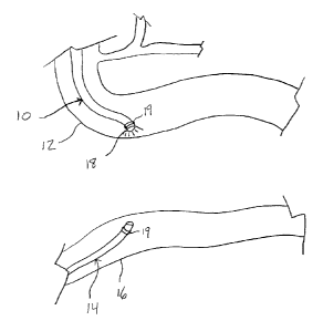
[0020] Figures 1 ¨ 7 illustrate steps in a method of the present invention;
[0021] Figure 8 is a cross-section of an embodiment of an anastomosis
device of the
present invention;
[0022] Figure 9 is a plan view of a component of the device of Figure 8;
[0023] Figure 10 is a cross-section of an embodiment of an anastomosis
device of the
present invention;
[0024] Figure 11 is a perspective view of a component of the device of
Figure 10;
[0025] Figure 12 is a cross-section of an embodiment of an anastomosis
device of the
present invention;
[0026] Figures 13 and 14 are depictions of anastomoses created by various
embodiments of the device of Figure 12;
[0027] Figures 15 ¨ 17 depict a deployment sequence of an embodiment of an
anastomosis device of the present invention;
[0028] Figures 18 ¨ 21 depict a deployment sequence of an embodiment of an
anastomosis device of the present invention;
[0029] Figures 22 ¨ 26 depict the use of a deployment device of the present
invention
being used to deploy an embodiment of the anastomosis device of the present
invention;
[0030] Figures 27 ¨ 29 depict a deployment sequence of an embodiment of an
anastomosis device of the present invention;
[0031] Figures 30 ¨ 33 depict expanded and collapsed configurations of an
embodiment
of an anastomosis device of the present invention;
¨6¨
CA 02911795 2015-11-12
[0032] Figure
34 is a perspective view of a collapsed configuration of an embodiment of
an anastomosis device of the present invention;
[0033] Figure
35 is a perspective view of an expanded configuration of the embodiment
of an anastomosis device of Figure 34;
[0034] Figure
36 is a perspective view of the anastomosis formed by the device of
Figures 34 and 35;
[0035] Figure
37 is a cross-sectional view of an embodiment of an anastomosis device
of the present invention;
[0036] Figure
38 is a cross-sectional view of an embodiment of a delivery device of the
present invention;
[0037] Figure
39 is a perspective view of an embodiment of an anastomosis device of
the present invention;
[0038] Figure
40 is a perspective view of an embodiment of an anastomosis device of
the present invention;
[0039] Figure
41 is a perspective view of an embodiment of an anastomosis device of
the present invention; and,
[0040] Figure 42 is a diagram showing various anastomosis sites.
DETAILED DESCRIPTION OF THE INVENTION
[0041] The
present invention includes a method of endoscopically creating an
anastomosis as well as several devices that can be used to form the
anastomosis. Figures
1-7 show a series of diagrams detailing the various steps of the method. The
remaining
figures depict several embodiments of various devices. By explaining the
method first, the
various embodiments of devices will be more easily understood. It is important
to note that
the method is described as forming an anastomosis between the duodenum and the
ileum.
¨7¨
CA 02911795 2015-11-12
These locations are provided by way of example only. One skilled in the art
will realize that
the sections of the digestive tract joined using the method of the present
invention is a
determination that is patient-dependent and is to be decided by a physician.
For example,
if a patient is extremely obese, it may be desired to an anastomosis between
the stomach
and the colon. Figure 42 shows several examples of anastomosis sites. Arrow 1
indicates
a duodenal-ileal anastomosis. Arrow 2 illustrates a duodenal-colic
anastomosis. Arrow 3
indicates a gastro-transverse colic anastomosis. Arrow
4 illustrates a gastro-colic
anastomosis. Not shown in this figure, but also envisioned, are jejenal-
jejenal anastomosis
and duodenal-jejenal anastomosis.
Method
[0042]
Referring first to Figure 1, the present invention provides a method by which
an
endoscope 10 is advanced from the stomach into the duodenum 12. A second
endoscope
14 is advanced from the large intestines into the ileum 16. The normal anatomy
in a human
is such that the third section of the duodenum 12 is in close proximity to the
ileum 16 and
thus if either structure is illuminated from within it can readily be seen
from the other.
Hence, the duodenum 12, for example, is illuminated with a light 18 at the tip
of the first
endoscope 10 and the light can be seen with an endoscope 14 in the ileum 16.
Alternatively or additionally, each endoscope 10 and 14 may be equipped with a
strong
magnet 19. The magnets 19 would then be able to automatically align and
connect the two
endoscopes 10 and 14 when in operational proximity to each other's magnetic
fields.
[0043] Next,
as shown in Figure 2, the ileum 16 is gently maneuvered such that it is
touching the duodenum 12. Though it is preferably to maneuver the ileum 16,
one skilled in
the art will understand that the duodenum 12 may be maneuvered as well,
although to a
lesser degree as this organ is not as mobile.
[0044]
Referring to Figure 3, once intimate contact has been confirmed from within
the
duodenum 12 and the ileum 16, a wire 20 is passed through the touching walls
of the
duodenum 12 and the ileum 16. This may be performed in a variety of ways. For
example,
the endoscope 10 may have a working channel containing a hollow needle, which
may be
¨8--
CA 02911795 2015-11-12
advanced to pierce the duodenum and ileum, and then the wire 20 may be passed
through
the needle. Alternatively, the wire 20 may be sharpened and act as a needle.
Once
passed through to the ileum, the wire 20 may enter a working channel of the
second
endoscope 14, obviating the need to re-navigate the small and large
intestines.
Alternatively, a sheath may be advanced over the endoscope 14 and the
endoscope
retracted as the wire is advanced until the distal end exits the rectum.
[0045] Referring to Figure 4, next first and second components 32 and 34 of
an
anastomosis device 30 are advanced over either end of the wire 20. Components
32 and
34 are depicted as generic blocks in these figures as the method is not to be
limited to any
single device. Rather, several embodiments of anastomosis devices are
described below
under the heading "Devices."
[0046] In Figure 5, the first and second components 32 and 34 of
anastomosis device
30 are drawn together and locked. Locking force may be provided by pushing on
the first
component 32 from the direction of the stomach, while pulling the second
component 34. It
is also envisioned that if the endoscopes 10 and 14 are equipped with strong
magnets 19,
the first and second components 32 and 34 may be placed around the end of the
endoscopes 10 and 14, distal of the magnets 19. This way, the magnets 19 may
be used
to provide the force necessary to lock the two components 32 and 34 together.
It is also
contemplated that this embodiment may completely obviate the need for the wire
20. It is
further contemplated that the magnets 19 could serve as the first and second
components
32 and 34. In this embodiment, the magnets are held onto the ends of the
endoscopes 10
and 14 with an adhesive or magnetic force that is easily overcome by the
attractive force
between the two magnets 19.
[0047] Once the components 32 and 34 are locked, the wire 20 may be
removed, as
shown in Figure 6. Locking the first and second components 32 and 34 of the
anastomosis
device 30 together creates intimate contact between the serosal surfaces of
the duodenum
12 and the ileum 16. The configuration of the contacting tissue can be
generally circular,
elliptical, diamond-shaped, elongate, or shaped like a cross, depending on the
¨9¨
CA 02911795 2015-11-12
configuration of the device 30. During the healing period the tissue is
compressed, limiting
or eliminating circulation, and the tissue becomes necrotic. The tissue around
the outside
of the anastomosis device is compressed at a lower force. This tissue forms a
ring of
healed tissue. Over time, an anastomosis 40 (Figure 7, for example) is formed
and the
device 30 is allowed to pass through the digestive system. There is no flow
through the
anastomosis 40 during the healing period. All chyme flows through the natural
distal
duodenum and, due to the relatively low profile of the various devices 30,
thus there is little
risk of obstructing flow. Human serosal tissue that is placed in intimate
contact has been
shown to heal within 7 days.
Devices
[0048] Referring now to Figures 8 and 9 there is shown an embodiment of an
anastomosis device 50 of the present invention. The device 50 includes a first
component
52 and a second component 54 that is configured to mate with the first
component 52. The
components 52 and 54 are generally circular or oval in shape. The second
component 54
is basically a cylindrical wall that forms a lip 56 at a mating end 58. The
wall is divided into
a plurality of fingers 60 by slots 62 that extend nearly to a non-mating end
64. The slots 62
are necessary to allow the fingers 60 to flex inward when the second component
54 is
being connected to the first component 52. The flexibility of the fingers 60
may be varied
by varying the length of the slots 62.
[0049] The first component 52 is a cylindrical cap that includes an
inwardly protruding lip
66 that forms a snap-fit with the lip 56 of the second component 54.
Preferably, the first
component 52 further includes a tapered extension 58 from the lip 56. The
tapered
extension 68 places a varying amount of squeezing force on the tissues of the
duodenum
and ileum such that some tissue necroses while adjacent serosal tissue heals
and fuses
together.
[0050] Figure 10 shows another embodiment of an anastomosis device 70 of
the
present invention. The device 70 includes a first component 72 and a second
component
74 that is configured to mate with the first component 72. The components 72
and 74 are
¨10¨
CA 02911795 2015-11-12
generally circular or oval in shape. Alternatively, the second component 74
could be one
shape, such as circular, while the first component 72 could be another shape
such as
spoked.
[0051] For example, Figure 11 shows a spoked embodiment of the first
component 72.
Generally, the first component 72 includes a center spike 76 with one or more
barbs 78.
The first component 72 also includes one or more downward extensions 80 that
include
inward mating surfaces 82 configured to mate with the second component 74.
These
inward mating surfaces 82 ensure there is sufficient downward force on the
radial extents
of the first component 72 to induce necrosis.
[0052] The second component 74 is basically a cylindrical wall that
includes an annular
indentation 84 that accepts the inward mating surfaces 82 of the first
component 72. The
second component 74 further includes one or more disks 86 each defining a hole
88
through which the spike 76 is inserted when the components 72 and 74 are
connected.
The barbs 78 act against the disks 86 to lock the first component 72 to the
second
component 74.
[0053] Figures 12-14 show another embodiment of an anastomosis device 90 of
the
present invention. The anastomosis device 90 includes a first component 92 and
a second
component 94. The first component 92 is a cap-like plate with a spike 96
extending
downwardly therefrom and having a plurality of barbs 98. At its radial
extents, the first
component 92 includes a short peripheral wall 100 that terminates in a shaped
surface 102.
The shaped surface 102 is designed to create a necrosis zone 104 and a healing
zone 106,
due to the varying pressures exerted by the shaped surface 102 on tissue
sandwiched
between the first and second components 92 and 94.
[0054] The second component 94 is basically a disk that defines a center
hole 108 for
accepting the spike 96 and providing a surface against which the barbs 98 can
act. The
second component 94 is stiff enough to exert a squeezing force on tissue when
connected
to the first component 92. The device 90 may be a variety of shapes, including
circular and
oval. A circular embodiment is advantageous in that it allows automatic
alignment of the
¨11¨
CA 02911795 2015-11-12
two components 92 and 94 once the spike 96 is inserted into the center hole
108.
However, a more elongate shape, such as an oval, may be more anatomically
suited to the
elongate configuration of the digestive tract.
[0055] Figures 13 and 14 show the resulting necrosis and healing zones 104
and 106
that result from circular and oval embodiments of the device 90. Because the
inner
necrosis zone 104 is continuous, only a thin band of tissue needs to necrose
in order to
create a comparatively large anastomosis.
[0056] Figures 15¨ 17 are sequential depictions of the deployment of an
embodiment of
an anastomosis device 110 of the present invention. The anastomosis device 110
includes
a first component 112 and a second component 114. The first and second
components
112 and 114 are both expandable mesh, umbrella-like devices that have
collapsed and
expanded configurations. It is envisioned that the first component 112 may
collapsed to a
point where it is possible to use the first component 112 to puncture through
the duodenum
and the ileum, possibly obviating the need to extend a guidewire 20 from the
mouth to the
rectum. Figure 15 shows the first component 112 passing through two layers of
tissue.
Figure 16 shows the first and second components 112 and 114 being expanded.
Figure 17
shows the fully expanded first and second components 112 and 114 being
compressed
against each other, thereby compressing the tissue therebetween to induce
necrosis and
create an anastomosis. The device 110 (and all of the devices described
herein) may be
constructed of any suitable material having sufficient strength to cause
necrosis, such as
stainless steel or Nitinol, for example. A fully expanded dome shape, such as
that shown in
Figure 17 is preferable to ensure sufficient strength at the periphery of the
device 110.
[0057] Figure 18 shows another embodiment of an anastomosis device 120 of
the
present invention. The device 120 includes a first component 122 and a second
component 124. The first component 122 is an elastomeric o-ring or band. The
second
component 124 is a continuous wall (such as a cylindrical wall, oval wall,
elliptical wall or
any desired shape) that includes an overhanging lip 126 around which the first
component
is stretched and secured, trapping tissue therebetween.
¨ 12 ¨
CA 02911795 2015-11-12
[0058] Figures 19 - 21 show a sequence of the first component 122 being of
device 120
being attached to the second component. In Figure 19, the second component 124
has
been positioned against a layer of tissue, such as the inside wall of the
ileum 16. Figure 20
shows the second component 124 being pushed against the inside wall of the
ileum 16
such that the ileum 16 comes in contact with another layer of tissue, such as
that from the
duodenum 12. Figure 21 shows that the first component 122 has been stretched
over the
lip 126 of the second component 124, locking the tissue 12 and 16 around the
device.
[0059] One advantage to using an anastomosis device with an elastomeric
component
providing squeezing force is that the force provided by an elastomeric
component remains
somewhat constant, even after necrosis begins to set in. In other words, if a
mechanical
device is used having first and second components that are a fixed distance
from each
other, the pressure placed on the tissue decreases as the tissue necroses and
shrinks. An
elastomeric component, on the other hand, will shrink with the tissue and
continue to apply
pressure.
[0060] Figures 22 ¨ 26 depict the use of a delivery device 130 of the
present invention
that may be used to connect the two components 122 and 124 of the anastonnosis
device
120. The delivery device 130 includes a centered spike 132 attached to the
second
component 124 and in the center thereof. The centered spike 132 is hollow and
able to be
advanced over the guidewire 20. The centered spike protrudes from the second
component 124 such that when the second component 124 contacts tissue walls 12
and
16, the centered spike 132 pierces through the tissue and is available for
connecting to a
receiving tube 134 on the other side of the tissue walls 12 and 16. This is
best shown in
Figures 22 and 23.
[0061] Referring to Figure 24, it is shown that the delivery device 130
also includes a
cone 136 that is able to slide over the receiving tube 134 and remain centered
thereon.
Because the centered spike 132 is concentric with the second component 124,
and
because the receiving tube 134 is concentric with both the centered spike 132
and the cone
136, the cone 136 may be advanced over the second component 124 without
concern for
¨13¨
CA 02911795 2015-11-12
alignment. Once the cone 136 is advanced over the second component, as shown,
the
elastomeric o-ring 122 may be stretched over the cone. This may be
accomplished, for
example, via a pusher sheath.
[0062] Figure 25 shows that the o-ring 122 has been advanced over the cone
136 and is
in place under the lip 126 of the second component 124. Figure 26 shows the
device 120
in place after the delivery device 130 and the guidewire 20 have been removed.
[0063] Figures 27 ¨ 29 show an embodiment of an anastomosis device 140. The
device
140 includes a first component 142 and a second component 144. The first
component
142 includes a cup 146 connected to an anchor bar 148 with an elastic
connector 150. The
elastic connector 150 may be an elastomeric band or a spring.
[0064] The second component 144 is a cylinder, preferably with a rounded
edge 152
that makes contact with tissue. In operation, as seen in the sequence shown in
Figures 27
¨ 29, the first component 142 is advanced through a first digestive passage,
such as the
duodenum, while the second component 144 is advanced through a second
digestive
passage, such as the ileum. The anchor bar 148 is aligned with the connector
150 and
both are passed through a small hole to the ileum and through the second
component 144.
The connector 150 is stretched sufficiently to allow the anchor bar 148 to
pass completely
through the second component and rotate to hold the second component 144
against the
cup 146 of the first component 142. Because the connector 150 is stretched,
constant
pressure is applied to the tissue trapped between the cup 146 and the first
component 142.
[0065] Figures 30 ¨ 33 show deployed and collapsed configurations of an
embodiment
of an anastomosis device 160 of the present invention. The device 160 includes
a first
component 162 and a second component, which is the essentially the same as the
first
component 162. The device 160 is similar to the device 110 of Figures 15 ¨ 17
except in
that a solid material is used instead of a mesh, thereby adding strength to
the device. The
component 162 is made up of a plurality of shaped plates 164 that slide
against each other
in order to transition from collapsed to deployed configurations. Figure 31
illustrates that in
¨14¨
CA 02911795 2015-11-12
the deployed configuration, the device 160 is domed, thereby providing
pressure against
the tissue at the periphery of the device 160.
[0066] Figures 34 ¨ 36 show deployed and collapsed configurations of an
embodiment
of an anastomosis device 170 of the present invention. The device 170 includes
a first
component 172 and a second component 174, which is the essentially the same as
the first
component 172. The device 170 demonstrates that shapes other than circular may
be
used to create anastomosis. The first and second components 172 and 174 of
device 170
each include a plurality of corresponding arms 176. Preferably, as shown in
Figure 35, in
the deployed configuration, the device 170 is domed, thereby providing
pressure against
the tissue at the distal ends of the arms 176. Figure 36 shows the resulting
anastomosis
40 created by the device 170. It is noted that the proximal side of the device
170, that is
the side of the device that is not passing through tissue, would not have to
be expandable.
[0067] Figure 37 shows an embodiment of an anastomosis device 180 of the
present
invention. The device 180 generally includes a first component 182, a second
component
184, and an elastic o-ring 186. The first component 182 has a center pin 188
extending
downwardly therefrom and is shaped to include a ramp 190 and a ledge 192,
which are
used to stretch and contain the o-ring 186. The center pin 188 also may
include a distal
bulb 198 for use in conjunction with a delivery device such as the device 200
shown in
Figure 38.
[0068] The second component 184 is a disk defining a center hole 194 for
accepting the
pin 188 and may include an annular pressure ridge 196 for exerting pressure on
the
compressed tissue between the two components 182 and 184. The elastomeric o-
ring 186
functions to lock the two components 182 and 184 together and also to exert
steady force
on the second component 184.
[0069] Figure 38 depicts an embodiment of a delivery device 200 of the
present
invention that may be used to connect two components of a delivery device
together,
wherein the second component is an o-ring. The delivery device 200 works in
conjunction
with a guidewire 20, which passes through the delivery device and through a
first
¨15¨
CA 02911795 2015-11-12
component 202 of an anastomosis device. The device 200 generally includes a
grabbing
device 204, which is used to grab a bulb 206 of the first component 202. The
grabbing
device 204 can then be used to pull the first component into the delivery
device 200, which
contains the second component, o-ring 208. It is understood that by pulling
the first
component 202 into the device 200, tissue is being trapped between the two
components
202 and 208.
[0070] The o-ring 208 is being held in an expanded state by an inner sheath
210. Once
the first component 202 is in place such that a receiving indentation 212 is
aligned with the
o-ring 208, the sheath 210 is retracted, thereby releasing the o-ring.
[0071] Figures 39 and 40 show an embodiment of an anastomosis device 220 of
the
present invention. The device 220 includes a first component 222 and a second
component 224. Both components 222 and 224 have a domed, clam-shell like
design
supported by braces 226. The first component 222 also includes a barbed spike
228. The
barbed spike 228 preferably extends from an inside surface of the clam shell
and slides
through an opening supported by the braces 226. The spike 228 is configured to
pass
through a similar opening 230 supported by the braces 226 of the second
component 224.
The two components 222 and 224 can thus be compressed together, as shown in
Figure
40. Each component 222 and 224 could be compressible or made of a material
such as
Nitinol that expands after reaching the implant site. In order to more easily
deliver the
components 222 and 224, the components may be equipped with attachment points
232
for a guide wire, and may also contain loops 234, through which the guide wire
or auxiliary
mandrel passes in order to maintain the components 222 and 224 in a sideways
configuration while navigating through the digestive tract to the target site.
The loops 234
are released upon reaching the target site, thereby allowing the components
222 and 224
to face each other. Though the curved "shell" portions of each component 222
and 224 are
shown as being solid, the device 220 would function with a mesh or skeletal
shell as well.
[0072] Figures 10-12 and 39 show embodiments of devices held together with
barbed
spikes. Figure 41 shows an alternative attachment mechanism 240. This
mechanism 240
¨16¨
CA 02911795 2015-11-12
includes a male component 242 and a female component 244. The male component
242
is a plug that has an circumferential lip 246. The female component 244
includes a plurality
of inward-projecting tabs 248. As the male component 242 is inserted into the
female
component 244, the tabs 248 expand over the lip 246 such that the male
component 242
cannot be retracted from the female component 244.
[0073]
Although the invention has been described in terms of particular embodiments
and applications, one of ordinary skill in the art, in light of this teaching,
can generate
additional embodiments and modifications without departing from the spirit of
or exceeding
the scope of the claimed invention. Accordingly, it is to be understood that
the drawings
and descriptions herein are proffered by way of example to facilitate
comprehension of the
invention and should not be construed to limit the scope thereof.
¨ 17 ¨