Note: Descriptions are shown in the official language in which they were submitted.
HERNIA MODEL
Field of the Invention
[0002] This application relates to surgical training tools, and in
particular,
to simulated tissue structures and models for teaching and practicing the
repair of a
hernia.
Background of the Invention
[0003] A hernia is the protrusion of an organ or the fascia of an
organ
through the abdominal wall. This occurs when the abdominal walls weaken either
from
incorrect formation at birth, recent surgery or trauma. The most common types
of
hernias are inguinal and incisional. Inguinal hernias occur in the groin area
in both
males and females but they are most common in men to the right and left of the
midline
where the spermatic cords and arteries pass through the spaces in the
abdominal wall.
There are three possible spaces in the abdominal wall for the hernia to pass
through:
direct, indirect and femoral. The direct space is medial to the epigastric
vessels while
the indirect space is lateral to the epigastric vessels. A femoral hernia
occurs when the
organs protrude through a large femoral ring into the femoral canal.
Incisional hernias
occur after a surgery when the abdominal wall does not heal correctly, causing
the
internal organs and fascia to push through.
[0004] Hernias can be repaired by either open or laparoscopic
surgery. In
laparoscopic surgery, a trocar is inserted to access a body cavity and to
create a
channel for the insertion of a camera, such as a laparoscope. The camera
provides a
live video feed capturing images that are then displayed to the surgeon on one
or more
monitors. Another trocar is inserted to create a pathway through which
surgical
instruments can be passed for performing procedures observed on the monitor.
The
1
CA 291-2069 2019-05-15
CA 02912069 2015-11-09
WO 2014/186574 PCT/US2014/038195
targeted tissue location such as the abdomen is typically enlarged by
delivering carbon
dioxide gas to insufflate the body cavity and create a working space large
enough to
accommodate the scope and instruments used by the surgeon. The insufflation
pressure in the tissue cavity is maintained by using specialized trocars.
Laparoscopic
repair has many advantages over the traditional open surgery repair including
quicker
recovery and less pain. Therefore, it is often more desirable for the patient
to undergo a
laparoscopic repair. However, laparoscopic repair requires an experienced
surgeon. In
order for surgeons to practice laparoscopic hernia repairs, a realistic,
anatomically
correct model for use in a laparoscopic training device is needed.
[0005] Generally, there are two ways to repair an inguinal hernia
laparoscopically. The first and more often taught way is called transabdominal
pre-
peritoneal (TAPP). The TAPP approach involves placing the laparoscopic
instruments
all the way into the insufflated abdominal cavity and approaching the hernia
from below
by cutting a hole in the peritoneum. The hernia is then resected, mesh is
placed over
the weakened abdominal wall and the peritoneum is closed. The second way of
reducing an inguinal hernia is called total extraperitoneal (TEP). The TEP
approach is
more difficult since it involves entering the space between the peritoneum and
the
abdominal wall without puncturing the peritoneum. Once the trocar has been
inserted
into that space, a balloon is used to open up the space to allow for easier
movement of
the instruments and less blunt dissection. When the balloon is removed, the
space is
insufflated and the hernia is found in that same plane. When the hernia is
found, it is
resected back into the abdominal cavity, the peritoneum laid flat and mesh
placed over
the weakened abdominal wall. When surgeons are learning how to perform
laparoscopic surgery, they are taught TAPP first since like most other
laparoscopic
procedures, it is performed inside the abdominal cavity. TEP is considered
more
advanced and surgeons need a way to safely learn and practice the procedure.
Due to
the need for a safe practice model for both beginner surgeons learning TAPP as
well as
more advanced surgeons learning TEP, a hernia model that allows for both
procedures
to be practiced is needed.
[0006] In order to help patient outcomes and recoveries, surgeons
need a
way to practice laparoscopic hernia repairs outside of the operating room. The
practice
2
CA 02912069 2015-11-09
WO 2014/186574 PCT/US2014/038195
model needs to be anatomically correct and include all important landmarks
normally
seen during surgery in order to give the surgeon or resident the most
realistic practice
possible. Additionally, the model should allow the surgeon to practice
incisional and
inguinal (TAPP and TEP) procedures.
Summary of the Invention
[0007] According to one aspect of the invention, an anatomical model
for
surgical training is provided. The model includes a simulated abdominal wall
located at
a first end of the model. The simulated abdominal wall has an inner surface
and an
outer surface. The simulated abdominal wall includes at least one opening
extending
between the inner surface and the outer surface defining a hernia opening. The
model
includes a simulated peritoneum located at a second end of the model. The
simulated
peritoneum has an inner surface and an outer surface. The simulated peritoneum
is
connected and adjacent to the simulated abdominal wall such that the simulated
abdominal wall and the simulated peritoneum are substantially coplanar when in
an
open configuration and the inner surface of the simulated abdominal wall and
the inner
surface of the peritoneum together define a common inner surface and an
overall
flexible model. The model further includes a first layer of synthetic tissue.
The first
layer of synthetic tissue has a bottom surface and a top surface. The first
layer of
synthetic tissue overlays at least a portion of the simulated abdominal wall.
At least part
of the first layer is selectively adhered to the simulated abdominal wall and,
in another
variation, at least part of the first layer is adhered to the simulated
abdominal wall and to
the simulated peritoneum. The model further includes a plurality of simulated
tissue
components positioned between the first layer and the simulated abdominal
wall. At
least some of the simulated tissue components are adhered, at least in part,
to at least
one of the first layer, the simulated peritoneum, and the simulated abdominal
wall. The
model has a curved configuration. When in the curved configuration, part of
the
simulated abdominal wall is located above the simulated peritoneum and a
cavity is
defined between the simulated abdominal wall and the simulated peritoneum with
the
first end and the second end defining, in part, an opening into the cavity. In
one
3
CA 02912069 2015-11-09
WO 2014/186574 PCT/US2014/038195
variation, the model includes a spring layer that extends through the
simulated
abdominal wall and the simulated peritoneum.
[0008] According to another aspect of the invention, an anatomical
model
for surgical training is provided. The model includes a simulated abdominal
wall located
at a first end of the model. The simulated abdominal wall has an inner surface
and an
outer surface. The simulated abdominal wall has at least one opening extending
between the inner surface and the outer surface. The model includes at least a
portion
of a simulated pelvis that is located at a second end of the model. The
simulated pelvis
has an inner surface and an outer surface. The simulated pelvis is connected
and
adjacent to the simulated abdominal wall such that the inner surface of the
simulated
abdominal wall and the inner surface of the simulated pelvis define a common
inner
surface of the model. The model further includes a first layer of synthetic
tissue having
a bottom surface and a top surface. The first layer of synthetic tissue
overlays at least a
portion of the simulated pelvis and at least a portion of the simulated
abdominal wall.
The first layer of synthetic tissue is adhered to at least a portion of the
simulated pelvis
and to at least a portion of the simulated abdominal wall. The first layer
includes at
least one opening aligned with the at least one opening in the simulated
abdominal wall.
The model includes a second layer of synthetic tissue having a bottom surface
and a
top surface. The second layer of synthetic tissue overlays at least a portion
of the top
surface of the first layer. The second layer includes at least one opening
aligned with
the at least one opening in the simulated abdominal wall. The model further
includes a
plurality of simulated tissue components positioned between the first layer of
synthetic
tissue and the second layer of synthetic tissue. At least some of the
plurality of
simulated tissue components is adhered, at least in part, to at least one of
the first layer
of synthetic tissue and the second layer of synthetic tissue. The model
further includes
a synthetic peritoneum overlaying at least one of the simulated abdominal wall
and the
simulated pelvis and is located above the second layer of synthetic tissue. At
least a
portion of the synthetic peritoneum is removably pushed into one of the
openings in the
simulated abdominal wall to simulate a hernia.
[0009] According to another aspect of the invention, a surgical
simulation
system for practicing hernia repair is provided. The surgical simulation
system includes
4
CA 02912069 2015-11-09
WO 2014/186574 PCT/US2014/038195
a hernia model placed inside a surgical training device. The hernia model
includes a
simulated abdominal wall located at a first end of the model. The simulated
abdominal
wall has an inner surface and an outer surface. The simulated abdominal wall
has at
least one opening extending between the inner surface and the outer surface.
The
hernia model includes at least a portion of a simulated pelvis located at a
second end of
the hernia model. The simulated pelvis has an inner surface and an outer
surface. The
simulated pelvis is connected to the simulated abdominal wall such that the
inner
surface of the simulated abdominal wall and the inner surface of the simulated
pelvis
define a common inner surface of the model. The hernia model includes a first
layer of
synthetic tissue having a bottom surface and a top surface. The first layer of
synthetic
tissue overlays at least a portion of the simulated pelvis and at least a
portion of the
simulated abdominal wall. The first layer is adhered to at least a portion of
the
simulated pelvis and to at least a portion of the simulated abdominal wall.
The first layer
includes at least one opening aligned with the at least one opening in the
simulated
abdominal wall. The model further includes a second layer of synthetic tissue
having a
bottom surface and a top surface. The second layer overlays at least a portion
of the
top surface of the first layer. The second layer includes at least one opening
aligned
with the at least one opening in the simulated abdominal wall and the at least
one
opening in the first layer. The hernia model also includes a plurality of
simulated tissue
components positioned between the first layer of synthetic tissue and the
second layer
of synthetic tissue. At least some of the plurality of simulated tissue
components are
adhered, at least in part, to at least one of the first layer of synthetic
tissue and the
second layer of synthetic tissue. The model further includes a synthetic
peritoneum
overlaying at least a portion of the simulated abdominal wall and at least a
portion of the
simulated pelvis. The synthetic peritoneum is positioned above the second
layer of
synthetic tissue. The surgical training device includes a base and a top cover
connected to and spaced apart from the base to define an internal cavity. The
internal
cavity is at least partially obstructed from direct observation by a user and
is configured
for practicing laparoscopic surgical techniques. The top cover includes an
aperture or
penetrable simulated tissue region for the passage of surgical instruments
into the
internal cavity. The hernia model is positioned inside the internal cavity.
CA 02912069 2015-11-09
WO 2014/186574 PCT/US2014/038195
[0010] According to another aspect of the invention, a model that
allows
surgeons and residents to practice incisional and inguinal hernia repairs is
provided.
The model is a clam-shaped and simulates the insufflated space between the
abdominal muscles and peritoneum. A hole is provided in the model from which a
simulated peritoneum and/or simulated bowel protrudes to create a simulated
hernia.
The model contains all important anatomical structures including Cooper's
ligament, the
iliopubic tract, the pubic ramus bone, the medial umbilical ligament, the
triangle of
doom, triangle of pain and the spermatic cords. The model is covered with a
layer of
simulated tissue to allow users to practice dissecting in order to find and
navigate the
important anatomical landmarks and to safely repair the hernia. Additionally,
the model
is designed with a thick abdominal wall to allow the surgeon to practice
tacking mesh to
repair the hernia. Silicone is used to create the thick abdominal walls,
simulated
anatomical structures and synthetic tissue. A spring layer may be incorporated
to
provide realistic resiliency to the model while maintaining a simulated
insufflated space
configuration or curved configuration. The model may be used to selectively
simulate
direct, indirect and femoral inguinal hernia repairs as well as incisional
hernia repairs by
removably placing the protruding simulated tissue into any one of three
openings in the
model. The model sits on a base or frame that imparts and maintains the clam
shape or
is connected to a rigid simulated pelvis. When located inside a laparoscopic
trainer with
an angled top cover to simulate a Trendelenburg position of the patient, the
model
provides an ideal simulation for teaching and practicing laparoscopic hernia
repair.
Brief Description of the Drawings
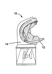
[0011] FIG. 1 is a side perspective view of a hernia model according
to the
present invention.
[0012] FIG. 2 is a front perspective view of a hernia model according
to the
present invention.
[0013] FIG. 3 is rear perspective view of a hernia model according to
the
present invention.
[0014] FIG. 4 is a top view of an anatomical portion of a hernia
model
according to the present invention.
6
CA 02912069 2015-11-09
WO 2014/186574 PCT/US2014/038195
[0015] FIG. 5 is a top view of an anatomical portion of a hernia
model
according to the present invention.
[0016] FIG. 6 is a top view of an anatomical portion of a hernia
model with
human hands shown retracting simulated tissue according to the present
invention.
[0017] FIG. 7 is a bottom perspective view of an anatomical portion
of a
hernia model according to the present invention.
[0018] FIG. 8 is bottom view of an anatomical portion of a hernia
model
according to the present invention.
[0019] FIG. 9 is a top perspective view of a frame of a hernia model
according to the present invention.
[0020] FIG. 10 is a side perspective view of a laparoscopic trainer.
[0021] FIG. 11 is a side perspective view of a laparoscopic trainer
with an
angled top cover.
[0022] FIG. 12 is a side perspective view of a laparoscopic trainer
with a
hernia model according to the present invention.
[0023] FIG. 13 is a rear perspective view of a laparoscopic trainer
with a
hernia model according to the present invention.
[0024] FIG. 14 is a front perspective view of a laparoscopic trainer
with a
hernia model according to the present invention.
[0025] FIG. 15 is a front perspective view of a hernia model with
human
hands shown retracting simulated tissue according to the present invention.
[0026] FIG. 16 is a front perspective view of a hernia model with
human
hands shown retracting simulated tissue according to the present invention.
[0027] FIG. 17 is a front top perspective view of a hernia model
according
to the present invention.
[0028] FIG. 18 is a rear top perspective view of a hernia model
according
to the present invention.
[0029] FIG. 19 is a top view of a hernia model according to the
present
invention.
[0030] FIG. 20 is a top view of a hernia model according to the
present
invention.
7
CA 02912069 2015-11-09
WO 2014/186574 PCT/US2014/038195
[0031] FIG. 21 is a rear top perspective view of a hernia model
according
to the present invention.
Detailed Description of the Invention
[0032] Referring to FIGs. 1-3, there is shown a side, front and rear
view,
respectively, of a hernia model 10 according to the present invention. The
hernia model
includes an anatomical portion 12 supported by a frame 14. As seen most
clearly in
FIG. 1, the substantially planar anatomical portion 12 is maintained in a
curved
configuration such that the major part of the anatomical portion 12 is
substantially C-
shaped forming a half or open generally cylindrical configuration. The
concavity formed
inside the C-shaped disposition of the anatomical portion 12 advantageously
simulates
an insufflated space between an artificial muscular abdominal wall generally
located at
the top of the C shape and the simulated peritoneum 18 generally located at
the bottom
of the C shape. The simulated muscular abdominal wall forms approximately the
top
half or more than the top half of the C-shaped curve; whereas, the bottom half
or less
than the bottom half of the C-shaped curve is formed by the simulated
peritoneum 18.
The open clamshell-like configuration advantageously provides a realistic
surgical
approach to repairing a hernia when viewed by the user from the front of the
hernia
model 10 as in FIG. 2.
[0033] The frame or stand 14 divides the hernia model 10 into an
upper
portion and a lower portion. The lower portion constitutes approximately one-
third of the
entire height of the hernia model 10 and simulates the abdominal cavity
beneath the
peritoneum. The lower portion contains that part of the anatomical portion 12
such as
the simulated bowel that protrudes through the simulated peritoneum 18 and
through
the simulated muscular abdominal wall. The upper portion contains the
anatomical
portion 12. FIGs. 1-3 illustrate a simulated bowel residing in the lower
portion and
extending upwardly through an opening in the peritoneum 18 into the concavity
of the
upper portion. The simulated bowel crosses the concavity of the insufflated
space and
exits through an opening in the muscular abdominal wall to simulate a hernia.
One or
more exit openings in the simulated muscular abdominal wall of the anatomical
portion
12 is provided to simulate the possible spaces in the abdominal wall for the
hernia to
8
CA 02912069 2015-11-09
WO 2014/186574 PCT/US2014/038195
pass through. Generally, there are three spaces through which a hernia may
pass.
These spaces are the direct space, the indirect space and the femoral space.
If all
three openings are provided in the hernia model, the distal end of simulated
bowel is
inserted into any one of the exit openings for practicing hernia repair
through any of the
three spaces. The surgeon practices approaching the simulated insufflated
space of
the hernia model 10 from the front, either from below the peritoneum or above
the
peritoneum for practicing TAPP or TEP, respectively. The surgeon visualizes
the
insufflated space, practices carefully dissecting simulated fascia layers,
identifying a
variety of visual anatomical markers, navigating around them to approach the
bowel,
resecting the hernia and placing mesh to patch and close any spaces.
[0034] The anatomical portion 12 of the hernia model 10 will now be
described in detail with reference to FIGs. 4-8. Turning to FIG. 4, there is
shown a top
view of an anatomical portion 12 of the hernia model 10. The anatomical
portion 12 is a
substantially planar object having varying thickness and materials. The
anatomical
portion 12 includes a simulated muscular abdominal wall portion 16
interconnected in
substantially the same plane to a simulated peritoneum portion 18. Aside from
the
relatively thicker abdominal wall portion 16 relative to the peritoneum
portion 18, both
the abdominal wall portion 16 and peritoneum portion 18 are substantially
coplanar. In
human anatomy, the layers of the abdominal wall are from superficial to deep:
1) skin,
2) fascia, 3) muscle, which includes the rectus abdominis, external oblique
muscle,
internal oblique muscle and transverse abdominal muscle, 4) fascia
transversalis, and
5) peritoneum. These abdominal layers are sandwiched or layered above each
other to
form part of the abdominal wall portion 16. In the present invention, one or
more layers
representing muscle are positioned substantially coplanar with or otherwise
adjacent to
the simulated peritoneum portion. In this arrangement, the top side (anterior
facing
surface) of the simulated peritoneum 18 is substantially coplanar or adjacent
to the
bottom side (posterior facing surface) of the simulated muscular abdominal
wall portion
16 such that when the substantially planar anatomical portion 12 is curved
into a C-
shape configuration the bottom side of the simulated muscular abdominal wall
portion
16 faces and is spaced apart from the top side of the simulated peritoneum 18.
The
interior portion of the C-shaped structure simulates an insufflated space. In
real
9
CA 02912069 2015-11-09
WO 2014/186574 PCT/US2014/038195
surgery, the insufflated space is created by inserting a trocar between the
muscle layer
and peritoneum and delivering fluid such as carbon dioxide gas under pressure
from the
proximal end of the trocar to the distal end of the trocar to spread apart the
muscle layer
from the peritoneum to create a working space. The simulated insufflation
cavity of the
present invention is the concavity of the C-shaped orientation which is
approximately 5
inches in height and approximately 10 inches in length. As can be seen in FIG.
4, the
simulated muscular abdominal wall portion 16 is approximately 8 inches long
and
approximately 7.5 inches wide and is adjacent to the simulated peritoneum 18
which is
approximately 3 inches long and approximately 7.5 inches wide. When formed
into a
clamshell configuration, the simulated muscular abdominal wall portion 16 is
disposed
at the top of the hernia model 10 and follows the C-shaped curve down beyond
the
halfway mark of the C-shape. The simulated peritoneum 18 is disposed at the
bottom
of the C-shape and curves upwardly approximately a third of the way along the
C-shape
when the anatomical portion 12 is formed into a clamshell. Overall, the
substantially
planar anatomical portion 12 is approximately 7.4 inches wide and
approximately 11
inches long. The anatomical portion 12 further includes a simulated fascia
layer 20
located on the inner surface of the anatomical portion 12. The simulated
fascia layer 20
is a thin layer that is partially translucent and draped over the simulated
muscular
abdominal wall 16. The simulated fascia layer 20 is glued with adhesive in one
or more
locations and generally does not extend to completely over the simulated
peritoneum 18
when laid flat as shown in FIG. 4. The simulated peritoneum 18 includes an
opening 22
simulating the location of a ruptured peritoneum through which a simulated
bowel 24
protrudes above the inner or top surface of the peritoneum 18. The simulated
bowel 24
is part of the anatomical portion 12 although it is loosely connected thereto
such that the
simulated bowel 24 may be moved, pulled and pushed through the opening 22 and
other spaces.
[0035] Turning to FIG. 5, there is shown a top view of the anatomical
portion 12 with the simulated fascia layer 20 uncovering the underlying
simulated
muscular abdominal wall 16. Various anatomical structures are provided on the
surface
of the simulated muscular abdominal wall 16. These landmarks include but are
not
limited to Cooper's ligament 72, vas deferens 88, external iliac vessels 74,
76,
CA 02912069 2015-11-09
WO 2014/186574
PCT/US2014/038195
spermatic vessels 78, 80, nerves 90, and iliopubic tract 86 arranged as
labeled in FIG.
5. A piece of hard plastic (not shown) may also be embedded to simulate a
femoral
bone. In addition to opening 22 in the simulated peritoneum 18, one or more
additional
openings are formed through the simulated muscular abdominal wall 16. These
additional openings define exit openings or spaces through which the bowel
protrudes
in a hernia. In FIG. 5, a first opening 26 and a second opening 28 are formed
through
the simulated muscular abdominal wall 16 to simulate the direct space and
indirect
space, respectively. FIG. 6 illustrates the first and second openings 26, 28
more
clearly. Also visible in both FIGs. 5 and 6 is the curved intersection between
the
simulated muscular abdominal wall 16 and the simulated peritoneum 18. The
simulated
bowel 24 is passed through the opening 22 in the simulated peritoneum 18 such
that
the distal end resides above the inner surface and at least a portion of the
simulated
bowel 24 is above the top surface of the peritoneum 18. The distal end of the
simulated
bowel 24 is then passed into either of the first opening 26 or second opening
28 to
simulate a hernia located in the direct or indirect space, respectively. In
FIG. 4, the
simulated bowel 24 is shown passed into the second opening 28 representing the
indirect space. The hernia model 10 simulates a portion of the anatomy lateral
to the
midline 45 of a patient.
[0036] Turning
now to FIGs. 7 and 8, there is shown a perspective and
bottom view of the outer surface of the anatomical portion 12. The anatomical
portion
12 is built upon a layer of flexible wire mesh 30 such as chicken wire. The
wire mesh
material 30 is made of thin, flexible galvanized steel wire crisscrossing to
form small
square or other-shaped windows. The outer surface of the wire mesh layer 30 is
covered with a first layer of silicone 32 which is glued to the wire mesh
layer 30. The
inner surface of the wire mesh layer 30 is covered with a second layer of
silicone 34
sandwiching the wire mesh layer 30 between the first and second layers of
silicone 32,
34 forming the simulated muscular abdominal wall 16 at one end of the
anatomical
portion 12. At the other end of the anatomical portion 12, the inner surface
of the wire
mesh 30 is covered with a yellow foam layer 36 forming the simulated
peritoneum 18.
The yellow foam layer 36 that is approximately 1/16 of an inch thick is
adhered to inner
surface of the mesh layer with adhesive with the outer edges of the yellow
foam layer
11
CA 02912069 2015-11-09
WO 2014/186574 PCT/US2014/038195
36 being wrapped over the outer edges of the mesh layer 30. The yellow foam
layer 36
forms the finished inner surface of one end of the anatomical portion 12. The
simulated
muscular abdominal wall 16 comprising the first and second silicone layers 32,
34 and
wire mesh layer 30 is approximately 0.75 inches thick. The same wire mesh
layer or
frame 30 extends throughout the anatomical portion 12 defining the general
plane of the
anatomical portion 12. The simulated peritoneum 18 is substantially thinner
than the
simulated muscular abdominal wall 16 although still generally coplanar and
adjacent to
the simulated abdominal wall 16. The thick simulated muscular abdominal wall
16
permits the surgeon to tack surgical mesh to the abdominal wall to practice
patching the
hernia.
[0037] With reference back to FIGs. 5-6, the inner surface of the
second
silicone layer 34 is populated with a variety of anatomical landmarks as
mentioned
above. The second silicone layer 34 is textured and additional silicone layers
may be
employed above the second layer 34 to complete the anatomical geography. The
tubular simulated vessels and nerves are made of silicone and have diameters
of
approximately 0.185 inches. The simulated Cooper's ligament 72, iliopubic
tract 86 and
vas deferens 88 are also made of silicone and have diameters of approximately
0.25
inches. The thick external iliac vessels 74, 76 are made of silicone and have
a diameter
of approximately 0.25-0.375 inches. These tubular structures are made by
pouring
uncured silicone into straw like tubes and removed them after they solidify.
The
simulated bowel 24 is made from a thin layer of pink-colored silicone. The
silicone
comprising the iliopubic tract 86, Cooper's ligament 72 and vas deferens 88 is
colored
white, the nerves are colored yellow, the external iliac vein 74 and spermatic
vein 78 are
blue, the external iliac artery 76 and the spermatic artery 80 are red and the
remaining
vessels are red or pink.
[0038] Turning now to FIG. 9, there is shown a perspective view of a
frame
14 configured to hold the anatomical portion 12 of the hernia model 10
according to the
present invention. The frame 14 includes a rectangular lower frame portion 38
and an
upper frame receiving portion 40. The lower frame portion 38 is configured to
house
excess simulated bowel 24 that is simulated to reside below the peritoneum.
The lower
frame portion 38 includes a base and two or more upwardly extending side walls
to form
12
CA 02912069 2015-11-09
WO 2014/186574 PCT/US2014/038195
a rectangular container with a top wall. At least one opening is provided, for
example
via an open side, into the lower frame portion 38. The upper frame portion 40
is
configured to receive the anatomical portion 12 and retain the anatomical
portion 12 in a
clamshell or C-shaped orientation. As such, the upper frame portion 40
includes a C-
shaped receiving portion to receive and retain the anatomical portion in a C-
shaped
configuration. In FIG. 9, the C-shaped receiving portion is formed by two
upwardly
extending C-shaped claws or prongs 42, 44 that are attached to a top wall of
the lower
frame portion 38. Any number of C-shaped prongs 42, 44 including a wide
singular
prong may be employed to retain the anatomical portion 12. The lower frame
portion 14
is approximately 10.5 inches wide, approximately 4 inches deep and 3.5 inches
tall.
The C-shaped prongs 42, 44 are approximately 6 inches in height and each have
a
concavity that is approximately 4 inches deep.
[0039] As described above, the anatomical portion 12 is substantially
planar and made of flexible silicone, flexible foam and flexible wire mesh.
The wire
mesh layer 30 advantageously imparts the anatomical portion 12 with a
resiliency that
permits the planar anatomical portion 12 to be bent into a substantially semi-
cylindrical
or C-shaped configuration and placed into the C-shaped receiving prong(s) of
the frame
14. The mesh layer 30 acts as a spring layer such that when the anatomical
portion 12
is bent and inserted into the frame 14, it exhibits a biasing force against
the frame 14
advantageously keeping the anatomical portion 12 in position. Removability of
the
anatomical portion 12 allows for interchangeability of the anatomical portion
12 after it
has been used several times for replacement, repair, reconstruction and
compact
transport. When the anatomical portion 12 is removed from the frame 14, the
resilient
mesh layer 30 aids in springing the anatomical portion 12 back to its
substantially planar
orientation. Hence, the mesh spring layer advantageously keeps the silicone
and foam
layers 32, 34 and 36 from collapsing onto itself while in the clam shape.
[0040] Although the hernia model 10 is described above to be
comprised
of an anatomical portion 12 that is separate from the frame 14, one skilled in
the art will
recognize that, in an alternative variation, the hernia model 10 can be
constructed such
that the frame 14 and anatomical portion 12 is formed integrally as one piece.
Furthermore, although the hernia model 10 of the present invention may be used
to
13
practice hernia repair in a simulated open surgical procedure, the hernia
model 10 is
also advantageously configured for practicing laparoscopic hernia repair, in
particular,
employing the TEP approach. As such, the hernia model 10 of the present
invention is
configured to function together with a specialized laparoscopic trainer which
will now be
discussed in detail.
[0041] Turning now to FIG. 10, there is shown a laparoscopic
trainer 46.
The laparoscopic trainer 46 is described in co-pending U.S. Patent Application
Serial
No. 13/248,449 entitled "Portable laparoscopic trainer" and filed on September
29, 2011
by Pravong et al. to Applied Medical Resources Corporation and published as
U.S.
Patent Publication No. 2012/0082970.
The laparoscopic trainer 46 includes a top cover 48 connected to a base 50 by
a pair of legs 52 spacing the top cover 48 from the base 50. The laparoscopic
trainer
46 is configured to mimic the torso of a patient such as the abdominal region.
The top
cover 48 is representative of the anterior surface of the patient and the
space between
the top cover 48 and the base 50 is representative of an interior of the
patient or body
cavity where organs reside. The laparoscopic trainer 46 is a useful tool for
teaching,
practicing and demonstrating various surgical procedures and their related
instruments
in simulation of a patient. Surgical instruments are inserted into the cavity
through pre-
established apertures 58, 60 in the top cover 48. These pre-established
apertures may
include seals that simulate trocars or may include simulated tissue 60 that
simulates the
patient's skin and abdominal wall portions. Various tools and techniques may
be used
to penetrate the top cover 48 to perform mock procedures on model organs
placed
between the top cover 48 and the base 50 such as the hernia model 10. When
placed
inside the cavity of the trainer 46, the hernia model 10 is generally obscured
from the
perspective of the user who can then practice performing surgical techniques
laparoscopically by viewing the surgical site indirectly via a video feed
displayed on a
video monitor.
[0042] A video display monitor 54 that is hinged to the top cover
48 is
shown in a closed orientation in FIG. 10 and in an open orientation in FIGs.
11-14. The
video monitor 54 is connectable to a variety of visual systems for delivering
an image to
the monitor 54. For example, a laparoscope inserted through one of the pre-
established
14
CA 2912069 2019-05-15
CA 02912069 2015-11-09
WO 2014/186574 PCT/US2014/038195
apertures 58, 60 or a webcam located in the cavity and used to observe the
simulated
procedure can be connected to the video monitor 54 and/or a mobile computing
device
to provide an image to the user. In another variation, the top cover 48 does
not include
a video display but includes means for supporting a laptop computer, a mobile
digital
device or tablet such as an IPAD and connecting it by wire or wirelessly to
the trainer
46.
[0043] When assembled, the top cover 48 is positioned directly above
the
base 50 with the legs 52 located substantially at the periphery and
interconnected
between the top cover 48 and base 50. The top cover 48 and base 50 are
substantially
the same shape and size and have substantially the same peripheral outline.
Although
the trainer 46 has no sidewalls, the legs 52 partially obscure the internal
cavity from
view from an otherwise open-sided trainer 46. The top cover 48 includes a
first insert
56 removable and replaceable with respect to the top cover 48, in particular,
insertable
into and removable from an opening formed in the top cover 48. The first
insert 56
includes a plurality of apertures 58 to serve as fixed insertion ports for a
variety of
instruments. The apertures 58 may include various seals. The first insert 56
also
includes a tissue simulation region 60 for simulating the skin or several
layers of tissue.
In one embodiment, the tissue simulation region 60 is configured as a second
insert
provided within the first insert 56. The second insert is removable and
replaceable via
snap-fit, friction fit or threaded engagement or other means with respect to
the top cover
48 or with respect to the first insert 56 if provided.
[0044] Turning now to FIG. 11, the laparoscopic trainer 46 includes a
top
cover 48 that angulates with respect to the base 50. The legs 52 are
configured to
permit the angle of the top cover 48 with respect to the base 50 to be
adjusted. FIG. 11
illustrates the trainer 46 adjusted to an angulation of approximately 30-45
degrees with
respect to the base 50 and in another variation approximately 30-35 degrees.
The
angulation of the trainer 46 advantageously simulates a patient in a
Trendelenburg or
reverse Trendelenburg position. In the Trendelenburg position the body is
tilted such
that it is laid flat on the back with the feet higher than the head or vice
versa. The
Trendelenburg position allows better access to the pelvic organs as gravity
pulls the
intestines away from the pelvis to thereby prevent encroachment of the
intestines upon
CA 02912069 2015-11-09
WO 2014/186574 PCT/US2014/038195
the pelvic operating field to provide more working space inside the abdominal
cavity in
which the surgeon can more easily manipulate organs. The selected angulation
of the
top cover 48 is locked by tightening thumbscrews provided on the legs 52. The
angulation of the top cover 48 of the trainer 46 with respect to the base 50
is particularly
advantageous with respect to accommodating the hernia model 10 of the present
invention.
[0045] With the top cover 48 angled as shown in FIG. 11, the hernia
model
is inserted into the cavity of the trainer 46 and positioned between the top
cover 48
and base 50 as shown in FIG. 12. The rear view of the trainer 46 with the
hernia model
10 inserted is shown in FIG. 13. As described above, the anatomical portion 12
of the
hernia model 10 is held in a C-shaped configuration in frame 14 such that the
opening
to the C-shape or opening to the clamshell is oriented approximately 90
degrees from
the vertical. In other words, if the anatomical portion 12 is considered to be
substantially U-shaped with the opening to the U facing upwardly, when the U
is turned
90 degrees on its side, a substantially C-shaped configuration is created.
With the
hernia model 10 inserted into the trainer 46, the opening of the C shape faces
the front
of the trainer 46 or, in other words, the opening or concavity of the C shape
faces the
top cover 48. If the top cover 48 was not angled, the concavity of the C shape
would
not face the top cover 48 and, instead, the opening of the C shape would face
the front
side between the top cover 48 and the base 50. The top cover 48 is angled such
that
the top cover 48 is positioned between the user and the hernia model 10
obscuring the
opening of the C shape from the user. The direction of approach by the user is
depicted
in FIG. 12 by the arrow 62. It is substantially along this direction 62 that
instruments will
be inserted through the tissue simulation region 60 and apertures 58 in the
top cover 48
to access the hernia model 10. In one variation, the simulated fascia layer 20
is
connected to the trainer 46 with clips (not shown) that are connected to the
trainer 46.
The clips may be retractable and attached to the top cover 48, base 50, or
legs 52.
When clipped with the clips, the simulated fascia layer 20 is suspended within
the cavity
of the trainer 46 between the top cover 48 and the base 50 such as from the
top cover
48. A gooseneck laparoscope holder 64 is provided on the trainer 46 to hold a
scope
(not shown). The scope is inserted into the trainer cavity via one of the
apertures 58 or
16
CA 02912069 2015-11-09
WO 2014/186574 PCT/US2014/038195
region 60 to capture video images of the obscured hernia model and display
them to the
user via the video monitor 54. Users practicing hernia repair will pass other
instruments
in addition to the scope into the cavity of the trainer to access the hernia
model inside
the trainer 46.
[0046] FIG. 14 is a front view of the laparoscopic trainer 46 with
the first
insert 56 removed to provide a view of the hernia model 10 from the
perspective of the
user. The combination of the hernia model 10 and trainer 46 is particularly
unique
because it permits hernia repair training in a laparoscopic simulation. The
hernia model
itself simulates an insufflation cavity formed between the muscular abdominal
wall
and the peritoneum via the C-shaped construct and without the need for any
insufflation
gas in the training simulation. This C-shaped construct is resiliently held in
position by
the reinforced metallic mesh layer 30 which provides support to the silicone
tissue
features attached thereto. The metallic mesh layer 30 and silicone layers 32,
34 further
provide a springy feel that is realistic to an abdominal wall distended
outwardly by
insufflation gas. The selected colors and materials employed in the anatomical
portion
12 including the yellow foam for the peritoneum and the pink silicone and
translucent
fascia layer and bowel mimic a real live surgical situation. Because the
hernia model 10
includes an anatomical portion 12 that is angled 90 degrees, the resulting
visual mimics
the angles encountered in a real hernia repair situation. Furthermore, the
angled top
cover 48 of the trainer 46 allows the tall hernia model 10 to be received with
ease. Also,
the angled top cover 48 further mimics the outer anterior body of the patient
with an
insufflated abdominal region that is enlarged in the area of the hernia.
[0047] The hernia model 10 combined with the angled trainer 46
provides
a unique wedge-shaped approach to the target site of hernia repair via arrow
62 into a
triangular or wedge-shaped cavity. This triangular shaped cavity is best seen
in FIG. 12
wherein one side of the triangle, generally the hypotenuse of the triangle, is
formed by
the top cover 48. The base 50 of the trainer 46 forms the other side of the
triangle that
is substantially perpendicular to the hernia model 10 which forms the third
side of the
triangle. This triangle across the width of the trainer 46 defines a wedge-
shaped cavity
inside the trainer 46. With the angle of the top cover 48 being less than 45
degrees, an
elongated wedge is created having a confined approach following arrow 62 or
narrow
17
CA 02912069 2015-11-09
WO 2014/186574 PCT/US2014/038195
cavity near the front of the trainer 46 that expands towards the rear of the
trainer 46
where the hernia model 10 is located. This wedge-shaped cavity provides for an
extremely realistic, confined and challenging surgical approach for the
surgeon to
practice both TEP and TAPP hernia repairs. FIG. 15 shows a view of the hernia
model
as a surgeon practitioner would see in practice. The simulated fascia layer 20
is
shown lifted by hand whereas, the surgeon practitioner would employ
instruments to lift
and dissect the simulated fascia layer 20. FIG. 15 illustrates a bowel portion
24
extending through the direct space 26. FIG. 16 illustrates a front view of the
hernia
model 10 with the simulated bowel portion 24 resected from the direct space 26
and still
protruding through the opening 22 in the peritoneum 18.
[0048] Turning now to FIGs. 17-21, there is shown another variation
of the
hernia model 10 where like reference numbers will be used to describe like
parts. The
hernia model 10 is substantially similar to the one described above and is
configured for
both practicing both the TEP and TAPP approaches. The model 10 of FIGs. 17-21
has
an inner surface and an outer surface and is also substantially C-shaped in
which the
inner surface is concave. A simulated muscular abdominal wall 16 is connected
to a
simulated pelvis 66. The simulated muscular abdominal wall 16 forms
approximately
the top half or more of the model 10 or C-shaped curve. Instead of the bottom
half or
less than the bottom half of the C-shaped curve being formed by a simulated
peritoneum as described above, it is formed by the simulated pelvis 66. The
pelvic
base 66 is molded and is shown in the figures to represent approximately half
of a
human pelvis approximately lateral to the midline 45 of the anatomy to
illustrate a right-
sided hernia model 10. The natural shape of the simulated pelvis 66
contributes to the
curvature of the C-shape of the model 10. The pelvic base 66 is connected to
the
simulated muscular abdominal wall 16 which is made of foam material and
reinforced
and connected to the simulated pelvis 66 with wires 70 as can be seen in FIG.
18.
[0049] The simulated pelvis 66 is covered with a first silicone layer
68.
The thin silicone layer 68 is not powdered and is cured after optionally being
calendared
over foam to impart the silicone layer 68 with at least one textured surface.
The silicone
layer 68 also covers the simulated muscular abdominal wall 16 at the inner
surface.
The silicone layer 68 is adhered to both the simulated pelvis 66 and to the
simulated
18
CA 02912069 2015-11-09
WO 2014/186574 PCT/US2014/038195
muscular abdominal wall 16 with adhesive. The silicone layer 68 is formed
around,
conformingly applied and adhered to the contours of both the simulated pelvis
66 and
the simulated abdominal wall 16 including the first opening 26 which simulates
the direct
space and the second opening 28 which simulates the indirect space through
which a
hernia may extend. The model 10 may also be provided with a third opening that
would
simulate a femoral space through which the hernia may extend. The first
silicone layer
68 includes two holes that are aligned with the first and second openings 26,
28. A third
opening is included in the first silicone layer 68 if a third opening is
formed in the
simulated abdominal wall 16 to simulate a femoral space.
[0050] With particular reference to FIG. 19, a variety of anatomical
structures or body tissue components are overlaid onto the first silicone
layer 68.
Included among them is a simulated Cooper's ligament 72. The simulated
Cooper's
ligament 72 is made of a strip of silicone material that is white in color and
overlaid onto
the silicone layer 68. A white tube 86 representing the iliopubic tract is
laid over the
silicone layer 68. Then a simulated external iliac vein 74, simulated external
iliac artery
76, simulated spermatic vein 78, simulated spermatic artery 80 are overlaid
onto the
silicone layer 68 and over the simulated iliopubic tract 86. A simulated
epigastric vein
82 and simulated epigastric artery 84 extend upwardly from the simulated
external iliac
vein 74 and simulated external iliac artery 76, respectively, and are overlaid
onto the
silicone layer 68. The model 10 includes a simulated vas deferens 88 made of
translucent silicone and additional nerves 90 also made of silicone that are
placed over
the silicone layer 68. The end of one or more of the simulated spermatic vein
78,
spermatic artery 80 and vas deferens 88 are placed inside the first opening
26.
[0051] A second silicone layer 92 is placed over the anatomical
structures
to sandwich them between the first silicone layer 68 and the second silicone
layer 92.
The second silicone layer 92 includes two holes aligned with the two holes in
the first
silicone layer 68 and aligned with the first opening 26 and second opening 28.
The
second silicone layer 92 includes a third hole in a variation that includes a
third opening
aligned with a third opening in the first silicone layer 68 and third opening
in the
simulated abdominal wall 16 for the femoral space. The second silicone layer
92 is
wrapped around the model 10 as shown in FIGs. 20 and 21 and attached with
adhesive
19
CA 02912069 2015-11-09
WO 2014/186574 PCT/US2014/038195
to the first silicone layer 68. The second silicone layer 92 may be
selectively adhered
along the edges such as to the back side of the model 10 and/or to the first
silicone
layer 92 between the anatomical landmarks and/or to the anatomical landmarks.
In one
variation, the second silicone layer 92 is attached to the spermatic vessels
78, 80 and to
the vas deferens 88. The second silicone layer 92 is attached closely to the
contours of
the model 10 and the layer is formed through the first and second openings 26,
28 as
shown in FIGs. 17-18. The second silicone layer 92 is translucent and thin and
may
include a textured outwardly-facing surface like the first silicone layer 68.
The layer 92
is unpowdered, clear, white or pink in color.
[0052] The model 10 further includes a third layer 94 of silicone
visible in
FIGs. 20 and 21. The third layer 94 is configured to simulate the peritoneum.
The third
layer 94 is also unpowdered, thin and red in color and may include a textured
outer-
facing surface formed by calendaring the uncured silicone between one or more
foam
surfaces. The third layer 94 is pushed through one of the first or second
opening 26, 28
or through the third opening that simulates the femoral space. In FIGs. 20-21,
the third
layer 94 is shown with a portion of the third layer 94 pushed through the
second
opening 28 to simulate the appearance of a hernia extending through the
indirect space.
The third layer 94 is attached with adhesive to the rest of the model 10. The
third layer
94 is wrapped and glued around its edges to the backside of the model 10 as
shown in
FIG. 21. The third layer 94 may also be selectively adhered to portions of the
underlying second silicone layer 92. The first silicone layer 68, second
silicone layer 92
and third silicone layer 94 are all incisable with a blade and configured in
thickness and
tear strength to mimic real human tissue.
[0053] With the model 10 assembled as described, it is then inserted
into
the laparoscopic trainer 46 with the trainer 46 top cover 48 being angled or
not angled
with respect to its base 50 or with respect to a table top. The model 10 is
inserted into
the trainer 46 such that the concavity of the C-shape is positioned facing the
first insert
56, apertures 58, and/or tissue simulation region 60 such that instruments
inserted
through these locations may readily observe or approach the concavity of the C-
shape.
The user will practice incising the second silicone layer 92 from the
spermatic vessels,
78, 80 and vas deferens 88. With the model 10 inserted into the trainer 48,
practitioners
may practice resolving the hernia employing the TAPP or TEP procedures. For
practicing TAPP procedures, the trainer 46 includes clips and the third layer
94 or
simulated peritoneum is clipped to the surgical training device. The top cover
of the
surgical trainer may be angled to form an inner acute angle with respect to a
horizontal
plane in order to simulate a Trendelenburg positioning of the patient. The
inner surface
of the model faces the inner acute angle such that the inner surface of the
model is
approachable with instruments inserted into the internal cavity through the
apertures 58
or penetrable simulated tissue region 60.
[0054] The hernia model 10 of the present invention is particularly
suited
for laparoscopic procedures; however, the invention is not so limited and the
hernia
model of the present invention can be used in open surgical procedures equally
effectively.
[0055] It is understood that various modifications may be made to
the
embodiments of the hernia model disclosed herein. Therefore, the above
description
should not be construed as limiting, but merely as exemplifications of
preferred
embodiments. Those skilled in the art will envision other modifications of the
present
disclosure.
21
CA 2912069 2019-05-15