Note: Descriptions are shown in the official language in which they were submitted.
CA 02912300 2015-11-12
WO 2014/170430 1 PCT/EP2014/057887
METHODS FOR THE EXPRESSION OF PEPTIDES AND PROTEINS
FIELD OF THE INVENTION
The present invention lies in the field of molecular biology, recombinant
peptide and protein
expression and relates to methods comprising nucleic acid sequences of
substrates/allocrites of Type 1
secretion systems or fragments thereof for the efficient production of
recombinant peptides and
proteins of interest. The allocrites or fragments thereof improve the
expression of peptides and protein
of interest as inclusion bodies (IB) and function as TB-tags.
BACKGROUND OF THE INVENTION
In recent years recombinant protein/enzyme production for use in industrial
processes has become
more and more important and it is expected that soon many industrial processes
will involve
recombinant technologies. Currently, bioactive peptides and proteins are used
as curative agents in a
variety of diseases such as diabetes (insulin), viral infections and leukemia
(interferon), diseases of the
immune system (interleukins), and red blood cell deficiencies (erythropoietin)
to name a few.
Additionally, large quantities of proteins and peptides are needed for various
industrial applications
including, for example, the pulp and paper industries, textiles, food
industries, personal care and
cosmetics industries, sugar refining, wastewater treatment, production of
alcoholic beverages and as
catalysts for the generation of new pharmaceuticals.
However, the expression of recombinant peptides and proteins is still limited,
as large efforts are
required in order to obtain the desired peptides and proteins with a native
fold, in high amounts and
high purity.
Generally, product purification is expensive and especially the final step to
100% purity tends to
increase the costs exponentially because proteins with similar characteristics
are difficult to separate
from one another (Hacking, A.I. (1986) Economic aspects of biotechnology,
Cambridge University
Press).
In many cases it is useful to express a protein or peptide in insoluble form,
particularly when the
peptide of interest (Pe0I) or protein of interest (PrOI) is rather short,
normally soluble, and/or subject
to proteolytic degradation within the host cell. Production of the peptide in
insoluble form both
facilitates simple recovery and protects the peptide from the undesirable
proteolytic degradation. One
means to produce the peptide in insoluble form is to recombinantly produce the
peptide as part of an
insoluble fusion peptide/protein by including in the fusion peptide at least
one solubility tag (i.e., an
CA 02912300 2015-11-12
WO 2014/170430 2 PCT/EP2014/057887
inclusion body (IB) tag) that induces TB formation. Typically, the fusion
protein is designed to include
at least one cleavable peptide linker so that the PeOI or PrOI can be
subsequently recovered from the
fusion protein. The fusion protein may be designed to include a plurality of
IB-tags, cleavable peptide
linkers, and regions encoding the PeOI or PrOI.
Fusion proteins comprising a peptide tag that facilitate the expression of
insoluble proteins are well
known in the art. Typically, the tag portion of the chimeric or fusion protein
is large, increasing the
likelihood that the fusion protein will be insoluble. Example of large
peptides typically used include,
but are not limited to chloramphenicol acetyltransferase (Dykes et al., (1988)
Eur. J. Biochem.,
174:411), P-galactosidase (Schellenberger et al., (1993) Int. J. Peptide
Protein Res., 41:326; Shen et
al., (1984) Proc. Nat. Acad. Sci. USA 281:4627; and Kempe et al., (1985) Gene,
39:239), glutathione-
S-transferase (Ray et al.. (1993) Bio/Technology, 11:64 and IIancock et al.
(W094/04688)), the N-
terminus of L-ribulokinase (U.S. Pat. No. 5,206,154 and Lai et al., (1993)
Antimicrob. Agents &
Chemo.), 37:1614, bacteriophage T4 gp55 protein (Gramm et al., (1994)
Bio/Technology. 12:1017).
bacterial ketosteroid isomerase protein (Kuliopulos et al., (1994) J Am. Chem.
Soc. 116:4599 and in
U.S. Pat. No. 5,648,244), ubiquitin (Pilon et al., (1997) Biotechnol. Prog.,
13:374-79), bovine
prochymosin (Haught et al., (1998) Biotechnol. Bioengineer. 57:55-61), and
bactericidal/permeability-
increasing protein ("BPI"; Better, M. D. and Gavit, P D., U.S. Pat. No.
6,242,219). The art is replete
with specific examples of this technology, see for example U.S. Pat. No.
6,037,145, teaching a tag that
protects the expressed chimeric protein from a specific protease; U.S. Pat.
No. 5,648,244, teaching the
synthesis of a fusion protein having a tag and a cleavable linker for facile
purification of the desired
protein; and U.S. Pat. Nos. 5,215,896; 5,302,526; 5,330,902; and U.S. Patent
Application Publication
No. 2005/221444, describing fusion tags containing amino acid compositions
specifically designed to
increase insolubility of the chimeric protein or peptide.
However, the methods known in the art do not provide any solution to refold
the PeOI or PrOI. Thus,
there is still need in the art for methods that allow improved production of a
recombinant Pe01 and
PrOI.
The present inventors found that methods comprising nucleic acid sequences
comprising hemolysin A
(HlyA) or lipase A (LipA) gene fragments overcome the above need in the art.
Both genes are part of a
Type 1 secretion system (T1SS), which mostly occur in Gram-negative bacteria
and export their
cognate substrates in a single step from the cytosol to the extracellular
medium without the formation
of periplasmic substrate intermediates. Among the family of Ti SS the Hly T1SS
described by Bakkes
et al. involving HlyA as transport substrate is of particular interest, as it
carries so-called GG repeats
with the consensus sequence GGxGxDxUx (x: any amino acid residue, U: large,
hydrophobic amino
acid residue) (Ostolaza, H. et al., (1995) Eur J Biochcm 228, 39-44). These GG
repeats bind Ca2+ ions
CA 02912300 2015-11-12
WO 2014/170430 3 PCT/EP2014/057887
with high affinity. This binding event happens after the secretion of the T1SS
allocrite to the exterior,
where the Ca2+ concentration is high (up to the mM range) compared to the Ca2+
concentration inside
the cells (high nM). Ca2+ binding to the GO repeats catalyzes the folding of
the allocrites into the
native, active conformation and Ca2+ ions act as a folding helper/chaperone
(Jumpertz, T. et al.,
Microbiology 156, 2495-2505, dokmicØ038562-0 [pill). Further components of
the HlyA T1SS of
E.coli are the inner membrane protein HlyB, which is an ATP binding cassette
(ABC) transporter, the
outer membrane protein (OMP) To1C and the membrane fusion protein (MFP) HlyD
in the inner
membrane.
SUMMARY OF THE INVENTION
In a first aspect, the present invention relates to a method for production of
a recombinant PeOI or
PrOI, wherein the method comprises (a) introducing a nucleic acid molecule
encoding a fusion protein
comprising at least one PeOI or PrOI, and at least one allocrite of a T1SS or
a fragment thereof, into a
host cell, wherein the host cell does not express a heterologous ABC
transporter, a heterologous MFP
and/or a heterologous OMP of the TI SS; (I)) cultivating the host cell under
conditions that allow
expression of the fusion protein, wherein the fusion protein is expressed in
the form of IB; (c) isolating
the recombinant fusion protein from said host cells; and (d) subjecting the
recombinant fusion protein
to conditions that allow the PeOI or PrOI to fold into a functional three-
dimensional conformation.
In various embodiments, the allocrit of a T1SS is selected from the group
consisting of of HlyA,
CyaA, EhxA, LktA, PILktA, PasA, PvxA, MnixA, LtxA, ApxIA, ApxIIA, ApxIIIA,
ApxIVA, ApxI,
ApxII, AqxA, VcRtxA, VvRtxA, MbxA, RTX cytotoxin, RtxL1, RtxL2, FrhA, LipA,
TliA, PrtA,
PrtSM, PrtG, PrtB, PrtC, AprA, AprX, ZapA, ZapE, Sap, HasA, colicin V. LapA,
ORF, RzcA, RixA,
XE2407, XE2759, RzcA, RsaA, Crs, CsxA, CsxB, SlaA, SwmA, S111951, NodO, P1yA,
PlyB, ErpA,
FrpC and fragments thereof.
In preferred embodiments, the allocrit of a Ti SS may be HlyA comprising or
consisting of the amino
acid sequence as set forth in SEQ Ill NO:2, a fragment thereof or a
polypeptide that has at least 80%
sequence identity to the amino acid sequence of SEQ ID NO:2 or the fragment
thereof.
In more preferred embodiments, the fragment of HlyA consists of the amino acid
sequence as set forth
in SEQ ID NO:4 or a polypeptide that has at least 80% sequence identity to the
amino acid sequence
of SEQ ID NO:4.
In various embodiments, the expression medium comprises 20.0 mM or less of Ca.
CA 02912300 2015-11-12
WO 2014/170430 4 PCT/EP2014/057887
In further embodiments, the present invention relates to a method wherein the
expression of the
endogenous ABC transporter gene, the endogenous MFP gene and/or the endogenous
OMP gene of
the Ti SS or the activity of the corresponding gene products in the host cell
is inhibited or the transport
is inefficient.
In other various embodiments, the host cell does not express endogenous ABC
transporter,
endogenous MFP and/or endogenous OMP of the T1SS.
In still further embodiments, the recombinant peptide or protein may be
exposed to a refolding buffer,
wherein the refolding buffer comprises at least 0.01 mM of Ca2+.
In various embodiments of the methods of the invention, (I) the
host cell is a prokaryotic cell;
and/or (II) the expression is performed in minimal culture medium; and/or
(III) the recombinant fusion
peptide or protein is purified using a method selected from affinity
chromatography, ion exchange
chromatography, reverse phase chromatography, size exclusion chromatography,
and combinations
thereof; and/or (IV) the method comprises the additional step (e) contacting
of the recombinant fusion
protein with a protease suitable for cleavage of the fusion protein to yield
the allocrite and the PeOI or
PrOI as separate molecules; and/or (V) the method comprises a step (e) as
defined in (IV) followed by
purification of the PeOI or PrOI.
In still other embodiments, the present invention may also relate to a method
wherein the at least one
PeOI or PrOI is selected from the group consisting of Nisin, HCRF, IFABP,
IFNA2, MBP, peptide
101, peptide 102, peptide 103, MAB-40 Mab-42, Fuzeon , salmon Calcitonin,
human Calcitonin,
peptides 1, 238, 239, 240 or 241.
Moreover, the nucleic acid molecule encoding the fusion protein further
comprises a regulatory
nucleotide sequence that modulates expression of the fusion protein in further
embodiments.
It is understood that all combinations of the above disclosed embodiments are
also intended to fall
within the scope of the present invention.
BRIEF DESCRIPTION OF THE DRAWINGS
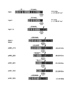
Figure 1 shows a schematic presentation of some of the used plasmid
constructs.
Figure 2 shows a SDS-PAGE gel (15%) of insoluble and the soluble fractions of
cell lysates of
IFABP and MBP (and the indicated mutations thereof) fused to the N-terminus of
HlyAl/HlyAc
CA 02912300 2015-11-12
WO 2014/170430 5 PCT/EP2014/057887
(Figure 2A). Samples were loaded on a SDS-PAGE and stained with Coomassie
Brilliant Blue (CBB).
Figure 2B shows cell lysate samples of E. coli expressing fusion proteins of
HlyAl and indicated PrOI
or Pe0I, wherein the PrOI or PeOI are C-terminally fused to HlyAl, analyzed by
SDS-PAGE and
visualized by CBB staining. Figure 2C depicts a SDS-PAGE (15 %) of soluble and
insoluble fractions
after cell disruption of cells producing IFABP wt (encoded by plasmid pQE-
IFABP wt) or HlyAl-
IFABP wt (encoded by pIAR_207). A soluble degradation product of HlyA 1 -IFABP
wt is indicated.
Figure 2D shows the expression of peptides 238, 239, 240 and 241 fused to
HlyAl and demonstrates
that the expression of peptides 240 and 241 fails without the fusion protein
HlyAl.
Figure 3 shows experiments of HlyAl being refolded in the presence of EDTA or
Ca2+ and applied to
an IMAC and SEC. A: In the presence of Ca, HlyAl, carrying an N-terminal His6-
tag, was loaded to
the IMAC (left lane) and bound proteins were eluted with an imidazole
gradient. B: SEC analysis
(Superdex 75 10/300 column, GE Healthcare) of HlyAl eluted from the IMAC. C &
D: HlyAl was
analyzed by IMAC and SEC in the presence of EDTA.
Figure 4 shows experiments of HlyAl -Nisin being refolded either in the
presence of Ca2+ or EDTA,
concentrated and applied to a SEC. A: Insoluble ("pellet") and soluble
("supernatant") fraction of
HlyAl-Nisin after refolding in the presence of Ca2+ and elution fractions of
SEC analysis. B: Insoluble
("pellet") and soluble ("supernatant") fraction of HlyAl-Nisin after refolding
in the presence of EDTA
.. and elution fractions of SEC analysis. C: SEC chromatograms of A and B. D:
HlyAl -Nisin was
incubated with Factor Xa (NEB), samples of the mixture were taken at various
time points (0, 20, 40,
60, 90, 120 min from left to right), loaded on a SDS-PAGE gel and stained with
CBB. E: HPLC
chromatograms of reference nisin (upper line) and nisin that was produced with
the invented
technology (lower line). F: SDS-PAGE analysis of HPLC elution fractions. Left
lane: Factor Xa
reaction after 90 min, other lanes: elution fractions of HPLC. Arrows indicate
the positions of Nisin
and HlyAl.
Figure 5 shows refolding experiments of LipA 1 -Nisin in the presence of Ca2+
and EDTA. LipAl-
Nisin was produced as lBs in L. coli and isolated lBs were refolded in the
presence of Ca2+ or EDTA.
In the presence of Ca2+, pure and soluble LipAl-Nisin was produced in a
homogeneous state. In
contrast, LipAl-Nisin was aggregated in the presence of EDTA. A and B: SDS-
PAGE analysis of
SEC elution fractions and the corresponding SEC chromatogram of LipA 1 -Nisin
refolded with Ca2+. C
and D: LipAl-Nisin was refolded in the presence of EDTA and analyzed as
described in A and B.
Figure 6 shows expression analyses of peptides encoded by pIAR_215, pIAR_220,
pIAR_221,
pIAR_222 and pIAR_223 and refolding experiments of expressed peptides (encoded
by pIAR_220,
pIAR_222 and pIAR_223) in the presence of Ca2+ and EDTA. The indicated
proteins were produced
CA 02912300 2015-11-12
WO 2014/170430 6 PCT/EP2014/057887
as IBs in E. coli and isolated IBs were refolded in the presence of Ca:2+ or
EDTA. A: SDS-PAGE
analysis of indicated samples. B-G: SEC analysis of refolded indicated
proteins.
Figure 7 shows the production of HCRF. A: IBs of HlyAl-HCRF. encoded by
plasmid pIAR_202.
were refolded either in the presence of Ca2+ or EDTA. B: HlyA 1 -HCRF,
refolded in the presence of
Ca2+, was incubated with Factor Xa for 10 min, 40 min and 120 min. Arrows
indicate the position of
HlyAl-HCRF, HlyA I and HCRF, respectively. HlyAl-HCRF, refolded in buffer
containing either
Ca2+ or EDTA, was applied to SEC analysis and elution fractions were analyzed
by SDS-PAGE. C:
Elution chromatograms of the above mentioned SEC analysis. D: CBB stained SDS-
PAGE gel after
SEC analysis of HlyAl-HCRF refolded in the presence Ca2+. E: CBB stained SDS-
PAGE gel after
SEC analysis of HlyAl-HCRF refolded in the presence of EDTA. Arrows indicate
the position of
HlyAl-IICRF. IIlyAl-IICRF was refolded in the presence of Ca2+, incubated with
Factor Xa for 2h
and the digestion mixture was purified by HPLC. F: HPLC chromatogram. G: CBB
stained SDS-
PAGE gel of IIPLC elution fractions. Arrows indicate the position of IICRF.
Figure 8 shows the production and functional studies of HlyAl -TFABP. SEC
analyses of refolded
HlyAl-IFABP. A and C: SDS-PAGE gel of elution fractions from SEC analysis of
HlyAl-IFABP
refolded with Ca2+ and elution chromatogram of the SEC. B and D: SDS-PAGE gel
of elution
fractions from SEC analysis of HlyAl-IFABP refolded with EDTA and elution
chromatogram of the
SEC. E: Purified HlyAl -IFABP in the presence of either Ca2+ or EDTA were
incubated with Factor
Xa and protein samples were analyzed by SDS-PAGE at indicated time points. The
arrow indicates the
position of IFABP. F and G: Functional studies of HlyAl-IFABP using titration
experiments with
DAUDA. F: HlyAl -IFABP was refolded in the presence of Ca2+ and purified by
SEC. DAUDA was
titrated to HlyAl-IFABP, the fluorescence signal at 500 nm was recorded and
plotted against the
DAUDA concentration. "[he black lane represents the curve of the theoretical
fit. G: Same experiments
as in F were repeated with HlyAl-IFABP purified in the presence of EDTA.
Figure 9 shows the production of H1yA1-IFNA2. A: SDS-PAGE analyses of H1yA1-
IFNA2 refolded
in the presence of 0.5 M ar2inine. Secreted IFNA2-HlyA1 served as reference
for oxidized protein
containing disulfide bonds (left lane). In the absence of DTT, HlyA1-IFNA2
migrates on the same
running height as the reference. In contrast, H1yA1-IFNA2 in the presence of
Drf migrates slower.
These results indicate the formation of disulfide bonds within refolded HlyA1-
IFNA2. B and C: SEC
analyses of H1yA1-IFNA2 after refolding in the presence of Ca2+ and EDTA,
respectively. B:
Refolding in the presence of Ca2+. C: Refolding in the presence of EDTA.
Figure 10 shows a binding experiment of refolded HlyAl-MBP and amylose resin.
HlyAl-MBP was
expressed in E. coli (lane "cell lysate") and IBs of HlyAl-MBP were prepared
(lane "denat. HlyAl-
CA 02912300 2015-11-12
WO 2014/170430 7 PCT/EP2014/057887
MBP"), denaturated and refolded in the presence of Ca2+. Some HlyAl-MBP
precipitated during
refolding (lane "precipitated") and soluble HlyAl-MBP (lane "refolded") was
loaded to amylose resin.
HlyA 1 -MBP bound to amylose and no protein remained within the "flow through-
. After washing,
HlyA 1 -MBP was eluted by maltose (lane "elution").
Figure 11 shows the production of peptides 101, 102 and 103. A: Expression of
HlyAl fused to
peptides 101, 102 and 103. B-D: Purification scheme of HlyAl fused to peptide
101, 102 and 103.
Cells expressing the corresponding fusion proteins were broken and cell
lysates (lane "cell lysates")
were centrifuged. No visible fusion proteins were in the soluble fraction
(lane "soluble fraction") and
fusion proteins aggregated as IBs. IBs were denaturated (lane "denat. Ills")
and refolded in the
presence of Ca. Fusion proteins were efficiently refolded with Ca2+ ("refolded
peptide 10X") and no
proteins precipitated ("pellet"). Renaturated fusion proteins were incubated
with Factor Xa and
peptides 101, 102 and 103 were separated from HlyA 1 (lane "Factor Xa"). An
unspecific cleavage
product occurred in all cases (see lane "+Factor Xa-).
Figure 12 shows Factor Xa digestion experiments with peptide 103 fused to HlyA
1 -R210D (encoded
by plasmid pIAR_112). Refolded Ifls (lane "-") were incubated with Factor Xa
("+") and samples
were analysed by SDS-PAGE. No unspecific cleavage product occurred (compared
to the results
shown in Figure 11).
Figure 13 shows the production of Fuzeon . HlyA1-Fuzeon was refolded in the
presence of Ca2 (A)
or EDTA (B) and loaded onto a Superclex 75 16/60 column. The arrow indicates
the position of
HlyAl-Fuzeon . C: HlyAl-Fuzeod) was refolded in the presence of Ca2+ and
incubated with Factor
Xa for 10 min, 40 mm and 120 min. The arrows indicate the position of the
cleavage products HlyAl
and Fuzeon .
Figure 14 shows experiments of lBs of HlyA 1 M88A-Met-peptide 103 that were
denaturated and
incubated with CNBr for indicated periods. Samples were analyzed by SDS-PAGE.
The cleavage
product HlyA 1 M88A-Met is visible on the gel. No unspecific cleavage products
were obtained. Since
peptide 103 was not stained by CBB (presumably due to its relative small
molecular weight), it was
purified by HPLC and de-novo sequenced by mass spectrometry. Using such
method, peptide 103
was identified.
Figure 15 shows the expression analyses of HlyA 1 M88A-Met-peptide 3 in batch
cultures and
fermentation. E. coli cells carrying plasmid pIAR_115 were incubated in batch
cultures (left lane), or
by fermentation with glucose (middle lane) or glycerol (right lane) as feed.
High cell densities (>50)
were obtained by fermentation with glucose and glycerin as feed. However,
glucose seems to repress
CA 02912300 2015-11-12
WO 2014/170430 8 PCT/EP2014/057887
the expression of the fusion protein under the used conditions. In the
presence of glycerol, in contrast,
high cell densities and high expression levels were achieved.
Figure 16 shows the secretion analysis of different peptides fused to HlyAl.
A: secretion analysis of
peptides 101, 102 and 103 fused to HlyAl. PeOI were fused C-terminal to HlyAl
(plasmids
pIAR_101, pIAR_102 and pIAR_103) and co-expressed with HlyB and HlyD (pK184-
HlyBD). Cell
lysate and supernatant samples were analyzed by SDS-PAGE. B: Secretion
analyses of HlyA1-Mab40.
Cells expressed HlyA1-Mab40 and, if indicated by a +, HlyB and HlyD (from
plasmid pK184-
H1yBD). Moreover, the influence of IPTG and baffles (different aeration) for
expression and secretion
is investigated. After cell growth, cell lysate and supernatant samples were
analyzed by SDS-PAGE.
The analysed peptides fused C-terminal to HlyAl were efficiently secreted by
the dedicated T1SS.
DETAILED DESCRIPTION OF THE INVENTION
The terms used herein have, unless explicitly stated otherwise, the following
meanings.
"At least one", as used herein, relates to one or more, in particular 1, 2, 3,
4, 5, 6, 7, 8, 9, 10 or more.
"Isolated" or "isolating", as interchangeably used herein in relation to a
molecule, means that said
molecule has been at least partially separated from other molecules that are
naturally associated with
said molecule. -Isolated" may mean that the molecule has been purified to
separate it from other
molecules and components, such as other proteins and nucleic acids and
cellular debris which may
originate from a host cell.
"Nucleic acid" as used herein includes all natural forms of nucleic acids,
such as DNA and RNA.
Preferably, the nucleic acid molecules are DNA. "Nucleic acid sequence
identity" as used herein,
means that the residue at a given position is identical to that at a
corresponding position of a reference
nucleic acid. The preferred nucleic acid sequence identity of the present
invention is 80%, more
preferred 90% or still more preferred 95%.
The term "fragment", as used herein in connection with a nucleic acid
molecule, relates to a nucleic
acid sequence which is compared to its reference nucleic acid sequence
shortened by one or more 3 or
5' terminal nucleotides. The shortening occurs at the 3'-end, the 5' -end or
both so that a contiguous
strand of nucleotides of the reference sequence remains. The fragment has
preferably a length of at
least 20, more preferably at least 50 nucleotides.
CA 02912300 2015-11-12
WO 2014/170430 9 PCT/EP2014/057887
The term "peptide" is used throughout the specification to designate a polymer
of amino acid residues
connected to each other by peptide bonds. A peptide according to the present
invention has 2-100
amino acid residues.
The terms "protein" and "polypeptide" are used interchangeably throughout the
specification to
designate a polymer of amino acid residues connected to each other by peptide
bonds. A protein or
polypeptide according to the present invention has preferably 100 or more
amino acid residues.
The terms "protein of interest", "PrOI" or "peptide of interest", "PeOI", as
used herein, relate to any
gene product that is expressed via recombinant expression. The term "a peptide
or protein of interest"
as disclosed herein covers any naturally or non-naturally occurring peptide or
protein. In some
embodiments, the PeOI or PrOI is a non-natural/synthetic peptide or protein.
Synthetic in this
connection means that the sequence of the peptide or protein has been
artificially designed. Thus, a
sequence encoding for a PeOI or PrOI may comprise a nucleic acid sequence
encoding for one, two or
more naturally occurring peptides or proteins. These naturally occurring
peptides or proteins may have
been further modified, e.g., by mutagenesis of the encoding sequence.
The term "an N-termi nal fragment" relates to a peptide or protein sequence
which is in comparison to
a reference peptide or protein sequence C-terminally truncated, such that a
contiguous amino acid
polymer starting from the N-terminus of the peptide or protein remains. In
some embodiments, such
fragments may have a length of at least 10 amino acids.
The term "a C-terminal fragment" relates to a peptide or protein sequence
which is in comparison to a
reference peptide or protein sequence N-terminally truncated, such that a
contiguous amino acid
polymer starting from the C-terminus of the peptide or protein remains. In
some embodiments, such
fragments may have a length of at least 10 amino acids.
The term "fusion protein" as used herein concerns peptides and proteins which
are N- or C-terminally
connected to each other. Such fusion proteins may be encoded by nucleic acid
sequences which are
operably fused to each other. In certain embodiments, a fusion protein refers
to at least one PeOI or
PrOI C-terminally fused to a polypeptide chain according to the invention, for
example a polypeptide
chain comprising HlyA or a fragment thereof or a homolog thereof.
Generally, the skilled person understands that for putting the present
invention into practice any
nucleotide sequence described herein may or must comprise an additional start
and/or stop codon or
that a start and/or stop codon of any of the sequences described herein may or
must be deleted
depending on the nucleic acid construct used. The skilled person will base
this decision, e.g., on
CA 02912300 2015-11-12
WO 2014/170430 10 PCT/EP2014/057887
whether a nucleic acid sequence comprised in the nucleic acid molecule of the
present invention is to
be translated and/or is to be translated as a fusion protein.
The term "introducing" in relation to a nucleic acid molecule, as used herein,
refers to the uptake and
incorporation of exogenous DNA into a host cell. Such uptake of the nucleic
acid molecule may
depend on the natural competence of the host cell or on transfcction methods
such as electroporation
or calcium chloride transformation which are well known in the art.
The term "host cell" as used herein relates to an organism that harbors the
nucleic acid molecule or a
vector encoding the recombinant PeOI or PrOI. In preferred embodiments the
host cell is a prokaryotic
cell. In more preferred embodiments the host cell is E. coli which may include
but is not limited to
BL21, DH1, DH5a, DM1, HB101, JM101-110, K12, Rosetta(DE3)pLysS, SURE, TOP10,
XL1-Blue,
XL2-Blue and XL10-Blue strains.
The terms "expression" or "expressed", as interchangeably used herein, relate
to a process in which
information from a gene is used for the synthesis of a gene product. In cell-
based expression systems
the expression comprises transcription and translation steps.
The term "recombinant expression", as used herein, relates to transcription
and translation of an
exogenous gene in a host organism. Exogenous DNA refers to any
deoxyribonucleic acid that
originates outside of said organism. The term "heterologous" as used herein in
relation to proteins
refers to a protein that is expressed from an exogenous DNA. This also
includes proteins that are
expressed from nucleic acid sequences which are identical to endogenous
nucleic acid sequences and
that were artificially duplicated.
The term "production", as used herein in relation to a recombinant peptide or
protein, means that a
recombinant peptide or protein is expressed in a host cell and is subsequently
isolated from other
molecules of the host cell.
"Culturing", "cultivating" or "cultivation", as used herein, relates to the
growth of a host cell in a
specially prepared culture medium under supervised conditions. The terms
"conditions suitable for
recombinant expression" or "conditions that allow expression" relate to
conditions that allow for
production of the PrOI in host cells using methods known in the art, wherein
the cells are cultivated
under defined media and temperature conditions. The medium may be a nutrient,
minimal, selective,
differential, or enriched medium. Preferably, the medium is a minimal culture
medium. Growth and
expression temperature of the host cell may range from 4 C to 45 C.
Preferably, the growth and
expression temperature range from 30 C to 39 C. The term "expression medium"
as used herein
CA 02912300 2015-11-12
WO 2014/170430 11 PCT/EP2014/057887
relates to any of the above media when they are used for cultivation of a host
cell during expression of
a protein.
The term "subjecting" as used herein means that various components, for
instance proteins and a
buffer, are brought into contact.
The terms "inclusion body" or "I13-, as interchangeably used herein, relate to
nuclear or cytoplasmic
aggregates of substances, for instance proteins. IBs are undissolved and have
a non-unit lipid
membrane. In the method of the present invention, the IBs mainly consist of
the fusion protein
comprising at least one PeOI or PrOI and at least one allocrite of a T1SS or a
fragment thereof.
The terms "substrate" or "allocrite", as interchangeably used herein, relate
to a solute that may be
cargo of a T1SS. The substrate or allocrite is a protein that contains
specific peptide sequence motives,
such as GG repeats and the secretion signal, that allow the transportation via
the T1SS.
The terms type 1 secretion system" or "Ti SS" as interchangeably used herein
relate to a protein
complex which consists of three protein subunits: an ABC transporter protein,
a MFP, and an OMP.
The ABC transporters are transmembrane proteins that utilize the energy of
adenosine triphosphate
(ATP) hydrolysis to carry out certain biological processes including
translocation of various substrates
across membranes. Proteins of the MFP family function as auxiliary proteins or
'adaptors', connecting
a primary porter in the cytoplasmic membrane of a Gram-negative bacterium with
an outer membrane
factor protein that serves a porin or channel function in the outer membrane.
Therefore, the tripartite
protein complex allows the transport of various molecules, such as ions, drugs
and proteins to pass the
inner and outer membrane of Grain-negative bacteria. A subgroup of T1SS
substrates are RTX
(repeats in toxins) toxins.
The term -functional three-dimensional conformation" as used herein in
relation to proteins refers to
the structure of a protein which allows said protein to have a specific
activity such as substrate
catalysis, protein specific localization or interaction with other proteins
that is at least 5%, 10%, 20%,
40% or 50%, or more preferably at least 80%, or even more preferably 100% of
the activity of the
same protein in its native conformation. A functional three-dimensional
conformation usually requires
that the protein is soluble. The native conformation of a protein is its
properly folded and/or assembled
form, which is operative and functional. The native state of a biomolecule may
possess all four levels
of biomolecular structure, with the secondary through quaternary structure
being formed from weak
interactions along the covalently-bonded backbone. This is in contrast to the
denatured state, in which
these weak interactions are disrupted, leading to the loss of these forms of
structure and retaining only
the biomolccule's primary structure.
12
The term "inhibiting", as used herein, relates to a detectable and significant
reduction of protein activity
or gene expression activity caused by an effector molecule. Methods to detect
protein activity or gene
expression are known in the art.
The present invention relates to methods comprising nucleic acid sequences of
substrates/allocrites of
Type 1 secretion systems or fragments thereof for the efficient production of
recombinant peptides and
proteins of interest. The allocrites or fragments thereof improve the
expression of peptides and protein of
interest as inclusion bodies (TB) and function as TB-tags. Importantly, the
allocrites and fragments thereof
allow the efficient renaturation of the inclusion bodies into a functional
three-dimensional conformation.
Therefore, the allocrites or fragments thereof combine the advantages of TB-
tags and solubility-tags
without the corresponding disadvantages.
The Hly secretion system is a protein secretion system, which mostly occurs in
Gram-negative bacteria.
This secretion system belongs to the family of T1SS, which transport their
substrates in an ATP driven
manner in a single step from the cytosol to the extracellular space without an
intermediate station in the
periplasm. The Hly secretion system comprises HlyB, which represents an ABC
transporter, the MFP
HlyD, and the universal OMP To1C. The ¨110 kDa hemolytic toxin HlyA is a
transport substrate of the
Hly secretion system. On genetic level, the components necessary for HlyA-
specific secretion are
organized in an operon structure. The nucleic acid sequence encoding for HlyC
also forms part of this
operon but is not required for HlyA secretion through the Hly secretion
system. HlyC catalyzes acylation
of HlyA, which renders HlyA hemolytic. HlyA is a protein, which consists of
1024 amino acid residues
and requires for its export via the Hly secretion system its C-terminus
comprising about 40-60 amino
acids called secretion signal. Furthermore, HlyA is characterized by a domain
comprising several glycine
rich (GG) repeats (GGXGXDXUX, wherein X can be any amino acid, U is a
hydrophobic, large amino
acid located N-terminal of the secretion signal (SEQ ID NO:67)). GG repeats
are the characteristic of the
repeats in toxin (RTX) family. The GG repeats bind Ca2 which induces their
folding. Hence, in absence of
Ca2 the domain comprising the GG repeats is unstructured. The amino acid
sequence of one HlyA
protein is set forth in SEQ ID NO:2, as encoded by the nucleotide sequence set
forth in SEQ ID NO:1. A
fragment of HlyA, which was expressed in enhanced levels compared to the
wildtype HlyA and lacks the
N-terminal part of HlyA (Figure 1) was named HlyAl. The amino acid sequence of
HlyAl is set forth in
SEQ ID NO:4 whereas the encoding nucleotide sequence is set forth in SEQ ID
NO:3.
The present invention is based on the inventors' surprising finding that a
PeOI or PrOI fused to at least
one allocrite of a T1SS or a fragment thereof leads to the expression of the
fusion protein in form of 1B
even if the non-conjugated PeOI or PrOI alone is expressed in a soluble form.
Further, it was found by
300577.00002/110620243.1
Date Recue/Date Received 2020-12-11
CA 02912300 2015-11-12
WO 2014/170430 13 PCT/EP2014/057887
the present inventors that Ca2+ induces the folding of the denaturated TB of
the fusion proteins
consisting of the allocrite and the PeOI or PrOI into a soluble and functional
three-dimensional
conformation. Therefore, the allocrites or fragments thereof are bifunctional
tags combining the
advantages of IB-tags (high yield, high initial purity, immunity against
proteolytic degradation) and
solubility-tags (soluble, bioactive products) without the corresponding
disadvantages (inclusions body-
tags: aggregated, non-active products; solubility-tags: rather low yields, low
purity, prone to
proteolytic degradation).
Thus, in a first aspect, the present invention relates to a method for
production of a recombinant PeOI
or PrOI, wherein the method comprises: (a) introducing a nucleic acid molecule
encoding a fusion
protein comprising at least one PeOI or PrOI, and at least one allocrite of a
T1SS or a fragment
thereof, into a host cell; (b) cultivating the host cell under conditions that
allow expression of the
fusion protein, wherein the fusion protein is expressed in the form of TB; (c)
isolating the recombinant
fusion protein from said host cells. Further embodiments may comprise step (d)
of subjecting the
recombinant fusion protein to conditions that allow the PeOI or PrOI to fold
into a functional three-
dimensional conformation. In various other embodiments of the first aspect,
the host cell does not
express a heterologous ABC transporter, a heterologous MFP and/or a
heterologous OMP of the T1SS.
In various embodiments, this aspect of the invention also includes allocrites
of a T1SS that are
.. selected from the group consisting of HlyA, CyaA, EhxA, LktA, PILkiA, PasA,
PvxA, MmxA,
ApxIA, ApxIIA, ApxIIIA, ApxIVA, ApxI, ApxII, AqxA, VcRtxA, VvRtxA, MbxA, RTX
cytotoxin,
RtxL1, RtxL2, FrhA, LipA, TliA, PrtA, PrtSM, PrtG, PrtB, PrtC, AprA, AprX,
Za.pA, ZapE, Sap,
HasA, colicin V, LapA, ORF, RzcA, RtxA, XF2407, XF2759, RzcA, RsaA, Crs, CsxA,
CsxB, SlaA,
SwmA, S111951, Nod , PlyA, PlyB, FrpA, FrpC, FrpC-like or other T1SS
allocrites as described in
Linhartova et al. (Linhartova, I. et al., FEMS Microbiol Rev 34, 1076-1112,
FMR231
ipiii10.1111/j.1574-6976.2010.00231.x) and fragments thereof. In various
preferred embodiments, the
allocrites are characterized by the presence of at least one GO repeat of the
consensus sequence
GGxGxDxUx (wherein X can be any amino acid, U is a hydrophobic, large amino
acid). In more
preferred embodiments the allocrit of a T1SS is HlyA comprising or consisting
of the amino acid
sequence as set forth in SEQ ID NO:2, a fragment thereof or a polypeptide that
has at least 80%
sequence identity to the amino acid sequence of SEQ ID NO:2 or the fragment
thereof. In other
various embodiments the fragment of HlyA consists of the amino acid sequence
as set forth in SEQ ID
NO:4 or a polypeptide that has at least 80% sequence identity to the amino
acid sequence of SEQ ID
NO:4.
In various embodiments, this aspect of the invention also includes homologs of
the afore-mentioned
sequences of SEQ ID Nos. 1-4. The term "homologous" or "homolog" as used
herein refers to a
CA 02912300 2015-11-12
WO 2014/170430 14 PCT/EP2014/057887
polynucleotide or polypeptide sequence that has a highly similar sequence to
or high sequence identity
(e.g. 70%, 80%, 90%, 95%, 97.5%, 99% or more) with another polynucleotide or
polypeptidc
sequence or part thereof. With regard to the above nucleic acid molecule, the
term homologs thus
includes nucleic acid sequences that have at least 70, preferably 80, more
preferably 90, even more
preferably 95, 97.5 or 99 % sequence identity to the nucleotide sequence of
the first nucleic acid
sequence as defined above. The sequence identity may occur over a continuous
stretch of nucleotides
or may be discontinuous.
In various embodiments of the first aspect of the invention, the allocrite of
a Ti SS can be fused to the
C-terminus of the PeOI or PrOI. In other various embodiments of the first
aspect, the allocrite can be
fused to the N-terminus of the PeOI or PrOI.
In one embodiment, the expression medium comprises 20.0 mM or less of Ca2+. In
a more preferred
embodiment the Ca2+ concentration in the expression medium is 0.1 mM or less.
In various embodiments, the expression of the endogenous ABC transporter gene,
the endogenous
MFP gene and/or the endogenous OMP gene of the Ti SS or the activity of the
corresponding gene
products in the host cell is inhibited. In various embodiments, the host cell
does not express
endogenous ABC transporter, endogenous MFP and/or endogenous 01VIP of the Ti
SS. Methods to
inhibit the expression of genes such as their deletion or insertion of
nucleotide sequences destroying
the integrity of the promoter sequence or the gene itself are known in the
art. A preferred gene
expression activity after deletion or disruption may be less than 35 %, 30 %,
25 %. 20 %, 15 %, 10 %
or 5 % of the activity measured in untreated cells. In other various
embodiments of the invention, the
endogenous ABC transporter, the endogenous MFP and/or the endogenous OMP of
the type 1
secretion system are inhibited by antibodies or small molecule inhibitors. In
preferred embodiments of
the invention, the ABC transporter activity is inhibited by orthovanadate or
an ATP homologous
inhibitor such as 8-azido-ATP. Such NIP mimetics are known in the art. The
preferred protein activity
after inhibitor treatment may be less than 35 %, 30 %, 25 %, 20 %, 15 %, 10 %
or 5 % of the activity
measured in untreated cells. In other embodiments of the invention, the
transport is inhibited or
blocked by the allocrite itself, for example by over-expressing the allocrites
or the attachment of
fusion peptides and proteins.
In other embodiments, the recombinant peptide or protein is exposed to a
refolding buffer, wherein the
refolding buffer comprises at least 0.01, more preferably 0.01-40 mM of Ca2+.
In various embodiments of the methods of the invention, (I) the
host cell is a prokaryotic cell;
and/or (II) the expression is performed in minimal culture medium; and/or
(III) the recombinant fusion
CA 02912300 2015-11-12
WO 2014/170430 15 PCT/EP2014/057887
peptide or protein is purified using a method selected from affinity
chromatography, ion exchange
chromatography, reverse phase chromatography, size exclusion chromatography,
and combinations
thereof; and/or (IV) the method comprises the additional step (e) of
contacting the recombinant fusion
protein with a protease suitable for cleavage of the fusion protein to yield
the allocritc and the PeOI or
PrOI as separate molecules; and/or (V) the method comprises a step (e) as
defined in (IV) followed by
purification of the PeOI or PrOI.
In still another embodiment, the present invention may also relate to a method
wherein the at least one
PeOI or PrOI is selected from the group consisting of Nisin, HCRF, IFABP,
IFNA2, MBP, peptide
101, peptide 102, peptide 103, MAB-40 Mab-42, Fuzeon , salmon Calcitonin,
human Calcitonin,
Inhibitor peptide 1, 238, 239, 240 or 241.
The nucleic acid molecule encoding the fusion protein further comprises a
regulatory nucleotide
sequence that modulates expression of the fusion protein in various
embodiments. A preferred
regulatory nucleic acid sequence is set forth in SEQ ID NO:39. The term
"regulatory nucleotide
sequence" as used herein relates to a nucleic acid sequences which are located
5' of a gene and
enhance the expression activity of said gene.
The terms "affinity tag" as used herein relates to entities,which are coupled
to the Pe01 or PrOI and
allow enrichment of the tagged PeOI or PrOI using an affinity tag receptor.
The term "affinity
chromatography" as used herein relates to the complex fomiation of the tagged
peptide or protein and
the receptor. In certain embodiments affinity tags may be selected front the
group consisting of the
Strep-tag or Strep-tag II, the myc-tag, the FLAG-tag, the His-tag, the small
ubiquitin-like modifier
(SUMO) tag, the covalent yet dissociable NurpD peptide (CYD) tag, the heavy
chain of protein C
(HPC) tag, the calmodulin binding peptide (CBP) tag, or the HA-tag or proteins
such as Streptavidin
binding protein (SBP), maltose binding protein (MBP), and glutathione-S-
transferase.
The term "protease cleavage site" refers to peptide sequence which can be
cleaved by a selected
protease thus allowing the separation of peptide or protein sequences which
are interconnected by a
protease cleavage site. In certain embodiments the protease cleavage site is
selected from the group
consisting of a Factor Xa-, a tobacco edge virus (TEV) protease-, a
enterokinase-, a SUMO Express
protease-, an IgA-Protease-, an Arg-C proteinase-, an Asp-N endopeptidases-,
an Asp-N
endopcptidase + N-terminal Glu -, a caspasel-, a caspase2-,a caspase3-, a
caspase4, a caspasc5, a
caspase6, a caspase7, a caspase8, a caspase9, a caspase10, a chymotrypsin-high
specificity, a
chymotrypsin-low specificity-, a clostripain (Clostridiopeptidase B)-, a
glutamyl endopeptidase-, a
granzymeB-, a pepsin-, a proline-endopeptidase-, a proteinase K-, a
staphylococcal peptidase I-, a
Thrombin-, a Trypsin-, and a Thermolysin-cleavage site.
CA 02912300 2015-11-12
WO 2014/170430 16 PCT/EP2014/057887
The term chemical cleavage refers to the cleavage of peptide bonds caused by a
chemical compound.
Such compounds may include, but are not limited to cyanogen bromid (CNBr)
cleaving C-terminal to
methionine residues, BNPS-skatole, NCS or TFA cleaving C-terminal to
tryptophane residues and
Ni2+ ions cleaving C-terminal to the tetrapeptide S/TXHZ (X and Z can be any
amino acid residues,
except X = prolinc) (Kopera et al., (2012), Plos One 7(5)).
"Fused", as used in the context of this invention, means that the resulting
peptides or proteins are
directly connected to each other or linked to each other by one or more amino
acids, peptides or
proteins, e.g., one or more protease cleavage sites and/or affinity tags.
If the PeOI or PrOI comprises two or more naturally occurring peptides or
proteins, the two or more
peptides or proteins may be separated by protease cleavage sites.
Generally, any peptide or protein may be chosen as PeOI or PrOI. In certain
embodiments, the PrOI is
a protein which does not form a homo-dimer or homo-multimer. The avoidance of
self-interacting
peptides or proteins may be advantageous if the recombinant peptide or protein
is to be secreted into
the cell culture supernatant, because the formation of larger protein
complexes may disturb an efficient
protein export. However, the PrOI may also be a peptide or protein, which is a
subunit of a larger
peptide or protein complex. Such a peptide or protein may be isolated after
expression and optionally
secretion and be suitable for an in vitro reconstitution of the multi peptide
or protein complex. In
certain embodiments, the PeOI or PrOI is a peptide having less than 100 amino
acid residues. If these
peptides comprise pre- and/or pro- sequences in their native state after
translation the nucleic acid
sequence encoding for the PeOI may be engineered to be limited to the sequence
encoding the mature
peptide. One exemplary peptide is insulin, e.g., human insulin. The secretion
of over-expressed
peptides and proteins is especially advantageous where the peptide or protein
is harmful to the host
cell. For this reason, the present invention is particularly advantageous for
expression of lipases and
proteases which are known to be toxic to the host cell and thus the expression
of these proteins by the
inventive systems and methods represents a specific embodiment of the present
invention.
In various embodiments, the Pe01 or PrOI is an enzyme.
The International Union of Biochemistry and Molecular Biology has developed a
nomenclature for
enzymes, the EC numbers; each enzyme is described by a sequence of four
numbers preceded by
"EC". The first number broadly classifies the enzyme based on its mechanism.
The complete nomenclature can be browsed at
http://www.chem.qmul.ac.ukhubmb/enzyme/.
CA 02912300 2015-11-12
WO 2014/170430 17 PCT/EP2014/057887
Accordingly, a PeOI or PrOI according to the present invention may be chosen
from any of the classes
EC 1 (Oxidoreductases), EC 2 (Transferases), EC 3 (Hydrolases), EC 4 (Lyases),
EC 5 (Isomerases),
and EC 6 (Ligases), and the subclasses thereof.
In certain embodiments, the PeOI or PrOI is cofactor dependent or harbors a
prosthetic group. For
expression of such peptides or proteins, in some embodiments, the
corresponding cofactor or
prosthetic group may be added to the culture medium during expression.
In certain cases, the PeOI or PrOI is a dehydrogenase or an oxidase.
In case the PeOI or PrOI is a dehydrogenase, in some embodiments, the PeOI or
PrOI is chosen from
the group consisting of alcohol dehydrogenases, glutamate dehydrogenases,
lactate dehyrogenases,
cellobiose dehydrogenases, formate dehydrogenases, and aldehydes
dehydrogenases.
In case the PeOI or PrOI is an oxidase, in some embodiments, the PeOI or PrOI
is chosen from the
group consisting of cytochrome P450 oxidoreductases, in particular P450 BM3
and mutants thereof,
perox idases, monooxygenases, hydrogen ases , monoamine oxidases, aldehydes
oxidases, x anthin
oxidases, amino acid oxidases, and NADH oxidases.
In further embodiments, the PeOI or PrOI is a transaminase or a kinase.
In case the PeOI or PrOI is a transaminase, in some embodiments, the PeOI or
PrOI is chosen from the
group consisting of alanine aminotransferases, aspartate aminotransferases,
glutamate-oxaloacetic
transaminases, histidinol-phosphate transaminases, and histidinol-pyruvate
transaminases.
In various embodiments, if the PeOI or PrOI is a kinase, the PeOI or PrOI is
chosen from the group
consisting of nucleoside diphosphate kinases, nucleoside monophosphate
kinases, pyruvate kinase,
and glucokinases.
In some embodiments, if the PeOI or PrOI is a hydrolase, the PeOI or PrOI is
chosen from the group
consisting of lipases, amylases, proteases, cellulases, nitrile hydrolases,
halogenases, phospholipases,
and estcrases.
In certain embodiments, if the PeOI or PrOI is a lyase, the PeOI or PrOI is
chosen from the group
consisting of aldolases, e.g., hydroxynitrile lyases, thiamine-dependent
enzymes, e.g., benzaldehyde
lyases, and pyruvate decarboxylases.
CA 02912300 2015-11-12
WO 2014/170430 18 PCT/EP2014/057887
In various embodiments, if the PeOI or PrOI is an isomerase, the PeOI or PrOI
is chosen from the
group consisting of isomerases and mutases.
In some embodiments, if the PeOI or PrOI is a ligase, the PeOI or PrOI may be
a DNA ligase.
In certain embodiments, the PeOI or PrOI may be an antibody. This may include
a complete
immunoglobulin or fragment thereof, which immunoglobulins include the various
classes and
isotypes, such as IgA, WD, IgE, IgG1, IgG2a, IgG2b and IgG3, IgM, etc.
Fragments thereof may
include Fab, Fy and F(ab')2, Fab', and the like.
Also contemplated herein are therapeutically active Pe0Is and PrOI. e.g., a
cytokine.
Thus, in certain embodiments the PeOI or PrOI is selected from the group
consisting cytokines, in
particular human or murine interferons, interleukins, colony-stimulating
factors, necrosis factors, e.g.,
tumor necrosis factor, and growth factors.
In some embodiments, if the PeOI or PrOI is an interferon, the PeOI or PrOI
may be selected from the
group consisting of interferon alpha, e.g., alpha-1, alpha-2, alpha-2a, and
alpha-2b, alpha-2, alpha-8,
alpha-16, alpha 21, bet a, e.g., beta-1, beta-1a, and beta- lb, or gamma.
In further embodiments, the PeOI or PrOI is an antimicrobial peptide, in
particular a peptide selected
from the group consisting of bacteriocines and lantibiotics, e.g., nisin,
cathelicidins, defensins, and
saposins.
In further embodiments, the PeOI or PrOI is an adhesive peptide with distinct
surface specificities, for
example for steel, aluminum and other metals or specificities towards other
surfaces like carbon,
ceramic, minerals, plastics, wood and other materials or other biological
materials like cells, or
adhesive peptides that function in aqueous environments and under anaerobe
conditions.
In further embodiments, the PeOI or PrOI has a length ranging from 2-100 amino
acids, wherein said
amino acids are selected from the group of the 20 proteinogenic amino acids.
More preferably, said
Pc0I or PrOI are selected from the group of peptides or proteins consisting of
DYKDDDDKMASMTGGQQMGHHHHHH (SEQ ID NO:
45),
MGSSAAAAAAAASGPGGYGPENQGPSGPGGYGPGGP (SEQ ID
NO:46),
ENREVPPGFTALIKTLRKCKII (SEQ ID NO:47), NLVSGLIEARKYLEWLHRKLKNCKV (SEQ
ID NO:48), IIIIIIIIIIIIIEGRAMSILKSPIDERSIEK (SEQ ID
NO:49).
- 19 -
HHHHHHIEGRPPGPPGPPGPPGPPGPPGPPGPPGPPG (SEQ ID
NO:50),
HHHHHHIEGRG APGAPGS QGAPGLQ (SEQ ID
NO:51),
GGGRGDMGSSAAAAAAAASGPGGYGPENQGPSGPGGYGPGGPRGDGGG (SEQ ID
NO:52).
Also disclosed herein are Pe0Is or PrOI, which are therapeutically active
peptides or proteins.
In certain embodiments, the PeOI or PrOI is a therapeutically active peptide.
In some
embodiments, a therapeutically active peptide may be selected from the group
consisting of
Fuzeon/T20, human calcitonin, salmon calcitonin, human corticotropin release
factor, Mab40,
Mab42, peptides associated with Alzheimer's disease, exenatide,
glatiramer/copaxone,
teriparatide/forsteo, romiplostim/nplate,
pramlintitde/symlin, thymalfasin/zadaxin,
enfuvirtide, andrenocorticotropin hormones (ACTH), brain natriuretic peptide,
nesiritide/natrecor, corticoliberin, sermorelin, somatorelin, secretin (human
and porcin),
terlipressin, sinapultide, teduglutide, vx-001, vasoactive intestinal peptide,
avipdadil,
linaclotide, and teduglutide.
In certain embodiments, the PeOI or PrOI is a type I secretion substrate. More
than 1000
proteins are annotated or have been described as type I secretion substrates
in the literature.
Many of them have interesting characteristics for the biotechnological usage,
in
particularhydrolascs like proteases and lipascs. Suitable proteases and
lipascs have been
described by Baumann et al. (1993) EMBO J 12, 3357-3364; and Meier et al.
(2007) J. BIOL.
CHEM.: 282(43), pp. 31477-31483.
Of course, the nucleic acid sequence encoding for the at least one PeOI or
PrOI may be
subjected to mutagenesis and thus lead to a mutated PeOI or PrOI on protein
level.
The term "mutation" as used herein relates to a variation in the nucleotide
and/or amino acid
sequence of a given nucleotide sequence or protein and includes substitutions,
deletions,
truncations, and insertions. In one specific example, the mutation is a point
mutation, i.e. the
replacement of one or more nucleotides and/or amino acids in a given sequence.
It is
understood that if the term "mutation" is used in relation to a protein
sequence, that the
nucleotide sequence encoding the protein can comprise multiple mutations or
modifications,
including silent mutations that, for example, serve the purpose to increase
expression
efficiency (codon-optimization) without changing the amino acid sequence. In
the present
invention, the mutation is preferably the substitution of one or two amino
acids by other amino
acids. Alternatively or in addition, the nucleic acid molecule may comprise
nucleotide
exchanges which do not alter the encoded protein sequence, so called silent
mutations. In some
embodiments, the mutations, e.g., silent mutations increase the expression
and/or secretion
efficiency of the peptide or protein encoded by the nucleic acid molecule.
Importantly,
mutations may be induced throughout the nucleic acid molecule of the present
invention. Thus,
the mutations may not be limited to sequences encoding for a peptide or
protein. Accordingly,
also non-coding sequence stretches may be subjected to mutagenesis. This type
of mutation also
falls within the scope of the term silent mutation. The mutagenesis of non-
coding sequences may
Date recue / Date received 2021-12-02
- 20 -
be advantageous, e.g., for the achievement of an improved expression and/or
secretion of a
peptide or protein encoded by a different sequence stretch within the nucleic
acid molecule.
The term "mutagenesis" as used herein means that the experimental conditions
are chosen such
that the amino acid naturally occurring at a given sequence position of a
protein sequence can
be substituted by at least one amino acid that is not present at this specific
position in the
respective natural polypeptide sequence. The term "mutagenesis" also includes
the (additional)
modification of the length of sequence segments by deletion or insertion of
one or more amino
acids. Thus, it is withinthe scope of the invention that, for example, one
amino acid at a chosen
sequence position is replaced by a stretch of three random mutations, leading
to an insertion of
two amino acid residues compared to the length of the respective segment of
the wild type
protein. Such an insertion or deletion may be introduced independently from
each other in any
of the peptide segments that can be subjected to mutagenesis in the invention.
The term "random mutagenesis" means that no predetermined single amino acid
(mutation) is
present at a certain sequence position but that one of at least two different
amino acids can
be incorporated with a certain probability at a predefined sequence position
during mutagenesis.
"Codon-optimized" means that codons encoding one amino acid residue are
replaced by a
different codon encoding the same amino acid, but being more frequently used
by a given host
organism for this particular amino acid. It is understood that such nucleotide
sequences that
encode a homologous polypeptide may have high sequence variability so that
sequence
identity between the nucleic acid molecules encoding the same or homologous
polypeptides
may be low.
The natural coding sequence of a protein sequence, i.e. the respective gene
segment of an
enzyme, can be used as a starting point for the mutagenesis of the amino acid
positions
selected in the present invention. For the mutagenesis of the recited amino
acid positions, the
person skilled in the art has at his disposal the various established standard
methods for site-
directed mutagenesis (Sambrook, J. et al. (2001) Molecular Cloning: A
Laboratory Manual, 3rd
Ed., Cold Spring Harbor Laboratory Press, Cold Spring Harbor, NY). A commonly
used
technique is the introduction of mutations by means of PCR (polymerase chain
reaction) using
mixtures of synthetic oligonucleotides, which bear a degenerate base
composition at the
desired sequence positions. Por example, use of the codon NNK or NNS (wherein
N = adenine,
guanine, cytosine or thymine; K = guanine or thymine; S = adenine or cytosine)
allows
incorporation of all 20 amino acids plus the amber stop codon during
mutagenesis, whereas the
Date recue / Date received 2021-12-02
CA 02912300 2015-11-12
WO 2014/170430 21 PCT/EP2014/057887
codon VVS limits the number of possibly incorporated amino acids to 12, since
it excludes the amino
acids Cys, Ile, Leu, Met, Phe, Trp, Tyr, Val from being incorporated into the
selected position of the
polypeptide sequence (V= adenine, guanine, or cytosine); use of the codon NMS
(wherein M
adenine or cytosine), for example, restricts the number of possible amino
acids to 11 at a selected
sequence position since it excludes the amino acids Arg, Cys, Gly, Ile, Leu,
Met, Phe, Trp, Val from
being incorporated at a selected sequence position. Another possibility is the
use of codons NDT or
NDC (wherein D = adenine, guanine, or thymine) as this provides a 1:1 ratio
between the number of
codons and the encoded amino acids, thus reduces the screening effort, and
leads to a balanced set of
12 polar, non-polar, aromatic, non-aromatic, hydrophilic and hydrophobic amino
acid residues (Arg,
Asn, Asp, Cys, Gly, His, Ile, Leu, Phe, Ser, Tyr, Val (Reetz MT et al., 2008,
ChemBioChem,
21 ;9(11): 1797-804)).
In certain embodiments, the PeOI or PrOI comprises a deletion of at least 10,
20, 30, 40, 50, or more
N- and/or C-terminal amino acid relative to the wildtype peptide or protein
sequence.
In various embodiments, the PeOI or PrOI is chosen from the group consisting
of MBP, lipase CalB,
protease SprP, hydrolase PlaB, hydrolase PlaK, hydrolase PlbF, lipase TesA,
Vif, human interferon
alpha-1, alpha-2, alpha-8, alpha-16, alpha-21, human interferon beta, human
interferon gamma,
murine interferon alpha, murine interferon gamma, IFABP, Cas2, affibody
protein ZA3, nisin,
corticotropin release factor, amyloid-beta peptide, exenatide, Fuzeon/T20,
salmon calcitonin, Mab40.
Mab42, lipase LipA, SprP, the HIV-1 protein Vif, and human calcitonin.
The PeOI or PrOI may be cloned into a vector. In certain embodiments, the
vector is selected from the
group consisting of a pSU-vector, pET-vector, a pBAD-vector, a pK184-vector, a
pMONO-vector, a
pSELECT-vector, pSELECI -Tag-vector, a pV1TRO-vector, a pVIVO-vector, a pORE-
vector, a
pBLAST-vector, a pUNO-vector, a pDUO-vector, a pZERO-vector, a pDeNy-vector, a
pDRIVE-
vector, a pDRIVE-SEAP-vector, a HaloTag Fusion-vector, a pIARGETTm-vector, a
Elexi0-vector, a
pDEST-vector, a pHIL-vector, a pPIC-vector, a pMET-vector, a pPink-vector, a
pLP-vector, a
pTOPO-vector, a pBud-vector, a pCEP-vector, a pCMV-vector, a pDisplay-vector,
a pEE-vector, a
pFL-vector, a pERT-vector, a pFastBac-vector, a pGAPZ-vector, a pIZ/V5-vector,
a pLenti6-vector, a
pMI13-vector, a p0G-vector, a pOpti-vector, a pREP4-vector, a pRSE'l -Nector,
a pSCREEN-vector, a
pSecTag-vector, a pTEF1-vector, a pTracer-vector, a pTrc-vector, a pUB6-
vector, a pVAX1-vector, a
pYC2-vector, a pYES2- vector, a pZeo-vector, a pcDNA-vector, a pFLAG-vector, a
pTAC-vector, a
pT7-vector, a gateway -vector, a pQE-vector, a pLEXY-vector, a pRNA-vector, a
pPK-vector, a
pUMVC-vector, a pLIVE-vector, a pCRUZ-vector, a Duet-vector, and other vectors
or derivatives
thereof. In preferred embodiments the vector is the pSU-vector.
CA 02912300 2015-11-12
WO 2014/170430 22 PCT/EP2014/057887
The vectors of the present invention may be chosen from the group consisting
of high, medium and
low copy vectors.
The above described vectors may be used for the transformation or transfection
of a host cell in order
to achieve expression of a peptide or protein which is encoded by an above
described nucleic acid
molecule and comprised in the vector DNA.
The host cell may be specifically chosen as a host cell capable of expressing
the gene. In addition or
otherwise, in order to produce a peptide or protein, a fragment of the peptide
or protein or a fusion
protein of the peptide or protein with another polypeptide, the nucleic acid
coding for the peptide or
protein can be genetically engineered for expression in a suitable system.
Transformation can be
performed using standard techniques (Sambrook, J. et al. (2001), supra).
Prokaryotic or eukaryotic host organisms comprising such a vector for
recombinant expression of a
PeOI or PrOI as described herein form also part of the present invention.
Suitable host cells can be
prokaryotic cell. In certain embodiments the host cells are selected from the
group consisting of gram
positive and gram negative bacteria. In some embodiments, the host cell is a
gram negative bacterium,
such as Ecoli. In certain embodiments, the host cell is E. coil, in particular
E. coli BL21 (DE3) or
other E. coli K12 or E. coli B834 or E. coli DH5a or XL-1 derivatives. In
further embodiments, the
host cell is selected from the group consisting of Escherichia coil (E. coil),
Pseudomonas, Serratia
marcescens, Salmonella, Shigella (and other enterobacteriaceae), Neisseria,
Hemophilus, Klebsiella,
Proteus, Enterobacter, Helicobacter, Acinetobacter, Moraxella, Helicobacter,
Stenotrophomonas,
Bdellovibrio, Legionella, acetic acid bacteria, Bacillus, Bacilli,
Carynebacterium, Clostridium,
ListeriaõStreptococcusõStaphylococcas, and Archaea cells. Suitable eukaryotic
host cells are among
others CHO cells, insect cells, fungi, yeast cells, e.g., Saccharomyces
cerevisiae, S. pombe, Pichia
pastoris.
The transformed host cells are cultured under conditions suitable for
expression of the nucleotide
sequence encoding a peptide or protein of the invention. In certain
embodiments, the cells are cultured
under conditions suitable for expression of the nucleotide sequence encoding a
PeOI or PrOI.
For producing the recombinant PeOI or PrOI, a vector is introduced into a
suitable prokaryotic or
eukaryotic host organism by means of recombinant DNA technology. For this
purpose, the host cell is
first transformed with a vector comprising a nucleic acid molecule according
to the present invention
using established standard methods (Sambrook, J. et al. (2001), supra). The
host cell is then cultured
under conditions, which allow expression of the heterologous DNA and thus the
synthesis of the
corresponding polypeptide. Subsequently, the polypeptide is recovered either
from the cell.
CA 02912300 2015-11-12
WO 2014/170430 23 PCT/EP2014/057887
For expression of the peptides and proteins of the present invention several
suitable protocols are
known to the skilled person.
Generally, any known culture medium suitable for growth of the selected host
may be employed in
this method. In various embodiments, the medium is a rich medium or a minimal
medium. Also
contemplated herein is a method, wherein the steps of growing the cells and
expressing the peptide or
protein comprise the use of different media. For example, the growth step may
be performed using a
rich medium, which is replaced by a minimal medium in the expression step. In
certain cases, the
medium is selected from the group consisting of LB medium, TB medium, 2YT
medium, synthetical
medium and minimal medium.
In some embodiments, the medium may be supplemented with IPTG, arabinose,
tryptophan and/or
maltose, and/or the culture temperature may be changed and/or the culture may
be exposed to IN
light. In various embodiments, the conditions that allow secretion of the
recombinant peptide or
protein are the same used for the expression of the peptide or protein.
In certain embodiments, the host cell is a prokaryotic cell, such as E.cm/i,
in particular E.coli BI21
(DE3) and E. coli DH5cc.
In some embodiments, the entire culture of the host cell, e.g., during growth
and expression, is carried
out in minimal medium. Minimal medium is advantageous for recombinant peptide
or protein
expression, as the protein, lipid, carbohydrate, pigment, and impurity content
in this medium is
reduced and thus circumvents or reduces the need of extensive purification
steps
Furthermore, the inventors found that a supplementation of the refolding
buffer with alkaline earth
metal salts is advantageous for subjecting the recombinant PeOI or PrOI to
fold into a functional three-
dimensional conformation. In some embodiments, the final concentration in the
refolding buffer is at
least 0.01 mM. In certain embodiments, the refolding buffer may be
complemented with at least one
alkaline earth metal salt selected from the group consisting of a magnesium
salt, calcium salt,
strontium salt, or barium salt. In some embodiments, the refolding buffer
comprises at least 0.01 mM
of a calcium salt. The total concentration of at least 0.01 mM earth alkaline
metal salt may be achieved
by combining several salts from different earth alkaline metals and/or the
same earth alkaline metal. If
the earth alkaline metal is selected from magnesium salt, calcium salt,
strontium salt, or barium salt,
the composition may comprise at least 0.01 HIM of a single calcium, strontium
or barium salt or
combinations of several magnesium, calcium, strontium or barium salts, leading
to a total
concentration of at least 0.01 mM. In particular, a calcium salt concentration
of at least 0.01 mM may
CA 02912300 2015-11-12
WO 2014/170430 24 PCT/EP2014/057887
be achieved by combining several calcium salts leading to a total
concentration of at least 0.01 mM. In
certain embodiments, the calcium salts are selected from the group consisting
of CaCl2, CaCO3,
Ca(OH)2, CaSO4.2f170, Ca3(PO4)2, Ca(CH3C00)24120, and Ca(C,H302)2. In specific
embodiments,
the buffer contains at least 0.01 mM Ca2+ ions. In various embodiments the
concentration of Ca2+ in
the refolding buffer is in the range of 20-100 mM. In a preferred embodiment,
the Ca2+ concentration
is 20 mM.
In various embodiments, the method also encompasses the purification the
recombinant peptide or
protein, wherein the recombinant peptide or protein is purified using a method
selected from affinity
chromatography, ion exchange chromatography, reverse phase chromatography,
size exclusion
chromatography, and combinations thereof.
In several embodiments, the method may comprise the treatment of the
recombinant peptide or protein
with a protease suitable for cleavage of a protease cleavage site within the
recombinant peptide or
protein. In some embodiments, the recombinant peptide or protein is purified
prior to proteolytic
cleavage using one or more methods disclosed above. Also after cleavage of the
recombinant peptide
or protein, the method may comprise a further purification step as defined
above. Thus, in some
embodiments the recombinant peptide or protein is purified, subjected to
proteolytic cleavage and the
PeOI or PrOI is further purified.
By the introduction of a site-specific protease cleavage site between HlyA or
fragments of HlyA and
the PeOI (e.g. the protease Factor Xa) or by chemical cleavage, the allocrit
and the PeOI or PrOI can
be separated and the resulting PrOI or PeOI is produced without any
additional/artificial amino acid.
Chemical cleavage may be processed by cyanogenbromid, B-bromosuccinimide, N-
chlorosuccinimide, BNPS-skatole (3-bromo-3-methyl-2-(o-
nitrophenylsulfenyl)indolenine) or Ni2+
ions. Such methods are well known in the art.
After the purification and/or secretion of the peptide or protein of the
present invention, in particular
of the Pe01 or Pr01, the peptide or protein may be fused to a moiety that
extends the serum half-life of
the peptide or protein. Such moieties are well-known in the art and those
skilled in the art may resort
to routine practice to identify suitable moieties. Exemplary moieties include,
but are not limited to an
Fe part of an immunoglobulin, a CH3 domain of an immunoglobulin, a CH4 domain
of an
immunoglobulin, albumin or an albumin fragment, an albumin binding peptide, an
albumin binding
protein, transferrin, a polyalkylene glycol molecule, hydroxyethyl starch,
palmitic acid and other fatty
acid molecules. The fusion may be to the N- or C-terminus, but also may be to
an amino acid side
chain that is amenable to such modification, including cysteine, lysine,
serine, threonine, glutamic acid
and aspartic acid side chains.
CA 02912300 2015-11-12
WO 2014/170430 25 PCT/EP2014/057887
In various other embodiments, cysteinc residues in the polypeptide sequence of
the peptide or protein
of the present invention, e.g., the PeOI or PrOI, may be mutated to other
amino acids or deleted, for
example to prevent disulphide bridge formation. In other embodiments, the
peptide or protein of the
invention may include one or more amino acids that are mutated to cysteine
residues in order to
generate coupling sites for modifications, for example for the conjugation to
other compounds, such as
polyethylene glycol (PEG), biotin, peptides or proteins, or for the formation
of non-naturally occurring
disulphide linkages. Thus, the above described method may also comprise the
coupling of compounds,
such as polyethylene glycol (PEG), biotin, peptides or proteins, or for the
formation of non-naturally
occurring disulphide linkages.
EXAMPLES
Materials and methods
Proteins were expressed in Escherichia coli BL21 (DE3) (Novagen). All
oligonucleotides were
purchased from Eurofins MWG. All enzymes were purchased from NEB, Clontech,
Invitrogen or
Fermentas.
1. In-Fusion HD Cloning
1.1 Linearization of the vector
The vector was linearized by PCR using oligonucleotides that anneal at the
desired positions. For
example, the plasmid pSU-HlyA I was amplified with the oligonucleotides
pSUrev_lin_for
TAATATATTAATTTAAATGATAGCAATCTTACT-3.) (SEQ ID NO:40) and pSUrev X rev (5'-
TGCTGATGIGGIVAGG-3) (SEQ Ill NO:41). If desired, the oligonucleotides encoded
a protease
cleavage site, i. e. the oligonucleotide
pSUrev_lin_Xa_rev (5'-
ACGGCCNICAAT1GCTGAIGTGGICACiG-3') (SEQ 11) NO:42) encodes a Factor Xa cleavage
site (primary sequence: IDGR). PCR products were treated with DpnI to destroy
the PCR template. If
desired, PCR products were purified with a PCR Purification Kit (Qiagen,
Hilden) or by Gel
Extraction (Qiagen, Hilden).
1.2. Amplification of the inserts
Genes of interest were amplified by PCR with oligonucleotides that anneal to
the gene of interest and
that carry 5' overhangs being complementary to the linearized vector. For
instance, the overhang of the
forward oligonucleotide (5'-GCAATTGATGGCCGT-3') (SEQ ID NO:43), was
complementary to the
oligonucleotide pSUrev_lin_Xa_rev, and the overhang of the reverse
oligonucleotide (5'-
TAAATTAATATATTA-3) (SEQ ID NO:44) was complementary to the oligonucleotide
CA 02912300 2015-11-12
WO 2014/170430 26 PCT/EP2014/057887
pSUrev_lin_for. If desired, the PCR products were purified with a PCR
Purification Kit (Qiauen,
Hilden) or by Gel Extraction (Qiagen, Hilden).
1.3. In-Fusion HD reaction
This reaction was performed in accordance to the manual (ClonTech).
1.4. Transformation
Suitable E. coli cells were transformed with the In-Fusion product, grown
overnight on agar plates
containing the desired selection marker, and single colonies were picked for
the preparation of the
plasmids. The sequences of all plasmids were verified by sequencing.
2. Insertion or deletion of nucleotide(s) by PCR and subsequent ligation
2.1. Phosphorylation
Oligonucleotides were ordered with a 5'-Phosphate or phosphorylated at their
5' with the T4
Polynucleotide Kinase (NEB) according to the manual.
2.2. Amplification of the plasmid
For the deletion of nucleotides, plasmids were amplified by PCR at the desired
nucleotides with
phosphorylated oligonucleotides. For insertions, plasmids were amplified by
PCR at the desired
nucleotides with 5' -phosphorylated oligonucleotides that carried the
nucleotides to be inserted at their
5'. PCR reactions were incubated with DpnI at 37 'V to destroy the template
DNA, gel-extracted and
50 ng linearized plasmids were ligated overnight at 4 C with Ligase (NEB) and
the recommended
reaction buffer. Ligation samples were used for the transformation of E. coli,
the plasmids were
isolated and the sequences were verified by sequencing.
3. Site-directed mutagenesis
Mutagenesis was performed according to the Site-Directed Mutagenesis Protocol
of (Agilent).
4. Overexpression of the genes of interests
E. coli was transformed with the desired plasmid and grown in 2YT medium at 37
C and shaking. The
expression was induced with 1 mM IPTG and the cells were grown for 4 h.
Typically, the 0D600 of
the cultures was ranging between 5 and 8. Cells were harvested by
centrifugation and stored at -20 C.
5. Preparation of IBs
Cells were resuspended in resuspension buffer (10 mM Tris-HC1, 120 mM NaCl, 1
mM EDTA, pH
7.3) and broken by three passages through a cell disruptor (2.5 kbar, Constant
Systems). Cell lysatcs
CA 02912300 2015-11-12
WO 2014/170430 27 PCT/EP2014/057887
were centrifuged for 20 min, 13.500 x g, 4 C, the pellet was washed with
resuspension buffer
supplemented with 0.5% Triton-X-100 and 1 mM DTT. After centrifugation for 20
mM, 13.500 x g, 4
C. the pellet was washed with resuspension buffer. After centrifugation, the
pellet containing the lBs
was solubilized/denaturated at 4 C/room temperature in denaturation buffer
(10 mM Tris-HC1, 120
mM NaCl, 0.1 mM ETDA, 10% glycerol, 6 M guanidinium-hydrochlorid,/8 M urea pH
7.3).
Denaturated lBs were stored at -20 C.
6. Chemical cleavage with cyanogen bromid (CNBr)
Denaturated lBs were supplemented with 0.1 N HCl and 100-fold molar excess of
CNBr (Sigma-
Aldrich). Reaction was incubated at room temperature.
7. Chemical cleavage with 3-Bromo-3-methy1-2-(2-nitrophenylthio)-31 1-
indole (BNPS-skatole)
lBs were solubilized in 50% H20 and 50% acetic acid and incubated with BNPS-
skatole (dissolved in
acetic acid) at room temperature or other temperatures. More details are
described in Vestling et al.
(1994) RCM 8,786-790; and Rabali et al. (1999) Journal of protein chemistry
18, 1-12.
8. Chemical cleavage with N-Chlorosuccinimide (NCS)
lBs were dissolved in 8 M urea, pH 7.3, and mixed with NCS (dissolved in
acetic acid). More details
are described in Shechter et al. (1976) Biochemistry 15, 5071-5075; and
Lischwe et al. (1977) The
Journal of biological chemistry 252, 4976-4980.
9. Chemical cleavage with trifluoroacetic acid (TFA)
lBs were dissolved in TFA. DMSO and HCl (37%) were added to start the cleavage
reaction. The
reaction mixture was incubated at different temperatures. More details are
described in Dimarchi et al.
(1987) Process for selective peptide bond cleavage, EP0288272A3.
10. Refolding of lBs
Denaturated lBs were diluted to 0.2 mg/mL in denaturation buffer and refolded
either while being
dialyzed against refolding buffer (10 mM Tris-HCI, 120 mM NaC1, pH 7.3)
supplemented with 20
rnM CaCl2 or 0.2 mM EDTA or by diluting denaturated lBs immediatly in
resuspension buffer
(supplemented with 20 mM CaC12 or 0.2 mM EDTA) to 0.2 mg/mL. Components of the
refolding
buffer might be altered and other buffers (Hepes, CAPS, Bicine, Citrate etc.),
pH values ranging from
0-14, salt concentrations and additional supplements might be used. The
protein concentration for the
refolding might also be altered, for example ranging from 0.01 -20 mg/mL.
Refolded lBs were
centrifuged for 20 min, 50.000 x g, 4 C and the supernatant was used for
further experiments.
11. Concentration of protein samples
CA 02912300 2015-11-12
WO 2014/170430 28 PCT/EP2014/057887
Proteins were concentrated by ultrafiltration (Amicon Filter devices,
Millipore) with the desired
molecular weight cut-offs (MWCO).
12. Immobilized metal-ion affinity chromatography (IMAC)
IMACs were performed with FPLC systems (Akta Systems, GE Healthcare). Refolded
proteins, which
contained a His-tag, were loaded to an IMAC column equilibrated in IMAC buffer
A (10 mM Tris-
HC1, 100 mM KO, 10 % glycerol, 10 mM imidazole, pH 7.3) supplemented with
either 0.1 mM
[DTA or 20 mM CaCl2. Proteins were eluted with a linear gradient from 10 to
503 mM imidazole.
13. Factor Xa digestion
50 lig proteins were incubated with 1 lug Factor Xa (NEB) either at 4 C, 20
C or 25 C and samples
were taken at various time points.
14. Size-exclusion chromatography (SEC)
Proteins were loaded onto a SEC column (Superdex 75 16/60, Superdex 75 10/300,
Superdex 200
16/60, Superdex 200 10/300, GE Healthcare) equilibrated in the corresponding
buffer.
15. HPI,C
Analytical RP-HPLC was performed with a LiChrospher WP 300 RP-18 end capped
column (Merck)
at room temperature. Refoldecl/denaturated Ms were injected and eluted by
mixing the aqueous buffer
A (0% acetonitrile, 0.1% (v/v) trifluoroacetic acid) with the organic solvent
buffer B (100%
acetonitrile, 0.1% (v/v) trifluoroacetic acid). Proteins or peptides were
eluted with a gradient of 0-
100% of buffer B. Chromatograms show the absorbance at 220 nm. The eluent was
fractionated,
collected and the solvent was evaporated.
16. Fluorescence spectroscopy with DAUDA
11-(Dansylamino)undecanoic acid (DAUDA, Cayman Chemical) was prepared as a 1
mM stock
solution in 50% isopropanol and 50% buffer (20 mM KH2PO4, 0.25 mM EDTA, pH
7.3) and further
diluted to the appropriate concentrations with buffer. 100 nM protein was used
in buffer and a 1 mL
silica glass cuvette. The fluorescence signal was recorded at a wavelength of
500 nm after excitation at
350 nm using a 1-4uor 1og -3 (Horiba) (Kim and Friedcn (1998) Protein Sci, 7,
1821-1828). The slit
width was adjusted to 4.3 nm for both, the excitation and emission, and the
signal was integrated for
0.5 s. Fluorescence signals of DAUDA in buffer in the absence of protein was
subtracted as
background. The corrected and normalized fluorescence signals were plotted
against the DAUDA
concentration. Data were fitted using the program Prism 5.
17. Binding assays to amylose resin
CA 02912300 2015-11-12
WO 2014/170430 29
PCT/EP2014/057887
Refolded HlyAl-MBP was loaded on amylose resin by gravity flow and after
extensive washing,
protein was eluted with refolding buffer containing 10 mM maltose.
18. Secretion assays
E. coli cells were transformed with plasmid pK184-HlyBD (encoding the ABC
transporter HlyB and
the membrane fusion protein HlyD to allow the assembly of the Hacmolysin A Ti
SS and the plasmid
encoding fusion proteins. pK184 HlyB,D has been previously described by Bakkes
et al. (2010) JBC;
285(52):40573-80. Cells were grown in 2xYT medium supplemented with (30
lag/mL) and ampicillin
(100 1-1 g/mL) and 5 mM CaCl2 overnight. 50 mL growth medium was inoculated
with cells of a start
OD600 of 0.1. Cells were grown at 37 C and agitation and expression was
induced with 1 mM IPTG
at an 0D600 of 0.4- 0.6. After 2-6 h, cultures were centrifuged (20 min,
50.000 x g, 4 C) and
supernatant samples (16 ittL) and cells (0Deq = 0.08) were analyzed by SDS-
PAGE and Coomassie
Brilliant Blue (CBB) staining.
19. Used vector and protein sequences encoded by the used vectors
The below DNA was cloned into the backbone of the pSU plasmid.
Vector Pe01/ PrOI Wildtype protein Construct Vector
Protein
name accession no., sequence sequence
version no., date encoding / SEQ ID
SEQ ID NO:
NO:
pIAR_101 peptide 101 HlyAl -peptide 1 5
6
pIAR_102 peptide 102 HlyAl -peptide 2 7
8
plAR_103 peptide 103 HlyAl -peptide 3 9
10
pIAR_112 peptide 103 H1yA1R210D- 11
12
peptide 3
pIAR_115 peptide 103 HlyA1M88A- 13 14
peptide 3
pIAR_201 Fuzeoe ACCESSION HlyAl -Fuzeoe 15
16
3HOO_A
VERSION 3HOO_A
GI:281307071,
VRE 19-DEC-2009
pIAR_202 human ACCESSION HlyAl-HCRF 17 18
CA 02912300 2015-11-12
WO 2014/170430 30
PCT/EP2014/057887
corticotropin AAX18228
release factor VERSION
AXX18228.1
GI:6978827,
ROD 14-AUG-2011
pIAR_207 IFABP ACCESSION HlyAl-IFABP 19 20
NP_037200
VERSION
NP_37200.1
GI:125973,
BCT 03-MAY-2011
pIAR_212 beta-amyloid ACCESSION HlyAl-Mab40 21 22
peptide precursor ABB26265
[horn sapiens], 40 VERSION
amino acids ABB26265.2
GI:8176534,
14-JUL-2000
pIAR_213 MBP HlyAl-MBP 23 24
pIAR_214 nisin ACCESSION HlyAl-nisin 25 26
P13068
VERSION P13068.1
G1:125973,
BCT 03-MAY-2011
pIAR_215 nisin H1yA1-nisin(3 27 28
GG repeats)
pIAR_220 nisin HlyAl -nisi n(2 29 30
GG repeats)
pIAR_221 nisin H1yA1-nisin(1 31 32
GG repeat)
pIAR_222 nisin HlyAl-nisin(no 33 34
GG repeat)
pIAR_223 nisin H1yA1-nisin(4 35 36
GG repeats
without the C-
terminal part of
HlyAl)
CA 02912300 2015-11-12
WO 2014/170430 31
PCT/EP2014/057887
pIAR_227 salmon calcitonin ACCESSION HlyAl-Met- 65
BAA00281 salmon calcitonin
VERSION
BAA00281.1
GI:220946, SYN 21-
MAY-2003
pIAR_228 human calcitonin ACCESSION HlyAl -Tyr-
60
ACB14881 human calcitonin
VERSION
ACB14881.1
GI:170320110,
SYN 24-MAR-2008
pIAR_229 beta-amyloid ACCESSION HlyAl -Tyr- 64
peptide precursor AAB26265 Mab42
[homo sapiens], 42 VERSION
amino acids AAB26265.2
GI:8176534, 14-
JUL-2000
pIAR_237 Inhibitor Peptide 1 Q06455 HlyAl-Met- 62
(MTG8_HUMAN) Inhibitor Peptide
1
pIAR 238 artificial sequence HlyAl -Try-
53
peptide 238
pIAR_239 artificial sequence HlyAl-Try-
54
peptide 239
pIAR_240 artificial sequence HlyA 1 -Try-
55
peptide 240
pIAR_241 artificial sequence HlyAl -Try-
56
peptide 241
pIAR_242 artificial sequence peptide 240
55
plAR_243 artificial sequence peptide 241
56
pIAR_302 nisin ACCESSION LipA-nisin 37 38
P13068
VERSION P13068.1
GI:125973,
BCT 03-MAY-2011
CA 02912300 2015-11-12
WO 2014/170430 32 PCT/EP2014/057887
LC
Example 1: Formation of IBs of PeOI or PrOI that are N-terminally or C-
terminally fused to
HlyA or fragments thereof
All indicated PrOI and PeOI fused N-terminal to HlyA or fragments of HlyA (for
instance HlyAl see
Figure 1 for a scheme of some constructs) aggregate in the cells as lBs
(Figure 2). Remarkably,
unconjugated IFABP is normally soluble inside cells (see Figure 2C). These
results indicate that HlyA
and fragments of HlyA induce the formation of IBs and transfer this
characteristic to fused peptides
and proteins.
PrOI or PeOI fused C-terminal to HlyA or fragments of HlyA (for instance
HlyAl, see Figure 1 for a
schematic view of the constructs) were expressed in E. coli (Figure 2B) and
are insoluble inside cells
(see exemplary HlyAl-IFABP, Figure 2C). 'fhus, HlyA and fragments of HlyA
force the aggregation
as IBs of PrOI and PeOI fused to its N- and C-terminus.
Fusion proteins of HlyA or fragments thereof and peptide oligomers consisting
of two, three, four,
five, six, seven, eight, nine, ten or more monomers of one peptide or
different peptides are also
expressed as insoluble aggregated inside the cells. Figure 2D shows the
expression of peptide 238
(SEQ ID NO: 53) as monomer, trimer (peptide 240 [SEQ ID NO:55]) and pentamer
(peptide241 [SEQ
ID NO:56]) fused to HlyAl. Without the fusion to HlyAl, the trimeric and
pentameric peptides were
not produced by the cells (see pET16b_240 and pET16b_241). The oligomeric
peptides are either not
expressed inside the cells or proteolytically degraded.
Similar expression data were obtained for the following peptides fused to
HlyAl: Mab40 (SEQ ID
NO:63), Mab42 (SEQ ID NO:64), salmon Calcitonin (SEQ ID NO:65), human
Calcitonin (SEQ ID
NO:60), inhibitor peptid 1 (SEQ ID NO:62) and others.
Example 2: Purification and refolding of HlyA and HlyAl
IllyA and IllyAl were produced in E. roll as IBs. The IBs were refolded in the
presence of EDTA or
CaCl2, respectively. Refolding efficiencies were about 35% with EDTA and
inhomogeneous, instable
HlyA or HlyAl were obtained. Figure 3A and B show results for the HlyA
fragment HlyAl. In
contrast, over 90% of HlyA or HlyAl were refolded in the presence of Ca24 and
proteins were stable
and homogeneous (Figure 3C and D).
Example 3: Purification and refolding of HlyAl-Nisin
From 5 g cells expressing HlyAl-Nisin (encoded by plasmid pIAR_214), about 140
mg crude IBs of
HlyAl-Nisin were prepared, denaturated and refolded. In the presence of EDTA,
most protein was
CA 02912300 2015-11-12
WO 2014/170430 33 PCT/EP2014/057887
insoluble and aggregated and only low amounts of inhomogeneous HlyAl-Nisin
were produced
(Figure 4A and C). Refolding with Ca2+, in contrast, produced a homogeneous
protein species with
high purity and in high yields (Figure 4B and C). The refolding efficiency was
higher than 95% in the
presence of Ca2+ and below 25 % in the presence EDTA.
Subsequent to refolding, Nisin was separated from HlyAl by site-specific
proteolysis with Factor Xa
(Figure 4D). After 90 mm, 100 1.1 L of the reaction mixture was purified by
HPLC (Figure 4E). Elution
fractions were analyzed by SDS-PAGE analysis (Figure 4F), Western Blotting and
mass spectrometry
(data not shown) and the complete Nisin molecule was identified.
Example 4: Purification and refolding of LipAl-Nisin
Lipase A (LipA) is another RTX protein that is secreted by a dedicated T1SS.
Comparable construct to
HlyAl-Nisin were cloned wherein Nisin is fused to a C-terminal fragment of
LipA. This fragment
covered the RTX domain and the secretion signal of LipA (encoded on plasmid
pIAR_302).
Subsequently, LipAl-Nisin was produced in E. coli and the formation of IBs was
examined. As shown
in Figure 5A,LipAl-Nisin forms IBs. Moreover, refolding of denaturated LipAl-
Nisin was analyzed
in the presence of Ca2+ and EDTA, respectively. SEC analyses of refolded LipAl-
Nisin demonstrate
that the fusion protein behaves similar to HlyAl -Nisi n and that Ca2+ induces
refolding of LipAl-Nisin
into a stable, homogeneous protein (Figure 5B). In the presence of EDTA, in
contrast, LipAl-Nisin is
refolded less efficient and the majority of LipAl-Nisin forms aggregates
(Figure SC and D).
Therefore, HlyA, LipA and likely other members of the RTX protein superfamily
might be used to
produce PrOI and PeOI as IBs and to refold fusion proteins by the addition of
Ca2+,other metal ions or
specific refolding conditions.
Example 5: Analysis of different HlyA fragments and their integrity to induce
refolding
HlyAl-Nisin was truncated stepwise by GG repeats from the N-terminus (Figure
1) to investigate the
influence of each GG repeat for the formation of IBs and the refolding.
Proteins expressed from the
plasmids pIAR_215, pIAR_220, pIAR_221, pIAR_222 were investigated. To examine
the influence
of the C-terminal part of HlyAl, plAR 223 was designed. plAR 223 corresponds
to Nisin fused C-
terminal to all 4 GG repeats of HlyAl lacking the C-terminus of HlyAl (which
includes the secretion
signal).
Only the protein expressed from plasmid pIAR_222 was expressed in E. coli in
amounts that were
visible in cell lysate samples (Figure 6A). Nevertheless, IBs of cells
expressing proteins encoded by
pIAR_215, pIAR_220-223 could be prepared, denaturated and refolded with either
Ca2+ or EDTA.
Refolded proteins were concentrated and analyzed by SEC. The cleavage of
concentrated proteins
with Factor Xa was analyzed. Only plasmids pIAR_220, pIAR_222 and pIAR_223
produced
CA 02912300 2015-11-12
WO 2014/170430 34 PCT/EP2014/057887
recombinant protein in significant amounts. Therefore, these proteins are
considered for the following
interpretations.
lBs of proteins encoded from plasmid pIAR_220 (two GG repeats), pIAR_222 (no
GG repeat) and
pIAR_223 (only four GG repeats, no C-terminal part of HlyAl) were produced.
Therefore, the GG
repeats and the C-terminal portion of HlyAl consisting of the secretion signal
are potent to induce the
formation of lBs.
The protein encoded by pIAR_220 (two GG repeats) was refolded and concentrated
more efficient in
the presence of Ca2+ compared to EDTA (Figure 611 and C), whereas the
refolding and concentration
was comparable efficient for the protein encoded by pIAR_222 (no GG repeat)
with both, Ca2+ and
EDTA. Thus, two GG repeats allow increasing the refolding efficiency in a Ca2+-
dependent manner.
SEC analysis of proteins encoded by pIAR_220 and pIAR_222 supported these
results. The protein
encoded by pIAR_220 showed a distinct and symmetric signal by SEC analysis
only in the presence of
Ca2+ (Figure 6C), whereas the chromatograms of SEC analysis with the protein
encoded by pIAR_222
are comparable in the presence of EDTA and Ca2+ (Figure 61) and E) and the
protein encoded by
pIAR_222 thus did not fold into a soluble, homogenous state. This demonstrates
that the GG repeats
are essential for the efficient renaturation.
The protein encoded by pIAR_223 (4 GG repeats without the C-terminal part of
HlyAl) elutes as a
symmetric signal from the SEC only in the presence of Ca'''. In contrast, in
the presence of EDTA the
recombinant protein expressed from pIAR_223 is non-homogenous (Figure 6F and
G).
Example 6: Purification and refolding of HlyAl-HCRF
Out of about 5 g cells, about 200 me crude lBs were purified for HlyAl-HCRF
(encoded by plasmid
pIAR_202) that were denaturated in guanidinium-hydrochlorid and subsequently
refolded (Figure
7A). The refolding of 40 mg HlyAl -HCRF in the presence of LDTA produced about
8.4 mg soluble
protein, however, the protein sample was instable and inhomogenous (Figure 7C
and E). In contrast,
refolding in the presence of Ca2+ yielded 34 mg soluble and homogeneous
protein (corresponding to a
.. refolding rate of about 85%) (Figure 7C and D).
Refolded HlyA 1 -HCRF was digested with Factor Xa (Figure 7G) and the reaction
mixture was
purified by HPLC (Figure 7F). With this strategy, HCRF was cleaved off and
separated from HlyAl.
Example 7: Production of IFABP
HlyA 1 -IFABP (encoded on plasmid pIAR_207) was expressed and from about 6 g
cells, 205 mg
denaturated ID s were prepared. HlyAl-IFABP was refolded (> 65%) in the
presence of Ca2+ and
CA 02912300 2015-11-12
WO 2014/170430 35 PCT/EP2014/057887
EDTA into a stable, homogenous state (Figure 8A-D). Despite forcing the
aggregation of IFABP into
IBs, HlyAl does not disturb refolding of denaturated IFABP in vitro. With this
strategy HlyAl-
IFABP was produced in high yields, high purity and a soluble form. Based on
SEC analysis, the
positive effect of Ca2+ was negligible for the renaturation of HlyAl-IFABP,
however, the biological
activity of HlyA 1 -IFABP is higher in the presence of Ca2+ (Figure 8F and G).
Therefore, in some
embodiments allocrites of Ti SS or fragments thereof function as IB -tags and
do not interfere with the
efficient renaturation of fused PeOI or PrOI. The presence of Ca2+ ions
increases the bio-activity of the
fusion partner, for example, by increasing its stability and solubility by
increasing the stability and/or
stability of the allocrites or fragments thereof.
IFABP was cleaved from HlyAl with Factor Xa. 1 it g Factor Xa digested >95% of
50 it g HlyAl-
IFABP in 3 h at 20 C (Figure 8E). The biological activity of IIlyAl-IFABP was
examined with 11-
(((5- (dimethylamino)- 1- naphthalenyl)sulfonyl)amino)- undecanoic acid
(DAUDA), a fluorescent
fatty acid analogon (Figure 8F and (1). The KD for HlyAl -IFABP refolded in
Ca2+ and EDTA were
determined to be 140.8 nM 13.1 nM and 264.3 nM 13.2 nM, respectively.
HlyAl-IFABP is
biological active demonstrating that the developed technology allows
production of active, soluble
protein. The activity is comparable to values in the literature for IFABP (KD
= 20.9 nM 0.6 nM).
Example 8: Production of IFNA2
HlyAl -IFNA2 (encoded by plasmid pIAR_210) was expressed in high levels in E.
co/i. 310 mg
denaturated IBs were purified from about 5 g cells. Refolding of HlyAl -IFNA2
was performed in the
presence of Ca2+ or EDTA. 3-4% of H1yA1-IFNA2 was refolded in the presence
of both reagents.
SDS-PAGE analyses with or without a reducing reagent (DTT) suggested an
oxidized state of HlyA1-
IFNA2 after refolding and therefore the formation of disulfide bonds (data not
shown). Refolding
efficiency was increased to nearly 100 % by the addition of 0.5 M arginine.
Again, HlyAl does not
interfere with the refolding of IFNA2, and IFNA2 was produced in high yields
with the presented
invention. Refolded HlyA1-IFNA2 was oxidized as demonstrated by SDS-PAGE
analysis in the
presence and absence of DTT (Figure 9A) and biological active (data not
shown).
HlyA 1 -IFNA2 refolded in the presence of arginine and Ca2+ was soluble and
homogeneous (Figure
9B). In contrast, two elution signals were observed for HlyAl-IFNA2 refolded
with arginine and
EDTA (Figure 9C). Therefore, refolding in the presence of Ca2+ increased the
homogeneity of HlyA1-
IFNA2 compared to refolding in the presence of EDTA.
Example 9: Production of MBP
MBP is well known as solubility-tag for the soluble expression of fused PrOI
within E. coli
(Nallamsetty, S. & Waugh, D.S.A. (2007) Nat Protoc, 2, 383-391). Nevertheless,
HlyAl forces the
CA 02912300 2015-11-12
WO 2014/170430 36 PCT/EP2014/057887
aggregation of MBP as lBs and HlyAl-MBP lBs (encoded by plasmid pIAR_213) were
prepared
(Figure 10). HlyAl-MBP was refolded and concentrated in the presence of Ca2+.
SEC analysis
demonstrated the production of soluble HlyAl-MBP (data not shown). Refolded
HlyAl-MBP bound
to immobilized amylose demonstrating the biological functionality of refolded
MBP (Figure 10).
Example 10: pIAR_101-103 - Refolding and Factor Xa digestion
Fusion proteins of HlyAl and peptides 101, 102 and 103 (encoded by plasmids
pIAR_101, pIAR_102
and pIAR_103) were expressed in E. coli as lBs (Figure 11A). Out of about 4.5
g cells 72 mg, 92 mg
and 108 mg crude lBs were prepared.
lBs were refolded with Ca2+ to nearly 100% and proteins were concentrated
(compare lanes "pellet"
and "refolded peptide 10X", Figure 11B-D). Fusion proteins were incubated with
Factor Xa and the
PeOI could be separated from HlyAl. Peptides were purified by HPLC. Peptide
103 was identified by
mass spectrometry (data not shown).
Example 11: Purification and refolding of HlyAl-Fuzeon
Out of about 4 g cells 130 mg crude lBs of HlyAl-Fuzeon() (encoded by plasmid
pIAR_201) were
purified, which were denaturated in guanidinium-hydrochlorid and subsequently
refolded. The
refolding efficiency was 30% with Ca2' and 11% with EDTA. SEC analysis
demonstrated that
HlyAl -Fuzeon could only be produced in a soluble state in the presence of
Ca2+ (Figure 12A and B).
Figure 12C shows the results of the proteolytic separation of Fuzeon from
HlyAl -Fuzeon with
Factor Xa.
Example 12: Degeneration of an unspecific cleavage site for Factor Xa in HlyAl
Mass spectrometry identified an unspecific cleavage site for Factor Xa in
HlyAl (primary sequence
SYGR, aa 207-210). The mutation of wild-type HlyAl to HlyA1-R210D in plasmic]
pIAR_112 was
constructed with the purpose to disrupt this cleavage site and to increase the
cleavage efficiency of
Factor Xa on intended cleavage sites between HlyAl and the PeOI or PrOI.
Factor Xa digestion of
HlyAl-R210D fused to peptide 103 indicated that no site products of HlyAl-
R210D were formed
(Figure 13).
Example 13: Chemical cleavage of fusion proteins
Chemical cleavage of peptide bonds is well known in the literature. The
applicability of such approach
to separate PrOI or PeOI from the bifunctional tag was investigated. Cyanogen
bromid (CNBr), a
chemical reagent that cleaves peptide bonds C-terminal to methionine residues,
and BNPS-skatole,
NCS and TFA, chemical reagents that cleave peptide bonds C-terminal to
tryptophane residues, were
chosen. The single methionine and tryptophane residues within HlyAl were
mutated to alanine
- 37 -
(M8 8A, W109A) and a methionine or tryptophane was placed N-terminal to the
PeOI or PrOI.
For example, a methionine was added N-terminal to peptide 103 (protein HlyAl
M88A-Met-
Peptide 3, encoded on plasmid pIAR 1 15). As shown by the SDS-PAGE analysis in
Figure
14, the peptides were successfully cleaved off from HlyAl. A: Peptide 103 was
separated
from HlyAl M88A-Met by CNBr. The identity of peptide 103 was confirmed by mass
spectrometry (data not shown). B: About 50% of the peptides encoded by pIAR
202w,
plAR 238, plAR 239, pIAR 240 and pIAR 241 were cleaved off from HlyAl by NCS
in
3h. pIAR 240 and pIAR 241 are clearly visible on the coomassie-stained gel
(indicated by
the arrow), whereas the other peptides were too small to be stained. The
species visible at
above 50 kDa in the Oh sample corresponds to the cysteine-bridged dimer of the
peptides.
Example 14: HlyAl fusion proteins expressed via fermentation
E. coil carrying plasmid pIAR. 115 was used for fermentation. When glucose was
used as
feed during fermentation the expression was repressed. This repression might
result from a
repression of the lac promoter that regulates the expression of plasmid pIAR
115 by glucose.
Therefore, glycerol was tested as feed during fermentation. With this
strategy, more than
2.5 kg wet cells were produced from 10 L fermentation broth. Expression levels
of HlyAl
M88A-Met-peptide 3 were comparable or even increased to batch cultures (Figure
15). Out
of 440 g cells (wet cell mass), 65 g IBs were prepared. Therefore, the
invented technology is
up-scalable and titers of IBs are increased by increasing the cell mass and/or
the expression
levels.
The inventions illustratively described herein may suitably be practiced in
the absence of any
element or elements, limitation or limitations, not specifically disclosed
herein. Thus, for
example, the terms "comprising", "including", "containing", etc. shall be read
expansively
and without limitation. Additionally, the terms and expressions employed
herein have been
used as terms of description and not of limitation, and there is no intention
in the use of such
terms and expressions of excluding any equivalents of the features shown and
described or
portions thereof, but it is recognized that various modifications are possible
within the scope
of the invention claimed. Thus, it should be understood that although the
present invention
has been specifically disclosed by preferred embodiments and optional
features, modification
and variation of the inventions embodied therein herein disclosed may be
resorted to by those
skilled in the art, and that such modifications and variations are considered
to be within the
scope of this invention. The invention has been described broadly and
generically herein.
Each of the narrower species and subgeneric groupings falling within the
generic disclosure
Date recue / Date received 2021-12-02
- 38 -
also form part of the invention. This includes the generic description of the
invention with a
proviso or negative limitation removing any subject matter from the genus,
regardless of
whether or not the excised material is specifically recited herein. In
addition, where features
or aspects of the invention are described in terms of Markush groups, those
skilled in the art will
recognize that the invention is also thereby described in terms of any
individual member or
subgroup of members of the Markush group. Further embodiments of the invention
will become
apparent from the following claims.
Date recue / Date received 2021-12-02