Note: Descriptions are shown in the official language in which they were submitted.
81791473
DESCRIPTION
TRANSPLANTAIION ADJUVANT IN Chl..L THERAPY
USING NEURAL PROGENITOR CPT .S
Technical Field
[0001) The present invention relates to a transplantation adjuvant in cell
therapy using neural progenitor cells.
Background Art
[0002] Parkinson's disease is a progressive neurodegenerative disease,
and is characterized by loss of nigrostriatal dopaminergic nerves
(dopaminergic neurons). It has been confirmed through clinical
studies made until now that a motor symptom of a Parkinson's disease
patient is improved by transplantation of fetal midbrain cells. Based
on this fact, cell replacement therapy is presumed to be employed as a
treatment method for Parkinson's disease. -
IS Citation List
Non Patent Literature
[0003] Non Patent Literature 1: Hsieh, J. et al., Proc. Natl. Acad. Sci.
USA (2004) 101, 16659-16664
Non Patent Literature 2: Abematsu, M. et al., J Clin. Invest,
(2010) 120, 3255-3266
Summary of Invention
Problems to be Solved by the Invention
[0004] Pluripotent stem cells, particularly induced pluripotent stem
cells (iPS cells), have a possibility of supplying a large amount of
dopaminergic neurons. Therefore, pluripotent stem cells are regarded
as a novel donor cell source. According to the 6naing,c obtained by the
CA 2912345 2020-03-04
CA 02912345 2015-11-12
FP13-0650-00
present inventors, however, neural progenitor cells and dopaminergic
neurons differentiated from stem cells like iPS cells have an extremely
low retention rate (hereinafter sometimes referred to as "survival rate")
after transplantation into a brain.
[0005] Accordingly, an object of the present invention is to provide a
transplantation adjuvant for neural progenitor cells with which a
retention rate of dopaminergic neurons induced from transplanted neural
progenitor cells can be improved.
Means for Solving the Problems
[0006] The present inventors found that a retention rate of
dopaminergic neurons after transplantation is improved by
administering, to a subject, valproic acid or zonisamide, used as an
antiepileptic drug, as adjuvant in transplantation of neural progenitor
cells, resulting in accomplishing the present invention.
[0007] It has been conventionally reported that valproic acid
differentiates hippocampal neural progenitor cells into neurons in an in
vitro system (Non Patent Literature 1), and that differentiation into
neurons is accelerated in a model mouse suffering from spinal injury by
administering valproic acid at the same time as transplantation of neural
stem cells (Non Patent Literature 2). It has not been known, however,
that valproic acid improves a retention rate of differentiation induced
dopaminergic neurons after transplantation. Also with respect to
zonisamide, that is, an antiepileptic drug, there have been no findings
about improvement of a retention rate of neural progenitor cells and
dopaminergic neurons after transplantation.
[0008] Specifically, the present invention pertains to the following:
2
81791473
[1] A transplanthtion adjuvant for neural progenitor cells
comprising valproic acid and/or zonisamide as an active ingredient
[2] The transplantation adjuvant according to [1] described
above, to be administered no sooner than two days before transplanting
the neural progenitor cells.
[3] The transplantation adjuvant according to [1] or [2]
described above, in which the neural progenitor cells are derived from
iPS cells.
[4] The transplantation adjuvant according to any one of [1] to
[3] described above, used for treating a degenerative disease of
doparainergic neurons.
[5] The transplantation adjuvant according to [4] described
above, in which the degenerative disease of dopaminergic neurons is
Parkinson's disease.
[6] The transplantation adjuvant according to any one of [1] to
[5] described above, used for administering, to a human, 100 to 1200
mg per day of the valproic acid or 10 to 600 rag per day of the
zonisamide.
[7] The transplantation adjuvant artrording to any one of [1] to
[6] described above, to be administered, to a human, no sooner than two
days before transplanting the neural progenitor cells.
[8] A method for improving a retention rate of dopaminergic
neurons induced from neural progenitor cells after transplantation,
including administering an effective amount of valproic acid and/or
zonisamide to a mammal into which the neural progenitor cells have
3
CA 2912345 2019-10-01
CA 02912345 2015-11-12
FP13-0650-00
been transplanted.
[9] The method according to [8] described above, in which the
mammal is a human.
[10] The method according to [8] or [9] described above, in
which the effective amount of valproic acid and/or zonisamide is
administered no sooner than two days before transplanting the neural
progenitor cells.
[11] The method according to any one of [8] to [10] described
above, in which the neural progenitor cells are derived from iPS cells.
[12] The method according to any one of [8] to [11] described
above, in which the mammal has a degenerative disease of
dopaminergic neurons.
[13] The method according to [12] described above, in which
the degenerative disease of dopaminergic neurons is Parkinson's
disease.
[14] The method according to any one of [8] to [13] described
above, in which the effective amount is 100 to 1200 mg per day of the
valproic acid or 10 to 600 mg per day of the zonisamide.
[15] The method according to any one of [8] to [14] described
above, in which the effective amount of valproic acid and/or zonisamide
is administered to a human no sooner than two days before transplanting
the neural progenitor cells.
[16] Valproic acid and/or zonisamide for use in improving a
retention rate of dopaminergic neurons induced from neural progenitor
cells transplanted into a mammal.
[17] The valproic acid and/or zonisamide according to [16]
4
81791473
described above, to be administered no sooner than two days before
transplanting the
neural progenitor cells.
[18] The valproic acid and/or zonisamide according to [16] or [17] described
above, in which the neural progenitor cells are derived from iPS cells.
[19] The valproic acid and/or zonisamide according to any one of [16] to [18]
described above, in which the mammal has a degenerative disease of
dopaminergic
neurons.
[20] The valproic acid and/or zonisamide according to [19] described above, in
which the degenerative disease of dopaminergic neurons is Parkinson's disease.
[21] The valproic acid and/or zonisamide according to any one of [16] to [20]
described above, used to be administered, to a human, 100 to 1200 mg per day
of the
valproic acid or 10 to 600 mg per day of the zonisamide.
[22] The valproic acid and/or zonisamide according to any one of [16] to [21]
described above, to be administered, to a human, no sooner than two days
before
transplanting the neural progenitor cells.
[0009] The present invention includes:
[1] A transplantation adjuvant for neural progenitor cells comprising
zonisamide.
[2] The transplantation adjuvant according to [1], wherein the
transplantation
adjuvant improves retention rate of dopaminergic nerve cells induced from
neural
progenitor cells after transplantation.
[3] The transplantation adjuvant according to [1] or [2], for
administration no
sooner than two days before transplantation of the neural progenitor cells.
[4] The transplantation adjuvant according to any one of [1] to [3],
wherein
the neural progenitor cells are derived from iPS cells.
[5] The transplantation adjuvant according to any one of [1] to [4], for
use in
treating a degenerative disease of dopaminergic neurons.
5
CA 2912345 2020-03-04
81791473
[6] The transplantation adjuvant for use according to [5], wherein the
degenerative disease of dopaminergic neurons is Parkinson's disease.
[7] Use of zonisamide for treating a degenerative disease of dopaminergic
neurons, wherein zonisamide is for administration to a subject in need thereof
before,
after, or simultaneously with transplantation of neural progenitor cells in
the subject.
[8] The use according to [7], for administration no sooner than two days
before transplantation of neural progenitor cells.
[9] The use according to [7] or [8], wherein the neural progenitor cells are
derived from iPS cells.
[10] The use according to any one of [7] to [9], wherein the degenerative
disease of dopaminergic neurons is Parkinson's disease.
[11] The use according to any one of [7] to [10], wherein the subject is
human.
Effects of the Invention
[0010] According to the present invention, a transplantation adjuvant for
neural
progenitor cells with which a retention rate of dopaminergic neurons induced
from
transplanted neural progenitor cells can be improved can be provided.
Brief Description of Drawings
[0011] [Figure 1] Figure 1 is a diagram illustrating a schedule for
differentiation
induction of mouse iPS cells through neural progenitor
5a
CA 2912345 2019-10-01
CA 02912345,2015-11-12
FP13-0650-00
cells into midbrain dopaminergic neurons.
[Figure 2] Figure 2 illustrates RT-PCR results showing
expression time-course of respective marker genes caused by the
differentiation of mouse iPS cells through neural progenitor cells into
midbrain dopaminergic neurons.
[Figure 3] Figure 3 illustrates graphs, obtained in in vitro
experiments, of expression levels of respective marker molecules of
neural progenitor cells derived from mouse iPS cells.
[Figure 4] Figure 4 illustrates graphs, obtained in in vitro
experiments, of expression levels of respective marker molecules of
midbrain dopaminergic neurons derived from mouse iPS cells.
[Figure 5] Figure 5 illustrates a graph, obtained in in vitro
experiments, of an expression level of caspase 3 of midbrain
dopaminergic neurons derived from mouse iPS cells.
[Figure 6] Figure 6 illustrates graphs, obtained in in vivo
experiments, of expression levels of respective marker molecules in
grafts derived from mouse iPS cells.
[Figure 7] Figure 7 illustrates graphs, obtained in in vivo
experiments, of the volumes of grafts derived from mouse iPS cells and
the numbers of midbrain dopaminergic neurons present in the grafts.
[Figure 8] Figure 8 illustrates graphs, obtained in in vivo
experiments, of the numbers of midbrain dopaminergic neurons present
in grafts derived from human iPS cells.
Embodiments for Carrying Out the Invention
[0012] Preferred embodiments of the present invention will now be
described in detail. It is noted, however, that the present invention is
6
CA 02912345 2015-11-12
FP13-0650-00
not limited to the following embodiments.
[0013] A transplantation adjuvant for neural progenitor cells
(hereinafter sometimes simply referred to as the "transplantation
adjuvant") of the present embodiment contains, as an active ingredient,
valproic acid (chemical name: sodium 2-propylpentanoate) and/or
zonisamide (chemical name: I,2-benzisoxazole-3-methanesulfonsmide)
(hereinafter, valproic acid and zonisamide are sometimes designated
respectively as "VPA" and "ZNS").
[0014] Here, a transplantation adjuvant means an agent for assisting
transplantation for attaining a desired effect of cell transplantation by
improving a retention rate of transplanted cells, by leading transplanted
cells into a desired cell type, by preventing tumorigenesis of
transplanted cells, or the like. A transplantation adjuvant can be
grasped as, for example, an agent for improving survival rate, an agent
for improving a graft survival rate or an agent for improving a
differentiation induction for neurons of interest after transplantation. It
can be determined whether or not a retention rate of phenotypic neurons
of interest (midbrain dopaminergic neurons) after transplantation has
been improved depending on, for example, whether or not dopamine
producing cells remaining seven days to four weeks after the
transplantation or an increase rate of production or the like of brain
dopamine is statistically significant as compared with that of a control,
or whether or not the size of a graft is unchanged with time. Here,
since there is no problem even if a duration from the transplantation to a
test for making the aforementioned determination is longer, the duration
from the transplantation to the test is not limited to "seven days to four
7
CA 02912345 2015-11-12
=
FP13-0650-00
weeks", but the upper limit is not specified.
[0015] The transplantation adjuvant may contain, as an active
ingredient, valproic acid or zonisamide singly, or both valproic acid and
zoni sami de . For example, valproic acid is available from
Sigma-Aldrich, and zonisamide is available from Sumitomo Dainippon
Pharma Co., Ltd.
[0016] The transplantation adjuvant may contain merely the active
ingredient of valproic acid and/or zonisamide alone, or may contain
both the active ingredient and another component. Examples of
another component include a pharmaceutically acceptable carrier, a
filler, a binder, a stabilizer, a buffer, a solubilizer, and an isotonic
agent.
In addition, suitable another component can be appropriately prepared
in accordance with oral or parenteral administration.
[0017] To "contain as an active ingredient" includes not only a case
where valproic acid or zonisamide is in a free form of an acid or the like
but also a case where the transplantation adjuvant contains such a
component in the form of a pharmaceutically acceptable salt. An
example of the pharmaceutically acceptable salt includes sodium salt.
[0018] Although it is varied depending on various conditions including
the symptom, the age and the weight of a patient, in a case of oral
administration to a human, the transplantation adjuvant can contain, as
the active ingredient, 100 to 1200 mg or 400 to 1200 mg of valproic
acid per daily dose. In the case of oral administration to a human, the
transplantation adjuvant can contain, as the active ingredient, 10 to 600
mg or 25 to 200 mg of zonisamide per daily dose.
[0019] The neural progenitor cells to which the transplantation adjuvant
8
CA 02912345 2015-11-12
=
FM-0650-00
is applied mean cells capable of differentiating into neurons.
[0020] The neural progenitor cells may be cells isolated from a brain
tissue of a mammal such as a human. The neural progenitor cells may
be cells obtained through differentiation induction from pluripotent stern
cells such as embryonic stem cells (ES cells) and iPS cells (which are
respectively sometimes designated as ES cell-derived cells and iPS-cell
derived cells). An example of the cells isolated from a brain tissue
includes cells contained in a midbrain tissue of an embryo described in,
for example, Nature Neuroscience, 2, 1137 (1999) or N. Engl. J. Med.;
334: 710-9 (2001). The neural progenitor cells may be
dopamine-producing progenitor cells.
[0021] If the neural progenitor cells are isolated from a brain tissue of a
mammal such as a human, they can be isolated by a known method such
as flow cytometry by using, as an index, a marker molecule specifically
expressed in the neural progenitor cells or neurons, such as
PSA-NCAM, CD24 or Corin.
[0022] If the neural progenitor cells are obtained through the
differentiation induction from stem cells such as ES cells or iPS cells,
any of known methods can be employed. Examples of a method for
differentiating the neural progenitor cells from iPS cells include: (1)
serum-free floating culture of embryoid bodies-like aggregates (SFEB)
(Watanabe K. et al., Nat. Neurosci. 8: 288-96, 2005), (2) a method for
differentiating pluripotent stem cells through culture on stromal cells
(SDIA method) (Kawasaki H. et al., Neuron. 28: 31-40, 2000), (3) a
method for culturing with an agent added to Matrigel (Chambers SM. et
al., Nat. Biotechnol. 27: 275-80, 2009), and (4) a method using a low
9
CA 02912345,2015-11-12
FP13-0650-00
molecular weight compound (Morizane A. et al., J. Neurosci, Res. 89:
117-126, 2011). As a method for isolating neural progenitor cells
differentiated from pluripotent stem cells, a method similar to one
employed for isolnting the neural progenitor cells from a brain tissue of
a mammal such as a human may be employed.
[0023] Here, a pluripotent stem cell means a stem cell that has
pluripotency capable of differentiating into any of all cells present in an
organism, and in addition, has proliferation potency. Examples of the
pluripotent stem cell include, but are not especially limited to, an
embryonic stem (ES) cell, a cloned embryo-derived embryonic stem
(ntES) cell obtained by nuclear transplantation, a spermatogonial stem
cell (GS cell), an embryonic germ cell (EG cell), an induced pluripotent
stem (TS) cell, and a cultured fibroblast- or a bone marrovv- stem
cell-derived pluripotent stem cell (Muse cell). The pluripotent stem
cell may be an ES cell, an ntES cell or an iPS cell. In consideration of
an ethical point, the pluripotent stem cell may be an iPS cell.
[0024] An ES cell can be produced from a fertilized egg derived from a
mammal. Examples of the mammal include a mouse, a rat, a guinea
pig, a hamster, a rabbit, a cat, a dog, a sheep, a pig, a bovine, a horse, a
goat, a monkey and a human. The mammal may be a human.
[0025] Specifically, a blastocyst developed from a fertilized egg is
cultured together with feeder cells to increase an inner cell mass.
Thereafter, an operation to isolate cells derived from the increased inner
cell mass into single cells and to subculture the resultant cells with the
feeder cells is repeated, and thus, an ES cell line can be obtained
(Thomson JA. et al. (1998), Science. 282: 1145-1147 and H. Suemori et
CA 02912345 2015-11-12
FP13-0650-00
al. (2006), Biochem. Biophys. Res. Commun., 345: 926-932).
[0026] An iPS cell can be produced from a somatic cell derived from a
mammal. Examples of the mammal include a mouse, a rat, a guinea
pig, a hamster, a rabbit, a cat, a dog, a sheep, a pig, a bovine, a horse, a
goat, a monkey and a human. The mammal may be a human.
[0027] A specific example includes a cell that is obtained by
introducing a plurality of prescribed reprogramming factors into a
somatic cell such as a skin cell and has acquired multipotency.
Examples of the reprogramming factors include 0ct3/4, Sox2, Soxl,
Sox3, Sox15, Sox17, K1f4, Klf2, c-Myc, N-Myc, L-Myc, Nanog, Lin28,
Fbx15, ERas, ECAT15-2, Tell, beta-catenin, Lin28b, SaIll, Sa114, Esrrb,
Nr5a2, Tbx3 and Glisl. One of these reprogramming factors may be
singly used or, a combination of these may be used. Examples of the
combination of the reprogramming factors include those described in
W02007/069666, W02008/118820, W02009/007852,
W02009/032194, W02009/058413,
W02009/057831,
W02009/075119, W02009/079007,
W02009/091659,
W02009/101084, W02009/101407,
W02009/102983,
W02009/114949, W02009/117439,
W02009/126250,
W02009/126251, W02009/126655, W02009/157593,
W02010/009015, W02010/033906,
W02010/033920,
W02010/042800, W02010/050626,
W02010/056831,
W02010/068955, W02010/098419,
W02010/102267,
W02010/111409, W02010/111422,
W02010/115050,
W02010/124290, W02010/147395, W02010/147612, Huangfu D., et
al. (2008), Nat. Biotechnol., 26: 795-797, Shi Y, et al. (2008), Cell Stem
11
CA 02912345t2015-11-12
FP13-0650-00
Cell, 2: 525-528, Eminli S., et al. (2008), Stem Cells. 26:2467-2474,
Huangfu D., et al. (2008), Nat. Biotechnol. 26:1269-1275, Shi Y., et al.
(2008), Cell Stem Cell, 3, 568-574, Zhao Y., et al. (2008), Cell Stem
Cell, 3:475-479, Marson A., (2008), Cell Stem Cell, 3, 132-135, Feng
B., et al. (2009), Nat. Cell Biol. 11:197-203, R.L. Judson et al., (2009),
Nat. Biotech., 27:459-461, Lyssiotis CA., et al. (2009), Proc. Natl.
Acad. Sci. USA. 106:8912-8917, Kim .113., et al. (2009), Nature.
461:649-643, Ichida JK., et al. (2009), Cell Stem Cell. 5:491-503, Heng
JC., et al. (2010), Cell Stem Cell. 6:167-74, Han J., et al. (2010), Nature.
463:1096-100, Mali P., et al. (2010), Stem Cells. 28:713-720, Maekawa
M., et al. (2011), Nature. 474:225-9. The combination of the
reprogramming factors may be a combination of 0ct3/4, Klf4 and Sox2.
[0028] Besides, iPS cells are available from specific institutions (such
as Kyoto University). For example, a 440A3 cell line, that is, a
mouse-derived iPS cell line obtained by introducing 0ct3/4 gene, Klf4
gene and Sox2 gene, is available from Kyoto University. Examples of
a human-derived iPS cell line include 201B7, 409B2 and 1039A1.
[0029] Also when the neural progenitor cells are ES cell-derived cells,
similar effects can be attained.
[0030] The transplantation adjuvant can be used to be administered to a
mammal of interest before or after transplanting the neural progenitor
cells into the mammal or at the same time as the transplantation. The
transplantation adjuvant may be used to be administered no sooner than
two days before transplanting the neural progenitor cells. If the
transplantation adjuvant is administered no sooner than two days before
transplanting the neural progenitor cells, the transplantation can be
12
CA 02912345 2015-11-12
FP13-0650-00
conducted in a state where the active ingredient retains an effective
blood concentration. Here, "two days before transplanting the neural
progenitor cells" means two days before a day when the neural
progenitor cells are transplanted into the mammal of interest.
[0031] Examples of the mammal of interest include a mouse, a rat, a
guinea pig, a hamster, a rabbit, a cat, a dog, a sheep, a pig, a bovine, a
horse, a goat, a monkey and a human. The transplantation adjuvant of
the present embodiment may be used in a human.
[00321 Although it is varied depending on the purpose of
administration, the method of administration and the situation of an
administration subject (such as the sex, the age, the weight and the
condition of a disease), in a case of administration to a human, for
example, the transplantation adjuvant may be used so that 100 to 1200
mg or 400 to 1200 mg of the valproic acid can be administered per day.
For example, the transplantation adjuvant may be used so that 10 to 600
mg or 25 to 200 mg of the zonisamide can be administered per day.
[0033j 1f the transplantation adjuvant is administered at the
above-described dose once a day, it may be used to be administered to
the mammal of interest at least once or more. If the transplantation
adjuvant is administered at the above-described dose once a day, it may
be used to be administered to the mammal of interest 60 through 180
times or 90 through 120 times.
[0034] The route of the administration of the transplantation adjuvant
may be either oral administration or parenteral administration, and may
be oral administration. Examples of usually employed dosage form
include tablets, capsules, granules, fine granules, powders, sublingual
13
CA 02912345.2015-11-12
=
F1113-0650-00
tablets, syrups and suspensions. The transplantation adjuvant in a
liquid form may be parenterally administered as an injection. The
above-described dosage forms can be produced by mixing valproic acid
and/or zonisamide as the active ingredient with acceptable usnal carrier,
filler, binder, stabilizer and the like. If the transplantation adjuvant is
used as an injection, an acceptable buffer, solubilizer, isotonic agent and
the like may be added thereto.
[0035] When the transplantation adjuvant is administered to the
mammal of interest, retention ratios of the neural progenitor cells and
dopaminergic neurons differentiated from the neural progenitor cells
after the transplantation are improved. Therefore, the transplantation
adjuvant may be also used for treating or preventing a degenerative
disease of dopaminergic neurons.
[0036] A degenerative disease of dopaminergic neurons refers to a
disease caused when the dopaminergic neurons are reduced, and the
examples include Parkinson's disease and dementia with Levvy body.
[0037] The present invention has been specifically described with
reference to the embodiment so far, but the present invention is not
limited to the above-described embodiment. For example, the
transplantation adjuvant of the present invention may be added to neural
progenitor cells in vitro to prepare cells for transplantation of
dopaminergic neurons or the like, and thereafter, the thus obtained cells
for transplantation may be transplanted into a brain region or the like.
In this case, the transplantation adjuvant is added in an amount
sufficient for differentiation into desired cells for transplantation, and
after retaining it for, for example, 48 to 192 hours, the resultant cells are
14
CA 02912345 2015-11-12
=
FP13-0650-00
transplanted into a target brain region. After transplantin2 the cells for
transplantation into the target brain region, the transplantation adjuvant
of the present invention may be further systemically administered to the
transplanted mammal.
Examples
[0038] Materials and Methods
Differentiation of Dopaminergic Neurons from Mouse iPS cells
The 440A3 cells, that is, a mouse iPS cell line, were used after
to 25 passages. The 440A3 cells produced by using a plasmid
10 vector having three genes of 0ct3/4, Klf4 and Sox2 had a green
fluorescent protein (GFP) and a puromycin resistance gene under
control of Nanog enhancer and promotor. The GFP gene and the
puromycin resistance gene are activated merely when the 440A3 cells
are not differentiated. There is no report on integration of an
exogenous gene in the 440A3 cells.
[0039] The undifferentiated 440A3 cells were maintained in a DMEM
(Dulbecco's Modified Eagle Medium, manufactured by Wako Pure
Chemical Industries, Ltd.) together with mouse embryo fibroblasts
(MEF) (feeder cells) having been treated with mitomycin C. In this
manner, unintentionally differentiated 440A3 cells were removed. The
DMEM contained 1% fetal bovine serum (FBS), 5% knockout serum
replacement (KSR-, manufactured by Invitrogen), 0.1 mM non-essential
amino acids, 1 mM sodium pyruvate, 0.1 mM 2-mercaptoethanol
(2-ME; manufactured by Invitrogen), 2000 Ural leukemia inhibitory
factor (manufactured by Invitrogen), and 1.5 11g/till puromycin. In
order to differentiate and induce the iPS cells into neural cells,
CA 02912345 2015-11-12
FP13-0650-00
serum-free floating culture of embryoid bodies-like aggregates (SFEB)
was employed. Specifically, aggregates of the 440A3 cells were
separated into individual cells by using 0.25% trypsin/1 mM EDTA
(ethylenediarninetetraacetic acid), and the resultant cells were seeded in
a 96-well low adhesion plate (product name: Lipidure-Coat Plate
A-US96, manufactured by NOF Corporation) at a concentration (cell
density) of 3000 cells/well. Thereafter, re-aggregation of the 440A3
cells was induced in a differentiation medium containing GMEM
(Glasgow Minimum Essential Medium), 5% KSR, 0.1 mM
non-essential amino acids, 1 mM sodium pyruvate and 0.1 mM 2-ME,
and this day was defmed as day 0 (Figure 1). During this
differentiation process, various factors were added to the differentiation
medium for inducing midbrain dopaminergic phenotypes as illustrated
in Figure 1. Specifically, from day 3 to day 7 after starting the SFEB,
20 ng/ml of mouse FGF-8b (manufactured by R & D Systems) was
added, from day 4 to day 7 after starting the SFEB, 10 ng/ml of
recombinant mouse sonic hedgehog (C2511) N-terminus (manufactured
by R & D Systems) was added, and on and after day 7 after starting the
SFEB, 1% N-2 supplement (manufactured by Gibco) and 200 nM
ascorbic acid were added. The KSR was removed from the
differentiation medium on day 7 after starting the SFEB.
[0040] Fluorescence-Activated Cell Sorting (FACS)
On day 9 after starting the SFEB, the 440A3 cells were washed
with phosphate buffered saline (PBS(-)) twice. Thereafter, the 440A3
cells were dissociated into single cells by five-minute incubation
performed at 37 C by using Accumax (manufactured by Innovate Cell
16
CA 02912345 2015-11-12,
FP13-0650-00
Technologies, product name). The cells were collected with a FACS
buffer. The FACS buffer was constituted by PBS(-) containing 2%
FBS, 20 mM D-glucose and 1% penicillin/streptomycin (P/S,
manufactured by Invitrogen). The collected cells were mechanically
dissociated into a single cell suspension by a gentle pipetting operation.
Next, the resultant cells were incubated with a mouse anti-PSA-NCAM
antibody (dilution rate of 1:200, manufactured by Millipore) at 4 C for
about 30 minutes. Thereafter, a washing operation using a centrifuge
was performed twice, and the resultant cells were further incubated for
30 minutes with a secondary antibody of a donkey anti-mouse igG
(dilution rate of 1:400, manufactured by Invitrogen) labeled with
AlexaFluor 594. Dead cells and cell debris were excluded by using
7-aminoactinomycin-D (7-AAD, manufactured by BD Pharmigen)
stain. The remaining living cells were suspended again at a final
concentration (cell density) of 1 x 10⁷ cells/ml. Cell sorting was
conducted by using a FACSAria II cell sorter (manufactured by Becton
Dickinson And Company) equipped with a 488 nm argon laser, a 633 =
run helium-neon laser, a 100 gm nozzle and a FACSDiva software
prop-am. A PSA-NCAM positive rate was determined on the basis of
a negative control not using a primary antibody.
[0041] In vitro Treatment for Differentiation Inducing Neural
Progenitor Cells into Dopaminergic Neurons by using Test Compound
After the cell sorting, in order to induce the reagaregation of the
cells, a PSA-NCAM+ cell group was seeded in a D1V1EM/F12 medium
(manufactured by Wako Pure Chemical Industries, Ltd.) in a 96-well
plate at a concentration (cell density) of 20000 cells/well. The
17
CA 02912345 2015-11-12,
FP13-0650-00
DMEM/F12 medium contained 1% N-2 supplement, 200 nM ascorbic
acid, 2% B27 supplement (manufactured by Invitrogen), 0.5 mM
L-glutamine and 1% P/S. In order to prevent apoptosis, a ROCK
inhibitor, Y-27632 (manufactured by Wako Pure Chemical Industries,
Ltd.), was used at a concentration of 30 p.M during the cell sorting
process and the following overnight cultivation. On day 10 after
starting the SFEB, any one of valproie acid (VPA) (manufactured by
Sigma-Aldrich), zonisamide (ZNS) sodium salt (manufactured by
Sumitomo Dainippon Pharma Co., Ltd.), 1713 estradiol (E2)
(manufactured by Sigma-Aldrich), a glial cell line derived neurotrophic
factor (GDNF) (manufactured by R & D Systems) and PBS(-) was
added to the medium for 4 days. Each of VPA, ZNS and E2 was used
at three different concentrations. Specifically, the concentration of
VPA was 0.01 mM, 0.1 mM or 1 mM, the concentration of ZNS was 1
p.M, 10 p.M or 100 M, and the concentration of E2 was 1 nM, 10 nM
or 100 nM. GDNF was added at a concentration of 20 mg/ml to be
used as a positive control. In order to neutralize the effect of VPA and
E2, 2,5-dideoxyadenosine (ddA, 100 11M; manufactured by Santa Cruz
Biotechnology, Inc.), that is, an adenylate cyclase inhibitor, and
IC1182780 (ICI, 2 !LIM; manufactured by Wako Pure Chemical
Industries, Ltd.), that is, an estrogen receptor antagonist, were
respectively added to the media on day 10 after starting the SFEB.
[0042] Transplantation Experiment of Mouse iPS Cell-derived
Dopamine Neural Progenitor Cells
Ten-week-old Sprague-Dawley rats (SD rats, available from
Shimizu Laboratory Supplies Co., Ltd.) were handled in accordance
18
CA 02912345 2015-11-12
FP13-0650-00
with the Guidelines for Animal Experiments of Kyoto University.
Each SD rat was anesthetized, and donor cells were transplanted by
stereotaxic injection into striatums on both sides. Two cell aggregates
(containing 3.1 x 10⁵ cells on average) obtained on day 9 after starting
the SFEB were collected in 1 1 of PBS(-) so as to be used as the donor
cells for the transplantation in each tract. To the PBS(-), Y-27632 was
added at a final concentration of 30 M. Thereafter, intraperitoneal
injection of VPA (150 mg/kg/day), ZNS sodium salt (30 mg/kg/day), E2
(80 M/kg/day) or a saline was conducted from two days before
transplanting the donor cells until a day of necropsy. For
immunosuppression, cyclosporin A (CsA, manufactured by Wako Pure
Chemical Industries, Ltd.) was administered to all the SD rats at a daily
dose of 10 mg/kg, Four weeks after the transplantation of the donor
cells, the brains of the SD rats were washed and fixed by intracardially
perfusing 4% paraformaldehyde under deep anesthesia. On the day of
necropsy, a blood sample was collected from each SD rat one hour after
the final injection of the test compound or CsA. Such a sample was
sent to SRL, Inc. (Tokyo, Japan) where the blood concentration of the
administered drug (test compound) was measured.
[0043] Transplantation Experiment of Human iPS Cell-derived
Dopamine Neural Progenitor Cells
Twelve-week-old SC1D rats (produced by Institute of
Laboratory Animals, Graduate School of Medicine, Kyoto University)
were handled in accordance with the Guidelines for Animal
Experiments of Kyoto University. The SCID rats were anesthetized,
and donor cells were transplanted by stereotaxic injection into striatums
19
CA 02912345 2015-11-12
F1113-0650-00
on both sides. Dopamine neural progenitor cells (2.7 x 10⁵ cells on
average) prepared from 1039A1 cell, that is, a human iPS cell line, were
collected in 2 pl of PBS(-) so as to be used as the donor cells for the
transplantation in each tract. To the PBS(-), Y-28632 was added at a
final concentration of 30 M. Thereafter, intraperitoneal injection of
VPA (150 mg/kg/day or 600 mg/kg/day, which are sometimes
designated respectively as a high dose and a low dose), ZNS sodium salt
(30 mg/kg/day or 60 mg/kg/day, which are sometimes designated
respectively as a high dose and a low dose), or a saline was conducted
from two days before transplanting the donor cells until a day of
necropsy. Four weeks after the transplantation of the donor cells, the
brains of the SCID rats were washed and fixed by intracardially
perfusing 4% paraforrnaldehyde under deep anesthesia. On the day of
necropsy, a blood sample was collected from each SCID rat one hour
after the final injection of the test compound, and the blood
concentration of the drug (test compound) was measured.
[0044] Reverse Transcriptase Polyrnerase Chain Reaction (RT-PCR)
Total RNA was extracted by using RNeasy Plus Mini Kit
(manufactured by Qiagen). The extracted total RNA was reverse
transcribed by using Super Script HI First-Strand Synthesis System
(manufactured by Invitrogen). Each PCR was perfoinied by using Hot
StartTaq DNA polymerase (manufactured by Qiagen). A reverse
transcriptase was not added so as to perform a control amplification
reaction for each primer. MEF was used as another negative control.
Gene sequences to be detected by the RT-PCR were all known, and on
the basis of the gene sequences, the primers were designed and the
CA 02912345,2015-11-12
FP13-0650-00
molecular weights of amplified products were estimated.
[0045] Immunoftuorescence
In an in vitro experiment, a cell aggregate treated with any of the
above-described test compounds on day 14 after starting the SFEB was
fixed with 4% paraformaldehyde. Thereafter, the fixed cell aggregate
was frozen and sliced into thin sections each having a thickness of 10
tilVI by using a microtome for immunocytochemistry. On the other
hand, in an in vivo experiment (transplantation experiment), the brain of
a SD rat or a SCID rat was taken out after the transplantation
experiment to be fixed again with 4% paraformaldehycle for 2 days.
Thereafter, the fixed brain of the SD rat or SCID rat was cryopreserved
in 30% sucrose for 3 days, frozen, and sliced into thin sections each
having a thickness of 40 uM for immunohistochemistry. The frozen
sections of the sphere (spherical cell mass) and the brain were subjected
to a permeabilization and blocking treatment in PBS(-) for 1 hour at
room temperature to be used as samples. The PBS(-) contained 03%
Triton-X and 2% donkey serum. Thereafter, each of the sections was
incubated overnight together with a primary antibody at 4 C. Primary
antibodies used in this example are a rabbit anti-tyrosine hydroxylase
antibody (dilution rate of 1:400, TH., manufactured by Millipore), a
mouse anti-TH antibody (dilution rate of 1:200, manufactured by
Millipore), a sheep anti-TH antibody (dilution rate of 1:400,
manufactured by Millipore), a mouse anti-Tub03 antibody (dilution rate
of 1:1000, Tuji; manufactured by Covance Inc.), a rat anti-NURR1
antibody (dilution rate of 1:1000, KAN Research Institute, Inc., Kobe,
Japan), a rabbit anti-Ki67 antibody (dilution rate of 1:1000,
21
CA 02912345 2015-11-12.
FP13-0650-00
manufactured by Novocastra: NCL-Ki67p), a rabbit anti-caspase 3
antibody (dilution rate of 1:500, manufactured by Santa Cruz
Biotechnology, Inc.), a rat anti-M2M6 antibody (dilution rate of 1:50,
manufactured by Developmental Study Hybridoma Bank), a mouse
anti-Nestin_ antibody (dilution rate of 1:50; manufactured by Millipore),
a rabbit anti-Pitx3 antibody (dilution rate of 1:500; manufactured by
Chemicori International Inc.), a goat anti-}{NF-3J3 antibody (dilution
rate of 1:500, Foxa2; manufactured by Santa Cruz Biotechnology, Inc.),
a mouse anti-human Nuclei antibody (dilution rate of 1:1000;
manufactured by Millipore), and a mouse anti-NeuN antibody (dilution
rate of 1:500, manufactured by Chemicon International Inc.). After
washing with PBS (0.05% Tween-20) three times, the resultant sample
was incubated with an Alexa Fluor-conjugated secondary antibody for 1
hour at room temperature. After washing three more times, the sample
was incubated with DAPI for nuclear staining and mounted using
Permaflow (Dako). Immunoreactive cells were visualized with a
confocal laser microscope (Fluoview FV1000D; manufactured by
Olympus Corporation). In order to determine the percentage of
positive cells for each marker, the number of labelled cells was
manually counted in at least three independent experiments. The
volume and the number of Ki67+/Nestin+ cells in each graft were
determined by using BZ-II analysis software program (Keyence). In
order to estimate the number of immunoreactive cells in each graft, the
number of cells in every six grafts was manually counted, with
Abercrombie Correction (Abercrombie, 1946) applied.
[0046] Statistical Analysis
22
CA 02912345 2015-11-12
FP13-0650-00
Statistical analysis was carried out by using GraphPad Prism
software program Ver. 5.0b (GraphPad Software). All quantitative
data was indicated in the form of mean SD (standard deviation), and
One-way ANOVA and Newman-Keuls post-hoc tests were used.
Differences were considered to be statistically significant for P< 0.05.
[0047] Results
Differentiation of Dopaminergic Neurons from Mouse iPS Cells
Dopaminergic neurons were differentiation induced from 440A3
cells, that is, a mouse iPS cell line, by employing the SFEB. The
440A3 cells proliferated continuously until day 14 after starting the
SFEB. In accordance with the differentiation induction, expression of
Nanog-GFP was gradually reduced, and the expression was not
substantially observed on day 9 after starting the SFEB. RT-PCR
photographs showing change with time of the expression of respective
gene markers are illustrated in Figure 2. It was confirmed that cell
aggregates having pluripotency (Oct3/4+, Nanog+) were differentiated
into immature neural progenitor cells (NPC) (Nestin+) on day 6 to 9
after starting the SFEB, and thereafter differentiated into Tuj 1+ neurons
(Figure 2). The Tuj1+ neurons also expressed Lmxla, Nurrl and TH,
that are, dopaminergic neuron-specific markers.
[0048] The obtained cells also contained undifferentiated cells and
non-neural cells. Therefore, in order to obtain a highly homogeneous
population of NPC, PSA-NCAM- cells were sorted by using the FACS.
PSA-NCAM is a cell adhesion molecule specifically expressed on the
surface of a neural cell. On day 9 after starting the SFEB, about 60%
of the cells were positive for PSA-NCAM (PSA-NCAM+). The
23
CA 02912345,2015-11-12
FP13-0650-00
PSA-NCAM+ cells sorted by the FACS were made to re-aggregate and
allowed to mature for another 5 days. The matured cells were
subjected to immunocytochemistry. When a section of the matured
cell aggregate was immunofluorescence stained, most of cells were
Tuji+ neurons, in which midbrain dopaminergic neurons were present.
The dopaminergic neurons simultaneously expressed TH, NURR1,
FOXA2 and PITX3.
[0049] Influence of VPA and E2 on Differentiation into Dopaminergic
Neurons in vitro
It was examined whether or not VPA, ZNS and E2 affect the
differentiation induction into dopaminergic neurons in vitro. From day
10 to day 14 after starting the SFEB, re-aggregated PSA-NCAM+ cells
were cultured in the presence of VPA, ZNS or E2. When the
immunocytochemistry was performed on day 14 after starting the
SFEB, 90% or more of cells expressed Tujl, that is, a neuronal marker,
in using any of the test compounds (Figure 3(A)). In control cells, 5.2
1.1% of cells were -arr. On the contrary, in cells cultured
respectively in the presence of VPA (0.01 mM and 0.1 mM) and E2 (10
nM), ratios of TH+ cells were increased about twice as large as that of
the control (which ratios were 12.1 1.5%, 11.7 0.4% and 12.2
2.3%, respectively) (Figure 3(B)). In order to investigate whether or
not such an effect of VPA and E2 was caused via the cyclic AMP
pathway or the estrogen receptor, ddA, that is, an adenylate cyclase
inhibitor, and ICI, that is, an estrogen receptor antagonist, were
respectively used. When cells were cultured with 100 i.tM of ddA and
2 [NI of ICI respectively added to 0.1 mM of VPA and 10 nIvI of E2, the
24
CA 02912345 2015-11-12
FP13-0650-00
ratios of increase in the Tir cells were remarkably reduced (Figure
3(C)). On the other hand, even if ddA or ICI was singly added to cells,
the ratio of TH+ cells was not changed as compared with that of the
control.
[0050] Next, double-label immunocytochemistry was conducted for
marker molecules of midbrain dopaminergic neurons such as FOXA2,
NURR1, PITX3 and TH. In cells cultured with 0.1 rnM of VPA added,
ratios of TH FOXA2+ cells and TIT- N1JRR11 cells were remarkably
increased as compared with those of controls (which ratios were
respectively 1.00 0.58% vs. 0.25 0.22% and 1.00 0.70% vs. 0.37
0.32%, Figure 4). Since a time period of the differentiation induction
was too short and the cells were cultured without adding a cytokine such
as GDNF, PITX3- cells were substantially not observed. These results
suggested that the differentiation into dopaminergic neurons and the
acquisition of midbrain-like dopaminergic neuron phenotype are
accelerated by culturing cells with VPA.
[0051] Next, with expression of caspase 3, that is, a marker of apoptosis
cells, used as an index, the influence of the above-described test
compounds on the survival rate of TI-1+ neurons in vitro was evaluated.
In a control sphere, 18.0 5.9% of TH+ neurons expressed caspase 3.
This result suggested that one-fifth of dopaminergic neurons were
undergoing apoptosis (Figure 5). On the other hand, in cells cultured
in the presence of VPA or E2, a ratio of dopaminergic neurons
undergoing apoptosis was low. Among four groups, however, there
was no significant difference.
[0052] Influence of VPA on Differentiation of Transplanted NPC into
CA 02912345 2015-11-12,
F1'13-0650-00
Neurons
Next, it was examined whether or not the systemic
administration of VPA, ZNS or E2 affects the survival rate and
differentiation of dopaminergic neurons in a graft. In this
transplantation experiment, on day 9 after starting the SFEB, a cell
population (3.1 x 10⁵ cells in two aggregates, in PBS) not sorted by the
FACS was transplanted into a striatum of a SD rat. To the SD rat used
for the transplantation, one of the above-described agents and CsA, that
is, an immunosuppressive agent, were intraperitoneally administered
every day from two days before the transplantation until a day of
sacrifice (for four weeks after the transplantation). On the day of
sacrifice, the blood concentration of CsA was 3700 898 ng/ml on
average. The blood concentrations of VPA, ZNS and E2 were
respectively 158.5 3.9 fighnl, 2.43 0.13 On] and 1141 926
Peml.
[0053] When the double-label immunohistochemistry was performed
for Nestin (a marker of NPC) and Ki67 (a marker of proliferating cells),
15 to 20% of transplanted cells were Nestin+ cells, but there was no
significant difference among four groups (Figure 6(A)). A ratio of
Ki67+ cells in Nestin+ cells was very low (< 0.1%) in all grafts. It was
suggested that the Nestin+ cells were mostly quiescent or becoming
post-mitotic at this time point. On the other hand, when the
immunohistochemistry was performed for NeuN, that is, a marker of
mature neurons, in a SD rat administered with VPA, a ratio of NeuN+
cells to the number of all the cells in a graft was significantly increased
as compared with that in a control SD rat (which ratio was 77.9 5.1%
26
CA 02912345,2015-11-12
FP13-0650-00
vs. 57.7 9.4%, Figure 6(B)). This result suggested that VPA
accelerates the differentiation of transplanted NPC into neurons. The
grafted cells were identified using immunofiuorescent staining for
M2M6. The M2M6 is expressed merely in grafted cells (mouse cells)
and is not expressed in cells of the SD rat, that is, a host. Four weeks
after the transplantation, grafts satisfactorily survived and no signs of
tumor formation were observed in all the groups. Regarding the
volume of graft, that of the SD rat administered with VPA was smallest
(4.33 2.14 mm3), and that of the control SD rat was largest (9.76
3.19 mm3). There was, however, no significant difference (Figure
7(A)).
[0054] Improvement, attained by Administration of VPA or ZNS, of
Retention Rate of Midbrain Dopaminergic Neurons in Graft containing
Mouse iPS Cell-derived Neural Progenitor Cells
The number of TH+ cells in a graft obtained four weeks after the
transplantation was compared among four groups. When the
double-label immunohistochernistry was performed, the number of TI-I+
cells in a graft of a SD rat administered with VPA was remarkably large
as compared with that in a graft of a control SD rat (which numbers
were respectively 1396 864 cells and 393 311 cells, Figure 7(B)).
In the graft of the control SD rat, merely a part of the 'FII+ cells
co-expressed FOXA.,_, that is, a raidbrain marker (24.7 9.3%). On
the contrary, in grafts of SD rats respectively administered with VPA
and ZNS, most of the TI-1 cells were FOXA2+ (the ratios of which were
respectively 81.8 33.6% and 80.4 21.1%). It was revealed, through
statistical analysis, that the number of midbrain dopaminergic neurons
27
CA 02912345 2015-11-12
FP13-0650-00
(TH- FOXA.2 ) in a gait of a SD rat administered with VPA or ZNS
was significantly increased as compared with that in a graft of a control
SD rat (the numbers of which were respectively 984 770 cells, 835
540 cells and 97 76 cells, Figure 7(C)). These results suggested that
the retention rate of dopaminergic neurons differentiated from
transplanted neural progenitor cells is improved by systemic
administration of VPA or ZNS. When ZNS was administered, a ratio
of the differentiation of the transplanted NPC into neurons was almost
equivalent to that of a control (Figure 6(B)), and hence, it was suggested
that there is a mutually independent relationship between a retention
rate attained after transplantation and efficiency of differentiation of
NPC into neurons.
[0055] Survival Rate, attained by Administration of VPA or ZNS, of
Midbrain Dopaminergic Neurons in Graft containing Human iPS
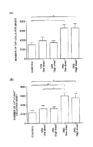
Cell-derived Neural Progenitor Cells
The number of TH+ cells in a graft obtained four weeks after
transplantation was compared among five groups. When the
double-label immunohistochemistry was performed, the number of Mr
cells in a graft of a SCID rat administered with ZNS at a high dose was
remarkably large as compared with that in a graft of a control SOD rat
(the numbers of which were respectively 6480 2145 cells and 3026
1349 cells, Figure 8(A). A symbol ''*" used in the drawing indicates p
< 0.05). While a ratio of cells co-expressing FOXA2, that is, a
midbrain marker, in the TH- cells was 763 9.7% in the graft of the
control SCID rat, 91.8 6.2% of the 'CH+ cells were FOXA2- in the
graft of the SCID rat administered with ZNS at a high dose. It was
28
CA 02912345 2015-11-12
= =
FP13-0650-00
revealed, through statistical analysis, that the number of midbrPin
dopaminergic neurons (MI+ FOXA2+) in a graft of a SCID rat
administered with ZNS at a high dose is significantly increased as
compared with that in a graft of a control SCID rat (the numbers of
which were respectively 5889 1821 cells and 2297 1116 cells,
Figure 8(B). Symbols "*" and "*" used in the drawing respectively
indicate p < 0.05 and p < 0.01.). These results suggested that the
retention rate of dopaminergic neurons differentiated from transplanted
neural progenitor cells is improved by the systemic administration of
ZNS.
Industrial Applicability
[0056] A transplantation adjuvant of the present invention is useful for
improving, in transplantation of neural progenitor cells, particularly iPS
cell-derived neural progenitor cells, a retention rate of dopaminergic
neurons in a transplantation site of a recipient's brain. Besides, if the
tronsplantation adjuvant contains valproic acid, the transplantation
adjuvant is also useful for accelerating differentiation of the neural
progenitor cells into dopaminergic neurons.
29