Note: Descriptions are shown in the official language in which they were submitted.
CA 02912379 2015-11-12
WO 2014/197616
PCT/US2014/040938
REPEATED ADMINISTRATION OF NON-IMMUNOSUPPRESSIVE ANTIGEN-
SPECIFIC IMMUNOTHERAPEUTICS
RELATED APPLICATIONS
This application claims the benefit under 35 U.S.C. 119 of United States
provisional
application 61/831,128, filed June 4, 2013, the entire contents of which are
incorporated
herein by reference.
FIELD OF THE INVENTION
This invention relates to repeated administration of antigen-specific
immunotherapeutics using protocols, or elements thereof, that do not induce
immunosuppression. In some embodiments, the protocol has been previously shown
not to
induce immunosuppression in a subject upon repeated administration.
BACKGROUND OF THE INVENTION
Certain diseases or conditions, such as autoimmune diseases, allergies, or
genetic or
acquired deficiencies requiring protein or enzyme replacement therapies, and
diseases
requiring biological therapies, often result in undesired immune responses.
Such undesired
immune responses may be reduced through the use of immunomodulator drugs.
Conventional immunomodulator drugs, however, are broad-acting. Additionally,
in order to
maintain immunosuppression, immunomodulator drug therapy is generally a life-
long
proposition. Unfortunately, the use of broad-acting immunomodulators are
associated with a
risk of severe side effects, such as tumors, infections, nephrotoxicity and
metabolic disorders.
Accordingly, new immunomodulator therapies would be beneficial.
SUMMARY OF THE INVENTION
In one aspect, a method comprising determining a protocol for repeatedly
administering an antigen-specific immunotherapeutic that does not result in
immunosuppression in a subject; and administering repeatedly the antigen-
specific
immunotherapeutic to another subject using one or more elements of the
protocol is provided.
In another aspect, a method comprising determining a protocol for repeatedly
administering an antigen-specific immunotherapeutic that does not result in
immunosuppression in a subject, wherein the determining comprises
administering repeatedly
the antigen-specific immunotherapeutic to a subject is provided.
2 con-mn 1 rst-lr,
CA 02912379 2015-11-12
WO 2014/197616
PCT/US2014/040938
- 2 -
In another aspect, a method comprising repeatedly administering to a subject
an
antigen-specific immunotherapeutic that comprises an antigen or an
immunomodulator,
wherein the antigen or immunomodulator is repeatedly administered according to
one or
more elements of a protocol that does not induce immunosuppression upon
repeated
administration of the antigen or an immunomodulator.
In one embodiment of any one of the methods provided, the method further
comprises
obtaining or providing an antigen-specific immunotherapeutic. In another
embodiment of
any one of the methods provided herein, the determining further comprises
demonstrating
that one or more elements of the protocol results in antigen-specific
tolerance in the subject.
In another aspect, a composition comprising an antigen-specific
immunotherapeutic
that comprises an exogenous antigen or an exogenous immunomodulator in an
amount
previously demonstrated in a protocol not to induce immunosuppression upon
repeated
administration is provided. In one embodiment of any one of the compositions
provided
herein, the composition is a kit.
In one embodiment of any one of the methods or compositions provided, the
antigen-
specific immunotherapeutic is any one of the antigen-specific
immunotherapeutics as
provided herein.
In another embodiment of any one of the methods or compositions provided
herein,
the protocol is one that has been previously shown not to induce
immunosuppression.
In another embodiment of any one of the methods or compositions provided
herein,
the antigen or immunomodulator is present in an amount further shown to result
in antigen-
specific tolerance.
In another embodiment of any one of the methods or compositions provided
herein,
the antigen-specific immunotherapeutic comprises an exogenous immunomodulator.
In
another embodiment of any one of the methods or compositions provided herein,
the
exogenous immunomodulator comprises a/an statin; mTOR inhibitor; TGF-I3
signaling
agent; TGF-I3 receptor agonist; histone deacetylase inhibitor; corticosteroid;
inhibitor of
mitochondrial function; P38 inhibitor; NF-K13 inhibitor; lectin receptor
ligand; adenosine
receptor agonist; prostaglandin E2 agonist; phosphodiesterase inhibitor;
proteasome inhibitor;
kinase inhibitor; G-protein coupled receptor agonist; G-protein coupled
receptor antagonist;
glucocorticoid; retinoid; cytokine inhibitor; cytokine receptor inhibitor;
cytokine receptor
activator; peroxisome proliferator-activated receptor antagonist; peroxisome
proliferator-
activated receptor agonist; histone deacetylase inhibitor; calcineurin
inhibitor; phosphatase
CA 02912379 2015-11-12
WO 2014/197616
PCT/US2014/040938
- 3 -
inhibitor; oxidized ATP; IDO; vitamin D3; cyclosporine A; aryl hydrocarbon
receptor
inhibitor; resveratrol; azathiopurine; 6-mercaptopurine; aspirin; niflumic
acid; estriol;
tripolide; interleukin; cyclosporine A, or siRNA targeting cytokines or
cytokine receptors. In
another embodiment of any one of the methods or compositions provided
herein,the
exogenous immunomodulator comprises rapamycin, mycophenolic acid or a CD22
ligand.
In another embodiment of any one of the methods or compositions provided
herein,
the antigen-specific immunotherapeutic comprises an exogenous antigen.
In another embodiment of any one of the methods or compositions provided
herein,
when the antigen-specific immunotherapeutic also comprises an exogenous
immunomodulator, the exogenous antigen and exogenous immunomodulator are not
coupled
to each other. In another embodiment of any one of the methods or compositions
provided
herein, the repeated administration comprises concomitant repeated
administration of the
exogenous antigen and exogenous immunomodulator.
In another embodiment of any one of the methods or compositions provided
herein,
the exogenous antigen comprises a therapeutic protein, modified antigen or
expressed
antigen. In another embodiment of any one of the methods or compositions
provided herein,
the expressed antigen is expressed from modified messenger RNA.
In another embodiment of any one of the methods or compositions provided
herein,
the antigen-specific immunotherapeutic results in antigen-specific tolerance
to an endogenous
antigen.
In another embodiment of any one of the methods or compositions provided
herein,
the endogenous antigen comprise an autoantigen.
In another embodiment of any one of the methods or compositions provided
herein,
the autoantigen comprises those found in Anklosing spondylitis; bulous
pemiphigous;
rheumatoid arthritis; multiple sclerosis; diabetes; excema; inflammatory bowel
disease; lupus
or systemic lupus erythematosus; multiple sclerosis; primary biliary
cirrhosis; psoriasis;
sarcoidosis; systemic sclerosis; scleroderma; thyroiditis; autoimmune thyroid
disease;
Hashimoto's thyroiditis; thyrotoxicosis; alopecia areata; Grave's disease;
Guillain-Barre
syndrome; celiac disease; Sjogren's syndrome; rheumatic fever; gastritis
autoimmune
atrophic gastritis; autoimmune hepatitis; insulitis; oophoritis; orchitis;
uveitis; phacogenic
uveitis; myasthenia gravis; primary myxoedema; pernicious anemia; primary
sclerosing
cholangitis; autoimmune haemolytic anemia; Addison's disease; scleroderma;
Goodpasture's
syndrome; nephritis; psoriasis; pemphigus vulgaris; pemphigoid; sympathetic
opthalmia;
CA 02912379 2015-11-12
WO 2014/197616
PCT/US2014/040938
- 4 -
idiopathic thrombocylopenic purpura; idiopathic feucopenia; Wegener's
granulomatosis or
poly/dermatomyositis.
In another embodiment of any one of the methods or compositions provided
herein,
the antigen-specific immunotherapeutic comprises an exogenous antigen and
results in
antigen-specific tolerance when administered in the presence of an endogenous
immunomodulator.
In another embodiment of any one of the methods or compositions provided
herein,
the endogenous immunomodulator comprises a substance and/or combination of
substances
involved in apoptosis or related signalling, a substance and/or combination of
substances
involved in T or B cell biology, or a substance and/or combination of
substances involved in
dendritic cell biology.
In another embodiment of any one of the methods or compositions provided
herein,
the repeated administration occurs 1 week to 10 years after an initial dose or
a previous
repeated administration of the antigen-specific immunotherapeutic. In another
embodiment of
any one of the methods or compositions provided herein, the repeated
administration occurs 1
week after an initial dose or a previous repeated administration of the
antigen-specific
immunotherapeutic. In another embodiment of any one of the methods or
compositions
provided herein, the repeated administration occurs 2 weeks after an initial
dose or a previous
repeated administration of the antigen-specific immunotherapeutic. In another
embodiment
of any one of the methods or compositions provided herein, the repeated
administration
occurs 1 to 12 months after an initial dose or a previous repeated
administration of the
antigen-specific immunotherapeutic.
In another embodiment of any one of the methods or compositions provided
herein,
the antigen-specific immunotherapeutic comprises an exogenous antigen and
exogenous
immunomodulator, the exogenous antigen is repeatedly administered by a route
different
from the exogenous immunomodulator. In another embodiment of any one of the
methods or
compositions provided herein, repeated administration comprises concomitant
repeated
administration.
In another embodiment of any one of the methods or compositions provided
herein,
the antigen-specific immunotherapeutic comprises more than one exogenous
antigen.
In another embodiment of any one of the methods or compositions provided
herein,
when the antigen-specific immunotherapeutic comprises an exogenous antigen and
exogenous immunomodulator, the exogenous antigens are repeatedly administered
by a route
CA 02912379 2015-11-12
WO 2014/197616
PCT/US2014/040938
- 5 -
different from the exogenous immunomodulator. In another embodiment of any one
of the
methods or compositions provided herein, the repeated administration comprises
concomitant
repeated administration.
In another embodiment of any one of the methods or compositions provided
herein,
the exogenous antigen and exogenous immunomodulator are coupled to each other.
In
another embodiment of any one of the methods or compositions provided herein,
the
exogenous immunomodulator comprises ERY1 peptide.
In another embodiment of any one of the methods or compositions provided
herein,
the antigen-specific immunotherapeutic is repeatedly administered to another
subject using
all or substantially all of the elements of the protocol.
In another embodiment of any one of the methods or compositions provided
herein,
the antigen-specific immunotherapeutic comprises polymeric synthetic
nanocarriers coupled
to an exogenous immunomodulator.
In another embodiment of any one of the methods or compositions provided
herein, a
load of immunomodulator attached to the polymeric synthetic nanocarriers, on
average across
the polymeric synthetic nanocarriers, is between 0.1% and 50% (weight/weight).
In another
embodiment of any one of the methods or compositions provided herein, the load
is between
0.1% and 20% (weight/weight).
In another embodiment of any one of the methods or compositions provided
herein,
the load of immunomodulator of the exogenous immunodulator on average is at
least 95%,
97%, 98% or 99% (weight/weight).
In another embodiment of any one of the methods or compositions provided
herein,
when the antigen-specific immunotherapeutic comprises an exogenous
immunomodulator
and exogenous antigen, the polymeric synthetic nanocarriers are further
coupled to the
exogenous antigen. In another embodiment of any one of the methods or
compositions
provided herein, when the antigen-specific immunotherapeutic comprises an
exogenous
immunomodulator and exogenous antigen, the polymeric synthetic nanocarrier is
concomitantly administered with an exogenous antigen.
In another aspect, a method of manufacturing any one of the antigen-specific
immunotherapeutics provided herein is provided. In one embodiment, the method
comprises
producing or obtaining an exogenous antigen or an exogenous immunomodulator in
an
amount that does not induce immunosuppression upon repeated administration. In
another
embodiment of any one of the methods provided, the amount is in an amount
previously
CA 02912379 2015-11-12
WO 2014/197616
PCT/US2014/040938
- 6 -
demonstrated in a protocol not to induce immunosuppression upon repeated
administration.
In another embodiment of any one of the methods provided, the method further
comprises
determining the amount or the protocol.
In another aspect, an antigen-specific immunotherapeutic comprising an
exogenous
immunomodulator or an exogenous antigen for the manufacture of a medicament
for
achieving antigen-specific tolerance but not induction of immunosuppression in
a subject is
provided.
In another aspect, an antigen-specific immunotherapeutic comprising an
exogenous
immunomodulator or an exogenous antigen, for achieving antigen-specific
tolerance but not
induction of immunosuppression in a subject is provided. In one embodiment,
the antigen-
specific immunotherapeutic is for use in any one of the methods provided
herein.
In another embodiment of any one of the methods or compositions provided
herein,
the antigen-specific immunotherapeutic is any one of the antigen-specific
immunotherapeutics provided herein.
BRIEF DESCRIPTION OF FIGURES
Fig. 1 shows results from repeated administration of an antigen- specifc
immunotherapeutic comprising antigen and rapamycin.
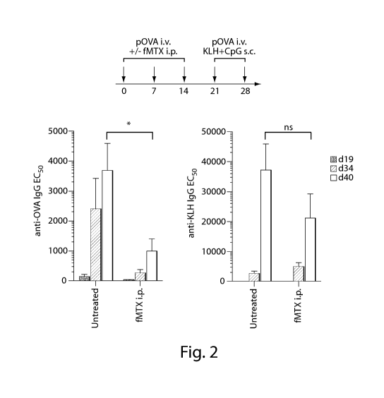
Fig. 2 shows results from repeated administration of an antigen-specific
immunotherapeutic comprising methotrexate, an exogenous immunomodulator.
Fig. 3 shows results from repeated administration of an antigen-specific
immunotherapeutic comprising methotrexate, an exogenous immunomodulator.
Fig. 4 demonstrates the deletion or anergy of CD8+ T cells with an exogenous
antigen
(OVA) attached to exogenous immunomodulator (ERY1 peptide).
DETAILED DESCRIPTION OF THE INVENTION
Before describing the present invention in detail, it is to be understood that
this
invention is not limited to particularly exemplified materials or process
parameters as such
may, of course, vary. It is also to be understood that the terminology used
herein is for the
purpose of describing particular embodiments of the invention only, and is not
intended to be
limiting of the use of alternative terminology to describe the present
invention.
All publications, patents and patent applications cited herein, whether supra
or infra,
are hereby incorporated by reference in their entirety for all purposes.
CA 02912379 2015-11-12
WO 2014/197616
PCT/US2014/040938
- 7 -
As used in this specification and the appended claims, the singular forms "a,"
"an"
and "the" include plural referents unless the content clearly dictates
otherwise. For example,
reference to "a polymer" includes a mixture of two or more such molecules or a
mixture of
differing molecular weights of a single polymer species, reference to "a
synthetic
nanocarrier" includes a mixture of two or more such synthetic nanocarriers or
a plurality of
such synthetic nanocarriers, reference to "a DNA molecule" includes a mixture
of two or
more such DNA molecules or a plurality of such DNA molecules, reference to "an
immunomodulator" includes a mixture of two or more such materials or a
plurality of
immunomodulator molecules, and the like.
As used herein, the term "comprise" or variations thereof such as "comprises"
or
"comprising" are to be read to indicate the inclusion of any recited integer
(e.g. a feature,
element, characteristic, property, method/process step or limitation) or group
of integers (e.g.
features, element, characteristics, properties, method/process steps or
limitations) but not the
exclusion of any other integer or group of integers. Thus, as used herein, the
term
"comprising" is inclusive and does not exclude additional, unrecited integers
or
method/process steps.
In embodiments of any of the compositions and methods provided herein,
"comprising" may be replaced with "consisting essentially of' or "consisting
of'. The phrase
"consisting essentially of" is used herein to require the specified integer(s)
or steps as well as
those which do not materially affect the character or function of the claimed
invention. As
used herein, the term "consisting" is used to indicate the presence of the
recited integer (e.g. a
feature, element, characteristic, property, method/process step or limitation)
or group of
integers (e.g. features, element, characteristics, properties, method/process
steps or
limitations) alone.
A. INTRODUCTION
As previously mentioned, current conventional immunomodulating compositions
are
broad acting and generally result in an overall systemic downregulation of the
immune
system. The compositions and methods provided herein allow for more targeted
immune
effects, particularly when the recited antigen-specific immunotherapeutics are
used in
repeated administration. Broad immunosuppression during repeated
administration is of
particular concern, because it generally would result in long-term
immunosuppression that
could lead to significant adverse events for the subjects receiving the
repeatedly administered
CA 02912379 2015-11-12
WO 2014/197616
PCT/US2014/040938
- 8 -
conventional immunomodulating compositions. Instead, the inventors have
discovered that it
is possible to provide antigen-specific immunomodulatory compositions and
methods that do
not result in long-term or broad immunosuppression during repeated
administration.
The inventors have unexpectedly and surprisingly discovered that the problems
and
limitations noted above can be overcome by practicing the invention disclosed
herein. In
particular, the inventors have unexpectedly discovered that it is possible to
provide methods
comprising determining a protocol for repeatedly administering an antigen-
specific
immunotherapeutic that does not result in immunosuppression in a subject; and
administering
repeatedly the antigen-specific immunotherapeutic to another subject using one
or more
elements of the protocol. Additionally, the inventors have unexpectedly
discovered that it is
possible to provide methods comprising: repeatedly administering to a subject
an antigen-
specific immunotherapeutic that comprises an antigen or an immunomodulator,
wherein the
antigen or immunomodulator is repeatedly administered according to one or more
elements
of a protocol that does not induce immunosuppression upon repeated
administration of the
antigen or immunomodulator. In some embodiments, the protocol is one that has
been
previously shown not to induce immunosuppression in a subject. Further, the
inventors have
unexpectedly discovered that it is possible to provide compositions
comprising: an antigen-
specific immunotherapeutic that comprises an exogenous antigen or an exogenous
immunomodulator in an amount that does not induce immunosuppression when
repeatedly
administered. In some embodiments, the amount is one that has been previously
demonstrated in a protocol not to induce immunosuppression upon repeated
administration in
a subject.
Various further embodiments and aspects of the invention, including different
types of
antigen-specific immunotherapeutics, different types of exogenous and
endogenous antigens,
and different types of exogenous and endogenous immunomodulators are disclosed
herein,
such as in the Examples.
The invention will now be described in more detail below.
B. DEFINITIONS
"Administering" or "administration" or "administer" means providing a material
to a
subject in a manner that is pharmacologically useful. The term is intended to
include causing
to be administered in some embodiments. "Causing to be administered" means
causing,
CA 02912379 2015-11-12
WO 2014/197616
PCT/US2014/040938
- 9 -
urging, encouraging, aiding, inducing or directing, directly or indirectly,
another party to
administer the material.
"An amount previously demonstrated in a protocol not to induce
immunosuppression
upon repeated administration" in the context of a composition, dosage form, or
method for
administration to a subject refers to an amount of the antigen or
immunomodulator that does
not induce immunosuppression upon repeated administration when administered
according to
a protocol previously demonstrated shown not to induce immunosuppression
Amounts effective will depend, of course, on the particular subject being
treated; the
severity of a condition, disease or disorder; the individual patient
parameters including age,
physical condition, size and weight; the duration of the treatment; the nature
of concurrent
therapy (if any); the specific route of administration and like factors within
the knowledge
and expertise of the health practitioner. These factors are well known to
those of ordinary
skill in the art and can be addressed with no more than routine
experimentation. It is
generally preferred that a maximum dose be used, that is, the highest safe
dose according to
sound medical judgment. It will be understood by those of ordinary skill in
the art, however,
that a patient may insist upon a lower dose or tolerable dose for medical
reasons,
psychological reasons or for virtually any other reason.
In certain embodiments, doses or amounts of the immunomodulators and/or
antigens
in the compositions of the invention can range from about 10 lig/kg to about
100,000 lig/kg.
In some embodiments, the doses can range from about 0.1 mg/kg to about 100
mg/kg. In still
other embodiments, the doses can range from about 0.1 mg/kg to about 25 mg/kg,
about 25
mg/kg to about 50 mg/kg, about 50 mg/kg to about 75 mg/kg or about 75 mg/kg to
about 100
mg/kg. Alternatively, the dose or amount can be administered based on the
number of
synthetic nanocarriers that provide the desired amount of immunomodulators
and/or antigens.
For example, useful doses or amounts include greater than 106, 107, 108, 109
or 1010 synthetic
nanocarriers (per dose). Other examples of useful doses or amounts include
from about
1x10⁶ to about 1x10¹⁰, about 1x10⁷ to about 1x10⁹ or about lx108 to about
1x10⁹ synthetic
nanocarriers (per dose).
"Antigen" means a B cell antigen or T cell antigen. "Type(s) of antigens"
means
molecules that share the same, or substantially the same, antigenic
characteristics. In some
embodiments, antigens may be proteins, polypeptides, peptides, lipoproteins,
glycolipids,
polynucleotides, polysaccharides or are contained or expressed in cells. In
some
embodiments, such as when the antigens are not well defined or characterized,
the antigens
CA 02912379 2015-11-12
WO 2014/197616
PCT/US2014/040938
- 10 -
may be contained within a cell or tissue preparation, cell debris, cell
exosomes, conditioned
media, etc. In some embodiments, the antigen can be endogenous or exogenous.
Endogenous antigen comprises antigen that is generated by a subject's own
body, and can
result in immune responses that can lead to antigen-specific tolerance with an
antigen-
specific immunotherapeutic, such as one that comprises exogenous
immunomodulator, upon
repeated administration as provided herein. In some embodiments, the
endogenous antigen
results in antigen-specific tolerance with the repeated administration of an
exogenous
immunomodulator as provided herein. Examples of endogenous antigen comprise
autoimmune antigens, some of which are disclosed elsewhere herein. Exogenous
antigen
comprises antigen that is administered as part of the antigen-specific
immunotherapeutic or as
part of some other therapeutic intervention, but is not generated by a
subject's own body.
Examples of exogenous antigens comprise environmental allergens, therapeutic
proteins or
polypeptides, etc. some of which are disclosed elsewhere herein.
"Antigen-specific" refers to any immune response that results from the
presence of
the antigen, or portion thereof, or that generates molecules that specifically
recognize or bind
the antigen. For example, where the immune response is antigen-specific
antibody
production, antibodies are produced that specifically bind the antigen. As
another example,
where the immune response is antigen-specific B cell or CD4+ T cell or CD8+ T
cell
activation, proliferation and/or activity, the activation, proliferation
and/or activity results
from recognition of the antigen, or portion thereof, alone or in complex with
MHC
molecules, B cells, etc.
"Antigen-specific immunotherapeutic" means a therapeutic agent that is capable
of
having a tolerogenic effect on a subject's immune response to an antigen of
interest. An
antigen-specific immunotherapeutic can comprise an antigen or an
immunomodulator. In
certain embodiments, antigen-specific immunotherapeutics can comprise both an
antigen and
an immunomodulator, wherein the antigen is coupled or uncoupled to the
immunomodulator.
In certain embodiments, antigen-specific immunotherapeutics can comprise an
antigen and an
immunomodulator that are not coupled to each other and the antigen and
immunomodulator
are repeatedly administered concomitantly. In such embodiments, the antigen
and
immunomodulator may be administered in the same composition or as separate
compositions,
and it is the totality of the compositions comprising the antigen or
immunomdoulator that
constitutes the antigen-specific immunotherapeutic. In embodiments, antigen-
specific
immunotherapeutics that comprise antigens (i.e. exogenous antigens) and/or
CA 02912379 2015-11-12
WO 2014/197616
PCT/US2014/040938
- 11 -
immunomodulators (i.e. exogenous immunomodulators) may interact with
endogenous
immunomodulators and/or endogenous antigens, respectively, to preferably
result in or lead
to immune responses that can result in antigen-specific tolerance.
"Antigen-specific immunotherapeutic efficacy" means that, for an antigen of
interest
(Agi) the Agi IgG titer (reported as EC50) changes from level of positive
control to a titer
(reported as EC50) at least 50% lower, with same Agi dosing. See generally J.
R. Crowther,
"ELISA: Theory and Practice" (1995 Humana Press).
"Average", as used herein, refers to the arithmetic mean unless otherwise
noted.
"B cell antigen" means any antigen that is recognized by and triggers an
immune
response in a B cell (e.g., an antigen that is specifically recognized by a B
cell or a receptor
thereon). In some embodiments, an antigen that is a T cell antigen is also a B
cell antigen. In
other embodiments, the T cell antigen is not also a B cell antigen. B cell
antigens include,
but are not limited to proteins, peptides, etc. In some embodiments, the B
cell antigen
comprises a non-protein antigen (i.e., not a protein or peptide antigen).
"Causing" means to make an action happened either directly or indirectly (for
example through a third party). In embodiments, the invention comprises
causing the
antigen-specific immunotherapeutic to be repeatedly administered to another
subject using
one or more elements of the protocol.
"Combination", as applied to two or more materials and/or agents (also
referred to
herein as the components), is intended to define material in which the two or
more materials
/agents are associated. Components may separately identified, e.g. first
component, second
component, third component, etc. The terms "combined" and "combining" in this
context are
to be interpreted accordingly.
The association of the two or more materials /agents in a combination may be
physical or non-physical. Examples of physically associated combined
materials/agents
include:
= compositions (e.g. unitary formulations) comprising the two or more
materials/agents
in admixture (for example within the same unit dose);
= compositions comprising material in which the two or more
materials/agents are
chemically/physicochemically linked (for example by crosslinking, molecular
agglomeration or binding to a common vehicle moiety);
CA 02912379 2015-11-12
WO 2014/197616
PCT/US2014/040938
- 12 -
= compositions comprising material in which the two or more
materials/agents are
chemically/physicochemically co-packaged (for example, disposed on or within
lipid
vesicles, particles (e.g. micro- or nanoparticles) or emulsion droplets);
= pharmaceutical kits, pharmaceutical packs or patient packs in which the
two or more
materials/agents are co-packaged or co-presented (e.g. as part of an array of
unit
doses);
Examples of non-physically associated combined materials/agents include:
= material (e.g. a non-unitary formulation) comprising at least one of the
two or more
materials/agents together with instructions for the extemporaneous association
of the
at least one compound/agent to form a physical association of the two or more
materials/agents;
= material (e.g. a non-unitary formulation) comprising at least one of the
two or more
materials/agents together with instructions for combination therapy with the
two or
more materials/agents;
= material comprising at least one of the two or more materials/agents
together with
instructions for administration to a patient population in which the other(s)
of the two
or more materials/agents have been (or are being) administered;
= material comprising at least one of the two or more materials/agents in
an amount or
in a form which is specifically adapted for use in combination with the
other(s) of the
two or more materials/agents.
As used herein, the term "combination therapy" is intended to define therapies
which
comprise the use of a combination of two or more materials/agents (as defined
herein). Thus,
references to "combination therapy", "combinations" and the use of
materials/agents "in
combination" in this application may refer to materials/agents that are
administered as part of
the same overall treatment regimen. As such, the posology of each of the two
or more
materials/agents may differ: each may be administered at the same time or at
different times.
It will therefore be appreciated that the materials/agents of the combination
may be
administered sequentially (e.g. before or after) or simultaneously, either in
the same
pharmaceutical formulation (i.e. together), or in different pharmaceutical
formulations (i.e.
separately). Simultaneously in the same formulation is as a unitary
formulation whereas
simultaneously in different pharmaceutical formulations is non-unitary. The
posologies of
CA 02912379 2015-11-12
WO 2014/197616
PCT/US2014/040938
- 13 -
each of the two or more materials/agents in a combination therapy may also
differ with
respect to the route of administration.
"Concomitantly" means administering two or more materials/agents to a subject
in a
manner that is correlated in time, preferably sufficiently correlated in time
so as to provide a
modulation in an immune response, and even more preferably the two or more
materials/agents are administered in combination. In embodiments, concomitant
administration may encompass administration of two or more materials/agents
within a
specified period of time, preferably within 1 month, more preferably within 1
week, still
more preferably within 1 day, and even more preferably within 1 hour. In
embodiments, the
materials/agents may be repeatedly administered concomitantly; that is
concomitant
administration on more than one occasion.
"Couple" or "Coupled" or "Couples" (and the like) means to chemically
associate one
entity (for example a moiety) with another. In some embodiments, the coupling
is covalent,
meaning that the coupling occurs in the context of the presence of a covalent
bond between
the two entities. In non-covalent embodiments, the non-covalent coupling is
mediated by
non-covalent interactions including but not limited to charge interactions,
affinity
interactions, metal coordination, physical adsorption, host-guest
interactions, hydrophobic
interactions, TT stacking interactions, hydrogen bonding interactions, van der
Waals
interactions, magnetic interactions, electrostatic interactions, dipole-dipole
interactions,
and/or combinations thereof. In embodiments, encapsulation is a form of
coupling.
"Determining" or "determine" or "demonstrating" or "demonstrate" means to
ascertain a factual relationship. These terms mean establishing a connection
between one or
more inputs, for example the elements of a protocol or the entire protocol,
and one or more
outputs, for example the presence or absence of immunosuppression or the
achievement of
antigen-specific tolerance. In embodiments, the invention encompasses
determining that one
or more elements of a protocol for repeatedly administering an antigen-
specific
immunotherapeutic do not result in immunosuppression in a subject.
Determining, etc. may be accomplished in a number of ways, including but not
limited to performing experiments, or making projections. For instance, one or
more
elements of a protocol, such as a dose of an immunomodulator, may be
determined by
starting with one or more elements of a test protocol, such as a test dose,
and using known
scaling techniques (such as allometric or isometric scaling) to determine the
protocol, such as
the dose, for administration. In another embodiment, one or more elements of a
protocol,
CA 02912379 2015-11-12
WO 2014/197616
PCT/US2014/040938
- 14 -
such as a dose, may be determined by testing variations in the one or more
elements, such as
various doses in a subject, e.g. through direct experimentation based on
experience and
guiding data. In embodiments, "determining" or "determine" or "demonstrating"
or
"demonstrate" comprises "causing to be determined" "or causing to be
demonstrated".
"Causing to be determined" "or causing to be demonstrated" means causing,
urging,
encouraging, aiding, inducing or directing or acting in coordination with an
entity for the
entity to ascertain a factual relationship; including directly or indirectly,
or expressly or
impliedly.
"Dosage form" means a pharmacologically and/or immunologically active material
in
a medium, carrier, vehicle, or device suitable for administration to a
subject. Any one of the
compositions or doses provided herein may be in a dosage form.
"Encapsulate" means to enclose at least a portion of a substance within a
synthetic
nanocarrier. In some embodiments, a substance is enclosed completely within a
synthetic
nanocarrier. In other embodiments, most or all of a substance that is
encapsulated is not
exposed to the local environment external to the synthetic nanocarrier. In
other
embodiments, no more than 50%, 40%, 30%, 20%, 10% or 5% (weight/weight) is
exposed to
the local environment. Encapsulation is distinct from absorption, which places
most or all of
a substance on a surface of a synthetic nanocarrier, and leaves the substance
exposed to the
local environment external to the synthetic nanocarrier.
"Immunomodulator" means a compound or combination of compounds that causes an
APC (Antigen Presenting Cell) to have a tolerogenic effect. A tolerogenic
effect generally
refers to the production or expression of cytokines or other factors by the
APC or changes in
the genetic expression profile of the APCs (e.g, changes in co-stimulatory
molecule
expression) that reduces, inhibits or prevents an undesired antigen-specific
immune response
or that promotes a desired antigen-specific tolerogenic immune response. In
some
embodiments, the immunomodulator can be endogenous or exogenous. Endogenous
immunomodulators comprise immunomodulators that are generated by a subject's
own body,
and can result in immune responses that can lead to antigen-specific tolerance
with an
antigen-specific immunotherapeutic, such as one comprising exogenous antigen,
upon
repeated administration as provided herein. In some embodiments, the
endogenous
immunomodulator can result in antigen-specific tolerance when an exogenous
antigen is
administered as provided herein. Examples of endogenous immunomodulators
comprise
apoptotic cells and other apoptotic ligands or markers, tolerogenic cytokines
such as IL-10,
CA 02912379 2015-11-12
WO 2014/197616
PCT/US2014/040938
- 15 -
and cell surface markers implicated in tolerogenic responses such as CD22.
Exogenous
immunomodulators comprise immunomodulators that are administered as part of
the antigen-
specific immunotherapeutic or as part of some other therapeutic intervention,
but are not
generated by a subject's own body. Examples of exogenous immunomodulators
comprise
rapamcycin and other immunomodulators disclosed herein.
In one embodiment, the immunomodulator is one that causes an APC to promote a
regulatory phenotype in one or more immune effector cells. For example, the
regulatory
phenotype may be characterized by the inhibition of the production, induction,
stimulation or
recruitment of antigen-specific CD4+ T cells or B cells, the inhibition of the
production of
antigen-specific antibodies, the production, induction, stimulation or
recruitment of Treg cells
(e.g., CD4+CD25highFoxP3+ Treg cells), etc. This may be the result of the
conversion of
CD4+ T cells or B cells to a regulatory phenotype. This may also be the result
of induction
of FoxP3 in other immune cells, such as CD8+ T cells, macrophages and iNKT
cells. In one
embodiment, the immunomodulator is one that affects the response of the APC
after it
processes an antigen. In another embodiment, the immunomodulator is not one
that
interferes with the processing of the antigen. In a further embodiment, the
immunomodulator
is not an apoptotic-signaling molecule. In another embodiment, the
immunomodulator is not
a phospholipid.
Immunomodulators include, but are not limited to, statins; mTOR inhibitors,
such as
rapamycin or a rapamycin analog; TGF-I3 signaling agents; TGF-I3 receptor
agonists; histone
deacetylase inhibitors, such as Trichostatin A; corticosteroids; inhibitors of
mitochondrial
function, such as rotenone; P38 inhibitors; NF-K13 inhibitors, such as 6Bio,
Dexamethasone,
TCPA-1, IKK VII; adenosine receptor agonists; prostaglandin E2 agonists
(PGE2), such as
Misoprostol; phosphodiesterase inhibitors, such as phosphodiesterase 4
inhibitor (PDE4),
such as Rolipram; proteasome inhibitors; kinase inhibitors; G-protein coupled
receptor
agonists; G-protein coupled receptor antagonists; glucocorticoids; retinoids;
cytokine
inhibitors; cytokine receptor inhibitors; cytokine receptor activators;
peroxisome proliferator-
activated receptor antagonists; peroxisome proliferator-activated receptor
agonists; histone
deacetylase inhibitors; calcineurin inhibitors; phosphatase inhibitors; P13 KB
inhibitors, such
as TGX-221; autophagy inhibitors, such as 3-Methyladenine; aryl hydrocarbon
receptor
inhibitors; proteasome inhibitor I (PSI); and oxidized ATPs, such as P2X
receptor blockers.
Immunomodulators also include IDO, vitamin D3, cyclosporins, such as
cyclosporine A, aryl
hydrocarbon receptor inhibitors, resveratrol, azathiopurine (Aza), 6-
mercaptopurine (6-MP),
CA 02912379 2015-11-12
WO 2014/197616
PCT/US2014/040938
- 16 -6-thioguanine (6-TG), FK506, sanglifehrin A, salmeterol, mycophenolate
mofetil (MMF),
aspirin and other COX inhibitors, niflumic acid, estriol and triptolide. In
embodiments, the
immunomodulator may comprise any one of the agents provided herein.
The immunomodulator can be a compound that directly provides the tolerogenic
effect on APCs or it can be a compound that provides the tolerogenic effect
indirectly (i.e.,
after being processed in some way after administration). Immunomodulators,
therefore,
include prodrug forms of any of the compounds provided herein.
Immunomodulators also include nucleic acids that encode the peptides,
polypeptides
or proteins provided herein that result in a tolerogenic immune response. In
embodiments,
therefore, the immunomodulator is a nucleic acid that encodes a peptide,
polypeptide or
protein that results in a tolerogenic immune response, and it is the nucleic
acid that is coupled
to the synthetic nanocarrier.
The nucleic acid may be DNA or RNA, such as mRNA. In embodiments, the
inventive compositions comprise a complement, such as a full-length
complement, or a
degenerate (due to degeneracy of the genetic code) of any of the nucleic acids
provided
herein. In embodiments, the nucleic acid is an expression vector that can be
transcribed when
transfected into a cell line. In embodiments, the expression vector may
comprise a plasmid
amongst others. Nucleic acids can be isolated or synthesized using standard
molecular
biology approaches, for example by using a polymerase chain reaction to
produce a nucleic
acid fragment, which is then purified and cloned into an expression vector.
Additional
techniques useful in the practice of this invention may be found in Current
Protocols in
Molecular Biology 2007 by John Wiley and Sons, Inc.; Molecular Cloning: A
Laboratory
Manual (Third Edition) Joseph Sambrook, Peter MacCallum Cancer Institute,
Melbourne,
Australia; David Russell, University of Texas Southwestern Medical Center,
Dallas, Cold
Spring Harbor.
In embodiments, the immunomodulators provided herein are coupled to synthetic
nanocarriers. In preferable embodiments, the immunomodulator is an element
that is in
addition to the material that makes up the structure of the synthetic
nanocarrier. For example,
in one embodiment, where the synthetic nanocarrier is made up of one or more
polymers, the
immunomodulator is a compound that is in addition and coupled to the one or
more
polymers. As another example, in one embodiment, where the synthetic
nanocarrier is made
up of one or more lipids, the immunomodulator is again in addition and coupled
to the one or
more lipids. In embodiments, such as where the material of the synthetic
nanocarrier also
CA 02912379 2015-11-12
WO 2014/197616
PCT/US2014/040938
- 17 -
results in a tolerogenic effect, the immunomodulator is an element present in
addition to the
material of the synthetic nanocarrier that results in a tolerogenic effect.
Other exemplary immunomodulators include, but are not limited, small molecule
drugs, natural products, antibodies (e.g., antibodies against CD20, CD3, CD4),
biologics-
based drugs, carbohydrate-based drugs, nanoparticles, liposomes, RNAi,
antisense nucleic
acids, aptamers, methotrexate, NSAIDs; fingolimod; natalizumab; alemtuzumab;
anti-CD3;
tacrolimus (FK506), etc. Further immunomodulators, are known to those of skill
in the art,
and the invention is not limited in this respect.
In embodiments of any one of the methods or compositions provided herein, the
immunomodulator is in a form, such as a nanocrystalline form, whereby the form
of the
immunomodulator itself is a particle or particle-like. In embodiments, such
forms mimic a
virus or other foreign pathogen. Many drugs have been nanonized and
appropriate methods
for producing such drug forms would be known to one of ordinary skill in the
art. Drug
nanocrystals, such as nanocrystalline rapamycin are known to those of ordinary
skill in the art
(Katteboinaa, et al. 2009, International Journal of PharmTech Resesarch; Vol.
1, No. 3;
pp682-694. As used herein a "drug nanocrystal" refers to a form of a drug
(e.g., an
immunomodulator) that does not include a carrier or matrix material. In some
embodiments,
drug nanocrystals comprise 90%, 95%, 98%, or 99% or more drug. Methods for
producing
drug nanocrystals include, without limitation, milling, high pressure
homogenization,
precipitation, spray drying, rapid expansion of supercritical solution (RESS),
Nanoedge
technology (Baxter Healthcare), and Nanocrystal TechnologyTm (Elan
Corporation). In some
embodiments, a surfactant or a stabilizer may be used for steric or
electrostatic stability of the
drug nanocrystal. In some embodiments the nanocrystal or nanocrytalline form
of an
immunomodulator may be used to increase the solubility, stability, and/or
bioavailability of
the immunomodulator, particularly immunomodulators that are insoluble or
labile.
"Immunosuppression" means (1) non-durable statistically-significant
downregulation
of an immune response as a result of repeated administration of an antigen-
specific
immunotherapeutic, or (2) the response of a non-human test subject to a KLH
challenge T-
cell dependent antibody response ELISA assay, assuming that KLH is not the
antigen of
interest, following at least one repeated administration of an antigen-
specific
immunotherapeutic, wherein the response is characterized as the KLH IgG titer
(reported as
EC50) changing from level of positive control to a titer (reported as EC50)
equivalent to, or
less than, 3 standard deviations above the mean negative control
("background"), with same
CA 02912379 2015-11-12
WO 2014/197616
PCT/US2014/040938
- 18 -
KLH dosing. See generally J. R. Crowther, "ELISA: Theory and Practice" (1995
Humana
Press). In a preferred embodiment, non-durable statistically-significant
downregulation
means that the downregulation (treatment arm measured against non-treatment
arm) does not
evidence a statistically-significant difference for longer than a week
following the last
repeated administration of the antigen-specific immunotherapeutic. Various
inventive
compositions, methods, protocols, and dosages forms do not result in, or do
not induce,
immunosuppression.
KLH challenge ELISA assays are described generally in the literature, for
example in
J.T. Brisbin et al., Influence of In-Feed Virginiamycin on the Systemic and
Mucosal
Antibody Response of Chickens, Poultry Science 87:1995-1999 (2008); or may be
obtained
commercially, for example from Stellar Biotechnologies (332 East Scott Street,
Port
Hueneme, California 93041 USA) as Item ELI-01G Mouse Anti-KLH IgG ELISA Kit,
or
ELI-03G NHP Anti-KLH IgG ELISA Kit.
An ELISA method for measuring an anti-KLH antibody titer, for example, a
typical
sandwich ELISA, may consist of the following steps (i) preparing an ELISA-
plate coating
material such that the antibody target of interest is coupled to a substrate
polymer or other
suitable material (ii) preparing the coating material in an aqueous solution
(such as PBS) and
delivering the coating material solution to the wells of a multiwell plate for
overnight
deposition of the coating onto the multiwell plate (iii) thoroughly washing
the multiwell plate
with wash buffer (such as 0.05% Tween-20 in PBS) to remove excess coating
material (iv)
blocking the plate for nonspecific binding by applying a diluent solution
(such as 10% fetal
bovine serum in PBS), (v) washing the blocking/diluent solution from the plate
with wash
buffer (vi) diluting the serum sample(s) containing antibodies and appropriate
standards
(positive controls) with diluent as required to obtain a concentration that
suitably saturates the
ELISA response (vii) serially diluting the plasma samples on the multiwell
plate such to
cover a range of concentrations suitable for generating an ELISA response
curve (viii)
incubating the plate to provide for antibody-target binding (ix) washing the
plate with wash
buffer to remove antibodies not bound to antigen (x) adding an appropriate
concentration of a
secondary detection antibody in same diluent such as a biotin-coupled
detection antibody
capable of binding the primary antibody (xi) incubating the plate with the
applied detection
antibody, followed by washing with wash buffer (xii) adding an enzyme such as
streptavidin-
HRP (horse radish peroxidase) that will bind to biotin found on biotinylated
antibodies and
incubating (xiii) washing the multiwell plate (xiv) adding substrate(s) (such
as TMB
CA 02912379 2015-11-12
WO 2014/197616
PCT/US2014/040938
- 19 -
solution) to the plate (xv) applying a stop solution (such as 2N sulfuric
acid) when color
development is complete (xvi) reading optical density of the plate wells at a
specific
wavelength for the substrate (450 nm with subtraction of readings at 570 nm)
(xvi) applying a
suitable multiparameter curve fit to the data and defining half-maximal
effective
concentration (EC50) as the concentration on the curve at which half the
maximum OD value
for the plate standards is achieved.
"Load" is the amount of the immunomodulator of an exogenous immunomodulator
composition (weight/weight). For example, when attached to a synthetic
nanocarrier, the
load is based on the total dry recipe weight of materials in an entire
synthetic nanocarrier
(weight/weight). Generally, such a load is calculated as an average across a
population of
synthetic nanocarriers. In one embodiment, the load on average across the
synthetic
nanocarriers is between 0.1% and 99%. In another embodiment, the load is
between 0.1%
and 50%. In another embodiment, the load of the immunomodulator is between
0.1% and
20%. In another embodiment, the load of the immunomodulator is no more than
25% on
average across a population of synthetic nanocarriers. In embodiments, the
load is calculated
as may be described in the Examples or as otherwise known in the art.
As another examples, when the form of the immunomodulator is itself a particle
or
particle-like, such as a nanocrystalline immunomodulator, the load of
immunomodulator is
the amount of the immunomodulator in the particles or the like
(weight/weight). In such
embodiments, the load can approach 90%, 95%, 97%, 98%, 99% or more.
"Maximum dimension of a synthetic nanocarrier" means the largest dimension of
a
nanocarrier measured along any axis of the synthetic nanocarrier. "Minimum
dimension of a
synthetic nanocarrier" means the smallest dimension of a synthetic nanocarrier
measured
along any axis of the synthetic nanocarrier. For example, for a spheroidal
synthetic
nanocarrier, the maximum and minimum dimension of a synthetic nanocarrier
would be
substantially identical, and would be the size of its diameter. Similarly, for
a cuboidal
synthetic nanocarrier, the minimum dimension of a synthetic nanocarrier would
be the
smallest of its height, width or length, while the maximum dimension of a
synthetic
nanocarrier would be the largest of its height, width or length. In an
embodiment, a
minimum dimension of at least 75%, preferably at least 80%, more preferably at
least 90%,
of the synthetic nanocarriers in a sample, based on the total number of
synthetic nanocarriers
in the sample, is equal to or greater than 100 nm. In an embodiment, a maximum
dimension
of at least 75%, preferably at least 80%, more preferably at least 90%, of the
synthetic
CA 02912379 2015-11-12
WO 2014/197616
PCT/US2014/040938
- 20 -
nanocarriers in a sample, based on the total number of synthetic nanocarriers
in the sample, is
equal to or less than 5 m. Preferably, a minimum dimension of at least 75%,
preferably at
least 80%, more preferably at least 90%, of the synthetic nanocarriers in a
sample, based on
the total number of synthetic nanocarriers in the sample, is greater than 110
nm, more
preferably greater than 120 nm, more preferably greater than 130 nm, and more
preferably
still greater than 150 nm. Aspects ratios of the maximum and minimum
dimensions of
inventive synthetic nanocarriers may vary depending on the embodiment. For
instance,
aspect ratios of the maximum to minimum dimensions of the synthetic
nanocarriers may vary
from 1:1 to 1,000,000:1, preferably from 1:1 to 100,000:1, more preferably
from 1:1 to
10,000:1, more preferably from 1:1 to 1000:1, still more preferably from 1:1
to 100:1, and yet
more preferably from 1:1 to 10:1. Preferably, a maximum dimension of at least
75%,
preferably at least 80%, more preferably at least 90%, of the synthetic
nanocarriers in a
sample, based on the total number of synthetic nanocarriers in the sample is
equal to or less
than 3 1.tm, more preferably equal to or less than 21.tm, more preferably
equal to or less than 1
1.tm, more preferably equal to or less than 800 nm, more preferably equal to
or less than 600
nm, and more preferably still equal to or less than 500 nm. In preferred
embodiments, a
minimum dimension of at least 75%, preferably at least 80%, more preferably at
least 90%,
of the synthetic nanocarriers in a sample, based on the total number of
synthetic nanocarriers
in the sample, is equal to or greater than 100 nm, more preferably equal to or
greater than 120
nm, more preferably equal to or greater than 130 nm, more preferably equal to
or greater than
140 nm, and more preferably still equal to or greater than 150 nm. Measurement
of synthetic
nanocarrier dimensions (e.g., diameter) is obtained by suspending the
synthetic nanocarriers
in a liquid (usually aqueous) media and using dynamic light scattering (DLS)
(e.g. using a
Brookhaven ZetaPALS instrument). For example, a suspension of synthetic
nanocarriers can
be diluted from an aqueous buffer into purified water to achieve a final
synthetic nanocarrier
suspension concentration of approximately 0.01 to 0.1 mg/mL. The diluted
suspension may
be prepared directly inside, or transferred to, a suitable cuvette for DLS
analysis. The cuvette
may then be placed in the DLS, allowed to equilibrate to the controlled
temperature, and then
scanned for sufficient time to acquire a stable and reproducible distribution
based on
appropriate inputs for viscosity of the medium and refractive indicies of the
sample. The
effective diameter, or mean of the distribution, is then reported. Determining
the effective
sizes of high aspect ratio, or non-spheroidal, synthetic nanocarriers may
require augmentative
techniques, such as electron microscopy, to obtain more accurate measurements.
CA 02912379 2015-11-12
WO 2014/197616
PCT/US2014/040938
- 21 -
"Dimension" or "size" or "diameter" of synthetic nanocarriers means the mean
of a particle
size distribution, for example, obtained using dynamic light scattering.
"Pharmaceutically acceptable excipient" or "pharmaceutically acceptable
carrier"
means a pharmacologically inactive material used together with a
pharmacologically active
material to formulate the compositions. Pharmaceutically acceptable excipients
comprise a
variety of materials known in the art, including but not limited to
saccharides (such as
glucose, lactose, and the like), preservatives such as antimicrobial agents,
reconstitution aids,
colorants, saline (such as phosphate buffered saline), and buffers.
"Protocol" means a pattern of repeatedly administering antigen-specific
immunotherapeutics to a subject. Protocols are made up of elements; thus a
protocol
comprises one or more elements. Such elements of the protocol can comprise
dosing
amounts, dosing frequency, routes of administration, dosing duration, dosing
rates, intervals
between dosing, combinations of any of the foregoing, and the like. In some
embodiments, a
protocol may be used to administer one or more compositions of the invention
to one or more
test subjects. Immune responses in these test subjects can then be assessed to
determine
whether or not the protocol was effective in generating a desired or desired
level of an
immunologic effect. One or more of the elements may have been previously
demonstrated in
test subjects, such as non-human subjects, and then translated into human
protocols. For
example, dosing amounts demonstrated in non-human subjects can be scaled as an
element of
a human protocol using established techniques such as alimetric scaling or
other scaling
methods. Whether or not a protocol had a desired effect can be determined
using any of the
methods provided herein or otherwise known in the art. For example, a
population of cells
may be obtained from a subject to which a recited composition and/or antigen-
specific
immunotherapeutic provided herein has been repeatedly administered according
to a specific
protocol in order to determine whether or not specific immune cells,
cytokines, antibodies,
etc. were reduced, generated, activated, etc. Useful methods for detecting the
presence and/or
number of immune cells include, but are not limited to, flow cytometric
methods (e.g.,
FACS) and immunohistochemistry methods. Antibodies and other binding agents
for
specific staining of immune cell markers, are commercially available. Such
kits typically
include staining reagents for multiple antigens that allow for FACS-based
detection,
separation and/or quantitation of a desired cell population from a
heterogeneous population of
cells. In embodiments, the antigen-specific immunotherapeutic is repeatedly
administered to
CA 02912379 2015-11-12
WO 2014/197616
PCT/US2014/040938
- 22 -
another subject using all or substantially all of the elements of which the
protocol is
comprised.
"Protocol previously shown not to induce immunosuppression upon repeated
administration" means a protocol wherein one or more of the elements of such
protocol (up to
and including the complete protocol) were demonstrated at a previous time not
to result in
immunosuppression during at least one point during, preferably the entirety
of, repeated
administration.
"Providing" means an action or set of actions that an individual performs that
supply a
needed item or set of items or methods for practicing of the present
invention. The action or
set of actions may be taken either directly oneself or indirectly.
"Providing a subject" is any action or set of actions that causes a clinician
to come in
contact with a subject and administer a composition provided herein thereto or
to perform a
method provided herein thereupon. Preferably, the subject is one who is in
need of a
tolerogenic immune response as provided herein. The action or set of actions
may be either
directly oneself or indirectly. In one embodiment of any one of the methods
provided herein,
the method further comprises providing a subject.
"Repeated administration" or "repeatedly administer"or "repeatedly
administering"
and the like means boosting or extending the persistence of a previously
established immune
tolerance. These embodiments generally involve one administration or a short
course of
treatment at a time when the established tolerance is declining or at risk of
declining.
Repeated administration begins upon the next dose or doses of the antigen-
specific
immunotherapeutic administered following administration of an initial dose of
an antigen-
specific immunotherapeutic. The initial antigen-specific immunotherapeutic
administered
may be the same or different (in terms of composition, dosing, etc.) from the
antigen-specific
immunotherapeutic administered during repeated administration. Boosting is
generally
performed 2 weeks to 1 year, and preferably 1 to 6 months after an initial
dose of the antigen-
specific immunotherapeutic or a previous repeated administration. This
invention also
includes embodiments that involve regular repeated administrations on a
schedule of
administrations that occur semiweekly, weekly, biweekly, or on any other
regular schedule.
"Subject" means animals, including warm blooded mammals such as humans and
primates; avians; domestic household or farm animals such as cats, dogs,
sheep, goats, cattle,
horses and pigs; laboratory animals such as mice, rats and guinea pigs; fish;
reptiles; zoo and
wild animals; and the like.
CA 02912379 2015-11-12
WO 2014/197616
PCT/US2014/040938
-23 -
"Synthetic nanocarrier(s)" means a discrete object that is not found in
nature, and that
possesses at least one dimension that is less than or equal to 5 microns in
size. Albumin
nanoparticles are generally included as synthetic nanocarriers, however in
certain
embodiments the synthetic nanocarriers do not comprise albumin nanoparticles.
In
embodiments, inventive synthetic nanocarriers do not comprise chitosan. In
other
embodiments, inventive synthetic nanocarriers are not lipid-based
nanoparticles. In further
embodiments, inventive synthetic nanocarriers do not comprise a phospholipid.
A synthetic nanocarrier can be, but is not limited to, one or a plurality of
lipid-based
nanoparticles (also referred to herein as lipid nanoparticles, i.e.,
nanoparticles where the
majority of the material that makes up their structure are lipids), polymeric
nanoparticles,
metallic nanoparticles, surfactant-based emulsions, dendrimers, buckyballs,
nanowires, virus-
like particles (i.e., particles that are primarily made up of viral structural
proteins but that are
not infectious or have low infectivity), peptide or protein-based particles
(also referred to
herein as protein particles, i.e., particles where the majority of the
material that makes up
their structure are peptides or proteins) (such as albumin nanoparticles)
and/or nanoparticles
that are developed using a combination of nanomaterials such as lipid-polymer
nanoparticles.
Synthetic nanocarriers may be a variety of different shapes, including but not
limited to
spheroidal, cuboidal, pyramidal, oblong, cylindrical, toroidal, and the like.
Synthetic
nanocarriers according to the invention comprise one or more surfaces.
Exemplary synthetic
nanocarriers that can be adapted for use in the practice of the present
invention comprise: (1)
the biodegradable nanoparticles disclosed in US Patent 5,543,158 to Gref et
al., (2) the
polymeric nanoparticles of Published US Patent Application 20060002852 to
Saltzman et al.,
(3) the lithographically constructed nanoparticles of Published US Patent
Application
20090028910 to DeSimone et al., (4) the disclosure of WO 2009/051837 to von
Andrian et
al., (5) the nanoparticles disclosed in Published US Patent Application
2008/0145441 to
Penades et al., (6) the protein nanoparticles disclosed in Published US Patent
Application
20090226525 to de los Rios et al., (7) the virus-like particles disclosed in
published US
Patent Application 20060222652 to Sebbel et al., (8) the nucleic acid coupled
virus-like
particles disclosed in published US Patent Application 20060251677 to Bachmann
et al., (9)
the virus-like particles disclosed in W02010047839A1 or W02009106999A2, (10)
the
nanoprecipitated nanoparticles disclosed in P. Paolicelli et al., "Surface-
modified PLGA-
based Nanoparticles that can Efficiently Associate and Deliver Virus-like
Particles"
Nanomedicine. 5(6):843-853 (2010), (11) apoptotic cells, apoptotic bodies or
the synthetic or
CA 02912379 2015-11-12
WO 2014/197616
PCT/US2014/040938
- 24 -
semisynthetic mimics disclosed in U.S. Publication 2002/0086049, or (12) those
of Look et
al., Nanogel-based delivery of mycophenolic acid ameliorates systemic lupus
erythematosus
in mice" J. Clinical Investigation 123(4):1741-1749(2013). In embodiments,
synthetic
nanocarriers may possess an aspect ratio greater than 1:1, 1:1.2, 1:1.5, 1:2,
1:3, 1:5, 1:7, or
greater than 1:10.
Synthetic nanocarriers according to the invention that have a minimum
dimension of
equal to or less than about 100 nm, preferably equal to or less than 100 nm,
do not comprise a
surface with hydroxyl groups that activate complement or alternatively
comprise a surface
that consists essentially of moieties that are not hydroxyl groups that
activate complement. In
a preferred embodiment, synthetic nanocarriers according to the invention that
have a
minimum dimension of equal to or less than about 100 nm, preferably equal to
or less than
100 nm, do not comprise a surface that substantially activates complement or
alternatively
comprise a surface that consists essentially of moieties that do not
substantially activate
complement. In a more preferred embodiment, synthetic nanocarriers according
to the
invention that have a minimum dimension of equal to or less than about 100 nm,
preferably
equal to or less than 100 nm, do not comprise a surface that activates
complement or
alternatively comprise a surface that consists essentially of moieties that do
not activate
complement. In embodiments, synthetic nanocarriers exclude virus-like
particles. In
embodiments, synthetic nanocarriers may possess an aspect ratio greater than
1:1, 1:1.2,
1:1.5, 1:2, 1:3, 1:5, 1:7, or greater than 1:10.
"T cell antigen" means a CD4+ T-cell antigen or CD8+ cell antigen. "CD4+ T-
cell
antigen" means any antigen that is recognized by and triggers an immune
response in a CD4+
T-cell e.g., an antigen that is specifically recognized by a T-cell receptor
on a CD4+T cell via
presentation of the antigen or portion thereof bound to a Class II major
histocompatability
complex molecule (MHC). "CD8+ T cell antigen" means any antigen that is
recognized by
and triggers an immune response in a CD8+ T-cell e.g., an antigen that is
specifically
recognized by a T-cell receptor on a CD8+T cell via presentation of the
antigen or portion
thereof bound to a Class I major histocompatability complex molecule (MHC). In
some
embodiments, an antigen that is a T cell antigen is also a B cell antigen. In
other
embodiments, the T cell antigen is not also a B cell antigen. T cell antigens
generally are
proteins or peptides.
A "therapeutic protein" refers to any protein or protein-based therapy that
may be
administered to a subject and have a therapeutic effect. Such therapies
include protein
CA 02912379 2015-11-12
WO 2014/197616
PCT/US2014/040938
- 25 -
replacement and protein supplementation therapies. Such therapies also include
the
administration of exogenous or foreign protein, antibody therapies, and cell
or cell-based
therapies. Therapeutic proteins include enzymes, enzyme cofactors, hormones,
blood clotting
factors, cytokines, growth factors, monoclonal antibodies and polyclonal
antibodies.
Examples of other therapeutic proteins are provided elsewhere herein.
Therapeutic proteins
may be produced in, on or by cells and may be obtained from such cells or
administered in
the form of such cells. In embodiments, the therapeutic protein is produced
in, on or by
mammalian cells, insect cells, yeast cells, bacteria cells, plant cells,
transgenic animal cells,
transgenic plant cells, etc. The therapeutic protein may be recombinantly
produced in such
cells. The therapeutic protein may also be produced in, on or by autologous
cells that have
been transfected, transduced or otherwise manipulated to express it.
Alternatively, the
therapeutic protein may be administered as a nucleic acid or by introducing a
nucleic acid
into a liposome, etc. Alternatively, the therapeutic protein may be obtained
from such forms
and administered as the therapeutic protein itself. Subjects, therefore,
include any subject
that has received, is receiving or will receive any of the foregoing.
"Undesired immune response" refers to any undesired immune response that
results
from exposure to an antigen, promotes or exacerbates a disease, disorder or
condition
provided herein (or a symptom thereof), or is symptomatic of a disease,
disorder or condition
provided herein. Such immune responses generally have a negative impact on a
subject's
health or is symptomatic of a negative impact on a subject's health. Undesired
immune
responses include antigen-specific antibody production, antigen-specific B
cell proliferation
and/or activity or antigen-specific CD4+ T cell proliferation and/or activity.
C. INVENTIVE COMPOSITIONS
Antigen-specific Immunotherapeutics
Synthetic Nanocarriers
In embodiments, the antigen-specific immunotherapeutics comprise synthetic
nanocarrier compositions that comprise an immunomodulator and/or an antigen,
together
with related methods.
A wide variety of synthetic nanocarriers can be used according to the
invention. In
some embodiments, synthetic nanocarriers are spheres or spheroids. In some
embodiments,
CA 02912379 2015-11-12
WO 2014/197616
PCT/US2014/040938
- 26 -
synthetic nanocarriers are flat or plate-shaped. In some embodiments,
synthetic nanocarriers
are cubes or cubic. In some embodiments, synthetic nanocarriers are ovals or
ellipses. In
some embodiments, synthetic nanocarriers are cylinders, cones, or pyramids.
In some embodiments, it is desirable to use a population of synthetic
nanocarriers that
is relatively uniform in terms of size, shape, and/or composition so that each
synthetic
nanocarrier has similar properties. For example, at least 80%, at least 90%,
or at least 95% of
the synthetic nanocarriers, based on the total number of synthetic
nanocarriers, may have a
minimum dimension or maximum dimension that falls within 5%, 10%, or 20% of
the
average diameter or average dimension of the synthetic nanocarriers.
Synthetic nanocarriers can be solid or hollow and can comprise one or more
layers. In
some embodiments, each layer has a unique composition and unique properties
relative to the
other layer(s). To give but one example, synthetic nanocarriers may have a
core/shell
structure, wherein the core is one layer (e.g. a polymeric core) and the shell
is a second layer
(e.g. a lipid bilayer or monolayer). Synthetic nanocarriers may comprise a
plurality of
different layers.
In some embodiments, synthetic nanocarriers may optionally comprise one or
more
lipids. In some embodiments, a synthetic nanocarrier may comprise a liposome.
In some
embodiments, a synthetic nanocarrier may comprise a lipid bilayer. In some
embodiments, a
synthetic nanocarrier may comprise a lipid monolayer. In some embodiments, a
synthetic
nanocarrier may comprise a micelle. In some embodiments, a synthetic
nanocarrier may
comprise a core comprising a polymeric matrix surrounded by a lipid layer
(e.g., lipid bilayer,
lipid monolayer, etc.). In some embodiments, a synthetic nanocarrier may
comprise a non-
polymeric core (e.g., metal particle, quantum dot, ceramic particle, bone
particle, viral
particle, proteins, nucleic acids, carbohydrates, etc.) surrounded by a lipid
layer (e.g., lipid
bilayer, lipid monolayer, etc.).
In other embodiments, synthetic nanocarriers may comprise metal particles,
quantum
dots, ceramic particles, etc. In some embodiments, a non-polymeric synthetic
nanocarrier is
an aggregate of non-polymeric components, such as an aggregate of metal atoms
(e.g., gold
atoms).
In some embodiments, synthetic nanocarriers may optionally comprise one or
more
amphiphilic entities. In some embodiments, an amphiphilic entity can promote
the production
of synthetic nanocarriers with increased stability, improved uniformity, or
increased
viscosity. In some embodiments, amphiphilic entities can be associated with
the interior
CA 02912379 2015-11-12
WO 2014/197616
PCT/US2014/040938
- 27 -
surface of a lipid membrane (e.g., lipid bilayer, lipid monolayer, etc.). Many
amphiphilic
entities known in the art are suitable for use in making synthetic
nanocarriers in accordance
with the present invention. Such amphiphilic entities include, but are not
limited to,
phosphoglycerides; phosphatidylcholines; dipalmitoyl phosphatidylcholine
(DPPC);
dioleylphosphatidyl ethanolamine (DOPE); dioleyloxypropyltriethylammonium
(DOTMA);
dioleoylphosphatidylcholine; cholesterol; cholesterol ester; diacylglycerol;
diacylglycerolsuccinate; diphosphatidyl glycerol (DPPG); hexanedecanol; fatty
alcohols such
as polyethylene glycol (PEG); polyoxyethylene-9-lauryl ether; a surface active
fatty acid,
such as palmitic acid or oleic acid; fatty acids; fatty acid monoglycerides;
fatty acid
diglycerides; fatty acid amides; sorbitan trioleate (Span 85) glycocholate;
sorbitan
monolaurate (Span 20); polysorbate 20 (Tween 20); polysorbate 60 (Tween 60);
polysorbate 65 (Tween 65); polysorbate 80 (Tween 80); polysorbate 85 (Tween
85);
polyoxyethylene monostearate; surfactin; a poloxomer; a sorbitan fatty acid
ester such as
sorbitan trioleate; lecithin; lysolecithin; phosphatidylserine;
phosphatidylinositol;sphingomyelin; phosphatidylethanolamine (cephalin);
cardiolipin;
phosphatidic acid; cerebrosides; dicetylphosphate;
dipalmitoylphosphatidylglycerol;
stearylamine; dodecylamine; hexadecyl-amine; acetyl palmitate; glycerol
ricinoleate;
hexadecyl sterate; isopropyl myristate; tyloxapol; poly(ethylene glycol)5000-
phosphatidylethanolamine; poly(ethylene glycol)400-monostearate;
phospholipids; synthetic
and/or natural detergents having high surfactant properties; deoxycholates;
cyclodextrins;
chaotropic salts; ion pairing agents; and combinations thereof. An amphiphilic
entity
component may be a mixture of different amphiphilic entities. Those skilled in
the art will
recognize that this is an exemplary, not comprehensive, list of substances
with surfactant
activity. Any amphiphilic entity may be used in the production of synthetic
nanocarriers to be
used in accordance with the present invention.
In some embodiments, synthetic nanocarriers may optionally comprise one or
more
carbohydrates. Carbohydrates may be natural or synthetic. A carbohydrate may
be a
derivatized natural carbohydrate. In certain embodiments, a carbohydrate
comprises
monosaccharide or disaccharide, including but not limited to glucose,
fructose, galactose,
ribose, lactose, sucrose, maltose, trehalose, cellbiose, mannose, xylose,
arabinose, glucoronic
acid, galactoronic acid, mannuronic acid, glucosamine, galatosamine, and
neuramic acid. In
certain embodiments, a carbohydrate is a polysaccharide, including but not
limited to
pullulan, cellulose, microcrystalline cellulose, hydroxypropyl methylcellulose
(HPMC),
CA 02912379 2015-11-12
WO 2014/197616
PCT/US2014/040938
- 28 -
hydroxycellulose (HC), methylcellulose (MC), dextran, cyclodextran, glycogen,
hydroxyethylstarch, carageenan, glycon, amylose, chitosan, N,0-
carboxylmethylchitosan,
algin and alginic acid, starch, chitin, inulin, konjac, glucommannan,
pustulan, heparin,
hyaluronic acid, curdlan, and xanthan. In embodiments, the inventive synthetic
nanocarriers
do not comprise (or specifically exclude) carbohydrates, such as a
polysaccharide. In certain
embodiments, the carbohydrate may comprise a carbohydrate derivative such as a
sugar
alcohol, including but not limited to mannitol, sorbitol, xylitol, erythritol,
maltitol, and
lactitol.
In some embodiments, synthetic nanocarriers can comprise one or more polymers.
In
some embodiments, the synthetic nanocarriers comprise one or more polymers
that is a non-
methoxy-terminated, pluronic polymer. In some embodiments, at least 1%, 2%,
3%, 4%, 5%,
10%, 15%, 20%, 25%, 30%, 35%, 40%, 45%, 50%, 55%, 60%, 65%, 70%, 75%, 80%,
85%,
90%, 95%, 97%, or 99% (weight/weight) of the polymers that make up the
synthetic
nanocarriers are non-methoxy-terminated, pluronic polymers. In some
embodiments, all of
the polymers that make up the synthetic nanocarriers are non-methoxy-
terminated, pluronic
polymers. In some embodiments, the synthetic nanocarriers comprise one or more
polymers
that is a non-methoxy-terminated polymer. In some embodiments, at least 1%,
2%, 3%, 4%,
5%, 10%, 15%, 20%, 25%, 30%, 35%, 40%, 45%, 50%, 55%, 60%, 65%, 70%, 75%, 80%,
85%, 90%, 95%, 97%, or 99% (weight/weight) of the polymers that make up the
synthetic
nanocarriers are non-methoxy-terminated polymers. In some embodiments, all of
the
polymers that make up the synthetic nanocarriers are non-methoxy-terminated
polymers. In
some embodiments, the synthetic nanocarriers comprise one or more polymers
that do not
comprise pluronic polymer. In some embodiments, at least 1%, 2%, 3%, 4%, 5%,
10%, 15%,
20%, 25%, 30%, 35%, 40%, 45%, 50%, 55%, 60%, 65%, 70%, 75%, 80%, 85%, 90%,
95%,
97%, or 99% (weight/weight) of the polymers that make up the synthetic
nanocarriers do not
comprise pluronic polymer. In some embodiments, all of the polymers that make
up the
synthetic nanocarriers do not comprise pluronic polymer. In some embodiments,
such a
polymer can be surrounded by a coating layer (e.g., liposome, lipid monolayer,
micelle, etc.).
In some embodiments, various elements of the synthetic nanocarriers can be
coupled with the
polymer.
The immunomodulators and/or antigens can be coupled to the synthetic
nanocarriers
by any of a number of methods. Generally, the coupling can be a result of
bonding between
the immunomodulators and/or antigens and the synthetic nanocarriers. This
bonding can
CA 02912379 2015-11-12
WO 2014/197616
PCT/US2014/040938
- 29 -
result in the immunomodulators and/or antigens being attached to the surface
of the synthetic
nanocarriers and/or contained within (encapsulated) the synthetic
nanocarriers. In some
embodiments, however, the immunomodulators and/or antigens are encapsulated by
the
synthetic nanocarriers as a result of the structure of the synthetic
nanocarriers rather than
bonding to the synthetic nanocarriers. In preferable embodiments, the
synthetic nanocarrier
comprises a polymer as provided herein, and the immunomodulators and/or
antigens are
coupled to the polymer.
When coupling occurs as a result of bonding between the immunomodulators
and/or
antigens and synthetic nanocarriers, the coupling may occur via a coupling
moiety. A
coupling moiety can be any moiety through which an immunomodulator and/or
antigen is
bonded to a synthetic nanocarrier. Such moieties include covalent bonds, such
as an amide
bond or ester bond, as well as separate molecules that bond (covalently or non-
covalently) the
immunomodulator and/or antigen to the synthetic nanocarrier. Such molecules
include
linkers or polymers or a unit thereof. For example, the coupling moiety can
comprise a
charged polymer to which an immunomodulator and/or antigen electrostatically
binds. As
another example, the coupling moiety can comprise a polymer or unit thereof to
which it is
covalently bonded.
In preferred embodiments, the synthetic nanocarriers comprise a polymer as
provided
herein. These synthetic nanocarriers can be completely polymeric or they can
be a mix of
polymers and other materials.
In some embodiments, the polymers of a synthetic nanocarrier associate to form
a
polymeric matrix. In some of these embodiments, a component, such as an
immunomodulator or antigen, can be covalently associated with one or more
polymers of the
polymeric matrix. In some embodiments, covalent association is mediated by a
linker. In
some embodiments, a component can be noncovalently associated with one or more
polymers
of the polymeric matrix. For example, in some embodiments, a component can be
encapsulated within, surrounded by, and/or dispersed throughout a polymeric
matrix.
Alternatively or additionally, a component can be associated with one or more
polymers of a
polymeric matrix by hydrophobic interactions, charge interactions, van der
Waals forces, etc.
A wide variety of polymers and methods for forming polymeric matrices
therefrom are
known conventionally.
Polymers may be natural or unnatural (synthetic) polymers. Polymers may be
homopolymers or copolymers comprising two or more monomers. In terms of
sequence,
CA 02912379 2015-11-12
WO 2014/197616
PCT/US2014/040938
- 30 -
copolymers may be random, block, or comprise a combination of random and block
sequences. Typically, polymers in accordance with the present invention are
organic
polymers.
In some embodiments, the polymer comprises a polyester, polycarbonate,
polyamide,
or polyether, or unit thereof. In other embodiments, the polymer comprises
poly(ethylene
glycol) (PEG), polypropylene glycol, poly(lactic acid), poly(glycolic acid),
poly(lactic-co-
glycolic acid), or a polycaprolactone, or unit thereof. In some embodiments,
it is preferred
that the polymer is biodegradable. Therefore, in these embodiments, it is
preferred that if the
polymer comprises a polyether, such as poly(ethylene glycol) or polypropylene
glycol or unit
thereof, the polymer comprises a block-co-polymer of a polyether and a
biodegradable
polymer such that the polymer is biodegradable. In other embodiments, the
polymer does not
solely comprise a polyether or unit thereof, such as poly(ethylene glycol) or
polypropylene
glycol or unit thereof.
Other examples of polymers suitable for use in the present invention include,
but are
not limited to polyethylenes, polycarbonates (e.g. poly(1,3-dioxan-2one)),
polyanhydrides
(e.g. poly(sebacic anhydride)), polypropylfumerates, polyamides (e.g.
polycaprolactam),
polyacetals, polyethers, polyesters (e.g., polylactide, polyglycolide,
polylactide-co-glycolide,
polycaprolactone, polyhydroxyacid (e.g. poly(I3-hydroxyalkanoate))),
poly(orthoesters),
polycyanoacrylates, polyvinyl alcohols, polyurethanes, polyphosphazenes,
polyacrylates,
polymethacrylates, polyureas, polystyrenes, and polyamines, polylysine,
polylysine-PEG
copolymers, and poly(ethyleneimine), poly(ethylene imine)-PEG copolymers.
In some embodiments, polymers in accordance with the present invention include
polymers which have been approved for use in humans by the U.S. Food and Drug
Administration (FDA) under 21 C.F.R. 177.2600, including but not limited to
polyesters
(e.g., polylactic acid, poly(lactic-co-glycolic acid), polycaprolactone,
polyvalerolactone,
poly(1,3-dioxan-2one)); polyanhydrides (e.g., poly(sebacic anhydride));
polyethers (e.g.,
polyethylene glycol); polyurethanes; polymethacrylates; polyacrylates; and
polycyanoacrylates.
In some embodiments, polymers can be hydrophilic. For example, polymers may
comprise anionic groups (e.g., phosphate group, sulphate group, carboxylate
group); cationic
groups (e.g., quaternary amine group); or polar groups (e.g., hydroxyl group,
thiol group,
amine group). In some embodiments, a synthetic nanocarrier comprising a
hydrophilic
polymeric matrix generates a hydrophilic environment within the synthetic
nanocarrier. In
CA 02912379 2015-11-12
WO 2014/197616
PCT/US2014/040938
- 31 -
some embodiments, polymers can be hydrophobic. In some embodiments, a
synthetic
nanocarrier comprising a hydrophobic polymeric matrix generates a hydrophobic
environment within the synthetic nanocarrier. Selection of the hydrophilicity
or
hydrophobicity of the polymer may have an impact on the nature of materials
that are
incorporated (e.g. coupled) within the synthetic nanocarrier.
In some embodiments, polymers may be modified with one or more moieties and/or
functional groups. A variety of moieties or functional groups can be used in
accordance with
the present invention. In some embodiments, polymers may be modified with
polyethylene
glycol (PEG), with a carbohydrate, and/or with acyclic polyacetals derived
from
polysaccharides (Papisov, 2001, ACS Symposium Series, 786:301). Certain
embodiments
may be made using the general teachings of US Patent No. 5543158 to Gref et
al., or WO
publication W02009/051837 by Von Andrian et al.
In some embodiments, polymers may be modified with a lipid or fatty acid
group. In
some embodiments, a fatty acid group may be one or more of butyric, caproic,
caprylic,
capric, lauric, myristic, palmitic, stearic, arachidic, behenic, or lignoceric
acid. In some
embodiments, a fatty acid group may be one or more of palmitoleic, oleic,
vaccenic, linoleic,
alpha-linoleic, gamma-linoleic, arachidonic, gadoleic, arachidonic,
eicosapentaenoic,
docosahexaenoic, or erucic acid.
In some embodiments, polymers may be polyesters, including copolymers
comprising
lactic acid and glycolic acid units, such as poly(lactic acid-co-glycolic
acid) and poly(lactide-
co-glycolide), collectively referred to herein as "PLGA"; and homopolymers
comprising
glycolic acid units, referred to herein as "PGA," and lactic acid units, such
as poly-L-lactic
acid, poly-D-lactic acid, poly-D,L-lactic acid, poly-L-lactide, poly-D-
lactide, and poly-D,L-
lactide, collectively referred to herein as "PLA." In some embodiments,
exemplary polyesters
include, for example, polyhydroxyacids; PEG copolymers and copolymers of
lactide and
glycolide (e.g., PLA-PEG copolymers, PGA-PEG copolymers, PLGA-PEG copolymers,
and
derivatives thereof. In some embodiments, polyesters include, for example,
poly(caprolactone), poly(caprolactone)-PEG copolymers, poly(L-lactide-co-L-
lysine),
poly(serine ester), poly(4-hydroxy-L-proline ester), poly[a-(4-aminobuty1)-L-
glycolic acid],
and derivatives thereof.
In some embodiments, a polymer may be PLGA. PLGA is a biocompatible and
biodegradable co-polymer of lactic acid and glycolic acid, and various forms
of PLGA are
characterized by the ratio of lactic acid:glycolic acid. Lactic acid can be L-
lactic acid, D-
CA 02912379 2015-11-12
WO 2014/197616
PCT/US2014/040938
- 32 -
lactic acid, or D,L-lactic acid. The degradation rate of PLGA can be adjusted
by altering the
lactic acid:glycolic acid ratio. In some embodiments, PLGA to be used in
accordance with
the present invention is characterized by a lactic acid:glycolic acid ratio of
approximately
85:15, approximately 75:25, approximately 60:40, approximately 50:50,
approximately
40:60, approximately 25:75, or approximately 15:85.
In some embodiments, polymers may be one or more acrylic polymers. In certain
embodiments, acrylic polymers include, for example, acrylic acid and
methacrylic acid
copolymers, methyl methacrylate copolymers, ethoxyethyl methacrylates,
cyanoethyl
methacrylate, aminoalkyl methacrylate copolymer, poly(acrylic acid),
poly(methacrylic acid),
methacrylic acid alkylamide copolymer, poly(methyl methacrylate),
poly(methacrylic acid
anhydride), methyl methacrylate, polymethacrylate, poly(methyl methacrylate)
copolymer,
polyacrylamide, aminoalkyl methacrylate copolymer, glycidyl methacrylate
copolymers,
polycyanoacrylates, and combinations comprising one or more of the foregoing
polymers.
The acrylic polymer may comprise fully-polymerized copolymers of acrylic and
methacrylic
acid esters with a low content of quaternary ammonium groups.
In some embodiments, polymers can be cationic polymers. In general, cationic
polymers are able to condense and/or protect negatively charged strands of
nucleic acids (e.g.
DNA, or derivatives thereof). Amine-containing polymers such as poly(lysine)
(Zauner et al.,
1998, Adv. Drug Del. Rev., 30:97; and Kabanov et al., 1995, Bioconjugate
Chem., 6:7),
poly(ethylene imine) (PEI; Boussif et al., 1995, Proc. Natl. Acad. Sci., USA,
1995, 92:7297),
and poly(amidoamine) dendrimers (Kukowska-Latallo et al., 1996, Proc. Natl.
Acad. Sci.,
USA, 93:4897; Tang et al., 1996, Bioconjugate Chem., 7:703; and Haensler et
al., 1993,
Bioconjugate Chem., 4:372) are positively-charged at physiological pH, form
ion pairs with
nucleic acids, and mediate transfection in a variety of cell lines. In
embodiments, the
inventive synthetic nanocarriers may not comprise (or may exclude) cationic
polymers.
In some embodiments, polymers can be degradable polyesters bearing cationic
side
chains (Putnam et al., 1999, Macromolecules, 32:3658; Barrera et al., 1993, J.
Am. Chem.
Soc., 115:11010; Kwon et al., 1989, Macromolecules, 22:3250; Lim et al., 1999,
J. Am.
Chem. Soc., 121:5633; and Zhou et al., 1990, Macromolecules, 23:3399).
Examples of these
polyesters include poly(L-lactide-co-L-lysine) (Barrera et al., 1993, J. Am.
Chem. Soc.,
115:11010), poly(serine ester) (Zhou et al., 1990, Macromolecules, 23:3399),
poly(4-
hydroxy-L-proline ester) (Putnam et al., 1999, Macromolecules, 32:3658; and
Lim et al.,
CA 02912379 2015-11-12
WO 2014/197616
PCT/US2014/040938
- 33 -
1999, J. Am. Chem. Soc., 121:5633), and poly(4-hydroxy-L-proline ester)
(Putnam et al.,
1999, Macromolecules, 32:3658; and Lim et al., 1999, J. Am. Chem. Soc.,
121:5633).
The properties of these and other polymers and methods for preparing them are
well
known in the art (see, for example, U.S. Patents 6,123,727; 5,804,178;
5,770,417; 5,736,372;
5,716,404; 6,095,148; 5,837,752; 5,902,599; 5,696,175; 5,514,378; 5,512,600;
5,399,665;
5,019,379; 5,010,167; 4,806,621; 4,638,045; and 4,946,929; Wang et al., 2001,
J. Am. Chem.
Soc., 123:9480; Lim et al., 2001, J. Am. Chem. Soc., 123:2460; Langer, 2000,
Acc. Chem.
Res., 33:94; Langer, 1999, J. Control. Release, 62:7; and Uhrich et al., 1999,
Chem. Rev.,
99:3181). More generally, a variety of methods for synthesizing certain
suitable polymers are
described in Concise Encyclopedia of Polymer Science and Polymeric Amines and
Ammonium Salts, Ed. by Goethals, Pergamon Press, 1980; Principles of
Polymerization by
Odian, John Wiley & Sons, Fourth Edition, 2004; Contemporary Polymer Chemistry
by
Allcock et al., Prentice-Hall, 1981; Deming et al., 1997, Nature, 390:386; and
in U.S. Patents
6,506,577, 6,632,922, 6,686,446, and 6,818,732.
In some embodiments, polymers can be linear or branched polymers. In some
embodiments, polymers can be dendrimers. In some embodiments, polymers can be
substantially cross-linked to one another. In some embodiments, polymers can
be
substantially free of cross-links. In some embodiments, polymers can be used
in accordance
with the present invention without undergoing a cross-linking step. It is
further to be
understood that inventive synthetic nanocarriers may comprise block
copolymers, graft
copolymers, blends, mixtures, and/or adducts of any of the foregoing and other
polymers.
Those skilled in the art will recognize that the polymers listed herein
represent an exemplary,
not comprehensive, list of polymers that can be of use in accordance with the
present
invention.
In some embodiments, synthetic nanocarriers do not comprise a polymeric
component. In some embodiments, synthetic nanocarriers may comprise metal
particles,
quantum dots, ceramic particles, etc. In some embodiments, a non-polymeric
synthetic
nanocarrier is an aggregate of non-polymeric components, such as an aggregate
of metal
atoms (e.g., gold atoms).
Compositions according to the invention may comprise synthetic nanocarriers in
combination with pharmaceutically acceptable excipients, such as
preservatives, buffers,
saline, or phosphate buffered saline. The compositions may be made using
conventional
pharmaceutical manufacturing and compounding techniques to arrive at useful
dosage forms.
CA 02912379 2015-11-12
WO 2014/197616
PCT/US2014/040938
- 34 -
In an embodiment, inventive synthetic nanocarriers are suspended in sterile
saline solution
for injection together with a preservative.
In embodiments, when preparing synthetic nanocarriers as carriers, methods for
coupling components to the synthetic nanocarriers may be useful. If the
component is a small
molecule it may be of advantage to attach the component to a polymer prior to
the assembly
of the synthetic nanocarriers. In embodiments, it may also be an advantage to
prepare the
synthetic nanocarriers with surface groups that are used to couple the
component to the
synthetic nanocarrier through the use of these surface groups rather than
attaching the
component to a polymer and then using this polymer conjugate in the
construction of
synthetic nanocarriers.
In certain embodiments, the coupling can be a covalent linker. In embodiments,
peptides according to the invention can be covalently coupled to the external
surface via a
1,2,3-triazole linker formed by the 1,3-dipolar cycloaddition reaction of
azido groups on the
surface of the nanocarrier with antigen or immunomodulator containing an
alkyne group or
by the 1,3-dipolar cycloaddition reaction of alkynes on the surface of the
nanocarrier with
antigens or immunomodulators containing an azido group. Such cycloaddition
reactions are
preferably performed in the presence of a Cu(I) catalyst along with a suitable
Cu(I)-ligand
and a reducing agent to reduce Cu(II) compound to catalytic active Cu(I)
compound. This
Cu(I)-catalyzed azide-alkyne cycloaddition (CuAAC) can also be referred as the
click
reaction.
Additionally, the covalent coupling may comprise a covalent linker that
comprises an
amide linker, a disulfide linker, a thioether linker, a hydrazone linker, a
hydrazide linker, an
imine or oxime linker, an urea or thiourea linker, an amidine linker, an amine
linker, and a
sulfonamide linker.
An amide linker is formed via an amide bond between an amine on one component
such as an antigen or immunomodulator with the carboxylic acid group of a
second
component such as the nanocarrier. The amide bond in the linker can be made
using any of
the conventional amide bond forming reactions with suitably protected amino
acids and
activated carboxylic acid such N-hydroxysuccinimide-activated ester.
A disulfide linker is made via the formation of a disulfide (S-S) bond between
two
sulfur atoms of the form, for instance, of R1-S-S-R2. A disulfide bond can be
formed by
thiol exchange of a component containing thiol/mercaptan group(-SH) with
another activated
CA 02912379 2015-11-12
WO 2014/197616
PCT/US2014/040938
- 35 -
thiol group on a polymer or nanocarrier or a nanocarrier containing
thiol/mercaptan groups
with a component containing activated thiol group.
R 1
N -N
c li'l
A triazole linker, specifically a 1,2,3-triazole of the form
R 2 , wherein R1 and R2
may be any chemical entities, is made by the 1,3-dipolar cycloaddition
reaction of an azide
attached to a first component such as the nanocarrier with a terminal alkyne
attached to a
second component such as the immunomodulator or antigen. The 1,3-dipolar
cycloaddition
reaction is performed with or without a catalyst, preferably with Cu(I)-
catalyst, which links
the two components through a 1,2,3-triazole function. This chemistry is
described in detail
by Sharpless et al., Angew. Chem. Int. Ed. 41(14), 2596, (2002) and Meldal, et
al, Chem.
Rev., 2008, 108(8), 2952-3015 and is often referred to as a "click" reaction
or CuAAC.
In embodiments, a polymer containing an azide or alkyne group, terminal to the
polymer chain is prepared. This polymer is then used to prepare a synthetic
nanocarrier in
such a manner that a plurality of the alkyne or azide groups are positioned on
the surface of
that nanocarrier. Alternatively, the synthetic nanocarrier can be prepared by
another route,
and subsequently functionalized with alkyne or azide groups. The component is
prepared
with the presence of either an alkyne (if the polymer contains an azide) or an
azide (if the
polymer contains an alkyne) group. The component is then allowed to react with
the
nanocarrier via the 1,3-dipolar cycloaddition reaction with or without a
catalyst which
covalently couples the component to the particle through the 1,4-disubstituted
1,2,3-triazole
linker.
A thioether linker is made by the formation of a sulfur-carbon (thioether)
bond in the
form, for instance, of R1-S-R2. Thioether can be made by either alkylation of
a
thiol/mercaptan (-SH) group on one component with an alkylating group such as
halide or
epoxide on a second component. Thioether linkers can also be formed by Michael
addition of
a thiol/mercaptan group on one component to an electron-deficient alkene group
on a second
component containing a maleimide group or vinyl sulfone group as the Michael
acceptor. In
another way, thioether linkers can be prepared by the radical thiol-ene
reaction of a
thiol/mercaptan group on one component with an alkene group on a second
component.
A hydrazone linker is made by the reaction of a hydrazide group on one
component
with an aldehyde/ketone group on the second component.
CA 02912379 2015-11-12
WO 2014/197616
PCT/US2014/040938
- 36 -
A hydrazide linker is formed by the reaction of a hydrazine group on one
component
with a carboxylic acid group on the second component. Such reaction is
generally performed
using chemistry similar to the formation of amide bond where the carboxylic
acid is activated
with an activating reagent.
An imine or oxime linker is formed by the reaction of an amine or N-
alkoxyamine (or
aminooxy) group on one component with an aldehyde or ketone group on the
second
component.
An urea or thiourea linker is prepared by the reaction of an amine group on
one
component with an isocyanate or thioisocyanate group on the second component.
An amidine linker is prepared by the reaction of an amine group on one
component
with an imidoester group on the second component.
An amine linker is made by the alkylation reaction of an amine group on one
component with an alkylating group such as halide, epoxide, or sulfonate ester
group on the
second component. Alternatively, an amine linker can also be made by reductive
amination
of an amine group on one component with an aldehyde or ketone group on the
second
component with a suitable reducing reagent such as sodium cyanoborohydride or
sodium
triacetoxyborohydride.
A sulfonamide linker is made by the reaction of an amine group on one
component
with a sulfonyl halide (such as sulfonyl chloride) group on the second
component.
A sulfone linker is made by Michael addition of a nucleophile to a vinyl
sulfone.
Either the vinyl sulfone or the nucleophile may be on the surface of the
nanocarrier or
attached to a component.
The component can also be conjugated to the nanocarrier via non-covalent
conjugation methods. For example, a negative charged antigen or
immunomodulator can be
conjugated to a positive charged nanocarrier through electrostatic adsorption.
A component
containing a metal ligand can also be conjugated to a nanocarrier containing a
metal complex
via a metal-ligand complex.
In embodiments, the component can be attached to a polymer, for example
polylactic
acid-block-polyethylene glycol, prior to the assembly of the synthetic
nanocarrier or the
synthetic nanocarrier can be formed with reactive or activatible groups on its
surface. In the
latter case, the component may be prepared with a group which is compatible
with the
attachment chemistry that is presented by the synthetic nanocarriers' surface.
In other
embodiments, a peptide component can be attached to VLPs or liposomes using a
suitable
CA 02912379 2015-11-12
WO 2014/197616
PCT/US2014/040938
- 37 -
linker. A linker is a compound or reagent that capable of coupling two
molecules together. In
an embodiment, the linker can be a homobifuntional or heterobifunctional
reagent as
described in Hermanson 2008. For example, an VLP or liposome synthetic
nanocarrier
containing a carboxylic group on the surface can be treated with a
homobifunctional linker,
adipic dihydrazide (ADH), in the presence of EDC to form the corresponding
synthetic
nanocarrier with the ADH linker. The resulting ADH linked synthetic
nanocarrier is then
conjugated with a peptide component containing an acid group via the other end
of the ADH
linker on NC to produce the corresponding VLP or liposome peptide conjugate.
For detailed descriptions of available conjugation methods, see Hermanson G T
"Bioconjugate Techniques", 2nd Edition Published by Academic Press, Inc.,
2008. In
addition to covalent attachment the component can be coupled by adsorption to
a pre-formed
synthetic nanocarrier or it can be coupled by encapsulation during the
formation of the
synthetic nanocarrier.
Modified Antigens
In some embodiments, any one of the recited compositions and/or the recited
antigen-
specific immunotherapeutics can comprise a modified antigen, wherein the
modification can
serve a variety of purposes, including but not limited to increased
circulation stability (such
as pegylation of protein or peptide antigens), reduced sensitivity to
peptidase degradation
(such as substitution of non-natural amino acids for natural amino acids), and
to enhance
tolergenic performance (such as attachment to erythrocytes).
In a preferred embodiment, the modified antigen comprises a fusion protein
that
comprises an antigen of interest fused with a binding moiety that binds
erythrocytes. An
example of such a binding moiety comprises a synthetic 12-aa peptide (ERY1)
described in
the literature as H2N-WMVLPWLPGTLDGGSGCRGCONH2(SEQ ID NO: 1), which
includes a 12-mer sequence described in the literature as a mouse glycophorin-
A binder. The
GGSG region was described to serve as a linker to the cysteine residue used
for conjugation,
and the the flanking arginine residue was described to serve to lower the pKa,
and thus to
increase the reactivity of the cysteine residue. In another embodiment, a
fusion protein can
be generated that comprises the antigen of interest fused with a binding
moiety such as a
murine glycophorin A-binding moiety or an equivalent such moiety for other
species (e.g.
humans). In a specific embodiment, murine glycophorin A-binding TER-119 Ab, or
fragments thereof (such as a TER-119 scFv, can be fused with the antigen of
interest. See,
CA 02912379 2015-11-12
WO 2014/197616
PCT/US2014/040938
- 38 -
generally, S. Kontos et al., "Engineering antigens for in situ erythrocyte
binding induces T-
cell deletion" Proc Natl Acad Sci US A. 2013 Jan 2;110(1):E60-8 ("Kontsos").
Additional
erythrocyte binding moieties can be generated using the phage display or
mAb/mAb fragment
approaches generally disclosed in the Kontos article and in the relevant
literature.
Modified antigens can be formulated in a variety of ways, for administration
using a
variety of routes. Appropriate formulation approaches, and useful routes, are
disclosed
elsewhere herein and can be applied to compositions and/or antigen-specific
imunnotherapeutics according to the present invention.
Expressed Antigen:
In some embodiments, the recited compositions and/or the recited antigen-
specific
immunotherapeutics can comprise an expressed antigen, wherein the expressed
antigen is
expressed following delivery of a genetic construct, preferably a non-highly
immunogenic
genetic construct. Examples of such genetic constructs are known in the art,
and include, but
is not limited to, direct injection, liposomal, cationic lipid; or condensed
DNA/RNA particles,
or gene gun delivery of: various constructs comprising DNA or RNA; plasmids;
or naked
DNA or RNA (including cDNA, messenger RNA, modified messenger RNA, and forms
of
RNAi). See generally J. R. Ohlfest et al., "Phenotypic correction and long-
term expression of
factor VIII in hemophilic mice by immunotolerization and nonviral gene
transfer using the
Sleeping Beauty transposon system" Blood 2005;105:2691-2698; A Tautzenberger
et al.,
"Nanoparticles and their potential for application in bone" Int'l. J. of
Nanomedicine 2012:7
4545-4557. Modified messenger RNAs, including direct injection thereof, are
disclosed in
Published US Patent application 2013/0115272 to de Fougerolles et al. and in
Published US
Patent application 2012/0251618 to Schrum et al. Any of the proteins listed
elsewhere
herein, or known generally in the art may be considered for delivery in the
context of an
expressed antigen.
Expressed antigens can be formulated in a variety of ways, for administration
using a
variety of routes. Appropriate formulation approaches, and useful routes, are
disclosed
elsewhere herein and can be applied to compositions and/or antigen-specific
imunnotherapeutics according to the present invention.
CA 02912379 2015-11-12
WO 2014/197616
PCT/US2014/040938
- 39 -
Antigens
Antigens useful in the practice of the present invention can be selected from
a broad
range of antigens, including exogenous and endogenous antigens.
Exogenous Antigens
Exogenous antigens, as noted elsewhere herein, can comprise therapeutic
proteins,
modified antigens, and expressed antigens.
Therapeutic proteins can comprise any of the therapeutic proteins, or
fragments or
derivatives thereof, provided herein. Therapeutic proteins include, but are
not limited to,
infusible therapeutic proteins, enzymes, enzyme cofactors, hormones, blood
clotting factors,
cytokines and interferons, growth factors, monoclonal antibodies, and
polyclonal antibodies,
and proteins associated with Pompe's disease (e.g., alglucosidase alfa, rhGAA
(e.g.,
Myozyme and Lumizyme (Genzyme)) (e.g., that are administered to a subject as a
replacement therapy). Therapeutic proteins also include proteins involved in
the blood
coagulation cascade. Therapeutic proteins include, but are not limited to,
Factor VIII, Factor
VII, Factor IX, Factor V, von Willebrand Factor, von Heldebrant Factor, tissue
plasminogen
activator, insulin, growth hormone, erythropoietin alfa, VEGF, thrombopoietin,
lysozyme,
antithrombin and the like. Therapeutic proteins also include adipokines, such
as leptin and
adiponectin. Other examples of therapeutic proteins are as described below and
elsewhere
herein. Also included are fragments or derivatives of any of the therapeutic
proteins provided
as the antigen.
Examples of therapeutic proteins used in enzyme replacement therapy of
subjects
having a lysosomal storage disorder include, but are not limited to,
imiglucerase for the
treatment of Gaucher's disease (e.g., CEREZYMETh4), a-galactosidase A (a-gal
A) for the
treatment of Fabry disease (e.g., agalsidase beta, FABRYZYMETm), acid a-
glucosidase
(GAA) for the treatment of Pompe disease (e.g., alglucosidase alfa,
LUMIZYMETh4,
MYOZYMETm), arylsulfatase B for the treatment of Mucopolysaccharidoses (e.g.,
laronidase, ALDURAZYMETh4, idursulfase, ELAPRASETm, arylsulfatase B,
NAGLAZYMETh4).
Examples of enzymes include oxidoreductases, transferases, hydrolases, lyases,
isomerases, and ligases.
Examples of hormones include Melatonin (N-acetyl-5-methoxytryptamine),
Serotonin, Thyroxine (or tetraiodothyronine) (a thyroid hormone),
Triiodothyronine (a
CA 02912379 2015-11-12
WO 2014/197616
PCT/US2014/040938
- 40 -
thyroid hormone), Epinephrine (or adrenaline), Norepinephrine (or
noradrenaline), Dopamine
(or prolactin inhibiting hormone), Antimullerian hormone (or mullerian
inhibiting factor or
hormone), Adiponectin, Adrenocorticotropic hormone (or corticotropin),
Angiotensinogen
and angiotensin, Antidiuretic hormone (or vasopressin, arginine vasopressin),
Atrial-
natriuretic peptide (or atriopeptin), Calcitonin, Cholecystokinin,
Corticotropin-releasing
hormone, Erythropoietin, Follicle-stimulating hormone, Gastrin, Ghrelin,
Glucagon,
Glucagon-like peptide (GLP-1), GIP, Gonadotropin-releasing hormone, Growth
hormone-
releasing hormone, Human chorionic gonadotropin, Human placental lactogen,
Growth
hormone, Inhibin, Insulin, Insulin-like growth factor (or somatomedin),
Leptin, Luteinizing
hormone, Melanocyte stimulating hormone, Orexin, Oxytocin, Parathyroid
hormone,
Prolactin, Relaxin, Secretin, Somatostatin, Thrombopoietin, Thyroid-
stimulating hormone (or
thyrotropin), Thyrotropin-releasing hormone, Cortisol, Aldosterone,
Testosterone,
Dehydroepiandrosterone, Androstenedione, Dihydrotestosterone, Estradiol,
Estrone, Estriol,
Progesterone, Calcitriol (1,25-dihydroxyvitamin D3), Calcidiol (25-
hydroxyvitamin D3),
Prostaglandins, Leukotrienes, Prostacyclin, Thromboxane, Prolactin releasing
hormone,
Lipotropin, Brain natriuretic peptide, Neuropeptide Y, Histamine, Endothelin,
Pancreatic
polypeptide, Renin, and Enkephalin.
Examples of blood and blood coagulation factors include Factor I (fibrinogen),
Factor
II (prothrombin), tissue factor, Factor V (proaccelerin, labile factor),
Factor VII (stable factor,
proconvertin), Factor VIII (antihemophilic globulin), Factor IX (Christmas
factor or plasma
thromboplastin component), Factor X (Stuart-Prower factor), Factor Xa, Factor
XI, Factor
XII (Hageman factor), Factor XIII (fibrin-stabilizing factor), von Willebrand
factor,
prekallikrein (Fletcher factor), high-molecular weight kininogen (HMWK)
(Fitzgerald
factor), fibronectin, fibrin, thrombin, antithrombin III, heparin cofactor II,
protein C, protein
S, protein Z, protein Z-related protease inhibitot (ZPI), plasminogen, alpha 2-
antiplasmin,
tissue plasminogen activator (tPA), urokinase, plasminogen activator inhibitor-
1 (PAI1),
plasminogen activator inhibitor-2 (PAI2), cancer procoagulant, and epoetin
alfa (Epogen,
Procrit).
Examples of cytokines include lymphokines, interleukins, and chemokines, type
1
cytokines, such as IFN-y, TGF-13, and type 2 cytokines, such as IL-4, IL-10,
and IL-13.
Examples of growth factors include Adrenomedullin (AM), Angiopoietin (Ang),
Autocrine motility factor, Bone morphogenetic proteins (BMPs), Brain-derived
neurotrophic
factor (BDNF), Epidermal growth factor (EGF), Erythropoietin (EPO), Fibroblast
growth
CA 02912379 2015-11-12
WO 2014/197616
PCT/US2014/040938
- 41 -
factor (FGF), Glial cell line-derived neurotrophic factor (GDNF), Granulocyte
colony-
stimulating factor (G-CSF), Granulocyte macrophage colony-stimulating factor
(GM-CSF),
Growth differentiation factor-9 (GDF9), Hepatocyte growth factor (HGF),
Hepatoma-derived
growth factor (HDGF), Insulin-like growth factor (IGF), Migration-stimulating
factor,
Myostatin (GDF-8), Nerve growth factor (NGF) and other neurotrophins, Platelet-
derived
growth factor (PDGF), Thrombopoietin (TPO), Transforming growth factor
alpha(TGF-a),
Transforming growth factor beta(TGF-I3), Tumour_necrosis_factor-alpha(TNF-a),
Vascular
endothelial growth factor (VEGF), Wnt Signaling Pathway, placental growth
factor (P1GF),
[(Foetal Bovine Somatotrophin)] (FBS), IL-1, IL-2, IL-3, IL-4, IL-5, IL-6, and
IL-7.
Examples of monoclonal antibodies include Abagovomab, Abciximab, Adalimumab,
Adecatumumab, Afelimomab, Afutuzumab, Alacizumab pegol, ALD, Alemtuzumab,
Altumomab pentetate, Anatumomab mafenatox, Anrukinzumab, Anti-thymocyte
globin,
Apolizumab, Arcitumomab, Aselizumab, Atlizumab (tocilizumab), Atorolimumab,
Bapineuzumab, Basiliximab, Bavituximab, Bectumomab, Belimumab, Benralizumab,
Bertilimumab, Besilesomab, Bevacizumab, Biciromab, Bivatuzumab mertansine,
Blinatumomab, Brentuximab vedotin, Briakinumab, Canakinumab, Cantuzumab
mertansine,
Capromab pendetide, Catumaxomab, Cedelizumab, Certolizumab pegol, Cetuximab,
Citatuzumab bogatox, Cixutumumab, Clenoliximab, Clivatuzumab tetraxetan,
Conatumumab, Dacetuzumab, Daclizumab, Daratumumab, Denosumab, Detumomab,
Dorlimomab aritox, Dorlixizumab, Ecromeximab, Eculizumab, Edobacomab,
Edrecolomab,
Efalizumab, Efungumab, Elotuzumab, Elsilimomab, Enlimomab pegol, Epitumomab
cituxetan, Epratuzumab, Erlizumab, Ertumaxomab, Etaracizumab, Exbivirumab,
Fanolesomab, Faralimomab, Farletuzumab, Felvizumab, Fezakinumab, Figitumumab,
Fontolizumab , Foravirumab, Fresolimumab, Galiximab, Gantenerumab,
Gavilimomab,
Gemtuzumab ozogamicin, GC1008, Girentuximab, Glembatumumab vedotin, Golimumab,
Gomiliximab, Ibalizumab, Ibritumomab tiuxetan, Igovomab, Imciromab,
Infliximab,
Intetumumab, Inolimomab, Inotuzumab ozogamicin, Ipilimumab, Iratumumab,
Keliximab,
Labetuzumab, Lebrikizumab, Lemalesomab, Lerdelimumab, Lexatumumab,
Libivirumab,
Lintuzumab, Lorvotuzumab mertansine, Lucatumumab, Lumiliximab, Mapatumumab,
Maslimomab, Matuzumab, Mepolizumab, Metelimumab, Milatuzumab, Minretumomab,
Mitumomab, Morolimumab, Motavizumab, Muromonab-CD3, Nacolomab tafenatox,
Naptumomab estafenatox, Natalizumab, Nebacumab, Necitumumab, Nerelimomab,
Nimotuzumab, Nofetumomab merpentan, Ocrelizumab, Odulimomab, Ofatumumab,
CA 02912379 2015-11-12
WO 2014/197616
PCT/US2014/040938
- 42 -
Olaratumab, Omalizumab, Oportuzumab monatox, Oregovomab, Otelixizumab,
Pagibaximab, Palivizumab, Panitumumab, Panobacumab, Pascolizumab, Pemtumomab,
Pertuzumab, Pexelizumab, Pintumomab, Priliximab, Pritumumab, Rafivirumab,
Ramucirumab, Ranibizumab, Raxibacumab, Regavirumab Reslizumab, Rilotumumab,
Rituximab, Robatumumab, Rontalizumab, Rovelizumab, Ruplizumab, Satumomab
pendetide,
Sevirumab, Sibrotuzumab, Sifalimumab, Siltuximab, Siplizumab, Solanezumab,
Sonepcizumab, Sontuzumab, Stamulumab, Sulesomab, Tacatuzumab tetraxetan,
Tadocizumab, Talizumab, Tanezumab, Taplitumomab paptox, Tefibazumab, Telimomab
aritox, Tenatumomab, Teneliximab, Teplizumab, Ticilimumab (tremelimumab),
Tigatuzumab, Tocilizumab (atlizumab), Toralizumab, Tositumomab, Trastuzumab,
Tremelimumab, Tucotuzumab celmoleukin, Tuvirumab, Urtoxazumab, Ustekinumab,
Vapaliximab, Vedolizumab, Veltuzumab, Vepalimomab, Visilizumab, Volociximab,
Votumumab, Zalutumumab, Zanolimumab, Ziralimumab, and Zolimomab aritox.
Examples of infusion therapy or injectable therapeutic proteins include, for
example,
Tocilizumab (Roche/Actemra ), alpha-1 antitryp sin (Kamada/AAT), Hematide
(Affymax
and Takeda, synthetic peptide), albinterferon alfa-2b (Novartis/ZalbinTm),
Rhucin
(Pharming Group, C 1 inhibitor replacement therapy), tesamorelin
(Theratechnologies/Egrifta,
synthetic growth hormone-releasing factor), ocrelizumab (Genentech, Roche and
Biogen),
belimumab (GlaxoSmithKline/Benlysta ), pegloticase (Savient
Pharmaceuticals/KrystexxaTm), pegsiticase, taliglucerase alfa
(Protalix/Uplyso), agalsidase
alfa (Shire/Replagal0), velaglucerase alfa (Shire).
Additional therapeutic proteins useful in accordance to aspects of this
invention will
be apparent to those of skill in the art, and the invention is not limited in
this respect.
Additional exogenous antigens may comprise modified antigens or expressed
antigens.
Modified antigens, such as fusion constructs between an antigen of interest
and a
moiety that targets erythrocytes, have been described elsewhere herein and can
be useful in
the practice of the present invention.
Expressed antigens, such as antigens generated by administration of non- or
poorly-
immunogenic gene vectors, plasmid DNA, nucleic acids (e.g. DNA or RNA), or
modified
nucleic acids, have been described elsewhere herein and can be useful in the
practice of the
present invention.
CA 02912379 2015-11-12
WO 2014/197616
PCT/US2014/040938
- 43 -
Endogenous Antigens
Some embodiments of the present invention comprise antigens that are
endogenous
antigens. Endogenous antigens comprise autoantigens, such as those found in
autoimmune
diseases. Autoantigens include, but are not limited to, those found in
Anklosing spondylitis;
bulous pemiphigous; rheumatoid arthritis; multiple sclerosis; diabetes,
including but not
limited to insulin-dependent diabetes mellitus, diabetes mellitus, juvenile
diabetes,
spontaneous autoimmune diabetes, immune-mediated or Type I diabetes mellitus;
excema;
inflammatory bowel disease (e.g., Crohn's disease or ulcerative colitis) such
as autoimmune
inflammatory bowel disease; lupus or systemic lupus erythematosus; multiple
sclerosis;
primary biliary cirrhosis; psoriasis; sarcoidosis; systemic sclerosis;
scleroderma; thyroiditis;
autoimmune thyroid disease; Hashimoto's thyroiditis; thyrotoxicosis; alopecia
areata; Grave's
disease; Guillain-Barre syndrome; celiac disease; Sjogren's syndrome;
rheumatic fever;
gastritis autoimmune atrophic gastritis; autoimmune hepatitis; insulitis;
oophoritis; orchitis;
uveitis; phacogenic uveitis; myasthenia gravis; primary myxoedema; pernicious
anemia;
primary sclerosing cholangitis; autoimmune haemolytic anemia; Addison's
disease;
scleroderma; Goodpasture's syndrome; nephritis, for example,
glomerulonephritis; psoriasis;
pemphigus vulgaris; pemphigoid; sympathetic opthalmia; idiopathic
thrombocylopenic
purpura; idiopathic feucopenia; Wegener's granulomatosis and
poly/dermatomyositis.
Some additional exemplary autoimmune diseases, associated autoantigens, and
autoantibodies, which are contemplated for use in the invention, are described
in Table 1
below:
Autoantibody Type Autoantibody Autoantigen Autoimmune disease or
disorder
Anti-SSA/Ro ribonucleoproteins Systemic lupus
erythematosus, neonatal
autoantibodies heart block, primary
Sjogren's syndrome
Anti-La/SS-B ribonucleoproteins Primary Sjogren's
syndrome
autoantibodies
Anti-centromere centromere CREST syndrome
antibodies
Antinuclear
Anti-neuronal Ri[disambiguation Opsoclonus
antibodies
nuclear antibody-2 needed]
Anti-dsDNA double-stranded SLE
DNA
Anti-Jol histidine-tRNA Inflammatory myopathy
ligase
Anti-Smith snRNP core proteins SLE
CA 02912379 2015-11-12
WO 2014/197616
PCT/US2014/040938
- 44 -
Anti- Type I Systemic sclerosis (anti-Sc1-70
antibodies)
topoisomerase topoisomerase
antibodies
Anti-histone histones SLE and Drug-induced LE[2]
antibodies
Anti-p62 nucleoporin 62 Primary biliary
cirrhosis[3][4][5]
antibodies [3]
Anti-sp100 Sp100 nuclear
antibodies [4] antigen
Anti-glycoprotein- nucleoporin 210kDa
210 antibodies [5]
Anti- Anti-tTG Coeliac disease
transglutaminase Anti-eTG Dermatitis
herpetiformis
antibodies
Anti-ganglioside ganglioside GQ1B Miller-Fisher Syndrome
antibodies ganglioside GD3 Acute motor axonal neuropathy
(AMAN)
ganglioside GM1 Multifocal motor neuropathy with
conduction block (MMN)
Anti-actin actin Coeliac disease anti-actin
antibodies
antibodies correlated with the level of
intestinal
damage [6][7]
Liver kidney Autoimmune hepatitis. [8]
microsomal type 1
antibody
Lupus anticoagulant Anti-thrombin thrombin Systemic lupus
erythematosus
antibodies
Anti-neutrophil phospholipid Antiphospholipid syndrome
cytoplasmic c-ANCA proteins in Wegener's granulomatosis
antibody neutrophil
cytoplasm
p-ANCA neutrophil Microscopic polyangiitis, Churg-
Strauss
perinuclear syndrome, systemic vasculitides
(non-
specific)
Rheumatoid factor IgG Rheumatoid arthritis
Anti-smooth muscle smooth muscle Chronic autoimmune hepatitis
antibody
Anti-mitochondrial mitochondria Primary biliary cirrhosis [9]
antibody
CA 02912379 2015-11-12
WO 2014/197616 PCT/US2014/040938
- 45 -
Anti-SRP signal recognition Polymyositis[10]
particle
exosome complex Scleromyositis
nicotinic Myasthenia gravis
acetylcholine
receptor
muscle-specific Myasthenia gravis
kinase (MUSK)
Anti-VGCC voltage-gated Lambert-Eaton myasthenic
syndrome
calcium channel
(P/Q-type)
thyroid peroxidase Hashimoto's thyroiditis
(microsomal)
TSH receptor Graves' disease
Hu Paraneoplastic cerebellar
syndrome
Yo (cerebellar Paraneoplastic cerebellar
syndrome
Purkinje Cells)
amphiphysin Stiff person syndrome,
paraneoplastic
cerebellar syndrome
Anti-VGKC voltage-gated Limbic encephalitis, Isaac's
Syndrome
potassium channel (autoimmune neuromyotonia)
(VGKC)
basal ganglia Sydenham's chorea, paediatric
autoimmune
neurons neuropsychiatric disease
associated with
Streptococcus (PANDAS)
N-methyl-D- Encephalitis
aspartate receptor
(NMDA)
glutamic acid Diabetes mellitus type 1,
stiff person
decarboxylase syndrome
(GAD)
aquaporin-4 Neuromyelitis optica (Devic's
syndrome)
Endogenous antigens may also include those associated with transplanted
tissue, such
as solid organ transplant or bone marrow transplant. Graft versus host disease
(GVHD) is a
complication that can occur after a pluripotent cell (e.g., stem cell) or bone
marrow transplant
in which the newly transplanted material results in an attack on the
transplant recipient's
body. In some instances, GVHD takes place after a blood transfusion.
CA 02912379 2015-11-12
WO 2014/197616
PCT/US2014/040938
- 46 -
Additional endogenous antigens comprise antigens associated with inflammatory
diseases. Such antigens include, but are not limited to, those associated with
Alzheimer's,
arthritis, asthma, atherosclerosis, Crohn's disease, colitis, cystic fibrosis,
dermatitis,
diverticulitis, hepatitis, irritable bowel syndrome (IBS), lupus erythematous,
muscular
dystrophy, nephritis, Parkinson's, shingles and ulcerative colitis.
Inflammatory disease
associated antigens also include, for example, those associated with
cardiovascular disease,
chronic obstructive pulmonary disease (COPD), bronchiectasis, chronic
cholecystitis,
tuberculosis, Hashimoto's thyroiditis, sarcoidosis, silicosis.
In some embodiments, the endogenous antigens can be those associated with non-
autoimmune inflammatory bowel disease, post-surgical adhesions, coronary
artery disease,
hepatic fibrosis, acute respiratory distress syndrome, acute inflammatory
pancreatitis,
endoscopic retrograde cholangiopancreatography-induced pancreatitis, burns,
atherogenesis
of coronary, cerebral and peripheral arteries, appendicitis, cholecystitis,
diverticulitis, visceral
fibrotic disorders, wound healing, skin scarring disorders (keloids,
hidradenitis suppurativa),
granulomatous disorders (sarcoidosis, primary biliary cirrhosis), asthma,
pyoderma
gandrenosum, Sweet's syndrome, Behcet's disease, or primary sclerosing
cholangitis.
Immunomodulators
Exogenous Immunomodulators
Exogenous immunomodulators useul in the practice of the present invention
include,
but are not limited to, statins; mTOR inhibitors, such as rapamycin or a
rapamycin analog;
TGF-I3 signaling agents; TGF-I3 receptor agonists; histone deacetylase (HDAC)
inhibitors;
corticosteroids; inhibitors of mitochondrial function, such as rotenone; P38
inhibitors; NF-K13
inhibitors; lectin receptor (e.g. CD22) ligands; adenosine receptor agonists;
prostaglandin E2
agonists; phosphodiesterase inhibitors, such as phosphodiesterase 4 inhibitor;
proteasome
inhibitors; kinase inhibitors; G-protein coupled receptor agonists; G-protein
coupled receptor
antagonists; glucocorticoids; retinoids; cytokine inhibitors; cytokine
receptor inhibitors;
cytokine receptor activators; peroxisome proliferator-activated receptor
antagonists;
peroxisome proliferator-activated receptor agonists; histone deacetylase
inhibitors;
calcineurin inhibitors; phosphatase inhibitors and oxidized ATPs.
Immunomodulators also
include IDO, vitamin D3, cyclosporine A, aryl hydrocarbon receptor inhibitors,
resveratrol,
CA 02912379 2015-11-12
WO 2014/197616
PCT/US2014/040938
- 47 -
azathiopurine, 6-mercaptopurine, aspirin, niflumic acid, estriol, tripolide,
interleukins (e.g.,
IL-1, IL-10), cyclosporine A, siRNAs targeting cytokines or cytokine receptors
and the like.
Examples of statins include atorvastatin (LIPITOR , TORVAST ), cerivastatin,
fluvastatin (LESCOL , LESCOL XL), lovastatin (MEVACOR , ALTOCOR ,
ALTOPREV ), mevastatin (COMPACTIN ), pitavastatin (LIVALO , PTA VA ),
rosuvastatin (PRAVACHOL , SELEKTINE , LIPOSTAT ), rosuvastatin (CRESTOR ),
and simvastatin (ZOCOR , LIPEX ).
Examples of mTOR inhibitors include rapamycin and analogs thereof (e.g., CCL-
779,
RAD001, AP23573, C20-methallylrapamycin (C20-Marap), C16-(S)-
butylsulfonamidorapamycin (C16-BSrap), C16-(S)-3-methylindolerapamycin (C16-
iRap)
(Bayle et al. Chemistry & Biology 2006, 13:99-107)), AZD8055, BEZ235 (NVP-
BEZ235),
chrysophanic acid (chrysophanol), deforolimus (MK-8669), everolimus (RAD0001),
KU-
0063794, PI-103, PP242, temsirolimus, and WYE-354 (available from Selleck,
Houston, TX,
USA).
Examples of TGF-I3 signaling agents include TGF-I3 ligands (e.g., activin A,
GDF1,
GDF11, bone morphogenic proteins, nodal, TGF-13s) and their receptors (e.g.,
ACVR1B,
ACVR1C, ACVR2A, ACVR2B, BMPR2, BMPR1A, BMPR1B, TGFI3RI, TGFpRII), R-
SMADS/co-SMADS (e.g., SMAD1, SMAD2, SMAD3, SMAD4, SMAD5, SMAD8), and
ligand inhibitors (e.g, follistatin, noggin, chordin, DAN, lefty, LTBP1,
THBS1, Decorin).
Examples of inhibitors of mitochondrial function include atractyloside
(dipotassium
salt), bongkrekic acid (triammonium salt), carbonyl cyanide m-
chlorophenylhydrazone,
carboxyatractyloside (e.g., from Atractylis gummifera), CGP-37157, (-)-
Deguelin (e.g., from
Mundulea sericea), F16, hexokinase II VDAC binding domain peptide, oligomycin,
rotenone, Ru360, SFK1, and valinomycin (e.g., from Streptomyces fulvissimus)
(EMD4Biosciences, USA).
Examples of P38 inhibitors include SB-203580 (4-(4-Fluoropheny1)-2-(4-
methylsulfinylpheny1)-5-(4-pyridy1)1H-imidazole), SB-239063 (trans-1-
(4hydroxycyclohexyl)-4-(fluoropheny1)-5-(2-methoxy-pyrimidin-4-y1) imidazole),
SB-
220025 (5-(2amino-4-pyrimidiny1)-4-(4-fluoropheny1)-1-(4-
piperidinyl)imidazole)), and
ARRY-797.
Examples of NF (e.g., NK-K13) inhibitors include IFRD1, 2-(1,8-naphthyridin-2-
y1)-
Phenol, 5-aminosalicylic acid, BAY 11-7082, BAY 11-7085, CAPE (Caffeic Acid
Phenethylester), diethylmaleate, IKK-2 Inhibitor IV, IMD 0354, lactacystin, MG-
132 [Z-Leu-
CA 02912379 2015-11-12
WO 2014/197616
PCT/US2014/040938
- 48 -
Leu-Leu-CH01, NFKB Activation Inhibitor III, NF-KB Activation Inhibitor II,
JSH-23,
parthenolide, Phenylarsine Oxide (PAO), PPM-18, pyrrolidinedithiocarbamic acid
ammonium salt, QNZ, RO 106-9920, rocaglamide, rocaglamide AL, rocaglamide C,
rocaglamide I, rocaglamide J, rocaglaol, (R)-MG-132, sodium salicylate,
triptolide (PG490),
wedelolactone.
Examples of lectin receptor ligands include CD22 ligands such as the 9-azido-9-
deoxy-sialyloligosaccharides and other CD22 ligands disclosed in B. E. Collins
et. al. "High-
Affinity Ligand Probes of CD22 Overcome the Threshold Set by cis Ligands to
Allow for
Binding, Endocytosis, and Killing of B Cells" J. Immunol September 1, 2006
vol. 177 no. 5
2994-3003; and the ligands disclosed in G-J Boons, "Liposomes Modified by
Carbohydrate
Ligands can Target B cells for the Treatment of B-Cell Lymphomas" Expert Rev
Vaccines.
2010 November; 9(11): 1251-1256).
Examples of adenosine receptor agonists include CGS-21680 and ATL-146e.
Examples of prostaglandin E2 agonists include E-Prostanoid 2 and E-Prostanoid
4.
Examples of phosphodiesterase inhibitors (non-selective and selective
inhibitors)
include caffeine, aminophylline, IBMX (3-isobuty1-1-methylxanthine),
paraxanthine,
pentoxifylline, theobromine, theophylline, methylated xanthines, vinpocetine,
EHNA
(erythro-9-(2-hydroxy-3-nonyl)adenine), anagrelide, enoximone (PERFANTh4),
milrinone,
levosimendon, mesembrine, ibudilast, piclamilast, luteolin, drotaverine,
roflumilast
(DAXASTm, DALIRESPTm), sildenafil (REVATION , VIAGRA ), tadalafil (ADCIRCA ,
CIALTS ), vardenafil (LEVITRA , STAXYN ), udenafil, avanafil, icariin, 4-
methylpiperazine, and pyrazolo pyrimidin-7-1.
Examples of proteasome inhibitors include bortezomib, disulfiram,
epigallocatechin-
3-gallate, and salinosporamide A.
Examples of kinase inhibitors include bevacizumab, BIBW 2992, cetuximab
(ERBITUX ), imatinib (GLEEVEC ), trastuzumab (HERCEPTIN ), gefitinib
(TRESS/60,
ranibizumab (LUCENTIS ), pegaptanib, sorafenib, dasatinib, sunitinib,
erlotinib, nilotinib,
lapatinib, panitumumab, vandetanib, E7080, pazopanib, mubritinib.
Examples of glucocorticoids include hydrocortisone (cortisol), cortisone
acetate,
prednisone, prednisolone, methylprednisolone, dexamethasone, betamethasone,
triamcinolone, beclometasone, fludrocortisone acetate, deoxycorticosterone
acetate (DOCA),
and aldosterone.
CA 02912379 2015-11-12
WO 2014/197616
PCT/US2014/040938
- 49 -
Examples of retinoids include retinol, retinal, tretinoin (retinoic acid,
RETIN-A ),
isotretinoin (ACCUTANE , AMNESTEEM , CLARAVIS , SOTRET ), alitretinoin
(PANRETIN ), etretinate (TEGISON17\4) and its metabolite acitretin (SORIATANE
),
tazarotene (TAZORAC , AVAGE , ZORAC ), bexarotene (TARGRETIN ), and adapalene
(DIFFERIN ).
Examples of cytokine inhibitors include ILlra, IL1 receptor antagonist, IGFBP,
TNF-
BF, uromodulin, Alpha-2-Macroglobulin, Cyclosporin A, Pentamidine, and
Pentoxifylline
(PENTOPAK , PENTOXIL , TRENTAL ).
Examples of peroxisome proliferator-activated receptor antagonists include
GW9662,
PPARy antagonist III, G335, T0070907 (EMD4Biosciences, USA).
Examples of peroxisome proliferator-activated receptor agonists include
pioglitazone,
ciglitazone, clofibrate, GW1929, GW7647, L-165,041, LY 171883, PPARy
activator, Fmoc-
Leu, troglitazone, and WY-14643 (EMD4Biosciences, USA).
Examples of histone deacetylase inhibitors include hydroxamic acids (or
hydroxamates) such as trichostatin A, cyclic tetrapeptides (such as trapoxin
B) and
depsipeptides, benzamides, electrophilic ketones, aliphatic acid compounds
such as
phenylbutyrate and valproic acid, hydroxamic acids such as vorinostat (SAHA),
belinostat
(PXD101), LAQ824, and panobinostat (LBH589), benzamides such as entinostat (MS-
275),
CI994, and mocetinostat (MGCD0103), nicotinamide, derivatives of NAD,
dihydrocoumarin,
naphthopyranone, and 2-hydroxynaphaldehydes.
Examples of calcineurin inhibitors include cyclosporine, pimecrolimus,
voclosporin,
and tacrolimus.
Examples of phosphatase inhibitors include BN82002 hydrochloride, CP-91149,
calyculin A, cantharidic acid, cantharidin, cypermethrin, ethyl-3,4-
dephostatin, fostriecin
sodium salt, MAZ51, methyl-3,4-dephostatin, NSC 95397, norcantharidin, okadaic
acid
ammonium salt from prorocentrum concavum, okadaic acid, okadaic acid potassium
salt,
okadaic acid sodium salt, phenylarsine oxide, various phosphatase inhibitor
cocktails, protein
phosphatase 1C, protein phosphatase 2A inhibitor protein, protein phosphatase
2A1, protein
phosphatase 2A2, sodium orthovanadate.
Endogenous Immunomodulators
In certain embodiments, the immunomodulators are available endogenously.
Endogenous immunomdulators are generated by a subject's own body, but are not
repeatedly
CA 02912379 2015-11-12
WO 2014/197616
PCT/US2014/040938
- 50 -
administered as part of the antigen-specific immunotherapeutic or as part of
some other
therapeutic intervention. Examples of endogenous immunomodulators comprise
substances
and/or combinations of substances involved in apoptosis or related signalling,
substances
and/or combinations of substances involved in T or B cell biology, and
substances and/or
combinations of substances involved in dendritic cell biology. In such
embodiments, supply
of the antigen through repeated administration of the antigen-specific
immunotherapeutic can
initiate, or sustain, a tolerogenic process specific to the antigen of
interest.
Specific examples of endogenous immunomodulators include apoptotic
erythrocytes
(disclosed in S. Kontos et al., "Engineering antigens for in situ erythrocyte
binding induces
T-cell deletion" Proc Natl Acad Sci USA. 2013 Jan 2;110(1):E60-8); particular
cytokine
combinations generated when antigen is presented without also supplying
molecules involved
in immune cell stimulation (e.g. MHC I/II or costimulatory molecules) or
without enabling
immune cells (such as T cells, particularly naïve T cells) to acquire effector
function
(disclosed in Published US Patent Application 2012/0076831 to Miller et al.);
and cytokine
combinations generated when antigen is presented in an MHC-antigen complex
that induces
profileration of tolerogenic antigen-specific T cells (disclosed in Published
US Patent
Application 2009/0155292 to Santamaria et. al.).
In some embodiments, a component of an antigen-specific immunotherapeutic,
such
as an antigen or immunomodulator, may be isolated. Isolated refers to the
element being
separated from its native environment and present in sufficient quantities to
permit its
identification or use. This means, for example, the element may be (i)
selectively produced
by expression cloning or (ii) purified as by chromatography or
electrophoresis. Isolated
elements may be, but need not be, substantially pure. Because an isolated
element may be
admixed with a pharmaceutically acceptable excipient in a pharmaceutical
preparation, the
element may comprise only a small percentage by weight of the preparation. The
element is
nonetheless isolated in that it has been separated from the substances with
which it may be
associated in living systems, i.e., isolated from other lipids or proteins.
Any of the elements
provided herein may be isolated and included in the compositions and/or
antigen-specific
immunotherapeutic in isolated form.
CA 02912379 2015-11-12
WO 2014/197616
PCT/US2014/040938
- 51 -
D. METHODS OF MAKING AND USING THE INVENTIVE COMPOSITIONS AND
RELATED METHODS
The inventive antigen-specific immunotherapeutics can be prepared in a variety
of
ways, depending on the nature of the composition. Specific elements of such
preparations
may be known in the art. Preparation methods for cetain preferred embodiments
of the
recited antigen-specific immunotherapeutics are presented below; other
preparation methods
for other embodiments may be found in the relevant literature.
Synthetic nanocarriers, useful in various embodiments of the present
invention, may
be prepared using a wide variety of methods known in the art. For example,
synthetic
nanocarriers can be formed by methods as nanoprecipitation, flow focusing
using fluidic
channels, spray drying, single and double emulsion solvent evaporation,
solvent extraction,
phase separation, milling, microemulsion procedures, microfabrication,
nanofabrication,
sacrificial layers, simple and complex coacervation, and other methods well
known to those
of ordinary skill in the art. Alternatively or additionally, aqueous and
organic solvent
syntheses for monodisperse semiconductor, conductive, magnetic, organic, and
other
nanomaterials have been described (Pellegrino et al., 2005, Small, 1:48;
Murray et al., 2000,
Ann. Rev. Mat. Sci., 30:545; and Trindade et al., 2001, Chem. Mat., 13:3843).
Additional
methods have been described in the literature (see, e.g., Doubrow, Ed.,
"Microcapsules and
Nanoparticles in Medicine and Pharmacy," CRC Press, Boca Raton, 1992;
Mathiowitz et al.,
1987, J. Control. Release, 5:13; Mathiowitz et al., 1987, Reactive Polymers,
6:275; and
Mathiowitz et al., 1988, J. Appl. Polymer Sci., 35:755; US Patents 5578325 and
6007845; P.
Paolicelli et al., "Surface-modified PLGA-based Nanoparticles that can
Efficiently Associate
and Deliver Virus-like Particles" Nanomedicine. 5(6):843-853 (2010)).
Various materials may be encapsulated into synthetic nanocarriers as desirable
using a
variety of methods including but not limited to C. Astete et al., "Synthesis
and
characterization of PLGA nanoparticles" J. Biomater. Sci. Polymer Edn, Vol.
17, No. 3, pp.
247-289 (2006); K. Avgoustakis "Pegylated Poly(Lactide) and Poly(Lactide-Co-
Glycolide)
Nanoparticles: Preparation, Properties and Possible Applications in Drug
Delivery" Current
Drug Delivery 1:321-333 (2004); C. Reis et al., "Nanoencapsulation I. Methods
for
preparation of drug-loaded polymeric nanoparticles" Nanomedicine 2:8¨
21(2006); P.
Paolicelli et al., "Surface-modified PLGA-based Nanoparticles that can
Efficiently Associate
and Deliver Virus-like Particles" Nanomedicine. 5(6):843-853 (2010). Other
methods
suitable for encapsulating materials into synthetic nanocarriers may be used,
including
CA 02912379 2015-11-12
WO 2014/197616
PCT/US2014/040938
- 52 -
without limitation methods disclosed in United States Patent 6,632,671 to
Unger October 14,
2003.
In certain embodiments, synthetic nanocarriers are prepared by a
nanoprecipitation
process or spray drying. Conditions used in preparing synthetic nanocarriers
may be altered
to yield particles of a desired size or property (e.g., hydrophobicity,
hydrophilicity, external
morphology, "stickiness," shape, etc.). The method of preparing the synthetic
nanocarriers
and the conditions (e.g., solvent, temperature, concentration, air flow rate,
etc.) used may
depend on the materials to be coupled to the synthetic nanocarriers and/or the
composition of
the polymer matrix.
If particles prepared by any of the above methods have a size range outside of
the
desired range, particles can be sized, for example, using a sieve.
Elements (i.e., components) of the inventive synthetic nanocarriers (such as
moieties
of which an immunofeature surface is comprised, targeting moieties, polymeric
matrices,
antigens, immunomodulators and the like) may be coupled to the overall
synthetic
nanocarrier, e.g., by one or more covalent bonds, or may be coupled by means
of one or more
linkers. Additional methods of functionalizing synthetic nanocarriers may be
adapted from
Published US Patent Application 2006/0002852 to Saltzman et al., Published US
Patent
Application 2009/0028910 to DeSimone et al., or Published International Patent
Application
WO/2008/127532 Al to Murthy et al.
Alternatively or additionally, synthetic nanocarriers can be coupled to
components, such as immunomodulators or antigens, directly or indirectly via
non-covalent
interactions. In non-covalent embodiments, the non-covalent coupling is
mediated by non-
covalent interactions including but not limited to charge interactions,
affinity interactions,
metal coordination, physical adsorption, host-guest interactions, hydrophobic
interactions, TT
stacking interactions, hydrogen bonding interactions, van der Waals
interactions, magnetic
interactions, electrostatic interactions, dipole-dipole interactions, and/or
combinations
thereof. Such couplings may be arranged to be on an external surface or an
internal surface
of an inventive synthetic nanocarrier. In embodiments, encapsulation and/or
absorption is a
form of coupling. In embodiments, the inventive synthetic nanocarriers can be
combined
with an antigen by admixing in the same vehicle or delivery system.
Pharmaceutical dosage
forms of synthetic nanocarriers may be produced according to the present
invention using
traditional pharmaceutical methods.
CA 02912379 2015-11-12
WO 2014/197616
PCT/US2014/040938
- 53 -
Modified or expressed antigens may be prepared according to the references
cited
elsewhere herein. In particular, fusion constructs for modified antigens may
be prepared
using conventional protein production techniques, as disclosed in Kontos.
Expressed
antigens may be prepared using a variety of techniques, depending on how the
nucleotide
material that will serve as the template for protein expression is to be
delivered. For instance,
techniques for delivery of nucleotide material can be found depending on the
delivery/dosage
form of the material (e.g. naked DNA/RNA, liposomal delivery, gene gun, etc.).
Typical inventive compositions and/or antigen-specific immunotherapeutics may
comprise inorganic or organic buffers (e.g., sodium or potassium salts of
phosphate,
carbonate, acetate, or citrate) and pH adjustment agents (e.g., hydrochloric
acid, sodium or
potassium hydroxide, salts of citrate or acetate, amino acids and their salts)
antioxidants (e.g.,
ascorbic acid, alpha-tocopherol), surfactants (e.g., polysorbate 20,
polysorbate 80,
polyoxyethylene9-10 nonyl phenol, sodium desoxycholate), solution and/or
cryo/lyo
stabilizers (e.g., sucrose, lactose, mannitol, trehalose), osmotic adjustment
agents (e.g., salts
or sugars), antibacterial agents (e.g., benzoic acid, phenol, gentamicin),
antifoaming agents
(e.g., polydimethylsilozone), preservatives (e.g., thimerosal, 2-
phenoxyethanol, EDTA),
polymeric stabilizers and viscosity-adjustment agents (e.g.,
polyvinylpyrrolidone, poloxamer
488, carboxymethylcellulose) and co-solvents (e.g., glycerol, polyethylene
glycol, ethanol).
Compositions and/or antigen-specific immunotherapeutics according to the
invention
can be formulated to comprise pharmaceutically acceptable excipients. The
compositions
and/or antigen-specific immunotherapeutics may be made using conventional
pharmaceutical
manufacturing and compounding techniques to arrive at useful dosage forms.
Techniques
suitable for use in practicing the present invention may be found in Handbook
of Industrial
Mixing: Science and Practice, Edited by Edward L. Paul, Victor A. Atiemo-
Obeng, and
Suzanne M. Kresta, 2004 John Wiley & Sons, Inc.; and Pharmaceutics: The
Science of
Dosage Form Design, 2nd Ed. Edited by M. E. Auten, 2001, Churchill
Livingstone. In an
embodiment, inventive synthetic nanocarriers are suspended in sterile saline
solution for
injection together with a preservative.
It is to be understood that the compositions and/or antigen-specific
immunotherapeutics of the invention can be made in any suitable manner, and
the invention
is in no way limited to compositions and/or antigen-specific
immunotherapeutics that can be
produced using the methods described herein. Selection of an appropriate
method may
require attention to the properties of the particular moieties being
associated.
CA 02912379 2015-11-12
WO 2014/197616
PCT/US2014/040938
- 54 -
In some embodiments, inventive compositions and/or antigen-specific
immunotherapeutics are manufactured under sterile conditions or are terminally
sterilized.
This can ensure that resulting compositions and/or antigen-specific
immunotherapeutics are
sterile and non-infectious, thus improving safety when compared to non-sterile
compositions
and/or antigen-specific immunotherapeutics. This provides a valuable safety
measure,
especially when subjects receiving inventive compositions and/or antigen-
specific
immunotherapeutics have immune defects, are suffering from infection, and/or
are
susceptible to infection. In some embodiments, inventive compositions and/or
antigen-
specific immunotherapeutics may be lyophilized and stored in suspension or as
lyophilized
powder depending on the formulation strategy for extended periods without
losing activity.
Administration
The compositions of the invention, including the antigen-specific
immunotherapeutic
as appropriate, can be administered by a variety of routes, including but not
limited to
subcutaneous, intranasal, oral, intravenous, intraperitoneal, intramuscular,
transmucosal,
transmucosal, sublingual, rectal, ophthalmic, pulmonary, intradermal,
transdermal,
transcutaneous or intradermal or by a combination of these routes. Routes of
administration
also include administration by inhalation or pulmonary aerosol. Techniques for
preparing
aerosol delivery systems are well known to those of skill in the art (see, for
example, Sciarra
and Cutie, "Aerosols," in Remington's Pharmaceutical Sciences, 18th edition,
1990, pp.
1694-1712; incorporated by reference).
The compositions and/or antigen-specific immunotherapeutics of the invention
can be
administered in effective amounts, such as the effective amounts described
elsewhere herein.
Doses of the inventive compositions and/or antigen-specific immunotherapeutics
can contain
varying amounts of immunomodulators and/or antigens. The amount of
immunomodulators
and/or antigens present in the inventive compositions and/or antigen-specific
immunotherapeutics can be varied according to the nature of the antigens
and/or
immunomodulators, the therapeutic benefit to be accomplished, and other such
parameters.
In embodiments, dose ranging studies can be conducted to establish optimal
therapeutic
amount of immunomodulators and/or antigens to be present in the inventive
compositions
and/or antigen-specific immunotherapeutics. In embodiments, the
immunomodulators and/or
antigens are present in the inventive compositions and/or antigen-specific
CA 02912379 2015-11-12
WO 2014/197616
PCT/US2014/040938
- 55 -
immunotherapeutics in an amount effective to generate a tolerogenic immune
response to
antigens of interest upon administration to a subject.
Repeated Administration
The compositions and methods described herein can be used to induce or enhance
a
tolerogenic immune response and/or to suppress, modulate, direct or redirect
an undesired
immune response.
"Repeated administration" or "repeatedly administer"or "repeatedly
administering"
and the like means boosting or extending the persistence of a previously
established immune
tolerance or an effect that is characteristic of tolerance. Repeated
administration
embodiments can involve one administration or a short course of treatment at a
time when the
established tolerance is declining or at risk of declining. Repeated
administration begins
upon the next dose or doses of the antigen-specific therapeutic administered
following
administration of an initial dose of an antigen-specific immunotherapeutic.
The initial
antigen-specific immunotherapeutic administered may be the same or different
(in terms of
composition, dosing, etc.) from the antigen-specific immunotherapeutic
administered during
repeated administration. Repeated dosing is preferably performed 1 week to 10
years, and
more preferably 1 to 12 months after an initial dose of the antigen-specific
therapeutic or a
previous repeated administration. This invention also includes embodiments
that involve
regular repeated administrations on a schedule of administrations that occur
semiweekly,
weekly, biweekly, or on any other regular schedule.
The inventive compositions and/or antigen-specific immunotherapeutics may be
administered at a variety of frequencies. In a preferred embodiment, at least
one
administration of the inventive compositions and/or antigen-specific
immunotherapeutics
within a repeated administration are sufficient to generate a
pharmacologically relevant
response. In more preferred embodiments, at least two administrations, at
least three
administrations, or at least four administrations, of the inventive
compositions and/or antigen-
specific immunotherapeutics are utilized to ensure a pharmacologically
relevant response
within the overall repeated administration.
Prophylactic repeated administration of the inventive compositions and/or
antigen-
specific immunotherapeutics can be initiated prior to the onset of disease,
disorder or
condition or therapeutic repeated administration can be initiated after a
disorder, disorder or
condition is established.
CA 02912379 2015-11-12
WO 2014/197616
PCT/US2014/040938
- 56 -
In some embodiments, administration of an immunomodulator is undertaken e.g.,
prior to administration of an exogenous antigen. In exemplary embodiments,
immunomodulators are administered at one or more times including, but not
limited to, 30,
25,20, 15, 14, 13, 12, 11, 10, 9, 8, 7, 6, 5, 4, 3, 2, 1, or 0 days prior to
administration of an
exogenous antigen. In addition or alternatively, immunomodulators can be
administered to a
subject following exogenous antigen administration. In exemplary embodiments,
immunomodulators are administered at one or more times including, but not
limited to, 1, 2,
3, 4, 5, 6, 7, 8, 9, 10, 11, 12, 13, 14, 15, 20, 25, 30, etc. days following
administration of an
exogenous antigen.
Demonstrating Protocols and Elements Thereof
Protocols, and elements making up the protocols, can be demonstrated in human
or
non-human subjects. In embodiments wherein protocols are demonstrated in non-
human
subjects, such protocols or elements thereof can be translated into human
protocols. For
example, test results from protocols carried out in rodents or non-human
primates can suggest
protocol elements such as frequency of repeated dosing, dose amounts of the
antigen-specific
immunotherapeutic, number of administrations of the the antigen-specific
immunotherapeutic
within each instance of repeated dosing, routes of administration, and
variations of the above
elements within each protocol. In an embodiment, rodent and/or non-human
primate protocol
results can suggest a dose amount (including maximum and minimum doses that
define a
therapeutic window) that is then scaled for use in a human protocol, based on
customary
scaling techniques, such as alimetric scaling. Non-human protocol elements can
also suggest
optimal frequency of the repeated dosing that can be translated to human
protocols; with
certain embodiments having approximately the same frequency, and other
embodiments
having an adjusted frequency based on differences between the non-human
species and
humans.
Such non-human protocols, or protocol elements, can be selected for use in the
present invention based on results that showed non-induction of
immunosuppression upon
repeated administration. Such non-human protocols, or elements thereof, can be
translated
for use in humans, to provide an expected safety (and possibly efficacy)
benefit in humans
when the compositions and/or antigen-specific immunotherapeutics are
repeatedly
administered. Non-human protocols, or elements thereof, can be translated into
human
CA 02912379 2015-11-12
WO 2014/197616
PCT/US2014/040938
- 57 -
protocols, or elements thereof, using the techniques and considerations noted
above,
elsewhere herein, and generally in the art.
Another aspect of the disclosure relates to kits. In some embodiments, the kit
comprises a dose or more than one dose of an antigen-specific
immunotherapeutic as
provided herein. In such embodiments, the kit comprises more than one dose of
an
immunomodulator. The kit may also comprise or further comprise more than one
dose of an
antigen. The doses of immunomodulator and/or antigen may be contained within
separate
containers or within the same container in the kit. In some embodiments, the
container is a
vial or an ampoule. In some embodiments, the doses of immunomodulator and/or
antigen are
contained within a solution separate from the containers, such that the doses
may be added to
the container at a subsequent time. In some embodiments, the doses of
immunomodulator
and/or antigen are in lyophilized form each in a separate container or in the
same container,
such that they may be reconstituted at a subsequent time. In some embodiments,
the kit
further comprises instructions for reconstitution, mixing, administration,
etc. In some
embodiments, the instructions include a description of the methods described
herein.
Instructions can be in any suitable form, e.g., as a printed insert or a
label. In some
embodiments, the kit further comprises one or more syringes.
EXAMPLES
Example 1: Demonstration of Non-Immunosuppressiye Protocol Using Antigen-
Specific Immunotherapeutic that is Repeatedly Administered
Synthetic Nanocarrier Materials
Rapamycin was purchased from TSZ CHEM (185 Wilson Street, Framingham, MA
01702; Product Catalog # R1017). PLGA of approximately 25,000 Da was purchased
from
Lakeshore Biochemicals (756 Tom Martin Dr Birmingham, AL 35211). Product code
5050
DLG 2.5A. PLA-PEG-0Me block co-polymer with a methyl ether terminated PEG
block of
approximately 5,000 Da and PLA block of 48,000 Da was purchased from Lakeshore
Biochemicals (756 Tom Martin Drive, Birmingham, AL 35211). Product Code 100 DL
mPEG 5000 SCE. OPII.323 was purchased from BACHEM (3132 Kashiwa Street,
Torrance,
CA 90505; Lot Number # B06481). EMPROVE Polyvinyl Alcohol 4-88, USP (85-89%
CA 02912379 2015-11-12
WO 2014/197616
PCT/US2014/040938
- 58 -
hydrolyzed, viscosity of 3.4-4.6 mPa.$) was purchased from EMD Chemicals Inc.
(480 South
Democrat Road Gibbstown, NJ 08027. Part Number 1.41354).
Synthetic Nanocarrier Method
Solutions were prepared as follows: Solution 1: PLGA at 100 mg/mL in methylene
chloride. The solution was prepared by dissolving PLGA in pure methylene
chloride.
Solution 2: PLA-PEG at 100 mg/mL in methylene chloride. The solution was
prepared by
dissolving PLA-PEG in pure methylene chloride. Solution 3: Rapamycin at 50
mg/mL in
methylene chloride. The solution was prepared by dissolving rapamycin in pure
methylene
chloride. Solution 4: OPII.323 at 20 mg/mL in 0.13 M HC1. The solution was
prepared by
dissolving OPII.323 in 0.13 M HC1. Solution 5: Polyvinyl alcohol at 50 mg/mL
in 100 mM
pH 8 phosphate buffer. Solution 6: 70 mM phosphate buffer, pH 8. A primary
(W1/0)
emulsion was first created by mixing Solutions 1 through 4. Solution 1 (0.75
mL), Solution 2
(0.25 mL), Solution 3 (0.20 mL), and Solution 4 (0.2 mL) were combined in a
small glass
pressure tube and sonicated at 50% amplitude for 40 seconds using a Branson
Digital Sonifier
250.
A secondary (W1/0/W2) emulsion was then formed by adding Solution 5 (3.0 mL)
to
the primary emulsion, vortexing to create a crude dispersion, and then
sonicating at 30%
amplitude for 60 seconds using the Branson Digital Sonifier 250. The secondary
emulsion
was added to an open 50 mL beaker containing Solution 6 (30 mL) and stirred at
room
temperature for 2 hours to allow the dichloromethane to evaporate and the
nanocarriers to
form in suspension. A portion of the suspended nanocarriers was then washed by
transferring
the nanocarrier suspension to a centrifuge tube, spinning at 75,600 rcf for 35
minutes,
removing the supernatant, and re-suspending the pellet in phosphate buffered
saline. This
washing procedure was repeated and then the pellet was re-suspended in PBS 1X
to achieve a
nanocarrier suspension having a nominal concentration of 10 mg/mL on a polymer
basis.
The suspension was stored frozen at -20 C until use.
Nanocarrier size was determined by dynamic light scattering. The amount
rapamycin
in the nanocarrier was determined by HPLC analysis. The amount of OPII.323 in
the
nanocarrier was determined by HPLC analysis. The total dry-nanocarrier mass
per mL of
suspension was determined by a gravimetric method.
Effective TLR Agonist, % Antigen, % w/w
CA 02912379 2015-11-12
WO 2014/197616
PCT/US2014/040938
- 59 -
Diameter (nm) w/w
211.6 Rapamycin, 8.63 OPII.323 peptide, 1.3
ELISA: Measurement of Anti-OVA IgG
The level of IgG antibodies were measured. Blocker Casein in PBS (Thermo
Fisher,
Catalog #37528) was used as diluent. 0.05% Tween-20 in PBS was used as wash
buffer,
prepared by adding 10 ml of Tween-20 ((Sigma, Catalog #P9416-100mL) to 2
liters of a 10x
PBS stock (PBS: OmniPur 10X PBS Liquid Concentrate, 4L, EMD Chemicals,
Catalog
#6505) and 18 Liters of deionized water.
OVA protein at a stock concentration of 5 mg/ml was used as a coating
material. A
1:1000 dilution to 5 tg/m1 was used as a working concentration. Each well of
the assay
plates was coated with 100 pi diluted OVA per well, plates were sealed with
sealing film
(VWR catalog #60941-120), and incubated overnight at 4 C. Costar9017 96-well
Flat
bottom plates were used as assay plates, Costar9017.
Low-binding polypropylene 96-well plate or tubes were used as set-up plates,
in
which samples were prepared before being transferred to the assay plate. The
setup plates did
not contain any antigen and, therefore, serum antibodies did not bind to the
plate during the
setup of the samples. Setup plates were used for sample preparation to
minimize binding that
might occur during preparation or pipetting of samples if an antigen-coated
plate was used to
prepare the samples. Before preparing samples in the setup plate, wells were
covered with
diluent to block any non-specific binding and the plate was sealed and
incubated at 4 C
overnight.
Assay plates were washed three times with wash buffer, and wash buffer was
completely aspirated out of the wells after the last wash. After washing, 300
pi diluent were
added to each well of assay plate(s) to block non-specific binding and plates
were incubated
at least 2 hours at room temperature. Serum samples were prepared in the setup
plate at
appropriate starting dilutions. Starting dilutions were sometimes also
prepared in 1.5 ml
tubes using diluent. Appropriate starting dilutions were determined based on
previous data,
where available. Where no previous data was available, the lowest starting
dilution was 1:40.
Once diluted, 200 pi of the starting dilution of the serum sample was
transferred from to the
appropriate well of the setup plate.
CA 02912379 2015-11-12
WO 2014/197616
PCT/US2014/040938
- 60 -
An exemplary setup plate layout is described as follows: Columns 2 and 11
contained
anti-Ovabumin monoclonal IgG2b isotype (AbCam, ab17291) standard, diluted to 1
i.tg/mL
(1:4000 dilution). Columns 3-10 contained serum samples (at appropriate
dilutions).
Columns 1 and 12 were not used for samples or standards to avoid any bias of
measurements
due to edge effect. Instead, columns 1 and 12 contained 200 pi diluent. Normal
mouse
serum diluted 1:40 was used as a negative control. Anti-mouse IgG2a diluted
1:500 from
0.5mg/mL stock (BD Bioscience) was used as an isotype control.
Once all samples were prepared in the setup plate, the plate was sealed and
stored at
4 C until blocking of the assay plates was complete. Assay plates were washed
three times
with wash buffer, and wash buffer was completely aspirated after the last
wash. After
washing, 100 [IL of diluent was added to all wells in rows B-H of the assay
plates. A 12-
channel pipet was used to transfer samples from the setup plate to the assay
plate. Samples
were mixed prior to transfer by pipetting 150 pi of diluted serum up and down
3 times. After
mixing, 150 1 of each sample was transferred from the setup plate and added to
row A of the
respective assay plate.
Once the starting dilutions of each sample were transferred from the setup
plate to
row A of the assay plate, serial dilutions were pipetted on the assay plate as
follows: 50 pi of
each serum sample was removed from row A using 12-channel pipet and mixed with
the 100
pi of diluent previously added to each well of row B. This step was repeated
down the entire
plate. After pipetting the dilution of the final row, 50 pi of fluid was
removed from the wells
in the final row and discarded, resulting in a final volume of 100 pi in every
well of the assay
plate. Once sample dilutions were prepared in the assay plates, the plates
were incubated at
room temperature for at least 2 hours.
After the incubation, plates were washed three times with wash buffer.
Detection
antibody (Goat anti-mouse anti-IgG, HRP conjugated, AbCam ab98717) was diluted
1:1500
(0.33 i.tg/mL) in diluent and 100 pi of the diluted antibody was added to each
well. Plates
were incubated for 1 hour at room temperature and then washed three times with
wash buffer,
with each washing step including a soak time of at least 30 seconds.
After washing, detection substrate was added to the wells. Equal parts of
substrate A
and substrate B (BD Biosciences TMB Substrate Reagent Set, catalog #555214)
were
combined immediately before addition to the assay plates, and 100 pi of the
mixed substrate
solution were added to each well and incubated for 10 minutes in the dark. The
reaction was
stopped by adding 50 pi of stop solution (2N H2504) to each well after the 10
minute period.
CA 02912379 2015-11-12
WO 2014/197616
PCT/US2014/040938
- 61 -
The optical density (OD) of the wells was assessed immediately after adding
the stop solution
on a plate reader at 450 nm with subtraction at 570 nm. Data analysis was
performed using
Molecular Device's software SoftMax Pro v5.4. In some cases, a four-parameter
logistic
curve-fit graph was prepared with the dilution on the x-axis (log scale) and
the OD value on
the y-axis (linear scale), and the half maximum value (EC50) for each sample
was
determined. The plate template at the top of the layout was adjusted to
reflect the dilution of
each sample (1 per column).
ELISA: Measurement of Anti-KLH IgG
The level of IgG antibodies were measured. Blocker Casein in PBS (Thermo
Fisher,
Catalog #37528) was used as diluent. 0.05% Tween-20 in PBS was used as wash
buffer,
prepared by adding 10 ml of Tween-20 ((Sigma, Catalog #P9416-100mL) to 2
liters of a 10x
PBS stock (PBS: OmniPur 10X PBS Liquid Concentrate, 4L, EMD Chemicals,
Catalog
#6505) and 18 Liters of deionized water.
KLH protein (Sigma, Catalog #H7127) at a stock concentration of 10 mg/ml was
used
as a coating material. A 1:2000 dilution to 5 tg/m1 was used as a working
concentration.
Each well of the assay plates was coated with 100 pi diluted KLH per well,
plates were
sealed with sealing film (VWR catalog #60941-120), and incubated overnight at
4 C. Costar
9017 96-well Flat bottom plates were used as assay plates (Costar 9017).
Low-binding polypropylene 96-well plate or tubes were used as set-up plates,
in
which samples were prepared before being transferred to the assay plate. The
setup plates did
not contain any antigen and, therefore, serum antibodies did not bind to the
plate during the
setup of the samples. Setup plates were used for sample preparation to
minimize binding that
might occur during preparation or pipetting of samples if an antigen-coated
plate was used to
prepare the samples. Before preparing samples in the setup plate, wells were
covered with
diluent to block any non-specific binding and the plate was sealed and
incubated at 4 C
overnight.
Assay plates were washed three times with wash buffer, and wash buffer was
completely aspirated out of the wells after the last wash. After washing, 300
pi diluent were
added to each well of assay plate(s) to block non-specific binding and plates
were incubated
at least 2 hours at room temperature. Serum samples were prepared in the setup
plate at
appropriate starting dilutions. Starting dilutions were sometimes also
prepared in 1.5 ml
tubes using diluent. Appropriate starting dilutions were determined based on
previous data,
CA 02912379 2015-11-12
WO 2014/197616
PCT/US2014/040938
- 62 -
where available. Where no previous data was available, the lowest starting
dilution was
1:40. Once diluted, 200 pi of the starting dilution of the serum sample was
transferred from
to the appropriate well of the setup plate.
An exemplary setup plate layout is described as follows: Columns 2 and 3
contained
anti-KLH mouse monoclonal IgG1 isotype (AbCam, ab34607) standard, diluted to
0.2 i.tg/mL
(1:5000 dilution from 1 mg/mL stock). Columns 4-12 contained serum samples (at
appropriate dilutions). Column 1 was not used for samples or standards so the
effect of
diluent alone on the coating material could be assessed. Instead, column 1
contained 200 pi
diluent. Normal mouse serum diluted 1:40 was used as a negative control. Anti-
mouse IgG2a
diluted 1:500 from 0.5mg/mL stock (BD Bioscience) was used as an isotype
control.
Once all samples were prepared in the setup plate, the plate was sealed and
stored at
4 C until blocking of the assay plates was complete. Assay plates were washed
three times
with wash buffer, and wash buffer was completely aspirated after the last
wash. After
washing, 100 [IL of diluent was added to all wells in rows B-H of the assay
plates. A 12-
channel pipet was used to transfer samples from the setup plate to the assay
plate. Samples
were mixed prior to transfer by pipetting 150 pi of diluted serum up and down
3 times. After
mixing, 150 1 of each sample was transferred from the setup plate and added to
row A of the
respective assay plate.
Once the starting dilutions of each sample were transferred from the setup
plate to
row A of the assay plate, serial dilutions were pipetted on the assay plate as
follows: 50 pi of
each serum sample was removed from row A using 12-channel pipet and mixed with
the 100
pi of diluent previously added to each well of row B. This step was repeated
down the entire
plate. After pipetting the dilution of the final row, 50 pi of fluid was
removed from the wells
in the final row and discarded, resulting in a final volume of 100 pi in every
well of the assay
plate. Once sample dilutions were prepared in the assay plates, the plates
were incubated at
room temperature for at least 2 hours.
After the incubation, plates were washed three times with wash buffer.
Detection
antibody (Goat anti-mouse anti-IgG, HRP conjugated, AbCam ab98717) was diluted
1:1500
(0.33 i.tg/mL) in diluent and 100 pi of the diluted antibody was added to each
well. Plates
were incubated for 1 hour at room temperature and then washed three times with
wash buffer,
with each washing step including a soak time of at least 30 seconds.
After washing, detection substrate was added to the wells. Equal parts of
substrate A
and substrate B (BD Biosciences TMB Substrate Reagent Set, catalog #555214)
were
CA 02912379 2015-11-12
WO 2014/197616
PCT/US2014/040938
- 63 -
combined immediately before addition to the assay plates, and 100 pi of the
mixed substrate
solution were added to each well and incubated for 10 minutes in the dark. The
reaction was
stopped by adding 50 pi of stop solution (2N H2SO4) to each well after the 10
minute
period. The optical density (OD) of the wells was assessed immediately after
adding the stop
solution on a plate reader at 450 nm with subtraction at 570 nm. Data analysis
was
performed using Molecular Device's software SoftMax Pro v5.4. A four-parameter
logistic
curve-fit graph was prepared with the dilution on the x-axis (log scale) and
the OD value on
the y-axis (linear scale), and the half maximum value (EC50) for each sample
was
determined. The plate template at the top of the layout was adjusted to
reflect the dilution of
each sample (1 per column).
Antigen-specific Tolerogenic Activity of Antigen-Specific Immunotherapeutics
Under
Repeated Administration
The purpose of this experiment was to assess the potential for
immunosuppression of
the effect of protocol of a repeatedly administered antigen-specific
immunotherapeutic on
nascent antibody responses by measuring antigen-specific immunogloblulins. One
group of
animals remained unimmunized as a control. All groups of animals were
immunized using
Chicken Ovalbumin (OVA) and CpG with 3 injections (initial treatment at dO,
d14 and d28)
in the right front and and hind footpads and with Key Limpet Hemocyanine (KLH)
in the left
front and hind foodpads. Antigen-specific immunotherapeutics (synthetic
nanocarriers made
according to the procedures above, and labeled "t2SVP") containing OPII were
injected on
day 0, and then repeatedly administered on days 14, 28, 42 and 56. For
immunization,
animals received 20 pl/limb of OVA+CpG , 12.5 g OVA+10 lug CpG (KLH as
indicated in
Fig. 1), both hind limbs s.c. t2SVP were diluted in such a manner that the
same amounts of
0VA323-339 were injected in the treated groups. The results in Fig. 1 show
that, following
repeated administration of an antigen-specific immunotherapeutic, the titers
against OVA are
greatly affected by treatment with t2SVP (five left set of columns) but not
the anti KLH titers
(five right set of columns). For each group of animals treated differently the
titers for days 21,
35, 49 and 63 (from left to right) are shown. Thus, the protocol was
demonstrated not to
induce immunosuppression upon repeated administration.
CA 02912379 2015-11-12
WO 2014/197616
PCT/US2014/040938
- 64 -
Non-Immunosuppressive Antigen-specific Immunotherapeutic Repeated
Administration Protocol (Prophetic)
In the practice of the present invention, this protocol, or elements thereof,
would be
used to generate a non-immunosuppressive protocol for use in other subjects.
The dose
amount element would be scaled, for instance, in humans by increasing the dose
using
alimetric scaling techniques to still preserve the non-immunosuppression of
the underlying
protocol established above.
Example 2: PLP-Coupled Tolerogenic Synthetic Nanocarriers Utilizing Endogenous
Antigen Repeated Administered (Prophetic)
PLP-coupled synthetic nanocarriers are prepared according to the methods laid
out in
Example 21 of Published US Patent Application 2012/0076831 to Miller et. al.
("Miller").
The synthetic nanocarriers are initially administered to SJL mice
intravenously at a dose of
10 mg nanocarriers/kg body weight on day 0, and then repeatedly administered
i.v. biweekly
for 6 weeks following initial administration. Blood samples are taken at day
0, immediately
prior to each repeated administration, and one week following the final repeat
administration.
The blood samples are analyzed to establish KLH IgG titers using a KLH IgG
ELISA
procedure as generally set forth in Example 1 above. The absence of
immunosuppression, as
evidenced by KLH IgG tiers being above background in one or more of the
samples taken
following a repeated administration of the synthetic nanocarriers, may be
noted.
Following an initial dose at 10 mg/kg, the synthetic nanocarriers are then
repeatedly
administered i.v. to human subjects in a dose amount scaled based on relative
mass between
mouse and human at the same dose of 10 mg/kg. Repeated dosing frequency is
weekly for 3
weeks following the initial administration and monthly thereafter. The human
subjects are
monitored for clinical signs of opportunistic infections or other symptoms of
a suppressed
immune system.
Example 3: Nanogel-type Tolerogenic Synthetic Nanocarriers Utilizing
Endogenous
Antigen Repeatedly Administered (Prophetic)
Mycophenolic acid containing nanogel-type synthetic nanocarriers are prepared
according to the methods disclosed in M. Look et. al. "Nanogel-based delivery
of
mycophenolic acid ameliorates systemic lupus erythematosus in mice" J Clin
Invest.
doi:10.1172/JCI65907 (2013). The synthetic nanocarriers are initially
administered to
CA 02912379 2015-11-12
WO 2014/197616
PCT/US2014/040938
- 65 -
C57BL/6 mice daily for 4 days at a dose of 0.625 mg of MPA per kilogram of
animal body
weight ("mpk") intravenously, and then repeatedly administered i.v. monthly
for 6 months
following initial administration. Blood samples are taken at day 0,
immediately prior to each
repeated administration, and one week following the final repeat
administration.
The blood samples are analyzed to establish KLH IgG titers using a KLH IgG
ELISA
procedure as generally set forth in Example 1 above. The absence of
immunosuppression, as
evidenced by KLH IgG tiers being above background in one or more of the
samples taken
following a repeated administration of the synthetic nanocarriers, may be
noted.
The synthetic nanocarriers are then repeatedly administered i.v. to human
subjects in a
dose amount, i.e. 0.625 mpk, scaled based on relative mass between mouse and
human. The
initial dose is daily for two days. Repeated dosing frequency is monthly for 6
months
following the initial administration, at 0.625 mpk. The human subjects are
monitored for
clinical signs of opportunistic infections or other symptoms of a suppressed
immune system.
Example 4: Antigen Fusion Protein Utilizing Endogenous Immunomodulator
Repeatedly Administered (Prophetic)
A fusion protein that combined erythropoietin with a murine erythrocyte-
specific
single-chain Fv (scFv) antibody fragment is generated using the disclosure of
Kontos et al.,
discussed elsewhere herein. The fusion protein is then initially administered
i.v. to 12-wk-old
female C57BL/6 mice daily for 3 days with the dose calculated to contain 10
[t.g of fusion
protein per dose, and then repeatedly administered i.v. biweekly for 6 months
following
initial administration. Blood samples are taken at day 0, immediately prior to
each repeated
administration, and one week following the final repeat administration.
The blood samples are analyzed to establish KLH IgG titers using a KLH IgG
ELISA
procedure as generally set forth in Example 1 above. The absence of
immunosuppression, as
evidenced by KLH IgG tiers being above background in one or more of the
samples taken
following a repeated administration of the synthetic nanocarriers, may be
noted.
A human fusion protein, including human erythropoietin and an scFV fragment
specific for human erythrocytes is then generated. The fusion protein is then
initially
administered i.v. daily for three days to human subjects in a dose amount
based on the mouse
10 [t.g dose, scaled based on the relative blood volume between mouse and
human. The
repeated dose is the same as the initial dose. Repeated dosing frequency is
monthly for 6
months following the initial administration, at half the initial dose. The
human subjects are
CA 02912379 2015-11-12
WO 2014/197616
PCT/US2014/040938
- 66 -
monitored for clinical signs of opportunistic infections or other symptoms of
a suppressed
immune system.
Example 5: Tolerogenic Synthetic Nanocarriers Utilizing Exogenous mRNA Antigen
and Exogenous Immunomodulator Repeatedly Administered (Prophetic)
A degradable synthetic nanocarrier system comprised of a pH-responsive poly(I3-
amino-ester) (PBAE) core and a phospholipid shell is prepared according to the
disclosure of
Su et al., "In vitro and in vivo mRNA delivery using lipid-enveloped pH
responsive polymer
nanoparticles" Mol Pharm. 2011 June 6; 8(3): 774-787 ("Su"). The double
emulsion
formulation strategy is pursued, and mycophenolic acid, present as a solution
having an MPA
concentration of 100 mg/ml is encapsulated in the primary emulsion and
subsequently in the
synthetic nanocarriers. See also Moon et al.," Interbilayer-Crosslinked
Multilamellar
Vesicles as Synthetic Vaccines for Potent Humoral and Cellular Immune
Responses" Nat
Mater. 2011 March; 10(3): 243-251 for further encapsulation strategies that
may be used.
mRNA for EPO is then coupled to the synthetic nanocarriers, according to the
methods
generally disclosed by Su. Alternatively, mRNA encoding other therapeutic
proteins, such as
mRNA-based vaccines or protein replacements, as set forth in Su, may be
utilized.
The synthetic nanocarriers are then initially administered via i.v. infusion
to Rhesus
monkeys with the dose calculated to contain 7 mg/kg of synthetic nanocarriers,
and then
repeatedly administered i.v. bimonthly for 6 months following initial
administration. Blood
samples are taken at day 0, immediately prior to each repeated administration,
and one week
following the final repeat administration.
The blood samples are analyzed to establish KLH IgG titers using a KLH IgG
ELISA
procedure as generally set forth in Example 1 above. The absence of
immunosuppression, as
evidenced by KLH IgG tiers being above background in one or more of the
samples taken
following a repeated administration of the synthetic nanocarriers, may be
noted.
The synthetic nanocarriers are then repeatedly administered i.v. to human
subjects in a
dose amount based on doubling the initial 7 mg/kg dose (i.e. 14 mg/kg), scaled
based on the
relative mass between monkey and human. Repeated dosing frequency is monthly
for 6
months following the initial administration. The human subjects are monitored
for clinical
signs of opportunistic infections or other symptoms of a suppressed immune
system.
CA 02912379 2015-11-12
WO 2014/197616
PCT/US2014/040938
- 67 -
Example 6: Tolerogenic Synthetic Nanocarriers Utilizing Exogenous cDNA Antigen
and
Exogenous Immunomoulator Repeatedly Administered (Prophetic)
Genosphere-format synthetic nanocarriers are prepared. Rapamycin is
encapsulated
in the synthetic nanocarriers by dissolving rapamycin in ethanol and combining
the
rapamycin solution with the lipid solution to arrive at a calculated rapamycin
load of 4%
w/w, based on the weight of the dry ingredients added to the nanocarrier
formulation. The
DNA phase comprises a plasmid incorporating cDNA coding for erythropoietin
("EPO"),
using a conventional plasmid technology that can be translated in both humans
and non-
human primates.
The synthetic nanocarriers are then initially administered via i.v. infusion
daily for
two days to Macaque monkeys with the dose calculated to contain 12 mg/kg of
synthetic
nanocarriers, and then repeatedly administered i.v. monthly for 6 months
following initial
administration starting at twice the initial dose (i.e 24 mg/kg) and then
tapering by 25% every
two months thereafter (18 mg/kg, 12 mg/kg, 6 mg/kg). Blood samples are taken
at day 0,
immediately prior to each repeated administration, and one week following the
final repeat
administration.
The blood samples are analyzed to establish KLH IgG titers using a KLH IgG
ELISA
procedure as generally set forth in Example 1 above. The absence of
immunosuppression, as
evidenced by KLH IgG tiers being above background in one or more of the
samples taken
following a repeated administration of the synthetic nanocarriers, may be
noted.
The synthetic nanocarriers are then repeatedly administered i.v. to human
subjects in a
dose amount based on the tapering 24 mg/kg monkey dose (e.g. 24, 18, 12, and 6
mg/kg, with
tapering occurring at two month intervals) which was repeatedly administered
in monkeys,
scaled based on the relative mass between monkey and human. Repeated dosing
frequency is
monthly for 6 months following the initial administration. The human subjects
are monitored
for clinical signs of opportunistic infections or other symptoms of a
suppressed immune
system.
CA 02912379 2015-11-12
WO 2014/197616
PCT/US2014/040938
- 68 -
Example 7: Tolerogenic Synthetic Nanocarriers Utilizing Exogenous mmRNA
Antigen
and Exogenous Immunomoulator Repeatedly Administered (Prophetic)
Materials
Rapamycin is purchased from TSZ CHEM (185 Wilson Street, Framingham, MA
01702; Product Catalog # R1017). PLGA of approximately 25,000 Da is purchased
from
Lakeshore Biochemicals (756 Tom Martin Dr Birmingham, AL 35211). Product code
5050
DLG 2.5A. PLA-PEG-0Me block co-polymer with a methyl ether terminated PEG
block of
approximately 5,000 Da and PLA block of 48,000 Da is purchased from Lakeshore
Biochemicals (756 Tom Martin Drive, Birmingham, AL 35211). Product Code 100 DL
mPEG 5000 SCE. EMPROVE Polyvinyl Alcohol 4-88, USP (85-89% hydrolyzed,
viscosity of 3.4-4.6 mPa.$) is purchased from EMD Chemicals Inc. (480 South
Democrat
Road Gibbstown, NJ 08027. Part Number 1.41354).
Recombinant human Granulocyte-Colony Stimulating Factor (rhuG-CSF) modified
mRNA is prepared according to the disclosure of Published US Patent
Application
2013/0115272 to de Fougerolles et al.
Method
Solutions are prepared as follows: Solution 1: PLGA at 100 mg/mL in methylene
chloride. The solution is prepared by dissolving PLGA in pure methylene
chloride. Solution
2: PLA-PEG at 100 mg/mL in methylene chloride. The solution is prepared by
dissolving
PLA-PEG in pure methylene chloride. Solution 3: Rapamycin at 50 mg/mL in
methylene
chloride. The solution is prepared by dissolving rapamycin in pure methylene
chloride.
Solution 4: rhuG-CSF modified mRNA at 20 mg/mL in 0.13 M HC1. The solution is
prepared by dissolving the mmRNA in 0.13 M HC1. Solution 5: Polyvinyl alcohol
at 50
mg/mL in 100 mM pH 8 phosphate buffer. Solution 6: 70 mM phosphate buffer, pH
8. A
primary (W1/0) emulsion is first created by mixing Solutions 1 through 4.
Solution 1 (0.75 mL), Solution 2 (0.25 mL), Solution 3 (0.20 mL), and Solution
4 (0.2
mL) are combined in a small glass pressure tube and sonicated at 50% amplitude
for 40
seconds using a Branson Digital Sonifier 250. The secondary (W1/0/W2) emulsion
is then
formed by adding Solution 5 (3.0 mL) to the primary emulsion, vortexing to
create a crude
dispersion, and then sonicating at 30% amplitude for 60 seconds using the
Branson Digital
Sonifier 250. The secondary emulsion is added to an open 50 mL beaker
containing Solution
CA 02912379 2015-11-12
WO 2014/197616
PCT/US2014/040938
- 69 -
6 (30 mL) and stirred at room temperature for 2 hours to allow the
dichloromethane to
evaporate and the nanocarriers to form in suspension. A portion of the
suspended
nanocarriers is then washed by transferring the nanocarrier suspension to a
centrifuge tube,
spinning at 75,600 rcf for 35 minutes, removing the supernatant, and re-
suspending the pellet
in phosphate buffered saline. This washing procedure is repeated and then the
pellet is re-
suspended in PBS 1X to achieve a nanocarrier suspension having a nominal
concentration of
mg/mL on a polymer basis. The suspension is stored frozen at -20 C until use.
The synthetic nanocarriers are then initially administered via i.v. infusion
daily for
two days to Macaque monkeys with the dose calculated to contain 12 mg/kg of
synthetic
10 nanocarriers, and then repeatedly administered i.v. monthly for 6 months
following initial
administration at the same dose (i.e 12 mg/kg). Blood samples are taken at day
0,
immediately prior to each repeated administration, and one week following the
final repeat
administration.
The blood samples are analyzed to establish KLH IgG titers using a KLH IgG
ELISA
procedure as generally set forth in Example 1 above. The absence of
immunosuppression, as
evidenced by KLH IgG tiers being above background in one or more of the
samples taken
following a repeated administration of the synthetic nanocarriers, may be
noted.
The synthetic nanocarriers are then repeatedly administered i.v. to human
subjects in a
dose amount based on the 12 mg/kg dose which was repeatedly administered in
monkeys,
scaled based on the relative mass between monkey and human. Repeated dosing
frequency is
monthly for 6 months following the initial administration. The human subjects
are monitored
for clinical signs of opportunistic infections or other symptoms of a
suppressed immune
system.
Example 8: Tolerogenic Synthetic Nanocarriers Utilizing Exogenous mmRNA
Antigen
and Exogenous Immunomoulator Repeatedly Administered (Prophetic)
The procedures of Example 7 are repeated, except that the recombinant human
Granulocyte-Colony Stimulating Factor (rhuG-CSF) modified mRNA is replaced
with
recombinant human erythropoietin (huEPO) modified mRNA. The human
erythropoietin
(rhuEPO) modified mRNA is prepared according to the disclosure of Published US
Patent
Application 2013/0115272 to de Fougerolles et al.
CA 02912379 2015-11-12
WO 2014/197616
PCT/US2014/040938
- 70 -
Example 9: Mesoporous Silica Nanoparticles with Coupled Ibuprofen (Prophetic)
Mesoporous Si02 nanoparticle cores are created through a sol-gel process.
Hexadecyltrimethyl-ammonium bromide (CTAB) (0.5 g) is dissolved in deionized
water (500
mL), and then 2 M aqueous NaOH solution (3.5 mL) is added to the CTAB
solution. The
solution is stirred for 30 min, and then Tetraethoxysilane (TEOS) (2.5 mL) is
added to the
solution. The resulting gel is stirred for 3 h at a temperature of 80 C. The
white precipitate
which forms is captured by filtration, followed by washing with deionized
water and drying
at room temperature. The remaining surfactant is then extracted from the
particles by
suspension in an ethanolic solution of HC1 overnight. The particles are washed
with ethanol,
centrifuged, and redispersed under ultrasonication. This wash procedure is
repeated two
additional times.
The Si02 nanoparticles are then functionalized with amino groups using (3-
aminopropy1)-triethoxysilane (APTMS). To do this, the particles are suspended
in ethanol
(30 mL), and APTMS (50 [t.L) is added to the suspension. The suspension is
allowed to stand
at room temperature for 2 h and then is boiled for 4 h, keeping the volume
constant by
periodically adding ethanol. Remaining reactants are removed by five cycles of
washing by
centrifugation and redispersing in pure ethanol.
In a separate reaction, 1-4 nm diameter gold seeds are created. All water used
in this
reaction is first deionized and then distilled from glass. Water (45.5 mL) is
added to a 100
mL round-bottom flask. While stirring, 0.2 M aqueous NaOH (1.5 mL) is added,
followed by
a 1% aqueous solution of tetrakis(hydroxymethyl)phosphonium chloride (THPC)
(1.0 mL).
Two minutes after the addition of THPC solution, a 10 mg/mL aqueous solution
of
chloroauric acid (2 mL), which has been aged at least 15 min, is added. The
gold seeds are
purified through dialysis against water.
To form the core-shell nanocarriers, the amino-functionalized Si02
nanoparticles
formed above are first mixed with the gold seeds for 2 h at room temperature.
The gold-
decorated Si02 particles are collected through centrifugation and mixed with
an aqueous
solution of chloroauric acid and potassium bicarbonate to form the gold shell.
The particles
are then washed by centrifugation and redispersed in water. Ibuprofen is
loaded by
suspending the particles in a solution of sodium ibuprofen (1 mg/L) for 72 h.
Free ibuprofen
is then washed from the particles by centrifugation and redispersing in water.
CA 02912379 2015-11-12
WO 2014/197616
PCT/US2014/040938
-71 -
Example 10: Liposomes Containing Cyclosporine A (Prophetic)
The liposomes are formed using thin film hydration. 1,2-Dipalmitoyl-sn-glycero-
3-
phosphocholine (DPPC) (32 [tmol), cholesterol (32 [tmol), and cyclosporin A
(6.4 [tmol) are
dissolved in pure chloroform (3 mL). This lipid solution is added to a 50 mL
round-bottom
flask, and the solvent is evaporated on a rotary evaporator at a temperature
of 60 C. The
flask is then flushed with nitrogen gas to remove remaining solvent. Phosphate
buffered
saline (2 mL) and five glass beads are added to the flask, and the lipid film
is hydrated by
shaking at 60 C for 1 h to form a suspension. The suspension is transferred
to a small
pressure tube and sonicated at 60 C for four cycles of 30s pulses with a 30 s
delay between
each pulse. The suspension is then left undisturbed at room temperature for 2
h to allow for
complete hydration. The liposomes are washed by centrifugation followed by
resuspension
in fresh phosphate buffered saline.
Example 11: Polymeric Nanocarrier Containing Polymer-Rapamycin Conjugate
(Prophetic)
Preparation of PLGA-rapamycin conjugate:
PLGA polymer with acid end group (7525 DLG1A, acid number 0.46 mmol/g,
Lakeshore Biomaterials; 5 g, 2.3 mmol, 1.0 eq) is dissolved in 30 mL of
dichloromethane
(DCM). N,N-Dicyclohexylcarbodimide (1.2 eq, 2.8 mmol, 0.57 g) is added
followed by
rapamycin (1.0 eq, 2.3 mmol, 2.1 g) and 4-dimethylaminopyridine (DMAP) (2.0
eq, 4.6
mmol, 0.56 g). The mixture is stirred at rt for 2 days. The mixture is then
filtered to remove
insoluble dicyclohexylurea. The filtrate is concentrated to ca. 10 mL in
volume and added to
100 mL of isopropyl alcohol (IPA) to precipitate out the PLGA-rapamycin
conjugate. The
IPA layer is removed and the polymer is then washed with 50 mL of IPA and 50
mL of
methyl t-butyl ether (MTBE). The polymer is then dried under vacuum at 35 C
for 2 days to
give PLGA-rapamycin as a white solid (ca. 6.5 g).
Preparation of nanocarrier containing PLGA-rapamycin conjugate and ovalbumin
peptide (323-339):
Nanocarrier containing PLGA-rapamycin is prepared according to the procedure
described in Example 1 as follows:
Solutions for nanocarrier formation are prepared as follows:
Solution 1: Ovalbumin peptide 323-339 @ 20 mg/mL in dilute hydrochloric acid
aqueous solution. The solution is prepared by dissolving ovalbumin peptide in
0.13 M
CA 02912379 2015-11-12
WO 2014/197616
PCT/US2014/040938
- 72 -
hydrochloric acid solution at room temperature. Solution 2: PLGA-rapamycin @
100 mg/mL
in methylene chloride. The solution is prepared by dissolving PLGA-rapamycin
in pure
methylene chloride. Solution 3: PLA-PEG @ 100 mg/mL in methylene chloride. The
solution is prepared by dissolving PLA-PEG in pure methylene chloride.
Solution 4:
Polyvinyl alcohol @ 50 mg/mL in 100 mM pH 8 phosphate buffer.
A primary water-in-oil emulsion is prepared first. W1/01 is prepared by
combining
solution 1 (0.2 mL), solution 2 (0.75 mL), and solution 3 (0.25 mL) in a small
pressure tube
and sonicating at 50% amplitude for 40 seconds using a Branson Digital
Sonifier 250. A
secondary emulsion (W1/01/W2) is then prepared by combining solution 4 (3.0
mL) with the
primary W1/01 emulsion, vortexing for 10 s, and sonicating at 30% amplitude
for 60 seconds
using the Branson Digital Sonifier 250. The W1/01/W2 emulsion is added to a
beaker
containing 70 mM pH 8 phosphate buffer solution (30 mL) and stirred at room
temperature
for 2 hours to allow the methylene chloride to evaporate and for the
nanocarriers to form. A
portion of the nanocarriers is washed by transferring the nanocarrier
suspension to a
centrifuge tube and centrifuging at 75,600xg and 4 C for 35 min, removing the
supernatant,
and re-suspending the pellet in phosphate buffered saline. The washing
procedure is
repeated, and the pellet is re-suspended in phosphate buffered saline for a
final nanocarrier
dispersion of about 10 mg/mL.
Example 12: Preparation of Gold Nanocarriers (AuNCs) Containing Rapamycin
(Prophetic)
Preparation of HS-PEG-rapamycin:
A solution of PEG acid disulfide (1.0 eq), rapamycin (2.0-2.5 eq), DCC (2.5
eq) and
DMAP (3.0 eq) in dry DMF is stirred at rt overnight. The insoluble
dicyclohexylurea is
removed by filtration and the filtrate is added to isopropyl alcohol (IPA) to
precipitate out the
PEG-disulfide-di-rapamycin ester and washed with IPA and dried. The polymer is
then
treated with tris(2-carboxyethyl)phosphine hydrochloride in DMF to reduce the
PEG
disulfide to thiol PEG rapamycin ester (HS-PEG-rapamycin). The resulting
polymer is
recovered by precipitation from IPA and dried as previously described and
analyzed by H
NMR and GPC.
Formation of Gold NCs (AuNCs):
An aq. solution of 500 mL of 1 mM HAuC14 is heated to reflux for 10 min with
vigorous stirring in a 1 L round-bottom flask equipped with a condenser. A
solution of 50
CA 02912379 2015-11-12
WO 2014/197616
PCT/US2014/040938
-73 -
mL of 40 mM of trisodium citrate is then rapidly added to the stirring
solution. The resulting
deep wine red solution is kept at reflux for 25-30 min and the heat is
withdrawn and the
solution is cooled to room temperature. The solution is then filtered through
a 0.8 p.m
membrane filter to give the AuNCs solution. The AuNCs are characterized using
visible
spectroscopy and transmission electron microscopy. The AuNCs are ca. 20 nm
diameter
capped by citrate with peak absorption at 520 nm.
AuNCs conjugate with HS-PEG-rapamycin:
A solution of 150 pi of HS-PEG-rapamycin (10 [t.M in 10 mM pH 9.0 carbonate
buffer) is added to 1 mL of 20 nm diameter citrate-capped gold nanocarriers
(1.16 nM) to
produce a molar ratio of thiol to gold of 2500:1. The mixture is stirred at
room temperature
under argon for 1 hour to allow complete exchange of thiol with citrate on the
gold
nanocarriers. The AuNCs with PEG-rapamycin on the surface is then purified by
centrifuge
at 12,000g for 30 minutes. The supernatant is decanted and the pellet
containing AuNC-S-
PEG-rapamycin is then pellet washed with lx PBS buffer. The purified Gold-PEG-
rapamycin
nanocarriers are then resuspend in suitable buffer for further analysis and
bioassays.
Example 13: Liposomes Containing Rapamycin and Ovalbumin (Prophetic)
The liposomes are formed by thin film hydration. 1,2-Dipalmitoyl-sn-glycero-3-
phosphocholine (DPPC) (32 [tmol), cholesterol (32 [tmol), and rapamycin (6.4
[tmol) are
dissolved in pure chloroform (3 mL). This lipid solution is added to a 10 mL
glass tube and
the solvent is removed under nitrogen gas stream and desiccated for 6 hr.
under vacuum.
Multilamellar vesicles are obtained by hydration of the film with 2.0 ml of 25
mM MOPS
buffer pH 8.5, containing excess amount of Ovalbumin. The tube is vortexed
until the lipid
film is peeled of from the tube surface. To break the multilamellar vesicles
into
monolamellar, ten cycles of freezing (liquid nitrogen) and thawing (30 C water
bath) are
applied. The sample is then diluted to 1 ml in 25 mM MOPS buffer pH 8.5. Size
of the
resulting liposome is homogenized by extrusion by passing the sample 10 fold
through a 200
nm pore polycarbonate filters. The resulting liposomes are then used for
further analysis and
bioassays.
CA 02912379 2015-11-12
WO 2014/197616
PCT/US2014/040938
- 74 -
Example 14: Various Additional Tolerogenic Synthetic Nanocarriers Utilizing
Endogenous Antigen Repeatedly Administered (Prophetic)
The synthetic nanocarriers disclosed in Examples 9, 10 and 12 are initially
administered to C57BL/6 mice intravenously daily for 4 days at a dose of
synthetic
nanocarriers that provides 30 [t.g of immunomodulator per dose, and then
repeatedly
administered i.v. monthly for 6 months following initial administration. Blood
samples are
taken at day 0, immediately prior to each repeated administration, and one
week following
the final repeat administration.
The blood samples are analyzed to establish KLH IgG titers using a KLH IgG
ELISA
procedure as generally set forth in Example 1 above. The absence of
immunosuppression, as
evidenced by KLH IgG tiers being above background in one or more of the
samples taken
following a repeated administration of the synthetic nanocarriers, may be
noted.
The synthetic nanocarriers are then repeatedly administered i.v. to human
subjects in a
dose amount scaled based on relative blood volume between mouse and human. The
initial
dose is daily for three days. Repeated dosing frequency is monthly for 6
months following
the initial administration, at the same dose as the initial dose. The human
subjects are
monitored for clinical signs of opportunistic infections or other symptoms of
a suppressed
immune system.
Example 15: Tolerogenic Synthetic Nanocarriers Utilizing Exogenous Antigen and
Exogenous Immunomoulator Repeatedly Administered (Prophetic)
The synthetic nanocarriers of Examples 11 and 13 are initially administered
via i.v.
infusion daily for two days to female beagles with the dose calculated to
contain 25 mg/kg of
synthetic nanocarriers, and then repeatedly administered i.v. monthly for 6
months following
initial administration at the same dose. Blood samples are taken at day 0,
immediately prior
to each repeated administration, and one week following the final repeat
administration.
The blood samples are analyzed to establish KLH IgG titers using a KLH IgG
ELISA
procedure as generally set forth in Example 1 above. The absence of
immunosuppression, as
evidenced by KLH IgG tiers being above background in one or more of the
samples taken
following a repeated administration of the synthetic nanocarriers, may be
noted.
The synthetic nanocarriers are then repeatedly administered i.v. to human
subjects in a
dose amount based on the 25 mg/kg repeatedly administed beagle dose, scaled
based on the
relative mass between beagles and humans. Repeated dosing frequency is monthly
for 6
CA 02912379 2015-11-12
WO 2014/197616
PCT/US2014/040938
-75 -
months following the initial administration. The human subjects are monitored
for clinical
signs of opportunistic infections or other symptoms of a suppressed immune
system.
Example 16: Various Additional Tolerogenic Synthetic Nanocarriers Utilizing
Exogenous Antigen Repeatedly Administered (Prophetic)
The synthetic nanocarriers disclosed in Examples 9, 10 and 12 are initially
administered to C57BL/6 mice intravenously daily for 4 days at a dose of
synthetic
nanocarriers that provides 30 lug of immunomodulator per dose, and then
repeatedly
administered i.v. monthly for 6 months following initial administration. The
recombinant
human erythropoietin (rhuEPO) modified mRNA of Example 7 is administered
concomitantly with the synthetic nanocarriers, specifically within 24 hours of
each dose of
synthetic nanocarriers. Sufficient mmRNA is administered to achieve 10
milliunits per
milliliter of huEPO. Blood samples are taken at day 0, immediately prior to
each repeated
administration, and one week following the final repeat administration.
The blood samples are analyzed to establish KLH IgG titers using a KLH IgG
ELISA
procedure as generally set forth in Example 1 above. The absence of
immunosuppression, as
evidenced by KLH IgG tiers being above background in one or more of the
samples taken
following a repeated administration of the synthetic nanocarriers, may be
noted.
The synthetic nanocarriers are then repeatedly administered i.v. to human
subjects in a
dose amount scaled based on relative blood volume between mouse and human.
Modified
mRNA coding for rhuEPO (as described above) is concomitantly dosed (in this
embodiment,
within 24 hours) in an amount administered to achieve 10-20 milliunits per
milliliter of
rhuEPO in the human subjects. The initial dose is daily for three days.
Repeated dosing
frequency is monthly for 6 months following the initial administration, at the
same dose as
the initial dose. The human subjects are monitored for clinical signs of
opportunistic
infections or other symptoms of a suppressed immune system.
Example 17: Methotrexate (MTX) Leads to Antigen-specific Immunological
Tolerance
C57BL/6 age-matched (5-6 weeks) female mice were injected 5 times
intravenously
in the tail vein with weekly injections of 2514 of an immunogenic, particulate
form of
chicken Ovalbumin (pOVA). One group of animals received 3 intraperitoneal
injections of
20014 of MTX along with the 3 first antigen injections the same day and the 2
following
days. The untreated group received only antigen whereas the treated group
received 9
CA 02912379 2015-11-12
WO 2014/197616
PCT/US2014/040938
- 76 -
injections total of MTX i.p. (days 1-3, 7-9, 14-16). On the 4th and 5th
injection days all
animals received 20 g of keyhole limpet hemocyanine (KLH) subcutaneously in
the hind
limbs admixed to 21..tg of CpG in addition to the i.v. injection of pOVA.
The anti-KLH and anti-OVA antibody responses were monitored in the blood in
these
animals at different time points. As shown in Fig. 2, in the absence of any
treatment the
animals developed a robust immune response against OVA. In contrast,
administration of
MTX blocked the antibody response and only minimal titers were detected, even
after 5
injections with the antigen. When KLH and CpG were injected after the MTX
treatments
(from d21), a robust anti-KLH response could be detected in all groups. These
results show
that the immunosuppressive effect of MTX was lifted after day 21 and that the
tolerogenic
effect of MTX injections was restricted to the concomitantly administered
antigen (OVA)
(not the antigen provided after the MTX-treatment period).
Accordingly, these results show that repeated administrations of concomitant
injections with MTX and antigen can prevent antigen-specific antibody
formation without
leading to immunosuppression. Thus, the protocol was demonstrated not to
induce
immunosuppression upon repeated administration.
Example 18: Methotrexate (MTX) Leads to Tolerance Induction to Multiple
Antigens
and Routes
In order to test whether multiple injections of MTX can lead to the
establishment of
immunological tolerance, C57BL/6 age-matched (5-6 weeks) female mice were
injected
intravenously in the tail vein with weekly injections of 20014 of keyhole
limpet hemocyanin
(KLH) and subcutaneously in the hind limbs with 2514 of a particulate form of
chicken
Ovalbumin (pOVA) admixed to 21..tg of CpG oligodeoxynucleotides (ODN). With
the first
three antigen injections, a group of animals received 3 intraperitoneal
injections of MTX on
the same day and the following 2 days. All animals received 5 injections of
antigen (dO, 7,
14, 21 and 28) and one group received 9 additional injections of MTX i.p.
(days 1-3, 7-9, 14-
16).
The anti-KLH and anti-OVA responses were monitored in the blood in these
animals
at different time points. As shown in Fig. 3, in the absence of any treatment
the animals
developed a robust immune response against KLH and OVA that can be measured by
the
anti-KLH and anti-OVA IgG antibody titers. In contrast, administration of MTX
blocked
both responses and the animals showed low titers even after 5 injections with
the antigen.
CA 02912379 2015-11-12
WO 2014/197616
PCT/US2014/040938
- 77 -
Accordingly, these results show that repeated administrations of concomitant
injections with MTX and antigen can prevent antigen-specific antibody
formation without
leading to immunosuppression. This was found with more than one antigen and
with
administration by different routes. Also, the protocol was demonstrated not to
induce
immunosuppression upon repeated administration.
Example 19: OTI Model using ERY1-OVA
Material:
Imject maleimide activated Ovalbumin: Thermo scientific, Product # 77126, Lot
#
OF185798, 10 mg, and ERY1 peptide (sequence: Trp-Met-Val-Leu-Pro-Trp-Leu-Pro-
Gly-
Thr-Leu-Asp-Gly-Gly-Ser-Gly-Cys-Arg-Gly-NH2) (SEQ ID NO: 1), CSBio, Product
#CS11662, Lot# M613, MW 2001, TFA salt, 6 mg, ultrapure water and lx PBS
buffer were
obtained.
Methods
Imject maleimide activated OVA (10 mg) was dissolved in 2 mL of ultrapure
water.
To this solution was added a solution of ERY1 peptide ( 6 mg) in 0.6 mL of
ultrapure water.
The resulting solution was stirred at ambient temperature for 1 h and then at
8 C overnight.
The slightly cloudy solution was diluted with 3 mL of 1xPBS and filtered
through a 0.45
micron filter. The filtrate was then washed on a 10 KD MWCO Amicon-15
diafiltration tube
with ultrapure water to remove excess ERY1 peptide. The concentrate was then
diluted with
ultrapure water to 1 mg/mL concentration (ca. 9 mL). The solution was finally
filtered
through a 0.2 micron filter to give the ERY1-OVA conjugate solution (1 mg/mL,
ca. 9 mL).
HPLC analysis confirmed the material as ERY1-0VA conjugate.
Induction of CM T Cell Tolerance using Erythrocyte Binding Peptide:
A synthetic12-aa peptide (ERY1) discovered by phage display to bind to mouse
glycophorin-A specifically ({ Kontos, 2013 #8387}) is present uniquely on
erythrocytes and
can be conjugated to antigens to target erythrocytes and induce immunological
tolerance.
Without being bound by any particular theory, it is thought that binding to
erythrocytes
results in the handling of the antigen as an autoantigen during erythrocyte
recycling and
turnover.
CA 02912379 2015-11-12
WO 2014/197616
PCT/US2014/040938
- 78 -
To observe deletion or anergy of CD8+ T cells after administration of ERY1
conjugated to Ovalbumin (OVA), T cells were isolated from OTI+ transgenic
animals that
express a TCR recognizing a peptide of OVA in the MHC-CLI complex. These cells
were
transferred on day 1 into animals with a minor mismatch in the CD45 molecules
(CD45.1+)
to be able to track the donor cells (CD45.2+). One group of animals (n=3)
remained
untreated and unimmunized (naïve), another group of animals remained untreated
but was
immunized (No Treatment) whereas the last group received OVA conjugated to
ERY1 at
days 0 and 5. All animals (except the first, "Naïve" group) were immunized
with OVA
(10 g) and CpG ODN (1014) in the hind limb subcutaneously on day 14. Five days
later
(day 19) all animals were sacrificed and the lymph nodes draining the site of
immunization
(popliteal) were excised and analyzed for the presence of the transferred
CD45.2+ OTI+
cells by FACS.
As shown in Fig. 4, animals that remained naïve have about 0.7x10⁴ OTI+ cells
in
their lymph nodes. Injections of OVA+CpG induce the activation and about a 30-
fold
expansion of these cells (21x104) whereas treatment with ERY1-OVA complexes
resulted in
the complete inhibition of proliferative capacities of OTT cells. These
results show that
compositions provided herein can annihilate the capacity of CD8+ T cells to
recognize and
get activated in the presence of the antigen. This illustrates the ability for
a non-durable and
antigen-specific downregulation of an immune response as a result of an
antigen-specific
immunotherapeutic as provided herein.