Note: Descriptions are shown in the official language in which they were submitted.
CA 2912602 2017-05-18
,
WO 2014197441 14 = 41
%2I,14:004.36
VACUUM-ASSISTED PANCRLATICOBILIARV CANNULAT1ON
10011 This paragraph intentionally left blank
'TECHNICAL HELD
10021 Various embodiments of the present di=closure relate generally to
medical
devices and related methods of use thereof. More specifically, the present
disclonrc relates
to devices and methods tor accessing the pancreaticobiliary s)stem. e.g., to
examine,
diagnose, andior treat a condition of the Nincre"atic, duct or the bile duct
BACKGROUND
10031 .,1ccess to the pancreaticobiliary system is required to (..agnose araor
treat a
variety of conditions, including tumors, gallstones, infection, sclerosis, and
psetidarysts One
method of gaining access is via endoscopie retrograde eholangiopanerentography
tERCP). ir
which a side-viewing endoseope is passed down the esophagus. through the
stomach, and
into the duodenum where the dtmdenal papilla leading into he pancreatic and He
ducts ma%.
he visualited. In U.12(71'. tools such as sphincterotomes are passed through
the working
channel of the scope to gain access to the papilla. e.g., to investigate
potential obstruction or
inflammation of the pancreatic or bile ducts. Fluotiscopic contrast may be
injected into
either duet and X-ray. images taken to determine the presence and location of
strictures or
stones.
10441 Cannulation of either the bile get or the pancreatic duct is a
significant
challenge in ERCP procedures. Factors that may complicate insertion into the
papilla include
sphincter orientation. floppy intraductal segments. bil;ary inane/v.:Mc take-
oft levels, and lho
: =
CA 02912602 2015-11-12
WO 2014/197444 PCT/US2014/040636
presence of stones or strictures. Difficult cannulations carry a high risk of
perforation or
other damage to tissue. For example, one technique physicians use to cannulate
the papilla is
to identify a bile trail, e.g., by pushing against the ampulla to encourage
bile from the duct.
Prolonged probing, however, may lead to inflammation of the papilla and
adverse effects for
the patient.
[005] Complications also may arise when the duct accessed first is not the
duct
desired for the procedure. When biliary access is desired, for example, a
physician first may
gain access to the pancreatic duct, e.g., via a guidewire. The physician then
would have to
remove the wire and attempt cannulation again. The pancreatic duct may be
entered
unintentionally several more times before access to the bile duct is finally
achieved. These
multiple pancreatic injections can irritate the tissue of the pancreatic duct
and cause post-
ERCP complications such as pancreatitis.
[006] Thus, there remains a need for alternative methods of accessing the
pancreaticobiliary system in order to improve efficacy of medical treatment
and increase
patient safety.
SUMMARY OF THE DISCLOSURE
[007] The present disclosure includes devices and methods of use thereof for
cannulating the papilla, such as during an ERCP procedure.
[008] The present disclosure includes a medical device comprising: a tube
having a
proximal end, a distal end, and at least one channel extending therebetween,
the at least one
channel in communication with a side aperture at the distal end of the tube;
and a cap
disposed on the distal end of the tube, the cap including an opening in
communication with
the aperture and an expandable appendage disposed around the opening, wherein
the
appendage is configured to foini a seal with a tissue surface.
- 2 -
CA 02912602 2015-11-12
WO 2014/197444
PCT/US2014/040636
[009] Embodiments of the present disclosure may include one or more of the
following features: the appendage may include an elastomeric material; a
distal most surface
of the appendage may include a surface feature to grip the tissue surface; the
cap may have a
retracted configuration for moving the medical device along a body lumen and
an expanded,
conical configuration for engaging the appendage with the tissue surface; the
appendage may
include doors that pivot outward from the opening to engage the tissue
surface; the cap may
be transparent; a distal most surface of the appendage may include a
deformable portion
capable of forming a seal with the tissue surface; the cap may be removable
from the tube;
the medical device may comprise at least one treatment instrument slidably
disposed in the
channel; the appendage may include at least one inflatable member; or the
appendage may
include a flexible membrane and a plurality of inflatable members attached to
the membrane,
wherein the inflatable members extend radially outward from the opening to
expand the
membrane into the conical shape.
[010] The present disclosure further includes a method of accessing the
pancreaticobiliary system, the method comprising: introducing a suction device
into a body
lumen, the suction device having a retracted configuration for moving along
the body lumen;
deploying the suction device into an expanded configuration to form a seal
with the tissue
surface, wherein the tissue surface includes a papilla; and applying suction
with the suction
device.
[011] Embodiments of the present disclosure may include one or more of the
following features: the suction device may include a tube and a cap disposed
on a distal end
of the tube, the cap including an expandable appendage for transitioning
between the
retracted configuration and the expanded configuration; the method may
comprise
introducing a guidewire into at least a portion of the papilla; and advancing
an instrument
along the guidewire and through the papilla; the method may comprise
interrupting the
- 3 -
CA 02912602 2015-11-12
WO 2014/197444 PCT/US2014/040636
suction to draw the guidewire into the papilla before advancing the instrument
along the
guidewire; applying suction may cause bile to exit through the papilla; the
instrument may be
a sphincterotome, the method further comprising cutting at least a portion of
the tissue
surface with a cutting wire of the sphincterotome; the instrument may be
advanced through
the papilla into a bile duct or a pancreatic duct; the method may comprise
inflating an
inflatable portion of the suction device; or the suction device may include an
end cap, the
method further comprising placing the end cap over a distal end of an
endoscope.
BRIEF DESCRIPTION OF THE FIGURES
[012] The accompanying drawings, which are incorporated in and constitute a
part
of this specification, illustrate various exemplary embodiments and together
with the
description, serve to explain the principles of the disclosed embodiments.
[013] FIG. 1 shows anatomical features of the pancreaticobiliary system.
[014] FIGS. 2A-2C illustrate a method of accessing the pancreaticobiliary
system, in
accordance with the present disclosure.
[015] FIGS. 3A-3E illustrate a method of accessing the pancreaticobiliary
system, in
accordance with the present disclosure.
[016] FIGS. 4A-4B show a device, in accordance with the present disclosure.
[017] FIGS. 5A-59 show a device, in accordance with the present disclosure.
[018] FIGS. 6A-6B show a device, in accordance with the present disclosure.
[019] FIGS. 7A-7B show a device, in accordance with the present disclosure.
[020] FIGS 8A-8B show a device, in accordance with the present disclosure.
[021] FIG. 9 shows a device, in accordance with the present disclosure.
DETAILED DESCRIPTION
[022] The pancreaticobiliary system, illustrated in FIG. 1, includes the
pancreas
(101), the pancreatic duct (102), the common bile or biliary duct (103), and
the gallbladder
- 4 -
CA 02912602 2015-11-12
WO 2014/197444 PCT/US2014/040636
(104). The pancreatic and binary ducts join at the hepatopancreatic ampulla
(105) (also
known as the ampulla of Vader), which lies just behind the major duodenal
papilla (106).
The papilla (106) is a small opening that leads into the duodenum (107) to
allow for the
release of pancreatic juice and bile into the duodenum to aid in digestion.
Smooth muscle of
the hepatopancreatic sphincter (108) (also known as the sphincter of Oddi)
regulates flow of
pancreatic juice and bile into the duodenum. The minor duodenal papilla (not
shown) is a
separate small opening in the duodenum, upstream of the major papilla (106),
that leads into
the accessory pancreatic duct. The minor papilla is usually nonfunctional
(i.e., does not
release pancreatic juice into the duodenum) and may be absent, for example in
patients
lacking an accessory pancreatic duct. While the present disclosure generally
relates to the
major duodenal papilla (referred to herein simply as "papilla"), it is
understood that the
present disclosure also may be useful in accessing the minor duodenal papilla.
[023] Referring again to FIG. 1, in an ERCP procedure, an endoscope may be
passed
down the esophagus (109), through the stomach (110), and into the duodenum
(107) to gain
access to the pancreatic duct (102) and/or bile duct (103) via the papilla
(106). The
passageway leading from the papilla (106) towards the pancreatic duct (102)
and bile duct
(103) tends to be tortuous and difficult to navigate, however, e.g., via a
guidewire, catheter,
or other medical device. In some patients, the papilla may also be obscured
from view by a
diverticulum. A physician may make several unsuccessful attempts at
cannulation,
increasing the risk of injury to the patient, before access is achieved.
[024] According to embodiments of the present disclosure, negative pressure or
suction may be applied to the papilla, e.g., duodenal tissue surrounding the
papilla, to
facilitate cannulation. For example, a device may be used to suction or exert
a pulling force
on the duodenal surface to straighten tissue folds and/or smooth muscle bands,
e.g., of the
sphincter, ampulla, bile duct, and/or pancreatic duct. Smoothing tissue and/or
muscles
- 5 -
CA 02912602 2015-11-12
WO 2014/197444 PCT/US2014/040636
surrounding the papilla may allow for better visualization of the papilla and
enable more
direct entry therein. Suction also may draw bile from the bile duct, providing
a visible bile
trail to assist in locating the papilla. Identifying a bile trail may also
enable a physician to
distinguish the bile duct from the pancreatic duct, thus facilitating the
introduction of a
guidewire, cannula, catheter, or other medical device into the desired duct,
e.g., for
visualization and/or treatment. Suction may also be used to remove material,
e.g., from the
pancreaticobiliary system, or as a pseudo cyst drainage system.
[025] In some embodiments of the present disclosure, a device configured to
apply
suction may be introduced into the working channel of an endoscope, e.g., a
side-viewing
endoscope, to reach the papilla. According to one embodiment shown in FIGS. 2A-
2C, the
device (200) comprises an elongate body (201) having a proximal end, a distal
end, and one
or more lumens (202) extending therebetween. The device (200) further
comprises an inner
tubular member (203) that is slidable within the lumen (202), wherein a distal
end of the
inner tubular member (203) includes a suction cup (205). The suction cup (205)
may be an
integral part of the inner tubular member (203), or may be a separate
component that is
fixedly or removably attached to the distal end of the inner tubular member
(203).
[026] In some embodiments, the suction cup (205) may be collapsible, e.g.,
having a
collapsed configuration and an expanded configuration. FIG. 2A shows suction
cup (205) in
a collapsed configuration, constrained within the elongate body (201). Moving
the suction
cup (205) in a distal direction beyond the end of the elongate body (201)
deploys the suction
cup (205) into an expanded configuration having a generally conical shape as
shown in
FIGS. 2B-2C. The suction cup (205) may be moved in a proximal direction back
within the
elongate body (201), e.g., into a collapsed configuration. In some embodiments
of the
present disclosure, however, the suction cup may not be collapsible and may
maintain a
conical shape.
- 6 -
CA 02912602 2015-11-12
WO 2014/197444 PCT/US2014/040636
[027] While FIG. 2A illustrates one mechanism for deploying the suction cup
(205),
e.g., a self-expansion, other mechanisms may be used. The suction cup (205)
may be
deployed or expanded via a push- or pull-wire, or a spring, for example, or
other suitable
mechanism. For example, a sphincterotome or other instrument may assist in
deploying
and/or retracting the suction cup (205). In some embodiments, the suction cup
(205) may be
an integral part of the distal end of the elongate body (201). In other
embodiments, the
suction cup (205) may be a separate component that is fixedly or removably
attached to the
distal end of the elongate body (201).
[028] The suction cup (205) may have a conical or funnel shape (e.g.,
generally
circular or oval cross-section) as shown in FIGS. 2A-2C, but may have any
other shape
appropriate for contacting a surface and applying suction. The device (200)
may include one
or more materials that provide flexibility as well as columnar integrity to
ensure that the
device (200) does not collapse when suction or vacuum is applied. The suction
cup (205) and
elongate body (201) may be formed from any suitable biocompatible materials,
including one
or more flexible, deformable, elastomeric, or expandable materials. Non-
limiting examples
of materials that may be used for the suction cup (205) and/or elongate body
(201) include
silicone, rubber, metals, plastics, and polymers or polymer mixtures (e.g.,
polyethylene,
polyurethane, polycarbonate, fluoropolymers, copolymers, etc.). The suction
cup (205) may
include one or more coatings, such as a lubricious coating.
[029] The suction cup (205) may provide a greater field of view and/or greater
region of access when placed against a tissue surface. In some embodiments,
the distal most
surface of the suction cup (205) may include a deformable portion such as,
e.g., a layer of
silicone or other deformable material to provide for more uniform contact with
a tissue
surface. The deformable portion may include an inflatable member such as a
balloon that is
capable of conforming to the contour of the tissue surface. ln some
embodiments, the distal
- 7 -
CA 02912602 2015-11-12
WO 2014/197444 PCT/US2014/040636
most surface of the suction cup may include one or more surface features to
grip the tissue
surface. The distal most surface of the suction cup may include, for example,
ridges,
grooves, barbs, hooks, and/or a coarse material to grip tissue and enhance
friction when
contacting the tissue surface.
[030] In at least some embodiments, the suction cup comprises a flexible
membrane,
e.g., a non-permeable or semi-permeable membrane. The membrane may include one
or
more support members such as, e.g., support arms extending radially outward or
circular
supports embedded within or otherwise attached to the circumference of the
membrane. The
support members may comprise a rigid material such as, e.g., metal or plastic,
to maintain a
predefined shape. In a collapsed configuration as shown in FIG. 2A, for
example, support
arms may be drawn close together in a confined space and expand into a conical
or funnel
shape together with the membrane in an expanded configuration as shown in
FIGS. 2B-2C.
[031] A split catheter tip may also be used according to some embodiments,
wherein
split or divided portions of a catheter tip may be molded into the desired
shape, e.g., a
concical shape, and coated with a web of material. The split catheter may be
deployed
similarly to the suction cup illustrated in FIG. 2A, e.g., by compressing the
catheter tip,
loading it into an endoscope, and deploying the tip into an expanded shape by
advancing the
catheter tip distally outside a sheath. The catheter tip may be deployed by an
alternative
mechanism such as, e.g., pull-wire, spring, or other suitable mechanism.
[032] In another embodiment, the suction cup may include a ring- or donut-
shaped
inflatable member. The inflatable member may be expanded to form a conical or
funnel
shape, e.g., by pressing the inflatable member from a proximal direction via a
sheath.
[033] In yet another embodiment, access to the pancreaticobiliary system may
be
facilitated by using a diverted catheter to apply pressure against the
ampullary wall while a
catheter or wire is advanced into the desired duct, e.g., a third hand
concept. The shape of the
- 8 -
CA 02912602 2015-11-12
WO 2014/197444 PCT/US2014/040636
third hand may be a halo hoop that encircles the ampulla (105), for example,
or a flip up
paddle that pushes one side but may be rotated around the circumference of the
ampulla
(105), or like fingers similar to a the feet of a lunar lander.
[034] According to some embodiments of the present disclosure, the device
(200)
may be used for a medical procedure, such as an ERCP procedure. As shown in
FIGS. 2B-
2C, the device (200) may be introduced into the working channel of an
endoscope (250) to
reach the papilla (106) through an aperture of the endoscope (250). The
endoscope (250)
may include a proximal end and a distal end, the working channel extending
therebetween,
wherein the aperture is located at the distal end of the endoscope (250). In
at least some
embodiments, the endoscope (250) may be a side-viewing endoscope, i.e., having
a side
aperture at the distal end, as shown in FIGS. 2B-2C. The side-viewing
endoscope (250) may
include a positioning mechanism such as, e.g., a ramp, elevator, or other
feature to assist in
deploying and/or orienting the device (200) towards the papilla (106). The
endoscope (250)
may also include one or more proximal ports for receiving instruments, such as
device (200)
in the working channel. In some embodiments, the endoscope (250) may supply
suction
and/or inflation air, or an inflation tube or vacuum/suction tube may be
attached to the
endoscope (250), e.g., via a removable adhesive strip or other suitable
material or mechanism
to provide inflation air and/or suction capability.
[035] The suction cup (205) may be deployed, e.g., by moving the inner tubular
member (203) distally through an opening in the elongate body (201) as shown
in FIG. 2A, to
bring the suction cup (205) into an expanded configuration. The suction cup
(205) may
brought into contact with the duodenal surface surrounding the papilla (106).
Suction may be
applied through a lumen (207) in communication with the suction cup (205) to
apply negative
pressure to smooth tissue folds around the papilla (106) and/or draw bile from
the bile duct
(103) to assist in identification and/or cannulation of the papilla (106).
- 9 -
CA 02912602 2015-11-12
WO 2014/197444 PCT/US2014/040636
[036] A guidewire (210) may pass through the device (200) via the lumen (207)
used for suction or another lumen in communication with the suction cup (205).
While FIGS.
2A-2C show a single lumen (207), in other embodiments, the device (200) may
include two
or more lumens to provide separate channels for applying suction and passage
of a guidewire.
The guidewirc (210) may be advanced through the suction cup (205) to enter the
papilla
(106) as shown in FIG. 2B. Guidewires are available in a variety of diameters,
e.g., ranging
from about 0.018" to about 0.035" outer diameter, and typically include a
solid metallic core
with an applied coating. The coating may have markings for visual indicators,
e.g.,
radiopaque markers, and may provide a lubricious surface for a catheter passed
over the wire.
The guidewire (210) may be of sufficient length to allow passage through the
working
channel of the endoscope (250), and the tip of the guidewire (210) may be
tapered and/or
constructed of a softer material to promote cannulation and minimize trauma to
the patient.
The guidewire (210) may be selectively introduced into the pancreatic duct
(102) or the bile
duct (103). For example, a physician may distinguish the bile duct from the
pancreatic duct
visually with the assistance of a visible bile trail, and advance the
guidewire (210) into the
desired duct for examination.
[037] The guidewire (210) may allow for exchange of a catheter (220) or other
treatment instrument introduced through the device (200) as shown in FIG. 2C.
While
FIGS. 2B-2C illustrate insertion of a guidewire (210) during cannulation of
the papilla (106),
in some embodiments cannulation may be achieved without the use of a guidewire
(210).
The catheter (220) may be flexible, and may include a tapered tip, typically
ranging from
about 3 Fr to about 6 Fr in diameter, to ease cannulation of the papilla. The
catheter (220)
may include one or more lumens, e.g., for receiving guidewire (210) and
injecting a contrast
agent for fluoroscopy or other imaging analysis. The catheter (220) may be
steerable, e.g., to
control movement of the distal end of the catheter (220). In some embodiments,
the distal
- 10 -
CA 02912602 2015-11-12
WO 2014/197444 PCT/US2014/040636
end of the catheter (220) may be deflected in one or more directions to align
the catheter tip
with the papilla (106).
[038] In some embodiments , the catheter (220) is a sphincterotome. For
example,
the sphincterotome may include an electrosurgical cutting wire at the distal
end to enable
deflection of the sphincterotome tip and to provide transmission of high
frequency electrical
current to incise the sphincter (108). In addition to aligning the
sphincterotome with the
papilla (106), deflection of the tip also may help to maintain contact with
tissue of the
ampulla (105) during incision. The physician may incise the sphincter to gain
access to the
pancreaticobiliary system according to some embodiments of the present
disclosure, but
incision may not be necessary.
[039] As an alternative, or in addition to use of a device including a suction
cup as in
FIGS. 2A-2C, the distal end of the endoscope may be configured to apply
suction, e.g., via a
cap configured to contact the wall of the duodenum. While the following
describes using cap
(355) to apply suction, the cap (355) may also be configured for insufflation,
e.g., to distend
the area around the papilla (106) to help in identifying the opening.
Insufflation may be
applied via the same channels used for suction, or additional or other
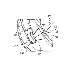
channels. FIGS. 3A-3D
illustrate an embodiment comprising a cap (355) that fits over the end of an
endoscope (350).
The cap (355) may be fixedly attached to the endoscope (350), or also may be
removable and
capable of sliding over the distal end of the endoscope (350), e.g., to form a
friction fit. The
cap (355) includes an appendage (360) configured for engaging with a tissue
surface. The
appendage (360) may form a conical or funnel shape (e.g., generally circular
or oval cross-
section) as shown in FIGS. 3A-3C, but may have any other shape appropriate for
contacting a
surface and applying suction. In some embodiments, for example, the appendage
(360) may
have a rectangular cross-section. The cap (355) may provide a greater field of
view and/or
greater region of access when placed against a tissue surface.
- 11 -
CA 02912602 2015-11-12
WO 2014/197444 PCT/US2014/040636
[040] In some embodiments, the appendage (360) of the cap (355) may include a
purse-string feature to vary a cross section of the appendage (360). For
example, the
appendage (360) may form a conical or funnel shape with a purse-string feature
at the distal
end of the funnel to allow for widening or narrowing the diameter of the
funnel in contact
with the tissue surface. The purse-string feature may help to grasp and
manipulate tissue, and
may also help to guide an instrument into position, e.g., to cannulate the
papilla. The purse
string may also be used to apply suction and enclose a portion of tissue,
e.g., for removal via
a snare. In such an embodiment, a purse-string may be looped around the distal
surface of
the appendage (360), and fed through a lumen of the endoscope (350) to the
proximal end. A
user may pull on the purse-string at the proximal end of the endoscope (350)
to reduce the
size, e.g., diameter, of the opening formed by appendage (360).
[041] The cap (355) may include a recessed area or window. In some
embodiments,
the window may include integrated cautery wire capability for cutting and/or
cauterizing
tissue pulled into the window via suction.
[042] The cap (355) may have a retracted configuration, e.g., for introducing
the
endoscope (350) into the duodenum as shown in FIG. 3A, and an expanded
configuration,
e.g., for engaging with the duodenal wall to contact the tissue surface and
form a seal with the
tissue surrounding the papilla (106) as shown in FIGS. 3B-3D. The retracted
configuration
may be a bellows shape. The cap (355) may be deployed from the retracted
configuration to
the expanded configuration via a pull wire, push wire, spring, or other
suitable mechanism.
The cap may include one or more support members, e.g., as discussed above in
connection
with suction cup (205), to support a membrane in a retracted configuration
and/or an
expanded configuration.
[043] Any of the materials and/or features described above in connection to
the
suction cup (e.g., suction cup (205) of FIGS. 2A-2C) may be used for the cap
(355). For
- 12 -
CA 02912602 2015-11-12
WO 2014/197444
PCT/US2014/040636
example, the cap (355) may be formed of one or more flexible and/or rigid
materials
including, e.g., silicone, rubber, metals, plastics, and polymers or polymer
mixtures. In some
embodiments, the cap (355) includes an elastomeric material. In some
embodiments, the cap
may be transparent, e.g., to peimit imaging and lighting functions of the
endoscope (350).
[044] The cap (355) may have a closed distal end. For example, in some
embodiments, the endoscope (350) and cap (355) are configured such that the
only openings
include the face of the appendage (360) and an opening at the proximal end of
the endoscope
(350) to allow for the cap (355) to fit over the endoscope (350). The proximal
end of the cap
(355) may include one or more elastic bands to secure the cap (355) over the
endoscope
(350), such as to provide a seal at the proximal end of the cap (355) so that
suction is applied
only at the face of the appendage (360). See also FIGS. 8A-8B and 9 below. The
cap (355)
may also include an open distal end to be deployed proximally, e.g., by
sliding the cap (355)
down the length of the endoscope into position distally, wherein the cap (355)
includes a
closing, sealing feature, e.g., a purse string or other mechanical mechanism,
to close the distal
end.
[045] The cap (355) may be sufficiently collapsible, flexible, and tearable
such that
it may be pulled through a working channel of the endoscope (350), if desired,
after
placement of a guidewire or other cannulation of the papilla (106),
pancreactic duct (102),
and/or bile duct (103). This may be done by, e.g., extending a grasper through
an endoscope
working channel, grasping the cap (355), and pulling it back through the
channel.
[046] The endoscope (350) and cap (355) according to the present disclosure
may be
used for a medical procedure, e.g., an ERCP procedure, as described above in
connection to
FIGS. 2A-2C. Thus, referring to FIGS. 3B-3C, the cap (355) may brought into
contact with
the duodenal surface surrounding the papilla (106) and suction applied to
smooth tissue folds
and/or muscles around the papilla (106), ampulla (105) and/or sphincter (108).
Suction may
- 13 -
CA 02912602 2015-11-12
WO 2014/197444 PCT/US2014/040636
also draw bile from the bile duct (103), providing a visible bile trail. A
guidewire (310) may
be introduced into the papilla (106) to assist in cannulation as shown in FIG.
3B, followed by
a catheter (320) such as a sphincterotome as shown in FIG. 3C, or other
treatment instrument
over the guidewire (310) for cannulation and/or examination, diagnosis,
treatment, etc.,
within the pancreatic duct (102) and/or bile duct (103). As noted above, a
guidewire (310)
may not be necessary for cannulation.
[047] In some embodiments, the cap (355) may include an inflatable member to
assist in securing the cap (355) against the tissue surface. As shown in FIG.
3D, for example,
the cap (355) may include an inflatable member such as a balloon (370) on the
back side of
the cap (355) directly opposite the appendage (360), wherein inflating the
balloon (370)
causes the balloon (370) to press against the duodenal wall opposite the
papilla (106) and
create forward pressure on the cap (355) to contact the tissue surface
surrounding the papilla
(106). In addition or alternatively, the cap (355) may include an inflatable
member such as
balloon (375) on the same side as the appendage (360), e.g., just below the
appendage (360)
(i.e., proximal to the appendage (360)), to press against the duodenal wall of
the papilla (106)
to create space between the cap (355) and the papilla (106). Embodiments of
cap (355) may
include one or both of these balloons (370, 375). To inflate the balloons
(370, 375), a
channel may extend through the endoscope (350) to provide inflation fluid to
the cap (355)
and its balloons (370, 375).
[048] In some embodiments, the cap (355) may provide more than one suction
area
or channel, e.g., for applying suction to two or more tissue surfaces
independently or in
combination with each other, and/or for guiding various instruments. For
example, the cap
(355) may include a suction area opposite the appendage (360), such as a
second appendage
(380) as illustrated in FIG. 3E, for applying suction against a tissue surface
opposite the
papilla (106). The second appendage (380) may help to maintain the position of
the
- 14 -
CA 02912602 2015-11-12
WO 2014/197444 PCT/US2014/040636
endoscope (350) and/or the main or first appendage (360) with respect to the
papilla (106), or
may draw tissue tight to create traction, smooth tissue folds, create
additional working space,
or help to open up the papilla (106). The second appendage may connect to a
suction channel
between the cap (355) and the endoscope (350), e.g., a suction channel
external to the
endoscope (350), or may connect to a working channel within the endoscope
(350). In some
embodiments, suction may be applied first to the tissue surface surrounding
the papilla (106)
via the first appendage (360), followed by suction applied to a tissue surface
of the duodenum
(107) opposite the papilla (106) via second appendage (380) to maintain the
position of the
first appendage (160). Alternatively, suction may first be applied to a tissue
surface opposite
the papilla (106) via second appendage (380), e.g., to draw suction and help
smooth tissue
surrounding the papilla (106), followed by suction applied to the papilla
surface. While the
second appendage (380) is illustrated as directly opposite the first appendage
(360) in FIG.
3E, the second appendage (360) may be located anywhere along the cap (355),
such as
adjacent, above, below, or at an angle with respect to the first appendage
(360). In some
embodiments, the cap (355) may include one or more working channels to guide
different
instruments to an area of interest, such as the papilla (106) and/or tissue
around the papilla
(106).
[049] Referring to FIG. 3B, a guidewire (310) may be advanced through the
working
channel of the endoscope (350), e.g., via a lumen of a catheter introduced
into the working
channel, through the cap to enter the papilla (106). A sphincterotome (320) or
other
cannulation catheter or treatment instrument may slide over the guidewire
(310) to cannulate
the papilla (106) as shown in FIG. 3C. The sphincterotome may be used to
incise the
sphincter (108) as described above, for example, and may also be used to
inject contrast into
the bile duct (103) and/or pancreatic duct (102) for fluoroscopy or other
imaging analysis.
- 15-
CA 02912602 2015-11-12
WO 2014/197444 PCT/US2014/040636
[050] FIGS. 4A and 4B illustrate similar retracted and expanded
configurations,
respectively, of a cap (455) including an expandable appendage (460), wherein
the
expandable appendage includes a feature (465) at the distal end of the
appendage (460)
configured to interface with the tissue surface. In some embodiments, for
example, the
feature (465) may comprise a deformable material or inflatable member to adapt
to the
contour of the tissue surface. In other embodiments, the feature (465) may
comprise a rigid
material such as a metal wire or plastic ring. For example, a rigid material
at or near the
distal end of the appendage (460) may be used to apply pressure against the
tissue surface,
e.g., to spread or smooth tissue. Further, the feature (465) may include a
rigid material to act
as a tissue stop, e.g., to prevent tissue from being drawn into the funnel of
the appendage
(460) by suction. The feature (465) may be continuous, e.g., covering the
entire distal end
circumference of the appendage (460), or may include one or more discrete
portions. In
some embodiments, the distal edge of the appendage (460) or the feature (465)
may include
one or more holes to help release or reduce the amount of suction. In some
embodiments, the
appendage (460) or the feature (465) may have a scalloped or thinned edge,
e.g., to help adapt
to the contour of the tissue surface and accommodate an irregular surface to
the tissue.
[051] In some embodiments, the feature (465) may be used to deploy the
appendage (460) into an expanded configuration. For example, the appendage
(460) may
include a flexible or thin film material with a distal end feature (465)
comprising a rigid
material such as a metal or plastic ring or wire. The appendage (460) may be
deployed into
an expanded configuration as shown in FIG. 4B via a spring or snare-like
mechanism coupled
to the rigid material of the feature (465).
[052] In some embodiments, the feature (465) may comprise a metal or plastic
ring
that includes one or more metal or plastic strips or wires across a diameter
of the ring and
parallel to the longitudinal axis of the cap (455), i.e., parallel to the
longitudinal axis of the
- 16-
CA 02912602 2015-11-12
WO 2014/197444 PCT/US2014/040636
endoscope. The wires may be concave and curved towards the face of the
endoscope,
allowing tissue to be partially drawn into the appendage (460) but not far
enough to interfere
with the field of view or field of access. In other embodiments, the feature
(465) may include
two or more concentric rings with radial arms connecting the rings to each
other and/or to the
main body of the cap (455), similar to the support arms described above.
[053] In another embodiment shown in FIGS. 5A-5B, the cap (555) includes as an
appendage one or more doors (560) configured to pivot or swing open and engage
with a
tissue surface. Each of the doors (560) may pivot along an axis parallel to
the central
longitudinal axis of the cap (555) as shown in FIGS. 5A-5B, or may also open
along another
direction, e.g., along an axis perpendicular or otherwise offset from the
longitudinal axis. In
some embodiments, the doors (560) may slide open rather than pivot open. The
edge(s) of
each door (560) may include a deformable or elastomeric material, e.g., to
form a seal
between the doors (560) in a retracted (i.e., closed) configuration and to
adapt to the tissue
surface in an expanded (i.e., open) configuration. Doors (560) may be opened
and closed via
a pull wire, push rod, or other suitable mechanism extending through the cap
and a channel of
the endoscope.
[054] FIGS. 6A-6B illustrate yet another embodiment of a device comprising a
plurality of inflatable members (665) attached to a membrane (660). The
membrane (660)
may comprise a flexible or elastomeric material such that upon inflation, the
inflatable
members (665) may expand radially outward to expand or stretch the membrane
(660) to
form a conical or funnel shape for engaging with the duodenal wall. The
embodiment shown
in FIGS. 6A-6B may be included in a device disposed in the working channel of
an
endoscope, e.g., a suction cup as discussed above, or may also comprise an
appendage of an
endoscope cap. In an embodiment shown in FIGS. 6A-6B, for example, the device
includes
four inflatable members (665), each of which is in fluid communication with an
inflatable
- 17 -
CA 02912602 2015-11-12
WO 2014/197444 PCT/US2014/040636
ring (670), wherein at least one inflatable member (665) receives an inflation
fluid via any
suitable mechanism. For example, a suction port 675 can connect to an
inflation lumen
extending through the endoscope to the proximal end, to receive inflation
fluid to inflate the
inflatable ring (670) and inflatable members (665), and expand the membrane.
[055] FIGS. 7A-7B illustrate another embodiment, comprising a cap (755) and
appendage (760) that includes a flexible material and one or more support arms
(770).
Support arms (770) may include a rigid material, and may pivot with respect to
the main
body of the cap (755) by manipulating a push or pull wire (775), (780). As
shown in FIG.
7A, pulling wire (775) in a proximal direction may cause support arms (770) to
pivot
downward, thus bringing the appendage (760) into a retracted and more
streamlined
configuration, e.g., to facilitate introducing and/or withdrawing the
endoscope from the body.
A second wire (780) may remain slack, or may have sufficient rigidity to push
the appendage
(760) downward into the retracted configuration. Tension on wire (775) may be
released
and/or tension on wire (780) may be increased to expand the appendage (760),
e.g., into a
conical shape as shown in FIG. 7B. Wire (775) may also have sufficient
rigidity to pivot
support arms (770) upward into the expanded configuration. In some
embodiments, the distal
end of the cap (755) may be configured to assist in advancing and withdrawing
the cap (755)
within the body. For example, the distal end of the cap (755) may include an
extension, such
as a pointed cone ribs extension. In some embodiments, a sphincterotome or
other instrument
may assist in deploying and/or retracting the appendage (760).
[056] While FIGS. 7A-7B illustrate an embodiment with support arms, other
embodiments may not include support arms, wherein one or more pull or push
wires connect
to the flexible material of the appendage (760) to expand and retract the
appendage (760).
Further, in some embodiments push or pull wires (775), (780) may be
manipulated to pivot
- 18 -
CA 02912602 2015-11-12
WO 2014/197444 PCT/US2014/040636
support arms (770) toward each other, e.g., to close the opening of the
appendage (760) to
provide a retracted and more streamlined configuration.
[057] It should be noted that while FIGS. 3-7 illustrate both a retracted
configuration
and an expanded configuration of the cap, in some embodiments, the cap may
have a single
configuration, e.g., for engaging with a tissue surface.
[058] FIGS. 8A-8B illustrate an embodiment of a cap (855) configured to fit
over
endoscope (850), wherein the cap (855) includes one or more protrusions (895)
or keying
features for aligning cap (855) with endoscope (850) and/or for forming a
friction fit or seal
against an outer surface of endoscope (850). The protrusions (895) may form an
integral part
of cap (855), or may comprise elements coupled to the cap (855), e.g.,
elastomeric rings for
forming a seal with endoscope (850). As shown in FIG. 8B, cap (855) may
include an
elongated portion (890) such as a pull-on tab to facilitate placement of the
cap (855) over the
endoscope (855).
[059] In another embodiment illustrated in FIG. 9B, cap (955) may have a
tapered
shape, wherein the outer diameter of the cap (955) narrows from a distal
portion (965) to a
proximal portion (960). For example, the outer diameter of the proximal
portion (960) may
be smaller than the outer diameter of the distal portion (965) such that the
proximal portion
(960) forms a friction fit against an endoscope. At least a portion of the
proximal portion
(960) may include a flexible material such that the proximal portion (960) may
be pulled
back or rolled up for placing the cap (955) around an endoscope, and then
pulled down to
form a seal around the endoscope.
[060] Any of the features discussed herein in connection to an embodiment may
be
used in combination with one or more features of any other embodiment.
Further, other
embodiments of the present disclosure will be apparent to those skilled in the
art from
consideration of the specification and practice of the embodiments disclosed
herein. It is
- 19 -
CA 02912602 2015-11-12
WO 2014/197444
PCT/US2014/040636
intended that the specification and examples be considered as exemplary only,
with a true
scope and spirit of the present disclosure being indicated by the following
claims.
- 20 -