Note: Descriptions are shown in the official language in which they were submitted.
CA 02913069 2015-11-20
WO 2014/191113
PCT/EP2014/001460
NOVEL ANTIBODIES
FIELD OF THE INVENTION
[0001] The present invention relates to novel antibodies, which combine high
affinity with high potency, particularly novel antibodies against a novel
epitope.
BACKGROUND OF THE INVENTION
[0002] This invention relates to novel anti-CD3 antibodies, which combine high
affinity with high potency, and in particular novel antibodies, which
specifically
recognize a novel CD3 epitope.
[0003] The T cell receptor or TCR is a molecule found on the surface of T
lymphocytes (or T cells) that is responsible for recognizing antigens bound to
major histocompatibility complex (MHC) molecules on the surface of antigen
presenting cells (APC). The binding between TCR and antigen is of relatively
low affinity. When the TCR engages with antigen and MHC, the T lymphocyte is
activated through a series of biochemical events mediated by associated
enzymes, co-receptors, specialized accessory molecules, and activated or
released transcription factors.
[0004] The TCR is associated with other molecules like CD3, which possess
three distinct chains (y, 6, and c) in mammals, and either a 42 (CD247)
complex
or a 4/n complex. These accessory molecules have transmembrane regions and
are vital to propagating the signal from the TCR into the cell; the
cytoplasmic tail
of the TCR is extremely short, making it unlikely to participate in signaling.
The
CD3- and 4-chains, together with the TCR, form what is known as the T cell
receptor complex.
[0005] CD3c is a type I transmembrane protein expressed on the surface of
certain T cells. It participates in the T cell receptor (TCR) complex and
interacts
1
CA 02913069 2015-11-20
WO 2014/191113
PCT/EP2014/001460
with other domains of this complex. One of these interaction partners is CD3y,
which binds to CD3E in a 1:1 stoichiometry (De la Hera et al, J. Exp.Med.1991;
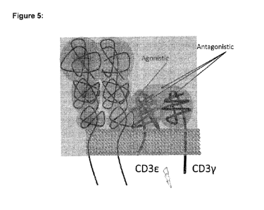
173: 7-17). Figure 5 shows a schematic view of the TCR complex, including
CD3E/CD3y. It is believed that binding of the TCR to the MHC-peptide complex
on the surface of an antigen presenting cell (APC) and subsequent movement
of the T cell along the APC leads to a certain rotation of the TCR complex
resulting in a dislocation of CD3E and CD3y relative to each other, which is
required for efficient TCR signaling and therefore activation of T-cells.
Certain
antibodies against CD3E have been demonstrated to induce TCR signaling
while others did not. TCR-activating antibodies typically bind to an exposed
epitope on CD3E (see Figure 5, "agonistic epitope"), whereas some non-
stimulatory antibodies have been demonstrated to bind to the interface between
CD3E and CD3y, or to concomitantly bind to CD3E and CD3y (see Figure 5,
"antagonistic epitope"), thus possibly interfering with the relative
displacement
of CD3E and CD3y (Kim et al, JBC.2009; 284: 31028-31037).
[0006] It is well established that peptide-MHC complexes bind TCR with low
affinity and fast off rate (Matsui et al, Science.1991; 254: 1788-1791; Weber
et
al, Nature.1992; 356: 793-796). It has been suggested that this low affinity
is
instrumental to allow a few peptide-MHC complexes to serially trigger many
TCRs (Valitutti et al, Nature.1995; 375: 148-151) by repeated binding and
dissociation. This serial triggering is critical to sustain signaling over
time,
allowing T cells to eventually reach the activation threshold (Valitutti et
al,
lmmunol. Today. 1997; 18: 299-304; Lanzavecchia et al, Cell. 1999; 96: 1-4).
This notion is supported by the finding that, when compared to peptide-MHC
complexes, high-affinity anti-CD3 antibodies do not efficiently stimulate T
cells,
since they trigger TCR with a 1:1 stoichiometry (Viola et al, Science 1996;
273:
104-106), suggesting that low-affinity antibodies may be more effective in
stimulating T cells via TCR signaling because of their ability to repeatedly
dissociate and re-bind to CD3E. Indeed, in a direct comparison of three
derivatives of the anti-CD3E antibody TR66, which all bind with different
affinities, wild-type TR66 having an intermediate affinity showed best
efficacy in
T cell activation when compared to its derivatives that have either higher or
2
CA 02913069 2015-11-20
WO 2014/191113
PCT/EP2014/001460
lower affinities (Bortoletto et al, J. Immuno.2002;32:3102-3107). Thus, a KD
at
around that of TR66 is ideal for the stimulation of T cells. The affinity of
TR66
has been determined by use of surface-plasmon resonance (SPR) technology
as well as by flow-cytometry, yielding equilibrium dissociation constants of
2.6 x
10-7 M (Moore et al, Blood.2011; 117: 4542-4551) and 1.0 x 10⁻⁷ M (Amann et
al, Cancer Res. 2008; 68: 143-151), respectively. In line with this, it has
been
recommended to use anti-CD3 antibodies with an affinity of less than 10-8 M
(US 7,112,324), and the T cell-stimulatory antibodies that have been published
for human therapeutic use, bind with affinities to human CD3E in the same
range. Therefore, according to the theory of serial TCR triggering and in
agreement with published results for anti-CD3c antibodies, monoclonal
antibodies with affinities significantly better than the ones published are
not
expected to be more potent stimulators of T cells but in contrast are expected
to
be weaker activators.
[0007] Some of the published antibodies against CD3E have been generated via
immunization of animals with T cell preparations and subsequent isolation of
monoclonal antibodies by the so-called hybridoma procedure. The weakness of
this approach is that the unselective immune response against various antigens
of foreign (human) T cells in the animal, on one hand, and the poor efficiency
of
the hybridoma procedure on the other hand, decrease the probability to
identify
monoclonal antibodies with T cell-stimulatory activity, also because these
agonistic antibodies may represent a minority in the entirety of anti-CD3E
antibodies. Immunization with a linear peptide spanning the targeted epitope
increases the selectivity of the immune response, may, however, result in
antibodies that do not recognize the native full-length CD3E or that may exert
non-optimal TCR stimulation.
[0008] For the immunization of animals with other type-I transmembrane
proteins it has been particularly useful to use the purified extracellular
domain
(ECD). However, purified ECD of CD3E tends to aggregate, and aggregates
may have an altered structure as compared to the native protein. Further this
approach may preferentially lead to antibodies binding to the interface
between
3
CA 02913069 2015-11-20
WO 2014/191113
PCT/EP2014/001460
CD3E and CD3y. In contrast, the complex of CD3E and CD3y produced as a
single-chain protein, connected by a flexible peptide linker, can be purified
in a
monomeric fraction and in its native conformation (Kim et al, JMB.2000; 302:
899-916). Immunization of animals with such a CD3E/y single-chain protein may
however lead to antibodies concomitantly binding to CD3E and CD3y, which
would result in antagonistic effects.
[0009] Several antibodies directed against human CD3E have been developed
in the past.
[0010] Monoclonal antibody SP34 is a murine antibody that cross-reacts with
non-human primate CD3, and that is also capable of inducing cell proliferation
on both human and non-human primate PBMCs (Pessano et al., The T3/T cell
receptor complex: antigenic distinction between the two 20-kD T3 (T38 and T3c)
subunits. EMBO J 4 (1985) 337-344).
[0011] WO 2007/042261 and WO 2008/119567, both assigned to Micromet,
disclose cross-reactive binders directed against the epitopes FSEXE and
QDGNE, respectively, in CD3E. In opposition proceedings filed by several
opponents against granted European patent EP 2 155 783 (based on the
regional phase of WO 2008/119567), it is submitted that SP34 is binding to
epitope QDGNE as well.
[0012] However, despite the fact that many attempts have been made to
address the issue of obtaining anti-CD3 antibodies, or to binding molecules in
general, with particularly advantageous properties, so far these attempts have
had limited success.
[0013] Thus, there remained still a large unmet need to develop novel CD3
binding molecules, in particular novel anti-CD3 antibodies, for high affinity,
which is not limiting for high potency. Additionally, there is still a large
unmet
need to develop novel CD3 binding molecules, in particular novel anti-CD3
antibodies, for high affinity, which are cross-reactive with other species, in
particular with non-human primates such as cynomolgUs monkeys.
4
CA 02913069 2015-11-20
WO 2014/191113
PCT/EP2014/001460
[0014] The solution for this problem that has been provided by the present
invention, i.e. CD3-binding molecules, in particular anti-CD3 antibodies
obtained
by genetic immunization of rabbits and screening of affinity matured memory B-
cells, and in particular CD3-binding molecules, in particular anti-CD3
antibodies,
with specificity for a novel agonistic epitope, has so far not been achieved
or
suggested by the prior art.
SUMMARY OF THE INVENTION
[0015] The present invention relates to novel isolated CD3-binding molecules,
in
particular isolated antibodies or functional fragments thereof, each
comprising a
binding region, particularly an antigen-binding region, wherein said binding
molecules, in particular said antibodies or functional fragments thereof, are
specific for an epitope of human CD3, particularly for a novel agonistic
epitope
of CD3, wherein said binding molecules, in particular said isolated antibodies
or functional fragments thereof, have a higher affinity than the prior art
antibodies, particularly OKT-3 and/or TR66, while simultaneously exhibiting a
higher potency.
[0016] Thus, in a first aspect, the present invention relates to an isolated
binding
molecule comprising a binding region that is specific for an epitope of human
CD3E, in particular to an isolated antibody or functional fragment thereof
comprising an antigen-binding region, wherein said epitope comprises amino
acid residue N4 as residue that is critical for binding.
[0017] In a second aspect, the present invention relates to a novel isolated
CD3-binding molecule that is specific for an epitope of human CD3, wherein
said isolated CD3-binding molecule is binding to human CD3 with a dissociation
constant for monovalent binding of less than 3.0 x 10⁻⁸ M, particularly less
than
1.5 x 10⁻⁸ M, more particularly less than 1.2 x 10⁻⁸ M, and most particularly
less
than 1.0 x 10⁻⁸ M, in particular to an isolated antibody or functional
fragment
CA 02913069 2015-11-20
WO 2014/191113
PCT/EP2014/001460
thereof comprising an antigen-binding region that is specific for an epitope
of
human CD3, wherein said antibody or functional fragment thereof, is binding to
human CD3 with a dissociation constant for monovalent binding of less than 3.0
x 10-8 M, particularly less than 1.5 x 10⁻⁸ M, more particularly less than 1.2
x 10-
8 M, and most particularly less than 1.0 x 10⁻⁸ M.
[0018] In a third aspect, the present invention relates to an isolated
antibody or
functional fragment thereof comprising an antigen-binding region that is
specific
for an epitope of human CD3, wherein said antibody or functional fragment
thereof, when tested in an IgG format, upon cross-linking, is inducing T-cell
activation at least 1.5-fold stronger than antibodies OKT-3 or TR66 after 24 h
of
stimulation at an IgG concentration of 1.25 pg/ml.
[0019] In a fourth aspect, the present invention relates to an isolated
antibody or
functional fragment thereof comprising an antigen-binding region that is
specific
for an epitope of human CD3, wherein said antibody or functional fragment
thereof, when tested in an IgG format upon cross-linking, is resulting in T-
cell
activation, which lasts longer than with antibodies OKT-3 or TR66 as indicated
by at least 1.5-fold greater increase in CD69 expression after 72 hours of
stimulation at an IgG concentration of 1.25 pg/ml.
[0020] In a fifth aspect, the present invention relates to an isolated
antibody or
functional fragment thereof comprising an antigen-binding region that is
specific
for an epitope of human CD3, wherein said antibody or functional fragment
thereof, when tested in an IgG format, upon cross-linking, is resulting in a
dose-
dependent homogeneous activation state of T-cells.
[0021] In a sixth aspect, the present invention relates to an isolated
antibody or
functional fragment thereof comprising an antigen-binding region that is
specific
for an epitope of human CD3, wherein said antibody or functional fragment
thereof, when tested in an IgG format, (i) is binding to human CD3 with a
dissociation constant for monovalent binding of less than 3.0 x 10⁻⁸ M,
particularly less than 1.5 x 10⁻⁸ M, more particularly less than 1.2 x 10⁻⁸ M,
and
most particularly less than 1.0 x 10⁻⁸ M; and (iia), upon cross-linking, is
inducing
6
CA 02913069 2015-11-20
WO 2014/191113
PCT/EP2014/001460
T-cell activation at least 1.5-fold stronger than antibodies OKT-3 or TR66
after
24 h of stimulation at an IgG concentration of 1.25 pg/ml; (iib) is resulting
in T-
cell activation, which lasts longer than with antibodies OKT-3 or TR66 as
indicated by at least 1.5-fold greater increase in CD69 expression after 72
hours
of stimulation at an IgG concentration of 1.25 pg/ml; (iic) is resulting in a
dose-
dependent homogeneous activation state of 1-cells; and/or (iid) is specific
for an
epitope of human CD3E, wherein said epitope comprises amino acid residue N4
as residue that is critical for binding.
[0022] In a seventh aspect, the present invention relates to an isolated
binding
molecule, particularly an isolated antibody or functional fragment thereof,
binding to essentially the same epitope as the isolated antibody or functional
fragment thereof of Sections [0078] to [0080], [0083] to [0085] and [0089].
[0023] In an eighth aspect, the present invention relates to a pharmaceutical
composition comprising a binding molecule of the present invention, in
particular an isolated antibody or functional fragment thereof, and optionally
a
pharmaceutically acceptable carrier and/or excipient.
[0024] In a ninth aspect, the present invention relates to a nucleic acid
sequence or a collection of nucleic acid sequences encoding a binding
molecule of the present invention, in particular an isolated antibody or
functional
fragment thereof.
[0025] In a tenth aspect, the present invention relates to a vector or a
collection
of vectors comprising the nucleic acid sequence or a collection of nucleic
acid
sequences of the present invention.
[0026] In an eleventh aspect, the present invention relates to a host cell,
particularly an expression host cell, comprising the nucleic acid sequence or
the
collection of nucleic acid sequences of the present invention, or the vector
or
collection of vectors of the present invention.
7
CA 02913069 2015-11-20
WO 2014/191113
PCT/EP2014/001460
[0027] In a twelfth aspect, the present invention relates to a method for
producing a binding molecule of the present invention, in particular an
isolated
antibody or functional fragment thereof, comprising the step of expressing the
nucleic acid sequence or the collection of nucleic acid sequences of the
present
invention, or the vector or collection of vectors of the present invention, or
the
host cell, particularly an expression host cell, of the present invention.
[0028] In a thirteenth aspect, the present invention relates to a method for
generating an isolated antibody or functional antibody fragment in accordance
with the present invention comprising the steps of:
a) Immunization of rabbits with a CD3E-expressing plasmid to present the
native full-length CD3E on the surface of host cells;
b) Clonal isolation of affinity matured memory B-cells that interact with the
CD3E/y single-chain using fluorescence activated cell-sorting;
c) Cultivation of single sorted B cells in a co-cultivation system that does
not
require immortalization of sorted clones;
d) Screening of B cell culture supernatants in a cell-based ELISA to identify
antibodies binding to the native CD3E embedded in the TCR complex on the
surface of T cells.
[0029] In a fourteenth aspect the present invention relates to a particular
epitope
of human CD3 epsilon comprising exclusively amino acid residues of CD3
epsilon that are not located in the interface between CD3 epsilon and CD3
gamma and that still can be bound by an antibody in the context of the native
TCR expressed on T cells, binding of which by a cross-linked antibody of the
invention is inducing T-cell activation at least 1.5-fold stronger than
antibodies
OKT-3 or TR66 after 24 h of stimulation at an IgG concentration of 1.25 pg/ml;
(iib) is resulting in T-cell activation, which lasts longer than with
antibodies OKT-
3 or TR66 as indicated by at least 1.5-fold greater increase in CD69
expression
after 72 hours of stimulation at an IgG concentration of 1.25 pg/ml; and/or
(iic) is
resulting in a dose-dependent homogeneous activation state of T-cells.
8
CA 02913069 2015-11-20
WO 2014/191113
PCT/EP2014/001460
[0030] In a fifteenth aspect, the present invention relates to a method for
identifying a binding molecule comprising a binding region that is specific
for a
novel epitope of human CD38, comprising the step of (a) selecting from one or
more molecules binding to human CD3 at least one binding molecule, which
comprises a binding region that is specific for an epitope of human CD3E,
wherein said epitope comprises amino acid residue N4 as residue that is
critical
for binding.
BRIEF DESCRIPTION OF THE DRAWINGS
[0031] Figure 1 shows the phylogenetic clustering of joined VH and VL CDR
Sequences from monoclonal rabbit antibodies.
[0032] Figure 2 shows binding of purified monoclonal rabbit antibodies to
Jurkat
T cells.
[0033] Figure 3 shows the stimulation of CD69 expression by cross-linked anti-
CD3c mAbs. The potential of purified monoclonal rabbit anti-CD3 antibodies
and comparator antibodies TR66 and OKT-3 to induce T-cell activation was
assessed by measurement of CD69 expression. Three different concentrations
of cross-linked antibodies were used to stimulate Jurkat cells and CD69
expression was assessed by flow-cytometry 24 h later. Antibody concentrations
were 1.25 pg/ml (a), 5.0 pg/ml (b) and 20 pg/ml (c).
[0034] Figure 4 shows the stimulation of CD69 by cross-linked rabbit mAbs
over time. The potential of purified monoclonal rabbit anti-CD3 antibodies to
induce T-cell activation was assessed by measurement of CD69 expression.
Cross-linked antibodies were used at a concentration of 5.0 pg/ml to stimulate
Jurkat cells and CD69 expression was assessed by flow-cytometry 0, 4, 15, 24,
48 and 72 h later. For the qualitative detection of CD69 expression the mean
fluorescence intensity (MFI), reflecting the signal intensity at the geometric
mean, was measured for both, the negative control as well as for the test
9
CA 02913069 2015-11-20
WO 2014/191113
PCT/EP2014/001460
antibodies. The difference of the MFI between test antibody and negative
control (AMFI) was calculated as a measure for CD69 expression.
[0035] Figure 5 shows a simplified schematic view of the TCR complex,
including CD3E/CD3y.
[0036] Figure 6 shows the results of epitope mapping experiments for prior art
antibodies: (a) epitope mapping of antibody SP34 (see file history of EP 2 155
788); (b) epitope mapping of Micromet antibody (see EP 2 155 788 / WO
2008/119567; Figure 6 shows the results of binding experiments of single
alanine mutants, where a decrease of binding for a given mutant indicates the
relevance of the corresponding wild-type amino acid residue for antibody
binding (i.e. low bar = highly relevant for binding).
[0037] Figure 7 shows the results of epitope mapping experiments by ELISA for
antibodies of the present invention (clone-02, clone-03, clone-06); Figure 7
shows the results of binding experiments in a peptide scan analysis. 15mer
linear arrays derived from human CD3E, residues 1 ¨ 15 in which each position
is substituted by 18 amino acids (all natural amino acids except cysteine)
were
probed with 0.1 pg/ml of each antibody to study amino acid specificities
affecting binding to the epitope. Decrease in binding signals in ELISA is
given,
(a) for each substitution individually, and (b) averaged over the 18 different
substitutions for each position. The height of a bar in Figure 7b indicates
the
relevance of the corresponding wild-type amino acid residue for antibody
binding (i.e. large bar = highly relevant for binding).
[0038] Figure 8 shows binding of anti-CD3 x anti-IL5R scDbs to Jurkat T-cells
and CH0-1L5R cells. Binding of A) Construct 1, B) Construct 2 and C) Construct
3 to Jurkat T-cells and CD3-negative Jurkat cells and binding of D) Construct
1,
E) Construct 2 and F) Construct 3 to IL5R-CHO cells as well as wild-type CHO
cells was assessed by flow cytometry. Construct 1, Construct 2 and Construct 3
have the same anti-IL5R moiety but 3 different anti-CD3 moieties that bind to
CD3 with diverse affinities (1.15 x 10⁻⁸ M for Construct 1, 2.96 x 10⁻⁸ M for
Construct 2, and 1.23 x 10⁻⁷ M for Construct 3); Construct 1 = comprises the
CA 02913069 2015-11-20
WO 2014/191113
PCT/EP2014/001460
humanized variable domain of clone-06; Construct 2 = comprises the
humanized variable domain of clone-02; Construct 3 = comprises the
humanized variable domain of clone-03.
[0039] Figure 9 shows the specific stimulation of interleukin-2 secretion by
cross-linking of cytotoxic T-cells with target cells by scDbs. CD8+ T-cells
were
incubated with increasing concentrations of scDbs in presence of CHO-1L5R or
CHO cells. Interleukin-2 concentrations in culture supernatants were measured
by ELISA after 16 hours of incubation; Construct 1 = comprises the humanized
variable domain of clone-06; Construct 2 = comprises the humanized variable
domain of clone-02; Construct 3 = comprises the humanized variable domain of
clone-03.
[0040] Figure 10 shows the specific lysis of human IL5R-expressing CHO cells
by anti-CD3 x anti-IL5R scDbs. CD8+ T-cells were incubated with increasing
concentrations of scDbs in presence of CHO-1L5R or CHO cells. Target cells
(CHO-1L5R and CHO) were labeled with cell tox green dye and cell lysis was
determined by measurement of fluorescence intensity after 88 hours of
incubation; Construct 1 = comprises the humanized variable domain of clone-
06; Construct 2 = comprises the humanized variable domain of clone-02;
Construct 3 = comprises the humanized variable domain of clone-03.
DETAILED DESCRIPTION OF THE INVENTION
[0041] The peculiarity of this invention compared to former anti-CD3
antibodies
is the fact that the novel isolated antibodies or functional fragments thereof
comprising antigen-binding regions that are specific for an epitope of human
CD3 have higher affinities than the prior art antibodies, particularly OKT-3
and/or TR66, while simultaneously exhibiting higher potencies..
[0042] Thus, in a first aspect, the present invention relates to an isolated
binding
molecule comprising a binding region that is specific for an epitope of human
CD3E, in particular to an isolated antibody or functional fragment thereof
11
CA 02913069 2015-11-20
WO 2014/191113
PCT/EP2014/001460
comprising an antigen-binding region, wherein said epitope comprises amino
acid residue N4 as residue that is critical for binding.
[0043] In the context of the present invention, an amino acid residue is to be
considered "critical for binding", when the binding affinity of a binding
molecule
to a peptide comprising said amino acid residue position is reduced to at
least
50%, particularly to at least 25%, more particularly to at least 10%, and most
particularly to at least 5% of the binding affinity to the wild-type peptide
sequence, when said critical amino acid residue is exchanged by alanine.
and/or when the average signal intensity resulting from binding to a peptide
comprising said amino acid residue position as determined by the ELISA of
Example 7 is reduced to at least 50%, particularly to at least 25%, and most
particularly to at least 10% of the binding signal to the wild-type peptide
sequence, when said critical amino acid residue is separately exchanged by
each of the other natural amino acid residues except cysteine.
[0044] In particular embodiments, said epitope further comprises amino acid
residue E6 as residue that is involved in binding. In particular embodiments,
said epitope further comprises amino acid residue E6 as residue that is
critical
for binding.
[0045] In the context of the present invention, an amino acid residue is to be
considered "involved in binding", when the binding affinity of a binding
molecule
is reduced to at least 80%, when said amino acid residue is exchanged by
alanine, and/or when the average signal intensity resulting from binding to a
peptide comprising said amino acid residue position as determined by the
ELISA of Example 7 is reduced to at least 80%, when said amino acid residue
is separately exchanged by each of the other natural amino acid residues
except cysteine.
[0046] In particular embodiments, said binding molecule is an antibody or
functional fragment thereof.
12
CA 02913069 2015-11-20
WO 2014/191113
PCT/EP2014/001460
[00471 In particular embodiments, said binding molecule, particularly said
isolated antibody or functional fragment thereof, is cross-reactive with
cynomolgus CD3, particularly cynomolgus CD3E, particularly having an affinity
to cynomolgus monkey CD3E that is less than 100-fold, particularly less than
30-fold, even more particularly less than 15-fold and most particularly less
than
5-fold different to that of human CD3E.
[0048] In particular embodiments, said binding molecule, in particular said
antibody or functional fragment thereof, is binding to human CD3 with an
equilibrium dissociation constant for monovalent binding of less than 3.0 x 10-
8
M, particularly less than 1.5 x 10⁻⁸ M, more particularly less than 1.2 x 10⁻⁸
M,
and most particularly less than 1.0 x 10⁻⁸ M.
[0049] In particular embodiments, said binding molecule is an antibody or
functional fragment thereof, which, when tested in an IgG format, upon cross-
linking, is inducing T-cell activation at least 1.5-fold stronger than
antibodies
OKT-3 or TR66 after 24 h of stimulation at an IgG concentration of 1.25 pg/ml.
[0050] In particular embodiments, said binding molecule is an antibody or
functional fragment thereof, which, when tested in an IgG format upon cross-
linking, is resulting in 1-cell activation, which lasts longer than with
antibodies
OKT-3 or 1R66 as indicated by at least 1.5-fold greater increase in CD69
expression after 72 hours of stimulation at an IgG concentration of 1.25
pg/ml..
[0051] In particular embodiments, said binding molecule is an antibody or
functional fragment thereof, which, when tested in an IgG format, upon cross-
linking, is resulting in a dose-dependent activation state of T-cells that is
less
heterogeneous when compared to activation by OKT-3 or 1R66.
[0052] In a second aspect, the present invention relates to a novel isolated
CD3-binding molecule that is specific for an epitope of human CD3, wherein
said isolated CD3-binding molecule is binding to human CD3 with a dissociation
constant for monovalent binding of less than 3.0 x 10⁻⁸ M, particularly less
than
1.5 x 10⁻⁸ M, more particularly less than 1.2 x 10⁻⁸ M, and most particularly
less
13
CA 02913069 2015-11-20
WO 2014/191113
PCT/EP2014/001460
than 1.0 x 10⁻⁸ M, in particular to an isolated antibody or functional
fragment
thereof comprising an antigen-binding region that is specific for an epitope
of
human CD3, wherein said antibody or functional fragment thereof, is binding to
human CD3 with a dissociation constant for monovalent binding of less than 3.0
x 10-8 M, particularly less than 1.5 x 10⁻⁸ M, more particularly less than 1.2
x 10-
8 M, and most particularly less than 1.0 x 10⁻⁸ M.
[0053] In particular embodiments, said binding molecule, particularly said
isolated antibody or functional fragment thereof, is cross-reactive with
cynomolgus CD3, particularly cynomolgus CD3E, particularly having an affinity
to cynomolgus monkey CD3E that is less than 100-fold, particularly less than
30-fold, even more particularly less than 15-fold and most particularly less
than
5-fold different to that of human CD3E.
[0054] In a third aspect, the present invention relates to an isolated
antibody or
functional fragment thereof comprising an antigen-binding region that is
specific
for an epitope of human CD3, wherein said antibody or functional fragment
thereof, when tested in an IgG format, upon cross-linking, is inducing T-cell
activation at least 1.5-fold stronger than antibodies OKT-3 or TR66 after 24 h
of
stimulation at an IgG concentration of 1.25 pg/ml.
[0055] In particular embodiments, said binding molecule, particularly said
isolated antibody or functional fragment thereof, is cross-reactive with
cynomolgus CD3, particularly cynomolgus CD3E, particularly having an affinity
to cynomolgus monkey CD3E that is less than 100-fold, particularly less than
30-fold, even more particularly less than 15-fold and most particularly less
than
5-fold different to that of human CD3E.
[0056] In a fourth aspect, the present invention relates to an isolated
antibody or
functional fragment thereof comprising an antigen-binding region that is
specific
for an epitope of human CD3, wherein said antibody or functional fragment
thereof, when tested in an IgG format, upon cross-linking, is resulting in T-
cell
activation, which lasts longer than with antibodies OKT-3 or TR66 as indicated
14
CA 02913069 2015-11-20
WO 2014/191113
PCT/EP2014/001460
by at least 1.5-fold greater increase in CD69 expression after 72 hours of
stimulation at an IgG concentration of 1.25 pg/ml..
[0057] In particular embodiments, said binding molecule, particularly said
isolated antibody or functional fragment thereof, is cross-reactive with
cynomolgus CD3, particularly cynomolgus CD3E, particularly having an affinity
to cynomolgus monkey CD3E that is less than 100-fold, particularly less than
30-fold, even more particularly less than 15-fold and most particularly less
than
5-fold different to that of human CD3E.
[0058] In a fifth aspect, the present invention relates to an isolated
antibody or
functional fragment thereof comprising an antigen-binding region that is
specific
for an epitope of human CD3, wherein said antibody or functional fragment
thereof, when tested in an IgG format, upon cross-linking, is resulting in a
dose-
dependent activation state of T-cells that is less heterogeneous when compared
to activation by OKT-3 or TR66.
[0059] In particular embodiments, said binding molecule, particularly said
isolated antibody or functional fragment thereof, is cross-reactive with
cynomolgus CD3, particularly cynomolgus CD3E, particularly having an affinity
to cynomolgus monkey CD3E that is less than 100-fold, particularly less than
30-fold, even more particularly less than 15-fold and most particularly less
than
5-fold different to that of human CD3E.
[0060] In a sixth aspect, the present invention relates to an isolated
antibody or
functional fragment thereof comprising an antigen-binding region that is
specific
for an epitope of human CD3, wherein said antibody or functional fragment
thereof, when tested in an IgG format, (i) is binding to human CD3 with a
dissociation constant for monovalent binding of less than 3.0 x 10⁻⁸ M,
particularly less than 1.5 x 10⁻⁸ M, more particularly less than 1.2 x 10⁸ M,
and
most particularly less than 1.0 x 10⁻⁸ M; and (iia), upon cross-linking, is
inducing
1-cell activation at least 1.5-fold stronger than antibodies OKT-3 or TR66
after
24 h of stimulation at an IgG concentration of 1.25 pg/ml; (iib) is resulting
in T-
cell activation, which lasts longer than with antibodies OKT-3 or TR66 as
CA 02913069 2015-11-20
WO 2014/191113
PCT/EP2014/001460
indicated by at least 1.5-fold greater increase in CD69 expression after 72
hours
of stimulation at an IgG concentration of 1.25 pg/ml; (iic) is resulting in a
dose-
dependent activation state of 1-cells that is less heterogeneous when compared
to activation by OKT-3 or TR66; and/or (iid) is specific for an epitope of
human
CD3E, wherein said epitope comprises amino acid residue N4 as residue that is
critical for binding. For the sake of clarity, according to this embodiment,
the
isolated antibody or functional fragment thereof has the property of (i) and
additionally at least one of the properties according to (iia) to (iid).
[0061] In particular such embodiments, said isolated antibody or functional
fragment thereof, is additionally cross-reactive with cynomolgus CD3,
particularly cynomolgus CD3E, particularly having an affinity to cynomolgus
monkey CD3E that is less than 100-fold, particularly less than 30-fold, even
more particularly less than 15-fold and most particularly less than 5-fold
different to that of human CD3E.
[0062] In the context of the present invention, the term "antibody" is used as
a
synonym for "immunoglobulin" (Ig), which is defined as a protein belonging to
the class IgG, IgM, IgB, IgA, or IgD (or any subclass thereof), and includes
all
conventionally known antibodies and functional fragments thereof. A
"functional
fragment" of an antibody/imrnunoglobulin is defined as a fragment of an
antibody/immunoglobulin (e.g., a variable region of an IgG) that retains the
antigen-binding region. An "antigen-binding region" of an antibody typically
is
found in one or more hypervariable region(s) of an antibody, i.e., the CDR-1, -
2,
and/or -3 regions; however, the variable "framework" regions can also play an
important role in antigen binding, such as by providing a scaffold for the
CDRs.
Preferably, the "antigen-binding region" comprises at least amino acid
residues
4 to 103 of the variable light (VL) chain and 5 to 109 of the variable heavy
(VH)
chain, more preferably amino acid residues 3 to 107 of VL and 4 to 111 of VH,
and particularly preferred are the complete VL and VH chains (amino acid
positions 1 to 109 of VL and Ito 113 of VH; numbering according to WO
97/08320). In the case of rabbit antibodies, the CDR regions are indicated in
Table 4 (see below). A preferred class of immunoglobulins for use in the
16
CA 02913069 2015-11-20
WO 2014/191113
PCT/EP2014/001460
present invention is IgG. "Functional fragments" of the invention include the
domain of a F(ab')2 fragment, a Fab fragment and scFv. The F(ab')2 or Fab
may be engineered to minimize or completely remove the intermolecular
disulphide interactions that occur between the CH1 and CL domains.
[0063] As used herein, a binding molecule is "specific to/for", "specifically
recognizes", or "specifically binds to" a target, such as human CD3 (or an
epitope of human CD3), when such binding molecule is able to discriminate
between such target biomolecule and one or more reference molecule(s), since
binding specificity is not an absolute, but a relative property. In its most
general
form (and when no defined reference is mentioned), "specific binding" is
referring to the ability of the binding molecule to discriminate between the
target
biomolecule of interest and an unrelated biomolecule, as determined, for
example, in accordance with a specificity assay methods known in the art. Such
methods comprise, but are not limited to Western blots, ELISA, RIA, ECL, IRMA
tests and peptide scans. For example, a standard ELISA assay can be carried
out. The scoring may be carried out by standard colour development (e.g.
secondary antibody with horseradish peroxide and tetramethyl benzidine with
hydrogen peroxide). The reaction in certain wells is scored by the optical
density, for example, at 450 nm. Typical background (= negative reaction) may
be about 0.1 OD; typical positive reaction may be about 1 OD. This means the
ratio between a positive and a negative score can be 10-fold or higher.
Typically, determination of binding specificity is performed by using not a
single
reference biomolecule, but a set of about three to five unrelated
biomolecules,
such as milk powder, BSA, transferrin or the like.
[0064] In the context of the present invention, the term "about" or
"approximately" means between 90% and 110% of a given value or range.
[0065] However, "specific binding" also may refer to the ability of a binding
molecule to discriminate- between the target biomolecule and one or more
closely related biomolecule(s), which are used as reference points.
Additionally,
"specific binding" may relate to the ability of a binding molecule to
discriminate
17
CA 02913069 2015-11-20
WO 2014/191113
PCT/EP2014/001460
between different parts of its target antigen, e.g. different domains, regions
or
epitopes of the target biomolecule, or between one or more key amino acid
residues or stretches of amino acid residues of the target biomolecule.
[0066] In the context of the present invention, the term "epitope" refers to
that
part of a given target biomolecule that is required for specific binding
between
the target biomolecule and a binding molecule. An epitope may be continuous,
i.e. formed by adjacent structural elements present in the target biomolecule,
or
discontinuous, i.e. formed by structural elements that are at different
positions in
the primary sequence of the target biomolecule, such as in the amino acid
sequence of a protein as target, but in close proximity in the three-
dimensional
structure, which the target biomolecule adopts, such as in the bodily fluid.
[0067] In one embodiment, the epitope is located on the epsilon chain of human
CD3.
[0068] In certain embodiments, said binding to human CD3E is determined by
determining the affinity of said antibody or functional fragment thereof in an
IgG
format to the purified extracellular domain of heterodimeric CD3cy of human
origin using a surface plasmon resonance experiment.
[0069] In a particular embodiment, the following conditions are used, as shown
in Example 1: MASS-1 SPR instrument (Sierra Sensors); capture antibody:
antibody specific for the Fc region of said IgG immobilized on an SPR-2
Affinity
Sensor chip, Amine, Sierra Sensors, using a standard amine-coupling
procedure; two-fold serial dilutions of human heterodimeric single-chain CD3E7
extracellular domain ranging from 90 to 2.81 nM, injection into the flow cells
for
3 min and dissociation of the protein from the IgG captured on the sensor chip
for 5 min, surface regeneration after each injection cycle with two injections
of
mM glycine-HCI, calculation of the apparent dissociation (kd) and
association (ka) rate constants and the apparent dissociation equilibrium
constant (KD) with the MASS-1 analysis software (Analyzer, Sierra Sensors)
using one-to-one Langmuir binding model.
18
CA 02913069 2015-11-20
WO 2014/191113
PCT/EP2014/001460
[0070] In particular embodiments, said inducing of T-cell activation according
to
(iia) and/or (iic) is determined by determining the stimulation of CD69
expression by said isolated antibody or functional fragment thereof in an IgG
format.
[0071] In a particular embodiment, the following conditions are used, as shown
in Example 3: stimulation of Jurkat cells (100,000 cells/well) for 24 h with
20
pg/ml, 5 pg/ml and 1.25 pg/ml of said isolated antibody or functional fragment
thereof in an IgG format after prior cross-linking by addition of 3-fold
excess of
an anti-IgG antibody (control: OKT3 (BioLegend, Cat. No. 317302) or TR66
(Novus Biologicals, Cat. No. NBP1-97446), cross-linking with rabbit anti-mouse
IgG antibody (Jacksonlmmuno Research, Cat. No. 315-005-008)); cell staining
for CD69 expression after stimulation using a Phycoerithrin (PE)-labeled
antibody specific for human CD69 (BioLegend, Cat. No. 310906), analysis with
a flow cytometer (FACS aria III, Becton Dickinson); negative control:
unstimulated Jurkat cells incubated with the cross-linking antibody stained
with
said anti-CD69 antibody.
[0072] In particular embodiments, said longer lasting T-cell activation
according
to (iib) is determined by determining the time course of stimulation of CD69
expression by said isolated antibody or functional fragment thereof in an IgG
format.
[0073] In a particular embodiment, the following conditions are used, as shown
in Example 3: stimulation of 100,000 Jurkat cells/well for 0 h, 4 h, 15 h, 24
h, 48
h and 72 h with 5 pg/ml of said isolated antibody or functional fragment
thereof
in an IgG format anti-CD3 antibodies that have been cross-linked as in [0071]
and analysis of CD69 expression by flow cytometry as in [0071].
[0074] In particular embodiments, said inducing of T-cell activation according
to
(iia) and/or (iic) is determined by determining the stimulation of IL-2
secretion by
said isolated antibody or functional fragment thereof in an IgG format.
19
CA 02913069 2015-11-20
WO 2014/191113
PCT/EP2014/001460
[0075] In a particular embodiment, the following conditions are used, as shown
in Example 4: stimulation of Jurkat cells (200,000 cells/well) with said
isolated
antibody or functional fragment thereof in an IgG format at a concentration of
5
pg/ml using 4 different assay setups: (a) stimulation of Jurkat cells with
said
isolated antibody or functional fragment thereof in an IgG format cross-linked
by
addition of 3-fold higher concentrations of an anti IgG antibody (control:
OKT3
(BioLegend, Cat. No. 317302) or TR66 (Novus Biologicals, Cat. No. NBP1-
97446), cross-linking with rabbit anti-mouse IgG antibody (Jacksonlmmuno
Research, Cat. No. 315-005-008)); (b) T-cell activation in absence of cross-
linking antibody; (c) immobilization of said cross-linking antibodies on the
tissue
culture plates by over-night incubation; (d) immobilization of said isolated
antibody or functional fragment thereof in an IgG format (or of control
antibodies) on the tissue culture plate by over-night incubation in absence of
cross-linking antibodies; in each setup, one hour after addition, stimulation
of
cells with 10 ng/ml PMA and collection of supernatant after 24, 48 and 72 h to
measure IL-2 release, quantified using a commercially available ELISA
(BioLegend, Cat. No. 431801).
[0076] In particular embodiments, the antibody or functional fragment thereof
is
(i) a rabbit antibody or functional fragment thereof, or (ii) an antibody or
functional fragment thereof obtained by humanizing the rabbit antibody or
functional fragment thereof of (i).
[0077] Methods for the humanization of rabbit antibodies are well known to
anyone of ordinary skill in the art (see, for example, Borras et al., J Biol
Chem.
2010 Mar 19;285(12):9054-66; Rader et al, The FASEB Journal, express article
10.1096/fj.02-0281fje, published online October 18, 2002; Yu et al (2010) A
Humanized Anti-VEGF Rabbit Monoclonal Antibody Inhibits Angiogenesis and
Blocks Tumor Growth in Xenograft Models. PLoS ONE 5(2): e9072.
doi:10.1371/journal.pone.0009072).
[0078] In particular embodiments, said isolated antibody or functional
fragment
thereof comprises an antigen-binding region comprising a VH domain
comprising a combination of one CDR1, one CDR2 and one CDR3 region
CA 02913069 2015-11-20
WO 2014/191113
PCT/EP2014/001460
present in SEQ ID NOs: 2, 4, 6, 8, 10, 12, 14, 16, 18, and 20, particularly
SEQ
ID NOs: 4, 6, and 10, more particularly SEQ ID NO: 10, particularly wherein
said VH domain comprises framework domains selected from the framework
domains present in SEQ ID NOs: 2, 4, 6, 8, 10, 12, 14, 16, 18, and 20,
particularly SEQ ID NOs: 4, 6, and 10, more particularly SEQ ID NO: 10, and a
VL domain comprising a combination of one CDR1, one CDR2 and one CDR3
region present in SEQ ID NOs: 1, 3, 5, 7, 9, 11, 13, 15, 17, and 19,
particularly
SEQ ID NOs: 3, 5, and 9, more particularly SEQ ID NO: 9, particularly wherein
said VL domain comprises framework domains selected from the framework
domains present in SEQ ID NOs: 1, 3, 5, 7, 9, 11, 13, 15, 17, and 19,
particularly SEQ ID NOs: 3, 5, and 9, more particularly SEQ ID NO: 9. In
particular embodiments, the VL domain comprises framework domains selected
from the framework domains present in SEQ ID NO: 21 and the VH domain
comprises framework domains selected from the framework domains present in
SEQ ID NO: 22. In other particular embodiments, the VL domain comprises
framework domains that are variants of the framework domains present in SEQ
ID NO: 21 and/or the VH domain comprises framework domains that are
variants of the framework domains present in SEQ ID NO: 22, particularly
variants comprising one or more non-human donor amino acid residues,
particularly donor amino acid residues present in one of the sequences
selected
from SEQ ID NOs: 1 to 20, instead of the corresponding human acceptor amino
residues present in SEQ ID NO: 21 and/or 22.
[0079] In particular embodiments, said isolated antibody or functional
fragment
thereof comprises an antigen-binding region comprising a VH domain
comprising the combination of CDR1, CDR2 and CDR3 present in one of SEQ
ID NOs: 2, 4, 6, 8, 10, 12, 14, 16, 18, and 20, particularly SEQ ID NOs: 4, 6,
and 10, more particularly SEQ ID NO: 10, particularly wherein said VH domain
comprises the combination of framework domains present in one of SEQ ID
NOs: 2, 4, 6, 8, 10, 12, 14, 16, 18, and 20, particularly SEQ ID NOs: 4, 6,
and
10, more particularly SEQ ID NO: 10, and a VL domain comprising the
combination of CDR1, CDR2 and CDR3 present in one of SEQ ID NOs: 1, 3, 5,
7, 9, 11, 13, 15, 17, and 19, particularly SEQ ID NOs: 3, 5, and 9, more
21
CA 02913069 2015-11-20
WO 2014/191113
PCT/EP2014/001460
particularly SEQ ID NO: 9, particularly wherein said VL domain comprises the
combination of framework domains present in one of SEQ ID NOs: 1, 3, 5, 7, 9,
11, 13, 15, 17, and 19, particularly SEQ ID NOs: 3, 5, and 9, more
particularly
SEQ ID NO: 9. In particular embodiments, the VL domain comprises framework
domains selected from the framework domains present in SEQ ID NO: 21 and
the VH domain comprises framework domains selected from the framework
domains present in SEQ ID NO: 22. In other particular embodiments, the VL
domain comprises framework domains that are variants of the framework
domains present in SEQ ID NO: 21 and/or the VH domain comprises framework
domains that are variants of the framework domains present in SEQ ID NO: 22,
particularly variants comprising one or more non-human donor amino acid
residues, particularly donor amino acid residues present in one of the
sequences selected from SEQ ID NOs: 1 to 20, instead of the corresponding
human acceptor amino residues present in SEQ ID NO: 21 and/or 22.
[0080] In particular embodiments, said isolated antibody or functional
fragment
thereof comprises an antigen-binding region comprising a VH domain selected
from SEQ ID NOs: 2, 4, 6, 8, 10, 12, 14, 16, 18, and 20, particularly SEQ ID
NOs: 4, 6, and 10, more particularly SEQ ID NO: 10, and a VL domain selected
from SEQ ID NOs: 1, 3, 5, 7, 9, 11, 13, 15, 17, and 19, particularly SEQ ID
NOs:
3, 5, and 9, more particularly SEQ ID NO: 9. In other particular embodiments,
the VH domain is a variant of a VH domain selected from SEQ ID NOs: 2, 4, 6,
8, 10, 12, 14, 16, 18, and 20, particularly SEQ ID NOs: 4, 6, and 10, more
particularly SEQ ID NO: 10, and/or the VL domain is a variant of a VL domain
selected from SEQ ID NOs: 1, 3, 5, 7, 9, 11, 13, 15, 17, and 19, particularly
SEQ ID NOs: 3, 5, and 9, more particularly SEQ ID NO: 9, particularly a
variant
comprising one or more amino acid residue exchanges in the framework
domains and/or in CDR residues not involved in antigen binding.
[0081] Methods for the identification of amino acid residues in framework
regions suitable for exchange, e.g. by homologous amino acid residues, are
well known to one of ordinary skill in the art, including, for example,
analysis of
groups of homologous sequences for the presence of highly conserved
residues (which are particularly kept constant) and variegated sequence
22
CA 02913069 2015-11-20
WO 2014/191113
PCT/EP2014/001460
positions (which may be modified, particularly by one of the residues
naturally
found at that position).
[0082] Methods for the identification of an amino acid residues in the CDR
regions suitable for exchange, e.g. by homologous amino acid residues, are
well known to one of ordinary skill in the art, including, for example,
analysis of
structures of antibody binding domains, particularly of structures of antibody
binding domains in a complex with antigens for the presence of antigen-
interacting residues (which are particularly kept constant) and sequence
positions not in contact with the antigen (which may be modified).
[0083] In particular other embodiments, said isolated antibody or functional
fragment thereof comprises an antigen-binding region comprising a VH domain
selected from SEQ ID NOs: 2, 4, 6, 8, 10, 12, 14, 16, 18, 20, and 22,
particularly
SEQ ID NOs: 4, 6, 10, and 22, more particularly SEQ ID NO: 10, and 22, and a
VL domain selected from SEQ ID NOs: 1, 3,5, 7, 9, 11, 13, 15, 17, 19, and 21,
particularly SEQ ID NOs: 3, 5, 9, and 21, more particularly SEQ ID NO: 9.and
21. In other particular embodiments, the VH domain is a variant of a VH domain
selected from SEQ ID NOs: 2, 4, 6, 8, 10, 12, 14, 16, 18, 20, and 22,
particularly
SEQ ID NOs: 4, 6, 10, and 22, more particularly SEQ ID NO: 10 and 22, and/or
the VL domain is a variant of a VL domain selected from SEQ ID NOs: 1, 3, 5,
7, 9, 11, 13, 15, 17, 19, and 21, particularly SEQ ID NOs: 3, 5, 9, and 21,
more
particularly SEQ ID NO: 9 and 21, particularly a variant comprising one or
more
amino acid residue exchanges in the framework domains and/or in CDR
residues not involved in antigen binding.
[0084] In particular embodiments, said isolated antibody or functional
fragment
thereof comprises an antigen-binding region comprising a VH/VL domain
combination selected from SEQ ID NO: 1/SEQ ID NO: 2; SEQ ID NO: 3/SEQ ID
NO: 4; SEQ ID NO: 5/SEQ ID NO: 6; SEQ ID NO: 7/SEQ ID NO: 8õ SEQ ID
NO: 9/SEQ ID NO: 10, SEQ ID NO: 11/SEQ ID NO: 12, SEQ ID NO: 13/SEQ ID
NO: 14, SEQ ID NO: 15/SEQ ID NO: 16, SEQ ID NO: 17/SEQ ID NO: 18, and
SEQ ID NO: 19/SEQ ID NO: 20, particularly SEQ ID NO: 3/SEQ ID NO: 4; SEQ
23
CA 02913069 2015-11-20
WO 2014/191113
PCT/EP2014/001460
ID NO: 5/SEQ ID NO: 6; and SEQ ID NO: 9/SEQ ID NO: 10, more particularly
SEQ ID NO: 9/SEQ ID NO: 10. In particular other embodiments, said isolated
antibody or functional fragment thereof comprises an antigen-binding region
comprising a variant of a VHNL domain combination selected from SEQ ID NO:
1/SEQ ID NO: 2; SEQ ID NO: 3/SEQ ID NO: 4; SEQ ID NO: 5/SEQ ID NO: 6;
SEQ ID NO: 7/SEQ ID NO: 8õ SEQ ID NO: 9/SEQ ID NO: 10, SEQ ID NO:
11/SEQ ID NO: 12, SEQ ID NO: 13/SEQ ID NO: 14, SEQ ID NO: 15/SEQ ID
NO: 16, SEQ ID NO: 17/SEQ ID NO: 18, and SEQ ID NO: 19/SEQ ID NO: 20,
particularly SEQ ID NO: 3/SEQ ID NO: 4; SEQ ID NO: 5/SEQ ID NO: 6; and
SEQ ID NO: 9/SEQ ID NO: 10, more particularly SEQ ID NO: 9/SEQ ID NO: 10,
wherein in such variant at least the VL or the VH domain is a variant of the
VL /
VH domain listed.
[0085] In a particular embodiment, said isolated antibody or functional
fragment
thereof comprises an antigen-binding region comprising the VHNL domain
combination SEQ ID NO: 21/SEQ ID NO: 22. In another embodiment, said
isolated antibody or functional fragment thereof comprises a variant of the
antigen-binding region comprising the VHNL domain combination SEQ ID NO:
21/SEQ ID NO: 22, wherein in such variant at least the VL or the VH domain is
a variant of the VL / VH domain listed.
[0086] In particular embodiments, said isolated antibody or functional
fragment
thereof comprises an antigen-binding region that is a variant of the sequences
disclosed herein. Accordingly, the invention includes isolated antibody or
functional fragment thereof having one or more of the properties of the
isolated
antibody or functional fragment thereof comprising SEQ ID NOs: 1 to 20,
particularly the properties defined in Sections [0042], [0047], and [0054] to
[0061], comprising a heavy chain amino acid sequence with: at least 60 percent
sequence identity in the CDR regions with the CDR regions comprised in SEQ
ID NO: 2, 4, 6, 8; 10, 12, 14, 16, 18, or 20, particularly SEQ ID NOs: 4, 6,
and
10, more particularly SEQ ID NO: 10, particularly at least 70 percent sequence
identity, more particularly at least 80 percent sequence identity, and most
particularly at least 90 percent sequence identity, and/or at least 80 percent
24
CA 02913069 2015-11-20
WO 2014/191113
PCT/EP2014/001460
sequence homology, more particularly at least 90 percent sequence homology,
most particularly at least 95 percent sequence homology in the CDR regions
with the CDR regions comprised in SEQ ID NO: 2, 4, 6, 8; 10, 12, 14, 16, 18,
or
20, particularly SEQ ID NOs: 4, 6, and 10, more particularly SEQ ID NO: 10,
and/or comprising a light chain amino acid sequence with: at least 60 percent
sequence identity in the CDR regions with the CDR regions comprised in SEQ
ID NO: 1, 3, 5, 7; 9, 11, 13, 15, 17, or 19, particularly SEQ ID NOs: 3, 5,
and 9,
more particularly SEQ ID NO: 9, particularly at least 70 percent sequence
identity, more particularly at least 80 percent sequence identity, and most
particularly at least 90 percent sequence identity, and/or at least 80 percent
sequence homology, more particularly at least 90 percent sequence homology,
most particularly at least 95 percent sequence homology in the CDR regions
with the CDR regions comprised in SEQ ID NO: 1, 3, 5,7; 9, 11, 13, 15, 17, or
19, particularly SEQ ID NOs: 3, 5, and 9, more particularly SEQ ID NO: 9.
Methods for the determination of sequence homologies, for example by using a
homology search matrix such as BLOSUM (Henikoff, S. & Henikoff, J. G.
(1992). Amino acid substitution matrices from protein blocks. Proc. Natl.
Acad.
Sci. USA 89, 10915-10919), and methods for the grouping of sequences
according to homologies are well known to one of ordinary skill in the art.
[0087] In particular embodiments, such a variant comprises a VL sequence
comprising the set of CDR1, CDR2 and CDR3 sequences according to the VL
sequence of SEQ ID NO: 19, and/or a VH sequence comprising the set of
CDR1, CDR2 and CDR3 sequences according to the VH sequence of SEQ ID
NO: 20, wherein in each case one of the indicated amino acid residues shown
at every degenerate position "X" in SEQ ID NO: 19 and/or 20 is selected. For
example, in the case of each of the positions shown as "X(S/N)" in the CDR1 of
SEQ ID NO: 19, any such variant comprises either amino acid residue "S" or
amino acid residue "N" at the corresponding positions.
[0088] In particular other embodiments, such a variant comprises a VL
sequence according to the sequence of SEQ ID NO: 19, and/or a VH sequence
according to the sequence of SEQ ID NO: 20, wherein in each case one of the
CA 02913069 2015-11-20
WO 2014/191113
PCT/EP2014/001460
indicated amino acid residues shown at every degenerate position "X" in SEQ
ID NO: 19 and/or 20 is selected. For example, in the case of the position
shown
as "X(P/A)" in framework 1 of SEQ ID NO: 19, any such variant comprises
either amino acid residue "P" or amino acid residue "A" at that position.
[0089] In particular embodiments, said isolated antibody or functional
fragment
thereof comprises an antigen-binding region which is obtained by humanizing
an antigen-binding region of Sections [0078] to [0080], and [0083] to [0085]
[0090] In a seventh aspect, the present invention relates to an isolated
antibody
or functional fragment thereof binding to essentially the same epitope as the
isolated antibody or functional fragment thereof of Sections [0078] to [0080],
[0083] to [0085] and [0089].
[0091] In particular embodiments, said isolated antibody or functional
fragment
thereof is cross-reactive with cynomolgus CD3, particularly cynomolgus CD3E,
particularly having an affinity to cynomolgus monkey CD3E that is less than
100-
fold, particularly less than 30-fold, even more particularly less than 15-fold
and
most particularly less than 5-fold different to that of human CD3E.
[0092] In an eighth aspect, the present invention relates to a pharmaceutical
composition comprising a binding molecule of the present invention, in
particular an isolated antibody or functional fragment thereof, and optionally
a
pharmaceutically acceptable carrier and/or excipient.
[0093] In a ninth aspect, the present invention relates to a nucleic acid
sequence or a collection of nucleic acid sequences encoding a binding
molecule of the present invention, in particular an isolated antibody or
functional
fragment thereof.
[0094] In a tenth aspect, the present invention relates to a vector or a
collection
of vectors comprising the nucleic acid sequence or a collection of nucleic
acid
sequences of the present invention.
26
CA 02913069 2015-11-20
WO 2014/191113
PCT/EP2014/001460
[0095] In an eleventh aspect, the present invention relates to a host cell,
particularly an expression host cell, comprising the nucleic acid sequence or
the
collection of nucleic acid sequences of the present invention, or the vector
or
collection of vectors of the present invention.
[0096] In a twelfth aspect, the present invention relates to a method for
producing a binding molecule of the present invention, in particular an
isolated
antibody or functional fragment thereof, comprising the step of expressing the
nucleic acid sequence or the collection of nucleic acid sequences of the
present
invention, or the vector or collection of vectors of the present invention, or
the
host cell, particularly an expression host cell, of the present invention.
[0097] In a thirteenth aspect, the present invention relates to a method for
generating an isolated antibody or functional antibody fragment in accordance
with the present invention comprising the steps of:
a) Immunization of rabbits with a CD3E-expressing plasmid to present the
native full-length CD3E on the surface of host cells;
b) Clonal isolation of affinity matured memory B-cells that interact with the
CD3E/y single-chain, preferably using fluorescence activated cell-sorting;
c) Cultivation of single sorted B cells, preferably in a co-cultivation system
that does not require immortalization of sorted clones;.
d) Screening of B cell culture supernatants to identify antibodies binding to
the native CD3E embedded in the TCR complex on the surface of T cells,
particularly by a cell-based ELISA.
[0098] In a fourteenth aspect the present invention relates to a particular
epitope
of human CD3 epsilon comprising exclusively amino acid residues of CD3
epsilon that are not located in the interface between CD3 epsilon and CD3
gamma and that still can be bound by an antibody in the context of the native
TCR expressed on T cells, binding of which by a cross-linked antibody of the
invention is inducing T-cell activation at least 1.5-fold stronger than
antibodies
OKT-3 or TR66 after 24 h of stimulation at an IgG concentration of 1.25 pg/ml;
(iib) is resulting in T-cell activation, which lasts longer than with
antibodies OKT-
27
CA 02913069 2015-11-20
WO 2014/191113
PCT/EP2014/001460
3 or TR66 as indicated by at least 1.5-fold greater increase in CD69
expression
after 72 hours of stimulation at an IgG concentration of 1.25 pg/ml; and/or
(iic) is
resulting in a dose-dependent homogeneous activation state of T-cells.
[0099] In a fifteenth aspect, the present invention relates to a method for
identifying a binding molecule comprising a binding region that is specific
for a
novel epitope of human CD3E, comprising the step of (a) selecting from one or
more molecules binding to human CD3 at least one binding molecule, which
comprises a binding region that is specific for an epitope of human CD3E,
wherein said epitope comprises amino acid residue N4 as residue that is
critical
for binding.
[00100] In particular embodiments, step (a) is performed by performing an
epitope mapping using overlapping peptides spanning the N-terminal part of
CD3E, to identify the critical linear binding region of the respective CD3E-
binder.
In step (b) derivatives of this linear binding region are generated in which
at
each position individually, the wild-type amino acid is exchanged by either
(i)
alanine, or (ii) any of the natural amino acids (except cysteine) separately.
The
resulting peptide library is screened by use of the ELISA described in
Examples
7 and 8 to assess the relevance of each position for binding. In particular, a
set
of peptides is used that is selected from the list of:
EXAMPLES
[00101] The following examples illustrate the invention without limiting
its
scope.
[00102] The approach used for the invention described herein is a step-
wise procedure to increase the probability of success to identify T cell
stimulatory antibodies. This approach encompasses the following procedure:
a) Using rabbits as a host for immunization, as rabbit antibodies generally
show greater clonal diversity as compared to rodents. Therefore, the use of
28
CA 02913069 2015-11-20
WO 2014/191113
PCT/EP2014/001460
rabbits increases the probability to identify binders against a particular
epitope and enhances the probability of identifying novel epitopes,
b) Immunizing rabbits with a CD3E-expressing plasmid to present the native
full-length CD3E on the surface of host cells. This approach leads to a
strong immune response against full-length CD3E and avoids the generation
of antibodies concomitantly binding to CD3E and CD3y;
c) Clonal isolation of affinity matured memory B-cells that interact with the
CD3E/y single-chain using fluorescence activated cell-sorting. This
procedure avoids the selection of antibodies binding to the interface
between CD3E and CD3y, thereby increasing the specificity of the selection.
d) Cultivation of single sorted B cells in a co-cultivation system that does
not
require immortalization of sorted clones, thereby overcoming the poor
efficiency of the hybridoma procedure.
e) Screening of B cell culture supernatants in a cell-based ELISA to identify
antibodies binding to the native CD3E embedded in the TCR complex on the
surface of T cells.
Example 1: Identification and selection of monoclonal antibodies binding
to a T cell-stimulatory epitope on CD3
[00103] Rabbit memory B cells binding to CD3E were isolated from one
immunized rabbit using fluorescence activated cell sorting. In order to
exclude
antibodies binding to the interface of CD3c and CD3y, a Phycoerythrin (PE)-
labeled single-chain protein construct was used consisting of the
extracellular
domains of CD3E and CD3y joined by a flexible peptide linker (scCD3yE). In
total, 4,270 memory B cells binding to PE-scCD3yE were individually sorted
into
96-well culture plates and cultured at conditions published elsewhere
(Lightwood et al, JIM 2006; 316: 133-143). All culture supernatants were first
29
CA 02913069 2015-11-20
WO 2014/191113
PCT/EP2014/001460
screened by ELISA for binding to scCD3ye, which yielded 441 hits. In a second
screening, positive supernatants from the first screening were tested for
their
ability to bind the native CD3E embedded in the TCR complex on the surface of
Jurkat cells (see Methods below). A total of 22 hits showed binding to CD3E
expressing Jurkat cells but not to cd3-/- Jurkat cells. The affinity to the
purified
extracellular domain of heterodimeric CD3Ey from human and cynomolgus
monkey origin was measured using SPR for the 22 hits. Affinities to human
CD3Ey as expressed by KD ranged from 0.16 to 9.28 nM (data not shown). One
of the screening hits did not show binding by SPR and was therefore not
considered for further analysis.
[00104] The DNA sequence encoding the variable domains of the
remaining 21 clones were retrieved by RT-PCR and DNA sequencing and
resulted in 18 independent clones. These rabbit IgGs were recombinantly
produced in a mammalian expression system and were characterized in terms
of affinity to scCD3yE from human and cynomolgus origin and their ability to
bind to Jurkat cells. Phylogenetic sequence analysis of these 18 sequences
revealed two main clusters, which clearly differed from each other, while
there
was significant homology within the two clusters (Figure 1). As all
representatives from one cluster presumably derive from the same antigen-
binding parent B cell they likely bind to the same epitope. Thus, in order to
cover the maximal diversity, the most diverse clones were selected from each
cluster resulting in 12 clones that were further tested for their ability to
bind and
activate T cells. T cell binding was assessed in a cell-based ELISA and T cell
stimulation was quantified by measuring expression of CD69 by FACS.
Representative antibodies were further characterized as shown in Examples 2
to 4.
CA 02913069 2015-11-20
WO 2014/191113
PCT/EP2014/001460
Example 2: Binding of purified monoclonal rabbit anti-CD3E antibodies to
Jurkat T cells and to cvnomolqus monkey HSC-F T cells
[00105] Jurkat human T cells and cynomolgus monkey HSC-F T cells
were incubated with increasing concentrations of the purified monoclonal
rabbit
antibodies, as described in the methods section. With all antibodies tested,
specific binding to human CD3E increased with increasing antibody
concentrations (Figure 2). The EC50 values, indicating half-maximal binding to
Jurkat human T cells, were very similar for all antibodies, ranging from 0.28
to
1.87 nM (see Table 1, which shows the pharmacodynamic characteristics of
purified monoclonal rabbit antibodies. For the qualitative detection of CD69
expression the mean fluorescence intensity (MFI), reflecting the signal
intensity
at the geometric mean, was measured for both, the negative control as well as
for the test antibodies. The normalized MFI was calculated by dividing the MFI
of the test antibody through the MFI of the negative control antibody.). EC50
values for binding to cynomolgus monkey HSC-F T cells are shown for 3
antibodies (clone-06, clone-02, clone-03) (see Table 2C)..
31
Table 1
1 SPR data human CD3ge SPR data cyno CD3ge Specific
binding to Jurkat cells Fold increase in CD69 expression:
0
[MFI normalized to neg. ctrl.]
t,.)
o
1-
.6.
Clone ID ka EM-1 kd Es- KD ka [M.' kd Es-
KD EC50 (nM) relative ECso 20 itg/m1 5 gg/m1 1.25 g/m1
1-
o
s-1] 1.1 [M] s'1] 1]
[KA] (ECK
done JEC50, anti-CD3 anti-CD3 anti-CD3 1-
1-
test)
IgG IgG IgG 1-
clone-01 5.36E+0 1.59E- 2.97E- 3.86E+0 3.92E- 1.02E- 0.58 0.88
ND ND ND
03 09 5 03 08
clone-02 8.69E+0 2.64E- 3.04E- 6.68E+0 2.58E- 3.86E- 0.71 0.59
7.4 4.6 3.3
_ 5 04 10 5 03 09
clone-03 5.51E+0 4.98E- 9.05E- 3.50E+0 4.03E- 1.15E- 1.45 0.37
6.6 4.6 2.6
5 04 10 5 03 08
P
clone-04 8.73E+0 9.88E- 1.13E- 6.46E+0 2.66E- 4.12E- 1.87 0.29
7.8 3.5 2.6 .
r.,
5 05 10 5 03 09
.
,
clone-06 6.18E+0 1.38E- 2.23E- 4.44E+0 3.97E- 8.95E- 0.67 0.76
5.3 5.1 2.7 .
n.)
5 03 09 5 03 09
,
u,
'
clone-09 6.01E+0 6.88E- 1.14E- 2.32E+0 2.69E- 1.16E- 0.82 0.90
ND ND ND ,
,
,
5 04 09 5 03 08
clone-10 7.57E+0 1.26E- 1.66E- 3.21E+0 3.49E- 1.09E- 0.35 2.10
6.2 4.2 2.6
5 03 09 5 03 08
clone-11 4.25E+0 1.33E- 3.13E- 3.63E+0 3.65E- 1.00E- 0.28 2.39
ND ND ND
5 03 09 5 03 08
clone-12 7.21E+0 7.98E- 1.11E- 1.42E+0 3.14E- 2.22E- 0.59 1.14
ND ND ND
_ 5 04 09 5 03 08
OKT3 ND ND ND
3.1 2.5 1.8 Iv
n
TR66 ND ND ND
3.0 2.2 1.6 1-3
t=1
Iv
n.)
o
1-,
.6.
'a
o
1-,
.6.
o,
o
CA 02913069 2015-11-20
WO 2014/191113
PCT/EP2014/001460
Table 2A:
Clone ID KD (human) / KD (cyno)
clone-01 3
clone-02 13
clone-03 13
clone-04 36
clone-06 4
clone-09 10
clone-10 7
clone-11 3
clone-12 20
Table 2B:
Clone ID Affinity to human CD3E Affinity to cyno CD3E
[KD] [KD]
clone-06 2.23x10⁻⁹ M 8.95x10⁻⁹ M
clone-02 3.04x10⁻¹ M 3.86x10⁻⁹ M
clone-03 9.05x10⁻¹ M 1.15x10⁻⁹ M
Table 2C: Rabbit IgG binding to cell surface
Clone ID Binding to human Jurkat Binding to cyno HSC-F T
T cells [EC50] cells [EC50]
clone-06 0.67 nM 1.6 nM
clone-02 0.71 nM 3.82 nM
clone-03 1.45 nM 23.9 nM
Example 3: Potential of purified monoclonal rabbit anti-CD3E antibodies to
stimulate CD69 expression on T cells
[00106] The potential of purified monoclonal rabbit anti-CD3 antibodies to
induce T-cell activation as assessed by measurement of CD69 expression (see
methods) was compared to the published antibodies OKT-3 and TR66. In the
33
CA 02913069 2015-11-20
WO 2014/191113
PCT/EP2014/001460
first approach, three different concentrations of cross-linked antibodies were
used to stimulate Jurkat cells and CD69 expression was assessed by flow-
cytometry 24 h later. A significant increase in CD69 expression was observed
with all tested antibodies at 1.25 pg/ml (Figure 3 and Table 1).
Interestingly, all
tested rabbit antibodies showed stronger stimulation of CD69 expression than
the published OKT-3 and TR66. This is unexpected as the rabbit antibodies
bind to human scCD3yE with much higher affinity than OKT-3 or TR66, which,
according to prior art, should negatively affect their ability to serially
trigger and
thereby enhance TCR signaling. With increasing concentrations of rabbit
antibodies the CD69 expression level further increased, while there was only a
moderate increase in CD69 expression with increasing concentrations of OKT-3
or TR66. Further with the rabbit antibodies, the peak in the histogram became
narrower indicating a more homogenous population of T cells, all expressing
CD69 at similarly high levels. In contrast there were broad distributions of
CD69
expression levels in the T cell populations stimulated with OKT-3 or TR66 at
each concentration tested. An antibody that leads to distinct and homogenous T
cell activation levels depending on the dose allows for better dose adjustment
to
optimize efficacy and to control side effects.
[00107] In the second approach, T-cell activation after different time
points
of stimulation by anti-CD3 antibodies was analyzed. Jurkat cells were
stimulated by cross-linked antibodies and CD69 expression was assessed as
described above after 0, 4, 15, 24, 48 and 72 h (Figure 4).
Example 4: Binding of anti-CD3 x anti-IL5R antibodies to Jurkat T cells
and CH0-1L5R cells
[00108] In order to show the benefit of the agonistic anti-CD3 antibodies,
a
set of bispecific anti-CD3 x IL5R single-chain diabodies (scDbs) were
constructed by standard methods (methods/data not shown; Construct 1 =
comprises the humanized variable domain of clone-06; Construct 2 = comprises
34
CA 02913069 2015-11-20
WO 2014/191113
PCT/EP2014/001460
the humanized variable domain of clone-02; Construct 3 = comprises the
humanized variable domain of clone-03).
[00109] Jurkat T cells and IL5R-expressing CHO cells (CHO-IL5R) are
incubated with 1 pg/ml and 10 pg/ml of the scDbs, as described in the methods
section. With all scDbs tested, specific binding to CD3E and IL5R expressing
cell lines but no unspecific binding to control cell lines is detected. The
three
different scDbs (Constructs 1 to 3) containing the identical anti-IL5R moiety
while the anti-CD3 moieties being different, were tested for specific binding
to
cells expressing either IL5R or CD3E. The anti-CD3 parts bind to overlapping
epitopes with variable affinities though (Table 1 and 3 and Figure 7). As
expected the binding to CHO-IL5R cells was similar for all scDbs tested
(Figure
8). In contrast, binding to Jurkat T-cells decreased with decreasing affinity
of the
CD3E binding domain. No binding to Jurkat T-cells was detected for the low
affinity binder Construct 3 at the highest concentration tested (Figure 8).
Example 5: Potential of bispecific anti-CD3 x IL5R scDbs to stimulate IL-2
secretion from T cells
[00110] The potential of scDbs bound to a target cell to induce T-cell
activation can be assessed by measurement of IL-2 secretion (see methods) by
cytotoxic T-cells purified from human blood. The different scDbs are incubated
with CD8+ cytotoxic T-cells in presence of target expressing CHO-IL5R cells at
an effector:target cell ratio of 10:1 and IL-2 secretion is analysed after 16
hours
of incubation. A dose-dependent stimulation of IL-2 secretion is observed in
presence of CHO-IL5R cells while essentially no IL-2 secretion is observed in
presence of wild-type CHO cells (see representative data in Table 3 and in
Figure 9). Therefore, T-cell activation is specifically induced in presence of
target expressing cells. Moreover, the potential to induce IL-2 secretion
correlates with binding affinity to recombinantly produced CD3ey and to the
capacity to bind to T-cells. In line with affinity analysis, Construct 1,
which is the
binder with the highest affinity, is a more potent inducer of IL-2 secretion
than
CA 02913069 2015-11-20
WO 2014/191113
PCT/EP2014/001460
Construct 2, while no IL-2 secretion is observed with the low affinity scDb
Construct 3 (Figure 9).
Table 3: Humanized anti-CD3E domains in the IL5RxCD3 scDb format
Clone ID Affinity human Potency to lyse Potency to
CD3E target cells [EC50] stimulate
IL-2
[KD] secretion by T cells
[EC50]
clone-06 1.15x10⁻⁸ M 0.1 nM 0.96 nM
clone-02 2.96x10⁻⁸ M 0.96 M 5.67 nM
clone-03 1.23x101 M no lysis no signal
Example 6: Specific scDb mediated tarciet cell lysis by cvtotoxic T-cells
[00111] Specific lysis of target cells by cytotoxic T-cells mediated by
anti-
CD3 x IL5R scDbs is analyzed with the CellToxTm green cytotoxicity assay (see
methods) after 88 hours of incubation. Similarly to results discussed above
for
T-cell activation, a dose-dependent target cell lysis is observed for
Construct 1
and Construct 2 in presence of CH0-1L5R cells while no lysis is observed in
presence of wild-type CHO cells (see representative data for constructs 1 to 3
in
Table 3 and in Figure 10). In line with results mentioned above, scDbs binding
with high affinity to CD3E shows more potent lysis compared to the lower
affinity
scDbs. No target cell lysis is observed for the low affinity scDb Construct 3.
We
further tested the potency to lyse target cells of a scDb containing the
humanized variant of an additional CD3e binding clone (clone-05) originating
from a different cluster (cluster 1) of antibodies that are also cross-
reactive to
cynomolgus monkey CD3E. Clone-05 binds with even higher affinity (KD = 8.45
x 10-10 M and 4.29 x 10⁻⁹ M for the rabbit IgG and humanized derivative
thereof,
respectively) as compared to clone-06, but importantly, binds to a different
epitope than the binders from cluster 2 (clone-06, clone-02 and clone-03).
Interestingly, we found that the respective anti-IL5RxCD3 scDb showed weaker
potency to induce T-cell dependent lysis of CH0-1L5R target cells (EC50 = 9.9
x
36
CA 02913069 2015-11-20
WO 2014/191113
PCT/EP2014/001460
10-9 M) than the scDb containing humanized clone-06, suggesting that the
epitope of clone-06 is particularly well suited for the redirection of
cytotoxic T
cells to lyse target cells. The superior potency of the cross-linked parental
IgGs
from cluster 2 versus OKT-3 and TR66 (example 3) confirm that even with
higher affinities than those tested in the scDb format no affinity optimum was
found after which the potency would decrease.
Example 7: Epitope mapping and fine-mapping
[00112] Epitope mapping and fine-mapping were performed essentially as
described (Timmerman et al., Functional reconstruction and synthetic mimicry
of a conformational epitope using CLIPS TM technology.J.Mol.Recognit.20
(2007) 283-99; Slootstra et al., Structural aspects of antibody antigen
interaction
revealed through small random peptide libraries, Molecular Diversity 1: 87
(1996) 96). In brief, CLIPS technology structurally fixes peptides into
defined
three-dimensional structures. This results in functional mimics of even the
most
complex binding sites. CLIPS technology is now routinely used to shape peptide
libraries into single, double or triple looped structures as well as sheet and
helix-
like folds.
[00113] CLIPS library screening starts with the conversion of the target
protein into a library of up to 10,000 overlapping peptide constructs, using a
combinatorial matrix design. On a solid carrier, a matrix of linear peptides
is
synthesized, which are subsequently shaped into spatially defined CLIPS
constructs. Constructs representing both parts of the discontinuous epitope in
the correct conformation bind the antibody with high affinity, which is
detected
and quantified. Constructs presenting the incomplete epitope bind the antibody
with lower affinity, whereas constructs not containing the epitope do not bind
at
all. Affinity information is used in iterative screens to define the sequence
and
conformation of epitopes in detail.
37
CA 02913069 2015-11-20
WO 2014/191113
PCT/EP2014/001460
[00114] The following clones were analyzed: clone-02, clone-03, clone-04,
clone-06, and clone-10. The following target sequences of CD3 (N-terminal
sequences) were used
HumanCD3:
2 DGNEEMGGIT
QTPYKVSISG TTVILTCPQY PGSEILWQHN DKNIGGDEDD 51
52 KNIGSDEDHL SLKEFSELEQ SGYYVCYPRG SKPEDANFYL YLRARVCENC 101
102 MEMD 105
Cynomolgus CD3:
2 DGNEEMGSIT QTPYQVSISG TTVILTCSQH LGSEAQWQHN GKNKEDSGDR 51
52 LFLPEFSEME QSGYYVCYPR GSNPEDASHH LYLKARVCEN CMEMD 96
Sequence alignments
CLUSTAL 2.1 multiple sequence alignment:
Human DGNEEMGGITQTPYKVSISGTTVILTCPQYPGSEILWQHNDKNIGGDEDDKNIG
1111111.111111:111111111111.1: III 1111.11 II. 1
Cynomolgus DGNEEMGSITQTPYQVSISGTTVILTCSQHLGSEAQWQHNGKNK---EDS---G
Human SDEDHLSLKEFSELEQSGYYVCYPRGSKPEDANFYLYLRARVCENCMEMD
1:1 1 1111:1111111111111:1111 :III:11111111111
Cynomolgus ---DRLFLPEFSEMEQSGYYVCYPRGSNPEDASHHLYLKARVCENCMEMD
Synthesis of peptides
[00115] To reconstruct discontinuous epitopes of the target molecule a
library of structured peptides was synthesized. This was done using the so-
called "Chemically Linked Peptides on Scaffolds" (CLIPS) technology. CLIPS
technology allows structuring peptides into single loops, double loops, triple
loops, sheet like folds, helix like folds and combinations thereof. CLIPS
templates are coupled to cysteine residues. The side chains of multiple
cysteines in the peptides are coupled to one or two CLIPS templates. For
example, a 0.5 mM solution of the T2 CLIPS template 1,3 bis (bromomethyl)
benzene is dissolved in ammonium bicarbonate (20 mM, pH 7.9)/acetonitrile
(1:1(v/v). This solution is added onto the peptide arrays. The CLIPS template
will bind to side chains of two cysteines as present in the solid phase bound
peptides of the peptide arrays (455 wells plate with 3 pl wells). The peptide
arrays are gently shaken in the solution for 30 to 60 minutes while completely
covered in solution. Finally, the peptide arrays are washed extensively with
excess of H20 and sonicated in disrupt buffer containing 1 percent SDS/0.1
percent beta mercaptoethanol in PBS (pH 7.2) at 70 C for 30 min, followed by
38
CA 02913069 2015-11-20
WO 2014/191113
PCT/EP2014/001460
sonication in H20 for another 45 min. The T3 CLIPS carrying peptides were
made in a similar way but now with three cysteines.
ELISA screening
[00116] The binding of antibody to each of arrays were incubated with
primary antibody solution (overnight at 4 C). After washing, the peptide
arrays
were incubated with a 1/1000 dilution of an antibody peroxidase conjugate
(SBA, cat.nr.2010 05) for one hour at 25 C. After washing, the peroxidase
substrate 2,2' azino di 3 ethylbenzthiazoline sulfonate (ABTS) and 2 p1/ml of
3%
H202 were added. After one hour, the color development was measured. The
color development was quantified with a charge coupled device (CCD) camera
and an image processing system.
DESIGN OF PEPTIDES
[00117] Chemically synthesized CLIPS peptides were synthesized as
described above according to the following designs.
Set 1
Mimic Type Linear peptides: Double sets of linear peptides for both human and
cynomolgus sequences. Length is 15 residues with an overlap of 14. Two of the
sets feature a double alanine mutation (shown in grey).
Sequences (first 10 of human sequences shown)
DGNEEMGGITQTPYK
GNEEMGGITQTPYKV
NEEMGGITQTPYKVS
EEMGGITQTPYKVSI
EMGGITQTPYKVSIS
MGGITQTPYKVSISG
GGITQTPYKVSISGT
GITQTPYKVSISGTT
ITQTPYKVSISGTTV
TQTPYKVSISGTTVI
DGNEEMGGITUPYK
GNEEMGGITYKV
NEEMGGITQTAVS
EEMGGITQTPAAVSI
EMGGITQTPNRSIS
MGGITQTPYR.P.A4:1ISG
GGITQTPYKVAAISGT
4-1
GITQTPYKVSAAGTT
39
CA 02913069 2015-11-20
WO 2014/191113
PCT/EP2014/001460
ITQTPYKVSITAITTV
--1 h
TQTPYKVSISAATVI
Set 2
Mimic Type Linear peptides with added charges
Description Control sets with added charges that are required for some
antibodies that strongly interact with the peptide array surface
Sequences (first 10 of human sequence shown)
PIDGNEEMGGITQTPYK
tEEMGGITQTPYKVSI
GGITQTPYKVSISGT
ETQT PYKVS I SGTTVI
EPYKVSISGTTVILTC
i 1
LEVSISGTTVILTCPQY
TSGTTVILTCPQYPGS
ETVILTCPQYPGSEIL
ELTCPQYPGSEILWQH
EPQYPGSEILWQHNDK
7DGNEEMGGITQTPYK
7EEMGGITQTPYKVSI
7GGITQTPYKVSISGT
7 TQTPYKVSISGTTVI
17PYKVSISGTTVILTC
EVS I SGT TVILTCPQY
RSGTTVILTCPQYPGS
7 TVILTCPQYPGSEIL
TLTCPQYPGSEILWQH
FPQYPGSEILWQHNDK
Set 3
Mimic Type Conformational peptides
Description Peptide sequence are similar to Set 1, but are constrained into a
CLIPS conformational loop.
Sequences (first 10 of unmodified human sequence shown)
DGNEEMGGITQTPYKD
7GNEEMGGITQTPYKV
--L,
ONEEMGGITQTPYKVSC
7EEMGGITQTPYKVS Ira'
`ThMGGITQTPYKVS IS
,....4,_,
OMGGITQTPYKVSISGC
7:1 jr:
-4:61GGITQTPYKVSISGTC
CGITQTPYKVSISGTTC
CITQTPYKVSISGTTVC
2TQTPYKVSISGTTVIC
CA 02913069 2015-11-20
WO 2014/191113
PCT/EP2014/001460
Set 4
Mimic Type CLIPS conformational peptides
Description Overlapping set of 20mer CLIPS conformational peptides
Sequences (first 10 of human sequence shown)
,DGNEEMGG I TQT PYKVS I SG
fjNEEMGGITQT PYKVS I SGTT
r:IEMGGITQT PYKVS I SGTTVI:c..
rfjGGITQT PYKVS I SGTTVILT:E;
]TTQTPYKVSI SGTTVI LT S
QT PYKVS I SGTTVI LT S PQYD
3PYKVS I SGTTVILTSPQYPG]
KVS I SGTTVILTSPQYPGSE]
gS I SGTTVI LT S PQY P GS E IL r_q
S GTTV I LT S PQY P GS E LWQ.'
Set 5
Mimic Type CLIPS discontinuous matrix peptides
Description Combinatorial set of 13mer peptides, constrained pairwise into a
double looped CLIPS structure. Human and Cynomolgus peptides are ordered
according to pairwise alignment to minimize technical variation.
Sequences (first 10 shown)
1DGNEEMGG I TQT PrdDGNEEMGG ITQT PP
DGNEEMGS ITQT P4GNEEMGS ITQT PC
EEMGGITQT PYK1DGNEEMGGITQT PC
EEMGS ITQTPYQV DGNEEMGS ITQT PC
,'OGG I TQT PYKVS IS DGNEEMGGITQT PC
CGSITQTPYQVSISODGNEEMGSITQTPC
T
qTQT PYKVS I SGT TUDGNEEMGG I TQT P
p
ITQT PYQVS I SGT TICIDGNEEMGS ITQT PC
idPYKVS ISGTTVILdDGNEEMGGITQTPC
CPYQVS I SGTTVIL2IDGNEEMGS ITQT PL4
IDENTIFICATION OF PUTATIVE EPITOPES
[00118] In general, all five antibodies showed very similar binding
characteristics. All binding took place on the N terminus of human CDD3e (data
not shown). Considering the binding strength and observations from
constrained and non-constrained peptides, it is most likely that all
antibodies
bind predominantly to linear epitopes as:.
= Binding was observed only to N-terminal sequences
= Loss of D2 or G3 does not strongly reduce binding
= Loss of 2DGN4 completely abolishes binding.
41
CA 02913069 2015-11-20
WO 2014/191113
PCT/EP2014/001460
CONCLUSION
[00119] The analysis identified binding regions for all five antibodies
tested. All antibodies were found to bind to a seemingly linear epitope on the
N
terminus. All antibodies were found to bind to a similar epitope that relied
strongly on 2DGN4 for binding.
Example 8: Epitope fine-mapping
Methods
[00120] 15mer linear arrays derived from human and cynomolgus CD3E,
residues 2 ¨ 16 and 5 ¨ 20, in which each position is substituted by 18 amino
acids (all natural amino acids except cysteine) were probed with the
antibodies
and specificities affecting the binding were found.
Results
[00121] All antibodies bind the N terminus with an absolute requirement
for N4 and an involvement of E6, and share significant similarities. All
antibodies bind both human and cynomolgus versions of CD3E, despite the
small differences in sequence adjacent to the core epitope.
TARGET PROTEIN
[00122] The initial mapping identified a linear stretch on the N terminus
of
CD3E as the core epitope for all antibodies tested. Residues 2 ¨ 20 of the
sequences below were used to design full substitution libraries of linear
15mer
peptides.
METHODS
Synthesis of peptides
[00123] Linear peptides were synthesized by standard Fmoc synthesis on
to the hydrogel of a Hi-Sense surface. After deprotection and washing, the
cards were extensively washed in a sonication bath with a proprietary washing
buffer.
42
CA 02913069 2015-11-20
WO 2014/191113
PCT/EP2014/001460
ELISA Screening
[00124] The binding of the antibodies to each of the synthesized peptides
was tested by ELISA. The peptide arrays were incubated with primary antibody
solution (overnight at 4 C). After washing, the peptide arrays were incubated
with a 1/1000 dilution of an antibody peroxidase conjugate (SBA, cat.nr.2010-
05) for one hour at 25 C. After washing, the peroxidase substrate 2,2'-azino-
di-
3-ethylbenzthiazoline sulfonate (ABTS) and 2 p1/ml of 3% H202 were added.
After one hour, the color development was measured. The color development
was quantified with a charge coupled device (CCD) - camera and an image
processing system.
DESIGN OF PEPTIDES
[00125] Chemically synthesized CLIPS peptides were synthesized (see
also Methods section) according to the following designs.
Set 1
Mimic Type
Linear peptides
Description
Linear 15mer peptides derived from human CD3E residues 2 ¨ 16. In each
peptide one of the residues is replaced by all naturally occurring amino acids
(except cysteine), creating a saturation mutagenesis library.
Sequences (first 10 shown)
AGNEEMGGITQTPYK
DGNEEMGGITQTPYK
GGNEEMGGITQTPYK
HGNEEMGGITQTPYK
LGNEEMGGITQTPYK
MGNEEMGGITQTEYK
NGNEEMGGITQTPYK
PGNEEMGGITQTPYK
QGNEEMGGITQTPYK
RGNEEMGGITQTPYK
Set 2
Mimic Type: Linear peptides
43
CA 02913069 2015-11-20
WO 2014/191113
PCT/EP2014/001460
Description
Linear 15mer peptides derived from cynomolgus CD3E residues 2 ¨ 16. In each
peptide one of the residues is replaced by all naturally occurring amino acids
(except cysteine), creating a saturation mutagenesis library.
Sequences (first 10 shown)
AGNEEMGSITQTPYQ
DGNEEMGSITQTPYQ
GGNEEMGSITQTPYQ
HGNEEMGSITQTPYQ
LGNEEMGSITQTPYQ
MGNEEMGSITQTPYQ
NGNEEMGSITQTPYQ
PGNEEMGSITQTPYQ
QGNEEMGSITQTPYQ
RGNEEMGSITQTPYQ
Set 3
Mimic Type: Linear peptides
Description
Linear 15mer peptides derived from human CD3E residues 5 ¨ 20. In each
peptide one of the residues is replaced by all naturally occurring amino acids
(except cysteine), creating a saturation mutagenesis library.
Sequences (first 10 shown)
REMGGITQTPYKVSI
DEMGGITQTPYKVSI
GEMGGITQTPYKVSI
:14EMGGITQTPYKVSI
dEMGGITQTPYKVSI
MEMGGITQTPYKVSI
NEMGGITQTPYKVSI
LPiEMGGITQTPYKVSI
pEMGGITQTPYKVSI
IREMGGITQTPYKVSI
[00126] Set 4
Mimic Type
Linear peptides
44
CA 02913069 2015-11-20
WO 2014/191113
PCT/EP2014/001460
Description
Linear 15mer peptides derived from cynomolgus CD3E residues 5 ¨ 20. In each
peptide one of the residues is replaced by all naturally occurring amino acids
(except cysteine), creating a saturation mutagenesis library.
Sequences (first 10 shown)
AEMGSITQTPYQVSI
DEMGSITQTPYQVSI
GEMGSITQTPYQVSI
HEMGSITQTPYQVSI
LEMGSITQTPYQVSI
MEMGSITQTPYQVSI
NEMGSITQTPYQVSI
PEMGSITQTPYQVSI
QEMGSITQTPYQVSI
REMGSITQTPYQVSI
Comparison of samples
[00127] All five antibodies bind to linear peptides derived from the human
and cynomolgus variant of the CD3E N terminus in a very similar fashion, by
absolutely requiring N4 (only to be supplanted by Histidine), and with a great
preference for E6, for which limited substitutions are tolerated, however it
seems that Glutamate is the most preferred residue at that position. None of
the
antibodies bound to peptides spanning residues 5-20. Within this group of
five,
three antibodies (Clone 2, Clone 3, and Clone 4) are more sensitive to
mutations in the Cyno sequence than the other two (Clone 6, and Clone 10), in
that the former group of three also is more sensitive to replacements of G3,
E5,
and/or G8. This observation is in line with the difference in affinity for the
human
and cynomolgus forms of the protein as determined by SPR (see Table 1).
CONCLUSION
The analysis fine mapped the epitopes of the five antibodies, which bind the N
terminus with an absolute requirement for N4 and E6, and share significant
similarities. All antibodies bind both human and cynomolgus versions of CD3E,
despite the small differences in sequence adjacent to the core epitope.
CA 02913069 2015-11-20
WO 2014/191113
PCT/EP2014/001460
General Methods:
Primary Sequence Analysis
[00128] The obtained sequence information of the corresponding heavy
and light chain variable domains (VL and VH) was aligned and grouped
according to sequence homology. The sets of rabbit variable domains were
analyzed to identify unique clones and unique sets of CDRs. A combined
alignment of the VL and VH domains was performed based on the joint amino
acid sequences of both domains to identify unique clones. In addition to the
alignment of the variable domains, the set of sequences of the six
complementarity determining regions (CDRs) of each rabbit IgG clone were
compared between different clones to identify unique sets of CDRs. These
unique CDR sets were aligned using the multiple alignment tool COBALT and a
phylogenetic tree was generated with the Neighbor Joining algorithm. The CDR
sets were grouped based on sequence homology of the joined CDR sequences
of each clone and a cluster threshold was determined based on sequence
homology and identity. Based on the screening assay results and the cluster
affiliation of the individual rabbit IgG clones candidates are selected for
further
analysis. Clones from different clusters were selected with the aim to proceed
with high sequence diversity.
Rabbit IgG manufacturing
[00129] The rabbit IgG variable domains were cloned by RT-PCR
amplification and ligation into a suitable mammalian expression vector for
transient heterologous expression containing a leader sequence and the
respective constant domains e.g. the pFUSE-rIgG vectors (Invivogen). The
transient expression of the functional rIgG was performed by co-transfection
of
vectors encoding the heavy and light chains with the FreeStyleTM MAX system
in CHO S cells. After cultivation for several days the supernatant of the
antibody
secreting cells was recovered for purification. Subsequently the secreted
rabbit
IgGs were affinity purified by magnetic Protein A beads (GE Healthcare). The
46
CA 02913069 2015-11-20
WO 2014/191113
PCT/EP2014/001460
IgG loaded beads were washed and the purified antibodies were eluted by a pH
shift. The elution fractions were analyzed by sodium dodecyl sulfate
polyacrylamide gel electrophoresis (SDS-PAGE), UV absorbance at 280 nm
and size-exclusion high performance liquid chromatography (SE-HPLC) to
ensure comparable quality of all samples.
Engineering and characterization of humanized single-chain Fv fragments
and IgGs
[00130] The humanization of rabbit antibody clone comprised the transfer
of the rabbit CDRs onto Numab's proprietary scFv acceptor framework of the
VONH3 type. In this process the amino acid sequence of the six CDR regions
of a given rabbit clone was identified on the rabbit antibody donor sequence
as
described elsewhere (Borras, L. et al. 2010. JBC;285:9054-9066) and grafted
into the Numab acceptor scaffold sequence. In the case of rabbit clone clone-
06, for example, the VL and VH sequences of the resulting humanized clone-06
are shown in SEQ ID NO: 21 and 22, respectively.
[00131] Humanized IgG constructs can be made in analogy to the method
described in [00129].
SPR Assay for Determination of Binding Kinetics and Species Cross-
reactivity of Monoclonal anti-CD3 Antibodies
[00132] Binding affinities of monoclonal rabbit anti-CD3 antibodies were
measured by surface plasmon resonance (SPR) using a MASS-1 SPR
instrument (Sierra Sensors). For affinity measurements, an antibody specific
for
the Fc region of rabbit IgGs (Bethyl Laboratories, Cat. No. A120-111A) was
immobilized on a sensor chip (SPR-2 Affinity Sensor, Amine, Sierra Sensors)
using a standard amine-coupling procedure. Rabbit monoclonal antibodies were
captured by the immobilized anti-rabbit IgG antibody. Two-fold serial
dilutions of
human heterodimeric single-chain CD3ey extracellular domain (produced in-
house) ranging from 90 to 2.81 nM were injected into the flow cells for 3 min
47
CA 02913069 2015-11-20
WO 2014/191113
PCT/EP2014/001460
and dissociation of the protein from the IgG captured on the sensor chip was
allowed to proceed for 5 min. After each injection cycle, surfaces were
regenerated with two injections of 10 mM glycine-HCI. The apparent
dissociation (kd) and association (ka) rate constants and the apparent
dissociation equilibrium constant (KD) were calculated with the MASS-1
analysis software (Analyzer, Sierra Sensors) using one-to-one Langmuir binding
model.
Determination of species cross-reactivity
[00133] Species cross-reactivity to cynomolgus monkey single-chain
CD3ey extracellular domain was measured using the same assay setup. Three-
fold serial dilutions of cynomolgus monkey heterodimeric CD367 extracellular
domain (produced in-house) ranging from 90 to 0.12 nM were injected into the
flow cells for 3 min and dissociation of the protein from the IgG captured on
the
sensor chip was allowed to proceed for 5 min. After each injection cycle,
surfaces were regenerated with two injections of 10 mM glycine-HCI. The
apparent dissociation (kd) and association (ka) rate constants and the
apparent
dissociation equilibrium constant (KD) were calculated with the MASS-1
analysis software (Analyzer, Sierra Sensors) using one-to-one Langmuir binding
model.
Cell-based ELISA for Determination of Binding of Monoclonal anti-CD3
Antibodies to CD3E Expressed on the Cell Surface of T-cells
[00134] Jurkat cells (clone E6-1), a human T cell line, were seeded at
300,000 cells/well in round bottom 96-well plates in 100 pl phosphate-buffered
saline (PBS) containing 10% FBS. Five-fold serial dilutions of anti-CD3 rabbit
monoclonal antibodies ranging from 90 nM to 0.0058 nM were added to the
plates in 100 pl PBS containing 10% FBS. Binding of rabbit antibodies to CD36
expressed on the surface of Jurkat cells was detected by a secondary antibody
specifically recognizing the Fc part of rabbit antibodies of the IgG subtype
(Jacksonlmmuno Research, Cat. No. 111-035-046). This secondary antibody
48
CA 02913069 2015-11-20
WO 2014/191113
PCT/EP2014/001460
was linked to the enzyme horseradish peroxidase (HRP). HRP activity was
measured by addition of TMB substrate (3,3',5,5'-tetramethylbenzidine, KPL,
Cat. No. 53-00-00), which in a colorimetric reaction is processed by the HRP.
The color intensity of the processed substrate is directly proportional to the
amount of anti-CD3 antibody bound to Jurkat cells. To quantify color
intensity,
light absorbance (optical density) at the respective wave length was measured
using a microtiter plate reader (Infinity reader M200 Pro, Tecan).
[00135] To correct for unspecific binding of the antibodies to unknown
components presented on the cell surface of Jurkat cells, a CD38 deficient
derivative of the Jurkat T cell line (J.RT3-T3.5) was used. Binding of the
monoclonal antibodies to this cell line was measured as described above for
the
Jurkat cells. For quantification of specific binding to Jurkat cells, the
optical
density for binding to the negative control was subtracted from the optical
density for binding to Jurkat cells. Data were analyzed using a four-parameter
logistic curve fit using the Softmax Data Analysis Software (Molecular
Devices),
and the molar concentration of anti-CD3 antibody required to reach 50% binding
(EC50, mid-OD of the standard curve) was derived from dose response curves.
Determination of species cross-reactivity
[00136] Binding to cynomolgus monkey CD3 presented on the cell surface
of HSC-F T cells was measured using the same assay setup. HSC-F cells, a
cynomolgus monkey T cell line, were seeded at 300,000 cells/well in round
bottom 96-well plates in 100 pl phosphate-buffered saline (PBS) containing 10%
FBS. Five-fold serial dilutions of anti-CD3 rabbit monoclonal antibodies
ranging
from 18 nM to 0.0058 nM were added to the plates in 100 pl PBS containing
10% FBS. Binding of rabbit antibodies to cynomolgus monkey CD3E expressed
on the surface of HSC-F cells was detected by a secondary antibody
specifically recognizing the Fc part of rabbit antibodies of the IgG subtype
(Jacksonlmmuno Research, Cat. No. 111-035-046). This secondary antibody
was linked to the enzyme horseradish peroxidase (HRP). HRP activity was
measured as described above.
49
CA 02913069 2015-11-20
WO 2014/191113
PCT/EP2014/001460
[00137] To correct for unspecific binding of the antibodies to unknown
components presented on the cell surface, a CD3E negative human B
lymphoblast cell line (DB) was used. Binding of the monoclonal antibodies to
this cell line was measured as described above. For quantification of specific
binding to HSC-F cells, the optical density for binding to the negative
control
was subtracted from the optical density for binding to HSC-F cells. Data were
analyzed using a four-parameter logistic curve fit using the Softmax Data
Analysis Software (Molecular Devices), and the molar concentration of anti-CD3
antibody required to reach 50% binding (EC50, mid-OD of the standard curve)
was derived from dose response curves.
T-cell activation by monoclonal anti-CD3 antibodies: induction of CD69
expression
[00138] The potential of monoclonal rabbit anti-CD3 antibodies to induce
T-cell activation was evaluated by measurement of induction of CD69
expression, an early T-cell activation marker, in Jurkat cells, described
elsewhere (Gil et al, Ce11.2002; 109: 901-912). For dose-response assays,
Jurkat cells (100,000 cells/well) were stimulated for 24 h with 20 pg/ml, 5
pg/ml
and 1.25 pg/ml of anti-CD3 antibodies. Prior to addition of anti-CD3
monoclonal
antibodies to Jurkat cells, anti-CD3 antibodies were cross-linked by addition
of
3-fold excess of a goat anti-rabbit IgG antibody (Bethyl Laboratories, Cat.
No.
A120-111A) and a rabbit anti-mouse IgG antibody (Jacksonlmmuno Research,
Cat. No. 315-005-008) respectively when OKT3 (BioLegend, Cat. No. 317302)
or TR66 (Novus Biologicals, Cat. No. NBP1-97446) were used. After
stimulation, cells were stained for CD69 expression using a Phycoerithrin (PE)-
labeled antibody specific for human CD69 (BioLegend, Cat. No. 310906) and
then analyzed with a flow cytonneter (FACS aria III, Becton Dickinson). As
negative control unstimulated Jurkat cells incubated with the cross-linking
antibody were stained with the anti-CD69 antibody described above. T-cell
activation over time was assessed with a similar assay setup as described
above. 100,000 Jurkat cells/well were stimulated for 0 h, 4 h, 15 h, 24 h, 48
h
and 72 h with 5 pg/ml anti-CD3 antibodies that have been cross-linked as
CA 02913069 2015-11-20
WO 2014/191113
PCT/EP2014/001460
described above. Identical to the dose-response assay, CD69 expression was
analyzed by flow cytometry.
Manufacturing of scDb constructs
[00139] The nucleotide sequences encoding the various anti-IL5R x
CDE3E scDb constructs were de novo synthesized and cloned into an adapted
vector for E.coli expression that is based on a pET26b(+) backbone (Novagen).
The expression construct was transformed into the E.coli strain BL12 (DE3)
(Novagen) and the cells were cultivated in 2YT medium (Sambrook, J., et al.,
Molecular Cloning: A Laboratory Manual) as a starting culture. Expression
cultures were inoculated and incubated in shake flasks at 37 C and 200 rpm.
Once an 0D600 nm of 1 was reached protein expression was induced by the
addition of IPTG at a final concentration of 0.5 mM. After overnight
expression
the cells were harvested by centrifugation at 4000 g. For the preparation of
inclusion bodies the cell pellet was resuspended in IB Resuspension Buffer
(50 mM Tris-HCI pH 7.5, 100 mM NaCI, 5 mM EDTA, 0.5% Triton X-100). The
cell slurry was supplemented with 1 mM DTT, 0.1 mg/mL Lysozyme, 10 mM
Leupeptin, 100 pM PMSF and 1 pM Pepstatin. Cells are lysed by 3 cycles of
ultrasonic homogenization while being cooled on ice. Subsequently 0.01 mg/mL
DNAse was added and the homogenate was incubated at room temperature for
20 min. The inclusion bodies were sedimented by centrifugation at 15000 g and
4 C. The !Bs were resuspended in IB resuspension Buffer and homogenized by
sonication before another centrifugation. In total a minimum of 3 washing
steps
with IB Resuspension Buffer were performed and subsequently 2 washes with
IB Wash Buffer (50 mM Tris-HCI pH 7.5, 100 mM NaCI, 5 mM EDTA) were
performed to yield the final [Bs.
[00140] For protein refolding the isolated IBs were resuspended in
Solubilization Buffer (100 mM Tris/HCI pH 8.0, 6 M Gdn-HCI, 2 mM EDTA) in a
ratio of 5 mL per g of wet lBs. The solubilization was incubated for 30 min at
room temperature until DTT was added at a final concentration of 20 mM and
the incubation was continued for another 30 min. After the solubilization was
completed the solution was cleared by 10 min centrifugation at 21500 g and
51
CA 02913069 2015-11-20
WO 2014/191113
PCT/EP2014/001460
4 C. The refolding was performed by rapid dilution at a final protein
concentration of 0.3 g/L of the solubilized protein in Refolding Buffer
(typically:
100 mM Tris-HCI pH 8.0, 5.0 M Urea, 5 mM Cysteine,1 mM Cystine). The
refolding reaction was routinely incubated for a minimum of 14 h. The
resulting
protein solution was cleared by 10 min centrifugation at 8500 g and 4 C. The
refolded protein was purified by affinity chromatography on Capto L resin (GE
Healthcare). The isolated monomer fraction was analyzed by size-exclusion
HPLC, SDS-PAGE for purity and UV/Ms spectroscopy for protein content.
Buffer was exchanged into native buffer (50 mM Citrate-Phosphate pH 6.4,
200 mM NaCI) by dialysis.
SPR assay for determination of binding kinetics of bispecific anti-CD3 x
IL5R scDbs
[00141] Binding affinities of anti-CD3 x IL5R scDbs were measured by
surface plasmon resonance (SPR) using a MASS-1 SPR instrument (Sierra
Sensors). For affinity measurements to CD3, human heterodimeric single-chain
CD3Ey extracellular domain (produced in-house) is immobilized on a sensor
chip (SPR-2 Affinity Sensor High Capacity, Amine, Sierra Sensors) using a
standard amine-coupling procedure. Three-fold serial dilutions of scDbs
ranging
from 90 to 0.1 nM were injected into the flow cells for 3 min and dissociation
of
the protein from the CD3Ey immobilized on the sensor chip was allowed to
proceed for 12 min. After each injection cycle, surfaces are regenerated with
two injections of 10 mM Glycine-HCI (pH 2.0). For affinity measurements
against IL5R, an antibody specific for the Fc region of human IgGs was
immobilized on a sensor chip (SPR-2 Affinity Sensor High Capacity, Amine,
Sierra Sensors) by amine-coupling. A human IL5R-Fc chimeric protein (Novus
Biologicals) was captured by the immobilized antibody. Three-fold serial
dilutions of scDbs specific for IL5R (90 nM -0.1 nM) are injected into the
flow
cells for three minutes and dissociation is monitored for 12 minutes. After
each
injection cycle, surfaces are regenerated with three injections of 10 mM
Glycine-
HCI (pH 1.5). The apparent dissociation (kd) and association (ka) rate
constants
and the apparent dissociation equilibrium constant (KD) are calculated with
the
52
CA 02913069 2015-11-20
WO 2014/191113
PCT/EP2014/001460
MASS-1 analysis software (Analyzer, Sierra Sensors) using one-to-one
Langmuir binding model.
Binding of bispecific anti-CD3 x IL5R scDbs to CD3e expressed on the cell
surface of T-cells and to IL5R expressed on the surface of CHO cells
(CHO-IL5R cells)
[00142] Binding of scDbs to CD3E expressed on the cell surface of Jurkat
cells (clone E6-1, ATCC), a human T cell line, was analyzed by flow cytometry.
To assess unspecific binding of the scDbs to unknown components presented
on the cell surface of Jurkat cells a CD3E deficient derivative of the Jurkat
T cell
line (J.RT3-T3.5, ATCC) was used. Binding of scDbs to IL5R expressed on the
cell-surface was analyzed using transgenic CHO-IL5R cells (generated at
ZHAW) and wild-type CHO cells (lnvitrogen) were used as controls for
unspecific binding. Both cell lines were incubated with 1 pg/mL and 10 pg/mL
of
scDbs for 1 hour and bound scDbs were detected by addition of RPE-labeled
protein L (BioVision) and then analyzed with a flow cytometer (FACS aria III,
Becton Dickinson). As negative control a scFv specific for an unrelated target
was used. For the qualitative assessment of binding to Jurkat and CHO-IL5R
cells the mean fluorescence intensity (MFI), reflecting the signal intensity
at the
geometric mean, was measured for both, the unspecific scFv as well as for the
test scDbs. The difference of the MFI between test antibody and negative
control antibody (AMFI) was calculated as a measure for binding. Furthermore,
the normalized MFI was calculated by dividing the MFI of the test scDb through
the MFI of the negative control scFv.
T-cell activation by bispecific anti-CD3 x IL5R scDbs: induction of IL-2
secretion
[00143] The potential of anti-CD3 x anti-IL5R scDbs to induce IL-2
expression in CD8+ cytotoxic T-cells in presence of target cells was evaluated
as follows. Cytotoxic T-cells were freshly isolated from human blood by using
the RosetteSepTM human CD8+ T-cell enrichment cocktail (STEMCELL
Technologies) according to the manufacturer's instructions. CHO-IL5R cells
53
CA 02913069 2015-11-20
WO 2014/191113
PCT/EP2014/001460
(10'000 cells/well) were incubated with CD8+ cytotoxic T-cells at an
effector:target ratio of 10:1 in presence of 10-fold serially diluted scDbs
(100 nM
to 0.001 nM) in 96 well microtiter plates. To assess unspecific stimulation of
T-
cells wild-type CHO cells were used as target cells. Supernatant was collected
after 16 hours of co-incubation to measure IL-2 release. IL-2 release was
quantified using a commercially available ELISA kit (BioLegend). Data were
analyzed using a four-parameter logistic curve fit using the SoftMax Pro data
analysis Software (Molecular Devices), and the molar concentration of scDb
required to induce half maximal IL-2 secretion (EC50) is derived from dose-
response curves.
scDb mediated lysis of IL5R expressing CHO cells by cytotoxic T cells
[00144] For assessment of the potential of bispecific anti-CD3 x IL5R
scDbs to induce target cell lysis a transgenic IL5R expressing CHO cell line
was
used (CHO-IL5R). Unstimulated human CD8+ T-cells isolated as described
above were used as effector cells. Target cells were labeled with cell tox
green
dye (Promega) according to the manufacturer's instructions. Cell lysis was
monitored by the CellToxIm green cytotoxicity assay (Promega). The assay
measures changes in membrane integrity that occur as a result of cell death.
The assay uses an asymmetric cyanine dye that is excluded from viable cells
but preferentially stains the dead cell DNA. When the dye binds DNA in
compromised cells, its fluorescence properties are substantially enhanced.
Viable cells produce no appreciable increases in fluorescence. Therefore, the
fluorescence signal produced by the binding interaction with dead cell DNA is
proportional to cytotoxicity. Similarly as described above, labeled CHO-IL5R
cells (10'000 cells/well) were incubated with CD8+ cytotoxic T-cells at an
effector:target ratio of 10:1 in presence of 10-fold serially diluted scDbs
(100 nM
to 0.001 nM) in 96 well microtiter plates. To assess. unspecific lysis of
cells that
do not express the target, T-cells were co-incubated with labeled wild-type
CHO
cells. Fluorescence intensity was analyzed after 88 h of incubation using a
multi-mode microplate reader (FlexStation 3, Molecular Devices). Data were
analyzed using a four-parameter logistic curve fit using the SoftMax Pro data
analysis Software (Molecular Devices), and the molar concentration of scDb
54
CA 02913069 2015-11-20
WO 2014/191113
PCT/EP2014/001460
required to induce half maximal target cell lysis (EC50) was derived from dose-
response curves.
Table 3: Residues most affecting binding of the different antibodies
Clone ID NO. SET 1 SET 2 SET 3 SET 4
clone-06 N4- E6 N4- E6 bindin = low bindins low
clone-02 N4. E6 N4; E6; (G8) binding low __ binding low
clone-03 N4. E6 _____ N4; E6; (G8) __ binding low __ binding low
clone-04 N4. E6 G3. E6 bindin = low bindin= low
clone-10 N4- E6 N4- E6 bindin e low bindin! low
Table 4: Sequences of anti-CD3 antibodies
SEQ Antibody Heavy Amino acid sequence
ID NO. clone Chain/
Light
Chain
1 clone-01 VL AQVLTQTASSVSAAVGGTVTISCQSSESVY
NNNRLSWFQQKPGQPPKQUYSASSLASG
VPSRFKGSGSGTQFTLTISDLECDDAATYY
CQGEFSCSSADCFTFGGGTEWVKGD
2 clone-01 VH QSVEESGGRLVTPGTPLTLTCTVSGFPLSS
YAMIVVVRQAPGKGLEWIGMILRAGNIYYAS
WAKGRFTISKTSTTVDLKITSPTTEDTATYF
CARR = YNTDGYPIGIGDLWGPGTLVTVSS
3 clone-02 VL AQVLTQTPSSVSAVVGGTVTISCQSSESVY
SNNRLSWFQQKPGQPPKLLIYSASTLASGV
PSRFKGSGSGTQFTLTITDLECDDAATYFC
QGEFSCSSVDCFSFGGGTEVVVKGD
4 clone-02 VH QSLEESGGRLVTPGTPLTLTCTVSGFPLSA
YAM IVVVRQAPG KGLEWIGM IIRSGTVYYAN
WAKGRFTISKTSTTVDLKITSPTTEDTATYF
CARRHYNADGYPIGIGDLWGPGTLVTVSS
clone-03 VL AQVLTQTPSSVSAAVGGIVTISCQSNENIYS
NNRLSWFQQKPGQPPNQUYSASSLASGV
PSRFKGSGSGTQFTLTISDLECDDAATYYC
QGEFNCNSADCFTFGGGTEVVVKGD
6 clone-03 VH QSLEESGGRLVTPGTPLTLTCTVSGFPLNR
YAMLVVVRQAPGKGLEWIGLITRADKKYYAS
WAKGRFTISKTSTTVD LE ITGPTTE DTATYF
CARRHYNTDGYPIAIGDLWGPGTLVTVSS
7 clone-04 VL AQVLTQTPSSVSAAVGGTVTINCQSSQSVY
NNNRLSWFQQKPGQPPKLLIYTTSSLASGV
PSRFKGSGSGTEFTLTISDLECADAATYYC
QGEFSCSRADCFNFGGGTEVVVKGD
CA 02913069 2015-11-20
WO 2014/191113
PCT/EP2014/001460
8 clone-04 VH QSLEESGGRLVKPDETLTLTCTVSGFPLSS
YAMGWFRQAPGKGLEWIGMILRSDNTYYA
SWAKGRFTISKTSTTVDLKITSPTTEDTATY
FCARRHYNASGNPIAIGDLWGPGTLVTVSS
9 clone-06 VL AQVLTQTPSSVSAAVGGTVTISCQSSESVY
NNKRLSWFQQKPGQPPKQUYTASSLASGV
PSRFKGSGSGTQFTLTISDLECDDAATYYC
QGEFTCSNADCFTFGGGTEVVVKGD
clone-06 VH QSVEESGGRLVTPGTPLTLTCTVSGFPLSS
YAM IVVVRQAPGKGLEWIGMILRAGNIYYAS
WVKGRVTISKTSTTVDLKITSPTTEDTATYF
CARRHYNREGYPIGIGDLWGPGTLVTVSS
11 clone-09 VL AQVLTQTPSSVSAAVGGTVTISCQSNENIYS
NNRLSWFQQKPGQPPNQUYSASSLASGV
PSRFKGSGSGTQFTLTISDLECDDAATYYC
QGEFNCNSADCFTFGGGTEVVVKGD
12 clone-09 VH QSLEESGGRLVTPGTPLTLTCTVSGFPLNR
YAMLVVVRQAPGKGLEWIGLITRADKKYYAS
WAKGRFTISKTSTTVDLEITGPTTEDTATYF
CARRHYNTDGYPVAIGDLWGPGTLVTVSS
13 clone-10 VL AQVLTQTPSSVSAAVGGTATISCQSNENIYS
NNRLSWFQQKAGQPPNQUYSASSLASGV
PSRFKGSGSGTQFTLTISDLECDDAATYYC
QGEFSCSSADCFTFGGGTEVVVKGD
14 clone-10 VH QSLEESGGRLVTPGTPLTLTCTVSGFPLSS
FAMLVVVRQAPGKGLEWIGMIMRAHNMYYA
SWAKGRFTISKTSTTVDLEITSPTTEDTATY
FCARRHYNTYGYPIAIGDLWGPGTLVTVSS
clone-11 VL AQVLTQTPSSVSAAVGGTVTINCQSSQSVY
NNNRLSWFQQKPGQPPKLLIYTASSLASGV
PSRFKGSGSGTEFTLTISDLECADAATYYC
QGEFSCSSADCFTFGGGTEVVVKGD
16 clone-11 VH QSLEESGGRLVTPGTPLTLTCTVSGFPLSS
YAMGWFRQAPGKGLEWIGMILRADNTYYA
SWVNGRFTISKTSTIVDLKITSPTTEDTATY
FCARRHYNTYGYPVAIGDLWGPGTLVTVSS
17 clone-12 VL AQVLTQTPSSVSATVGGTVTISCQSNENIYS
NNRLSWFQQKPGQPPKLLIYSASSLASGVP
SRFKGSGSGTQFTLTISDLECDDAATYYCQ
GEFNCNSADCFTFGGGTEVVVKGD
18 clone-12 VH QSLEESGGRLVTPGTPLTLTCTVSGFPLSR
YAMLVVVRQAPGKGLEWIGLITRADNKYYAS
WAKGRFTISKTSTTVDLEITSPTTEDTATYF
CARRHYNTDGYPIAIGDLWGPGTLVTVSS
19 consensus VL AQVLTQTX(P/A)SSVSAX(AN/T)VGGTX(V/A
)TIX(S/N)CQSX(S/N)X(E/Q)X(S/N)X(V/I)YX(S
IN)NX(N/K)RLSWFQQKX(P/A)GQPPX(K/N)X
(Q/L)LIYX(S/T)X(ATT)SX(S/T)LASGVPSRFK
GSGSGTX(Q/E)FTLTIX(S/T)DLECX(D/A)DA
56
CA 02913069 2015-11-20
WO 2014/191113
PCT/EP2014/001460
ATYX(Y/F)CQGEFX(S/N/T)CX(S/N)X(S/N/R)
X(AN)DCFX(T/S/N)FGGGTEVVVKGD
20 consensus VH QSX(LN)EESGGRLVX(T/K)PX(G/D)X(T/E)X(
PIT)LTLTCTVSGFPLX(S/N)X(S/A/R)X(Y/F)A
MX(L/I/G)WX(V/F)RQAPGKGLEWIGX(M/L)1
X(LiT/M/I)RX(A/S)X(D/G/H)X(N/KiT)X(K/T/1/M
)YYAX(S/N)WX(A/V)X(K/N)GRX(FN)TISKTS
TTVDLX(K/E)ITX(S/G)PTTEDTATYFCARRX(
H/Q)YNX(T/A/R)X(DN/S/E)GX(Y/N)PX(IN)X(
A/G)IGDLWGPGTLVTVSS
21 humanized VL DI QMTQSPSSLSASVG D RVTITCQSSESVY
clone-06 NNKRLSINYQQKPGKAPKLL1YTASSLASGV
PSRFSGSGSGTDFTLTISSLQPEDFATYYC
QGEFTCSNADCFTFGQGTKLTVLG
22 humanized VH EVQLVESGGGLVQPGGSLRLSCAASGFPL
clone-06 SSYAMIVVVRQAPGKG LEWIGM I LRAGN IYY
ASVVVKGRFTISRDNSKNTVYLQMNSLRAED
TAVYYCARRHYN REGYPI G I G DLWGQGTLV
TVSS
[CDR1 to 3 shown in bold and underlined in SEQ ID NOs: 1 and 2 as
representatives for all sequences]
[in SEQ ID NOs: 19 and 20: positions "X" are degenerate positions: respective
degeneracy provided in square brackets behind individual "X"]
[00145] The present invention is not to be limited in scope by the
specific
embodiments described herein. Indeed, various modifications of the invention
in
addition to those described herein will become apparent to those skilled in
the
art from the foregoing description. Such modifications are intended to fall
within
the scope of the appended claims.
[00146] To the extent possible under the respective patent law, all
patents,
applications, publications, test methods, literature, and other materials
cited
herein are hereby incorporated by reference.
57