Note: Descriptions are shown in the official language in which they were submitted.
CA 02913338 2015-11-23
WO 2014/190302
PCT/US2014/039417
HYDROPHOBIC TISSUE ADHESIVES
STATEMENT REGARDING FEDERALLY SPONSORED
RESEARCH OR DEVELOPMENT
This invention was made with government support under Agreement
Numbers GM086433 and DE013023 awarded by the National Institutes of
Health. The government has certain rights in the invention.
FIELD OF THE INVENTION
This invention is in the field of surgical glues and adhesives,
especially those that can be used in cardiac chambers and major blood
vessels.
CROSS-REFERENCE TO RELATED APPLICATIONS
This application claims priority to and the benefit of U.S. Provisional
Application Serial No. 61/924,864, filed January 8, 2014, entitled
"Hydrophobic Tissue Adhesives, Methods of Making, and Methods of Using
Thereof" and U.S. Provisional Application Serial No. 61/827,240, filed May
24, 2013, entitled "Hydrophobic Tissue Adhesives, Methods of Making, and
Methods of Using Thereof".
BACKGROUND OF THE INVENTION
Cardiovascular defects are the most common birth defects in children
and a major cause of death in infants under one year of age. Since most
congenital cardiovascular defects are structural, involving abnormalities in
the cardiac chambers, valves, or great vessels, surgical intervention is
required to repair holes, reconnect abnormal vessels and reconstruct valves to
achieve normal physiology. Surgery is also necessary to treat acquired
cardiovascular diseases in adults, including valve pathology secondary to
degenerative or rheumatic heart disease, ventricular septal or free-wall
rupture following myocardial infarction, and aortic dissection.
Open heart surgery typically relies on a suture-based closure or
attachment of cardiovascular structures; however, this can be technically
challenging due to the fragility of young infant tissue and diseased/damaged
adult tissue, leading to longer operative times, increased risk of
complications of bleeding or dehiscence, and therefore worse outcomes.
Furthermore, cardiopulmonary bypass (CPB) is required for open-heart
CA 02913338 2015-11-23
WO 2014/190302
PCT/US2014/039417
surgery, which can have significant adverse effects, including an
inflammatory response and potential neurological complications.
While catheter-based interventions for closure of cardiac defects (e.g.
atrial and ventricular septal defects (ASDs and VSDs)) have recently
emerged in an effort to reduce the invasiveness of the procedures, major
challenges remain with securing devices inside the beating heart. Fixation of
devices for catheter-based closure of cardiac septal defects currently relies
on
mechanical means of gripping tissue. This can cause injury to critical
structures, such as heart valves or specialized conduction tissue.
Furthermore, if inadequate tissue rims exist around defects, the prosthesis
may dislodge, damaging the neighboring structures and also leaving residual
defects, limiting device application. Therefore, such methods can only be
applied in select patients, depending on the anatomic location and the
geometric shape of the defect.
Soft and compliant tissue adhesives that cure rapidly, have significant
adhesive strength, and work in the presence of blood offer a potential
solution. They could be used to attach tissue surfaces together or prosthetic
devices to tissue without the need for mechanical entrapment or fixation,
thereby avoiding tissue compression and erosion, and may also be utilized in
minimally invasive surgical procedures. Such materials could find a broad
range of applications not only in minimally invasive cardiac repair, but also
in the repair of soft tissues, potentially with minimal scarring and damage.
For example, in vascular surgery, suture-based anastomosis does not always
result in an instantaneous hemostatic seal, and can create irregularities in
the
endothelium that predispose to thrombosis. Furthermore, the presence of
permanent sutures can cause a foreign body reaction with further
inflammation and scarring at the repair site, which may increase the risk of
late vessel occlusion. Tissue adhesives could accomplish such repairs with an
instantaneous seal and with minimal scarring or tissue damage.
An ideal tissue adhesive, especially for cardiovascular and/or
gastrointestinal applications, should have the following properties: (1) low
viscosity or liquid-like properties prior to curing to enable easy application
to
a desired area, (2) minimum washout by body fluids and activation only
2
CA 02913338 2015-11-23
WO 2014/190302
PCT/US2014/039417
when desired to facilitate delivery and repositioning of implanted devices
during minimally invasive procedures, (3) significant adhesive strength,
especially in the presence of blood and/or other body fluids, (4) ability to
resist the mechanical loads from adhesion to highly mobile tissue (e.g.
contractions of the heart, or pulsations in large vessels), (5) ability to
form a
hemostatic seal, (6) minimal inflammatory response, and (7)
biodegradability, which is especially important for pediatric applications
since the long-term consequences of foreign materials in the growing body
are uncertain.
Unfortunately, current clinically-available adhesives, such as medical
grade cyanoacrylate (CA) or fibrin sealant, are easily washed out under
dynamic conditions, are toxic and therefore cannot be used internally, and/or
exhibit weak adhesive and/or physical strength under physiological
conditions, so that they cannot withstand the forces inside the cardiac
chambers and major blood vessels. Also, many of these adhesives exhibit
activation properties that make fine adjustments or repositioning of the
devices very difficult. Moreover, many adhesives under development
achieve tissue adhesion only through chemical reaction with functional
groups at the tissue surface, and thus become ineffective in the presence of
blood.
Alternatives to cyanoacrylate have been explored. U.S. Patent No.
8,143,042 to Bettinger et al. describes biodegradable elastomers that can be
used for a variety of applications, such as surgical glues. The elastomers are
prepared by crosslinking a pre-polymer containing crosslinkable functional
groups, such as acrylate groups. The pre-polymer can have a molecular
weight of between about 300 Daltons and 75,000 Daltons. The '042 patent
discloses that the degree of acrylation can range from 0.3 to 0.8 and defines
"low degree of acrylation" as 0.31 and "high degree of acrylation" as 0.41.
The crosslink density can affect the mechanical properties and/or
adhesive strength of the crosslinked/cured polymer. The '042 patent
discloses that when the pre-polymers described therein are used as a surgical
glue or sealant, the crosslink density in the cured polymer, i.e. the percent
of
activated functional group on the corresponding pre-polymer backbone, is
3
CA 02913338 2015-11-23
WO 2014/190302
PCT/US2014/039417
preferably low, less than 1%, in order to increase the number of free-
hydroxyl groups and render the product exceedingly sticky. The '042 patent
discloses that it is desirable to increase the number of free hydroxyl groups
on the polymer in order to increase the stickiness of the polymer. This
suggests that the primary mechanism of adhesion of the polymer disclosed in
the '042 patent, as many other adhesives in the art, is chemical interactions
between functional groups (e.g. free hydroxyl groups) on the polymer and
the tissue to which it is applied. This type of chemical interaction becomes
ineffective in the presence of body fluids, especially blood (Artzi et al.,
Adv.
Mater. 21, 3399-3403 (2009)).
Similarly, Mahdavi, et al., PNAS, 2008, 2307-2312 describes
nanopatterned elastomeric PGSA polymer with a thin layer of oxidized
dextran with aldehyde functionalities (DXTA) to increase adhesion strength
of the adhesive by promoting covalent cross-linking between terminal
aldehyde group in DXTA with amine groups in proteins of tissue.
This adhesion mechanism based essentially on covalent bonding
between the radicals generated during the curing process and functional
groups of the tissue has several limitations. The use of adhesives with
reactive chemistry requires tissue surfaces to be dried prior to application
of
the pre-polymer, which makes it very challenging to use in cardiac
application, such as during emergency procedures. Additionally, reactive
chemistry can denature proteins or tissue and promote undesirable immune
reaction such as local inflammation that can lead to adhesive rejection.
Moreover, reactive chemistry that only bonds to the surface of tissue would
likely have lower adhesion as the interface would be more distinct, and thus
there would be a mismatch in mechanical properties at the interface between
the glue and tissue.
There exists a need for an improved tissue sealant/adhesive that can
be readily applied to the desired site, remains in place at the desired site
prior
to curing/crosslinking, is not washed away by bodily fluids, is biocompatible
(non-toxic), and exhibits strong adhesive forces, such as those encountered
inside the cardiac chambers and major blood vessels even in the presence of
bodily fluids, such as blood.
4
CA 02913338 2015-11-23
WO 2014/190302
PCT/US2014/039417
Therefore, it is an object of the invention to provide improved tissue
sealants/adhesives that can be readily applied to the desired site and remain
in place at the desired site prior to curing/crosslinking and are not washed
away by bodily fluids.
It is a further object of the invention to provide these improved tissue
sealants/adhesives that are biocompatible (non-toxic).
It is also an object of the invention to provide these improved tissue
sealants/adhesives that exhibit strong adhesive forces and withstand
mechanical disruption, such as those encountered inside the cardiac
chambers and major blood vessels.
It is an additional object of the invention to provide methods of
making these improved tissue sealants/adhesives and methods of using
improved tissue sealants/adhesives.
SUMMARY OF THE INVENTION
Pre-polymers for use as tissue sealants and adhesives, and methods of
making and using thereof, are described. The pre-polymers have flow
characteristics such that they can be applied to the desired area through a
syringe or catheter (e.g., relatively low viscosity) but are sufficiently
viscous
to remain in place at the site of application and not run off the tissue. The
pre-polymer is also sufficiently hydrophobic to resist washout by bodily
fluids, such as blood. This facilitates delivery to the desired site as well
as
repositioning of implanted devices during minimally invasive surgery. The
pre-polymer is stable in bodily fluids; does not spontaneously crosslink in
bodily fluids absent the presence of an intentionally applied stimulus (e.g.,
UV light, heat, chemical initiator) to initiate crosslinking. The molecular
weight of the pre-polymer can vary. In some embodiments, the molecular
weight of the pre-polymer is from about 1,000 Daltons to about 10,000
Daltons, preferably about 3,000 Daltons. Upon crosslinking, the cured
polymer exhibits significant adhesive strength in the presence of blood and
other bodily fluids. The pre-polymer can be incubated in bodily fluids, such
as blood, prior to administration and crosslinking, without a substantial
decrease in adhesive strength when crosslinked.
5
CA 02913338 2015-11-23
WO 2014/190302
PCT/US2014/039417
The adhesive strength of bioadhesive polymers can be improved as a
function of the mechanical properties of adhesive cured polymer and the
degree of interdigitation or entanglement of the cured polymer with the tissue
to which it is applied. The degree of entanglement and mechanical properties
are a function of the molecular weight of the precursor, the degree of
activation of the pre-polymer (e.g. activation by acrylation), and the percent
crosslinking of the cured polymer. In one embodiment, the pre-polymer is
activated by introduction of one or more functional groups that can be
reacted to form crosslinks between polymer chains. The pre-polymer is
preferably activated. This means that reactive functional groups are
incorporated on the pre-polymer backbone. The activation according to the
preferred embodiment includes introducing substituted or unsubstituted vinyl
groups in the pre-polymer backbone. In more preferred embodiments, it
includes the introduction of substituted or unsubstituted acrylate groups,
using techniques known in the art. According to another embodiment, the
activation includes introducing vinyl ester, vinyl carbamates, vinyl ketones,
vinyl amide, vinyl carbonate, vinyl ether groups or vinyl groups in the form
of allyl. In some embodiments, the polymer chain is a polyester formed from
a substituted or unsubstituted polyol, such as a triol, and a substituted or
unsubstituted diacid. In particular embodiments, the triol is glycerol. Free
functional groups on the pre-polymer can be activated by introducing
reactive functional groups that can be reacted to form crosslinks to form the
tissue sealant or adhesive. For example, in some embodiments, free hydroxy
groups on the polyol can be acrylated by introducing acrylate groups. The
acrylate groups are subsequently reacted to form crosslinks to form the
adhesive or sealant. In some embodiments, the degree of activation,
preferably acrylation, of the pre-polymer is from about 0.2 to about 0.9, more
preferably from about 0.4 to about 0.6. The crosslink density in the cured
polymer can be varied by varying the degree of activation, preferably
acrylation, and/or the crosslinking conditions, such as time. In some
embodiments, the crosslink density is at least about 1% up to 40%, or
greater. In particular embodiments, the activation of the pre-polymer is
6
CA 02913338 2015-11-23
WO 2014/190302
PCT/US2014/039417
acrylation and the crosslinks in the cured polymer contain single dioic acid
ester functionality. The crosslink density is a function of the actual degree
of
activation, preferably acrylation, of the pre-polymer (e.g., theoretical
number
of crosslinking sites). It can be further improved by modulating the
crosslinking reaction time (e.g., how many groups actually reacted) and/or
energy.
The pre-polymer is sufficiently hydrophobic such that upon
application or administration to the desired site, the pre-polymer repels
water
and is not washed away by bodily fluids, such as blood. The pre-polymer
can also be incubated in bodily fluids, such as blood, without reacting (e.g.,
crosslinking). Once applied and crosslinked, the cured polymer exhibits no
loss or minimal loss in adhesive properties due to the incubation in bodily
fluids, especially blood. Mechanical properties of the adhesive or sealant are
dependent on the crosslink density of the cured polymer.
In some embodiments, the degree of activation, preferably acrylation,
is from about 0.2 to about 0.9, Values below this range tend to form adhesive
that is not mechanically robust enough, particularly for applications where
the adhesive must withstand high pressures, such as cardiac chambers or
blood vessels and/or where the adhesive is in contact, especially prolonged
contact, with bodily fluids, such as blood. Values above this range tend to
form adhesives with a higher degree of stiffness. This can be problematic for
applications where the adhesive needs to flex and move with the movement
of the patient.
In some embodiments, the activated pre-polymer is applied directly
to the desired site, such as by injection or through a catheter. The pre-
polymer should be sufficiently non-viscous as to be injectable through a
syringe needle having a gauge of about 14-20, preferably 14-18 but
sufficiently viscous to remain in place at the site of administration. The pre-
polymer should also be sufficiently hydrophobic to repel water and not be
washed away by bodily fluids. The pre-polymer can be mixed with a
photoinitiator, therapeutic, prophylactic, and/or diagnostic agent, and/or one
or more excipients and the mixture applied via injection or a catheter. In
some embodiments, the activated pre-polymer is cured in the presence of
7
CA 02913338 2015-11-23
WO 2014/190302
PCT/US2014/039417
electromagnetic radiation (e.g. UV light) to form an adhesive (cured
polymer). According to alternative embodiments, the polymerization
similarly can be initiated thermally or chemically, e.g., by using a redox
initiator. In other embodiments, the activated pre-polymer is applied to a
patch, which is applied to the desired site. The patch is sufficiently
transparent (as described above) to allow electromagnetic radiation (e.g., UV
light) to pass through the patch material and initiate photopolymerization of
the pre-polymer to form an adhesive (cured polymer) in those embodiments
where a photoinitiator is used to initiate polymerization. In other
embodiments, the polymerization can be initiated thermally or chemically,
e.g., redox initiator, in which case transparency of the patch is not
important.
The glue layer should be in such a quantity to maximize adhesion. In
preferred embodiments the glue layer thickness is above 74 lam, more
preferably above 200 lam.
The adhesive (cured polymer) is sufficiently elastic that it is able to
resist movement of the underlying tissue (e.g., contractions of the heart,
blood vessels, etc.). The adhesive (cured polymer) can provide a hemostatic
seal and is biodegradable and biocompatible, causing minimal inflammatory
response. In particular embodiments, the crosslinked polymer (or cured
polymer) in stand-alone or as applied to a patch has a 90 pull off adhesive
strength of at least about 0.5 N/cm2, at least about 1 N/cm2, more preferably
at least about 2 N/cm2, and one or more of the following characteristics: (1)
molecular weight of the pre-polymer is from about 1,000 Daltons to about
10,000 Daltons; (2) the degree of activation, preferably acrylation, is from
about 0.2 to about 0.9; and/or (3) the crosslink density is at least about 1%
to
40%, or greater. In particular embodiments, the cured polymer in stand-
alone or as applied to a patch exhibits burst strengths of at least 100 mmHg
to 200 mm Hg.
The materials can be used in a variety of indications where a sealant
or adhesive or barrier is desired. Exemplary indications include, but are not
limited to, surgery, such as cardiovascular surgery (e.g., areas that have
high
pressures, such as cardiac chambers and/or major blood vessels), stopping
8
CA 02913338 2015-11-23
WO 2014/190302
PCT/US2014/039417
bleeding due to a wound or trauma (battlefield injuries, car accidents, etc.),
treating wounds that are hard to close or that fail to heal properly through
normal physiologic mechanisms, for example, diabetic ulcers, repair of
aneurisms, tissue closure (GI tract, lung, etc.), preventing the formation of
holes in tissue, preventing the formation of adhesions, enhancing/augmenting
mechanical properties of tissues, etc. The materials described can also be
used for drug delivery alone or as part of the use of the material as a
sealant,
adhesive, or barrier.
In preferred embodiments the patch material is soft and elastic.
Preferably, the patch material has an elongation of at least 50%, more
preferably above 100% and more preferably above 150%. The patch should
also preferably have a Young's modulus below 20 MPa, more preferably
below 10 MPa and more preferably 5 MPa. In some embodiments, the
thickness of the patch is less than about 500 p.m, more preferably less than a
bout 400 p.m, more preferably less than about 300 p.m and more preferably
less than about 200 p.m. Patches are useful as hernia meshes, drug delivery
patches, patches to prevent infection (i.e. blocking bacteria/fungi entry into
tissue), augmenting sutures / staples or replacing them, delivery of agents
locally onto tissue, i.e. chemotherapeutics delivered to tumor, or chemo
delivered to site to prevent recurrence, to promote wound
healing/regeneration, glues / patches for dental applications for guided bone
regeneration or gingival grafts, patches for sealing bones together, patches
affixing devices or grafts to cartilage or bone, replacement of screws into
bone), etc. The patch can be applied to any organ or site where an adhesive
or sealant is required, such as stomach, lung, heart, pancreas, intestine,
colon,
liver, kidney, orthopedic applications, craniofacial applications, and dental
applications.
In some embodiments, the patch can be double sides, i.e., pre-
polymer applied to both sides. In other embodiments, the material can be
part of a barrier membrane, where one side is adhesive and the other side is
not. The patch can contain topography, e.g., microscale or nanoscale
features created on the patch surface to enhance adhesion. These features
can be prepared using techniques in the art, such as lithography. The
9
CA 02913338 2015-11-23
WO 2014/190302
PCT/US2014/039417
features can have any shape and/or size provided they enhance adhesion
compared to a patch without the features.
Non-medical applications include, but are not limited to, underwater
adhesion, for example to seal holes in boats or apply coatings to boats to
prevent barnacle attachment.
BRIEF DESCRIPTION OF THE DRAWINGS
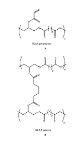
Figure lA depicts the chemical structure of a hydrophobic light-
activated adhesive (HLAA) pre-polymer before exposure to UV light. Figure
1B is the chemical structure of the HLAA after exposure to UV light. Figure
1C is a bar graph of the adhesive strength (N/cm2) of a poly(glycerol
sebacate urethane) (PGSU) patch as a function of adhesive for (from left to
right) cyanoacrylate (CA, DERMABONDO), fibrin (TISSUESEALO) and
HLAA that is uncured (0 s) or is cured for 1 s, 5 s, and 30 s respectively.
Figure 1D is a bar graph showing the adhesive strength of an HLAA
adhesive as a function of the patch material and curing time. The patch
materials include, from left to right, poly(glycerol sebacate urethane)
(PGSU), bovine pericardium, porcine small intestine submucosa, and
polyethylene terephthalate (PET). The UV curing times are 5 s or 30 s as
indicated below each bar. Figure lE is a bar graph of the relative adhesive
strength for patches coated with HLAA (left bar) or CA (right bar) after
being exposed to blood prior to adhesion testing. No significant change in
adhesion strength was observed for the HLAA patches. In contrast, the CA
patch is immediately activated upon contact with blood, losing almost all
ability to adhere to its intended substrate
Figure 2 is a graph showing the stress (MPa)-strain (%) curve for
compression of a cured HLAA over 100 cycles.
Figure 3A is a graph showing the loss modulus (G") and the storage
modulus (G') as a function of angular frequency for the HLAA pre-polymer
prior to curing. Figure 3B is a graph showing the viscosity (Pa*s) as a
function of shear rate (1/s) for the HLAA pre-polymer.
Figure 4 is a graph of the adhesive strength (N/cm2) as a function of
degree of acrylation (mol acrylate/mol-glycerol) for HLAA.
CA 02913338 2015-11-23
WO 2014/190302
PCT/US2014/039417
Figure 5A is a bar graph of the adhesive strength (N/cm2) of HLAA
cured for 5 seconds with 365 nm light and a pre-load of 3 Newtons as
function of the light intensity (W/cm2). Figure 5B is a bar graph of the
adhesive strength (N/cm2) of the HLAA cured for 5 seconds with 365 nm
light at an intensity of 0.38 W/cm2 as a function of pre-load (N) during
curing.
Figure 6 is a graph of the percent of UV light transmitted through
patch materials, going from left to right, poly(glycerol sebacate urethane)
(PGSU), bovine pericardium (BP), porcine small intestine submucosa (SIS),
and polyethylene terephthalate (PET).
Figure 7 is a bar graph of the thickness of an HLAA adhesive-coated
patch before (left bar) and after (right bar) exposure to blood.
Figure 8 depicts the chemical structures representing the acrylated
poly(glycerol subarate) (PGSubA) and acrylated poly(glycerol
dodecanedoate) (PGDoA) evaluated in adhesion measurements.
Figure 9 is a bar graph of the adhesion force (N/cm2) for GSubA (left
bar) and PGDoA (right bar). The dashed line represents the average value
obtained for adhesion of the HLAA (PGSA)
Figure 10 depicts the chemical structures of the different acrylate
derivatives produced from the PGS pre-polymer backbone.
Figure 11 is a bar graph of the adhesion force (N/cm2) for the
different acrylate derivatives produced from a PGS pre-polymer backbone.
The dashed line represents the average value obtained for adhesion of the
HLAA (PGSA)
Figure 12 depicts the chemical structure for a vinyl derivative
produced from the PGS pre-polymer (PGS-AI).
Figure 13 is a bar graph showing the adhesion force (N/cm2) of PGS-
AI. The dashed line represents the average value obtained for adhesion of the
HLAA (PGSA)
Figure 14 is a bar graph of the fold increase in adhesive strength of
HLAA on a blank (left bar) and a collagen-coated (right bar) substrate.
11
CA 02913338 2015-11-23
WO 2014/190302
PCT/US2014/039417
Figures 15A-15B are bar graphs of the degree of necrosis (Figure
15A) and the degree of inflammation (15B) as scored by a subjective
evaluation performed by a blinded pathologist of explanted hearts 7 days and
14 days after implantation with HLAA (left bars) and CA (right bars)
implants.
Figure 16 is a graph showing the number of deposited platelets as a
function of material.
DETAILED DESCRIPTION OF THE INVENTION
I. Definitions
"900 pull off adhesion" or "90 pull off adhesive strength" as used
herein refers to the adhesion value obtained by attaching an adhesive article
or sample to wet tissue, such as epicardial surface of cardiac tissue, blood
vessels, or the serosol side of porcine intestine tissue, immobilized on a
flat
substrate, such as a metallic stub. The 90 pull off adhesion test determines
the greatest perpendicular force (in tension) that a surface area can bear
before adhesive detachment.
The term "biomolecules", as used herein, refers to molecules (e.g.,
proteins, amino acids, peptides, polynucleotides, nucleotides, carbohydrates,
sugars, lipids, nucleoproteins, glycoproteins, lipoproteins, and small
molecules) whether naturally-occurring or artificially created (e.g., by
synthetic or recombinant methods) that are commonly found in cells and
tissues. Specific classes of biomolecules include, but are not limited to,
enzymes, receptors, neurotransmitters, hormones, cytokines, cell response
modifiers such as growth factors and chemotactic factors, antibodies,
vaccines, haptens, toxins, interferons, ribozymes, anti-sense agents,
plasmids, DNA, and RNA.
The terms "polynucleotide", "nucleic acid", or "oligonucleotide"
refer to a polymer of nucleotides. The terms "polynucleotide", "nucleic
acid", and "oligonucleotide", may be used interchangeably. Typically, a
polynucleotide comprises at least three nucleotides. DNAs and RNAs are
polynucleotides. The polymer may include natural nucleosides (i.e.,
adenosine, thymidine, guanosine, cytidine, uridine, deoxyadenosine,
12
CA 02913338 2015-11-23
WO 2014/190302
PCT/US2014/039417
deoxythymidine, deoxyguanosine, and deoxycytidine), nucleoside analogs
(e.g., 2-aminoadenosine, 2-thiothymidine, inosine, pyn-olo-pyrimidine, 3-
methyl adenosine, C5-propynylcytidine, C5-propynyluridine, C5
bromouridine, C5 fluorouridine, C5 iodouridine, C5 methylcytidine, 7
deazaadenosine, 7 deazaguanosine, 8 oxoadenosine, 8 oxoguanosine, 0(6)
methylguanine, and 2-thiocytidine), chemically modified bases, biologically
modified bases (e.g., methylated bases), intercalated bases, modified sugars
(e.g., 2'-fluororibose, ribose, 2'-deoxyribose, arabinose, and hexose), or
modified phosphate groups (e.g., phosphorothioates and 5 '-N
phosphoramidite linkages).
As used herein, a "polypeptide", "peptide", or "protein" comprises a
string of at least three amino acids linked together by peptide bonds. The
terms "polypeptide", "peptide", and "protein", may be used interchangeably.
Peptide may refer to an individual peptide or a collection of peptides.
Inventive peptides preferably contain only natural amino acids, although
non-natural amino acids (i.e., compounds that do not occur in nature but that
can be incorporated into a polypeptide chain) and/or amino acid analogs as
are known in the art may alternatively be employed.
The terms "polysaccharide", "carbohydrate", or "oligosaccharide"
refer to a polymer of sugars. The terms "polysaccharide", "carbohydrate",
and "oligosaccharide", may be used interchangeably. Typically, a
polysaccharide comprises at least three sugars. The polymer may include
natural sugars (e.g., glucose, fructose, galactose, mannose, arabinose,
ribose,
and xylose) and/or modified sugars (e.g., 2'-fluororibose, 2'-deoxyribose,
and hexose).
The term "biocompatible", as used herein, is intended to describe
materials that do not elicit a substantial detrimental response in vivo. For
example, cyanoacrylate glues are not approved for use in vivo due to
significant inflammation and toxicity and therefore are not considered to be
biocompatible.
As used herein, "biodegradable" polymers are polymers that degrade
to oligomeric and/or monomeric species under physiological or endosomal
conditions. In various preferred embodiments, the polymers and polymer
13
CA 02913338 2015-11-23
WO 2014/190302
PCT/US2014/039417
biodegradation byproducts are biocompatible. Biodegradable polymers are
not necessarily hydrolytically degradable and may require enzymatic action
to fully degrade.
The phrase "physiological conditions", as used herein, relates to the
range of chemical (e.g., pH, ionic strength) and biochemical (e.g., enzyme
concentrations) conditions likely to be encountered in the intracellular and
extracellular fluids of tissues. For most tissues, the physiological pH ranges
from about 7.0 to 7.4.
"Hydrophobic", as used herein, means that the pre-polymer
sufficiently repels water to remain in place at the site of
application/administration prior to crosslinking.
"Injectable", as used herein, means the pre-polymer composition is
sufficiently less viscous that is can applied through a syringe need, for
example, a needle having a gauge from 14-20, preferably 14-18, more
preferably 16-18. In some embodiment, the needle is 18 gauge.
"Degree of activation", as used herein, refers to the actual amount of
activation/functionalization on the pre-polymer. The degree of activation is
typically expressed as moles of activating agent per mole of moiety to be
acrylated. For example, acrylation (e.g., degree of acrylation) is expressed
as
moles of acrylating agent (e.g., acryl chloride) per mole of moiety to be
acrylated (e.g., glycerol). In other embodiments, the degree of activation
(e.g., degree of acrylation) can be expressed as a percent of the available
moieties that have been activated and are available for crosslinking. The
actual percent of moieties that are crosslinked is typically less than the
percent of moieties that are activated since the degree of crosslinking is
depending on the stimulus time (e.g., irradiation time, heating time, etc.).
"Crosslinked in the presence of blood", as used herein, means that the
pre-polymer can be incubated in blood or other bodily fluids before
application/administration with little or no crosslinking. The pre-polymer is
substantially crosslinked only after the intentional application of an
external
stimulus, such as UV light, heat, chemical initiator, etc. This is contrasted
with other known adhesives, such as cyanoacrylates, which are highly
reactive to moisture in the surrounding atmosphere and must be stored in an
14
CA 02913338 2015-11-23
WO 2014/190302
PCT/US2014/039417
inert, dry environment prior to use. Such adhesives cannot be exposed to
bodily fluids prior to application at the desired site.
As used herein, "bioactive agents" is used to refer to compounds or
entities that alter, inhibit, activate, or otherwise affect biological or
chemical
events.
As used herein, the term "tissue" refers to a collection of similar cells
combined to perform a specific function, and any extracellular matrix
surrounding the cells.
The term "substituted" as used herein means replacing a hydrogen or
one or more atoms, e.g., carbon, nitrogen, oxygen, etc., of a molecule.
Substituents can include, for example, alkyl, alkenyl, alkynyl, halogen,
hydroxyl, alkylcarbonyloxy, arylcarbonyloxy, alkoxycarbonyloxy,
aryloxycarbonyloxy, carboxylate, alkylcarbonyl, arylcarbonyl,
alkoxycarbonyl, aminocarbonyl, alkylaminocarbonyl, dialkylaminocarbonyl,
alkoxyl, cyano, amino (including alkyl amino, dialkylamino, arylamino,
diarylamino, and alkylarylamino), acylamino (including alkylcarbonylamino,
arylcarbonylamino, carbamoyl and ureido), amidino, imino, sulfhydryl,
alkylthio, arylthio, nitro, trifluoromethyl, cyano, azido, heterocyclyl,
alkylaryl, or an aromatic or heteroaromatic group. Accordingly, the phrase
"a substituent as described herein" or the like refers to one or more of the
above substituents, and combinations thereof
The term "alkyl" includes saturated aliphatic groups, which includes
both "unsubstituted alkyls" and "substituted alkyls", the latter of which
refers
to alkyl groups having substituents replacing a hydrogen on one or more
carbons of the hydrocarbon backbone. The term "alkyl" includes straight-
chain alkyl groups (e.g., methyl, ethyl, propyl, butyl, pentyl, hexyl, heptyl,
octyl, nonyl, decyl, etc.), branched-chain alkyl groups (isopropyl, tert-
butyl,
isobutyl, etc.), cycloalkyl (alicyclic) groups (cyclopropyl, cyclopentyl,
cyclohexyl, cycloheptyl, cyclooctyl), and cycloalkyl substituted alkyl groups.
The term "alkyl" also includes the side chains of natural and unnatural amino
acids.
An "alkylaryl" or an "aralkyl" group is an alkyl substituted with an
aryl (e.g., phenylmethyl (benzyl)).
CA 02913338 2015-11-23
WO 2014/190302
PCT/US2014/039417
The term "aryl" includes 5- and 6-membered single-ring aromatic
groups, as well as multicyclic aryl groups, e.g., tricyclic, bicyclic, e.g.,
naphthalene, anthracene, phenanthrene, etc.). The aromatic ring(s) can be
substituted at one or more ring positions with such substituents as described
above. Aryl groups can also be fused or bridged with, e.g., alicyclic or
heterocyclic rings which are not aromatic so as to form, e.g., a polycycle.
The term "alkenyl" includes unsaturated aliphatic groups analogous
in length and possible substitution to the alkyls described above, but which
contain at least one double bond. For example, the term "alkenyl" includes
straight-chain alkenyl groups (e.g., ethenyl, propenyl, butenyl, pentenyl,
hexenyl, heptenyl, octenyl, nonenyl, decenyl, etc.), branched-chain alkenyl
groups, cycloalkenyl (alicyclic) groups (cyclopropenyl, cyclopentenyl,
cyclohexenyl, cycloheptenyl, cyclooctenyl), alkyl or alkenyl substituted
cycloalkenyl groups, and cycloalkyl or cycloalkenyl substituted alkenyl
groups. The term alkenyl includes both "unsubstituted alkenyls" and
"substituted alkenyls", the latter of which refers to alkenyl groups having
substituents replacing a hydrogen on one or more carbons of the hydrocarbon
backbone.
The term "alkynyl" includes unsaturated aliphatic groups analogous
in length and possible substitution to the alkyls described above, but which
contain at least one triple bond. For example, the term "alkynyl" includes
straight-chain alkynyl groups (e.g., ethynyl, propynyl, butynyl, pentynyl,
hexynyl, heptynyl, octynyl, nonynyl, decynyl, etc.), branched-chain alkynyl
groups, and cycloalkyl or cycloalkenyl substituted alkynyl groups. The term
alkynyl includes both "unsubstituted alkynyls" and "substituted alkynyls",
the latter of which refers to alkynyl groups having substituents replacing a
hydrogen on one or more carbons of the hydrocarbon backbone.
The term "acyl" includes compounds and groups which contain the
acyl radical (CH3C0-) or a carbonyl group. The term "substituted acyl"
includes acyl groups having substituents replacing a one or more of the
hydrogen atoms.
16
CA 02913338 2015-11-23
WO 2014/190302
PCT/US2014/039417
The term "acylamino" includes groups wherein an acyl group is
bonded to an amino group. For example, the term includes
alkylcarbonylamino, arylcarbonylamino, carbamoyl and ureido groups.
The term "aroyl" includes compounds and groups with an aryl or
heteroaromatic group bound to a carbonyl group. Examples of aroyl groups
include phenylcarboxy and naphthyl carboxy.
The terms "alkoxyalkyl", "alkylaminoalkyl" and "thioalkoxyalkyl"
include alkyl groups, as described above, which further include oxygen,
nitrogen or sulfur atoms replacing one or more carbons of the hydrocarbon
backbone, e.g., oxygen, nitrogen or sulfur atoms.
The term "alkoxy" includes substituted and unsubstituted alkyl,
alkenyl, and alkynyl groups covalently linked to an oxygen atom. Examples
of alkoxy groups include methoxy, ethoxy, isopropyloxy, propoxy, butoxy,
and pentoxy groups and may include cyclic groups such as cyclopentoxy.
The term "amine" or "amino" includes compounds where a nitrogen
atom is covalently bonded to at least one carbon or heteroatom. The term
"alkyl amino" includes groups and compounds wherein the nitrogen is bound
to at least one additional alkyl group. The term "dialkyl amino" includes
groups wherein the nitrogen atom is bound to at least two additional alkyl
groups. The term "arylamino" and "diarylamino" include groups wherein
the nitrogen is bound to at least one or two aryl groups, respectively. The
term "alkylarylamino," "alkylaminoaryl" or "arylaminoalkyl" refers to an
amino group that is bound to at least one alkyl group and at least one aryl
group. The term "alkaminoalkyl" refers to an alkyl, alkenyl, or alkynyl
group bound to a nitrogen atom that is also bound to an alkyl group.
The term "amide" or "aminocarboxy" includes compounds or groups
that contain a nitrogen atom that is bound to the carbon of a carbonyl or a
thiocarbonyl group. The term includes "alkaminocarboxy" groups that
include alkyl, alkenyl, or alkynyl groups bound to an amino group bound to a
carboxy group. It includes arylaminocarboxy groups that include aryl or
heteroaryl groups bound to an amino group which is bound to the carbon of a
carbonyl or thiocarbonyl group. The terms "alkylaminocarboxy,"
"alkenylaminocarboxy," "alkynylaminocarboxy," and "arylaminocarboxy"
17
CA 02913338 2015-11-23
WO 2014/190302
PCT/US2014/039417
include groups wherein alkyl, alkenyl, alkynyl and aryl groups, respectively,
are bound to a nitrogen atom which is in turn bound to the carbon of a
carbonyl group.
The term "carbonyl" or "carboxy" includes compounds and groups
which contain a carbon connected with a double bond to an oxygen atom,
and tautomeric forms thereof Examples of groups that contain a carbonyl
include aldehydes, ketones, carboxylic acids, amides, esters, anhydrides, etc.
The term "carboxy group" or "carbonyl group" refers to groups such as
"alkylcarbonyl" groups wherein an alkyl group is covalently bound to a
carbonyl group, "alkenylcarbonyl" groups wherein an alkenyl group is
covalently bound to a carbonyl group, "alkynylcarbonyl" groups wherein an
alkynyl group is covalently bound to a carbonyl group, "arylcarbonyl"
groups wherein an aryl group is covalently attached to the carbonyl group.
Furthermore, the term also refers to groups wherein one or more heteroatoms
are covalently bonded to the carbonyl group. For example, the term includes
groups such as, for example, aminocarbonyl groups, (wherein a nitrogen
atom is bound to the carbon of the carbonyl group, e.g., an amide),
aminocarbonyloxy groups, wherein an oxygen and a nitrogen atom are both
bond to the carbon of the carbonyl group (e.g., also referred to as a
"carbamate"). Furthermore, aminocarbonylamino groups (e.g., ureas) are
also include as well as other combinations of carbonyl groups bound to
heteroatoms (e.g., nitrogen, oxygen, sulfur, etc. as well as carbon atoms).
Furthermore, the heteroatom can be further substituted with one or more
alkyl, alkenyl, alkynyl, aryl, aralkyl, acyl, etc. groups.
The term "ether" includes compounds or groups that contain an
oxygen bonded to two different carbon atoms or heteroatoms. For example,
the term includes "alkoxyalkyl" which refers to an alkyl, alkenyl, or alkynyl
group covalently bonded to an oxygen atom that is covalently bonded to
another alkyl group.
The term "ester" includes compounds and groups that contain a
carbon or a heteroatom bound to an oxygen atom that is bonded to the carbon
of a carbonyl group. The term "ester" includes alkoxycarboxy groups such
as methoxycarbonyl, ethoxycarbonyl, propoxycarbonyl, butoxycarbonyl,
18
CA 02913338 2015-11-23
WO 2014/190302
PCT/US2014/039417
pentoxycarbonyl, etc. The alkyl, alkenyl, or alkynyl groups are as defined
above.
The term "hydroxy" or "hydroxyl" includes groups with an ¨OH or ¨
0-.
The term "halogen" includes fluorine, bromine, chlorine, iodine, etc.
The term "perhalogenated" generally refers to a group wherein all hydrogens
are replaced by halogen atoms.
The term "heteroatom" includes atoms of any element other than
carbon or hydrogen. Preferred heteroatoms are nitrogen, and oxygen. The
term "heterocycle" or "heterocyclic" includes saturated, unsaturated,
aromatic ("heteroaryls" or "heteroaromatic") and polycyclic rings which
contain one or more heteroatoms. The heterocyclic may be substituted or
unsubstituted. Examples of heterocyclics include, for example,
benzodioxazole, benzofuran, benzoimidazole, benzothiazole,
benzothiophene, benzoxazole, chromene, deazapurine, furan, indole,
indolizine, imidazole, isoxazole, isoindole, isoquinoline, isothiaozole,
methylenedioxyphenyl, napthridine, oxazole, purine, pyran, pyrazine,
pyrazole, pyridazine, pyridine, pyrimidine, pyrrole, quinoline, tetrazole,
thiazole, thiophene, and triazole. Other heterocycles include morpholino,
piprazine, piperidine, thiomorpholino, and thioazolidine.
The terms "polycyclic ring" and "polycyclic ring structure" include
groups with two or more rings (e.g., cycloalkyls, cycloalkenyls,
cycloalkynyls, aryls and/or heterocyclyls) in which two or more carbons are
common to two adjoining rings, e.g., the rings are "fused rings". Rings that
are joined through non-adjacent atoms are termed "bridged" rings. Each of
the rings of the polycyclic ring can be substituted with such substituents as
described above.
The term "about" or "approximately" as used herein generally means
within 20%, preferably within 10%, and more preferably within 5% of a
given value or range. The term "about x" further includes x.
II. Pre-polymers
Pre-polymers for use as tissue sealants and adhesives have flow
characteristics such that they can be applied to the desired area through a
19
CA 02913338 2015-11-23
WO 2014/190302
PCT/US2014/039417
syringe or catheter (e.g., relatively low viscosity) but are sufficiently
viscous
to remain in place at the site of application and not run off the tissue. Pre-
polymer", as used herein, refers to the activated polymer prior to
crosslinking. The pre-polymer is also sufficiently hydrophobic to resist
washout by bodily fluids, such as blood. This facilitates delivery to the
desired site as well as repositioning of implanted devices during minimally
invasive surgery. The pre-polymer is stable in bodily fluids; does not
spontaneously crosslink in bodily fluids absent the presence of an
intentionally applied stimulus (e.g., UV light, heat, chemical initiator) to
initiate crosslinking. The molecular weight of the pre-polymer can vary. In
some embodiments, the molecular weight of the pre-polymer is from about
1,000 Daltons to about 10,000 Daltons, from about 2,000 Daltons to about
10,000 Daltons, from about 3,000 Daltons to about 10,000 Daltons from
about 5,000 Daltons to about 10,000 Daltons. In some embodiments the
molecular weight of the pre-polymer is about 3,000 Daltons. Upon
crosslinking, the cured polymer exhibits significant adhesive strength in the
presence of blood and other bodily fluids. The pre-polymer can be incubated
in bodily fluids, such as blood, prior to administration and crosslinking,
without a substantial decrease in adhesive strength when crosslinked. The
adhesive (cured polymer) is sufficiently elastic that it is able to resist
movement of the underlying tissue (e.g., contractions of the heart, blood
vessels, etc.). The adhesive (cured polymer) can provide a hemostatic seal
and is biodegradable and biocompatible, causing minimal inflammatory
response.
The adhesive strength of bioadhesive polymers can be improved as a
function of the mechanical properties of adhesive cured polymer and the
degree of interdigitation or entanglement of the cured polymer with the tissue
to which it is applied. The degree of entanglement and mechanical properties
are a function of the molecular weight of the precursor, the degree of
activation of the pre-polymer (e.g. activation by acrylation), and the percent
crosslinking of the cured polymer. In one embodiment, the pre-polymer is
activated by introduction of one or more functional groups that can be
reacted to form crosslinks between polymer chains. The resulting material is
CA 02913338 2015-11-23
WO 2014/190302
PCT/US2014/039417
preferably biodegradable and elastomeric. In some embodiments, the
polymer chain is a polyester formed from a substituted or unsubstituted
polyol, such as a triol, and a substituted or unsubstituted diacid. In
particular
embodiments, the triol is glycerol. Free functional groups on the pre-
polymer can be activated by introducing reactive functional groups that can
be reacted to form crosslinks to form the tissue sealant or adhesive. For
example, in some embodiments, free hydroxy groups on the polyol can be
acrylated by introducing acrylate groups. The acrylate groups are
subsequently reacted to form crosslinks to form the adhesive or sealant. In
some embodiments, the degree of activation, preferably acrylation, of the
pre-polymer is from about 0.2 to about 0.9, preferably from about 0.3 to
about 0.7, more preferably from about 0.4 to about 0.6. In particular
embodiments, the degree of activation, preferably acrylation, is about 0.5.
The crosslink density in the cured polymer can be varied by varying the
degree of activation, preferably acrylation, and/or the crosslinking
conditions, such as time. In some embodiments, the crosslink density is at
least about 1%, 2%, 3%, 4%, 5%, 6%, 7%, 8%, 9%, 10%, 12%, 15%, 18%,
20%, 22%, 25%, 28%, 30%, 32%, 35%, 38%, 4,,o/
/0
u or greater. In particular
embodiments, the activation of the pre-polymer is acrylation and the
crosslinks in the cured polymer contain single dioic acid ester functionality.
In particular embodiments, the crosslinked polymer (or cured
polymer) in stand-alone or as applied to a patch has a 90 pull off adhesive
strength of at least about 0.5 Nlem2, at least about 1 N/cm2, or even at least
about 2 Nictn2, and one or more of the following characteristics: (I)
molecular weight of the pre-polymer is from about 1,000 Daltons to about
10,000 Daltons, from about 2,000 Daltons to about 10,000 Daltons, from
about 3,000 Daltons to about 10,000 Daltons, from about 5,000 Daltons to
about 10,000 Daltons, or according to preferred embodiments, about 3,000
Daltons; (2) the degree of activation, preferably acrylation, is from about
0.2
to about 0.9, from about 0.3 to about 0.8, from about 0.4 to about 0.6, or
about 0.5; and/or (3) the crosslink density is at least about 1%, 2%, 3%, 4%,
5%, 6%, 7%, 8%, 9%, 10%, 12%, 15%, 18%, 20%, 22%, 25%, 28%, 30%,
21
CA 02913338 2015-11-23
WO 2014/190302
PCT/US2014/039417
32%, 35%, 38%, 40%, or greater. In particular embodiments, the cured
polymer in stand-alone or as applied to a patch exhibits burst strengths of at
least 100 mmHg, 140 mm Hg, 150 mm Hg, 160 mm Hg, 170 mm Hg, 180
mm Hg, 190 mm Hg, 200 mm Hg, or greater than 200 mm Hg,
The pre-polymer is sufficiently hydrophobic so that upon application
or administration to the desired site, the pre-polymer repels water and is not
washed away by bodily fluids, such as blood. This is contrasted with
hydrophilic tissue sealants/adhesives in the art, such as polyethylene glycol
(PEG)-based materials, which are washed away by bodily fluids after
application/administration. The pre-polymer can also be incubated in bodily
fluids, such as blood, without reacting (e.g., crosslinking). Once applied and
crosslinked, the cured polymer exhibits no loss or minimal loss in adhesive
properties due to the incubation in bodily fluids, especially blood. This is
contrasted with known adhesives, such as cyanoacrylates, which are highly
reactive and must be stored in an inert, dry environment prior to use since
the
materials will react due to moisture in the surrounding environment (e.g.,
air,
bodily fluids, etc.). These materials cannot be incubated in bodily fluids
prior to use.
The pre-polymer is preferably activated. This means that reactive
functional groups are incorporated on the pre-polymer backbone. The
activation according to the preferred embodiment includes introducing
substituted or unsubstituted vinyl groups in the pre-polymer backbone. In
more preferred embodiments, it includes the introduction of substituted or
unsubstituted acrylate groups, using techniques known in the art. In one
embodiment, the activation includes introducing vinyl ester, vinyl
carbamates, vinyl ketones, vinyl amide, vinyl carbonate, vinyl ether groups
or vinyl groups in the form of allyl. ..
Mechanical properties of the adhesive or sealant are dependent on the
crosslink density of the cured polymer. In some embodiments, the crosslink
density in the cured polymer is greater than 1%, for example, greater than
5%, 8%, 10%, 12%, 15%, 18%, 20%, 22%, 25%, 28%, 30%, 32%, 35%,
38%, 40%, or greater. The crosslink density is a function of the actual
degree of activation, preferably acrylation, of the pre-polymer (e.g.,
22
CA 02913338 2015-11-23
WO 2014/190302
PCT/US2014/039417
theoretical number of crosslinking sites). It can be further improved by
modulating the crosslinking reaction time (e.g., how many groups actually
reacted) and/or energy.
In some embodiments, the degree of activation, preferably acrylation,
is from about 0.2 to about 0.9, preferably from about 0.4 to about 0.75, more
preferably about 0.5. Values below this range tend to form adhesive that is
not mechanically robust enough, particularly for applications where the
adhesive must withstand high pressures, such as cardiac chambers or blood
vessels and/or where the adhesive is in contact, especially prolonged contact,
with bodily fluids, such as blood. Values above this range tend to form
adhesives with a higher degree of stiffness. This can be problematic for
applications where the adhesive needs to flex and move with the movement
of the patient.
The adhesive is sufficiently elastic that it is able to resist movement
of the underlying tissue (e.g., contractions of the heart, blood vessels,
etc.).
The adhesive can provide a hemostatic seal. The adhesive is biodegradable
and biocompatible, causing minimal inflammatory response. The adhesive is
preferably elastomeric.
In some embodiments, the pre-polymers are prepared by reacting a
polyol, such as a diol, triol, tetraol, or greater with a polyacid, such as
diacid
or higher order acid to form a polyester. Other pre-polymer backbones can
also be used to form activated pre-polymers including, but not limited to
poly(ester amides) poly(urethanes) and/or other elastomeric materials. The
free hydroxy groups of the pre-polymer can be activated, such as by
acrylation or vinylation, to form the activated pre-polymer. In some
embodiments the acrylation reaction occurs through acylation of the free
hydroxyl groups. In other embodiment, free hydroxyl groups in the pre-
polymer can be activated via an isocyanate linker generating urethane bonds.
Other functional groups (e.g., carboxylic acid, amine, etc.) can be activated
in place of or in addition to free hydroxy groups.
A. Polyol
"Polyol", as used herein, means a molecule or moiety containing two
or more hydroxy groups. If only one type of polyol is used, the polyol
23
CA 02913338 2015-11-23
WO 2014/190302
PCT/US2014/039417
contains three or more hydroxy groups. In other embodiments, a mixture of
different polyols can be used where some of the polyols contain two or more
hydroxy groups and the other polyols contain three or more hydroxy groups.
Suitable polyols include diols, such as alkane diols; triols, such as
glycerol,
trimethylolpropane, triethanolamine; tetraols, such as erythritol,
pentaerythritol; and higher polyols, such as sorbitol. Unsaturated diols, such
as tetradeca-2,12-diene-1,14-diol, or other diols including macromonomer
diols such as, e.g., polyethylene oxide, and N-methyldiethanoamine (MDEA)
can also be used. In one embodiment, the polyol is substituted or
unsubstituted glycerol.
In addition to incorporation into the pre-polymer, the polyols can be
incorporated into the resultant cross-linked polymer through, e.g., acrylate
chemistry. For example, the diols could be first acrylated and then combined
with acrylated pre-polymer using a free radical polymerization reaction. In
various embodiments, aldehydes and thiols can be used, e.g., for attaching
proteins and growth factors to the pre-polymer.
B. Polyacid
A wide variety of diacid, or higher order acids, can be used in the
formation of the elastic biodegradable polymer compositions. Exemplary
acids include, but are not limited to, glutaric acid (5 carbons), adipic acid
(6
carbons), pimelic acid (7 carbons), suberic acid (8 carbons), and azelaic acid
(nine carbons). Exemplary long chain diacids include diacids having more
than 10, more than 15, more than 20, and more than 25 carbon atoms. Non-
aliphatic diacids can be used. For example, versions of the above diacids
having one or more double bonds can be used to produce polyol-diacid co-
polymers.
Amines and aromatic groups can be incorporated into the carbon
chain. Exemplary aromatic diacids include terephthalic acid and
carboxyphenoxy-propane. The diacids can also include substituents as well.
For example, in various embodiments, reactive groups like amine and
hydroxyl can be used increase the number of sites available for cross-linking.
In various embodiments, amino acids and other biomolecules can be used to
modify the biological properties of the polymer. In various embodiments,
24
CA 02913338 2015-11-23
WO 2014/190302
PCT/US2014/039417
aromatic groups, aliphatic groups, and halogen atoms can be used to modify
the inter-chain interactions within the polymer. In one embodiment, the
diacid is substituted or unsubstituted sebacic acid.
C. Activated pre-polymer
The pre-polymer is preferably activated. It can be activated by
introducing functional groups that can react or be reacted to form crosslinks.
The pre-polymer is activated by reacting one or more functional groups on
the polymer backbone with one or more functional groups that can react or
be reacted to form crosslinks resulting in cured polymer. In some
embodiments, the reactive functional group to be crosslinked in the pre-
polymer is a substituted or unsubstituted vinyl group. In some embodiments,
the crosslink in the corresponding cured polymer is or contains a single dioic
ester functionality.
Suitable functional groups to be activated on the pre-polymer
backbone include hydroxy groups, carboxylic acid groups, amines, and
combinations thereof In particular embodiments, the functional group to be
activated is hydroxy and/or carboxylic acid. In more particular
embodiments, it is hydroxy. The free hydroxyl groups on the pre-polymer
can be activated by functionalizing the hydroxy groups with a moiety which
can form a crosslink between polymer chains. In some embodiment, the
groups that are activated are free hydroxyl groups on A and/or B moieties in
pre-polymer.
The free hydroxy groups can be functionalized with a variety of
functional groups. In one embodiment, the free hydroxy groups are
functionalized with vinyl groups. Vinyl groups can be introduced by a
variety of techniques known in the art, such as by vinylation or acrylation.
Vinyl groups contain the following structure -CR1=CR2R3whereinR1, R2, R3
are independently from one another, selected from H, alkyl (e.g. methyl,
ethyl), aryl (e.g. phenyl), substituted alkyl, substituted aryl, carboxylic
acid,
ester, amide, amine, urethane, ether, and carbonyl
In one embodiment, the functional group is or contains an acrylate
group. Acrylate group are moieties containing substituted or unsubstituted
acryloyl group. According to specific embodiment, it contains the following
CA 02913338 2015-11-23
WO 2014/190302
PCT/US2014/039417
group: , -C(=0)-CR1=CR2R3, whereinRi, R2, R3 are independently from one
another, selected in the group consisting of H, alkyl (e.g. methyl, ethyl),
aryl
(e.g. phenyl), substituted alkyl, substituted aryl, carboxylic acid, ester,
amide, amine, urethane, ether, and carbonyl
Preferred embodiments include where R1, R2 and R3 are H; R1 is
CH3, R2 and R3 are H ; R1 and R2 are H and R3 is CH3 ; and R1 and R2 are H
and R3 is phenyl. Vinyl groups can also be incorporated in the backbone of
the pre-polymer using free carboxyl groups on the pre-polymer. For
example, hydroxyethyl methacrylate can be incorporated through the COOH
groups of the pre-polymer using carbonyl diimidazole activation chemistry.
The degree of activation can vary. In some embodiments, the degree
of activation is from about 0.2 to about 0.9, preferably from about 0.3 to
about 0.8, most preferably from about 0.4 to about 0.6. In particular
embodiments, the degree of activation, preferably of acrylation, is about 0.5.
In particular embodiments, the degree of activation is as described above and
the reactive functional group is acrylate (degree of acrylation).
In addition to acrylates or other vinyl groups, other agents can be
used to activate the pre-polymer. Examples of such agents include, but are
not limited to, glycidyl, epichlorohydrin, triphenylphosphine, diethyl
azodicarboxylate (DEAD), diazirine, divinyladipate, and divinylsebacate
with the use of enzymes as catalysts, phosgene-type reagents, di-acid
chlorides, bis-anhydrides, bis-halides, metal surfaces, and combinations
thereof
The activated pre-polymer should have a viscosity which allows the
pre-polymer to stay in place at the site of administration without being
washed away by bodily fluids, such as water and/or blood. In some
embodiments, the viscosity of the pre-polymer is between about 0.5 to about
100 Pas, preferably between about 1.0 to about 50 Pas, more preferably
between about 2.0 to about 40 Pas, and most preferably between about 2.5 to
about 25 Pas. The viscosity of the pre-polymer is determined in part by the
molecular weight of the pre-polymer. In some embodiments, the weight
average molecular weight of the pre-polymer is between about 5,000 Daltons
to about 1,000,000 Daltons, between about 10,000 Daltons to about
26
CA 02913338 2015-11-23
WO 2014/190302
PCT/US2014/039417
1,000,000 Daltons, preferably between about 10,000 Daltons to about
500,000 Daltons, more preferably between about 10,000 Daltons to about
250,000 Daltons, and most preferably between about 10,000 Daltons to
100,000 Daltons. In particular embodiments, the weight average molecular
weight is less than about 100,000 Daltons, less than about 75,000 Daltons,
less than about 50,000 Daltons, less than about 40,000 Daltons, less than
30,000 Daltons, or less than 20,000 Daltons. In other embodiments, the
molecular weight is between about 1000 Daltons to about 10,000 Daltons,
between about 2000 Daltons to about 10,000 Daltons, between about 3000
Daltons to about 10,000 Daltons, or between about 5,000 Daltons to about
10,000 Daltons. In a preferred embodiment, it is about 3000 Daltons.
The hydrophobic nature of the pre-polymer functions to keep the pre-
polymer in place at the site of administration by repelling water.
Hydrophobicity is dependent on the chemical composition of the pre-
polymer, including the hydrophobic nature of the polymer backbone (e.g.,
longer alkyl chain are more hydrophobic than shorter chains) and the degree
of activation.
In some embodiments, the pre-polymer has the following chemical
structure:
o o
(7N7oN7)/n
P
0
0 ____________________
where p is an integer from 1-20, preferably 2-20, more preferably
from 2-10, most preferably from 4-10 and n is an integer from 1-10,000. In
some embodiments, the crosslinks are between a portion of the A moieties.
In other embodiments, the crosslinks can be between a portion of the B
moieties. In still other embodiments, the crosslinks can be between a portion
27
CA 02913338 2015-11-23
WO 2014/190302
PCT/US2014/039417
of the A and B moieties. A "portion", as used herein, means some amount
less than the total amount, for example, less than 10%, 15%, 20%, 25%,
30%, 35%, 40%, 45%, 50%, 55%, 60%, 65%, 70%, or 75%. In some
embodiments, the portion of functional groups that are activated is less than
60%, preferably less than 55%, more preferably less than 50%.
The activated pre-polymer can be further reacted with one or more
additional materials to modify the crosslinks between the polymer chains.
For example, prior to or during curing/crosslinking, one or more hydrogel or
other polymeric precursors (e.g., precursors that may be modified to contain
acrylate groups) such as poly(ethylene glycol), dextran, chitosan, hyaluronic
acid, and alginate, other acrylate based precursors such as acrylic acid,
butyl
acrylate, 2-ethylhexyl acrylate, methyl acrylate, ethyl acrylate,
acrylonitrile,
n-butanol, methyl methacrylate, and trimethylol propane trimethacrylate
("TMPTA"), pentaerythritol trimethacrylate, pentaerythritol
tetramethacrylate, ethylene glycol dimethacrylate. dipentaerythritol penta
acrylate, (Bis phenol A glycidal methacrylate) ("Bis-GMA") and (tri-
ethylene, glycol dimethacrylate) ("TEGDMA"), sucrose acrylate, and
combinations thereof, can be reacted with the acrylated pre-polymer (e.g.,
PGSA).
III. Methods of making the pre-polymers
Crosslinkable groups, such as vinyl groups, can be incorporated in
the backbone of the pre-polymer with or with-out the use of catalyst,
although the use of a catalyst is preferred. A wide variety of catalysts can
be
used, including, but not limited to, 4-(dimethylamino)pyridine, N-hydroxy
succinimide, carbodiimides, and pyridine. Preferably, the reaction is carried
out in a solvent. Examples of suitable solvents include, but are not limited
to, benzene, toluene, chloroform, dichloromethane, ethyl acetate, and
tetrahydrofuran.
In some embodiments, activation of the pre-polymer through
vinylation can be carried out. Examples of suitable vinyl groups to activate
the pre-polymer include substituted or unsubstituted vinyl ester, substituted
or unsubstituted vinyl carbamates, substituted or unsubstituted vinyl ketones,
substituted or unsubstituted vinyl amides, substituted or unsubstituted vinyl
28
CA 02913338 2015-11-23
WO 2014/190302 PCT/US2014/039417
carbonates, substituted or unsubstituted vinyl ether groups, and substituted
or
unsubstituted vinyl groups in the form of allyl. Vinyl groups can be
introduced in the pre-polymer through a variety of techniques known in the
prior art. These can be, but are not limited to, acylation or urethanization
reactions.
In some embodiments, free hydroxyl groups (or other functional
groups, such as amines or carboxylic acids) can be activated through
acrylation, generating acrylate groups.. Examples of suitable acrylates
include, but are not limited to, methacrylate, 3-phenylacrylate, beta-
methylacrylate vinyl methacrylate, maleic methacrylate, and those having the
structure:
R1 R1 R1
,...."---....,..-0,..
R,....-5,.._ ,....Ø, 0,.......õ---.
2 `= R2......-
0, 0 0
,or
0 R1
/0
R1 R2 R1
\
R2 R2
0 0 /
where R1 can be methyl or hydrogen; and R2, R2', and R2" can be alkyl, aryl,
heterocycles, cycloalkyl, aromatic heterocycles, multicycloalkyl, hydroxyl,
ester, ether, halide, carboxylic acid, amino, alkylamino, dialkylamino,
trialkylamino, amido, carbamoyl thioether, thiol, alkoxy, or ureido groups.
R2, R2', and R2" may also include branches or substituents including alkyl,
aryl, heterocycles, cycloalkyl, aromatic heterocycles, multicycloalkyl,
hydroxyl, ester, ether, halide, carboxylic acid, amino, alkylamino,
dialkylamino, trialkylamino, amido, carbamoyl, thioether, thiol, alkoxy, or
ureido groups.
Further examples of suitable acrylate monomers include, but are not
limited to,
29
CA 02913338 2015-11-23
WO 2014/190302
PCT/US2014/039417
0 ,
OH
0 0 0 0 0
F F 0
F F
F F
0 F F
0
0 0
0
,and
OH
0
Activation of the pre-polymer through acrylation can be carried out
by reacting the pre-polymer with an acryloyl, such as acryloyl chloride,
generating an acrylate group through an acylation. The reaction can be
carried out in the presence of catalysts, such as triethylamine and 4-
(dimethylamino)pyridine ("4-DMAP"). The reaction can be carried in an
organic solvent, such as anhydrous dichloromethane. It is preferred that that
this reaction is carried out under dry conditions using these reagents. Free
carboxylic acid groups may also be acrylated in this reaction.
The degree of activation, preferably degree of acrylation, of the pre-
polymer can be used to adjust the properties of the resultant cross-linked
polymer.
In alternative embodiments, the activated, preferably acrylated, pre-
polymer is a viscous liquid that can be cured without solvent.
III. Methods of making the adhesives
CA 02913338 2015-11-23
WO 2014/190302
PCT/US2014/039417
In various embodiments, the activated pre-polymers can be
crosslinked to form a cured polymeric network using a free radical initiated
reaction, such as, for example, by photo-initiated polymerization, thermally-
initiated polymerization, and redox initiated polymerization.
The acrylated pre-polymer can be irradiated with light (typically
ultraviolet (UV) light) in the presence of a photoinitiator to facilitate the
reaction. Examples of suitable photoinitiators include, but are not limited
to,
2-dimethoxy-2-phenyl-acetophenone, 2-hydroxy-1-[4-
(hydroxyethoxy)pheny1]-2-methyl-l-propanone (IRGACUREO 2959), 1-
hydroxycyclohexyl-l-phenyl ketone (IRGACUREO 184), 2-hydroxy-2-
methyl-l-pheny1-1-propanone (DAROCURO 1173), 2-benzy1-2-
(dimehylamino)-1-[4-morpholinyl) phenyl]-1-butanone (Irgacure 369),
methylbenzoylformate (DAROCURO MBF), oxy-phenyl-acetic acid-242-
oxo-2-phenyl-acetoxy-ethoxy]-ethyl ester (IRGACUREO 754), 2-methyl-1-
[4-(methylthio)pheny1]-2-(4-morpholiny1)-1-propanone (IRGACUREO 907),
dipheny1(2,4,6-trimethylbenzoy1)-phosphine oxide (DAROCURO TPO),
phosphine oxide, phenyl bis(2,4,6-trimethyl benzoyl) (IRGACUROE 819),
and combinations thereof
In various preferred embodiments, activated pre-polymer is irradiated
with visible light (typically blue light or green light) in the presence of a
photoinitiator to facilitate the reaction. Examples of photoinitiators for
visible light include, but are not limited to, eosin Y disodium salt, NVP and
triethanolamine, and camphorquinone
In some embodiments, the pre-polymer is crosslinked by
photopolymerization. In order for the photopolymerization to occur, the pre-
polymer (and the substrate to which is it applied) must be sufficiently
transparent to the UV light. In some embodiments, the pre-polymer (and
substrate) transmits at least 5, 10, 12, 20, 25, 30, 35, 40, 45, 50, 55, 60,
65,
70, 75, or 80% or greater of the UV light. The time period of irradiation can
be varied in order to achieve the desired mount of crosslinking. In some
embodiments, the irradiation time is about 1 second, 5 seconds, 10 seconds,
15 seconds, 20 seconds, 30 seconds, 45 seconds, one minute, 90 seconds, or
two minutes or greater. The intensity of the light can be varied as needed to
31
CA 02913338 2015-11-23
WO 2014/190302
PCT/US2014/039417
achieve sufficient crosslinking. In some embodiments, the intensity is less
than about 0.45 W/cm2. In some embodiments, the pre-polymer is applied to
a patch, wherein the patch is transparent to the radiation use to crosslink
the
pre-polymer to form the adhesive.
In those embodiments involving in vivo photopolymerization and
other medical applications, the use of cytocompatible photoinitiators is
preferred and may be required by regulatory agencies. It has been reported
that the photoinitiator IRGACUREO 2959 causes minimal cytotoxicity (cell
death) over a broad range of mammalian cell types and species.
In some embodiments, the activated pre-polymer is crosslinked in
vivo. The temperature at which crosslinking occurs has to be controlled to
not damage the tissue on which the pre-polymer has been applied. In some
embodiments, the pre-polymer mixture is not heated above about 45 C
during irradiation, preferably not above about 37 C, and more preferably not
above about 25 C.
In some embodiments, the cured polymer has the following structure:
o o
,
)(*oo4n
P
0
0
0
0
x /q m
0 o
where p and q are independently an integer from 1-20, preferably 2-20, more
preferably from 2-10, most preferably from 4-10 and m and n are
independently an integer from 1-10,000.
32
CA 02913338 2015-11-23
WO 2014/190302
PCT/US2014/039417
In other embodiments, the cured polymer has the following structure:
1%0
õ
wherein m, n and p each independently represent an integer greater than 1,
and R1 is hydrogen or methyl.
In addition to photochemical crosslinking, the pre-polymer can be
crosslinked: thermally, by Mitsunobu-type reaction, by redox-pair initiated
polymerization (e.g., benzoyl peroxide, N,N,-dimethyl-p-toluidine,
ammonium persulfate, "TEMED"), or by a Michael-type addition reaction
using a bifunctional sulfhydryl compound. A Mitsunobu type reaction can
be used to cross-link the pre-polymer. For example, a PGS pre-polymer
dissolved in THF is reacted, at room temperature and pressure conditions,
with diisopropyl azodicarboxylate and triphenylphosphine. Within about 1
hour of reaction time the final elastomeric cross-linked polyester
composition product is formed. The mild conditions of this reaction permit
the incorporation of a variety of functional groups, such as, e.g., esters,
epoxides, halides into the elastomeric cross-linked polyester composition. In
other embodiments, mono-acids can be used to introduce ester linked side-
chains, and mono-alcohols can be used to create ether linked side-chains.
The links and polymer strands of the network are not homogeneous
in a cured polymer network. The formation of different cross-links in the
cured polymer network can exploited to adjust or optimize the properties of
the resultant cured polymer. For example, polymer networks, such as those
formed by the photopolymerization PGSA and acrylated polyethylene glycol
(PEGD) contain both single dioic ester crosslinks and crosslinks formed
from PEGD.
33
CA 02913338 2015-11-23
WO 2014/190302
PCT/US2014/039417
The mechanical properties of the materials can be varied to suit the
desired application by varying the chemical composition of the polymer
backbone and/or crosslinks, the molecular weight of the polymer backbone
and/or crosslink, the degree of activation (e.g., degree of acrylation),
and/or
the crosslink density. In some embodiments, the materials exhibit a
maximum compression strain greater than about 30%, such as greater than
35%, 40%, 45%, 50%, or greater. In other embodiments, the crosslinked
materials exhibit a maximum compressive strength greater than about 0.5
MPa, such as greater than 0.6, 0.7, 0.8, 0.9, 1.0, 1.25, or 1.5 MPa.
In some embodiments, the cured polymer is biodegradable.
Biodegradability can be evaluated in vitro, such as in phosphate buffered
saline (PBS) or in acidic alkaline conditions. In other embodiments,
biodegradability can be evaluated in vivo, such as in an animal (e.g., mice,
rats, dogs, pigs, humans). The rate of degradation can be evaluated by
measuring the loss of mass of the polymer over time in vitro or in vivo. The
rate of degradation is dependent on a variety of factors, including molecular
weight of the polymer, chemical composition of the polymer backbone
and/or crosslinks, and/or crosslink density.
In some embodiments, the crosslink density (after crosslinking of the
pre-polymer) is greater than 1%, 2%, 3%, 4%, 5%, 6%, 7%, 8%, 9%, 10%,
12%, 15%, 18%, 20%, 22%, 25%, 28%, 30%, 32%, 35%, 38%, or 40%.
Higher crosslink densities allow one to obtain material cohesion upon
crosslinking. It is believe that the principle mechanism of adhesion is chain
entanglement. By increasing the degree of crosslinking, the degree of chain
entanglement and polymer cohesion is increased thereby contributing to an
increase in the adhesive strength. The necessary crosslink density is
achieved by the optimal combination of degree of activation (e.g., acrylation)
and exposure time or reaction time and/or energy for the crosslinking
reaction. For example, in those embodiments where the pre-polymer is
photopolymerized, the time of exposure of the pre-polymer to the applied
electromagnetic radiation affects crosslink density. In contrast, the prior
art
teaches much lower crosslink density, less than 1%, preferably less than
34
CA 02913338 2015-11-23
WO 2014/190302
PCT/US2014/039417
0.5%, more preferably less than 0.05% in order to maximize the number of
free hydroxy groups available to interact with the tissue surface.
IV. Methods of Use
Unlike conventional tissue adhesives that spontaneously activate
during application or in the presence of water, or adhesives that are
hydrophilic and thus are subject to washout prior to curing, the materials
described here can be applied to wet substrates without activation or
displacement. The materials can also be applied to dry substrates.
While light activated adhesives have been previously described, most
of these adhesives are hydrophilic leading to substantial swelling and quick
washout in the presence of shear stress. UV crosslinkable biocompatible and
biodegradable hydrophobic pre-polymers, such as poly(glycerol sebacate
acrylate) composed of two naturally-occurring monomers: (1) glycerol ¨ a
basic building block of lipids, and (2) sebacic acid ¨ a metabolic
intermediate
of fatty acids, were studied. Both glycerol and sebacic acid exist in US Food
and Drug Administration approved products for medical applications.
The materials can be used in a variety of indications where a sealant
or adhesive or barrier is desired. Exemplary indications include, but are not
limited to, surgery, such as cardiovascular surgery (e.g., areas that have
high
pressures, such as cardiac chambers and/or major blood vessels), stopping
bleeding due to a wound or trauma (battlefield injuries, car accidents, etc.),
treating wounds that are hard to close or that fail to heal properly through
normal physiologic mechanisms, for example, diabetic ulcers, repair of
aneurisms, tissue closure (GI tract, lung, etc.), preventing the formation of
holes in tissue, preventing the formation of adhesions, enhancing/augmenting
mechanical properties of tissues, etc. The materials described can also be
used for drug delivery alone or as part of the use of the material as a
sealant,
adhesive, or barrier.
In some embodiments, the activated pre-polymer is applied directly
to the desired site, such as by injection or through a catheter. The pre-
polymer should be sufficiently non-viscous as to be injectable through a
syringe needle having a gauge of about 14-20, preferably 14-18 but
sufficiently viscous to remain in place at the site of administration. The pre-
CA 02913338 2015-11-23
WO 2014/190302
PCT/US2014/039417
polymer should also be sufficiently hydrophobic to repel water and not be
washed away by bodily fluids. The pre-polymer can be mixed with a
photoinitiator, therapeutic, prophylactic, and/or diagnostic agent, and/or one
or more excipients and the mixture applied via injection or a catheter. In
some embodiments, the activated pre-polymer is cured in the presence of
electromagnetic radiation (e.g. UV light) to form an adhesive (cured
polymer).
Alternatively, polymerization can be initiated thermally or
chemically, e.g., by using a redox initiator. In other embodiments, the
activated pre-polymer is applied to a patch, which is applied to the desired
site. The patch is sufficiently transparent (as described above) to allow
electromagnetic radiation (e.g., UV light) to pass through the patch material
and initiate photopolymerization of the pre-polymer to form an adhesive
(cured polymer) in those embodiments where a photoinitiator is used to
initiate polymerization. In other embodiments, the polymerization can be
initiated thermally or chemically, e.g., redox initiator, in which case
transparency of the patch is not important.
The glue layer should be in such a quantity to maximize adhesion. In
preferred embodiments the glue layer thickness is above 74 lam, more
preferably above 200 lam
In preferred embodiments the patch material is soft and elastic.
Preferably, the patch material has an elongation of at least 50%, more
preferably above 100% and more preferably above 150%. The patch should
also preferably have a Young's modulus below 20 MPa, more preferably
below 10 MPa and more preferably 5 MPa. In some embodiments, the
thickness of the patch is less than about 500 lam, more preferably less than a
bout 400 lam, more preferably less than about 300 lam and more preferably
less than about 200 lam.
Suitable applications include, but are not limited to, hernia meshes,
drug delivery patches, patches to prevent infection (i.e. blocking
bacteria/fungi entry into tissue), augmenting sutures / staples or replacing
them, delivery of agents locally onto tissue, i.e. chemotherapeutics delivered
to tumor, or chemo delivered to site to prevent recurrence (i.e. glioblastoma)
36
CA 02913338 2015-11-23
WO 2014/190302
PCT/US2014/039417
+ promote wound healing/regeneration, glues / patches for dental
applications for guided bone regeneration or gingival grafts, patches for
sealing bones together, patches affixing devices or grafts to cartilage or
bone,
replacement of screws into bone), etc. The patch can be applied to any organ
or site where an adhesive or sealant is required, such as stomach, lung,
heart,
pancreas, intestine, colon, liver, kidney, orthopedic applications,
craniofacial
applications, and dental applications.
The material can be mixed with therapeutic, prophylactic and/or
diagnostic agents at the time of administration or onto the agents at the time
of administration. The material can also be used to coat or adhere the agents
to a device for implantation or injection, for example, a stent or heart
valve,
where the agent is an anti-inflammatory, anti-infective, or antithrombotic.
The materials are flexible and elastic allowing the
glue/sealant/adhesive to move with the movement of the patient as the
patient moves, e.g., sits, walks, runs, etc. The materials are flexible while
maintaining the necessary mechanical properties (e.g., Young's modulus,
maximum elongation, etc.) for the specific application. In specific
embodiments, the materials described are able to withstand the pressures
exerted in the cardiac chambers and/or major blood vessels. For example,
HLAA-treated patches exhibited burst strengths of at least 100 mm Hg, 110
mm Hg, 120 mm Hg, 130 mm Hg, 140 mm Hg, 150 mm Hg, 160 mm Hg,
170 mm Hg, 180 mm Hg, 190 mm Hg, or 200 mm Hg. In some
embodiments, the burst strength is greater than 200 mm Hg, which is
significantly higher than physiological systolic arterial pressure (90-130 mm
Hg).
After 24 hours of implantation, the formation of a thin fibrin capsule
as part of the normal wound healing process, likely helps to further secure
the patch in place Thus, a dislodgement of the patch at later time points is
unlikely. Our experience with the HLAA based patch attachment contrasts
with reports utilizing other adhesives such as CA or BSA-glutaraldehyde
glue for similar procedures, where all required invasive open heart surgery.
In some embodiments, the patch can be double sides, i.e., pre-
polymer applied to both sides. In other embodiments, the material can be
37
CA 02913338 2015-11-23
WO 2014/190302
PCT/US2014/039417
part of a barrier membrane, where one side is adhesive and the other side is
not. The patch can contain topography, e.g., microscale or nanoscale
features created on the patch surface to enhance adhesion. These features
can be prepared using techniques in the art, such as lithography. The
features can have any shape and/or size provided they enhance adhesion
compared to a patch without the features.
As shown in the examples, the pre-polymer is not easily washed out
or away from a tissue surface and remains crosslinkable in the presence of
bodily fluids. Upon crosslinking, the result material is flexible/elastic and
exhibits excellent adhesive strength even after prolonged contact with blood.
In some embodiments, the adhesion strength of the cured polymer is greater
than 0.2, 0.3, 0.4, 0.5, 0.6, 0.7, 0.8, 0.9, 1.0, 1.1, 1.2, 1.3, 1.4, or 1.5
N/cm2 as
measured by the pull-off assay described in the examples. In other
embodiments, the adhesive strength of the adhesive after incubation in a
bodily fluid, such as blood, is at least 50%, 55%, 60%, 65%, 70%, 75%,
80%, 81%, 82%, 83%, 84%, 85%, 86%, 87%, 88%, 89%, 90%, 91%, 92%,
93%, 94%, 95%, 96%, 97%, 98%, or 99%. The examples suggest that the
predominant mechanism of adhesion is physical entanglement of the polymer
chains with the underlying tissue (e.g., collagen in the epicardium) upon
crosslinking. Some covalent interaction may also occur.
The materials are biocompatible with mammalian tissue. As
demonstrated by the examples, after seven days, the degree of necrosis for
HLAA and the control CA (cyanoacrylate) was equivalent. At seven days,
the degree of inflammation was slightly less for HLAA compared CA
(although with the margin of error). In contrast, after 14 days, the degree of
necrosis was not only substantially less than CA, but in fact had decreased
from the degree observed after 7 days. The same trend was observed in the
degree of inflammation.
In some embodiments, the materials can be used in medical devices,
e.g., either as part or all of a device or to adhere a device to tissue. In
other
embodiments, the materials described herein can be used to join tissue (e.g.,
one or more tissue in vivo). Conformal sealing of damaged tissue can be
challenging due to the requirement of good surface adhesion as well as shear
38
CA 02913338 2015-11-23
WO 2014/190302
PCT/US2014/039417
strength during tension loading. For example, lung punctures, punctured
blood vessels and anastomosis of the intestine can be challenging wounds to
seal. Adhesives/sealants can be designed to match tissue mechanical
properties to provide conformal wound closure and sealing. Such adhesives
can be particularly useful in applications where there is considerable tissue
movement.
The materials can be used directly, i.e., applied directly to the site to
be sealed. Alternatively, the materials can be applied to a device, such as a
patch or tape, to adhere the patch to the desired site. Conventional patch
and/or patch materials known in the art can be used. Patches for use with
major blood vessels, cardiac tissue, and/or hard to treat wounds (e.g.,
diabetic ulcers) are known in the art. Biocompatible, biodegradable surgical
tape can be used, for example, to stop bleeding during surgery. Since the
tape is biodegradable, it does not need to be removed before the surgeon
sutures the wound closed.
In some embodiments, the cured polymer, alone or coated on a patch,
exhibits a 90 pull off adhesive strength of at least about ft 5 N/cm2,
preferably at least about 1 N/cm2 and even more preferably at least about 2
N/cm2. In other embodiments, the 900 pull off adhesive strength is from
about 0.5 N/cm2 to about 2.5 N/cm2, preferably between about 0.7 N/cm2 to
about 2.5 N/cm2, more preferably from about 1 N/cm2 to about 2 NIcm.2.
The adhesive strength may be improved by subjecting the cured
polymer to preload. This may be particularly useful for those embodiments
involving a patch where the pre-polymer is applied to a substrate and, the
patch is applied to a tissue. The preload can vary provided it results in an
improvement in adhesive strength. In some embodiments, the preload is
from about 0.5 N to about 10 N, preferably from about 1 N to about 8 N,
more preferably from about 2 N to about 8 N, most preferably from about 3
N to about 7 N. The application of preload may help the adhesive penetrate
into the tissue.
The thickness of the adhesive layer can be varied depending on the
application and site of administration. In some embodiments, the thickness
39
CA 02913338 2015-11-23
WO 2014/190302
PCT/US2014/039417
of the coatings is at least about 50 microns, 60 microns, 70, microns, 74
microns, 75 microns, 80 microns, 100 microns, 125 microns, 150 microns,
175 microns, 200 microns, 225 microns, 250 microns, 275 microns, 300
microns, 325 microns, 350 microns, 375 microns, 400 microns, 425 microns,
450 microns, 475 microns, 500 microns, 525 microns, 550 microns, 575
microns, 600 microns, 625 microns, 650 microns, 675 microns, 700 microns,
or 725 microns. For those embodiments where the pre-polymer is applied to
a patch, the thickness of the adhesive may be less than 75, 70, 65, 60, 55,
50,
45, 40, 35, 30, 25, 20, 15, or 10 microns.
A. Minimally invasive cardiac surgery
Minimally invasive reconstructive cardiovascular surgery is actively
being pursued to avoid complications from invasive open heart procedures
and cardiopulmonary bypass. However, one of the main challenges is the
lack of successful technologies to rapidly reconnect tissue or attach
prosthetic materials in a highly dynamic environment in the presence of
blood, which are amenable to minimally invasive procedures. Furthermore,
despite their routine use, sutures and staples are associated with tissue
damage caused by deep piercing and ischemia. This becomes critical when
addressing friable tissue (e.g. after myocardial infarction, or in young
infants) or structures near specialized tissue (e.g. heart conduction system),
where damage can compromise organ function.
Given the favorable viscous and hydrophobic properties of the HLAA
pre-polymer, it exhibits minimal surface washout upon exposure to blood
flow as shown by in vitro and in vivo experiments. Moreover, its UV light
activation permits repositioning of patches or devices following delivery
without substantially increasing temperature. Other materials that rely on
reactive chemistries, such as dextran-aldehyde glues react with blood
proteins and therefore adhesion can be compromised by exposure to blood,
whereas the HLAA retains its function in the presence of blood.
The considerably short curing time to achieve adhesion limits the
amount of energy to which the biologic tissues are exposed and minimizes
the risk of destabilization of the adhesive-tissue interface during the curing
process. This is especially critical in dynamic environments, such as the
CA 02913338 2015-11-23
WO 2014/190302
PCT/US2014/039417
vasculature and the beating heart. Upon activation, the glue-tissue interface
has minimal discontinuity and maximal physical entanglement between the
HLAA and collagen fibers present on the cardiac surface, both relevant
characteristics for stronger tissue adhesives. To achieve this conformal
contact, the viscous and flowing properties of the pre-polymer play a major
role. The HLAA presented a maximum adhesive strength at a specific
crosslinking degree, revealing the importance of balancing the viscoelastic
properties of the material. A low DA results in cohesive failure of the
material at lower forces due to its limited crosslinking. Despite generating
more reactive free radicals during UV exposure, increasing the DA above 0.5
mol per mol of glycerol did not result in enhanced tissue adhesion. Likely, a
higher crosslinking degree stiffens the polymeric network reducing its
compliance.
Surgical glues/adhesives can also be used to stop bleeding, for
example, due to a wound or trauma (battlefield injuries, car accidents, etc.)
or during surgery. The glue does not need to be removed before the surgeon
sutures the wound closed since it will degrade over time. Other types of
wounds that can be treated include, but are not limited to, wounds that are
hard to close or that fail to heal properly through normal physiologic
mechanisms. For example, diabetics often get skin injuries ("diabetic
ulcers"), especially in the lower extremities that take a long time to heal or
fail to heal properly due to poor circulation. The use of the materials to
deliver antibiotics or anti-inflammatory agents to these wounds can aid
healing and provide a cover for the wound.
The materials exhibit mechanical properties in compliance with the
tissue to be treated. For example, the peripheral nerve has a Young's
modulus of approximately 0.45 MPa and the thoracic aorta has a Young's
modulus of 0.53 MPa. In various embodiments, the materials achieve
mechanical compliance with such biological structures. In addition, in
various embodiments, the swelling and/or degradation of the materials can be
adjusted without substantially changing one or more mechanical properties,
such as the Young's modulus.
41
CA 02913338 2015-11-23
WO 2014/190302
PCT/US2014/039417
B. Stents, Grafts and Valves
In some embodiments, the materials can be fabricated into a
biodegradable stent, mesh, graft or valve. The stent can increase the diameter
of a blood vessel to increase flow through the vessel, but since the stent is
biodegradable, the blood vessel can increase in diameter with a reduced risk
of thrombosis or covering the stent with scar tissue, which can re-narrow the
blood vessel. The time a stent remains in place and retains its shape before
degradation can vary from patient to patient and depend partially on the
amount of blockage and the age of the patient (e.g., older patients may need
more time to heal). In certain embodiments, the materials can cover an outer
surface of a stent to help adhere the stent to a vessel wall in a manner that
is
less damaging to the tissue than an uncovered stent. Similarly, the materials
can cover the surface of devices which are in contact with tissue to provide a
suitable interface that can be adhesive to tissue.
C. Other in vivo applications
The materials can be used in a variety of other applications where an
adhesive or sealant is required. Indications include, but are not limited to,
air
leaks following a lung resection; to reduce the time for surgical procedures
(e.g., sutures may require aligning tissue with each stitch, but an adhesive
tape may be able to align the tissue once); to seal dura; to ease laproscopic
procedures (e.g., it can be difficult to tie knots in small spaces, but a tape
can
be rolled up and placed through a large bore needle or trocar, and unfolded
on the surgical site); as a degradable skin adhesive (e.g., that can release
agents as it degrades); as a hernia matrix to prevent or to reduce the need
for
stables or tacks; to prevent blood loss; to manipulate organs or tissues
during
surgical procedures (e.g., to push the liver aside and hold it in place); to
secure corneal transplants in place; to patch a heart to deliver drugs and/or
to
reduce growth of the heart after myocardial infarction; to attach another
material to a tissue (e.g., to enhance engraftment of graft tissue, or to bond
a
drug delivery device or scaffold or other construct to a tissue or organ); to
augment sutures or staples; to distribute forces across tissue; to prevent
leaks;
as a barrier membrane on the skin to prevent evaporation of water from burnt
skin; as a patch for delivery of anti-scar medication; to attached devices
(e.g.,
42
CA 02913338 2015-11-23
WO 2014/190302
PCT/US2014/039417
drug delivery devices, sensors) to tissue; to attach devices (e.g., a drug
delivery device) to mucus membrane (e.g., mouth, gut, anus, nostrils, vagina,
etc.); to prevent adhesion of brain tissue to the skull after brain surgery or
implantation of devices; as adhesive barriers (as applies to surgical
applications) for tissue-tissue adhesion and/or tissue-device adhesion; to
prevent blood loss from blood vessels; as a tape to secure devices within an
oral cavity, such as to hold dentures and oral appliances; as a tape to anchor
soft tissue to bone; and to prevent peritoneal adhesion (e.g., where one side
is
adhesive and other is not), preventing the formation of holes in tissue,
preventing the formation of adhesions, enhancing/augmenting mechanical
properties of tissues, etc.
In some embodiments, the activated pre-polymer is applied directly
to the desired site, such as by injection or through a catheter. The pre-
polymer should be sufficiently non-viscous as to be injectable through a
syringe needle having a gauge of 14-20, preferably 14-18, but sufficiently
viscous to remain in place at the site of administration. The pre-polymer can
be mixed with a photoinitiator, therapeutic, prophylactic, and/or diagnostic
agent, and/or one or more excipients and the mixture applied via injection or
a catheter.
In other embodiments, the activated pre-polymer is applied to a
patch, which is applied to the desired site. The patch is sufficiently
transparent (as described above) to allow electromagnetic radiation (e.g., UV
light) to pass through the patch material and initiate photopolymerization of
the pre-polymer to form an adhesive.
D. Drug Delivery
The materials can contain one or more therapeutic, prophylactic,
and/or diagnostic agents that are released during the time period that the
material functions as a sealant/adhesive. The agent may be a small molecule
agent (e.g., molecular weight less than 2000, 1500, 1000, 750, or 500 am), a
biomolecule (e.g., peptide, protein, enzyme, nucleic acid, polysaccharide,
growth factors, cell adhesion sequences (e.g., RGD sequence, integrin's),
extracellular matrix components), or combinations thereof
43
CA 02913338 2015-11-23
WO 2014/190302
PCT/US2014/039417
Exemplary classes of small molecule agents include, but are not
limited to, anti-inflammatories, analgesics, antimicrobial agents, and
combinations thereof
Exemplary growth factors include, without limitation, TGF-13, acidic
fibroblast growth factor, basic fibroblast growth factor, epidermal growth
factor, IGF-I and II, vascular endothelial-derived growth factor, bone
morphogenetic proteins, platelet-derived growth factor, heparin-binding
growth factor, hematopoietic growth factor, and peptide growth factor.
Exemplary extracellular matrix components include, but are not limited to,
collagen, fibronectin, laminin, elastin and combinations thereof
Proteoglycans and glycosaminoglycans can also be covalently or non-
covalently associated with the materials.
Functional groups on the pre-polymer that were not activated for
crosslinking may be used to covalently attach one or more agents, such as
small molecule agents and/or biomolecules. Alternatively, the one or more
agents can be physically entrapped within the cured polymer by crosslinking
the pre-polymer in the presence of the agent.
The material may also contain one or more types of cells, such as
connective tissue cells, organ cells, muscle cells, nerve cells, and
combinations thereof In some embodiments, the material is seeded with one
or more of tenocytes, fibroblasts, ligament cells, endothelial cells, lung
cells,
epithelial cells, smooth muscle cells, cardiac muscle cells, skeletal muscle
cells, islet cells, nerve cells, hepatocytes, kidney cells, bladder cells,
urothelial cells, chondrocytes, and bone-forming cells.
E. Other applications
The materials can also be used to coat tools, such as surgical
instruments (e.g., forceps, retractors), to enhance the ability of the tools
to
manipulate (e.g., grip) objects (e.g., tissue). The materials can also be in
industrial applications where it is useful to have a degradable adhesive that
is
biocompatible (e.g., to reduce potential toxicity of the degradation products,
such as marine applications (e.g., underwater use, attach to surface of boats,
44
CA 02913338 2015-11-23
WO 2014/190302
PCT/US2014/039417
etc.).
Non-medical applications include, but are not limited to, underwater
adhesion, for example to seal holes in boats or apply coatings to boats to
prevent barnacle attachment.
Examples
Example 1. Engineered HLAA tissue adhesion
Materials and Methods
Synthesis of the HLAA
All chemicals were acquired from Sigma-Aldrich and used as
received, unless specified. A poly (glycerol sebacate) (PGS) pre-polymer
was prepared through polycondensation of equimolar amounts of glycerol
and sebacic acid. The formed pre-polymer, had an approximate weight
average molecular weight of 5500 g/mol, determined through gel permeation
chromatography (VISCOTEKO TDA 305 with Agilent 1260 pump and
autos ampler, Malvern Instruments). The pre-polymer was acrylated with
acryloyl chloride and purified as described. Different degrees of acrylation
(DA) were tested. Prior to use, the HLAA pre-polymer was mixed with the
photoinitiator Irgacure 2959 (0.2% w/w) and cured with a spot curing UV
light source (OMNICUREO S1000, Lumen Dynamics Group Inc.) equipped
with a filter in the range 320 to 390 nm.
Synthesis of HLAA derivatives with variable polyester backbones
All chemicals were acquired from Sigma-Aldrich and used as
received, unless specified.
Polyester pre-polymer backbones were synthesized through
polycondensation of glycerol and a different diacid, suberic acid or
dodecanedioic acid), resulting in poly(glycerol subarate) (PGSub) or
poly(glycerol dodecanedoate) (PGDo). The formed pre-polymer, had an
approximate weight average molecular weight of 3677 g/mol for PGSub and
3371 g/mol for PGD. These pre-polymer were acrylated with acryloyl
chloride, targeting an acrylation degree of 0.5, and purified. Prior to use,
the
photocurable pre-polymer was mixed with the photoinitiator IRGACUREO
2959 (0.2% w/w) and cured for 5 seconds with a spot curing UV light source
(OMNICUREO S1000, Lumen Dynamics Group Inc.) equipped with a filter
CA 02913338 2015-11-23
WO 2014/190302
PCT/US2014/039417
in the range 320 to 390 nm.
Synthesis of HLAA derivatives with variable photocurable functional
groups
All chemicals were acquired from Sigma-Aldrich and used as
received, unless specified. A poly (glycerol sebacate) (PGS) pre-polymer
was prepared through polycondensation of equimolar amounts of glycerol
and sebacic acid. The formed pre-polymer, had an approximate weight
average molecular weight of 5500 g/mol, determined through gel permeation
chromatography (VISCOTEKO TDA 305 with AGILENTO 1260 pump and
autosampler, Malvern Instruments). The pre-polymer was acrylated with
methacryloyl chloride, cinnamoyl chloride or crotonoyl chloride, targeting an
acrylation degree of 0.5, and purified. Different degrees of acrylation (DA)
were tested. Prior to use, the photocurable pre-polymers were mixed with the
photoinitiator Irgacure 2959 (0.2% w/w)42 and cured for 5 seconds with a
spot curing UV light source (OMNICUREO S1000, Lumen Dynamics
Group Inc.) equipped with a filter in the range 320 to 390 nm.
Synthesis of HLAA derivatives with variable other vinyl functional
groups All chemicals were acquired from Sigma-Aldrich and used as
received, unless specified. A poly (glycerol sebacate) (PGS) pre-polymer
was prepared through polycondensation of equimolar amounts of glycerol
and sebacic acid. One grams pf PGS pre-polymer was reacted with 178 .L of
the vinyl containing molecule ally' isocyanate (0.5 mol/mol of free
hydroxyl groups in PGS) in the presence of tin(II). Prior to use, the
photocurable pre-polymer was mixed with the photoinitiator Irgacure 2959
(0.5% w/w)42 and cured for 30 seconds with a spot curing UV light source
(OMNICUREO S1000, Lumen Dynamics Group Inc.) equipped with a filter
in the range 320 to 390 nm.
Chemical and mechanical characterization of the HLAA
The DA of PGSA networks after purification (n=3) was evaluated
through NMR (Bruker AVANCEO 400 MHz), and calculated as described in
SI. The stiffness and elasticity of cured PGSA networks (n=5) were
evaluated through a compression test at a rate of 1 mm/min (eXpert 3600
Biaxial, ADMET). The tested samples were cured for a total of 5 seconds at
46
CA 02913338 2015-11-23
WO 2014/190302
PCT/US2014/039417
a light intensity of 0.38 W/crd and in the presence of the UV transparent
borosilicate glass. Samples were 6 mm in diameter and 1 mm height. The
compressive modulus was calculated as the slope observed for the initial
15% of strain.
PGSU 1:0.5 patch synthesis
A Poly(glycerol sebacate urethane) (PGSU) patch was synthesized for
use with the HLAA. PGSU was selected because it biodegrades slowly in
vivo through a surface erosion mechanism and undergoes minimal swelling
upon exposure to physiologic conditions. Prior to in vivo use the patch
material was sterilized by autoclave (121 C, 100 kPa for 15 minutes).
Animals
Male Wistar rats (300-350g, Charles River Laboratories International)
and Yorkshire pigs (70-80 kg for the intracardiac study and 40-50 kg for the
vascular study, Parsons Em & Sons Inc.) were used. The in vivo studies were
conducted in accordance with the Guide for the Care and Use of Laboratory
Animals. Euthanasia of rats and pigs was performed with CO2 and
FATALPLUSO, respectively. The animal protocols were reviewed and
approved by the Animal Care Committee at Boston Children's Hospital.
Statistical analysis
Data are expressed as mean s.d. Statistical analysis was performed
using SigmaStat software. One-way ANOVA with post hoc Tukey testing
and unpaired t-test were used to examine statistical differences. Results were
considered significant when a P-value <0.05 was obtained.
Results
Prior to curing, HLAA is a highly viscous, water-immiscible pre-
polymer (Figure 1A) that can be easily spread over a surface. Rheological
characterization of the material demonstrated its viscous behavior for lower
shear rates, and a viscosity of approximately 14 Pas (Figure 3B). Upon
exposure to UV light and in the presence of a photoinitiator, crosslinking
occurs, and the HLAA becomes a flexible polymeric film (Figure 1B). This
crosslinking occurs through free radical polymerization due to the presence
of acrylate moieties in the pre-polymer. Initially, multiple compositions of
the HLAA were evaluated to maximize adhesive strength under wet
47
CA 02913338 2015-11-23
WO 2014/190302
PCT/US2014/039417
conditions. A controlled test apparatus was established to assure consistent
compression of the HLAA coated patch against cardiac tissue during curing.
A biocompatible patch was coated with the HLAA pre-polymer and
compressed on the tissue surface using a transparent non-adhesive rod
attached to the end of the UV light guide, the HLAA pre-polymer was cured
and a compressive force applied using the transparent non-adhesive rod
during the curing process, and the adhesion force was measured as the
maximum force observed during the pull-off procedure involving the
controlled application of a pre-load followed by grip separation causing
uniform patch detachment from the tissue surface.
It was found that 0.5 mol of acrylate groups per glycerol molecules in
the pre-polymer provided the strongest adhesion to cardiac tissue (Figure 4).
A lower degree of acrylation resulted in cohesive failure of the material at
lower
forces owing to its limited crosslinking. A higher degree of acrylation
resulted in
decreased adhesion, likely due to the high stiffness of the polymer network
which is too brittle and less compliant with tissue softness and therefore
more
prone to failure at low forces. For a degree of acrylation (DA) of 0.5
mol/mol-glycerol, the polymeric networks generated were elastic and could
be compressed to 61 11% of their initial dimensions. The networks can be
cyclically compressed for at least 100 cycles (Figure 2) with minimal
changes in the compression modulus of the material. The cured HLAA had a
compression modulus of 3.8 0.8 (n = 4) MPa during the first cycle of
compression. The modulus increased to 4.2 0.6 MPa for the second
compression and remained relatively constant for subsequent cycles (Figure
2). After 24 hours of immersion in PBS at 37 C, the cured HLAA had a
compressive modulus of 2.9 1.2 MPa, and an ultimate tensile strength of
6.4 1.7 MPa.
HLAA pre-polymers with an acrylation degree of 0.5 were used for
all the remainder experiments. The capacity of the HLAA to secure
prosthetic patch materials was evaluated through pull-off adhesion testing
(Figure 1C).
Pull-off adhesion testing (at 90 ) was performed on an ADMET
eXpert 7601 universal tester using fresh porcine epicardial tissue. The tissue
48
CA 02913338 2015-11-23
WO 2014/190302
PCT/US2014/039417
was kept in phosphate buffer saline (PBS) to assure that it remained wet
during testing. Unless specified, a PGSU patch was used for testing, and was
approximately 200 !um thick and 6 mm in diameter.
A thin layer of the HLAA, with a thickness of approximately 300 litm,
was applied to the patch material prior to adhesion testing. During the curing
process, a compressive force of -3N was applied to the HLAA-coated patch
using a non-adhesive material (borosilicate glass rod with 9 mm in height)
connected to the UV light guide (Lumen Dynamics Group Inc.; light
intensity 0.38 W/cm2 measured at a wavelength of 365 nm). The
interposition of the borosilicate glass rod facilitates the release of the
curing
system from the patch without disturbing the patch/adhesive ¨ tissue
interface.
The pull-off procedure involved the controlled application of a pre-
load (-1N) to the adherent PGSU patch followed by grip separation at a rate
of 8 mm/min causing uniform patch detachment from the tissue surface.
Adhesion force was recorded as the maximum force observed. To compare
with conventional tissue adhesives, the adhesive force of fibrin
(TISSUSEALO, n=4) and cyanoacrylate (CA, DERMABONDO, n=3)
coatings on PGSU 1:0.5 patches were measured. The effect of curing time
(1, 5 and 30 seconds, n=4 per condition) on the adhesive strength of the
HLAA was tested. Adhesion of different patch materials clinically used in
cardiovascular surgery (SUPPLE PERT-GUARD ; CORMATRIXO;
DACRON n>4 per patch material) coated with the HLAA was also tested.
To examine the ability of adhesives to resist washout and cure
following exposure to flowing blood, PGSU patches coated with HLAA pre-
polymer or CA were exposed to heparinized blood for 5 minutes in an
incubated shaker at 500 RPM and 37 C (n= 3) followed by pull-off adhesion
testing. The adhesive strength of PGSU patches coated with HLAA pre-
polymer or CA against wet epicardial tissue was used as a control. The
adhesive strength of HLAA-coated and CA-coated PGSU patches on wet
epicardial tissue without flowing blood served as a control.
Poly(glycerol sebacate urethane) (PGSU) was selected as the patch
material given its superior UV light transparency. A magnified FTIR
49
CA 02913338 2015-11-23
WO 2014/190302
PCT/US2014/039417
spectrum shows the vinyl group stretch peak of HLAA prior to UV
activation and after 5 seconds of exposure to UV light. The reduction in peak
area upon light exposure reveals a decrease in the number of acrylate
moieties in the pre-polymer due to crosslinking.. Curing times, light
intensities and pre-load during the curing process were varied to determine
optimal conditions for maximal adhesive strength. The HLAA reached its
maximum adhesion force after 5 seconds of UV light exposure, when using a
light intensity of 0.38 W/cm2 (Figure 1D). After 5 seconds of UV light
exposure, the HLAA had approximately half of the adhesive strength of CA,
and was approximately three times stronger than commercially available
fibrin sealant (Figure 1C).
The networks of the HLAA were evaluated through FTIR before and
after 5 seconds of UV curing. The intensity of the peak at 1635 cm-1,
corresponding to the absorption of acrylate groups, decreases upon exposure
to UV light. Upon variation of the light intensity, no major difference in
adhesive strength was observed (Figure SA). Thus, a light intensity of 0.38
W/cm2 was selected. In addition, increasing preload was correlated with an
increase in adhesive strength (Figure 5B), likely due to the displacement of
water and enhanced contact between patch and tissue surface. A compressive
force of 3N was selected given the ability to apply this in vivo to ensure
tight
contact between patch and tissue during the curing process.
The versatility of the HLAA was explored for clinically available
patch materials. After 5 seconds of curing, the measured pull-off adhesion
forces against fresh cardiac tissue were lower for these materials than for
PGSU patches (Figure 1D). This is likely due to inefficient curing of the
HLAA in the setting of these materials having inferior UV light transparency
compared to PGSU (Figure 6). This was overcome by increasing the curing
time from 5 to 30 seconds to achieve similar adhesive strength to PGSU
patches (Figure 1D).
A major advantage of employing an in situ curable adhesive is the
possibility to activate it with an external stimulus once correctly
positioned.
However, while navigating to the targeted site, adhesive washout can occur
upon contact with blood or other fluids and potentially compromise its
CA 02913338 2015-11-23
WO 2014/190302
PCT/US2014/039417
efficiency. This is especially relevant if adhesive-coated patches are exposed
to blood flow, for example, inside the chambers of the heart. Thus, the
resistance of CA- and HLAA-coated patches was evaluated in an
experimental setup that mimicked dynamic exposure to blood prior to
adhesion testing. CA is immediately activated upon contact with blood,
losing its ability to adhere to its intended substrate (Figure 1E). In
contrast,
following exposure of the HLAA pre-polymer to flowing blood, no
significant washout or change in adhesion strength was observed (Figure
1E). This was verified by measuring the thickness of the HLAA pre-polymer
before and after exposure to blood (see Figure 7).
Example 2. Properties of HLAA derivatives with variable PGS
backbone
Materials and Methods
The performance of acrylated PGDo (PGDoA) and acrylated PGSu
(PGSu) (Figure 8) was evaluated through pull off testing and compared to
the results obtained for the acrylated polymer derived from glycerol and
sebacic acid (HLAA) as described above.
Results
Significant pull off adhesion was achieved for all derivatives,
indicating that adhesive hydrophobic polymers can be achieved for different
pre-polymer backbone structures, as shown in Figure 9.
Example 3. Properties of HLAA derivatives with variable acrylate
functional groups
Materials and Methods
The performance of PGS acrylated using methacroloyl ("MA"),
cynammoyl ("CA"), and crotonoyl ("CinA") groups, shown in Figure 10,
was evaluated through pull off testing and compared to the results obtained
for the acrylated polymer derived from glycerol and sebacic acid (HLAA),
using the methods described above.
Results
Significant pull off adhesion was achieved for all derivatives,
indicating that adhesive hydrophobic polymers can be achieved for acrylate
derivatives chemistries, as demonstrated in Figure 11.
51
CA 02913338 2015-11-23
WO 2014/190302
PCT/US2014/039417
Example 4. Properties of HLAA derivatives with variable vinyl
functional groups
Materials and Methods
The performance of PGS vinylated using ally' isocyanate ("AI"),
shown in Figure 12, was evaluated as described above.
Results
Significant pull off adhesion could be achieved upon 30 seconds of
UV light in the presence of 0.5%w/w of photoinitiator, as demonstrated by
Figure 13. Adhesion was not significant upon exposure to 5 seconds of UV
light, as performed for the acrylated HLAA (dotted line)
Example 5 Evaluation of the interaction of the HLAA and biological
tissues
Materials and Methods
Adhesion tests of the HLAA against functionalized coverslips with
collagen was performed to understanding how the HLAA interacts and
adheres to the tissue surface. The adhesion of the HLAA on functionalized
glass collagen (BD biosciences) was examined through pull-off testing as
described above. Unmodified glass surfaces served as a control. In addition,
HLAA-coated patches were attached to fresh pig epicardial tissue and
Masson Trichrome (MT) staining was performed to characterize the tissue-
material interface.
Results
The HLAA showed strong adhesion against collagen coated slides
(Figure 14). The interaction of the HLAA with collagen was further
confirmed ex vivo through Masson & Trichrome (MT) staining of the
interface between the HLAA adhesive and cardiac tissue as well as by
scanning electron microscopy. Epicardium has a layer of collagen that
physically connects to the HLAA adhesive layer. Similar behavior was
observed for the adventia of pig carotid artery. The contribution of covalent
bonding between the radicals generated during the curing process and
functional groups present at the tissue surface cannot be discounted;
however, the entanglement observed between the HLAA and collagen
suggests that interlocking may play a major role.
52
CA 02913338 2015-11-23
WO 2014/190302
PCT/US2014/039417
Adhesive strength as a function of the average thickness of the HLAA
patch was also evaluated. The results are shown in Figure 7. Maximal
adhesion was obtained when a 200 nm-thick layer of HLAA was applied to
the patch and increasing the HLAA thickness did not impact adhesion.
Example 6. In vivo biocompatibility study
Materials and Methods
Preoperative echocardiography (VEVO0 2100 System, VisualSonics
Inc.) was performed. A left anterior thoracotomy in the 4th intercostal space
was performed to gain access to the left ventricle (LV). After opening the
pericardium, a HLAA-coated PGSU patch (diameter = 6 mm) was attached
to the epicardium. While activating the HLAA with UV light, the patch was
pressed against the epicardial surface with the light guide and an interposed
borosilicate glass cylinder. CA-coated patches were used as a positive
control. At defined survival time points (7 and 14 days, n=8 for the HLAA
and n=7 for CA) echocardiography was performed and animals were
euthanized. Hearts were explanted, fixed in 4% paraformaldehyde (PFA) and
hematoxylin and eosin (H&E) and MT staining was performed.
To evaluate biocompatibility and the adhesive potential of the HLAA
under wet and dynamic conditions, PGSU patches were coated with the
HLAA and attached to the epicardium of the heart in an in vivo rat model.
Results
HLAA and CA coated patches were successfully attached to the
epicardial surface of the rat heart in all cases (PGSA: n=8; CA: n=7). Patch
repositioning was not possible in case of CA because it immediately cures
upon contact with water. In contrast, due to its "on-demand" adhesion via
polymerization upon UV light exposure, the HLAA-coated patches could be
repositioned in situ prior to activation.
Following 7 days of implantation, 100% of the patches were attached
in both groups (n=3). After 14 days of implantation, the degree of necrosis
and inflammation was significantly less in the HLAA group compared to the
CA group, based upon analysis of H&E stained tissue sections (Figures 15A
and 15B). The nature of the inflammatory reaction was similar in the two
groups. There were predominantly lymphocytes and macrophages
53
CA 02913338 2015-11-23
WO 2014/190302
PCT/US2014/039417
surrounding the patches at 7 and 14 days. In contrast to CA, the infiltrate
was
reduced in size at 14 days for the HLAA. Cardiac function, determined by
echocardiography, did not change over the course of the study for either
group.
Example 7. Functional closure of transmural Left Ventricular wall
defects
Materials and Methods
To further evaluate the adhesive potential of the HLAA, in particular
its ability to achieve a hemostatic seal under dynamic conditions in the
presence of blood and systemic pressures, an in vivo rat model of a
transmural Left Ventricular (LV) wall defect was used.
Preoperative echocardiography, anesthesia and surgical preparation
were performed as described above. After exposure of the LV, a transmural
LV wall defect was created using a 2 mm puncher (INTEGRATm Miltex0).
Prior to defect creation, a purse-string suture was applied at the desired
position to prevent bleeding. The defects were closed with a HLAA-coated
PGSU patch (diameter = 6 mm). Subsequently, the purse string suture was
removed. In some cases, an immediate hemostatic seal was not achieved
because the patch was not exactly centered exactly over the defect and
bleeding at the edges of the patch was observed. To achieve a complete seal,
additional glue was applied to the edges of the patch using a pipette tip and
then cured for 5 seconds. A separate group of animals underwent purse-
string suture closure of the LV wall defect without the HLAA. Postoperative
echocardiography and euthanasia were performed after 7, 28, 90 and 180
days (HLAA: n=6 for 7, 28 and 90 days, n=4 for 180 days; CA: n=5 for 7
days, n=3 for 28 and 180 days, and n=4 for 90 days). Hearts were explanted,
fixed in 4% PFA and H&E and MT staining was performed.
Results
HLAA-coated patches were used to close LV wall defects in one
animal group, and this was compared to conventional suture-based closure in
a control group. Successful immediate closure of the transmural LV wall
defect was achieved in 17 of 19 animals who received the HLAA-coated
patch, with one additional animal dying of bleeding complications on the
54
CA 02913338 2015-11-23
WO 2014/190302
PCT/US2014/039417
fourth postoperative day. The 3 instances where the patch was not secured
resulted in part from the inability to center the small (6 mm diameter) patch
over the rapidly moving 2 mm defect. The heart rate of rats is 6-7 times
higher than that of humans, complicating the application of the patch, which
should not be an issue in humans. Closure of the transmural wound with
sutures was successful in 14 of 15 cases. One animal was sacrificed due to
depressed LV function postoperatively. While echocardiographic analysis 28
days after LV puncture and closure revealed a reduced cardiac function in
the area of the transmural LV wall defect, there was no significant difference
in global cardiac function between the HLAA coated-patch and suture
groups. Tissue scarring, with accumulation of organized collagen, was
visible in both groups as a result of the damage to the tissue during defect
creation.
Example 8. Attachment of a patch with the HLAA to the septum of
the beating heart
Materials and Methods
To demonstrate the ability of the HLAA to be used in the setting of
beating heart intra-cardiac procedures, such as closure of VSDs, a technique
to attach a patch coated with the HLAA onto the interventricular septum of a
pig's heart while in an in vivo beating heart procedure was developed.
Anesthesia and surgical preparation were performed as previously
described. Briefly, a left thoracotomy in the fifth or sixth intercostal space
was performed to expose the heart. The entire procedure was performed
without CPB. 2D and 3D epicardial echocardiography with an X4 matrix
probe on a SONOS 7500 system (Philips Medical Systems) was used for
imaging inside the beating heart. HLAA-coated patches were attached to the
ventricular septum with a specifically developed technique (SI). Two
animals were monitored for 4 hours post-procedure. An epinephrine bolus
was then administered and patch position was monitored via
echocardiography to evaluate the effect of elevated blood pressure and heart
rate on the performance of the HLAA (n=2). Following this, the animals
were euthanized. Another two animals were monitored for 24 hours and then
euthanized (n=2). The hearts were explanted, fixed with 4% PFA and H&E
CA 02913338 2015-11-23
WO 2014/190302
PCT/US2014/039417
staining was performed.
A patch delivery system which consists of a nitinol frame and a patch
that can be released by withdrawing the nitinol wires holding the patch to the
frame was used. For this procedure, a 90 angle to the delivery system was
introduced to comply with the position of the septum with respect to the
angle of approach. HLAA coated PGSU patches (diameter = 10 mm) were
delivered by the thin nitinol frame of the patch delivery system and attached
on the interventricular septum of the beating heart.
Results
Successful attachment of the patch was achieved in all 4 animals
tested with this device. After 24 hours of patch implantation, no
displacement of the patch could be detected by echocardiography. Following
administration of epinephrine 4 hours after patch placement, supra-normal
heart rate and blood pressures were achieved: the peak heart rate averaged
186 beats per minute (range 173 ¨ 200/min), and the peak systolic blood
pressure averaged 204 mmHg (range: 166 ¨236 mmHg; n=2). The patch
remained adherent to the tissue under this extremely dynamic environment.
Upon heart explantation, the patches were found to be well-affixed to the
septum in all 4 animals. Histopathological analysis revealed the formation of
a thin fibrin capsule around the patch after 24 hours that likely helps to
further secure the patch in place.
The degree of necrosis and the degree of inflammation as scored by a
subjective evaluation performed by a blinded pathologist of explanted hearts
7 days and 14 days after implantation with HLAA and CA implants, showed
minimal necrosis and inflammation for the HLAA, especially as compared to
the control.
Example 9. Closure of carotid artery defects with the HLAA
Materials and Methods
The use of the HLAA is not limited to the attachment of patches for
defect closure. If the defect size allows, the HLAA can be used on its own to
create a leak-proof seal. To study this, the in vitro burst pressure strength
of
the HLAA on an explanted porcine carotid artery was evaluated.
56
CA 02913338 2015-11-23
WO 2014/190302
PCT/US2014/039417
In vitro burst pressure testing (n=3) was performed on freshly
explanted swine carotid arteries. Briefly, one end of the vessel was connected
to a syringe driver and a pressure transducer (Honeywell T&M) and the other
side was closed using a custom made plug. A 3 to 4 mm full-thickness
longitudinal incision was made in the vessel wall. The incision and
surrounding vessel wall (covering an area of- lcm2) were then coated with
the HLAA and subsequently cured for 20 seconds without pressure
application. Saline was infused at 60 ml/min and the burst pressure was
recorded (eXpert 3600 Biaxial, ADMET). For the in vivo study (n=4),
anesthesia was performed as described above. Ultrasound with color Doppler
of the left carotid artery was performed preoperatively to confirm normal,
laminar blood flow. The left neck was then incised, and the carotid artery
was exposed and controlled proximally and distally with vascular clamps.
Then a 2 mm full-thickness longitudinal incision was made in the vessel. The
incision was closed with the HLAA as described above. The vascular clamps
were then released and the carotid artery was inspected for up to 10 minutes
to detect bleeding. After 24 hours of monitoring, ultrasound with color
Doppler was performed to evaluate blood flow. Subsequently, the animals
were euthanized, and the carotid artery was fixed with 4% PFA. H&E
staining was performed on cross-sections of the center and edges of the
defect.
The defect (length 3-4 mm) was covered with the viscous HLAA pre-
polymer followed by curing without application of pressure during curing.
The average burst pressure was 203.5 28.5mmHg, significantly greater than
physiological systolic arterial pressure (90 - 130 mmHg).
Results
To further examine the ability of the HLAA to create a leak-proof
seal, 2 mm diameter defects were created in the carotid artery in an in vivo
pig model and closed with the HLAA without a patch. All animals (n=4)
survived the procedure. Postoperative bleeding was not detected in any of the
animals. Doppler imaging revealed blood flow postoperatively. 24 hours
following carotid artery defect closure, no thrombus formation was identified
upon vessel explantation, and the endothelium was intact, as confirmed by
57
CA 02913338 2015-11-23
WO 2014/190302
PCT/US2014/039417
H&E staining of the carotid arteries.
Example 10. Evaluation of thrombogenic potential of HLAA containing
PGSU
Materials and Methods
The thrombogenic potential of HLAA- and PGSU-coated patches and
compared to a thrombogenic material, glass, was evaluated. A lactate
dehydrogenase assay was used to determine platelet attachment.
Circular patches (diameter = 12 mm) of HLAA, PGSU, and glass
were incubated with heparinized porcine blood for 1 h at 37 C on a
hematology mixer. The surfaces were rinsed thoroughly after blood contact
with 50 mL of PBS and immersed in 1 ml of 2% Triton X-100 solution for
min to lyse surface adherent platelets. The number of deposited platelets
on each sample was then quantified by a lactate dehydrogenase (LDH) assay
with an LDH Cytotoxicity Detection Kit (Promega).
15 Results
HLAA exhibited 46% less platelet adhesion and PGSU patches
exhibited 65% less platelet adhesion compared to glass, as shown by Figure
16. These data are in line with previous reports for the hemocompatibility of
PGSU.
20 Unless defined otherwise, all technical and scientific terms used
herein have the same meanings as commonly understood by one of skill in
the art to which the disclosed invention belongs. Those skilled in the art
will
recognize, or be able to ascertain using no more than routine
experimentation, many equivalents to the specific embodiments of the
invention described herein. Such equivalents are intended to be
encompassed by the following claims.
58