Note: Descriptions are shown in the official language in which they were submitted.
APPARATUS AND METHODS FOR DETECTING OPTICAL SIGNALS FROM IMPLANTED
SENSORS
[0001] __
Background
[0002] Some embodiments described herein relate to apparatus and methods for
monitoring
an implant, and in particular to apparatus and methods for detecting optical
signals emitted
from an implant with restriction of off-axis light.
[0003] Some embodiments described herein relate to apparatus and methods for
monitoring
an implant, and in particular to apparatus and methods for detecting optical
signals through a
relatively large surface area of tissue relative to a surface area of tissue
through which an
excitation optical signal is supplied.
[0004] The monitoring of the level or concentration of an analyte, such as
glucose, lactate,
oxygen, etc., in certain individuals is important to their health. High or low
levels of glucose,
or other analytes, may have detrimental effects or be indicative of specific
health states. The
monitoring of glucose is particularly important to persons with diabetes, a
subset of whom
must determine when insulin is needed to reduce glucose levels in their bodies
or when
additional glucose is needed to raise the level of glucose in their bodies.
[0005] A conventional technique used by many persons with diabetes for
monitoring their
blood glucose level includes the periodic drawing of blood, the application of
that blood to a
test strip, and the determination of the blood glucose level using
calorimetric,
electrochemical, or photometric detection. This technique does not permit
continuous or
automatic monitoring of glucose levels in the body, but typically must be
performed
manually on a periodic basis. Unfortunately, the consistency with which the
level of glucose
is checked varies widely among individuals. Many persons with diabetes find
the periodic
1
Date Recue/Date Received 2020-10-19
CA 02913474 2015-11-24
WO 2014/197786
PCT/1JS2014/041284
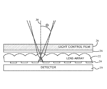
testing inconvenient, and they sometimes forget to test their glucose level or
do not have time
for a proper test. In addition, some individuals wish to avoid the pain
associated with the test.
Unmonitored glucose may result in hyperglycemic or hypoglycemic episodes. An
implanted
sensor that monitors the individual's analyte levels would enable individuals
to monitor their
glucose, or other analyte levels, more easily.
[0006] Some known devices perform in situ monitoring of analytes (e.g.,
glucose) in the
blood stream or interstitial fluid of various tissues. A number of these
devices use sensors that
are inserted into a blood vessel or under the skin of a patient. Communicating
and/or
retrieving data from such known and/or proposed devices, however, can be
challenging. For
example, an implanted sensor may be able to communicate with a detector or
receiver using
radio frequency (RF) transmissions. Such a sensor, however, may require
electronics,
batteries, antennae, and/or other communication hardware which may increase
the bulk of the
implanted sensor, may require frequent inconvenient recharging, and/or may
decrease the
longevity or reliability of the implant.
[0007] A need therefore exists for apparatus and methods for detecting optical
signals from
an implanted sensor, such that a fluorescent sensor can be used. A fluorescent
sensor may
not require electric charging and/or transmission electronics. Such implanted
sensors,
however, may be difficult to read or to monitor optically because of low
levels of florescence
in the presence of high scatter due to dynamic changes in skin conditions
(e.g., blood level
and hydration). The skin is highly scattering, and the scattering may dominate
the optical
propagation. Scatter is caused by index of refraction changes in the tissue,
and the main
components of scatter in the skin are due to lipids, collagen, and other
biological components.
The main absorption is caused by blood, melanin, water, and other components.
[0008] Devices and apparatus described herein are suitable for providing
accurate and
consistent measurement of an analyte by monitoring an implantable sensor in
such low-
s i gn al, high-scattering environments.
2
CA 02913474 2015-11-24
WO 2014/197786
PCT/1JS2014/041284
Summary
[0009] Some embodiments described herein relate to an apparatus including a
light source
configured to transmit an excitation optical signal to an implanted sensor and
a detector
configured to detect an analyte-dependent optical signal emitted from an
implanted sensor.
The apparatus can include a lens configured to focus at least a portion of the
analyte-
dependent optical signal onto the detector.
[0010] Some embodiments described herein relate to an array of lenses. Each
lens from the
array of lenses can be configured to transmit an analyte-dependent optical
signal from an
implanted sensor to a detector. A plurality of light-blocking elements can be
disposed within
a substrate of the array of lenses. Each light blocking element from the array
of light-
blocking elements can be configured to prevent or inhibit a photon having an
angle of
incidence greater than a predetermined angle of incidence from passing through
the substrate.
[0011] Some embodiments described herein relate to an apparatus including a
detector
configured to detect an analyte-dependent optical signal from an implanted
sensor. A lens
can be configured to focus at least a portion of the analyte-dependent optical
signal onto the
detector. A filter can be configured to attenuate light having wavelengths
shorter than the
analyte-dependent optical signal.
[0012] Some embodiments described herein relate to an implant capable of
emitting, in
response to excitation light in at least one excitation wavelength range, at
least one analyte-
dependent optical signal in at least one emission wavelength range. A device
including at
least one light source can be arranged to transmit the excitation light
through tissue
surrounding the implant. The device can include at least one detector arranged
to detect light
emitted from implanted sensor and transmitted through the tissue in the
emission wavelength
range. The device can also include an array of lenses arranged with an array
of apertures to
restrict transmission of off-axis light to the detector. The arrays of lenses
and the array of
apertures can be positioned with respect to the detector to restrict the light
emitted from the
tissue that travels to the detector based on the incidence angle of the
emitted light. At least
one layer of light control film can be arranged with the lens and aperture
arrays to restrict the
light emitted from the tissue that travels to the detector based on the
incidence angle of the
emitted light relative to the film. The device can further include at least
one filter positioned
3
CA 02913474 2015-11-24
WO 2014/197786
PCT/1JS2014/041284
to restrict transmission of light to the detector to wavelengths substantially
within the
emission wavelength range.
[0013] Some embodiments described herein relate to an optical detection device
is for
monitoring an implant embedded in tissue of a mammalian body. The implant is
capable of
emitting, in response to excitation light in at least one excitation
wavelength range, at least
one analyte-dependent optical signal in at least one emission wavelength
range. The device
can include at least one light source arranged to transmit the excitation
light through the
tissue to the implant. At least one detector is arranged to detect light
emitted from the tissue
in the emission wavelength range. The device can also include an array of
lenses arranged
with an array of apertures to restrict transmission of off-axis light to the
detector. The arrays
of lenses and the array of apertures are positioned with respect to the
detector to restrict the
light emitted from the tissue that travels to the detector according to an
input angle of the
emitted light. Light-blocking elements are arranged between the apertures to
block
propagation of incident light rays through the apertures. The light-blocking
elements are
positioned to block the incident light rays in accordance with an increase in
incident angle of
the light rays with respect to optical axes of the apertures. The device
further comprises at
least one filter arranged to restrict the transmission of the emitted light to
the detector to
wavelengths substantially within the emission wavelength range.
[0014] Some embodiments described herein relate to a method for monitoring an
implant
embedded in tissue of a mammalian body. The implant is capable of emitting, in
response to
excitation light in at least one excitation wavelength range, at least one
analyte-dependent
optical signal in at least one emission wavelength range. The method can
include transmitting
the excitation light through the tissue to the implant and detecting light
emitted from the
tissue in the emission wavelength range. The light in the emission wavelength
range is
transmitted through an array of lenses and an array of apertures arranged to
restrict the light
emitted from the tissue that travels to at least one detector according to an
input angle of the
emitted light. The light in the emission wavelength range is also transmitted
through at least
one layer of light control film arranged with the lens and aperture arrays to
restrict the light
emitted from the tissue that travels to the detector according to an incident
angle of the
emitted light relative to the film. The light in the emission wavelength range
is also
transmitted through at least one filter positioned to restrict transmission of
light to the
detector to wavelengths substantially within the emission wavelength range.
4
CA 02913474 2015-11-24
WO 2014/197786
PCT/1JS2014/041284
[0015] Some embodiments described herein relate to a method for monitoring an
implant
embedded in tissue of a mammalian body. The implant is capable of emitting, in
response to
excitation light in at least one excitation wavelength range, at least one
analyte-dependent
optical signal in at least one emission wavelength range. The method can
include transmitting
the excitation light through the tissue to the implant and detecting light
emitted from the
tissue in the emission wavelength range. An array of apertures arranged with
an array of
lenses restricts the light emitted from the tissue that travels to at least
one detector according
to an input angle of the emitted light. The method can also include blocking
propagation of
incident light rays through the apertures using light-blocking elements
positioned between the
apertures to block the incident light rays having an angle of incidence
greater than a threshold
angle of incidence based on, for example, the optical axes of the apertures.
The method can
further include filtering the emitted light to wavelengths substantially
within the emission
wavelength range.
[0016] Some embodiments described herein relate to an optical detection device
for
monitoring an implant embedded in tissue under skin. The implant is capable of
emitting, in
response to excitation light in at least one excitation wavelength range, at
least one analyte-
dependent optical signal in at least one emission wavelength range. The device
can include at
least one light source arranged to transmit the excitation light through a
first surface area of
the skin to the implant embedded in the tissue. One or more detectors can be
arranged to
detect light that is emitted from at least a second surface area of the skin,
wherein the light
source and one or more detectors are arranged such that the ratio of the
surface area of the
skin through which the detected light passes as it travels to the one or more
detectors to the
surface area of the skin through which the excitation light is transmitted is
at least 4:1.
[0017] Some embodiments described herein relate to a method for monitoring an
implant
embedded in tissue under skin. The implant can be capable of emitting, in
response to
excitation light in at least one excitation wavelength range, at least one
analyte-dependent
optical signal in at least one emission wavelength range. The method can
include
transmitting the excitation light through a first surface area of the skin to
the implant
embedded in the tissue and detecting light that is emitted from at least a
second surface area
of the skin. The ratio of the surface area of the skin through which the
detected light passes
as it travels to one or more detectors to the surface area of the skin through
which the
excitation light is transmitted is at least 4:1.
CA 02913474 2015-11-24
WO 2014/197786
PCT/1JS2014/041284
Brief Description of the Drawings
[0018] Fig. 1 is a schematic side view of an optical detection device for
monitoring an
implant, according to an embodiment.
[0019] Fig. 2 is a schematic side view of an optical detection device for
monitoring an
implant, according to an embodiment.
[0020] Fig. 3 is a plan view of an aperture array, according to an embodiment.
[0021] Fig. 4 is a schematic plan view of an optical detection device,
according to an
embodiment.
[0022] Fig. 5 is a schematic exploded view of an optical detection device,
according to an
embodiment.
[0023] Fig. 6 is a schematic side view of an optical detection device for
monitoring an
implant, according to an embodiment.
[0024] Fig. 7 is a schematic side view of an optical detection device for
monitoring an
implant, according to an embodiment.
[0025] Figs. 8A-8D depict a lens and aperture array with light-blocking
elements in various
stages of fabrication, according to an embodiment.
[0026] Fig. 9 is a schematic plan view of an optical detection device,
according to an
embodiment.
[0027] Fig. 10 is a schematic plan view of an optical detection device,
according to an
embodiment.
[0028] Fig. 11 is a schematic plan view of an optical detection device,
according to an
embodiment.
Detailed Description
[0029] According to some embodiments described herein, an optical detection
device is
provided for monitoring an implant embedded in tissue of a mammalian body. The
implant
can include a fluorophore-labeled target capable of emitting, in response to
excitation light in
at least one excitation wavelength range, at least one analyte-dependent
optical signal in at
6
CA 02913474 2015-11-24
WO 2014/197786
PCMJS2014/041284
least one emission wavelength range. The optical detection device can be
operable to
illuminate the implant with light whose wavelength content falls within an
absorption band
and/or collect light whose wavelength content is in an emission band.
[0030] The optical detection device can include excitation optics including a
light source
and/or optics operable to generate illumination in the absorption band. The
optical detection
device can also include emission optics operable to collect fluorescent
emissions from the
implant. Because in some instances, it may be difficult to obtain, design,
and/or implement a
light source that has a spectral content (i.e., wavelength range) that exactly
matches every
fluorophore absorption band, an optical filter or filters (usually band-pass
filters) can be used
along with the light source to limit the range of illuminating wavelengths to
that of the
absorption band and/or to reduce illuminating wavelengths of the emission
band. Similarly,
the emission optics can include another filter or filters operable to allow
substantially only
light with wavelengths in the emission band to reach the detector and/or to
attenuate light
with other wavelengths (e.g., light in the absorption band). Similarly stated,
the optical
detection device can include an optical system design operable to allow
substantially only
photons with wavelengths in the absorption band reach the target, and
substantially only
photons with wavelengths in the emission band reach the detector. Without
proper optics,
photons from the light source may be reach the detector and induce a
measurement error.
[0031] Properly designing an optical system for an optical detection device
can be
complicated in instances in which the amount of emitted fluorescence to be
detected is much
less than the amount of excitation light scattered (e.g., not absorbed) by an
intermediate
surface (e.g., skin or tissue disposed between the optical detection device
and the implant).
One challenge is that the amount of excitation light that reaches the implant
may be low
because of the absorption and scattering caused by the various body parts
(skin, tissue, etc.).
The low amount of emitted fluorescence is further reduced by absorption and
scattering as it
makes its way out of the body towards the detector. Existing optical filter
technology, which
may provide rejection of unwanted photons on the order of (10-6) may be
insufficient in these
situations. Another challenge is that the difference between excitation and
detection
wavelengths (e.g., Stokes shift) may be quite small. A further challenge is
that dichroic filters
cause shifting (e.g., the "blue shift") of filter wavelengths as a function of
the angle of light
rays transmitted through the filter. Because of these challenges, standard
fluorescence
7
CA 02913474 2015-11-24
WO 2014/197786
PCMJS2014/041284
methods would allow through high background levels and, in turn, result in low
Signal-to-
Background (SBR) and Signal-to-Noise (SNR) ratios.
[0032] Some embodiments described herein relate to a compact device that can
accurately
and consistently monitor an implanted sensor. Such a device can be worn by a
user
substantially continuously and/or may not substantially restrict movements or
activities of the
user. The device and the sensor can collectively allow for continuous and/or
automatic
monitoring of an analyte and can provide a warning to the person when the
level of the
analyte is at or near a threshold level. For example, if glucose is the
analyte, then the
monitoring device might be configured to warn the person of current or
impending
hyperglycemia or hypoglycemia. The person can then take appropriate actions.
[0033] In the description contained herein, it is understood that all recited
connections
between structures can be direct operative connections or indirect operative
connections
through intermediary structures. A set of elements includes one or more
elements. Any
recitation of an element is understood to refer to at least one element. A
plurality of elements
includes at least two elements. Unless clearly indicated otherwise, any
described method
steps need not be necessarily performed in a particular or illustrated order.
A first element
(e.g. data) derived from a second element encompasses a first element equal to
the second
element, as well as a first element generated by processing the second element
and optionally
other data. Making a determination or decision according to a parameter
encompasses
making the determination or decision according to the parameter and optionally
according to
other data. Unless otherwise specified, an indicator of some quantity/data may
be the
quantity/data itself, or an indicator different from the quantity/data itself.
Some embodiments
described herein reference a wavelength, such as an excitation wavelength or
an emission
wavelength. Unless clearly indicated otherwise, a wavelength should be
understood as
describing a band of wavelengths including the wavelength. Computer programs
described in
some embodiments of the present invention may be stand-alone software entities
or sub-
entities (e.g., subroutines, code objects) of other computer programs.
Computer readable
media encompass non-transitory media such as magnetic, optic, and
semiconductor storage
media (e.g. hard drives, optical disks, flash memory, DRAM), as well as
communications
links such as conductive cables and fiber optic links. According to some
embodiments, the
present invention provides, inter alia, computer systems comprising hardware
(e.g. one or
more processors and associated memory) programmed to perform the methods
described
8
CA 02913474 2015-11-24
WO 2014/197786
PCMJS2014/041284
herein, as well as computer-readable media encoding instructions to perform
the methods
described herein.
[0034] The following description illustrates embodiments of the invention by
way of
example and not necessarily by way of limitation.
[0035] Fig. 1 is a schematic side view of an optical detection device 10 for
monitoring an
implanted sensor or implant 12, according to an embodiment. The implant 12 is
embedded in
tissue 15 of a mammalian body (which may be a piece of tissue that is attached
or unattached
to the rest of the body in various embodiments). The implant 12 can be
embedded under a
surface of skin 14. The implant 12 can be embedded and/or positioned in the
subcutaneous
tissue (e.g., in the range of 1 to 4 mm under the surface of the skin 14). The
implant 12 is
capable of emitting, in response to excitation light within an excitation
wavelength range, at
least one analyte-dependent optical signal within an emission wavelength
range. The analyte
may be, for example, glucose or other analytes in the tissue 15. Suitable
optical signals
include, without limitation, luminescent, bioluminescent, phosphorescent,
autoluminescence,
and diffuse reflectance signals. In some embodiments, the implant 12 contains
one or more
luminescent dyes (e.g., fluorescent dyes) whose luminescence emission
intensity varies in
dependence upon the amount or presence of target analyte in the body of the
individual (e.g.,
in tissue 15).
[0036] A light source 18 is arranged to transmit excitation light within the
excitation
wavelength range from the surface of the skin 14, through the tissue 15, and
to the implant
12. Suitable light sources include, without limitation, lasers, semi-conductor
lasers, light
emitting diodes (LEDs), and organic LEDs. Detectors 16, 20 are arranged with
the light
source 18 to detect light emitted from the tissue in the emission wavelength
range. Suitable
detectors include, without limitation, photodiodes, complementary metal¨oxide¨
semiconductor (CMOS) detectors or charge-coupled device (CCD) detectors.
Although
multiple detectors are shown, a single and/or universal detector can be used.
[0037] The detectors can be 16, 20 filtered (e.g., with dichroic filters or
other suitable filters)
to measure the optical signals emitted within the wavelength ranges. For
example, a suitable
luminescent dye sensitive to glucose concentration is Alexa Flour 647
responsive to
excitation light (absorption) in the range of about 600 to 650 nm (absorption
peak 647 nm)
and within an emission wavelength range of about 670 to 750 nm with an
emission peak of
9
CA 02913474 2015-11-24
WO 2014/197786
PCMJS2014/041284
about 680 nm. Thus, in an embodiment in which the sensor includes Alexa Flour
4) 647, the
detectors 16, 20 can be filtered from light having a wavelength shorter than
about 650 nm or
shorter than about 670 nm.
[0038] In some embodiments, the implant 12 is further capable of emitting, in
response to
excitation light within a second excitation wavelength range, at least one
analyte-independent
optical signal within a second emission wavelength range. For example, the
implant 12 can
contain an analyte-independent luminescence dye that functions to control for
non-analyte
physical or chemical effects on a reporter dye (e.g., photo bleaching or pH).
Multiple dyes
may be used. The analyte-independent optical signal is not modulated by
analyte present in
the tissue 15 and provides data for normalization, offset corrections, or
internal calibration.
The analyte-independent signal may compensate for non-analyte affects that are
chemical or
physiological (e.g., oxygen, pH, redox conditions) or optical (e.g., water,
light
absorbing/scattering compounds, hemoglobin). Alternatively, the analyte-
independent signal
may be provided by a stable reference dye in the implant 12. Suitable stable
reference
materials include, but are not limited to, lanthanide-doped crystals,
lanthanide-doped
nanoparticles, quantum dots, chelated lanthanide dyes, and metal (e.g., gold
or silver)
nanoparticles. The stable reference dye may provide a reference signal for
other signals (e.g.,
to determine photo bleaching).
[0039] In the operation of device 10, the light source 18 is activated to
transmit excitation
light within the excitation wavelength range from the surface of the skin 14,
through the
tissue 15, and to the implant 12. The dye in the implant 12 absorbs some of
the excitation
light and emits fluorescence that depends on glucose or other analyte
properties. The light
may be emitted from the implant 12 in all directions, and scattered by the
tissue 15. Some of
the light that is emitted from the implant 12 is transmitted through the
tissue 15 and detected
by at least one of the detectors 16, 20. This can provide the primary analyte-
dependent optical
signal. In embodiments in which a reference optical signal is used for
normalization, the light
source 18 (or a second light source) is activated to transmit second
excitation light from the
surface of the skin 14 to the implant 12. At least one of the detectors 16, 20
measures, in
response to the second excitation light, a second optical signal emitted from
the tissue 15
through the surface of the skin 14.
CA 02913474 2015-11-24
WO 2014/197786
PCMJS2014/041284
[0040] The second optical signal may be used to normalize the primary analyte-
dependent
optical signal for scattering of light emitted from the implant 12. At least
one corrected signal
value may be calculated in dependence upon the measured optical signals. In
one example,
the primary analyte-dependent signal from the implant may be normalized by the
analyte-
independent optical signal emitted from the implant 12. Prior to executing
optical reads for
the analyte-dependent signal and/or the analyte-independent signal, a dark
reading may be
taken to account for background or ambient light, and this reading may be used
to further
correct the signals, e.g., by background subtraction.
[0041] In some embodiments, an analyte value (e.g., glucose concentration) is
determined
from the analyte-dependent signal and/or a ratio of multiple optical signals
including one or
more reference signals. In one example, the signal from the glucose sensitive
fluorophore
(e.g., Alexa Flour Og 647) is normalized by the signal from a glucose
insensitive fluorophore
(e.g., Alexa Flour 700). One suitable dye for the analyte-independent signal
is Alexa Flour
750 which is responsive to excitation light within an excitation wavelength
range of about
700 to 760 nm (excitation peak 750 nm) and has an emission wavelength range of
about 770
to 850 nm with an emission peak of about 780 nm.
[0042] An analyte value can be determined based on the optical signal(s)
using, for example,
a look-up table or calibration curve. Determining the analyte value can be
implemented in
software (executing on a processor) and/or hardware. For example, the optical
device 10 can
include a microprocessor. In some embodiments, the microprocessor is
programmed to store
measured optical signal values in a memory and/or to calculate normalized
signal values and
analyte concentrations. Alternatively, these functions may be performed in a
separate
processor or external computer in communication with the optical device 10.
The external
processor or computer can receive data representative of the measured optical
signals and
calculates the corrected signal value and analyte concentration.
Alternatively, multiple
processors may be provided, e.g., providing one or more processors in the
optical device that
communicate (wirelessly or with wires) with one or more external processors or
computers.
[0043] In some embodiments in which two implant dyes (e.g., luminescent dyes)
are utilized,
it is possible that the implant dyes may share or overlap excitation
(absorption) or emission
wavelength ranges. In one example, the emission wavelength range of the first
dye, which
provides the analyte-dependent luminescence signal, shares or overlaps the
excitation
11
CA 02913474 2015-11-24
WO 2014/197786
PCMJS2014/041284
wavelength range of the second dye, which provides the analyte-independent
luminescence
signal. In another embodiment, the first and second dyes may share or overlap
excitation
wavelength ranges (so that a common light source may be used) and emit optical
signals
within different emission wavelength ranges. In another embodiment, the first
and second
dyes may be excited by light within different excitation wavelength ranges and
emit optical
signals within the same or overlapping emission wavelength range(s).
[0044] The implant 12 can be embedded in subcutaneous tissue (e.g., 1 to 4 mm
under the
surface of the skin 14). In some embodiments, the implant 12 comprises
hydrogel scaffolds
embedded with glucose-sensing nanospheres. The design of the implant 12 can
use injectable,
tissue-integrating, vascularizing scaffolds as the sensor. Embedded
nanospheres emit
luminescence that changes intensity and lifetime in response to the presence
or concentration
of the analyte (e.g., interstitial glucose). The spacing distances between
each of the detectors
16, 20 and the light source 18 determine the depths of the respective light
paths for detecting
optical signals from the implant 12. The combination of an excitation light
source and a
detection band is an optical channel. The light source 18 and detectors 16, 20
can be arranged
such that a surface area of skin 14 through which the excitation light is
transmitted is located
between substantially surrounding surface areas of skin 14 through which the
detected light
passes as it travels from the tissue 15 to one or more detectors 16, 20.
[0045] Although only one light source 18 and two detectors 16, 20 are shown in
Fig. 1, in
some embodiments, the optical device 10 can have any number of light sources
and any
number of detectors. The optical device 10 can have multiple possible
combinations of
spacing distances between multiple light sources and detectors. Such a
multiple light source
and/or multiple detector implementation can allow increased flexibility of the
optical device
10. For example, since the depth of the implant 12 may be application-
specific, an optical
device 10 having multiple light sources and/or multiple detectors can be used
for multiple
applications.
[0046] The optical device 10 can be configured to ensure that substantially
only photons with
wavelengths in the excitation wavelength range(s) reach the implant 12, and
substantially
only photons with wavelengths in the emission wavelength ranges(s) reach at
least one of the
detectors 16, 20. Such an arraignment can minimize photons from the light
source 18
reaching the detectors 16, 20, which can result in measurement error.
12
CA 02913474 2015-11-24
WO 2014/197786
PCMJS2014/041284
[0047] Fig. 2 is a schematic side view of an optical detection device for
monitoring an
implant, according to an embodiment. An array of lenses 22 is aligned with an
array of
apertures 24 to restrict transmission of off-axis light to the detector 16.
The lens arrays 22 and
the aperture array 24 are positioned with respect to the detector 16 to
collectively restrict the
light emitted from the tissue that travels to the detector 16 based on an
input angle 0 (also
referred to herein as incident angle) of the emitted light relative to optical
axes 30 of the
apertures. The optical axes 30 of the apertures can be substantially
perpendicular to the
surface of the detector 16. Each aperture from the array of apertures 24 can
be substantially
aligned with a lens from the array of lenses 22. Similarly stated the optical
axes 30 of the
apertures can be substantially coaxial with the center and/or axes of the
lenses. For example,
a substantially opaque portion of the array of apertures 24 can be positioned
below the edges
of the lenses.
[0048] At least one layer of light control film 26 is arranged with the lens
array 22 and the
aperture array 24. The light control film 26 can restrict the light emitted
from the tissue from
entering the lens array 22 and/or the aperture array 24 based on the incident
angle of the
emitted light relative to the film 26. In one example, the light control film
26 is VikutiTM
optical grade micro-louver privacy film commercially available from 3MTm,
which can block
light having an incident angle greater than desired (e.g., greater than 24
degrees) relative to a
perpendicular line through the film 26. This privacy film comprises a set of
microlouvers that
prevent light from large incident angles from reaching the lens array 22. In
other
embodiments, the film 26 comprises alternating transparent and opaque layers
in an
arrangement which is similar to a Venetian blind. Light propagating from
angles greater than
a desired incident angle can be absorbed and/or reflected.
[0049] At least one filter 28 (e.g., a dichroic or dielectric filter) is
positioned to restrict
transmission of light to the detector 16 to wavelengths substantially within
the desired
emission wavelength range. Because the detection of optical signals is
dominated by low
levels of return signals relative to the excitation light, the filter 28 can
prevent scattered
excitation light from blinding the detector 16. Suitable filters include band-
pass, low-pass,
and high pass filters depending upon the desired emission wavelength range for
an
application. Some modern optical filters have demonstrated 10-9 light
rejection due to
improvements in coating technologies. Additionally, intermediate layers of the
optical
13
CA 02913474 2015-11-24
WO 2014/197786
PCMJS2014/041284
detection system (e.g., the lens array 22, the aperture array 24, etc.) can
include anti-reflective
coatings to reduce or prevent light leaking through to the detector 16.
[0050] Due to fundamental properties of dichroic filters, maintaining a high
level of light
rejection requires careful design. One property of dichroic filters that
detracts from light
rejection is the "blue shift" as a function of input angle, where the
transmittance wavelengths
of dichroic filters change as a function of input angle. For the detection
light emitted from the
implant, there is a trade off between the input angle and the absolute optical
signal. The light
leaving the tissue is highly scattered and may form a lambertian distribution
by the time it
reaches the surface of the skin. The collection efficiency of the emitted
light is proportional to
--NA2, where NA=Numerical Aperture = n sin 0, and 0 is the input angle. To
improve the
collection efficiency, the allowable input angle 0 can be increased without
increasing the
angle so much to allow excitation light though the filter 28.
[0051] The lens array 22 and the aperture array 24 control the input angle 0
of light traveling
to the detector 16. The lens array 22 and an aperture array 24 restrict the
light to an input
angle less than 0, which in some embodiments is selected to be +/- 20 degrees.
The input
angle 0 can be controlled by varying the size of the apertures and the focal
length of the
micro lenses in the lens array 22. The smaller the aperture, then the smaller
is input angle 0.
The longer the focal length, then the smaller is input angle 0. Although not
shown, a spacer
can be used to maintain separation between the surface of the aperture array
24 and the lens
array 22.
[0052] Fig. 3 is a plan view of the aperture array 24 having a plurality of
apertures 25. In
some embodiments, the aperture array 24 is constructed by patterning a metal
mask on the
surface of a silicon detector, such as the detector 16 shown in Fig. 2. The
lens array 22 may
be fabricated as etched glass or molded plastic. In some embodiments, the lens
array 22 is a
custom micro-lens array commercially available from JENOPTIK Optical Systems.
[0053] Fig. 4 is a schematic plan view of an optical detection device,
according to an
embodiment. The optical detection device of Fig. 4 is configured as a patch
32. At least one
light source (not shown in Fig. 4) and detector 38 are arranged in an optical
reader, such that
the patch 32, that is configured to be placed on the skin. A light source is
arranged to transmit
the excitation light through a central via 34 in the patch 32, and a single
universal detector 38
substantially surrounds the central via 34. In other embodiments, instead of
the single
14
CA 02913474 2015-11-24
WO 2014/197786
PCMJS2014/041284
detector 38, a plurality of detectors can be used, for example, substantially
encircling the
central via 34 to detect the emitted light in a plurality of emission
wavelength ranges. In some
embodiments, the optical detection device includes at least one light guiding
component 36 in
the central via 34. The light guiding component 36, such as a waveguide or
optical fiber, is
arranged to guide the excitation light to the skin. In some embodiments, a
plurality of light
sources (not shown for clarity in Fig. 4) are arranged to transmit the
excitation light through
the central via 34 (e.g., by means of one or more waveguides or optical
fibers) in a plurality
of different excitation wavelength ranges.
[0054] As one possible example, one or more light sources may be arranged to
transmit
excitation light to the skin through the central via 34 having a circular
cross-section to
transmit the excitation light through a substantially circular surface area of
the skin having a
diameter of about 1 mm and a corresponding excitation surface area of about
0.8 mm2. The
detector 38 has a square cross-section and is positioned to detect light
emitted from a
substantially square surface area of the skin through which the detected light
passes as it
travels to the detector 38. The detection surface area is substantially square
with sides of 10
mm length, so that the total detection surface area is (10 mm x 10 mm) - 1 mm2
= 99 mm2.
Accordingly, in this example, the ratio of detection surface area to
excitation surface area is
greater than 120:1.
[0055] Fig. 5 is a schematic exploded view of the patch 32. The patch 32
includes multiple
layers. Dimensions of the patch 32 may be, for example, a diameter of about 16
mm and a
thickness T of about 1.6 mm. In some embodiments, the layers may include a
plastic cover 40
having a thickness of about 200 um, the light control film 26 having a
thickness of about 100
um, the filter 28 having a thickness of about 200 um, the lens array 22 having
a thickness of
about 100 um, and the aperture array 24 patterned on a silicon detector layer
48 having a
thickness of about 200 um. The layers can also include a printed circuit board
(PCB) 50
having a thickness of about 400 um, a battery 52 having a thickness of about
300 um, and a
case 54 having a thickness of about 200 um. The PCB 50 can include one or more
light
sources. The PCB 50 can also include processing electronics and/or a
microprocessor in
communication with one or more detectors in the detector layer 48 to receive
data
representative of the light detected in the emission wavelength range and
programmed to
determine at least one analyte value in dependence upon the data. The central
via 34 may be
CA 02913474 2015-11-24
WO 2014/197786
PCT/1JS2014/041284
formed through a stack of the layers (e.g., etched or drilled through the
stack in the assembly
process).
[0056] Fig. 6 is a schematic side view of an optical detection device for
monitoring an
implant showing an arrangement of detection optics 60, according to an
embodiment. In this
embodiment, light emitted from the implant and tissue in the emission
wavelength range is
transmitted through at least two layers of light control films 62, 64. The two
layers of light
control films 62, 64 can restrict the light emitted from the tissue from
entering the lens array
22 and/or the aperture array 24 based on the incident angle of the emitted
light relative to the
films 62, 64. In one example, the light control film 62 comprises alternating
transparent and
opaque layers in an arrangement which is similar to a Venetian blind. Light
propagating from
angles greater than a desired incident angle is absorbed. The light control
film 64 may include
VikutiTM optical grade micro-louver privacy film commercially available from
3MTm, which
blocks light having an incident angle greater than desired (e.g., greater than
24 degrees)
relative to a perpendicular line through the film 64.
[0057] In some embodiments, the light control film 62 and/or 64 may be
operable to restrict
light emitted from the tissue from entering the lens array 22 and the aperture
array 24 based
on a combination of incident angle and azimuth. For example, in an embodiment
where the
light control film 62 and/or 64 includes multiple micro-louvers, the light
control film 62
and/or 64 may be effective at blocking high angle-of-incidence light having an
azimuth
substantially perpendicular to the micro-louvers, but may be relatively
ineffective at blocking
high angle-of-incidence light having an azimuth substantially parallel to the
micro-louvers.
In some such embodiments, two layers of light control film 62, 64 can be cross-
hatched or
otherwise disposed such that louvers or other light control elements are non-
parallel such that
the light control film 62, 64 are collectively effective at blocking high
angle-of-incidence
light having different azimuths.
[0058] In some embodiments, the films 62, 64 may be substantially the same as
each other,
or comprises different types of privacy film. Additionally, the filter 28
(e.g., a dichroic or
dielectric filter) may be positioned between the aperture array 24 and the
detector 16 to
restrict the transmission of the emitted light to the detector 16 to
wavelengths substantially
within the emission wavelength range(s). The operation of the embodiment of
Fig. 6 can be
similar to the operation of the embodiment of Figs. 1-2 previously described.
16
CA 02913474 2015-11-24
WO 2014/197786
PCMJS2014/041284
[0059] Fig. 7 is a schematic side view of an optical detection device for
monitoring an
implant. An array of lenses 122 is aligned with an array of apertures 24 to
restrict the
transmission of off-axis light to the detector 16. The lens arrays 122 and the
aperture array 24
are positioned with respect to the detector 16 to restrict the light emitted
from the tissue that
travels to the detector 16 according to an input angle 0 of the emitted light
relative to optical
axis 30 of the apertures. The optical axis 30 of the apertures can be
substantially
perpendicular to the surface of the detector 16.
[0060] The lens array 122 includes light-blocking elements 72. The light-
blocking elements
72 can be disposed between the apertures 25 to block propagation of off-axis
light rays 74, 76
through the apertures 25. The light-blocking elements 72 can include black
resin, metal,
and/or metal film deposited in cavities of a substrate 123 of the lens array
122 positioned. At
least one filter 28 is positioned to restrict the transmission of the emitted
light to the detector
16 to wavelengths substantially within the emission wavelength range.
Optionally, one or
more layers of light control film may be included in this embodiment. The
operation of the
embodiment of Fig. 7 can be similar to the operation of the embodiment of
Figs. 1-2
previously described.
[0061] Figs. 8A-8D depict a lens array 122 with light-blocking elements in
various stages of
fabrication, according to an embodiment. Fig. 8A shows a side view of the lens
array 122
which may be fabricated as etched glass or molded plastic. In some
embodiments, the lens
array 122 is a micro-lens array commercially available from JENOPTIK Optical
Systems.
Fig. 8B shows cavities 78 which can be, for example, etched or integrally
molded into a
substrate portion 123 of the lens array 122. As shown in Fig. 8C, the cavities
78 can be filled
with a substantially opaque material to form light-blocking elements 72. The
light-blocking
elements 72 can be constructed of, for example, black resin, metal, and/or
metal film. As
shown in Fig. 8D, the aperture array 24 may be positioned adjacent to the lens
array 122
(with a spacer in some embodiments) such that light-blocking elements 72 are
positioned
between the apertures 25. In some embodiments, the aperture array 24 is
constructed by
patterning a metal mask on the surface of a silicon detector and positioning
the detector with
aperture array 24 adjacent to the lens array 22 with light-blocking elements
72 such that the
light-blocking elements 72 are positioned between the between apertures 25.
17
CA 02913474 2015-11-24
WO 2014/197786
PCMJS2014/041284
[0062] Fig. 9 is a schematic plan view of an optical detection device 210,
according to an
embodiment. The optical detection device 210 includes four detectors 216, 220,
222, and 224
and a light source 218. The optical detection device 210 has a relatively
large ration of
detector surface area to light source surface area (also referred to herein as
"surface area
ratio"). The large surface area ratio can improve detection of implant
signals, when the
implant is embedded in subcutaneous tissue (e.g., 1-4 mm under the surface of
the skin). In
particular, the light source 218 and four detectors 216, 220, 222, 224 are
arranged such that
the ratio of the surface area of the skin through which the detected light
passes as it travels to
the detectors 216, 220, 222, 224 to the surface area of the skin through which
the excitation
light is transmitted is at least 4:1. For example, in one embodiment the light
source 218 has a
circular cross-section and is positioned to transmit the excitation light
through a substantially
circular surface area of the skin having a diameter of about 3 mm, a radius of
about 1.5 mm,
and an excitation surface area of about 7 mm2. The four detectors 216, 220,
222, 224 have
square cross-sections and are positioned to detect light emitted from four
substantially square
surface areas of the skin, through which the detected light passes as it
travels to the detectors.
Each of the four detection surface areas is substantially square with sides of
3 mm, so that the
total detection surface area is 4 x 9 mm2 = 36 mm2. Accordingly, in this
example, the ratio of
detection surface area to excitation surface area is slightly greater than
5:1.
[0063] In some embodiments, the optical detection device 210 can be configured
to detect
implant signals at a lateral distance at least twice the depth of the implant.
For example, at
least a portion of at least one of the detectors 216, 220, 222, 224 can be at
least twice as far
away from the implant laterally as that portion is from the implant distally.
For example, in
an instance where the light source 218 is centered over an implant that is
embedded under 4
mm of tissue, at least a portion of at least one of the detectors 216, 220,
222, 224 can be 8
mm away from the center of the light source 218. Similarly stated, the
furthermost edge or
corner of at least one of the detectors 216, 220, 222, 224 can be at least
twice as far away
from the center of the light source 218 as the implant is deep. In an
alternate embodiment,
such as an embodiment having a single or universal detector, the detector can
have a radius at
least twice the depth of the implant. In other embodiments, the optical
detection device 210
can be configured to detect implant signals at a lateral distance at least
three times, at least
five times, or any other suitable multiple of the depth of the implant. An
optical detector
device 210 operable to detect implant signals a relatively large lateral
distance from the
implant may be able to detect a larger portion of an emitted signal,
particularly in a high-
18
CA 02913474 2015-11-24
WO 2014/197786
PCT/1JS2014/041284
scattering environment. Capturing a larger portion of the emitted signal can
improve
detection accuracy.
[0064] Fig. 10 is a schematic plan view of an optical detection device 310,
according to an
embodiment. As compared to the optical detection device 210, in this
embodiment, the four
detectors 316, 320, 322, 324 are positioned closer to the light source 318 as
they surround or
encircle the light source 318, and the ratio of detection surface area to
excitation surface area
is larger. For example, the light source 318 may have a circular cross-section
and is arranged
to transmit the excitation light through a substantially circular surface area
of the skin having
a diameter of about 2 mm, a radius of about 1 mm, and an excitation surface
area of about
3.14 mm2. The four detectors 316, 320, 322, 324 have square cross-sections and
are
positioned to detect light emitted from four substantially square surface
areas of the skin,
through which the detected light passes as it travels to the detectors. Each
of the four
detection surface areas is substantially square with sides of 6 mm, so that
the total detection
surface area is 4 x 36 mm2 = 144 mm2. Accordingly, in this example, the ratio
of detection
surface area to excitation surface area is slightly greater than 45:1.
[0065] Fig. 11 is a schematic, plan view of aspects of an optical detection
device 410,
according to another embodiment. In this embodiment, five circular-shaped
detectors 428A,
428B, 428C, 428D, and 428E surround or encircle a central via 434. The central
via 434
may be a hole in the device 410. A plurality of light sources 426 are arranged
to transmit
excitation light in a plurality of different wavelength ranges through the
central via 434. As
one possible example, the light sources 426 may be arranged to transmit
excitation light to
the skin through the central via 434 having a circular cross-section to
transmit the excitation
light through a substantially circular surface area of the skin having a
diameter of about 3 mm
and a corresponding excitation surface area of about 7 mm2. The five detectors
428A, 428B,
428C, 428D, and 428E have circular cross-sections and are positioned to detect
light emitted
from five substantially circular surface areas of the skin, through which the
detected light
passes as it travels to the detectors. Each of the five detection surface
areas is substantially
circular with a diameter of 5 mm, so that the total detection surface area is
5 x 19.6 mm2 = 98
mm2. Accordingly, in this example, the ratio of detection surface area to
excitation surface
area is slightly greater than 13:1.
19
CA 02913474 2015-11-24
WO 2014/197786
PCMJS2014/041284
[0066] It will be clear to one skilled in the art that the above embodiments
may be altered in
many ways without departing from the scope of the invention. For example, many
different
permutations or arrangements of one or more light sources, one or more
detectors, filters,
and/or light guiding elements connecting the optical components may be used to
realize the
device and method of the invention. For example, alternative embodiments may
have
different dimensions and/or wavelengths. Embodiments may include cabled or
wireless hand-
held readers, wireless skin patch readers, bench-top instruments, imaging
systems,
smartphone attachments and applications, or any other configuration that
utilizes the
disclosed optics and algorithms.
[0067] In some embodiments described herein, a monitoring device can be
operable to
simultaneously emit an excitation optical signal and detect an emission
signal. For example,
the detector of such a monitoring device can be shielded from reflected or
back-scattering
excitation light using apertures, light-blocking elements, filters, light
control film, etc. In
other embodiments, a monitoring device can be operable to emit an excitation
optical signal
during one period of time, and detect an emission signal during another period
of time in
which the excitation optical signal is deactivated.
[0068] Tissue optical heterogeneity in some cases may be significant. Thus, it
may be
advantageous to utilize a single light source and a single detector to assure
that every color
passes through the same optical pathway through the tissue. In one embodiment,
a light
source can be positioned with a set of moveable filters between the light
source and the
surface of the skin. Similarly a single photodetector can be utilized in place
of separate
discrete detector elements. The detector may be used to detect different
wavelength ranges by
using moveable or changeable filters to enable multiple wavelengths to be
measured.
Changing or moving filters may be accomplished by a mechanical actuator
controlling a
rotating disc, filter strip or other means. Alternatively, optical filters may
be coated with a
material that, when subjected to current, potential, temperature or another
controllable
influence, changes optical filtering properties, so that a single
photodetector can serve to
detect multiple wavelength ranges.
[0069] In some embodiments, the devices and methods of the present invention
make use of
wafer-based micro-optics. These systems are created lithographically, but can
be replicated at
low cost. The technology allows for layers of optics and detectors to be
bonded at the wafer
CA 02913474 2015-11-24
WO 2014/197786
PCT/1JS2014/041284
level and then diced into individual detector systems. Suitable components
include etched
refractive lenses, polymer replicated refractive lenses, etched binary lenses,
replicated binary
lenses, replicated holograms, and replicated volume holograms.
[0070] In some embodiments, a complementary metal¨oxide¨semiconductor (CMOS)
detector may be used as an integral part of the optical system. The advantage
of a CMOS
sensor is the ability to integrate the detection, excitation, and digital
filtering circuitry into a
single piece of silicon. A new technology was recently announced, sCMOS, where
researchers have been able to greatly reduce the noise in CMOS detectors to be
comparable
to charge charge-coupled device (CCD) detectors. Another benefit to a CMOS
integrated
solution is the ability to perform lock-in detection and digital filtering on
the signals to reduce
or eliminate the impact of ambient light.
[0071] While various embodiments have been described above, it should be
understood that
they have been presented by way of example only, not limitation, and various
changes in
form and details may be made. Any portion of the apparatus and/or methods
described herein
may be combined in any combination, except mutually exclusive combinations.
The
embodiments described herein can include various combinations and/or sub-
combinations of
the functions, components and/or features of the different embodiments
described.
21