Note: Descriptions are shown in the official language in which they were submitted.
1
PERFORATING TROCAR
The present invention relates to a device that can be used in surgery
and in interventional radiology, and more particularly to a perforating trocar
that can be used especially in the field of percutaneous procedures for bone
or
marrow biopsy, vertebroplasty, cementoplasty of the skeletal areas, and more
generally the treatment of bone damage.
Various types of perforating trocars are known, which are surgical
instruments used to drill bone in order to reach a zone where a bone biopsy is
to be performed. These trocars are composed of a hollow outer tube, of which
the end is more or less sharp, and of a rod, of which the end is ground in
order to perforate the bone and which slides in the tube.
Thus, the patent application WO 2006/061514 describes a trocar
intended for bone biopsy and comprising an outer tube, of which the distal end
is divided into two segments with a helical cutting edge, in which a ground
rod
slides. This type of instrument is used manually by way of a handle.
The patent US 7850620 describes a trocar intended for bone marrow
biopsy and composed of an outer tube, of which the distal end has a
traditional grinding for this type of instrument, combined with a ground rod.
This instrument is used by coupling it to a drill.
The patent applications US 2003/225411 Al and US 2009/0204024 Al
concern trocars intended for bone marrow biopsy. These trocars each have a
rod with a notch permitting the removal of pieces of bone and of tissue. The
patent US 6,575,919 81 describes a trocar with an inner opening permitting
the passage of a needle.
The known perforating trocars are able to drill bone but cannot be
guided on a pin at the same time. This shortcoming has two major
disadvantages: the lack of precision at the moment of reaching the bone, and
the risk of accidents. Percutaneous procedures are mainly performed with
imaging and therefore based on images. The point of entry and the trajectory
can thus be visualized and defined in advance. The importance of the precision
Date Recue/Date Received 2020-06-26
2
of the point of entry is self-evident in the case of small bone lesions, since
these have diameters of sometimes less than a millimetre; matters are of
course more difficult when targeting a lesion located within a deep bone.
Therefore, in obese patients, it is not uncommon to have to pass through 150
mm to 200 mm of soft tissue before reaching the cortical bone. Under such
conditions, it is particularly difficult to keep the trajectory of the trocar,
of
which the diameter is approximately 2 mm, to within a millimetre. Moreover,
there are risks associated with direction introduction of a perforating trocar
when this bone is situated in a dense zone comprising organs, veins and
nerves. Confronted by such situations, the practitioner will accept the need
to
perform more manoeuvres and will first of all manually insert a thin and
minimally invasive rigid pin, which will serve as a guide for the various
coaxial
trocars and instruments in the course of the surgical procedure.
Perforated drill-bits are also known which are used in orthopaedics and
which can drill the bone while being guided on a pin. However, these drill-
bits
are not used, or not often used, in a percutaneous procedure, even less so
when the latter is performed manually. The use of these drill-bits requires
that
a pin first be inserted into the bone by means of a drill. This is because
these
drill-bits do not have a distal tip, since they are perforated all the way
through
so as to slide on the pin. Therefore, in the absence of an inserted pin, they
risk
sliding on the bone and deviating from the point of entry defined by the
practitioner.
The present invention relates to a device, in particular for surgery and
interventional radiology, which efficiently provides good perforation while at
the same time being entirely suitable to be guided on a rigid pin until
contact
with the bone. The invention also relates to a device with which the
practitioner can choose to commence a surgical procedure manually and
complete it using an automatic rotational drive means, such as a drill, if the
hardness of the bone so demands, and this without losing the point of entry to
the bone.
The device according to the present invention, in the form of a
perforating trocar, comprises an outer sleeve having a rigid tube, and a
Date Recue/Date Received 2020-06-26
3
mandrel having a rod. The rod is suitable for sliding in the outer sleeve and
has, at its distal end, a perforating tip. The rod additionally has a
longitudinal
groove arranged on the surface of the rod, and extending from the distal end
to the proximal end of the rod, in order to allow the device to slide on a
guide
pin.
According to an embodiment of the invention, the groove is V-shaped.
Preferably, the perforating tip is formed by bevelled grinding or
sharpening of the distal end of the rod. According to a preferred variant, the
distal end of the rod comprises three bevels, of which one is less inclined
than
the two others with respect to the axis of the rod, and a perforating tip
centred with respect to the axis of the rod.
Advantageously, the distal end of the rod comprises at least one
cutting ridge extending from the perforating tip to the edges of the rod.
According to one embodiment, this cutting ridge is situated on one of the
faces
of the groove. Preferably, this cutting ridge is more inclined than another
cutting ridge with respect to the axis of the rod.
The bevelled pyramidal grinding, with the cutting ridge whose angle is
greatest with respect to the axis of the rod situated on one of the faces of
the
longitudinal groove, permits better penetration into the bone. Indeed, tests
carried out on the bone marrow of cattle have shown that the tip thus formed
penetrates up to 5 times more deeply than a traditional triangular tip.
According to a feature of the invention, the distal end of the tube
comprises at least two segments with a helically shaped cutting edge.
Advantageously, the outer sleeve additionally comprises a connector
piece at the proximal end of the tube, and the mandrel additionally comprises
a stopper at the proximal end of the rod. The stopper is suitable for
receiving
the connector piece in order to form an integral assembly. The connector piece
is perforated in order to permit the sliding of the rod, and the stopper is
perforated in order to permit the sliding of the guide pin.
According to some embodiments, the assembly formed by the outer
sleeve and the mandrel is mounted in a handle or in an automatic rotational
Date Recue/Date Received 2020-06-26
4
drive means. For example, the automatic rotational drive means can be a drill
having an
endpiece in which the assembly is mounted.
Advantageously, the automatic drive means can be provided with a protective
envelope.
The protective envelope can, for example, be attached to the endpiece of a
drill by means of a
connector piece which is at the same time adapted to receive the outer sleeve
of the
perforating trocar.
For example, in the case of a bone lesion located in a dense cortical area,
the
practitioner, after providing local anaesthesia by conventional techniques,
introduces the guide
pin parallel to or through the anaesthetic needle in place until contact is
made with the bone.
The anaesthetic needle is then removed white leaving the guide pin in place.
The device
according to the invention, equipped with a removable handle, is introduced
through the
tissues, until in contact with the bone, by being guided on the pin. The pin
is removed once the
surface of the cortical bone is reached, then the practitioner drills the bone
by turning the
trocar manually, without losing the point of entry. In 80% to 90% of cases,
the drilling will be
performed manually, but if the wall of the bone is very hard, the practitioner
may remove the
removable handle and connect the trocar to a drill in order to complete the
surgical procedure.
The simplicity of the structure of the device according to the invention means
that,
depending on the depth or location of the lesion, the user can be provided
with a single
instrument which permits a choice between a direct route and one guided on the
pin, and of
which the drilling can be manual or done using the drill, or else can be
commenced manually
and completed using the drill. The combination of drilling by hand and
drilling with a drill
permits a high level of precision of the surgical manoeuvre and also
considerable power
regardless of the hardness of the bone.
Also disclosed is a trocar comprising:
- an outer sheath comprising a rigid tube; and
- a mandrel comprising a rod, the rod being adapted to slide in the outer
sheath and
comprising a perforating tip at a distal end thereof, wherein the perforating
tip comprises three
bevels, a first bevel being more inclined than a second bevel with respect to
an axis of the rod,
wherein the rod comprises a longitudinal groove formed in and along an
exterior surface
of the rod, the groove in the exterior surface of the rod extending from the
distal end to a
proximal end of the rod, wherein the distal end of the rod comprises a first
and second cutting
ridges, each of which extends from the perforating tip to an edge of the rod,
wherein the first
cutting ridge is located on a first face of the groove and is more inclined
with respect to the
axis of the rod than the second cutting ridge located on a second face of the
groove.
Other features and advantages of the present invention will become clear from
the
following description of a preferred embodiment and by reference to the
attached drawings, in
which:
Date Recue/Date Received 2021-05-06
5
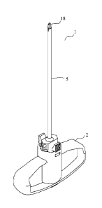
- Figure 1 shows a perspective view of a device according to the
invention mounted in a handle;
- Figures 2 to 3 show perspective views of a tube of the device
according to the invention mounted in a connector piece, forming
an outer sleeve;
- Figures 4 to 5 show perspective views of a grooved rod of the
device according to the invention mounted in a stopper, forming a
mandrel;
- Figures 6 to 9 show views of an example of the distal end of the
grooved rod;
- Figures 10 and 11 show views of an example of the distal end of
the rod provided with a hole;
- Figure 12 shows a perspective view of a handle for receiving the
assembly formed by the sleeve and the mandrel of the device
according to the invention; and
- Figure 13 shows a perspective view of a drill for receiving the
assembly formed by the sleeve and the mandrel of the device
according to the invention.
The trocar 1, shown in Figure 1, is composed of an outer sleeve 5, in
which is mounted a mandrel (of which only the distal end of the rod 18 is
visible in Figure 1), and of a handle 2, in which the sleeve/mandrel assembly
is inserted.
The outer sleeve 5, shown in Figures 2 and 3, has a connector piece 7
in which the bevelled tube 8 is accommodated. The tube 8 has a distal end 9
divided into two segments with a helical cutting edge as described in the
application WO 2006/061514. The connector piece 7 has a hexagonal shape
cooperating with hexagonal cavities 12 of the handle 2 and hexagonal cavities
13 of the endpiece 3 of a drill 4 (see also Figures 12 and 13). The connector
piece 7 has a bore 10, in which the tube 8 is accommodated, and two flexible
Date Recue/Date Received 2020-06-26
6
parts 11 that latch into recesses 14 of the handle 2 or into recesses 15 of
the
endpiece 3 of the drill 4.
The mandrel 6, shown in Figures 4 et 5, comprises a grooved rod 18
accommodated in a stopper 17. The grooved rod 18 has a V-shaped
longitudinal groove 20 and a distal end of pyramidal shape. The stopper 17
has a hexagonal shape cooperating with the hexagonal cavities 12 of the
handle 2 and the hexagonal cavities 13 of the endpiece 3 of the drill 4. The
stopper 17 has a bore 45 in which the grooved rod 18 is accommodated, and a
bore 41 permitting the passage of the guide pin.
The connector piece 7 of the outer sleeve 5 is completely perforated 43
in such a way as to permit the sliding of the rod 18. The end 16 of the outer
sleeve 5 is in the form of a Luer connector permitting the screwing of the
stopper 17. The stopper 17 has a thread 46 cooperating with the thread 16 of
the connector piece 7, allowing it to be screwed in order to assemble the
outer
sleeve 5 and the mandrel 6.
Figures 6 to 9 shows views of an example of the distal end of the rod
18. The distal end of the rod 18 comprises a tip 21 having a pyramidal
grinding with three bevels. Two of the bevels 22, 23 have the same inclination
(OA preferably 133 = 30 , with respect to the axis of the rod 18 and extend
through approximately 120 of the cross section of the rod 18, generating a
cutting ridge 24. The third bevel 25, inclined preferably by pl and at 40
with
respect to the axis of the rod 18, generates a second cutting ridge 26. The
second cutting ridge 26 is situated on one of the faces of the groove 20 and
has an inclination 131 greater than the inclination 02 of the ridge 24 with
respect to the axis of the rod 18. This permits better cutting of the bone
since
the second cutting ridge 26 has a greater angle of attack, generated by one of
the faces of the groove 20. The inclination 13, of the cutting ridge 26 is
greater
than the inclination 132 of the cutting ridge 24 and than the inclination 134
of the
other cutting ridge 28 situated on the other face of the longitudinal groove.
The intersection of the three bevels generates the tip 21 allowing the mandrel
6 not to slide on the bone.
Date Recue/Date Received 2020-06-26
7
The views in Figures 10 and 11 show a distal end of the rod 18
according to a variant. The rod 18 has, centred on its axis, a hole 34
allowing
it to be guided on a pin. The distal end of the rod 18 has a pyramidal
grinding
with three bevels of the same inclination, of which one bevel 31 is greater
than the other two bevels 32, in such a way as to generate a tip 33 situated
on the edge of the hole 34.
The perspective view in Figure 12 shows the handle 2 with a hexagonal
cavity 12 in which is mounted the outer sleeve 5 equipped with the mandrel 6.
The flexible parts 11 of the connector piece 7 latch into the recesses 14
provided for this purpose.
The perspective view in Figure 13 shows a drill 4 on which is mounted
the endpiece 3 having a hexagonal cavity 13, for the assembly of the outer
sleeve 5 equipped with the mandrel 6, and recesses 15 in which the flexible
parts 11 of the connector piece 7 latch.
Referring to Figures 2, 3, 12 and 13, the chamfer 35 of the connector
piece 7 of the outer sleeve 5 cooperates with the chamfers 36 of the handle 2
and the chamfers 37 of the endpiece 3 of the drill 4 in such a way as to
reduce
play during the drilling of the bone.
Date Re9ue/Date Received 2020-06-26
