Note: Descriptions are shown in the official language in which they were submitted.
DEVICE AND METHOD FOR FITTING AN ARTIFICIAL KNEE JOINT USING
UNIVERSAL ELECTRONIC TEMPLATES WHICH CAN BE ADAPTED TO ALL
ARTIFICIAL JOINTS
Technical Field:
Total knee arthroplasty (TKA) is the standard treatment for advanced knee
osteoarthritis.
The aim of a TKA surgery is to achieve long-term implant survival and
successful
functional outcome with cost effectiveness and minimal complications. The
success of a
TKA is dependent on surgical techniques that require a high degree of accuracy
and
reproducibility. Technical errors can have detrimental effects on function and
survival.
Component malpositioning may lead to wear and loosening, or patellar
instability which
results in early failure and revision surgery. Current surgical techniques
rely on plain
radiographs for preoperative planning and standardized conventional
instrumentation for
performing the procedure. Plain radiographs have limited accuracy.
Conventional
instrumentations have been reported to have limitations that affect the
ultimate accuracy of
surgery, especially bone cutting and implant alignment. Conventional
instrumentations are
complex tools with numerous jigs and fixtures. Their assembly is time
consuming and may
lead to errors. Their repeated use carries a theoretical risk of
contamination. The use of
alignment guides involves violating the IM canal. This can lead to a greater
risk of bleeding,
infection, fat embolism, and fractures. Each knee prosthesis has its own
instrumentation. In
the United Kingdom, there are more than 30 knee prostheses, and it is not
unusual to have
different prostheses used by different surgeons in the same department. This
may overload
hospital inventory, sterilization services, nurses learning curves, and
operating room time.
Although conventional surgical instrumentations have been refined, additional
technologic
improvements have been limited. Computer-assisted navigation and robotic
techniques have
proved to be more accurate than conventional instrumentations. However, the
broad
application of such techniques is limited by cost, complexity, set-up time,
and long learning
curve. Recently, a new technique was introduced to overcome the listed
drawbacks. This
technique called "Patient Specific Instruments for knee arthroplasty", which
is a new
concept of utilizing computer-assisted preoperative planning to provide custom
made
surgical guides that can partly or completely replace conventional
instrumentation systems. This new
technique of patient specific instruments (PSI) was first reported by Hafez et
al' 3
Background Art:
Patient Specific Instruments (PSI) for knee arthroplasty involves image-based
preoperative
1
Date Recue/Date Received 2020-06-29
planning, followed by the production of templates that match the surface
geometry of the patients
bony structures .The templates are designed to transfer the preoperative
planning to the
intraoperative performance. The production machines range from computer
numeric-controlled
(CNC) to a more sophisticated technology of rapid prototyping (RP) which acts
as a three-
dimensional printer to produce physical objects from the three-dimensional
computer-aided
designs (virtual templates). A preoperative CT or MRI scan is imported to a
special software
system that has three-dimensional data of TKA implants to be used. Planning
and virtual surgery
is performed on the computer before it is done on real patients. This includes
sizing, alignment,
bone cutting and verification of optimal implantation and positioning. Two
virtual templates are
designed and transformed into surgical guides using rapid prototyping
technology. Information
built into these guides make them patient specific and can be used by surgeons
as cutting guides
or cutting blocks. Therefore, TKA can be done without using conventional intra
or extra
medullary guides. This revolutionary technique has potential advantages over
conventional
systems as it improves short term recovery, reduces operative time and the
risk of bleeding and
fat embolism as well as maintaining accuracy. It is particularly useful for
cases of extra articular
deformities, especially in elderly patients. It is a midway between
conventional techniques and
other more complex computer assisted system such as navigation and robotics.
Problems with the current art:
The PSI technique is now produced by some implant companies manufacturing knee
prosthesis.
The planning process and PSI for each company is based on the implant of that
producing
company; which implies that the PSI of one company cannot be used for the
implant of another
company. Thus, these PSI's are company specific and they are very expensive,
which is a
significant disadvantage since this limits the wide spread application of
current availability of
PSI. Furthermore, there are manufactured implants used by many surgeons around
the world that
does not have PSI, which deprives patients of the privilege of PSI. In
addition, a serious limitation
of current PSI technique is that the planning is done by technicians not by
the surgeon, himself
2
Date Recue/Date Received 2020-06-29
and the whole process in under the control of the implant company. Although
the final planning may
be made available for surgeons to review, surgeons are not in control of the
planning. For this reason
PSI technique is used for the straight forward knee replacement and not for
complex cases of either
intra-articular or extra-articular deformity.
References
1. Hafez MA, Chelule K, et al. Computer assisted total knee replacement: Could
a two-
piece custom template replace the complex conventional instrumentations?
Computer
Aided Surgery. 2004;9(3):93-4
2. Hafez MAJaramaz B, et al. Computer Assisted Surgery of the Knee: An
overview. In
Surgery of the Knee (4th Ed.). Install JN, Scott N (Eds), Philadelphia,
Churchill
Livingston. 2006, 1655-1674
3. Hafez MA. Chelule K, Seedhom BB, Sherman KP. Computer-assisted total knee
arthroplasty using patient-specific templating. Clinical Orthopaedic and
Related
Research. 2006;444:184-192
Disclosure of Invention:
The invention is a device and a method for preparing a knee joint for a
prosthesis in a patient
undergoing TKA surgery for any knee implant (prosthesis) in a universal and an
open platform
fashion. The universal device and method are suitable to be used for any
commercially and
currently available knee implant. The device and the method are used for all
on-shelf implants
and all patient specific implants. The universal and an open platform device
and method are
furthermore suitable to be used for any on- shelf and any patient specific
knee implants that
could be produced in future.
The device is a patient specific instrument, which is based on a method
comprising of image based
(CT, MRI or computed X-ray) 3-D preoperative planning to design the virtual
templates, which
are then converted to physical templates using computer aided manufacturing
such as
computer numerical control (CNC) or additive manufacturing such as rapid
prototyping
technologies. A method of planning and complete virtual surgery of TKA
includes several steps:
3-D reconstruction and segmentation of computed tomography (CT) or MRI scan
data. The
preoperative planning that is comprised of 3-D evaluation of the anatomy and
pathology of the
knee joint and identification of landmarks and axes followed by complete
planning including
sizing alignment, bone cutting and positioning of implants(prosthetic
components), simulation of
surgery and template designing. The aforementioned method leads to the
production of the
3
Date Recue/Date Received 2020-06-29
templates (instruments). One femoral (figure 1) and one tibial template
(figure 2) were designed in
the form of cutting blocks to allow bone preparation based on preoperative
planning. This was
achieved by creating slits in the templates to allow bone cuts in the distal
femur and the
proximal tibia. The template was designed to have five cylindrical locators
for the femur
(labels 6 and 10 in figure 1-A) and four for the tibia (label 6 in figure 2-
A). These locators
were created in the internal surfaces of the templates, matching the surface
shape of the
distal femur and proximal tibia. Because these locators were patient-specific,
they could only
allow the templates to be placed in a unique and secure position. The locators
were
cannulated to allow passage of the fixation pins that provided additional
stability to the
templates over the bone. The femoral template is meant to allow the surgeon to
perform the
distal cut through the specified slit and guide the surgeon to mark the distal
femur for the
position of and direction of anterior cut and rotation. Then a conventional
cutting guide is
used to make the remaining 3 cuts of the femur (posterior, anterior chamfer
and posterior
chamfer cuts). There are slits (labels 2 and 5 in figure 2-B) in the upper
part of the tibial
template to indicate the direction of tibial rotation and keel if needed.
There is a projection in
the front of the lower part of tibial template (label 13 in figures 2-F, 2-G,
2-H) to allow the
attachment of a rod that goes down to the ankle as a double check for tibial
rotation.
Surgical simulation of bone cutting and implant positioning were performed
using virtual
templates (figure 1-F, 1-G, 2-F and 2-G). The final design of the custom
templates (patient-
specific instruments) was transferred electronically to a rapid prototyping
machine. Patient-
specific instruments then were produced from a material that is biocompatible
and durable.
Once manufactured, the templates were sterilized and used by the surgeon to
perform TKA.
The device is specifically designed for TKA and the planning is based on the
3D files
of a universal TKA prosthesis. There are four standard sizes of the universal
TKA prosthesis
which was built depending on the average bone geometry. These 4 sizes arc 55,
60, 65 and
70 mm. These sizes are consistent with the five most common implants available
today:
NexGen Zimmer, PFC Depuy, Triathlon Stryker, Vanguard Biomet, and Smith &
Nephew
Proflex. However, for extreme cases, one size above or below the maximum and
minimum
range can be used. The device has 2 parts; a femoral part (figure 1) and a
tibial part (figure 2)
both of which are independent of any commercially available knee implants.
They are
universally functional with all currently available implants or any implants
introduced in the
future.
The device has locating probes that are cannulated and have optional metallic
sleeves to
allow fixation pins to pass through and securely fix the instrument to the
bone (labels 7 in
figure 1 and 2). The method involves planning for the whole TKA surgery
including sizing,
4
Date Recue/Date Received 2020-06-29
alignment and rotation with simulation of the virtual surgical result. No need
for
preoperative coupling with other surgical instruments such as drills or
sleeves that are
related to a particular company. The preoperative plan is transferred to a two
piece
instrument that can replace the conventional instrumentation for TKA such as
intramedullary, extra-medullary guides, sizing, rotation guides for both
tibial and femur.
The instrument is designed in the form of a cutting block, through which most
of bone
machining steps are carried out. The paths for the bone machining steps such
as saw and
drills in addition to the path for fixation pins are multidirectional but they
are accurately
positioned to prevent any intersection. Surface matching of the templates
relies on
protruding locating probes that match bony surfaces away from the cartilage
i.e. positioned
on a "cartilage free area" (label 12 in figure 1-F,1-G and 2-F,2-G and 2-H ).
There is also
one protruding removable probe (label 10 in figure 1-A) for the femur that is
positioned on a
cartilage free area. The instrument is positioned directly to the bone with no
other interfaces
(guides, sleeves or cutting blocks).
The same technique can be applied for other knee procedures such as
unicompartmental,
bicondylar and patellofemoralarthroplasty.
Description of Drawings:
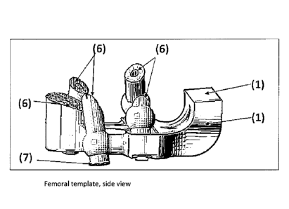
Figure 1: Femoral template
All views (1-A, 1-B, I-C, 1-D, 1-E, 1-F, 1-G)
The figure has several views of the femoral template from (1-A to 1-G).
The template was designed to have five cylindrical locators for the femur
(labels 6 and 10 in
figures 1-A, 1-D and 1-E). These locators were created in the internal
surfaces of the
templates, matching the surface shape of the distal femur. Because these
locators were
patient-specific, they could only allow the templates to be placed in a unique
and secure
position (figure 1-F and 1-G). The locators were cannulated to allow passage
of the fixation
pins that provided additional stability to the templates over the bone (label
7 in figures 1- F
and 1-G). The femoral template is meant to
5
Date Recue/Date Received 2020-06-29
allow the surgeon to perform the distal cut through the specified slit (label
4 in figure 1-B).
The template also guides the surgeon to mark the distal femur for the position
of and
direction of anterior cut and rotation at the margin (free end) of the
template (figure 1-C and
1-D).
Figure 2: Tibial template
AH views (2-A, 2-B, 2-C, 2-D, 2-E, 2-F, 2-G, 2-H)
The figure has several views of the tibial template from 2-A to 2-H.
The template was designed to have four locators for the tibia as indicated by
label 6 in figure
2. These locators were created in the internal surfaces of the templates
(figures 2-A, 2-D and
2-E), matching the surface shape of the proximal tibia (Figures 2-F, 2-G and 2-
H). Because
these locators were patient-specific, they could only allow the templates to
be placed in a
unique and secure position (Figures 2-F, 2-G and 2-H). The locators were
cannulated to
allow passage of the fixation pins which provided additional stability to the
templates over
the bone. A slit in the upper part of tibial template indicates the direction
of tibial rotation
and guides the position of the stem (label 3, Figure 2-B) and keel (label 2,
Figure 2-B) if
needed. Another slit in the front of the tibial template allows the attachment
of a rod that
goes down to the ankle as a double check for tibial rotation (label 5 in
Figure 2-B).
6
Date Recue/Date Received 2020-06-29