Note: Descriptions are shown in the official language in which they were submitted.
CA 02914743 2015-12-10
AUTOTITRATING METHOD AND APPARATUS
RELATED APPLICATIONS
This application is a divisional of Canadian Application Serial No. 2,576,171
filed
August 6, 2005 and which has been submitted as the Canadian national phase
application
corresponding to International Patent Application No. PCT/NZ2005/000196 filed
August 6,
2005.
FIELD OF INVENTION
This invention is generally directed to a method and apparatus for controlling
the
positive air pressure applied to a patient undergoing positive airway pressure
therapy.
BACKGROUND OF THE INVENTION
Obstructions in some patients' airways during sleep can cause limited airflow,
leading to apnoea, hypopnoea, or snoring. The obstruction is often a collapsed
pharynx.
The obstruction may be a partial airway obstruction, leading to altered
characteristics of the
airflow. A hypopnoea is a reduction of flow that is greater than fifty
percent, but not
complete. An apnoea, however, is a complete cessation of airflow. Each of
these conditions
frequently leads to sleep deprivation.
It is well known to treat patients suffering from sleep deprivation with
positive
airway pressure therapy ("PAP"). This therapy can be Continuous Positive
Airway Pressure
("CPAP"), Variable Positive Airway Pressure ("VPAP"), Bi-level Positive Airway
Pressure
("BiPAP"), or any of numerous other forms of respiratory therapy. The
application of
positive pressure to the patient's pharynx helps minimize or prevent this
collapse. Positive
airway pressure therapy is currently applied by means of an apparatus
containing a pressure
source, typically a blower, through a tube to a mask, which the patient wears
in bed.
It is desired to control the applied pressure. Too little pressure tends not
to solve the
problem. Too much pressure tends to cause discomfort to the patient, such as
drying out of
the mouth and pharynx, as well as difficulty in exhaling against the applied
pressure. The
difficulty in applying optimum pressure is that incidents of airway
obstruction come and go
through the course of a night's sleep. One solution is to try to find an
optimum pressure for
a particular
1
CA 02914743 2015-12-10
WO 2006/014114 PCT/NZ2005/000196
patient and maintain that pressure. This method requires the patient's stay at
a sleep clinic,
where sleep specialists can monitor the patient's course of breathing
throughout one or more
night's sleep, prescribe the appropriate pressure for that patient, and then
set the apparatus to
deliver the appropriate pressure. This method is, of course, inconvenient as
well as expensive to
the patient and tends to be inaccurate, as a typical patient will not sleep
the same when away
from familiar bedding and surroundings.
Accordingly, it is desirable to be able to adjust the applied pressure without
requiring the
patient to attend at a sleep center. Various methods of in-home adjustments
have been
considered. One method generally thought to be effective is to monitor the
patient to try to
anticipate the onset of an obstructed airway, and to adjust the pressure in
response. When an
elevated upper airway resistance or flow obstruction is anticipated or
underway, the apparatus
increases the applied pressure. When the patient returns to normal sleep, the
applied pressure is
reduced. The problem then, is to determine when a flow obstruction is
occurring or is about to
occur. It is desired to anticipate correctly in order to avoid the problems
set forth above for when
.. too much or too little pressure is applied.
Various methods have been proposed to solve this problem. In United States
Patent No.
5,107,831 to Halpern, an apparatus monitors the airflow to the patient and
posits an event of
airway obstruction when the patient's breath fails to meet a predetermined
threshold of flow rate
or duration. In United States Patent No. 5,1345,995 to Gruenke, an apparatus
monitors the
airflow to the patient and analyzes the shape of the flow versus time
waveform. If the shape of
this waveform tends to be flattened, that is, more similar to a plateau than
to a sinusoid, the
apparatus posits an event of airway obstruction. In United States Patent No.
5,245,995 to
Sullivan, an apparatus monitors the patient's sound with a microphone. If
audible snores are
-2-
detected, the apparatus posits an event of airway obstruction. Similarly, in
United States Patent
No. 5,953,713 to Behbehani, an apparatus measures the total pressure within an
interface placed
over a patient's airway and inputs frequency data in the range 100 to 150 Hz
into a neural
network to determine the presence of a pharyngeal wall vibration (a snore)
which, according to
Behbehani, is a precursor to sleep disorder breathing.
These methods have not proven totally satisfactory in controlling the applied
pressure
during PAP therapy. For example, the '713 patent, by measuring in the range of
100 to 150 Hz,
essentially tests for snoring and does not measure or analyze any information
concerning partial
airway obstruction (as described within the present application), as this
information is found in
the lower frequency range 0 to 25 Hz. The inventors have appreciated that
there is a marked
difference between normal breathing and breathing characterized by a partial
airway obstruction,
all in low frequencies. The present application exploits this difference to
control the delivery of
therapeutic gas.
Moreover, the methods of the prior art are unsatisfactory in analyzing a
signal in a high-
noise environment. The inventors herein have discovered an alternate way to
detect the onset of
an event of airway obstruction and to control the applied pressure from a high-
noise signal such
as results from a person's breathing over the course of a night. Accordingly,
the method and
apparatus of the present invention fulfill the need for analyzing a signal
from a patient in order to
control the applied pressure during PAP therapy.
SUMMARY OF THE INVENTION
The present invention in one embodiment is a method of controlling positive
airway
pressure therapy by providing a flow of gas to a patient's airway at a
pressure, obtaining
-3-
Date Recue/Date Received 2020-12-22
CA 02914743 2015-12-10
information from the frequency range of zero to 25 HZ in the frequency domain
of the flow,
and adjusting the pressure based on the information. In another embodiment,
the present
invention is an apparatus for providing controlled positive airway pressure
therapy, having a
blower for providing a flow of gas to a patient's airway, a sensor to measure
a characteristic of
the flow, a controller to obtain information from the frequency range of zero
to 25 HZ in the
frequency domain of the characteristic, and a pressure regulator for adjusting
the pressure
based on the information.
The present invention in a further embodiment provides a method of controlling
a
positive airway pressure apparatus for providing a flow of gas to a patient's
airway,
comprising the steps of: controlling the positive airway pressure apparatus to
provide a flow of
gas at a pressure; obtaining a signal relating to the flow; obtaining
information from the signal
from a frequency range of 0 to 25 Hz in a frequency domain of the signal, the
information
relating to an energy spectrum of inspiration of the patient; and generating
at least one
information-bearing value from said information, the at least one information-
bearing value
relating to an energy in a first harmonic of said energy spectrum and/or an
energy in a second
harmonic of said energy spectrum, and adjusting said pressure based on said
information-
bearing value.
In yet another aspect, the present invention provides an apparatus for
controlling
positive airway pressure therapy, comprising: a blower for providing a flow of
gas to a patient
at a pressure; a sensor to measure a characteristic of said flow; a controller
to: obtain a signal
from the sensor during inspiration of the flow of gas by the patient,
transform the signal into
the frequency domain and generate an energy spectrum of said characteristic,
and generate at
least one information-bearing value from a frequency range of 0 to 25 Hz of
said energy
spectrum, the at least one information-bearing value relating to an energy in
a first harmonic
of said energy spectrum and/or an energy in a second harmonic of said energy
spectrum; and a
- 4 -
pressure regulator controlled by said controller for adjusting said pressure
based on said at least
one information-bearing value.
In yet another aspect, the present invention provides a method of controlling
positive
airway pressure therapy, comprising: providing gas to an airway of a patient
at a pressure;
determining a flow of the gas provided to the airway of the patient; and
adjusting the pressure of
the gas based in part on determining that the flow does not exceed a minimum
flow threshold over
at least a first duration of time and that the flow exceeds the minimum flow
threshold during a
second duration of time.
In yet another aspect, the present invention provides a positive airway
pressure therapy
apparatus comprising: a blower configured to provide a flow of gas to an
airway of a patient at a
pressure; a pressure regulator configured to adjust the pressure; a pressure
sensor positioned in a
flow path of the gas; and a controller configured to: determine the flow of
the gas based on
pressure measured by the pressure sensor; and control the pressure regulator
to adjust the pressure
of the gas in response to a determination that the flow does not exceed a
minimum flow threshold
over at least a first duration of time and that the flow exceeds the minimum
flow threshold during
a second duration of time.
In yet a further aspect, the present invention resides in a positive airway
pressure therapy
apparatus comprising: a blower configured to provide a flow of gas to an
airway of a patient at a
pressure; a pressure regulator configured to adjust the pressure; a pressure
sensor positioned in a
flow path of the gas; and a controller configured to: determine the flow of
the gas based on
pressure measured by the pressure sensor; and store flow values of a plurality
of last breaths that
are not part of a hypopnea, and determine a minimum flow threshold based at
least in part on an
average of the flow values of a plurality of oldest breaths of the plurality
of last breaths, and
control the pressure regulator to adjust the pressure of the gas in response
to detecting a hypopnea
based on a determination that the flow does not exceed the minimum flow
threshold over at least a
first duration of time and that the flow exceeds the minimum flow threshold
during a second
duration of time.
- 4a -
CA 2914743 2017-07-10
In yet another aspect, the present invention provides a positive airway
pressure therapy
apparatus comprising: a blower configured to provide a flow of gas to an
airway of a patient
at a pressure; a pressure regulator configured to adjust the pressure; a
pressure sensor
positioned in a flow path of the gas; and a controller configured to:
determine the flow of the
gas based on pressure measured by the pressure sensor; store flow values of a
plurality of last
breaths that are not part of a hypopnea; determine a minimum flow threshold
based at least in
part on an average of the flow values of a plurality of oldest breaths of the
plurality of last
breaths, and excluding one or more most recent breaths of the plurality of
last breaths; and
control the pressure regulator to adjust the pressure of the gas in response
to detecting a
hypopnea based on a determination that the flow does not exceed the minimum
flow threshold
over at least a first duration of time and that the flow exceeds the minimum
flow threshold
during a second duration of time.
In yet another aspect, the invention provides a positive airway pressure
therapy
apparatus comprising: a blower configured to provide a flow of gas to an
airway of a patient
at a pressure; a pressure regulator configured to adjust the pressure; a
pressure sensor
positioned in a flow path of the gas; and a controller configured to:
determine the flow of the
gas; store flow values of a plurality of last breaths that are not part of a
hypopnea; determine
a minimum flow threshold based at least in part on an average of the flow
values of a plurality
of oldest breaths of the plurality of last breaths, and excluding one or more
most recent
breaths of the plurality of last breaths; and control the pressure regulator
to adjust the pressure
of the gas in response to detecting a hypopnea based on a determination that
the flow does not
exceed the minimum flow threshold over at least a first duration of time and
that the flow
exceeds the minimum flow threshold during a second duration of time.
BRIEF DESCRIPTION OF THE DRAWINGS
The organization and manner of the structure and operation of the invention,
together
with further objects and advantages thereof, may best be understood by
reference to the
following description, taken in connection with the accompanying drawings,
wherein like
reference numerals identify like elements in which:
-4b-
Date Recue/Date Received 2020-12-22
FIGURE 1 is a plot in the frequency domain of energy v. frequency for normal
breathing and breathing characterized by partial airway obstruction;
FIGURE 2 is also a plot in the frequency domain of energy v. frequency for
normal
breathing and breathing characterized by partial airway obstruction;
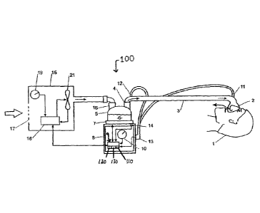
FIGURE 3 is a diagram of an exemplary positive airway pressure apparatus of
the
preferred embodiment of the present invention;
FIGURE 4 is a block diagram of the main algorithm of the method of the
preferred
embodiment of the present invention, showing the interaction of the five
algorithms;
FIGURE 5 is a block diagram of the Breath Detection Algorithm of the preferred
embodiment of the present invention;
-4c-
Date Recue/Date Received 2020-12-22
CA 02914743 2015-12-10
WO 2006/014114 PCT/NZ2005/000196
FIGURE 6 is a block diagram of the Partial Airway Obstruction Algorithm of the
preferred embodiment of the present invention;
FIGURE 7 is a block diagram of the Apnoea Detection Algorithm of the preferred
embodiment of the present invention;
FIGURE 8 is a block diagram of the Hypopnoea Detection Algorithm of the
preferred
embodiment of the present invention;
FIGURES 9a, 9b, 9c, and 9d are block diagrams of the Pressure Adjusting
Algorithm of
the preferred embodiment of the present invention;
FIGURE 10 is a diagram of airflow versus time, illustrating Tsui and Mf; and
FIGURE 11 is a diagram of airflow versus time, illustrating Tend,11,12 and Mtl-
t2-
DETAILED DESCRIPTION
While the invention may be susceptible to embodiment in different forms, there
is shown
in the drawings, and herein will be described in detail, a specific embodiment
with the
understanding that the present disclosure is to be considered an
exemplification of the principles
of the invention, and is not intended to limit the invention to that as
illustrated and described
herein.
A positive airway pressure apparatus 100 of the preferred embodiment of the
present
invention is shown in FIGURE 3 in which the patient 1 receives humidified,
pressurized gas
through an inspiratory conduit 3. It should be understood that the delivery
systems could be
CPAP (Continuous Positive Airway Pressure), 'VPAP (Variable Positive Airway
Pressure),
BiPAP (Bi-level Positive Airway Pressure), or any of numerous other forms of
respiratory
therapy. The apparatus 100 and method 200 of the present invention will be
described as used
-5-
CA 02914743 2015-12-10
WO 20061014114 1'CT/NZ2905/000196
for CPA but an artisan of ordinary skill in the art will readily adapt both
for use with VPAP,
BiPAP, or another positive airway pressure therapeutic system.
Inspiratory conduit 3 is attached at one end to a mask 2, preferably one such
as is
described in United States Patent No. 6,662,803. Inspiratory conduit 3
connects at its other end
to the outlet 4 of a humidification chamber 5, which contains a volume of
water 6. Inspiratory
conduit 3 may contain heating heater wires (not shown) or other suitable
heating elements that
beat the walls of the conduit to reduce condensation of humidified gases
within the conduit.
Humidification chamber 6 is preferably formed from a plastic material and may
have a highly
heat-conductive base (for example an aluminum base) that is in direct contact
with a heater plate
7 of humidifier 8.
Electronic controller 9 controls the various components of the apparatus 100.
Controller
9 may be a microprocessor-based controller containing, as is well known in the
art, RAM, ROM,
an ALU, one or more registers, a data bus, counters (including at least a
breath number counter
120 and a pressure decrease counter 130), and one or more buffers (including
at least a circular
buffer 110). Controller 9 executes computer software commands stored in its
RAM and ROM.
Controller 9 receives input from sources such as user input dial 10 through
which a user
of the device may, for example, set a predetermined required value (preset
value) of various
characteristics of the gases supplied to the patient 1, such as initial
airflow, pressure, humidity, or
temperature of the gases. Controller 9 preferably receives input relating to
airflow from
differential pressure sensor 11, which is preferably located in blower 15.
Differential pressure
sensor 11 could alternatively be located elsewhere, upstream of mask 2, such
as within conduit 3
or anywhere on mask 2. Alternatively, controller 9 may receive input related
to airflow by direct
measurement of flow at any point from blower 15 to mask 2. Controller 9 may
also receive input
-6-
CA 02914743 2015-12-10
WO 2006/014114 PCT/NZ2005/000196
from other sources, for example temperature sensors 12 through connector 13
and heater-plate
temperature sensor 14.
In response to the user-set inputs and the other inputs, controller 9
determines when (or to
what level) to energize heater plate 7 to heat the water 6 within
humidification chamber 5. As
the volume of water 6 within humidification chamber 5 is heated, water vapor
begins to fill the
volume of the chamber 5 above the water's surface and is passed out of the
outlet 4 of
humidification chamber 5 with the flow of gases (for example air) provided
from a gas supply
device such as blower 15, which gases enter the chamber 5 through inlet 16.
Exhaled gases from
the patient 1 are passed directly to ambient surroundings in FIGURE 3.
Blower 15 is provided with a variable-pressure regulating device such as
variable speed
fan 21, which draws air or other gases through blower inlet 17. The speed of
variable speed fan
21 is controlled by electronic controller 9 in response to inputs from the
various components of
apparatus 100 and by a user-set predetermined required value (preset value) of
pressure or fan
speed via dial 19.
Controller 9 is programmed with five algorithms:
1. Breath Detection Algorithm;
2. Apnoea Detection Algorithm;
3. Hypopnoea Detection Algorithm;
4. Partial Airway Obstruction Detection Algorithm; and
5. Pressure Adjusting Algorithm.
These algorithms interact as diagramed in FIGURE 4. When the patient 1 turns
on the
apparatus 100, the controller 9 receives data from pressure sensor 11 and
starts the main
algorithm (step 200). Pressure sensor 11 is preferably a differential pressure
sensor, and
-7-
CA 02914743 2015-12-10
WO 2006/014114 PCT/NZ2005/000196
controller 9 converts differential pressure data to airflow data. Controller 9
samples the raw I
analogue flow signal at 50 Hz (step 202) and calculates bias flow (step 204).
Bias flow, such as
occurs from leaks in the mask 2 or elsewhere in the apparatus 100, is obtained
preferably via a
Butterworth low-pass filter (with a rise time of approximately thirty
seconds). The information
stored in circular buffer 110 in controller 9 is therefore net airflow data,
as the controller 9
removes the bias flow (step 206). Circular buffer 110 in controller 9 is
continuously updating
data and storing that data for 15 seconds (step 208). Accordingly, throughout
these algorithms,
the flow being analyzed does not contain the bias flow. That is, the flow
oscillates about zero
flow.
The incoming flow data is continuously checked for the presence of a leak
(step 210). If a
significant leak is detected the algorithm is paused until the leak is
resolved.
If there are no leaks and fewer than ten breaths have passed, the current data
is analyzed
by the Breath Detection Algorithm (step 300), as will be described in
connection with FIGURE
5. The Breath Detection Algorithm determines where the oldest breath begins
and ends.
If no breath is detected (step 212), the main algorithm starts over with
sampling the raw
analogue flow signal (step 202). If a breath is detected (step 212), a breath
number counter 120
is incremented (step 214), and the main algorithm starts over with sampling
the raw signal (step
202).
Since it is assumed that the patient I will breathe a minimum of ten breaths
before any
apnoeas or hypopnoeas occur, the main algorithm of the preferred embodiment
counts to
determine if at least ten breaths have occurred (step 216). If more than ten
breaths have
occurred, the apparatus proceeds to the Apnoea Detection Algorithm (step 500),
as will
-8-
CA 02914743 2015-12-10
WO 2006/014114 PCUNZ2005/000196
hereinafter be described in connection with FIGURE 7. If fewer than ten
breaths have occurred,
circular buffer 110 in controller 9 continues to sample raw analogue data
(step 202).
Once ten breaths have occurred, the main algorithm proceeds as diagramed in
FIGURE 4.
The Apnoea Detection Algorithm (step 500) constantly checks the real-time
incoming flow to
see if an apnoea is occurring (step 218), as will be described in greater
detail in connection with
FIGURE 7. If an apnoea is occurring, the Pressure Adjusting Algorithm (step
700) is called, as
will be described in connection with FIGURE 9. Once the apnoea has finished
(step 220), the
main algorithm starts over with sampling raw data (step 202). If no apnoea is
occurring, the
Breath Detection Algorithm (step 300) is called.
If no new breath has been detected (step 222), the algorithm checks to see if
2.5 minutes
have passed since the last partial obstruction or apnoea (step 224). If not,
the main algorithm
starts over with sampling raw data (step 202). If so, the Pressure Adjusting
Algorithm (step 700)
is called. If a breath is detected, the Hypopnoea Detection Algorithm is
called (step 600), as will
be described in connection with FIGURE 8, followed by the Partial Airway
Obstruction
Algorithm (step 400) as will be explained in connection with FIGURE 6.
The Hypopnoea Detection Algorithm (step 600) checks to see if a breath is
possibly part
of a hypopnoea. The Partial Airway Obstruction Algorithm (step 400) is called
to check for
partial airway obstruction (step 232). If the Hypopnoea Detection Algorithm
finds that a
hypopnoea has occurred (step 226), the main algorithm checks to see if any
breaths in the
hypopnoea showed partial airway obstruction (step 228). If so, the Pressure
Adjusting
Algorithm is called (step 700). If not, the main algorithm checks to see if
2.5 minutes have
passed since the last partial obstruction or apnoea event (step 230). If so,
the Pressure Adjusting
-9-
CA 02914743 2015-12-10
WO 2006/014114 PCT/NZ2005/000196
Algorithm is called (step 700). If not, the main algorithm starts over with
sampling raw data
(step 202).
The Partial Airway Obstruction Algorithm checks for partial airway obstruction
(step
232) in the event a hypopnoea has not occurred. If the current breath shows a
partial airway
obstruction, the main algorithm checks to see if the previous two breaths have
shown a partial
airway obstruction (step 234). If so, the Pressure Adjusting Algorithm (step
700) is called. If the
current breath does not show a partial airway obstruction (step 232) or if the
previous two
breaths do not show a partial airway obstruction (step 234), the main
algorithm checks to see if
2.5 minutes have passed since the last partial obstruction or apnoea event
(step 224). If so, the
Pressure Adjusting Algorithm (step 700) is called; if not, the main algorithm
starts over with
sampling raw data.
Using the above algorithms, the applied positive airway pressure is at the
lowest pressure
required by the patient Ito achieve therapeutic treatment. The details of the
algorithms will now
be explained.
Breath Detection Algorithm
Two routines are used, as diagramed in FIGURE 5, depending on how many breaths
have
been detected since the program was initiated. The two routines differ, in
that one incorporates
the breathing period of the patient 1.
The Breath Detection Algorithm (step 300) initially determines if a previous
breath's end
is still contained within the flow buffer (step 302). If a previous breath's
end point is still in the
flow buffer, the start of the next breath (beginning of inspiration, or Lbw)
will be the data point
following the end point of the previous breath (step 304). If the previous
breath's end point is
not in the buffer (such as if an apnoea occurred), the new end point is
determined, once a piece
-10-
CA 02914743 2015-12-10
WO 2006/914114 PCT/NZ2005/000196
of flow data greater than five liters per minute is immediately followed by a
piece of flow data
less than 5 liters per minute has occurred (step 306), by searching the flow
buffer to find Ef
where Ef is 0.15 times the maximum flow in the buffer (step 308), and where
flow is increasing,
that is, flow is less than Et followed by flow greater than Ef (step 310). The
new end point Tend
is then set as the start of the next breath (step 304).
At this point, the algorithm determines whether more than twenty breaths have
occurred
(step 312). If so, the algorithm searches to find Mf, the maximum flow over
the last one-quarter
of the average breathing period after 'Led (step 314). If twenty or fewer
breaths have occurred,
M1 is defined as the maximum flow in the next second after Tgart (step 316).
The end point of expiration, Tend, is determined by searching between two
reference
points (step 318) (reference points t1, t2 are shown in FIGURE 10, a plot of
airflow to the patient,
as determined from differential pressure sensor 11, versus time). Each
reference point ti, t2 is
identified by determining the occurrence of a flow data value greater than the
reference value
followed by a flow data value less than the reference value (steps 320, 322),
where t2 is after ti,
which is after Tstart.
The reference value is given by:
reference value = 0.2 x I\41
where Mf is the maximum flow in 0.25 x average breathing period since the
beginning of
inspiration (as found in step 314).
Mt is illustrated in FIGURE 10, also a plot of airflow to the patient 1, as
determined from
differential pressure sensor 11, versus time.
-11-
CA 02914743 2015-12-10
WO 2006/014114 PCT/NZ2005/000196
The period between tl and t2 should be greater than 0.5 sec (step 326). If
not, t2 is found
again (step 322). The maximum flow, greater than zero, between the two
reference points ti, t2 is
calculated and used to determine the end of expiration Ef (step 328). The end
of expiration is:
Ef= 0.15 x Mu-t2
where
M1-t2 = maximum flow between t: and t2
Mti_t2, th and t2 are illustrated in FIGURE 11.
A flow data value less than Ef immediately followed by a flow data value
greater than Ef
indicates the end of the breath Tend (step 330). The breath is therefore from
Lt./ to Tend (step
332). The apparatus then stores the maximum flow Mti.t.2, provided the breath
is not part of a
hypopnoea, as determined by the Hypopnoea Detection Algorithm (step 600), as
will be
hereinafter described, and stores the period of the breath (step 334).
The period of the breath and the maximum inspiratory flow are used by the
Apnoea
Detection Algorithm (step 500) and the Hypopnoea Detection Algorithm (step
600), as will be
described.
Partial Airway Obstruction Algorithm
The Partial Airway Obstruction Detection Algorithm (step 400) is diagramed in
FIGURE
6. It analyzes a breath, previously detected by the Breath Detection Algorithm
(step 300), for the
presence of a partial airway obstruction (partial obstruction of the upper
airway).
When the patient 1 breathes, pressure gradients are generated between the
lungs and
atmosphere. The physiology of the upper airway combined with these pressure
gradients and
Bernoulli's Effect can result in partial collapse of the upper airway during
inspiration. This
partial collapse is prevalent in people with obstructive sleep apnoea.
-12-
CA 02914743 2015-12-10
WO 2006/014114 PCT/NZ2005/000196
In order to determine if a breath contains a partial airway obstruction,
Fourier analysis is
used to analyze the inspiratory flow for features specific to partial airway
obstruction. Once a
signal has been mapped to the frequency domain via a Fourier transform, there
are many ways to
represent and analyze the frequency domain information. One could analyze the
direct result of
the Fourier transform, which would give the amplitude of the Fourier
transform's sine
component (information representative of the odd component of the original
signal) and the
amplitude of the Fourier transform's cosine component (information
representing the even
component of the original signal). Alternatively, from the Fourier transform,
one could construct
a phase v. frequency plot and an energy v. frequency plot (energy spectrum).
The phase and
energy information could be used to analyze the original waveform. An
alternative to the energy
v. frequency plot is to construct a magnitude v. frequency plot. In the
preferred embodiment an
energy spectrum is used to determine the presence of partial airway
obstruction. Partial airway
obstructions can be detected from analysis of the energy spectrum at low
frequencies, as
illustrated in FIGURES 1 and 2.
In particular, energy statements involving groupings of the frequency
harmonics of the
Fourier transform of the flow of therapeutic gas to the patient are generated
from frequency-
domain considerations. This technique allows analysis of signals that might
have a considerable
amount of background noise. All processing and analysis is done in the
frequency domain based
upon observed relationships between the patients responses and the character
of the energy
spectrum in the frequency domain.
Additionally, severe airway obstruction often results in a reduced peak flow-
rate during
inspiration, which results in a prolongation of time spent inspiring relative
to expiring. This
-13-
CA 02914743 2015-12-10
WO 2006/014114 PCT/NZ2005/000196
increase in inspiratory time is incorporated in the Partial Airway Obstruction
Detection
Algorithm.
To obtain information solely from the inspiratory phase of the respiratory
cycle, Fourier
analysis is performed on a waveform consisting of two inspiratory phases
oppositely combined.
The result is an odd function defined as
(1)
The standard Fourier series definition is
n
(2)
f (x) = Aõ cos + õ sin fl 2t
where n is the number of hannonics, An are the harmonic cosine coefficients,
B, are the harmonic
sine coefficients, and T is the period of cycle. Modifying Equation (2)
according to Equation (1)
gives
co
(3) f (x) = B siiln Irx
n=-1
as all An, which represent the even part of the function, are zero.
To apply Fourier analysis to the inspiratory waveform, the algorithm of the
preferred
embodiment of the present invention first samples the incoming flow signal.
Inspiration is then
separated from expiration and manipulated as in Equation (1) to give a vector
of N data points, y
- I 4-
CA 02914743 2015-12-10
WO 2006/014114 PCT/NZ2005/000196
= [yi y2 ... yr,], that represent a single period of a cyclic function. The
data is sampled evenly in
time, hence tfil = T./ where r is the sampling interval between data points] =
0, ... ¨I. The
discrete Fourier transform of y is defined as
(4) N -1
Y- mo.e.= E
y +1 / N
k+1
j =0
where i is the square root of negative one and k = 0, , N ¨1. Each point Yk+1
of the transform
has an associated frequency,
(5) fk+1 = khN
In the preferred embodiment, the fundamental frequency, k = is defined as f2 =
1/T N
and the first harmonic frequency, k = 2, is defined as f3 = 2/T N.
In order to determine whether a breath is a partial airway obstruction, the
relative energy
of specific frequencies and groups of frequencies is analyzed. To do this the
energy spectrum is
calculated,
2
(6) TIT
1 k+1
and normalized such that the total energy equals one.
In the preferred embodiment, the first 13 harmonics are considered for
analysis, as the
relative power in the higher harmonics is minuscule. The analyzed harmonies
are in the
frequency range of zero to 25 Hz. The energy distribution of an inspiratory
contour of a normal
-15-
CA 02914743 2015-12-10
WO 2006/014114 PCT/NZ2005/000196
breath generally will have a majority of energy situated at W2, which is
associated with the
fundamental frequency, and a small amount of energy is distributed among the
harmonics. The
present invention uses this characteristic of the energy spectrum as developed
through Fourier
analysis to posit that if the relative energy situated at a particular
frequency or group of
frequencies is above an empirically-observed threshold, the breath is deemed
to be a partial
airway obstruction.
Generally, for a normal breath the percentage of time spent inspiring is 40
percent and
expiring is 60 percent. The patient 1 with a partially collapsed airway cannot
achieve maximum
inspiratory flow. Accordingly, the patient 1 extends the time spent inspiring
relative to expiring.
The time spent inspiring increases to 50 percent or more of the total breath
during a partial
airway obstruction.
Accordingly, the Partial Airway Obstruction Detection Algorithm first
calculates an
initial ratio, Iinsp, which is the portion of the entire breath spent on
inspiration greater than the
mean (step 402). Note that bias flow has been previously removed (steps 204,
206), so the mean
of the breath should be zero or very close to zero. Next, the algorithm
determines the inspiratory
part of the breath and constructs a waveform consisting of two inspiratory
phases oppositely
combined (step 404). Then, the algorithm calculates the discrete energy
spectrum of the
oppositely combined waveform as a function of frequency f (step 406):
W(f) =-- FFT(waveform) )2
It is assumed that no significant energy is contained in the frequencies (or
harmonics)
above a predetermined level, preferably 13 times the fundamental frequency.
Therefore, the
energy spectrum is only retained, in the preferred embodiment, up to 13 times
the fundamental
-16-
CA 02914743 2015-12-10
WO 2006/014114 PCT/NZ2005/000196
frequency (step 408). Next, the algorithm normalizes the energy spectrum such
that the total
energy equals one (step 410):
normalized energy spectrum = W(f) / E'W(f)
This calculation is done so that all breaths will be analyzed the same, even
though each
breath may differ from another breath in duration, tidal volume, and maximum
flow.
Next, the algorithm groups energies corresponding to different harmonic
frequencies into
information-bearing values (step 412). These information-bearing values are
compared to
threshold values that are calculated in accordance with the percentage of the
breath that is spend
on inspiration (step 414). The information-bearing values and the threshold
values are
determined empirically.
In the preferred embodiment, four information-bearing values are used: W
first, Wsecond,
Wfreri, and W
high_freq, as follows:
Wfirst = W3
Wsecond = W4
14
frfivg TwA,41
IF high_ iht; =1W k41
ket6
According, Wfirst corresponds to the energy in the first harmonic, Wsou
corresponds to the
ecn
energy in the second harmonic, Wfreq corresponds to the energy in the first 13
harmonics, and
-17-
CA 02914743 2015-12-10
WO 2006/014114 PCT/NZ2005/000196
Whighfreq corresponds to the energy in the harmonics five through 13. Other
information-bearing
values can be obtained from the energies corresponding to different harmonic
frequencies using
other mathematical operations.
In the preferred embodiment, two thresholds are used, Tfrezi and Thigh_freq.
These values
vary depending on the value of Lisp, the percentage of the breath spend
inspiring (calculated at
step 402) and have been determined empirically to be:
Threshold linsp Value
Tfreq Lisp < 40 0.15
Tfõq 40 < Lisp 50 -0.005 x Iinsp + 0.35
Tfreq 'imp >50 0.1
Thigh freq iinsp < 40 0.03
Thigh_freq 40 < Iõ,3), 60 -0.001 x 1,,,31, +0.07
Thigh_freq Tinsp >60 0.01
Using these empirically-determined values, the algorithm computes the
information-
bearing summations to the thresholds. If Wsecond is greater than or equal to
0.1 (step 416), the
breath is a partial airway obstruction (step 418). If Wfirst is greater than
or equal to 0.02, Wsecond
is greater than or equal to 0.02, and Wfreq is greater than or equal to 0.12
(step 420), the breath is
a partial airway obstruction (step 422). If the sum of Wfirst and W. second is
greater than or equal to
0.06 and Wfreq is greater than or equal to 0.12 (step 424), the breath is a
partial airway
obstruction (step 426). If the sum of Wrirst and Wsecond is greater than or
equal to 0.07 and Wfreq is
-18-
CA 02914743 2015-12-10
WO 2006/014114 PCT/NZ2005/000196
greater than or equal to 0.11 (step 428), the breath is a partial airway
obstruction (step 430). If
Wfreq is greater than or equal to Tfreq (step 432), the breath is a partial
airway obstruction (step
434). If Whigh_freq is greater than or equal to Thfreq (step 436), the breath
is a partial airway
obstruction (step 438). If none of these comparisons is true, the breath is
normal (step 440).
Apnoea Detection Algorithm
The Apnoea Detection Algorithm (step 500) is diagramed in FIGURE 7. In order
to
detect an apnoea (cessation of flow), the controller 9 compares the incoming
flow data (minus
bias flow) with a threshold, Ili, determined by the previous peak inspiratory
flow. The Breath
Detection Algorithm (step 300) had previously stored the maximum or peak
inspiratory flow, not
part of a hypopnoea. The Apnoea Detection Algorithm calculates the threshold,
I, as 20
percent of the average peak inspiratory flow of the oldest five breaths of the
last ten breaths (step
502). The algorithm then calculates Tapnoea (step 504):
Tapnoea = 1.7 x (breathing period averaged over last 50 breaths)
Tapnoea, however, must be between ten and fifteen seconds.
If the incoming flow is less than the threshold, 41, an apnoea may be
occurring. If this
condition is met for time greater than Tax,. (step 506), then an apnoea is
occurring (step 508),
otherwise, no apnoea occurred (step 510). If an apnoea is occurring, the
algorithm checks to see
when the flow has increased to more than the threshold, (step 512), indicating
that the apnoea
has finished.
Hypopnoea Detection Algorithm
In order to detect a hypopnoea (reduction of flow), the Hypopnoea Detection
Algorithm
(step 600), as diagramed in FIGURE 8, compares the stored breath with a
threshold, 112,
determined by the previous peak inspiratory flow (step 602). Similar to the
Apnoea Detection
-19-
CA 02914743 2015-12-10
WO 2006/014114 PCT/N22005/000196
Algorithm (step 500), the threshold, 112, is calculated from the peak
inspiratory flow for the
oldest five breaths of the last ten that did not constitute part of a
hypopnoea. The threshold (p.2)
is then taken as 60 percent of the average peak inspiratory flow of the oldest
five breaths (step
602).
If incoming flow is less than the threshold (12), for a period of time greater
than 12
seconds (step 604), then a possible hypopnoea has occurred; otherwise, no
hypopnoea is
occurring (step 606). For the event to be classified as a hypopnoea, there
must be an increase in
flow such that flow is greater than J.12 within 30 seconds since the flow was
less than 112 (step
608). If this increase in flow is detected, a hypopnoea occurred (step 610);
otherwise, the event
was not a hypopnoea (step 612).
Pressure Adjusting Algorithm
If an apnoea was detected during the Apnoea Detection Algorithm (step 500),
the
Pressure Adjusting Algorithm (step 700) is called. Also, if a hypopnoea was
detected during the
Hypopnoea Detection Algorithm (step 600), and there were partial airway
obstruction breaths in
the hypopnoea (step 228), or if there was no hypopnoea but the current breath
and two previous
breaths were partial airway obstructions (steps 226, 232, 234), the Pressure
Adjusting (step 700)
algorithm is called. If there was no hypopnoea, and either the current breath
does not show a
partial airway obstruction or the previous two breaths did not show a partial
airway obstruction,
but is has been 2.5 minutes since the last partial airway obstruction (steps
226, 232, 234, 224),
the Pressure Adjusting Algorithm is called. Also, if there was a hypopnoea,
but without any
partial airway obstruction breaths, and it has been longer than a
predetermined period since the
last partial airway obstruction event or apnoea, preferably 2.5 minutes (steps
226, 228, and 230),
-20..
CA 02914743 2015-12-10
WO 2006/014114 PCT/NZ2005/000196
the Pressure Adjusting Algorithm (step 700) is called. The Pressure Adjusting
Algorithm is
diagramed in FIGURES 9a through 9d.
The Pressure Adjusting Algorithm (step 700) determines whether to adjust the
pressure
and by how much, in order to control the therapeutic pressure delivered to the
patient. As an
initial rule of the preferred embodiment, this algorithm will only increase
pressure to a maximum
of 10 cm 1-120 on an event classified as an apnoea (step 702).
The algorithm first checks to determine if there have been any pressure
decreases since
the beginning of the period of sleep (step 704). If there have not been any
such decreases, the
algorithm determines if an obstructive event of any sort has been detected and
whether the
pressure is under a predetermined maximum, preferably ten cm H20 (step 706).
If these
conditions are met, the algorithm determines whether the obstructive event was
a partial airway
obstruction, an apnoea, or a hypopnoea with a partial airway obstruction (step
708). In the event
of a hypopnoea with a partial airway obstruction, the controller 9 increases
pressure by one cm
H20 (step 710) and waits ten seconds before allowing another pressure change
(step 712). If the
event was an apnoea, the controller 9 increases pressure by two cm H20 (step
714) and waits 60
seconds before allowing another pressure change (step 716). If the event was a
partial airway
obstruction, the controller 9 increases pressure by one cm H20 (step 718) and
waits ten seconds
before allowing another pressure change (step 720).
If there have been previous pressure decreases since the beginning of the
period of sleep
(step 704), or if the conditions of a detected obstructive event and the
pressure being less than ten
cm H20 have not been met (step 706), the algorithm determines if there have
been six
consecutive pressure decreases. If so, total consecutive pressure-decrease
counter 130 is reset to
zero (step 722).
-21-
CA 02914743 2015-12-10
WO 2006/014114 PCT/NZ2005/000196
The algorithm next determines if there has been normal breathing for a
predetermined
period of time, preferably 2.5 minutes (step 724). If so, the controller 9
decreases the pressure
by 0.5 cm H20 (step 726) (and increments pressure-decrease counter 130 by
one).
If there has not been normal breathing for the predetermined period of time
(step 724),
then either a partial airway obstruction, an apnoea, or a hypopnoea with
partial airway
obstruction has occurred (step 728). The next step depends on the previous
pressure changes. If
the previous consecutive pressure changes have been increases totaling greater
than or equal to a
total of one cm 1120, and the current pressure is less than ten cm H20 (step
730), the algorithm
proceeds to step 708 as described above. If not, the controller 9 proceeds to
increase the pressure
by an amount depending on the nature of the obstructive event and the amount
of previous
pressure decreases, as diagramed in FIGURES 9b, 9c, and 9d.
If the total previous pressure decreases were more than one cm 1120 (step
732), the
algorithm determines if the obstructive event was a partial airway
obstruction, an apnoea, or a
hypopnoea with partial airway obstruction (step 734). In the event of a
hypopnoea with a partial
airway obstruction, the controller 9 increases pressure by one cm H20 (step
736) and waits ten
seconds before allowing another pressure change (step 738). If the event was
an apnoea, the
controller 9 increases pressure by two cm H20 (step 740) and waits 60 seconds
before allowing
another pressure change (step 742), If the event was a partial airway
obstruction, the controller 9
increases pressure by 0.5 cm H20 (step 744) and waits ten seconds before
allowing another
pressure change (step 746).
If the previous pressure decreases were more than one cm H20 but not more than
1.5 cm
1120 (step 748), the algorithm determines if the obstructive event was a
partial airway
obstruction, an apnoea, or a hypopnoea with partial airway obstruction (step
750). In the event
-22-
CA 02914743 2015-12-10
WO 2006/014114 PCT/NZ2005/000196
of a hypopnoea with partial airway obstruction, the controller 9 increases
pressure by one cm
H20 (step 752) and waits ten seconds before allowing another pressure change
(step 754). If the
event was an apnoea, the controller 9 increases pressure by two cm H20 (step
756) and waits 60
seconds before allowing another pressure change (step 758). If the event was a
partial airway
obstruction, the controller 9 increases pressure by 0.5 cm H20 (step 760) and
waits ten seconds
before allowing another pressure change (step 762).
If the previous pressure decreases were more than 1.5 cm 1120 but not more
than two cm
H20 (step 764) (FIGURE 9c), the algorithm determines if the obstructive event
was a partial
airway obstruction, an apnoea, or a hypopnoea with partial airway obstruction
(step 766). In the
event of a hypopnoea with partial airway obstruction, the controller 9
increases pressure by 1.5
cm H20 (step 768) and waits ten seconds before allowing another pressure
change (step 770). If
the event was an apnoea, the controller 9 increases pressure by two cm H20
(step 772) and waits
60 seconds before allowing another pressure change (step 774). If the event
was a partial airway
obstruction, the controller 9 increases pressure by one cm 1120 (step 776) and
waits ten seconds
before allowing another pressure change (step 778).
If the previous pressure decreases were more than two cm H20 but less than or
equal to
3.5 cm 1120 (step 780), the algorithm determines if the obstructive event was
a partial airway
obstruction, an apnoea, or a hypopnoea with partial airway obstruction (step
782). In the event
of a hypopnoea with partial airway obstruction, the controller 9 increases
pressure by 1.5 cm
H20 (step 784) and waits ten seconds before allowing another pressure change
(step 754). If the
event was an apnoea, the controller 9 increases pressure by two cm 1120 (step
788) and waits 60
seconds before allowing another pressure change (step 790). If the event was a
partial airway
-23-
CA 02914743 2015-12-10
obstruction, the controller 9 increases pressure by 1.5 cm H20 (step 792) and
waits ten
seconds before allowing another pressure change (step 794).
If the previous pressure decreases were more than 3.5 cm H20 (step 796), the
algorithm determines if the obstructive event was a partial airway
obstruction, an apnoea, or a
hypopnoea with partial airway obstruction (step 798). In the event of a
hypopnoea with partial
airway obstruction, the controller 9 increases pressure by one-half the total
pressure decrease
(step 800) and waits ten seconds before allowing another pressure change (step
802). If the
event was an apnoea, the controller 9 increases pressure by one-half the total
pressure decrease
(step 804) and waits 60 seconds before allowing another pressure change (step
806). If the
event was a partial airway obstruction, the controller 9 increases pressure by
one-half the total
pressure decrease (step 808) and waits ten seconds before allowing another
pressure change
(step 810).
Although the present invention has been described in connection with certain
preferred embodiments, it is to be understood that the scope of the claims
should not be
.. limited by the preferred embodiments set forth in the example, but should
be given the
broadest interpretation consistent with the description as a whole.
- 24 -