Note: Descriptions are shown in the official language in which they were submitted.
CA 02914778 2015-12-08
WO 2013/190391 PCT/1B2013/001934
TITLE OF THE INVENTION
QUANTIFICATION AND ANALYSIS OF ANGIOGRAPHY AND PERFUSION
CROSS REFERENCE TO RELATED APPLICATION
[0001] This application claims the benefit of U.S. Provisional Application
No. 61/662,885, filed June 21,
2012, the disclosure of which is incorporated herein by reference.
BACKGROUND OF THE INVENTION
[0002] Variations in tissue perfusion have critically important
consequences throughout medicine. This
can be evident when not enough perfusion is available to keep tissues alive,
when perfusion is restored to
tissue after an acute event interrupting flow to that tissue, and when an
additional source of blood flow, such
as a bypass graft, is created to increase perfusion to the tissue supplied by
a diseased vessel.
[0003] There are two general classifications of tissue perfusion variation,
revascularization and
devascularization.
[0004] Revascularization occurs when an intervention is performed to
increase or restore blood flow to
tissue, either by pharmacologic, catheter-based, or surgical interventions.
The physiological benefit of
successful revascularization is not only angiographic vessel patency, but in
addition a demonstrable increase
in tissue perfusion in the tissue supplied by flow within that vessel. In both
circumstances, angiographic
patency (vessel or graft) is one traditional marker of success. A more
recently emerging consideration in the
literature is the functional or physiologic success of revascularization,
which is an index of the increase in
perfusion to the tissue supplied by the vessel that was revascularized.
[0005] Devascularization is when tissue is deprived, either artificially or
through a disease process, of
enough blood flow and perfusion to compromise tissue viability. This can occur
in a wide variety of surgical
procedures, such as when tissue reconstruction flaps are created, or when a
bowel tumor is removed and an
anastomosis is performed. In these cases, maintenance of a normal threshold of
perfusion to all parts of the
tissue is critical to overall clinical procedural success, and to the
avoidance of complications.
[0006] An example of revascularization that illustrates this principle is
the setting of coronary artery
bypass grafs (CABG). Here, where a stenotic area of narrowing in the vessel is
bypassed, the increase in
tissue perfusion results from a combination of flow down the bypass graft and
the native vessel.
[0007] An example of devascularization that illustrates this principle is
breast reconstruction after
mastectomy, where removal of all or part of the breast is performed because of
cancer. The remaining skin
1
CA 02914778 2015-12-08
WO 2013/190391 PCT/1B2013/001934
and underlying tissue needs to be stretched ("expanded") to create a new
breast; these skin and tissue edges
can be devascularized in this process, resulting in wound breakdown and scar
tissue formation.
[0008] In both these examples, the ability to directly assess perfusion at
the time of surgery creates the
opportunity to generate new, important information for decision-making.
Examples include 1) measurement of
the physiologic benefit of revascularization in CABG in a way quite
distinctive and supplemental to
angiographic graft patency alone; and 2) measurement leading to the avoidance
of areas of tissue
devascularization, which would decrease the incidence of complications from
this surgical procedure.
[0009] Accordingly, there is a need for an analysis platform to intra-
operatively visualize, display, analyze,
and quantify angiography, perfusion, and the change in angiography and
perfusion in real-time in tissues
imaged by indocyanine green (ICG) near-infrared (NIR) fluorescence angiography
technology (ICG-NIR-FA).
BRIEF SUMMARY OF THE INVENTION
[00010] Some embodiments of the present invention provide for the
derivation of unique analyzed data
from ICG-NIR-FA that describes simple and complex angiography and perfusion,
and their combination,
across multiple clinical applications of the imaging technology.
[00011] In all embodiments, we define the term Full Phase Angiography EPA)
as consisting of three
phases: 1) an arterial phase, 2) a micro-vascular phase, and 3) a venous
phase. More specifically, the arterial
phase is an arteriographic inflow phase, 2) the micro-vascular phase is a
tissue perfusion phase in between
phases 1 and 3, and 3) the venous phase is the venous outflow phase.
[00012] In some embodiments, Full Phase Angiography can be derived from any
ICG-NIR-FA video, if
properly captured. A properly captured video in this context would be one
captured according to a protocol
standardized with respect to time, dosage and image parameters.
[00013] In further embodiments, it has been determined that these three
phases can be captured and
elucidated in essentially all applications of the ICG-NIR-FA studied
clinically thus far, and should be present in
all applications of the technology assessing tissue perfusion with
angiography. The characteristics of the real-
time video generated by the NIR-FA system will vary according to the clinical
application, in terms of length
and image capture characteristics, but included in each image video are data
for these three phases in all
application areas. Importantly, the image capture characteristics need to be
optimized in order to capture data
from all three phases for the subsequent analysis platform to be accurate in
its application. Therefore, the
specific image capture characteristics are linked to the subsequent analysis.
This approach substantially
reduces the need for surgeons to make subjective judgments regarding perfusion
and patency.
2
CA 02914778 2015-12-08
WO 2013/190391 PCT/1B2013/001934
[00014] In still other embodiments, using our discovery of these full phase
angiographic characteristics in
fluorescent angiography, we have developed a core analytic platform for
combined angiography and perfusion
analysis, using these and other embodiments described herein. The core
analytic platform is the basis for all
assessments of perfusion across surgical specialties.
[00015] In still other embodiments, the core platform has been and can be
extended to be applicable
across Clinical Application Areas studied to date, and has been designed to be
extended to new Clinical
Application Areas where angiography and perfusion are important for
intraoperative and experimental
decision-making. Examples of Clinical Application Areas, not intended to be
limiting in any way, are plastic
and reconstructive surgery, wound care, vascular surgery and GI surgery.
[00016] In still other embodiments, this core analytic platform and its
Clinical Application Area-specific
component secondary applications are based on the following principles:
[00017] 1) In some embodiments, by analyzing the arterial phase,
angiographic inflow can be assessed
(similar to conventional angiography). However, unlike some conventional
angiography studies, the real-time
characteristics of this inflow under true physiologic conditions can be
readily imaged, assessed and evaluated.
An example of this type of analysis is the real-time, intra-operative imaging
of competitive flow in the context of
CABG.
[00018] 2) In some embodiments, by analyzing both the arterial phase and
the microvascular phase, tissue
perfusion can be imaged, assessed and quantified. An example in this context
is the imaging of limb perfusion
in vascular surgery.
[00019] 3) In some embodiments, by analyzing the venous phase, venous
congestion and outflow from
tissue problems can be imaged, assessed and quantified. An example in this
context is the assessment of
possible venous congestion in breast reconstruction surgery.
[00020] 4) In some embodiments, by capturing all three phases, with the
appropriate image acquisition
protocol, a complete description of the combination of angiography and
perfusion as applied to that clinical
application setting can be acquired and analyzed in real-time. This type of
analysis might be performed in the
context of esophageal or GI surgery.
[00021] 5) In some embodiments, by capturing all three phases, with the
appropriate image acquisition
protocol, this complete description of the combination of angiography and
perfusion can be evaluated against
important, physiologic changes in hemodynamics and/or other conditions that
would affect these angiography
and perfusion comparison results.
[00022] 6) In some embodiments, because this NIR imaging technology allows
for capture of real-time
physiology and changes over time, a dynamic analysis platform is necessary to
fully describe these changes
3
CA 02914778 2015-12-08
WO 2013/190391 PCT/1B2013/001934
over time and accurately reflect physiology. A static, single "snapshot"
analytical approach can't and won't
accurately describe these physiologic changes, and is not representative of
the physiologic changes that are
captured by this full phase angiography analysis.
[00023] In still other embodiments, each Clinical Application Area and
procedure within that Clinical
Application Area relies on a certain combination of phase information derived
from the FPA; this combination
may be relatively specific for that procedure. All Clinical Application Areas
and procedures, however, rely at a
minimum on information from at least two phases, emphasizing the requirement
for a dynamic analytical
approach.
[00024] In further embodiments, because the anatomy and physiology varies
across these Clinical
Application Areas, a core analytic platform has been developed with
characteristics that are applicable across
all applications; additions to this core analytic platform make up the
specific analytical toolkits used in each of
the Clinical Application Areas.
[00025] In further embodiments, because this fluorescence technology
captures information in the near-
infrared (NIR) spectrum, the standard display is in 255 grey scale black and
white. With the development of
the analysis platform, new color schemes based on the full phase angiography
components have been
developed to highlight the arterial, microvascular (perfusion) and venous
phases differently, based on the
same NIR image. An accurate depiction of the underlying physiology requires
more than just the NIR black
and white image display.
[00026] In still further embodiments, because in some Clinical Application
Areas there is a need to evaluate
perfusion to multiple anatomic areas at the same operative setting, capturing
the metadata imbedded in each
of the individual analyses and combining these data into 2-D and 3-D
representations is an important
component and attribute of the analytic platform. These representations, in
turn, are best presented as
dynamic displays. Solely by way of example, in the cardiac surgery context,
NIR fluorescence imaging can be
performed on multiple coronary artery grafts and the data can be aggregated
together to produce a dynamic
3D image of the heart showing all of the grafts and the resulting changes in
perfusion of the heart muscle.
[00027] It is noted that aspects of the invention described with respect to
some embodiments, may be
incorporated in different embodiments although not specifically described
relative thereto. That is, all
embodiments and/or features of any embodiment can be combined in any way
and/or combination. These
and other objects and/or aspects of the present invention are explained in
detail in the specification set forth
below. Further features, advantages and details of the present invention will
be appreciated by those of
ordinary skill in the art from a reading of the figures and the detailed
description of the embodiments that follow,
such description being merely illustrative of the present invention.
4
CA 02914778 2015-12-08
WO 2013/190391 PCT/1B2013/001934
BRIEF DESCRIPTION OF THE DRAWINGS
[00028] The patent or application file contains at least one drawing
executed in color. Copies of this patent
or patent application publication with color drawing(s) will be provided by
the Office upon request and payment
of the necessary fee.
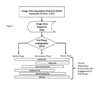
[00029] Figure 1 is a block flow chart diagram of how the Image Data
Acquisition Protocol (IDAP) and the
Image Data Sequence (IDS) are critically linked to Full Phase Angiography
(FPA), in accordance with various
embodiments of the present invention.
[00030] Figure 2 is an illustration of ICG Fluorescence imaging full phase
angiography EPA) in the cardiac
application, in accordance with various embodiments of the present invention.
[00031] Figure 3 illustrates full phase angiography EPA) in the GI surgery
application, in accordance with
various embodiments of the present invention.
[00032] Figure 4 illustrates full phase angiography (FPA) in the esophageal
surgery application, according
to various embodiments of the present invention.
[00033] Figure 5 illustrates the full phase angiography EPA) in the context
of breast reconstruction
surgery in accordance with various embodiments of the present invention.
[00034] Figure 6 shows a definition of FPA using an average intensity vs.
time curve. Figure 6 is an
idealized FPA curve indicating the necessary parameters to determine the three
phases (arterial,
microvascular and venous), in accordance with various embodiments of the
present invention.
[00035] Figure 7 is a block flow diagram of how different CAAs rely on the
same Combined Angiography
and Perfusion Analysis (CAPA) core platform, in accordance with various
embodiments of the present
invention.
[00036] Figure 8 illustrates the combined analytical components (baseline
correction, synchronization
accuracy check, angiography characteristic assessment, and dynamic perfusion
comparison) that are part of
the CAPA core platform, in accordance with various embodiments of the present
invention.
[00037] Figure 9 illustrates in the cardiac application the 'aortic root
shot," identifying an air bubble in a
bypass graft attached to the ascending aorta, in accordance with various
embodiments of the present
invention.
[00038] Figure 10 shows the Coronary Bypass Graft Image Protocol (CBGIP)
according to various
embodiments of the present invention.
[00039] Figure 11 is an illustration of the CAW (clinical application
window) and CAWT (CAW target) as
applied to a variety of CAAs identified thus far, in accordance with various
embodiments of the present
invention.
[00040] Figure 12 is an illustration of a method for Saturation Correction,
according to various
embodiments of the present invention.
CA 02914778 2015-12-08
WO 2013/190391 PCT/1B2013/001934
[00041] Figure 13 is an illustration of the static vs. dynamic analytical
approach, in accordance with various
embodiments of the present invention.
[00042] Figure 14 illustrates a method for the formatting the comparative
display of perfusion, applied to a
segment of bowel pre and post operatively, in accordance with various
embodiments of the present invention.
[00043] Figure 15 is an illustration of a method for synchronization
according to peak fluorescence
intensity, in accordance with various embodiments of the present invention.
[00044] Figure 16 illustrates a method for Fluorescence Baseline
Correction, in accordance with various
embodiments of the present invention.
[00045] Figure 17 is an illustration of one type of Complex Angiography
Analysis, namely, competitive flow,
in accordance with various embodiments of the present invention.
[00046] Figure 18 illustrates another type of Complex Angiography Analysis,
namely collateral flow, in
accordance with various embodiments of the present invention.
[00047] Figure 19 is an illustration that compares perfusion visualization
with the NIR B & W (left), a
standard RGB (middle), and the perfusion visualization scheme (right) used as
part of the CAPA core analysis
platform, in accordance with various embodiments of the present invention.
[00048] Figure 20 illustrates the Overview Display as used in the cardiac
application, in accordance with
various embodiments of the present invention.
[00049] Figure 21 A-D illustrates the CAPA core platform report format in
accordance with various
embodiments of the present invention.
[00050] Figure 21, Panel A shows the Overview Display of the synchronized
IDSs in standard color display
(both angiography and perfusion), in accordance with various embodiments of
the present invention.
[00051] Figure 21, Panel B includes all the analysis results in accordance
with various embodiments of the
present invention.
[00052] Figure 21, Panel C is the Quality Report for the data and analysis
in accordance with various
embodiments of the present invention.
[00053] Figure 21, Panel D offers an explanation for the different
perfusion comparison results as shown in
Panel B in accordance with various embodiments of the present invention.
[00054] Figure 22 illustrates one application of the cumulative and
additive presentation capabilities of the
CAPA analysis and display in accordance with various embodiments of the
present invention.
DETAILED DESCRIPTION OF THE PREFERRED EMBODIMENTS
[00055] Embodiments of the present invention will now be described more
fully hereinafter with reference
to the accompanying figures, in which preferred embodiments of the invention
are shown. The invention may,
however, be embodied in many different forms and should not be construed as
limited to the embodiments set
forth herein.
6
CA 02914778 2015-12-08
WO 2013/190391 PCT/1B2013/001934
[00056] The terminology used herein is for the purpose of describing
particular embodiments only and is
not intended to be limiting of the invention. As used herein, the singular
forms "a", "an" and "the" are intended
to include the plural forms as well, unless the context clearly indicates
otherwise. It will be further understood
that the terms "comprises" and/or "comprising," when used in this
specification, specify the presence of stated
features, integers, steps, operations, elements, and/or components, but do not
preclude the presence or
addition of one or more other features, integers, steps, operations, elements,
components, and/or groups
thereof. As used herein, the term "and/or" includes any and all combinations
of one or more of the associated
listed items. As used herein, phrases such as "between X and Y" and "between
about X and Y" should be
interpreted to include X and Y. As used herein, phrases such as "between about
X and Y" mean "between
about X and about Y." As used herein, phrases such as "from about X to Y" mean
"from about X to about Y."
[00057] Unless otherwise defined, all terms (including technical and
scientific terms) used herein have the
same meaning as commonly understood by one of ordinary skill in the art to
which this invention belongs. It
will be further understood that terms, such as those defined in commonly used
dictionaries, should be
interpreted as having a meaning that is consistent with their meaning in the
context of the specification and
relevant art and should not be interpreted in an idealized or overly formal
sense unless expressly so defined
herein. Well-known functions or constructions may not be described in detail
for brevity and/or clarity.
[00058] It will be understood that when an element is referred to as being
"on", "attached" to, "connected"
to, "coupled" with, "contacting", etc., another element, it can be directly
on, attached to, connected to, coupled
with or contacting the other element or intervening elements may also be
present. In contrast, when an
element is referred to as being, for example, "directly on", "directly
attached" to, "directly connected" to,
"directly coupled" with or "directly contacting" another element, there are no
intervening elements present. It
will also be appreciated by those of skill in the art that references to a
structure or feature that is disposed
"adjacent" another feature may have portions that overlap or underlie the
adjacent feature.
[00059] It will be understood that, although the terms first, second, etc.
may be used herein to describe
various elements, components, regions, layers and/or sections, these elements,
components, regions, layers
and/or sections should not be limited by these terms. These terms are only
used to distinguish one element,
component, region, layer or section from another element, component, region,
layer or section. Thus, a first
element, component, region, layer or section discussed below could be termed a
second element, component,
region, layer or section without departing from the teachings of the
invention. The sequence of operations (or
steps) is not limited to the order presented in the claims or figures unless
specifically indicated otherwise.
[00060] As will be appreciated by one of skill in the art, embodiments of
the present invention may be
embodied as a method, system, data processing system, or computer program
product. Accordingly, the
present invention may take the form of an embodiment combining software and
hardware aspects.
7
CA 02914778 2015-12-08
WO 2013/190391 PCT/1B2013/001934
Furthermore, the present invention may take the form of a computer program
product on a non-transitory
computer usable storage medium having computer usable program code embodied in
the medium. Any
suitable computer readable medium may be utilized including hard disks, CD
ROMs, optical storage devices,
or other electronic storage devices.
[00061] Computer program code for carrying out operations of the present
invention may be written in an
object oriented programming language such as Matlab, Mathematica, Java,
Smalltalk, C or C++. However,
the computer program code for carrying out operations of the present invention
may also be written in
conventional procedural programming languages, such as the "C" programming
language or in a visually
oriented programming environment, such as Visual Basic.
[00062] Certain of the program code may execute entirely on one or more of
a user's computer, partly on
the user's computer, as a standalone software package, partly on the user's
computer and partly on a remote
computer or entirely on the remote computer. In the latter scenario, the
remote computer may be connected
to the user's computer through a local area network (LAN) or a wide area
network (WAN), or the connection
may be made to an external computer (for example, through the Internet using
an Internet Service Provider).
[00063] The invention is described in part below with reference to
flowchart illustrations and/or block
diagrams of methods, devices, systems, computer program products and data
and/or system architecture
structures according to embodiments of the invention. It will be understood
that each block of the illustrations,
and/or combinations of blocks, can be implemented by computer program
instructions. These computer
program instructions may be provided to a processor of a general-purpose
computer, special purpose
computer, or other programmable data processing apparatus to produce a
machine, such that the instructions,
which execute via the processor of the computer or other programmable data
processing apparatus, create
means for implementing the functions/acts specified in the block or blocks.
[00064] These computer program instructions may also be stored in a
computer readable memory or
storage that can direct a computer or other programmable data processing
apparatus to function in a particular
manner, such that the instructions stored in the computer-readable memory or
storage produce an article of
manufacture including instruction means which implement the function/act
specified in the block or blocks.
[00065] The computer program instructions may also be loaded onto a
computer or other programmable
data processing apparatus to cause a series of operational steps to be
performed on the computer or other
programmable apparatus to produce a computer implemented process such that the
instructions which
execute on the computer or other programmable apparatus provide steps for
implementing the functions/acts
specified in the block or blocks.
8
CA 02914778 2015-12-08
WO 2013/190391 PCT/1B2013/001934
[00066] Figure 1 shows the relationship between FPA and existing ICG-NIR-FA
technology. In addition to
the IDAP and IDS links to the FPA, Figure 1 illustrates the FPA phase
components of Arterial, Microvascular
(Perfusion), and Venous Phases. Each of the currently known Clinical
Application Areas (CAA) engages a
minimum of two of these phases, illustrating the need for a dynamic analysis
of FPA to assess both
angiography and perfusion. In addition, the FPA characteristics specific for
that CAA acts as a 'filter' for the
IDS video loops captured for analysis.
[00067] The IDSs produced are DICOM or AVI video loops of variable
duration, depending upon the
Clinical Application of the imaging technology. The invention is applicable to
IDSs generated from ICG-NIR-
FA clinical and non-clinical research applications of the imaging technology
where arteriography and/or
perfusion assessment is important.
[00068] Figure 2 demonstrates the average intensity vs. time curve (one FPA
cycle) of the 34 sec
fluorescent angiography Image Data Sequence (IDS) video loop in the cardiac
context. These data are
fundamental to the Combined Angiographic and Perfusion Analysis (CAPA) core
analysis platform. Five
individual frames from the total of 1020 frames in the video loop are
illustrated to illustrate the phases (1 =
baseline, 2 = arterial phase, 3 = micro-vascular phase, 4 = venous phase, 5 =
residue of florescent dye). The
ECG (green tracing) and BP (red tracing) from the continuous 26 cardiac cycles
are shown.
[00069] Figure 3 illustrates FPA in the GI surgery context. Here, the three
FPA phases are shown as
follows: panels A-D are baseline background fluorescence; panels E-G are the
arterial phase; panel H is the
microvascular phase; panels I-L are the venous phase. These data are
fundamental to the Combined
Angiographic and Perfusion Analysis (CAPA) core platform. Shown is a segment
of large bowel being imaged
at the time of surgery, in the near-infrared 255 grey scale black and white
Overview Display format. The peak
of average fluorescence intensity for this Clinical Application Window (CAW)
is in panel H. Note the IDS in this
case is 45 seconds.
[00070] Figure 4 illustrates FPA in the esophageal surgical application. In
similar fashion to Figure 3, in
Figure 4 the peak of the average fluorescence intensity for this CAW is in
panel H. These data are
fundamental to the Combined Angiographic and Perfusion Analysis (CAPA) core
analysis platform. This
Overview Display uses the color scheme designed to highlight perfusion. The
image data from the IDS are the
same, however, regardless of the display presentation color scheme. Note the
IDS in this case is 16 seconds.
[00071] Figure 5 illustrates the FPA in the plastic surgery breast
reconstruction application. As in Figure
4, in Figure 5 the Overview Display uses the color scheme designed to
highlight perfusion, and again, the
peak of average fluorescence intensity for this CAW is in Panel H. These data
are fundamental to the
Combined Angiographic and Perfusion Analysis (CAPA) core analysis platform.
Note compared to Figure 2 -
9
CA 02914778 2015-12-08
WO 2013/190391 PCT/1B2013/001934
Figure 4, the venous phase of the FPA doesn't fall, suggesting venous
congestion in this breast
reconstruction. Note the IDS in this case is 35 seconds.
[00072] The modeling of a generic FPA, and the modifications for its
application to a specific CAA, is as
follows. The definition of FPA using an average intensity over time curve is
detailed in Figure 6.
[00073] Let B1 = the average baseline intensity before the arterial phase,
let P = the peak intensity, and let
B2 = the average baseline intensity after the venous phase. The Arterial phase
starting time is defined as
when the average intensity first increases to
B1 + (P ¨ B1) x k1 Equation 1
And the Arterial phase ending time is defined as when the average intensity
first increases to
B1 + (P ¨ B1) x k2 Equation 2
Where k1 is a percentage defining the beginning of arterial phase (e.g. 5%),
and k2 is a percentage defining
the ending of the arterial phase (e.g. 95%).
[00074] The Venous phase starting time is defined as when the average
intensity first decreases to
B2 + (P ¨ B2) x k3 Equation 3
And the Venous phase ending time is defined as when the average intensity
first decreases to
B2 + (P ¨ B2) x k4 Equation 4
Where k3 is a percentage defining the beginning of arterial phase (e.g. 95%),
and k4 is a percentage defining
the ending of the arterial phase (e.g. 5%).
[00075] The Micro-vascular phase is defined as when the average intensity
ranges between
B1 + (P ¨ B1) x k2 Equation 5
and
B2 + (P ¨ B2) x k3 Equation 6
nearby the peak.
[00076] The percentages will be somewhat different across different CAAs.
The collection and analysis of
clinical data is used to validate these percentages and to increase the
specificity of these percentage values
for each CAA utilization of the FPA 'filter.'
CA 02914778 2015-12-08
WO 2013/190391 PCT/1B2013/001934
[00077]
Figure 7 illustrates the Core Platform and all its component parts, including
the FPA, the
CAW/CAWT, the synchronization, and the analysis and results reporting. In
Figure 7, on the left side,
sequential IDSs are obtained, 'filtered' through the same FPA intensity vs.
time curve, synchronized, and
matched according to the same Clinical Application Window (CAW). This process
allows for a post- vs. pre-
comparison between two IDSs to quantify the perfusion change. On the right, a
single IDS in a different CAA
can be 'filtered' with the FPA, and within the same CAW two different targets
(CAWTs) (usually different areas)
can be compared after synchronization, using the same core platform. The
result output from the CAPA core
platform analysis is then formatted specifically for the appropriate CAA.
[00078]
Figure 7 illustrates how the FPA acts as a 'filter' for the IDS data in
particular CAAs. In some
CAAs, an angiographic and perfusion comparison is made by comparing two (or
more) sequential IDSs (left
side of diagram), as for example before and after coronary bypass grafting. It
is important that these two IDSs
be captured using the same Image Data Acquisition Protocol (IDAP), and are
'filtered' with the same, CAA-
specific FPA. Furthermore, the Clinical Application Window (CAW) for both
needs to be the same, that is, the
camera window and position of the camera (CAW) needs to be consistent between
the two IDSs. This
illustrates the need for a detailed and specific IDAP, since this CAW
application cannot occur accurately if the
IDAP generated two IDSs with different CAW information. More importantly, in
the next step the core CAPA
analysis cannot be reliably executed and a quantitative analysis comparison
performed if this CAW isn't
equally applied to both IDS + FPA datasets.
[00079]
Figure 7 also shows a different CAA on the right, where comparative
angiography and perfusion
information is derived from a single IDS (such as the GI CAA). In this
instance, the CAA-specific FPA 'filter'
information is applied to two or more Clinical Application Window Targets; a
target can be a specific area or
region of the CAW (see Figure 11), and can be manually selected or
automatically selected by the FPA, and
then analyzed with the platform.
[00080]
Importantly, the IDS synchronization step occurs before the CAW/CAWT step, to
avoid comparing
data that are inadequate for analysis.
[00081]
The Results of the analysis are reported in a format that is most applicable
to the specific CAA, to
assist the surgeon with new, real-time information in the operating room with
which to make better decisions
and decrease the incidence of complications.
[00082]
Figure 8 illustrates the unique attributes of this analysis platform. These
include: 1) baseline
correction algorithm; 2) synchronization validation; 3) saturation correction;
4) CAW/CAWT component
application(s); 5) angiography analyses (where applicable); and 6) the dynamic
and quantitative perfusion
comparison(s).
Importantly, this CAPA is a dynamic, as opposed to static, analysis platform,
accurately
reflecting the underlying physiology as captured in the FPA construct. It
contains in addition the following
11
CA 02914778 2015-12-08
WO 2013/190391 PCT/1B2013/001934
attributes: 1) a dynamic analysis of both angiography and perfusion in the
same construct; 2) real-time,
intraoperative image analysis capabilities, based on unmodified image data
captured with the ICG
Fluorescence system; 3) built-in image data quality checks and evaluation
processes with which to frame the
analysis results; 4) image and analytical results displays that reflect the
concept and principles of FPA as
critical to understanding and visualizing the underlying physiology being
studied and evaluated during these
surgical procedures; and 5) real-time 2-D and 3-D displays of the analyzed
data for rapid, visual-based
documentation of the analytical results, some in the format of a dynamic
movie. Additionally, the CAPA
analysis and display can be used for new and technologically-sophisticated
information documentation in
healthcare. This includes information sharing among healthcare professionals
and with patients and their
families, in which the dynamic visualization of the revascularization and/or
devascularization conditions of the
surgical procedure can be displayed. In addition, this CAPA infrastructure
creates the opportunity for
longitudinal analysis of the metadata contained in the analyzed information.
[00083] In Figure 8, some of these combined analytical components (baseline
correction, synchronization
accuracy check, angiography characteristic assessment, and dynamic perfusion
comparison) are used across
all CAAs; others are specifically emphasized for other CAAs because of the
underlying physiology being
imaged. The IDS Quality check was placed post-analysis, so as not to place the
surgeon in the position of
having no analysis generated following data acquisition; however, if the
IDS(s) do not meet the data quality
checks, assuring that the IDAP was adhered to and that other physiological
conditions were met as well, the
Report will contain and Error Warning indicating that the following image
quality metrics were not met.
[00084] Fluorescence angiography relies on low-energy, NIR laser excitation
of ICG in blood vessels and
perfused tissues, with capture of the intensity of fluorescence based upon the
ICG infrared absorption and
emission spectra. Importantly and in addition, imaging and its interpretation
are influenced by a number of
physiologic and/or pathophysiologic circumstances. The imaging data are
captured as standard AVI and/or
DICOM video loops at 30 fps, which can be directed imported into the core
analytical platform. These
standard image formats make the analytical platform widely applicable from a
technical perspective. The
frame rate was accounted for in the development of the CAPA core platform, as
it limits the fidelity of the
image analysis. An example of this is shown in Figure 21 A, where the
"movement" in the images on the
Display results from the movement of the heart exceeding the frame rate of the
camera at that point in the IDS
video.
[00085] The known behavior of ICG dye in the blood has established that on
the first pass through the
heart, the fluorescence intensity is proportional to the concentration of ICG,
which in turn is directly related to
the injected dose. This allows for tailoring of the ICG dosage/injection for
specific Clinical Application Areas
and procedures within those areas. Importantly, this behavior also creates the
possibility of fluorescence
saturation, where the quantification of the intensity exceeds the 0-255 scale.
This creates a problem of being
12
CA 02914778 2015-12-08
WO 2013/190391 PCT/1B2013/001934
unable to quantify how much greater than 255 the actual fluorescence intensity
actually is; this is particularly a
problem in other ICG-NIR-FA analysis approaches. As demonstrated, the CAPA
analysis accounts for
saturation correction when it does occur.
[00086] The known behavior of ICG dye as a bolus injection, with or without
a saline flush, allows for
specific detailing of how the ICG injection should be administered in order to
optimize image quality. This
understanding has specific importance in those CAAs where the angiography
analysis is of particular
relevance. The ICG bolus stays relatively undispersed as it passes through the
central cardiac circulation, and
ultimately out to the peripheral tissue microvasculature. Even at this
anatomic location extremely distant
physiologically from the heart, the FPA and its phase components can be
readily identified in the ICG-NIR-FA
IDS sequences. This documented discovery creates the opportunity to establish
the CAPA core platform as
an independent claim applicable across all ICG-NIR-FA applications involving
angiography and perfusion.
Now and in the future, supplemental analytical components that are specific to
the existing and new CAAs can
and will be developed as dependent claims.
[00087] The known behavior of ICG dye in blood and in circulation is
fundamental to this imaging
technology and analysis. ICG binds to the circulating proteins in serum, and
to endothelial proteins attached
to the inner surface of arterial and venous blood vessels. The half-life of
ICG in humans is about 3 minutes,
and the dye is metabolized by the liver and excreted in the kidney. Because
the surface area on the venous
side of the circulation is so much greater than the arterial side, there is
more endothelial binding on the venous
side, creating residual fluorescence, which typically is 'washed out' in 4-5
minutes after an injection. As
demonstrated, our discovery and analysis of FPA, however, led to the
understanding of how to deal with
residual, background fluorescence in a physiologically-accurate manner that
meets the time frame for this
imaging technology to be adopted and used clinically by surgeons during
complex operative procedures.
[00088] As with any imaging technology, image data acquisition is key to
sustained, successful analysis
across multiple providers in multiple settings. The standardization of these
image acquisition parameters for
each Clinical Application Area is critical for the analysis claim of the
invention to be used appropriately and for
the results to be used accurately in the clinical setting. As related to the
invention, it is critically important that
the image acquisition process for each CAA enables the complete capture of the
FPA information, which is, as
demonstrated, a key component for the CAPA platform analysis of angiography
and perfusion in that CAA,
and that surgical procedure.
[00089] We have defined the term Image Data Sequence (IDS) as the captured
video loop with all the
imbedded metadata. This IDS may be of variable duration, depending upon the
application. As shown in
Figure 7, the use and management of the IDS is specific for each CAA.
13
CA 02914778 2015-12-08
WO 2013/190391 PCT/1B2013/001934
[00090] We have defined the term Image Data Acquisition Protocol (IDAP) as
the specific, step-by-step
process of coordinated capture of the IDS. This includes: 1) machine setup and
positioning of the field of view,
specific to the application and procedure; 2) the dosage, administration route
and timing of administration of
the ICG fluorescent dye coupled with management of the data capture software
on the ICG Fluorescence
machine; and 3) any specific technical, clinical or hemodynamic management
processes necessary for
optimization of the IDAP.
[00091] In addition, there are specific subset applications of the IDAP,
depending upon the relative
predominance of the arterial, microvascular and venous phases in that
particular CAA and surgical procedure
application. In these cases, the IDAP needs to be designed and executed so as
to assure the time frame of
data capture encompasses the necessary FPA spectrum. For example, in a CAA
that is dependent upon the
arterial phase, starting the video capture without a stable baseline makes a
comparative analysis unfeasible.
Similarly, truncating the video capture, or moving the machine, or shining the
surgeon's headlight into the field,
before the necessary venous phase information is captured creates an
analytical problem. The specific IDAP
must reflect a very real understanding of the FPA, its principles, and the
CAPA platform.
[00092] In certain CAAs, specific IDAPs are developed for imaging purposes
specific to either angiography
or perfusion. For example, in the cardiac application, at the end of the
revascularization procedure, with the
heart in the anatomic position in the mediastinum, the 'aortic root shot" is
obtained, to illustrate flow and
subjective rate of flow down the graft conduits, and to assess the anastomoses
constructed to the ascending
aorta, and to identify subtle technical issues (air bubble, low flow rate vs.
other grafts) (Figure 9).
[00093] As is demonstrated in Figure 9, this bubble could not have been
recognized without ICG-NIR-FA
imaging, and was aspirated before it could embolize down the bypass graft to
the heart and cause heart
damage.
[00094] Also in certain CAAs, intraoperative techniques have been developed
to specifically facilitate IDS
capture in a framework that enables subsequent analysis. For example, in the
cardiac application, we have
determined that the most reliable approach to consistent angiography and
perfusion analysis is the following
Coronary Bypass Graft Image Protocol (CBG IP) (Figure 10). In Figure 10, the
CBG IP sequence consists of:
a) graft anastomosis construction; b) first IDS acquisition with a soft-jawed
clamp on the bypass conduit ("dog
on") to assess visually native coronary flow and perfusion to confirm that the
native circulation has not been
interrupted by the anastomosis, reflux up the bypass conduit as an index of
anastomotic patency, and any
other technical issues (air bubble, dissection flap in epicardial coronary
artery); c) saving the first IDS image
with removal of the soft-jawed clamp from the graft conduit; then d) second
IDS acquisition with both the native
coronary flow and graft flow together. In this way, all of the following FPA-
derived and related important
information can be captured by adhering to the IDAP, CAA-specific protocol: 1)
visual assessment of evidence
14
CA 02914778 2015-12-08
WO 2013/190391 PCT/1B2013/001934
for adequate flow down the conduit; 2) the presence of competitive flow
between the native and graft conduits;
3) the briskness of washout of ICG-blood from the graft conduit; 4) any other
technical issues (air bubble, poor
outflow, dye 'hang up' at the anastomosis); and 5) the subsequent CAPA
platform analyses.
[00095] Figure 10 illustrates the important connectivity between the
present invention(s) of the FPA and
CAPA analysis platform, and the methodology for collecting the ICG-NIR-FA
image data. These two
processes must be aligned by the clinical/experimental providers to optimize
the accuracy and fidelity of the
analytical and display results, as is the case with any imaging and analytical
technologies.
[00096] As shown in Figure 1 and Figure 7, the CAPA platform extends across
all the CAAs identified thus
far. Moreover, since the principle of FPA embodiments has been identified in
all applications of ICG-NIR-FA
thus far studied, we expect that it will apply to any ICG-NIR-FA application
area where angiography and
perfusion are critically important. The dynamic and flexible nature of the FPA
in this context is reflected in
various embodiments of the embodiments in the present invention(s).
[00097] We define the image area to which the FPA 'filter' intensity vs.
time curve is applied as the Clinical
Application Window (CAW), and/or to a sub-set of this window, termed the
Clinical Application Window
Target (CAWT).
[00098] Figure 11 is an illustration of the CAW and CAWT as applied to a
variety of CAAs identified thus
far. As shown in Figure 11, the CAWT can be selected automatically (as in
cardiac by the analysis algorithm)
or manually.
[00099] This CAW is the area of clinical interest for imaging, and will be
variable from application to
application, but as shown in Figure 7 the core CAPA platform uses information
from this CAW to further define
the parameters of the analysis beyond the FPA 'filter,' and to make sure that
the comparisons being made are
accurate and reflective of the underlying physiology.
[000100] The CAWT can be individual image pixels in a CAW, a certain selection
and/or identified grouping
of pixels, or an anatomic subset of the CAW as defined by the clinical
application. The target can be manually
selected, or automatically computer generated. The physiology of arterial flow
and perfusion predicts that
different CAWTs will, at any point in time, have different intensity vs. time
curve characteristics.
[000101] Because the opportunity inherent in FPA and the CAPA is a dynamic
analysis that reflects
physiology, an important observational finding present in all CAAs studied
thus far and critical for the analytical
platform is that the predominant blood supply source engages the tissue being
imaged be identified. This
allows identification of a proximal (nearest to the blood supply origin) and a
distal end (farthest away from the
proximal end). The perfusion analysis must account for the entirety of the
arterial and micro vascular phases
in real time rather than just a single static frame from the image sequence.
As mentioned, if the CAWT is
CA 02914778 2015-12-08
WO 2013/190391 PCT/1B2013/001934
defined as a certain selection and/or identified grouping of pixels in a CAW,
during a single ICG injection that
selection/grouping of pixels image arterial, micro-vascular and venous phases
of full phase angiography. For
that pixel CAWT and for the CAW as a whole, the image characteristics are very
different from phase to
phase. Since adjacent CAWT will have different characteristics, these
differences in intensity and time can be
used to derive comparative and contrasting data throughout the CAW.
[000102] Due to the limitations of 8-bit cameras, the intensity of
fluorescence measurement in any IDS is
limited to 255. At times, based on physiological or pathophysiologic
circumstances, the same dose and
concentration of ICG dye could in theory create saturation (intensity > 255)
in the IDS for part of the sequence.
This saturation effect has been observed, especially with multiple injections,
and this might jeopardize the
accuracy of the perfusion comparison. To address this, we created an algorithm
to estimate t ear intensity of
the saturated pixels from the image histogram and approximate their
distribution above intensity 255 by
estimating the distribution of the pixels with intensity smaller than 255.
Their geographical locations can be
also estimated using non-saturated frames previous to the saturated frame.
[000103] Figure 12 illustrates the method for saturation correction. In this
figure, the blue color curve is the
histogram of a saturated still frame and the red color curve is the estimated
intensity distribution of the
saturated pixels.
[000104] In Figure 13, for this example, the fluorescence progresses from left
to right of this large bowel
IDS. The same IDS and data are shown in both panels (note the intensity vs.
time curves). The CAW is the
segment of large bowel, and the CAWTs are each of the green linear points
along the long axis. The blue line
is the intensity vs time curve for the red reference point at the extreme
left; the red line is the intensity vs time
curve for the farthest right green box. The static black line (at 33 sec on
the top panel, and at 41 sec on the
bottom panel) represent what would be 'static snapshots' taken at these two
points in this dynamic imaging an
analysis process. At the 33 second mark on the top panel, the fluorescence
wavefront has reached the left
part of the bowel (blue curve red curve) so the intensities of the right
side the bowel are smaller compared
to the left side. At the 42 second mark on the bottom panel, the fluorescence
wave front has passed the left
part of the bowel and reached right part (red curve blue curve); the
fluorescence intensities of the left side
are now relatively smaller compared to the right side.
[000105] The same imaged segment of large bowel is analyzed to emphasize this
point. The bowel
segment takes 12 seconds to perfuse the left-sided CAWT reference point (red
box) to the CAWT point on the
far right. The blue curve is the intensity vs time curve for the left-sided
CAWT, and the red curve is the right-
sided CATW. In the top panel, if a static reference point is chosen (black
line at 46 sec), then the red CATW is
higher than the blue CATW, reflected by the normalized percentage of 156% for
this point. However, on the
bottom panel, if the reference point is chosen at the 32 second point, a
completely different normalize result
16
CA 02914778 2015-12-08
WO 2013/190391 PCT/1B2013/001934
occurs, despite the fact that the same blue CATW reference was used in both
analyses. The visual
appearance of the dynamic image sequence is dependent upon these physiologic
arteriography and perfusion
characteristics, depending on which part of the tissue the fluorescent wave
front will reach first.
[000106] Only by synchronizing these CAVVT curves by some parameter (time,
distance) can the perfusion
of different part of tissue can be quantified and validly compared in a
dynamic manner. Figure 14 illustrates
this same principle with the analytical output from the CAPA. As shown in
Figure 14, on the upper Left are the
average intensity vs. time curves; as applied to this GI large bowel
evaluation, there is an 11-sec delay
between the CAWC on the lower Left panel (blue oval) and the CAWC on the right
panel (red oval), pre-
synchronization. In this case, the fluorescence intensity of the CAWC on the
right panel is less than 50% of
the CAWC on the left panel, as shown by the green line in the upper right
panel. This is also displayed by the
relative perfusion bar data in the upper middle, where the 'post-graft' right
panel of 0.42 is compared to the
normalized value of 1 for the left (pre-graft) value.
[000107] Also as shown here in Figure 14, at first glance there appears to be
a substantial difference
between these two CAWTs in this bowel segment, where the 'quantified
perfusion' to the right (red) CATW is
0.42, compared to the normalized value of 1.0 for the left CAVVT. Because this
analysis result didn't include
the synchronization step, however, these results are invalid. Our definition
of FPA provides the basis to
synchronize the ICG dye fluorescence peak in different parts of the tissue, at
different times and combinations
of arterial microvascular, and venous phases of angiography and perfusion, as
appropriate.
[000108] Therefore, for a valid perfusion comparison, the corresponding phases
have to be accurately
aligned by a common parameter, whether the comparison is between different
IDSs with the same CAW, or
between different CAWCs within the CAW, derived from a single IDS (Figure 7).
This synchronization of
phases, possible only with the recognition of the multiple phases in the FAP
embodiments. Importantly, this
recognition and incorporation of the FPA embodiments also greatly improves the
visual display as well as the
analysis.
[000109] An illustration of a method for synchronization is shown in Figure
15. This figure is an illustration
of using the FPA cycle average intensity vs. time curves to synchronize two
IDS obtained with the appropriate
IDAP. The blue curve is pre-grafting, while the red curve is post-grafting.
Synchronization is based on the
peak fluorescence intensity for the CAW, or for each component of the CAW. Top
panel: average intensity
vs. time curves of Pre (blue) and post (red) IDSs before synchronization;
bottom panel: average intensity vs.
time curves of Pre (blue) and post (red) IDSs after synchronization.
[000110] The effect of curve synchronization impacts on both analysis and
display components of CAPA.
Using average intensity vs. time curves, a correlation coefficient is
calculated at each alignment time position
and the largest correlation coefficient yields the optimal synchronization
result. The extra segments in the
17
CA 02914778 2015-12-08
WO 2013/190391 PCT/1B2013/001934
beginning and/or end of the IDSs will be truncated.
Therefore a fundamental principle of this present
invention(s) is that the intensity vs time curve is the basis for
synchronization of the phases of F PA.
[000111] This venous residual creates the need to account for residual
fluorescence in any type of
comparative analysis. In this core analytical platform, we define the baseline
as described in Figure 6 and the
present disclosure. The management solution inherent in the CAPA platform
allows for accounting of the
residual fluorescence when sequential injections are compared, and/or when
multiple injections are used
during a procedure. Moreover, this solution allows for the data capture and
analysis to be performed in a time-
frame that is critical for surgeons collecting image data in real time during
complex surgical procedures.
[000112] During multiple ICG-NIR-FA dye injections, the residue of dye
accumulates and images acquired
later tend to be brighter than the previous ones, mostly due to binding in the
venules.
[000113] To investigate how residue of fluorescent dye from the previous
injection affects intensity of the
current IDS, we performed multiple sequential, paired IDSs without any change
to the tissue or position of the
camera. Since these two IDSs are recorded under same physiologic and CAW
conditions, by studying their
average intensity vs. time curves the optimal baseline management strategy was
developed.
[000114] In Figure 16, the top panel is an illustration of average intensity
vs. time curves of the pre (blue
color) and post (red color) fluorescence IDSs. The bottom panel is an
illustration of baseline difference
between average intensity vs. time curves of the pre and post IDSs.
[000115]
Importantly, from Figure 16 we can tell that baseline difference between two
CAWs/ CAWTs is
not constant across the IDS acquisition window. As the FPA average intensity
vs. time curve is increasing and
approaching the peak intensity, the baseline difference keeps decreasing.
Based on these observations, we
use Equation 7 to estimate the change of the baseline fluorescence intensity
difference over the IDS time:
.1A1Cpõt(0)
BD (x, y, t) = C(x, y) x, _____________________________________ Equation 7
VA/Cpõt(t)
Where BD is the baseline difference between pre and post IDSs with x, y as
pixel coordinates and t as time;
C(x, y) is the constant background difference between pre and post images
estimated from the first few
õ./Aicpost(0)
seconds of the IDSs; A/Cpost (t) is the average intensity curve of the post
image acquisition and
õ/Arcpost(t)
is used to adjust the baseline difference across time. From Figure 16 we can
tell that treating the baseline line
difference as a constant will lead to "over subtraction" causing loss of
useful signal from the post image
acquisition sequence.
18
CA 02914778 2015-12-08
WO 2013/190391 PCT/1B2013/001934
[000116] Examples of two important novel paradigms are documented herein.
These are 1) the ability to
recognize and document arterial-phase competitive flow between native and
grafted sources of blood flow
under physiologic conditions, and 2) the ability to recognize microvascular-
phase collateral flow in adjacent
and/or related areas of perfused tissues.
[000117] In Figure 17, visual documentation of competitive flow between a
native epicardial coronary artery
and a patent bypass graft to that artery, beyond what was thought to be a flow-
limiting stenosis is presented.
The physiology-based and dynamic analysis using the FPA embodiment makes the
documentation of
competitive and potentially-significant competitive flow identification at
CABG a reality for the first time.
[000118] Competitive flow is currently most appropriately understood in the
context of the arterial phase of
FPA, although extension into the microvascular phase is being examined. Figure
17 shows documentation of
competitive flow in man in real time at CABG. This figure clearly illustrates
the reversal of flow between the
native coronary and the widely patent bypass graft in early and late systole
that is diagnostic of competitive
flow. In these sequential frames from the IDS separated by 24 sec intervals,
there is washout of the ICG +
blood in the native coronary by the blood without ICG from the graft; the
competition also causes the ICG +
blood to ref lux back across the anastomosis into the distal end of the bypass
graft. This is new and very
important information to now have available in real time, at the setting of
surgical revascularization.
[000119] In, Figure 18 visual documentation and quantification of the effect
of collateral flow in the heart as
a result of bypass grafts and increases in perfusion to territories supplying
the collateral flow, is presented.
The top panel shows the comparison of the two, sequential IDSs, pre-grafting
(left) and post-grafting (right).
The bottom panel is the quantification display (see Figure 21 A for full
explanation of the display). Note, in
this case there was a 2.5-fold increase in the inferior wall of the heart as a
result of bypass grafts placed to the
anterior and lateral walls. The ability to use ICG-NIR-FA to capture and then
to analyze these images to
document in real-time this collateral flow is dependent upon the FPA
embodiment.
[000120] Collateral flow is currently most appropriately understood in the
context of the microvascular phase
of FPA. Again the cardiac application is used as an example, in part because
the heart is typically able to
develop collaterals with non-acute, regional occlusions of the blood supply to
a territory of the heart. Figure
18 shows collateral flow imaged in real time in man at CABG. The top panel
shows the same CAW from two
sequential IDSs; the CAW is imaging the inferior wall of the heart, before and
after placing bypass grafts to the
anterior and lateral walls of the heart. The left panel images the native
coronary perfusion to the inferior wall
(with the grafts temporarily occluded), while the right panel images the
inferior wall, with the grafts to the
anterior and lateral walls open and perfusing their respective territories.
Visually, there is a substantial
increase in fluorescence and hence perfusion to this inferior wall as a result
of these bypass grafts; this
increase in perfusion comes from collateral flow from the anterior and lateral
territories to the inferior territory in
19
CA 02914778 2015-12-08
WO 2013/190391 PCT/1B2013/001934
this patient's heart. The bottom panel shows the CAPA platform analysis and
quantification of the perfusion
difference before and after bypass grafting. There was a 2.5-fold increase in
perfusion to the inferior wall as a
result of this collateral perfusion increase. This is new and very important
information to have available in real
time, at the setting of surgical revascularization.
[000121] The CAPA perfusion quantification is a relative measurement based on
a comparison, as
illustrated in Figure 7 and Figure 8. To increase the sensitivity of the
analysis results, only pixels with
intensity above certain value are used to estimate relative perfusion. The
still frame located at the peak of the
average intensity vs. time curve in one IDS is used to determine this
threshold by
k = mean(Imax) + m x std(Imax) Equation 8
Where Iõõ is the still frame that has the maximum average intensity in one
IDS; mean is the average
function; std is the standard deviation function; m is a constant parameter
between 0-1 to adjust this
Equation 8. The threshold k is used in one or several IDSs depending on the
application and only pixels with
intensity above the value are used in the perfusion calculation.
The arterial phase of IDS records perfusion as a process of blood being
delivered by arteries to the tissue.
Correspondingly, this process starts from the beginning (baseline part) to the
peak (maximum) of the average
intensity vs. time curve. Visually, this process includes arterial and part of
micro vascular phases in the IDS.
We are assuming not only the 'perfusion strength" (corresponds to the average
intensity above the threshold)
but also the 'perfusion area" (corresponds to the number of the pixels with
intensity above the threshold)
should be included in estimation of the perfusion level. Equation 9 is applied
in all the still frames of the IDSs
till the maximum of the average intensity curve is reached.
Al (t) = Num(I (x, y, t) > k) x mean(I (x, y, t) > k) Equation 9
Where Al is a number representing combination of perfusion strength and area
at time t. 1(x, y, t) is a still
frame at one time location of an IDS; Num is the function to calculate the
number of pixels; mean is the
average function.
Then we estimate the accumulation effect of the Al (t) from the beginning
(baseline part) to the peak
(maximum) of the average intensity vs. time curve as
T
AI(T) = 1[AI (t) ¨ Al (0)] Equation 10
0
Where T is any time at the peak (maximum); Al (0) is the residue from
baseline. In the cardiac application we
calculate this area-intensity value in sequential IDSs of the same CAW tissue
area. In other CAAs identified
CA 02914778 2015-12-08
WO 2013/190391 PCT/1B2013/001934
thus far, we calculate this area-intensity value relatively across two or more
CAWTs identified in one CAW
identified in one IDS.
Notice that this is a relative value in both cases, and it does not reflect
the estimation of perfusion directly. In
the cardiac application, to estimate the perfusion change, we normalized the
post area-intensity value by the
pre one by
AI (T)0
Al = Equation 11
AI (T) põ
In the other CAAs identified thus far, to estimate the perfusion change, we
normalize the current CAVVT by the
reference CAWT
Al = Al(T)
.- ,CAWT-current
Equation 12
Al (T)CAWT-ref
[000122] The opportunity inherent in FPA and CAPA extends to image and image
analysis display. The NIR
part of the spectrum is outside the visible color spectrum, and therefore is
inherently a black and white, 255-
level grey scale image. This is actually quite sufficient for imaging the
arterial phase of full phase imaging, but
is not optimal or optimized for microvascular or venous phase imaging. We have
developed different color
schemes to optimize the display for combined (arteriography and perfusion)
display using a modified RGB
format, and for the microvascular (perfusion) image display and analysis. This
in turn means that in many
CAAs combination of displays of the same NIR image data is optimal for
understanding the context and
content of the image(s) and analyses for decision-making.
[000123] As illustrated in Figure 19, our experience has demonstrated that the
NIR is more optimized for
angiography, the RGB presentation is more optimized for BOTH angiography and
perfusion, and the BI-Y-R-
G-B-W display is optimized for perfusion. On the top panel is shown a segment
of large colon. On the bottom
panel is shown is a segment of stomach used to create a neo-esophagus in the
esophageal application (same
as Figure 4).
[000124] Figure 19 also shows the comparison of these three displays. It is
important to understand that
these displays all render the same image metadata; the NIR B & W is the 'raw'
NIR presentation; the same
image data are simply colorized according to the different 0-255 scales,
optimized for combined (arterial and
perfusion) and microvascular (perfusion) presentation and display.
Specifically the perfusion display range is
black, yellow, orange, red, green, blue and white for intensity of
fluorescence ranging from 0 - 255.
Comparably, the NIR grey scale and other RGB-based ranges are too narrow
between the low and high
intensities that they are not visually sensitive enough to reflect the subtle
but important perfusion changes.
21
CA 02914778 2015-12-08
WO 2013/190391 PCT/1B2013/001934
[000125] We also designed an Overview Display as a unique way to visualize the
IDS + FPA data. In
Figure 20, this Overview Display compares pre-grafting perfusion with post-
grafting perfusion, after
synchronization of the two IDSs. Panel H in each sequence again reflects the
peak average intensity in the
two CAWs, which by design and by the Image Data Acquisition Protocol (focusing
on both Angiography and
Perfusion) used in cardiac, image the same area on the anterior perfusion
territory of the heart. In this case,
an internal mammary artery was grafted to the left anterior descending
coronary artery. Note the obvious
increase in fluorescence intensity in the panel H post-grafting (bottom)
compared to pre-grafting (panel H, top).
The quantified difference in fluorescence intensity is directly proportional
to the difference in myocardial
perfusion.
[000126] However, as previously articulated, to visually capture the inference
of the FPA and CAPA
construct requires that two points can be accurately compared. As depicted in
Figure 13 and Figure 14,
(colon), however, we CANNOT use time alone to establish this comparison.
Therefore this Overview Display
uses the same IDS synchronization described above to accurately provide this
intuitive visual comparison. The
frame in the red box (labeled H) represents the peak intensity on the average
intensity vs. time curve, which
corresponds to the micro vascular phase. The frames before it (labeled from A
to G) are the baseline and
arterial phase and the frames after it are the venous phase and the
fluorescent dye residue; each frame is
separated by 1.5 sec from the peak, in either direction. This display is
physiologically organized, and because
of the synchronization technique is possible to reliably make visual
comparisons to accompany the CAPA
platform analyses. This same principle is used in the analysis display.
[000127] Figure 21, panels A-D, show the display format as applied to the
cardiac CAA. There are four
components to the analysis presentation. Panel A comes up first, and is the
Overview Display discussed
above. Panel B is the Quantified result display.
[000128] In Figure 21, Panel A is the Overview Display of the synchronized
IDSs in standard color display
(both angiography and perfusion). The pre images are in the upper panel and
post images are in the lower one
(see synchronization section for details). Compare the fluorescence intensity
in the panels labeled H, top vs.
bottom. There is visually much more fluorescence intensity post-grafting than
pre-grafting to the perfused
territory supplied by this grafted vessel on the anterior wall of the heart.
[000129] Figure 21, Panel B, includes all the analysis results. In the upper
left hand corner are displayed
the synchronized average intensity vs. time curves with time line indicating
the peaks. The left and right bottom
panels correspond to the colorized pre- and post- images at the peak of the
curves with time labels on the
upper left hand corners. Note these time labels are identical, indicating the
time synchronization between the
pre- and post- images is based on peak intensity, even though the image
sequences are not synchronized
based on the cardiac cycle. The two bars on the upper panel are calculated
from the accumulated area and
22
CA 02914778 2015-12-08
WO 2013/190391 PCT/1B2013/001934
intensity curves. The pre-graft perfusion status is represented by the blue
colored bar, which is always
normalized to one for comparison to the post graft perfusion status,
represented by the red color bar. To better
illustrate the quantification of change in perfusion over time, and to
illustrate the contribution of the bypass
graft, the perfusion changes over time are generalized in the chart at the
upper right hand corner with blue, red
and green color curves representing the accumulated perfusion changes over
time caused by native, native
plus graft and bypass graft respectively. Also shown in Panel B is the final
quantification result at 13.4 sec,
which is the time point of peak fluorescence. Finally, the pre-graft perfusion
level is normalized to 1, for
comparison to the post-graft perfusion (in this case, 1.28) in a bar chart
format.
[000130] Figure 21, Panel C is the Quality Report for the data and analysis.
This includes all the quality
criteria that each IDS is subjected to in order to further support and
validate the CAPA results. If there is an
IDS quality issue, the error warning message displays on this page and on Page
B as well, to avoid mis-
interpretation of the results.
[000131] Figure 21, Panel D provides Explanation data, including Error Warning
feedback on the Data
Quality check.
[000132] An additional opportunity inherent in the FPA and CAPA invention is
to analyze angiography and
perfusion as a dynamic process, rather than assuming that a selected static
image accurately represents
these physiologic processes. In some CAAs, multiple CAWs (for example, bypass
grafts to the anterior, lateral
and inferior territories of the heart) can be captured and analyzed
individually; following this, the CAPA
analysis metadata can be combined into 2-D and 3-D reconstructions to more
accurately display the
physiologic effects of perfusion increases or decreases, reperfusion, and/or
devascularization.
[000133] The importance of this component of the present invention is in the
ability to modify the CAPA core
analysis display capabilities to specifically represent the critical
information display that is necessary to
optimize real-time decision-making by the surgeons in the operating room. The
display results must be
entirely accurate, intuitively presented, and simple enough to be grasped and
understood in a visual display
format from across the operating room.
[000134] As an example of this display capability, we can use the cardiac
application of the 3-D model for
revascularization-induced change in myocardial perfusion (Figure 22). We
typically measure the perfusion
change in anterior, lateral and inferior territories of the heart after a 3-
vessel CABG procedure. We can map
the perfusion change onto each specific territory of the 3D heart model, along
with the corresponding grafts.
This creates a complete physiologic picture (combined anatomic and functional
changes as a result of CABG),
illustrating the global change in myocardial perfusion that results from the
illustrated grafts after CABG. We
use colorization to represent the results of perfusion analysis in each
different territory, derived from the
individual perfusion analyses obtained on a per-graft basis. In our
methodology, we can visualize anatomy
23
CA 02914778 2015-12-08
WO 2013/190391 PCT/1B2013/001934
(3D structure of the heart and grafts) and physiology (perfusion change in
each different area of the heart in
color representation) at the same time.
[000135] As an image analysis platform, it is necessary to be able to assess
the quality of the IDSs for
subsequent analysis. This is part of the analytical platform, and consists of
the IDS Image Quality Test
(Figure 8) As discussed, the validity of the perfusion analysis depend on if
IDAP criteria has been met.
Practically, in clinical setting sometimes a case failed the IDAP standard
might go unnoticed and the following
invalid analysis result could be confusing and misleading. To prevent this,
quality of IDS is examined before
the final report is generated. The following components of the Image Data
Quality are automatically tested:
[000136] Baseline test
a. Check if baseline is smooth enough.
b. In cardiac application, baseline of post graft image should be larger
than the one of pre graft image.
[000137] Timing test
c. If image acquisition starts too late thus arterial phase gets truncated.
d. If image acquisition ends too early thus venous phase gets truncated.
[000138] Brightness test
e. Check if image is too dark.
f. Check if image gets saturated.
[000139] IDS overall quality test
g. Check the shape and smoothness of the average intensity over time curve.
The quality of the curve could
be potentially compromised by external factors such as fluorescence from the
lung or contamination from
the headlight.
[000140] The foregoing is illustrative of the present invention and is not to
be construed as limiting thereof.
Although a few exemplary embodiments of the present invention have been
described, those skilled in the art
will readily appreciate that many modifications are possible in the exemplary
embodiments without materially
departing from the novel teachings and advantages of this invention.
Accordingly, all such modifications are
intended to be included within the scope of the invention as defined in the
claims. In the claims, means-plus-
function clauses, where used, are intended to cover the structures described
herein as performing the recited
function and not only structural equivalents but also equivalent structures.
Therefore, it is to be understood
that the foregoing is illustrative of the present invention and is not to be
construed as limited to the specific
embodiments disclosed, and that modifications to the disclosed embodiments, as
well as other embodiments,
are intended to be included within the scope of the appended claims. The
invention is defined by the following
claims, with equivalents of the claims to be included therein.
24