Note: Descriptions are shown in the official language in which they were submitted.
CA 02915060 2015-12-10
WO 2014/201183
PCT/US2014/042012
PREDICTING HYPOVOLEMIC HYPOTENSIVE
CONDITIONS USING A PULSE VOLUME WAVEFORM
CLAIM OF PRIORITY
[0001] This application claims priority to and benefit of U.S. Provisional
Application
Serial No. 61/833,680 filed June 11, 2013 entitled "Detection and
Quantification of
Bradycardia Behavior Using a Pulse Volume Waveform," the disclosure of which
is
incorporated herein by reference in its entirety.
BACKGROUND
[0002] Cardiac electrophysiology refers to the orchestration of electrical
pulses that
cause the myocardium to contract in a coordinated manner to efficiently pump
blood into the
arterial tree. Suboptimal physiological alterations that effect the cardiac
myocyte milieu can
compromise the myocyte function and adversely affect the electrical conduction
tissue. As a
result, the electrical pulse sequences of the heart may be altered leading to
abnormal cardiac
sinus rhythms that can cause dysynchronous or suboptimal myocardial
contractile behaviors.
[0003] Contractile abnormalities, as observed in electrocardiography (ECG)
traces,
can be characterized as irregular heartbeats or arrhythmias that may manifest
as tachycardia,
bradycardia, palpitations, or fibrillation. Practitioners having domain
expertise in
electrocardiology may be able to differentiate abnormal ECG patterns from
normal ECG
patterns. Practitioners may also be adept at recognizing specific types of
arrhythmias via
PQRST ECG tracing patterns or behaviors. These ECG patterns provide
information
regarding the nature or cause of the arrhythmia thereby enabling more
effective cardiac health
treatment management. For example, arrhythmias can be used to identify
numerous forms of
CA 02915060 2015-12-10
WO 2014/201183
PCT/US2014/042012
physiological dysfunction that include thryroid dysfunction, anemia,
myocardial ischemic
conditions, and multiple electrical pathways that result in poor cardiac
function. In these
examples, the recognition of an arrhythmia serves as part of a patient
assessment to either
diagnose a pathology, thereby enabling its treatment, or to predict onset of a
pathology,
thereby enabling overall patient management.
[0004] Alternatively, cardiac arrhythmias can result from myocardial ischemic
conditions and result in decreased cardiac output. Decreased cardiac output
may contribute to
a hemodynamically unstable physiological state and predispose a patient to
life threatening
conditions. As such, a second purpose of arrhythmia detection may be to serve
as part of a
real-time hemodynamic monitoring tool. Integral to facilitating this clinical
utility is the
ability to characterize the dysrhythmia behavior in terms of the severity of
its adverse effect
on cardiovascular hemodynamics. Use of physiological feedback of dysfunctional
cardiac
behavior in concert with other hemodynamic parameters can provide valuable
information to
characterize the overall physiologic behavior or state of a patient. Measures
related to
severity of cardiac related hemodynamic instability measures can provide
valuable real-time
feedback as a part of a hemodynamic monitor to manage patient stability and/or
determine
appropriate intervention for this purpose.
[0005] Cardiac dysrhythmia may also manifest as bradycardia that can result
from a
hypovolemic state of the patient. The pathogenesis of a hypovolemic response
may initially
begin with a rapid parasympathetic response to activate the cardiac
compensatory mechanism
to defend the arterial system against fluid translocation as a basis to
preserve pressure and
flow. The rapid parasympathetic response may continue until longer term
baroreceptor
instigated neural activation occurs and more sustained cardiac and vasomotor
compensatory
mechanisms are engaged. In some instances, a paradoxical bradycardic response
can occur
reflective of a sympathetic inhibition (also referred to as a Bezold-Jarische
reflex) and
2
CA 02915060 2015-12-10
WO 2014/201183
PCT/US2014/042012
vasodilation, which exacerbates the hypotensive response. Such vasodilation
can occur in
response to various forms of shock. In addition, the vasodilation may occur in
end-stage renal
disease patients undergoing fluid removal during hemodialysis treatments
during which a
bradycardia-like response can be observed accompanying an induced hypotensive
acute
condition.
[0006] The pulse waveform obtained from a pulse oximeter, also referred to as
a
photoplethysmograph, is a mature technology that can be used as a standalone
monitor or
readily integrated as part of a hemodynamic monitoring system. The
photoplethysmograph is
not capable of capturing electrophysiological signals. However, measures
derived from the
pulse waveform can be used to assess changes in tissue perfusion and autonomic
nervous
system stress patterns based upon temporal alterations of the pulse waveform
features. The
degree of specific waveform feature abnormality and the frequency of incidence
of such
anomalous waveform features can be used to recognize patient specific levels
of decreasing
compensation. Decreased hydrodynamic compensation may be indicative of the
severity of
the adverse hemodynamic impact resulting from cardiac dysfunction. The
resultant clinical
utility may be to provide either a standalone hemodynamic monitoring device or
a component
of a hemodynamic monitoring device that enables real-time feedback as a
hemodynamic
instability monitor based upon detecting threshold limits in pre-identified
photoplethysmograph pulse waveform features.
SUMMARY
[0007] In an embodiment, a method for predicting a hypovolemic hypotensive
condition resulting from cardiac bradycardia behavior may include, receiving,
by a
computing device, a biological signal emulating an arterial pulse wave from a
sensor in data
communication with a human body, determining, by the computing device, a
plurality of
3
CA 02915060 2015-12-10
WO 2014/201183
PCT/US2014/042012
pulse rate metrics from the biological signal, determining, by the computing
device, a
plurality of pulse strength metrics from the biological signal, determining,
by the computing
device, a plurality of pulse rate differences, wherein each pulse rate
difference is determined
from a first pulse rate metric and a pulse rate baseline, determining, by the
computing device,
a plurality of pulse strength differences, wherein each pulse strength
difference is determined
from a first pulse strength metric and a pulse strength baseline, and
predicting, by the
computing device, a hypovolemic hypotensive condition resulting from cardiac
bradycardia
behavior in the human body in response to at least one anomalous pulse rate
difference and at
least one anomalous pulse strength difference.
[0008] In an embodiment, a system for predicting a hypovolemic hypotensive
condition resulting from cardiac bradycardia behavior may include at least one
sensor in data
communication with a human body, the at least one sensor configured to receive
a biological
signal emulating an arterial pulse wave from the human body, a computing
device in operable
communication with the at least one sensor, a non-transitory, computer-
readable storage
medium in operable communication with the computing device, an input device in
operable
communication with the computing device, and an output device in operable
communication
with the computing device. Further, the computer-readable storage medium of
the computing
device may contain one or more programming instructions that, when executed,
cause the
computing device to receive a biological signal emulating an arterial pulse
wave from the
sensor, determine a plurality of pulse rate metrics from the biological
signal, determine a
plurality of pulse strength metrics from the biological signal, determine a
plurality of pulse
rate differences, wherein each pulse rate difference is determined from a
first pulse rate
metric and a pulse rate baseline, determine a plurality of pulse strength
differences, wherein
each pulse strength difference is determined from a first pulse strength
metric and a pulse
strength baseline, and predict a hypovolemic hypotensive condition resulting
from cardiac
4
CA 02915060 2015-12-10
WO 2014/201183
PCT/US2014/042012
bradycardia behavior in the human body in response to at least one anomalous
pulse rate
difference and at least one anomalous pulse strength difference.
BRIEF DESCRIPTION OF THE DRAWINGS
[0009] FIG. 1A depicts a normal human ECG tracing in accordance with some
embodiments.
[0010] FIG. 1B depicts a human ECG tracing illustrating bradycardia in
accordance
with some embodiments.
[0011] FIGS. 2A depicts a normal human pulse volume waveform in accordance
with
some embodiments.
[0012] FIG. 2B depicts a human pulse volume waveform showing bradycardia in
accordance with some embodiments.
[0013] FIGS. 3A and 3B depict a human pulse volume waveform in the time domain
and its respective spectral analysis in the frequency domain in accordance
with some
embodiments.
[0014] FIG. 4 is a flow chart for a method of predicting a hypovolemic
hypotensive
condition resulting from cardiac bradycardia behavior in accordance with some
embodiments.
[0015] FIG. 5 depicts a schematic of a computing device in accordance with
some
embodiments.
[0016] FIG. 6 depicts an output display of patient data for a patient
undergoing
dialysis therapy in accordance with some embodiments.
CA 02915060 2015-12-10
WO 2014/201183
PCT/US2014/042012
DETAILED DESCRIPTION
[0017] As disclosed above, hypovolemia may be one of the frequent causes of
arrhythmias. In some instances, hypovolemic shock may be induced during
hemodialysis.
Although tachycardia may frequently be present during hemodialysis,
bradycardia may also
be manisfested due to volemic loss. While an ECG trace may be used by a health
care
provider to monitor and diagnose the specific electrocardio-behavior
responsible for specific
arrhythimias, such a device may not provide information regarding anomalies in
the
hemodynamics of patient blood-flow.
[0018] A pulse oximeter is a sensor capable of detecting the pulsatile flow of
blood
through the vasculature and producing a pulse waveform that can emulate an
arterial pulse
wave from a patient. Such a sensor can be used as a standalone monitoring
device or may be
readily integrated in a hemodynamic monitoring system. One non-limiting
example of a pulse
oximeter may include a photoplethysmograph. The pulse oximeter may not be
capable of
capturing cardiac electrophysiology signals. However, cardiac dysrhythmia,
such as
bradycardia, may be deduced from alterations in normal pulse waveform patterns
due to the
effects of cardiac dysrhythmia on blood flow. The severity of the impact of
such cardiac
dysrhythmia on patient hemodynamic functions may be characterized by anomalous
features
in the pulse waveform patterns. In some non-limiting examples, the impact of
cardiac
dysrhythmia on hemodynamic functions may be characterized by specific
anomalous pulse
waveform features and the frequency of their occurrence. Methods of analyzing
pulse volume
waveform features derived from pulse oximeters (or similar devices) may be
used by a health
care provider to monitor hemodynamic instability in a patient, such as during
a therapeutic
procedure. Such methods may be embodied either in a standalone device or as a
non-limiting
component of a hemodynamic monitoring system.
6
CA 02915060 2015-12-10
WO 2014/201183
PCT/US2014/042012
[0019] An ECG or other heart rate monitoring source alone or in concert with a
blood
pressure measurement device, including one or more hemodynamic measurement
devices,
has been used to detect bradycardic behavior. Presently, techniques have been
developed
solely to recognizes bradycardia behavior, for example when the heart rate has
dropped
below 50 bpm (beats per minute) as may occur during a sinus bradycardia
condition. The
methods and systems disclosed herein, however, may be useful in recognizing
pre-
symptomatic conditions that, if left unchecked, may dispose a patient to
bradycardic behavior
and the hemodynamic impacts thereof.
[0020] Thus, disclosed herein are embodiments of a real-time method to detect
and
quantify cardiac bradycardia by applying an algorithm-based "toolkit" to a
pulse waveform
captured from a photoplethysmograph (PPG) or other source producing signals
related to a
pulse volume waveform, such as an ECG or blood pressure cuff. The toolkit may
include
functions to assess changes in one or more features of a patient's pulse
volume waveform
morphology to identify bradycardia patterns typically recognized using an ECG
trace. Non-
limiting examples of pulse waveform features may include a pulse amplitude and
an inter-
pulse occurrence time.
[0021] In some embodiments, such features may be compared to one or more pulse
waveform features maintained in one or more feature databases or feature
libraries. Such
feature databases or libraries may be stored in a device used to monitor the
hemodynamic
status of one or more patients. Alternatively, such feature databases or
libraries may be stored
in devices accessible to the device used to monitor the hemodynamic status of
one or more
patients. Such storage devices may include removable storage media, such as a
disk or a
thumb drive, or a server remote from the monitoring device. A remote server
may be in data
communication with the monitoring device over the internet, an intranet, a
local personal
network, or over wireless connection such as a telephonic connection or an RF
connection.
7
CA 02915060 2015-12-10
WO 2014/201183
PCT/US2014/042012
In one non-limiting example, a feature database may be derived from data
obtained from a
population of patients demonstrating such features. In another non-limiting
example, a
feature database may be derived from one or more animal models. In yet another
non-limiting
example, a feature database may be derived from data obtained from the same
patient being
monitored. In still another non-limiting example, a feature database may be
derived from one
or more mathematical models.
[0022] FIG. 1A depicts a typical normal human ECG trace, illustrating features
often
used by health care providers to assess the nature of cardiac contractility.
The ECG trace is
frequently described in terms of the PQRST features, as indicated in FIG. 1A.
The P feature
generally corresponds to the depolarization of the atria of the heart, and is
typically initiated
at the sinoatrial node. The QRS complex typically corresponds to ventricular
depolarization
and typically is initiated at the atrioventricular node. The P-R time interval
generally
represents an electrical conduction time lag between the onset of atrial
contraction and the
onset of ventricular contraction. The Q-R time interval generally is the total
time required for
complete ventricular electrical depolarization and hence ventricular
contraction. The T
feature corresponds to the repolarization of the ventricular tissue, and the S-
T interval is a lag
time between ventricular depolarization and the onset of ventricular re-
polarization. Other
features may be found in an abnormal ECG depending on the pathology. Not shown
in FIG.
1A is an R-R interval that generally corresponds to the time between
successive ventricular
contractions. For a normally functioning heart, the R-R interval is associated
with the heart
rate.
[0023] FIG. 1B illustrates an ECG trace characteristic of bradycardia. In FIG.
1B, two
PQRST features may be observed. Although the PQRST features in FIG. 1B appear
superficially the same as depicted in FIG. 1A, the R-R interval 110 appears
significantly
longer than may be found in normative heart rhythms. Typically, the resting
heart rate is
8
CA 02915060 2015-12-10
WO 2014/201183
PCT/US2014/042012
about 50 bpm (beats per minute) to about 60 bpm, providing an R-R interval of
about 1000
msec to about 1200 msec. It may be understood that athletically trained
individuals may
display unusually long R-R intervals, such as about 2200 msec. Clinically,
however, a
waking heart beat below 40 bpm (R-R interval of about 1500 msec) is frequently
considered
pathological.
[0024] FIGS. 2A and 2B illustrate human pulse volume waveforms (for example,
from a plethysmograph) of normative heart rates and bradycardic heart rates,
respectively.
The pulse volume waveform in FIG. 2A illustrates normal pulse volume waveforms
that may
be characterized by a series of pulse volume peak amplitudes 210a and a
difference in the
occurrence time between success peaks 220a. It may be understood that a
difference in the
occurrence time between success peaks 220a is related to the R-R interval
directly observable
in an ECG trace. The structure of the pulse volume waveforms that may be
present during a
bradycardic event is depicted in FIG. 2B. The bradycardic pulse volume
waveforms may also
be characterized by pulse volume peak amplitudes 210b and differences in the
occurrence
time between successive peaks 220b. It may be observed that the amplitudes of
the
bradycardic wave forms 220b appear significantly smaller than the amplitudes
of the normal
waveforms 220a. Additionally, the normative difference in the occurrence time
between
successive peaks 220a (a measure of the normative R-R interval) appears less
than the
bardycardial difference in the occurrence time between successive peaks 220b
(a measure of
the bradycardic R-R interval).
[0025] The methods disclosed herein may incorporate data derived from time
domain
data or frequency domain data obtained from the biological signal. Time domain
data may
include data from the biological signal that may be characterized by an
amplitude measure of
the signal that may change over time. Frequency domain data may include data
derived from
a frequency analysis of the biological signal limited to within one or more
time windows. In
9
CA 02915060 2015-12-10
WO 2014/201183
PCT/US2014/042012
various embodiments, a Fast-Fourier Transform (FFT) algorithm may be applied
to the
biological signal in one or more time windows, thereby producing one or more
power spectra.
Each power spectrum may be characterized by one or more frequency bands, each
band
having a frequency band power. The one or more frequency bands within a power
spectrum
may be further filtered using one or more filtering or smoothing techniques as
known in the
art. Such smoothing filters may include, without limitation, a Butterworth
filter, a Chebyshev
filter, a Bessel filter, an elliptical filter, a custom low pass filter, and
techniques using moving
averages. In alternative embodiments, a wavelet transformation may be used for
such a
frequency domain determination. One skilled in the art of signal processing
would recognize
that such a frequency analysis may further include pre-processing the
biological signal data
within the one or more time windows to reduce effects of finite window
aliasing on the
biological signal.
[0026] FIGS. 3A and 3B depict a trace of pulse volume waveforms and a power
spectrum analysis of the same waveforms, respectively. The pulse volume
waveforms in FIG.
3A may be characterized by peak amplitudes 310a and differences in occurrence
times 320a
between successive waveform peaks (corresponding to an ECG R-R interval). It
may be
understood that the pulse volume waveforms in FIG. 3A correspond to data
received in the
time domain from a pulse volume sensor such as a photoplethysmograph.
[0027] The power spectrum analysis graph in FIG. 3B may be characterized by a
series of peaks occurring at specific frequencies such as a primary frequency
corresponding
to a heart rate 320b. Each frequency peak may further be characterized by its
peak power
310b. It may be understood that the power spectrum graph in FIG. 3B presents
equivalent
data in the frequency domain to the time domain data in FIG. 3A.
[0028] In some embodiments, a pulse rate metric may be calculated from a
plurality
of time difference values 320a in the time domain. Alternatively, the pulse
rate metric may be
CA 02915060 2015-12-10
WO 2014/201183
PCT/US2014/042012
calculated from a primary power spectrum frequency 320b in the frequency
domain.
Similarly, a pulse strength metric may be calculated from a plurality of pulse
volume
waveform peak amplitudes 310a in the time domain or from the peak power 310b
at the
primary power spectrum frequency 320b in the frequency domain. It may be
appreciated that
the choice of time domain or frequency domain calculations may be dependent on
the quality
of data from the pulse volume sensor, the speed at which the calculations may
be made, or
other factors. It may also be recognized that more complex methods may use
both time
domain and equivalent frequency domain data together for improved system
performance.
[0029] FIG. 4 constitutes a flow chart of a method for predicting a
hypovolemic
hypotensive condition resulting from cardiac bradycardia behavior from a
plurality of pulse
volume waveforms.
[0030] A biological signal, emulating a plurality of arterial pulse volume
waveforms, may be received 410 by a computing device from a sensor associated
with a
human body such as from a patient undergoing a therapeutic procedure. Non-
limiting
embodiments of such a sensor may include one or more of a plethysmograph, a
photoplethysmograph, a transmittance photo-optic sensor, a reflective photo-
optic sensor, a
pressure transducer, a tonometry device, a strain gauge, an ultrasound device,
an electrical
impedance measurement device, a radar device, a sphygmomanometer, and an ECG
device.
Such sensors may be in physical contact with the patient's skin surface,
within the patient, or
may be placed at some distance from the patient.
[0031] The biological signal received 410 by the computing device may be
processed
by the computing device according to any method known in the arts of
electronic signal
acquisition. Post-acquisition conditioning of the acquired biological signal
may include any
of a variety of methods implemented in circuitry, firmware, software, or any
combination
thereof to improve signal quality and sensitivity. In various non-limiting
embodiments, such
11
CA 02915060 2015-12-10
WO 2014/201183
PCT/US2014/042012
conditioning may include one or more of noise filtering, signal amplification,
and signal
conversion from an analog to a digital format.
[0032] The biological signal, either in a raw form (without post-acquisition
conditioning) or in a conditioned form may be used by the computing device to
determine a
plurality of pulse strength metrics 420 as well as a plurality of pulse rate
metrics 450. The
computing device may determine a plurality of pulse strength differences 480,
wherein each
pulse strength difference is determined from a first pulse strength metric and
a pulse strength
baseline. The computing device may further determine a plurality of pulse rate
differences
485, wherein each pulse rate difference is determined from a first pulse rate
metric and a
pulse rate baseline.
[0033] In some non-limiting embodiments, the pulse strength baseline may be a
value
chosen by a computing device operator or a health care provider. In
alternative non-limiting
embodiments, the pulse strength baseline may be determined by the computing
device. The
pulse strength baseline may be determined in the time domain or in the
frequency domain.
[0034] In the time domain, a non-limiting example of determining the pulse
strength
baseline may include identifying a plurality of signal peaks occurring within
a data window
within the biological signal received from the patient, identifying an
amplitude for each of the
plurality of signal peaks, and determining a pulse strength baseline from the
plurality of
signal peaks. In one non-limiting example, the pulse strength baseline may be
determine from
an average peak amplitude of the plurality of signal peaks. In another non-
limiting example,
the pulse strength baseline may be determine from a maximum peak amplitude of
the
plurality of signal peaks. In yet another non-limiting example, the pulse
strength baseline
may be determined from a plurality of biological signals, wherein each
biological signal may
be obtained from one of a plurality of patients or normal humans. Thus,
average or maximal
peak amplitude values over a number of humans may be used to obtain the pulse
strength
12
CA 02915060 2015-12-10
WO 2014/201183
PCT/US2014/042012
baseline. In one non-limiting example, the windowed biological signal may be
chosen during
a period of normative (non-pathological) cardiac activity of the patient.
[0035] The data window for acquiring the biological signal used to determine
one or
more baselines may be characterized by one or more of a start time, a stop
time, and a
window duration. In some non-limiting examples, the data window may have a
window
duration of about 1 minute to about 24 hours. Non-limiting examples of such
time window
durations may include time durations of about 1 minute, about 2 minutes, about
5 minutes,
about 10 minutes, about 20 minutes, about 30 minutes, about 1 hour, about 2
hours, about 5
hours, about 10 hours, about 20 hours, about 24 hours, and ranges between any
two of these
values including endpoints. Values characterizing the data window may include
static values
accessible by the computing device, one or more values supplied by a computing
device user
or health care provider, or a combination thereof. In some non-limiting
examples, the data
window may be chosen to include at least one respiratory period, in which the
respiratory
period may be calculated as an average respiratory period of the patient or an
average
respiratory period of a plurality of patients.
[0036] In the frequency domain, a non-limiting example of determining the
pulse
strength baseline may include determining a spectrum analysis of a portion of
the biological
signal within a data window that includes a period of a normative cardiac
rhythm of the
human body, filtering one or more spectral peaks from the spectrum analysis,
identifying a
spectral peak having a central frequency of about a pulse rate of the human
body from the
spectrum analysis, and determining the pulse strength baseline from a spectral
power of the
spectral peak. In an alternative example, the pulse strength baseline may be
determined from
a plurality of spectrum analyses, each spectral analysis corresponding to a
portion of the
biological signal within each of a plurality of data windows, wherein each of
the plurality of
data windows includes a period of a normative cardiac rhythm of the human
body, filtering
13
CA 02915060 2015-12-10
WO 2014/201183
PCT/US2014/042012
one or more of a plurality of spectral peaks, each of the plurality of
spectral peaks being
obtained from one of the plurality of spectrum analyses, identifying a
plurality of spectral
peaks, each spectral peak having a central frequency of about a pulse rate of
the human body
from one of the plurality of spectrum analyses, and determining the pulse
strength baseline
from an average of a plurality of spectral powers, each spectral power being
determined from
one of the plurality of spectral peaks. Alternatively, spectrum analyses may
be performed on
a portion of biological signals obtained from a plurality of patients, and the
pulse strength
baseline may be determines from an average spectral power of the plurality of
spectral
powers corresponding to the pulse rates of each of the patients.
[0037] It may be understood that the window used to acquire the biological
signal or
signals for a spectrum analysis may have the same characteristics disclosed
above for a data
window used with respect to the time domain determination of the pulse
strength baseline.
[0038] In some non-limiting embodiments, the pulse rate baseline may be a
value
chosen by a computing device operator or a health care provider. In
alternative non-limiting
embodiments, the pulse rate baseline may be determined by the computing
device. The pulse
rate baseline may also be determined in the time domain or in the frequency
domain.
[0039] In the time domain, a non-limiting example of determining the pulse
rate
baseline may include identifying a plurality of signal peaks within a data
window of the
biological signal, wherein the data window includes a period of a normative
cardiac rhythm,
identifying a time occurrence for each of the plurality of signal peaks,
determining a plurality
of time differences, wherein each time difference is determined from a first
time occurrence
of the first peak and a second time occurrence of a second peak, determining
an average time
difference from the plurality of time differences, and determining an inverse
(or mathematical
reciprocal) of the average time difference. Thus, the method may include
determining a
plurality of peak-to-peak time differences (equivalent to a plurality of R-R
intervals of an
14
CA 02915060 2015-12-10
WO 2014/201183
PCT/US2014/042012
ECG), calculating an average peak-to-peak time difference, and inverting the
average time
difference to produce an average rate.
[0040] In an alternative time domain method, a method of determining a pulse
rate
baseline may include determining a plurality of peak-to-peak time differences
(equivalent to a
plurality of R-R intervals of an ECG), determining an inverse of each of the
time differences
to form a plurality of rates, and calculating an average of the rates.
[0041] In yet another alternative time domain method, the pulse rate baseline
may
include identifying a plurality of signal peaks within a data window of the
biological signal,
wherein the data window includes a period of a normative cardiac rhythm of the
human body,
identifying a time occurrence for each of the plurality of signal peaks,
determining plurality
of time differences, wherein each time difference is determined from a first
time occurrence
of the first peak and a second time occurrence of a second peak, identifying a
maximum time
difference of the plurality of time differences and determining an inverse of
the maximum
time difference.
[0042] In still another non-limiting embodiment, determining the pulse rate
baseline
may include determining an average pulse rate baseline from a plurality of
biological signals,
wherein each of the plurality of biological signals is obtained from one of a
plurality of
human bodies, thereby creating a baseline across a number of patients. In
still another non-
limiting embodiment, determining the pulse rate baseline may include
determining an
average of a normative pulse rate obtained from the human body using non-
volumetric data,
such as from an ECG device. In still another non-limiting embodiment,
determining the pulse
rate baseline comprises determining an average of a plurality of normative
pulse rates,
wherein each of the plurality of normative pulse rates is obtained from one of
a plurality of
human bodies.
CA 02915060 2015-12-10
WO 2014/201183
PCT/US2014/042012
[0043] In the frequency domain, a non-limiting example of determining the
pulse rate
baseline may include determining a spectrum analysis of a portion of the
biological signal
within a data window that includes a period of a normative cardiac rhythm of
the human
body, filtering one or more spectral peaks from the spectrum analysis, and
identifying a
spectral peak having a central frequency of about a pulse rate of the human
body from the
spectrum analysis. In an alternative example, the pulse rate baseline may be
determined from
a plurality of spectrum analyses, each spectral analysis corresponding to a
portion of the
biological signal within each of a plurality of data windows, wherein each of
the plurality of
data windows includes a period of a normative cardiac rhythm of the human
body, filtering
one or more of a plurality of spectral peaks, each of the plurality of
spectral peaks being
obtained from one of the plurality of spectrum analyses, identifying a
plurality of spectral
peaks, each spectral peak having a central frequency of about a pulse rate of
the human body
from one of the plurality of spectrum analyses, and determining the pulse rate
baseline from
an average of a plurality of central peak frequencies. Alternatively, spectrum
analyses may be
performed on a portion of biological signals obtained from a plurality of
patients, and the
pulse rate baseline may be determines from an average peak frequency
corresponding to the
pulse rates of each of the patients.
[0044] It may be understood that the window used to acquire the biological
signal or
signals for a time domain or frequency domain determination of the pulse rate
baseline may
have the same characteristics disclosed above for a data window used with
respect to the time
domain determination of the pulse strength baseline.
[0045] Values for the pulse rate baseline and pulse strength baseline may be
determined from average values of their respective metrics over one or more
data windows.
In some non-limiting examples, a variance measurement may be determined for an
average
pulse rate baseline value and a variance measurement may also be determined
for an average
16
CA 02915060 2015-12-10
WO 2014/201183
PCT/US2014/042012
pulse strength baseline value. In some non-limiting examples, a pulse rate
baseline value
derived from an average pulse rate value may be rejected if the equivalent
variance is greater
than an acceptance criterion. Similarly, in some non-limiting examples, a
pulse strength
baseline value derived from an average pulse strength value may be rejected if
the equivalent
variance is greater than an acceptance criterion. Under such rejection
conditions, new data
windows may be chosen for determining average values for one or more of the
pulse rate
baseline and pulse strength baseline.
[0046] Based on the pulse strength differences and the pulse rate difference,
the
computing device may predict 490 a hypovolemic hypotensive condition resulting
from
cardiac bradycardia behavior in the human body based on at least one anomalous
value of the
pulse rate difference and at least one anomalous value of the pulse strength
difference. In one
non-limiting example, an anomalous value of a pulse rate difference may be a
value of a
pulse rate difference greater than a pulse rate threshold value. In another
non-limiting
example, an anomalous value of a pulse strength difference may be a value of a
pulse
strength difference greater than a pulse strength threshold value.
[0047] In some non-limiting embodiments, one or more of the pulse strength
threshold value and the pulse rate threshold value may be chosen by a
computing device
operator or a health care provider. In alternative non-limiting embodiments,
such threshold
values may be determined by the computing device.
[0048] In some non-limiting examples, the pulse rate threshold may be
determined by
subtracting a pulse rate factor times the pulse rate baseline from the pulse
rate baseline. In
some non-limiting examples, the pulse rate factor may have a value greater
than zero and less
than or equal to 1. Examples of such pulse rate factors may include 0.05, 0.1,
0.15, 0.2, 0.25,
0.3, 0.35, 0.4, 0.45, 0.5, 0.55, 0.6, 0.65, 0.7, 0.75, 0.8, 0.85, 0.9, 0.95,
1, and ranges between
any two of these values including endpoints. In some non-limiting examples,
the pulse rate
17
CA 02915060 2015-12-10
WO 2014/201183
PCT/US2014/042012
factor may have a value of about 0.15. In some non-limiting embodiments, the
pulse rate
factor may be stored in a library of pulse rate baseline factors. Such a
library of pulse rate
baseline factors may be stored in one or more memory devices in data
communication with
the computing device.
[0049] In some non-limiting examples, the pulse strength threshold may be
determined by subtracting a pulse strength factor times the pulse strength
baseline from the
pulse strength baseline. In some non-limiting examples, the pulse strength
factor may have a
value greater than zero and less than or equal to 1. Examples of such pulse
strength factors
may include 0.05, 0.1, 0.15, 0.2, 0.25, 0.3, 0.35, 0.4, 0.45, 0.5, 0.55, 0.6,
0.65, 0.7, 0.75, 0.8,
0.85, 0.9, 0.95, 1, and ranges between any two of these values including
endpoints. In some
non-limiting examples, the pulse strength factor may have a value of about
0.15. In some
non-limiting embodiments, the pulse strength factor may be stored in a library
of pulse
strength baseline factors. Such a library of pulse strength baseline factors
may be stored in
one or more memory devices in data communication with the computing device.
[0050] Returning to FIG. 4, the computing device may determine 420 a pulse
strength
metric and determine 450 a pulse rate metric.
[0051] As disclosed herein, a pulse strength metric may be determined 420 from
time
domain data or frequency domain data. In some non-limiting examples, a time
domain pulse
strength metric may be determined by identifying 415, by the computing device,
a plurality of
signal peaks within the biological signal; and identifying 430, by the
computing device, an
amplitude for each of the plurality of signal peaks. In some embodiments, the
computing
device may identify 415 a plurality of signal peaks by determining a maximum
amplitude
within a time window that moves along the received 410 biological signal. In
another
embodiment, the computing device may identify 415 a plurality of signal peaks
by fitting a
portion of the biological signal within a window to a peak function, such as a
parabola. In
18
CA 02915060 2015-12-10
WO 2014/201183
PCT/US2014/042012
another embodiment, the computing device may identify 415 a plurality of
signal peaks by
calculating a time derivative of a portion of the biological signal within a
window and
determine the position of zero-crossing points.
[0052] In some embodiments the computing device may identify 430 an amplitude
for each of the plurality of signal peaks by identifying the maximum amplitude
of the peak.
In another embodiment, computing device may identify 430 an amplitude for each
of the
plurality of signal peaks by smoothing the data around the peak using a
smoothing filter and
identifying the maximum amplitude of the smoothed peak. In some non-limiting
examples,
the computing device may identify 430 an amplitude for each of the plurality
of signal peaks
by calculating an average amplitude of a plurality of amplitudes around each
of the signal
peaks.
[0053] In one example, the computing device may calculate an average amplitude
of
a plurality of amplitudes around each of the signal peaks within a data
window. In one non-
limiting example, the data window may comprise a time duration equal to at
least one
respiratory cycle of a patient being monitored. In one non-limiting example,
the data window
may have a duration of about 5 seconds to about 30 seconds. Non-limiting
examples of such
a window durations may include about 5 seconds, about 10 seconds, about 15
seconds, about
20 seconds, about 25 seconds, about 30 seconds, and ranges between any two of
these values
including endpoints. In one non-limiting example, the data window may have a
duration of
about 10 seconds.
[0054] In some non-limiting examples, a frequency domain pulse strength metric
may
be determined by choosing 455 a data window to delimit a portion of the
biological signal,
determining 460 a spectrum analysis of the portion of the biological signal
delimited by the
data window, filtering 465 one or more spectral peaks calculated from the
spectrum analysis,
identifying 465 a spectral peak having a central frequency of about a pulse
rate from the
19
CA 02915060 2015-12-10
WO 2014/201183
PCT/US2014/042012
spectrum analysis, identifying a spectral peak having a central frequency of
about a
respiration rate from the spectrum analysis, and identifying, 475 a spectral
power of the
spectral peak having a central frequency of about a pulse rate of the human
body.
[0055] In some non-limiting examples, the data window may have a fixed value
of
time or number of digitized samples of the biological signal. In one non-
limiting example, the
data window may have a duration of about 5 seconds to about 30 seconds. Non-
limiting
examples of such a window durations may include about 5 seconds, about 10
seconds, about
15 seconds, about 20 seconds, about 25 seconds, about 30 seconds, and ranges
between any
two of these values including endpoints. In one non-limiting example, the data
window may
have a duration of about 10 seconds. In other non-limiting examples, the data
window may be
calculated by the computing device. In one non-limiting example, the data
window may be
calculated from a respiratory period. It may be understood, that the
respiratory period may be
calculated from the inverse of the frequency of the respiration rate. The
respiratory period
may be determined from a respirometer or from the peak at about the
respiratory frequency
determined by the power spectrum.
[0056] In some non-limiting examples, a time domain pulse rate metric may be
determined by identifying 415 a plurality of signal peaks within the
biological signal,
identifying 435 a time occurrence for each of the plurality of signal peaks,
and determining
440 a plurality of time differences, wherein each time difference is
determined from a first
time occurrence of the first peak and a second time occurrence of a second
peak. In some
embodiments, the method may additionally include determining, an average time
difference
of a portion of the plurality of time differences, and determining 445 an
inverse (or
reciprocal) of the average time difference. In an alternative embodiment, the
computing
device may determine an inverse (or reciprocal) of each time difference of the
plurality of
time difference and calculate an average of the inverse time differences.
CA 02915060 2015-12-10
WO 2014/201183
PCT/US2014/042012
[0057] In one example, the computing device may calculate an average time
difference of a plurality of time differences around each of the signal peaks
within a data
window. In another example, the computing device may calculate an average
inverse time
difference of a plurality of inverse time differences around each of the
signal peaks within a
data window. In one non-limiting example, the data window may comprise a time
duration
equal to at least one respiratory cycle of a patient being monitored. In one
non-limiting
example, the data window may have a duration of about 5 seconds to about 30
seconds. Non-
limiting examples of such a window durations may include about 5 seconds,
about 10
seconds, about 15 seconds, about 20 seconds, about 25 seconds, about 30
seconds, and ranges
between any two of these values including endpoints. In one non-limiting
example, the data
window may have a duration of about 10 seconds.
[0058] In some non-limiting examples, a frequency domain pulse rate metric may
be
determined by choosing 455 a data window to delimit a portion of the
biological signal,
determining 460 a spectrum analysis of the portion of the biological signal
delimited by the
data window, filtering 465 one or more spectral peaks calculated from the
spectrum analysis,
and identifying 465 a spectral peak having a central frequency of about a
pulse rate from the
spectrum analysis.
[0059] In some non-limiting examples, the data window may have a fixed value
of
time or number of digitized samples of the biological signal. In one non-
limiting example, the
data window may have a duration of about 5 seconds to about 30 seconds. Non-
limiting
examples of such a window durations may include about 5 seconds, about 10
seconds, about
15 seconds, about 20 seconds, about 25 seconds, about 30 seconds, and ranges
between any
two of these values including endpoints. In one non-limiting example, the data
window may
have a duration of about 10 seconds. In other non-limiting examples, the data
window may be
calculated by the computing device. In one non-limiting example, the data
window may be
21
CA 02915060 2015-12-10
WO 2014/201183
PCT/US2014/042012
calculated from a respiratory period. It may be understood, that the
respiratory period may be
calculated from the inverse of the frequency of the respiration rate. The
respiratory period
may be determined from a respirometer or from the peak at about the
respiratory frequency
determined by the power spectrum.
[0060] FIG. 5 is a block diagram of an embodiment of at least some components
that may compose the computing device. Referring to FIG. 5, a bus 528 may
serve as the
main information highway interconnecting the other illustrated components of
the hardware.
CPU 502 is the central processing unit of the system, performing calculations
and logic
operations required to execute at least some calculations for the method. Read
only memory
(ROM) 518 is one non-limiting example of a static or non-transitory memory
device, and
random access memory (RAM) 520 is one non-limiting example of a transitory or
dynamic
memory device.
[0061] A controller 504 may interface the system bus 528 with one or more
optional
disk drives 508. These disk drives may include, for example, external or
internal DVD
drives, CD ROM drives, or hard drives.
[0062] Program instructions for calculations or other computing device
functions
may be stored in the ROM 518 and/or the RAM 520. Optionally, program
instructions may
be stored on one or more computer readable media such as a compact disk, a
digital disk, and
other recordable media. Alternatively, program instructions may be provided to
the
computing device via a communications signal or a carrier wave. Additionally,
pulse volume
waveform data or other data used by the computing device may be stored on one
or more
removable memory devices that may include, as non-limiting examples, a
removable disc, a
removable card, a removable memory stick, a flash drive, a removable SIM chip,
a writable
CD-ROM or DVD disk, and/or a miniature data tape. Such devices may be used to
transfer
data from the computing device to another data receiving device such as a home
computer.
22
CA 02915060 2015-12-10
WO 2014/201183
PCT/US2014/042012
[0063] An optional display interface 522 may permit information from the bus
528
to be displayed on a display device 524 in audio, graphic, or alphanumeric
format.
Additional output interface devices may include a printer, a barcode printer,
an LCD panel
device, a touch screen device, an audio device, an LED panel, an OLED panel
device, one or
more individual LEDs, either as separate displays or grouped together, and a
haptic device.
Communication with external devices may occur using various communication
ports 526.
[0064] In addition to the components disclosed above, the computing device may
also include an interface 512 which may allow for receipt of data from input
devices such as
a keyboard 514 or other input devices 516 such as a touch screen, a mouse, a
remote control,
a pointing device, a pushbutton, a haptic device, a voice recognition device,
a proximity
sensor, a motion detection sensor, a directional pad, and/or a joystick.
[0065] In addition, biological signals acquired by a pulse volume sensor or
other
sensors of biological signals may be communicated to the computing device via
a sensor
input 515 through the interface 512 to the bus 528. Such biological signals
may be presented
to the computing device as either analog signals or digital signals. If the
pulse volume sensor
provides analog biological signals, the computing device may also include
hardware
components configured to convert the analog signals into digital signals. Non-
limiting
examples of such hardware components may include one or more of a sample and
hold
device, an analog-to-digital converter, and a voltage reference. Such hardware
components
may be present as independent devices, one or more combination devices, or one
or more
detachable modules that may be placed in data communication with the sensor
input 515, the
interface 512,or the bus 528. If the pulse volume sensor provides digital
biological signals,
the computing device may include one or more separate digital interfaces to
receive the
digital biological signals. Such digital interfaces may include, without
limitation, one or more
23
CA 02915060 2015-12-10
WO 2014/201183
PCT/US2014/042012
of a parallel interface, a serial interface, an IR interface, a radio
frequency interface, and a
personal area network interface.
[0066] It may be appreciated that such a computing device may receive sensor
data
from additional biological signal detectors including, without limitation, an
ECG device, a
patient temperature measurement device, a patient respiratory measurement
device, a patient
blood pressure measurement device, a patient pulse rate measurement device,
and a patient
heart rate measurement device. In some embodiments, biological signal data
from these or
other biological signal detecting devices may be used as part of the method
for identifying or
characterizing cardiac bradycardia behavior.
[0067] It may be recognized that a computing device such as one depicted in
FIG. 5
may be used as a basis for system for predicting a hypovolemic hypotensive
condition
resulting for cardiac bradycardia behavior. Such a system may include, without
limitation at
least one sensor in data communication with a human body, the at least one
sensor configured
to receive a biological signal emulating an arterial pulse wave from the human
body, a
computing device in operable communication with the at least one sensor, a non-
transitory,
computer-readable storage medium in operable communication with the computing
device,
an input device in operable communication with the computing device, and an
output device
in operable communication with the computing device. The computer-readable
storage
medium may also contain one or more programming instructions that, when
executed, cause
the computing device to receive a biological signal emulating an arterial
pulse wave from the
sensor, determine a plurality of pulse rate metrics from the biological
signal, determine a
plurality of pulse strength metrics from the biological signal, determine a
plurality of pulse
rate differences, wherein each pulse rate difference is determined from a
first pulse rate
metric and a pulse rate baseline, determine a plurality of pulse strength
differences, wherein
each pulse strength difference is determined from a first pulse strength
metric and a pulse
24
CA 02915060 2015-12-10
WO 2014/201183
PCT/US2014/042012
strength baseline, and predict a hypovolemic hypotensive condition resulting
from cardiac
bradycardia behavior in the human body in response to at least one anomalous
pulse rate
difference and at least one anomalous pulse strength difference. Additionally,
the one or more
programming instructions may include programming instructions that, when
executed, cause
the computing device to determine one or more of the pulse strength baseline,
the pulse
strength threshold, the pulse rate baseline, and the pulse rate threshold.
[0068] The computing device may also be configured to receive data from
additional devices such as from one or more therapeutic devices including, for
example, a
dialysis device or a ventilator. Data from such therapeutic devices may be
included in one or
more output displays by the computing device to assist a health care
professional in
correlating a cardiac dysrhythmia behavior with the operation of the one or
more therapeutic
devices. In some non-limiting examples, the computing device may include
instructions to
predict possible cardiac dysrhythmia behavior based on data from the one or
more therapeutic
devices along with biological signal data from the one or more biological
signal detecting
devices.
[0069] It may be further understood that biological signal data and parameters
derived therefrom, including pulse rate metrics, pulse strength metrics,
baseline values,
threshold values, event warning annotations associated with patient data, and
other
calculated, determined, or derived values, may all be stored in one or more
memory devices,
removable memory devices, or disk drives included in the computing device.
Alternatively,
all such data may be stored in one or more server devices accessible by the
computing device
over one or more of internet, intranet, and personal network interfaces.
EXAMPLES
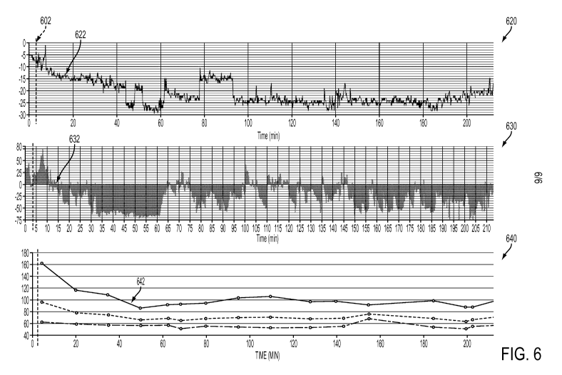
[0070] Example 1: An Output Display of Patient Data for a Patient Undergoing
Dialysis Therapy
CA 02915060 2015-12-10
WO 2014/201183
PCT/US2014/042012
[0071] It may be understood that an output display of patient data by a
computing
device may include data related to patient physiological status in addition to
annotations
related to, but not limited to, date and time, patient identification
information, patient
diagnosis information, warning indicators, arrhythmia event indicators, and
data associated
with a therapeutic device if the patient is undergoing a therapeutic procedure
during pulse
wave monitoring. In some embodiments, the computing device may display on an
output
device a representation of a portion of the biological signal along with at
least one annotation
identifying the cardiac bradycardia behavior. In some embodiments the
biological signal
displayed on the output device may be updated over time. In some embodiments,
the
computing device may display on the output device one or more annotations
including a
hypovolemia indicator and a hypotensive indicator. In still other embodiments,
the computing
device may provide one or more warnings to a user if the cardiac bradycardia
behavior
indicates an emergent condition associated with the human body.
[0072] FIG. 6 illustrates a non-limiting example of a computing device real-
time
output display to indicate the status of an end-stage renal disease patient
undergoing dialysis.
Exemplary data presented on such a display may include a trace of the percent
change in a
patient pulse rate 620, a trace of the percent changes in a patient pulse
strength 630, and a
trace of the patient blood pressure 640. The time axis of each display is
indicated as time, in
minutes, after the start of the dialysis treatment 602.
[0073] An indicator regarding patient status, such as a warning indicator, may
also be
provided to a user of the computing device. The warning indicator may be
triggered if any
data associated with patient status, including data associated with pulse
waveform peak
amplitude differences, pulse waveform peak time differences, and one or more
time
difference dispersion metrics meet one or more warning criteria. The warning
criteria may be
used by the health care provider as an indicator of a potential hypovolemic
hypotensive
26
CA 02915060 2015-12-10
WO 2014/201183
PCT/US2014/042012
condition resulting from cardiac bradycardia behavior. The health care
provider may then
assess the usefulness of continuing the therapeutic procedure or stop the
procedure depending
on the hemodynamic instability risk of the procedure to the patient.
[0074] Additional metrics associated with patient status, such as metrics
associated
with patient ventilation and patient blood chemistry (for example, additional
blood gas
metrics), may also be displayed. In one non-limiting example, such displays
may be
presented in real time by scrolling the data presented on the display.
[0075] Such a patient status display may also permit a health care provider
and
system user to display selected data presented during defined time windows.
Such time
windows may include an entire therapeutic session, a portion of a therapeutic
session, or a
time window including pre-therapy time, therapy time, and post-therapy time.
Thus, such a
display window may display data generally over any time interval, including,
without
limitation, a time window for intervals of about 1 minute to about 24 hours.
Non-limiting
examples of such time window intervals may include time intervals of about 1
minute, about
2 minutes, about 5 minutes, about 10 minutes, about 20 minutes, about 30
minutes, about 1
hour, about 2 hours, about 5 hours, about 10 hours, about 20 hours, about 24
hours, and
ranges between any two of these values including endpoints.
[0076] For example, in FIG. 6, an anomalous decrease in the percent change in
the
pulse rate 620 may be observed at an early time in the dialysis procedure. In
some
embodiments, the pulse rate metric may be calculated as a difference between
the pulse rate
and the pulse rate baseline. In alternative embodiments, the pulse rate metric
may be
calculated as a percent change in pulse rate defined as a difference between
the pulse rate and
the pulse rate baseline, the difference being normalized to (divided by) the
pulse rate baseline
value. In one non-limiting example, a percent change in pulse rate threshold
may be set as a
fraction of the value of the pulse rate baseline. In the trace of the percent
change in a patient
27
CA 02915060 2015-12-10
WO 2014/201183
PCT/US2014/042012
pulse rate 620, the pulse rate threshold is set to -15%. It may be observed
that the percent
change in pulse rate decreases to the threshold 622 at around minute 15 of the
procedure.
[0077] Similarly, an anomalous decrease in the percent change in the pulse
strength
630 may be observed at an early time in the dialysis procedure. In some
embodiments, the
pulse strength metric may be calculated as a difference between the pulse
strength and the
pulse strength baseline. In alternative embodiments, the pulse strength metric
may be
calculated as a percent change in pulse strength defined as a difference
between the pulse
strength and the pulse strength baseline, the difference being normalized to
(divided by) the
pulse strength baseline value. In one non-limiting example, a percent change
in pulse
strength threshold may be set as a fraction of the value of the pulse strength
baseline. In the
trace of the percent change in a patient pulse strength 630, the pulse
strength threshold is also
set to -15%. It may be observed that the percent change in pulse strength
decreases to the
threshold 632 at around minute 15 of the procedure.
[0078] It may be observed that the trace of the patient blood pressure 640
depicts a
drop in systolic blood pressure (top line in the trace 640) to less than 100
mm Hg (about 13
kPa) 642 at about minute 50 of the procedure. Such a low systolic blood
pressure, indicative
of a hypotensive state in the patient, may result from a therapeutic
procedure, such as induced
hypovolemia during dialysis. The percent change in pulse rate and the percent
change in
pulse strength reach their threshold values (622 and 632, respectively) about
30 minutes
before the blood pressure measurement indicates a potential hypotensive
condition 642. A
health care provider, thereby forewarned of possible hypotensive events, may
adjust or even
terminate the therapy to prevent additional trauma to the patient.
[0079] It may be understood that a user may control the display of patient
status
information provided by the computing device, such as a display of status
data, types of data
analysis results, and annotations of data analysis results. In one non-
limiting example, a drop-
28
CA 02915060 2015-12-10
WO 2014/201183
PCT/US2014/042012
down menu may be used by a user to indicate which types of information,
analyses, and
annotations may be displayed.
[0080] The present disclosure is not to be limited in terms of the particular
embodiments described in this application, which are intended as illustrations
of various
aspects. Many modifications and variations can be made without departing from
its spirit and
scope, as will be apparent to those skilled in the art. Functionally
equivalent methods and
apparatuses within the scope of the disclosure, in addition to those
enumerated in this
disclosure, will be apparent to those skilled in the art from the foregoing
descriptions. Such
modifications and variations are intended to fall within the scope of the
appended claims. The
present disclosure is to be limited only by the terms of the appended claims,
along with the
full scope of equivalents to which such claims are entitled. It is to be
understood that this
disclosure is not limited to particular methods, reagents, compounds, or
compositions, which
can, of course, vary. It is also to be understood that the terminology used in
this disclosure is
for the purpose of describing particular embodiments only, and is not intended
to be limiting.
[0081] With respect to the use of substantially any plural and/or singular
terms in this
disclosure, those having skill in the art can translate from the plural to the
singular and/or
from the singular to the plural as is appropriate to the context and/or
application. The various
singular/plural permutations may be expressly set forth in this disclosure for
sake of clarity.
[0082] It will be understood by those within the art that, in general, terms
used in this
disclosure, and especially in the appended claims (for example, bodies of the
appended
claims) are generally intended as "open" terms (for example, the term
"including" should be
interpreted as "including but not limited to," the term "having" should be
interpreted as
"having at least," the term "includes" should be interpreted as "includes but
is not limited to,"
etc.). While various compositions, methods, and devices are described in terms
of
29
CA 02915060 2015-12-10
WO 2014/201183
PCT/US2014/042012
"comprising" various components or steps (interpreted as meaning "including,
but not limited
to"), the compositions, methods, and devices can also "consist essentially or
or "consist of
the various components and steps, and such terminology should be interpreted
as defining
essentially closed-member groups.
[0083] It will be further understood by those within the art that if a
specific number of
an introduced claim recitation is intended, such an intent will be explicitly
recited in the
claim, and in the absence of such recitation no such intent is present. For
example, as an aid
to understanding, the following appended claims may contain usage of the
introductory
phrases at least one and one or more to introduce claim recitations. However,
the use of
such phrases should not be construed to imply that the introduction of a claim
recitation by
the indefinite articles "a" or an limits any particular claim containing such
introduced claim
recitation to embodiments containing only one such recitation, even when the
same claim
includes the introductory phrases one or more or at least one and indefinite
articles such
as "a" or an (for example, "a" and/or "an" should be interpreted to mean "at
least one" or
"one or more"); the same holds true for the use of definite articles used to
introduce claim
recitations. In addition, even if a specific number of an introduced claim
recitation is
explicitly recited, those skilled in the art will recognize that such
recitation should be
interpreted to mean at least the recited number (for example, the bare
recitation of two
recitations," without other modifiers, means at least two recitations, or two
or more
recitations). Furthermore, in those instances where a convention analogous to
"at least one of
A, B, and C, etc." is used, in general such a construction is intended in the
sense one having
skill in the art would understand the convention (for example, " a system
having at least one
of A, B, and C" would include but not be limited to systems that have A alone,
B alone, C
alone, A and B together, A and C together, B and C together, and/or A, B, and
C together,
etc.). It will be further understood by those within the art that virtually
any disjunctive word
CA 02915060 2015-12-10
WO 2014/201183
PCT/US2014/042012
and/or phrase presenting two or more alternative terms, whether in the
description, claims, or
drawings, should be understood to contemplate the possibilities of including
one of the terms,
either of the terms, or both terms. For example, the phrase "A or B" will be
understood to
include the possibilities of "A" or "B" or "A and B."
[0084] As will be understood by one skilled in the art, for any and all
purposes, such
as in terms of providing a written description, all ranges disclosed in this
disclosure also
encompass any and all possible subranges and combinations of subranges
thereof. Any listed
range can be easily recognized as sufficiently describing and enabling the
same range being
broken down into at least equal halves, thirds, quarters, fifths, tenths, etc.
As a non-limiting
example, each range discussed in this disclosure can be readily broken down
into a lower
third, middle third and upper third, etc. As will also be understood by one
skilled in the art all
language such as "up to," "at least," and the like include the number recited
and refer to
ranges which can be subsequently broken down into subranges as discussed
above. Finally,
as will be understood by one skilled in the art, a range includes each
individual member.
[0085] From the foregoing, it will be appreciated that various embodiments of
the
present disclosure have been described for purposes of illustration, and that
various
modifications may be made without departing from the scope and spirit of the
present
disclosure. Accordingly, the various embodiments disclosed are not intended to
be limiting,
with the true scope and spirit being indicated by the following claims.
31