Note: Descriptions are shown in the official language in which they were submitted.
CA 02915073 2015-12-10
WO 2014/201452
PCT/US2014/042465
- 1 -
MITRAL VALVE SPACER AND SYSTEM AND METHOD FOR
IMPLANTING THE SAME
CROSS-REFERENCE AND RELATED APPLICATIONS
The present application claims benefit of U.S. Provisional Patent No.
61/835,093,
filed June 14, 2013, the entire content of the application is hereby
incorporated by
reference.
FIELD OF THE APPLICATION
The present disclosure relates to the repair and/or correction of
dysfunctional heart
valves, and more particularly pertains to heart valve implants and systems and
methods for
delivery and implementation of the same.
BACKGROUND
The human heart has four chambers, the left and right atrium and the left and
right
ventricles. The chambers of the heart alternately expand and contract to pump
blood
through the vessels of the body. The cycle of the heart includes the
simultaneous
contraction of the left and right atria, passing blood from the atria to the
left and right
ventricles. The left and right ventricles then simultaneously contract forcing
blood from the
heart and through the vessels of the body. In addition to the four chambers,
the heart also
includes a check valve at the upstream end of each chamber to ensure that
blood flows in
the correct direction through the body as the heart chambers expand and
contract. These
valves may become damaged or otherwise fail to function properly, resulting in
their
inability to properly close when the downstream chamber contracts. Failure of
the valves to
properly close may allow blood to flow backward through the valve resulting in
decreased
blood flow and lower blood pressure.
Mitral regurgitation is a common variety of heart valve dysfunction or
insufficiency.
Mitral regurgitation occurs when the mitral valve separating the left coronary
atrium and
the left ventricle fails to properly close. As a result, upon contraction of
the left ventricle
blood may leak or flow from the left ventricle back into the left atrium,
rather than being
forced through the aorta. Any disorder that weakens or damages the mitral
valve can
prevent it from closing properly, thereby causing leakage or regurgitation.
Mitral
CA 02915073 2015-12-10
WO 2014/201452
PCT/US2014/042465
- 2 -
regurgitation is considered to be chronic when the condition persists rather
than occurring
for only a short period of time.
Regardless of the cause, mitral regurgitation may result in a decrease in
blood flow
through the body (cardiac output). Correction of mitral regurgitation
typically requires
surgical intervention. Surgical valve repair or replacement may be carried out
as an open
heart procedure. The repair or replacement surgery may last in the range of
about three to
five hours, and may be carried out with the patient under general anesthesia.
The nature of
the surgical procedure requires the patient to be placed on a heart-lung
machine. Because
of the severity, complexity, and/or danger associated with open heart surgical
procedures,
corrective surgery for mitral regurgitation may not be recommended in certain
patients.
SUMMARY OF THE INVENTION
Described herein is a heart valve implant and methods of delivering the same
to an
individual's heart. The heart valve implant is configured to be delivered to
the heart trans-
apically (i.e., through the apex of the heart) or trans-femorally. The heart
valve implant is
configured to be implanted at least partially within the heart.
Accordingly, in one aspect, the invention is directed to a heart valve
implant. The
heart valve implant comprises an inflatable valve body, a shaft, an anchor
assembly, and an
inflation (e.g., injection) port. The inflatable valve body defines a cavity
comprising a
proximal end and a distal end. The shaft extends from the proximal end of the
inflatable
valve body and comprises a lumen in fluid communication with said cavity. The
anchor
assembly is attached to the shaft, proximal to the inflatable valve body. The
inflation (e.g.,
injection) port can include one or more lumens in fluid communication with the
shaft
lumen.
In some aspects, the heart valve implant comprises one or more radiopaque
markers.
In another aspect, the heart valve implant comprises one or more radiopaque
markers that
are located at or near the proximal end of the inflatable valve body.
In some aspects, the inflation (e.g., injection) port comprises a pierceable
septum
configured to fluidly seal the inflation port. In some embodiments, the
pierceable septum
can be self-sealing. In a related aspect, the inflation port is substantially
hollow. The
pierceable septum can be formed of a variety of different materials. By way of
example, in
some embodiments, the pierceable septum comprises silicone. In one embodiment,
the
CA 02915073 2015-12-10
WO 2014/201452
PCT/US2014/042465
- 3 -
pierceable septum is liquid-tight. In some embodiments, the inflation port
comprises one or
more suture holes.
In some aspects, the anchor assembly comprises a passageway and one or more
arms. For example, the anchor assembly can include a central opening and two
opposed
peripheral arms that extend outwardly from the central opening. In one aspect,
the
passageway is configured to receive and advance the shaft. In one aspect, each
of the one
or more arms defines an opening and the anchor assembly is configured to be
secured to an
exterior surface of an individual's heart. In one aspect, one or more sutures
are placed
around each of the one or more arms of the anchor assembly. The sutures can be
employed
to secure (e.g., fixate) the anchor assembly to an exterior surface of an
individual's heart.
In some aspects, the inflatable valve body is partially inflated with an
expansion
medium (inflation fluid). In another aspect, the inflatable valve body is
completely inflated
with an expansion medium (inflation fluid). A variety of expansion media can
be
employed. In some embodiments, the expansion medium is a gas, a liquid or a
gel. By way
of example, in some embodiments, the expansion medium can be deionized water,
saline,
or contrast medium.
In some aspects, the shaft of the heart valve implant is attached to the
distal end of
the inflatable valve body. In some embodiments, the shaft of the heart valve
extends
partially through the inflatable valve body. In other embodiments, the shaft
of the heart
valve implant extends across a length of the inflatable valve body and is
attached to both
the proximal and distal ends of the inflatable valve body.
In some aspects, the lumen of the shaft, in fluid communication with the
inflatable
valve body, comprises one or more openings. The one or openings provide the
fluid
communication between the lumen of the shaft with the inflatable valve body.
In some embodiments, the inflatable valve body can be formed of any of
material
suitable for the implant described herein. The material can be able to
withstand
physiological conditions (e.g., the conditions in a heart). The material can
also be elastic
and pliably deformable.
In some embodiments, the shaft can be formed of any of material suitable for
the
implant described herein. The shaft can be flexible or pliably deformable so
that it can
bend to accommodate the delivery systems described herein. The shaft can also
have
sufficient rigidity as not to collapse onto itself
CA 02915073 2015-12-10
WO 2014/201452
PCT/US2014/042465
- 4 -
In some embodiments, the anchor assembly can be formed of any of material
suitable for the implant and methods described herein.
In another aspect, the invention is directed to a method of trans-apically
delivering a
heart valve implant within a heart. The method comprises trans-apically
advancing an
introducer comprising a lumen through an incision in an apex of a heart into a
left ventricle,
advancing said introducer through a mitral valve into a left atrium, advancing
the heart
valve implant through the introducer lumen into the left atrium, wherein a
shaft of the heart
valve implant extends from an inflatable valve body to beyond the incision in
the apex, and
removing the introducer from said heart, thereby delivering the valve body and
at least a
portion of the shaft within the heart.
In one aspect, the method of trans-apically delivering a heart valve implant
comprises placing the inflatable valve body at least a portion of one or more
cusps or
leaflets of the heart valve may interact with, engage, and/or seal against at
least a portion of
the heart valve implant when the heart valve is in a closed condition. The
interaction,
engagement and/or sealing between at least a portion of at least one cusp or
leaflet and at
least a portion of the heart valve implant may reduce and/or eliminate
regurgitation in a
heart valve, for example, providing insufficient sealing, including only a
single cusp, e.g.,
following removal of a diseased and/or damaged cusp, and/or having a ruptured
chordae.
In some aspects of the method of trans-apically delivering a heart valve
implant, the
implant comprises an anchor assembly. In one embodiment, the anchor assembly
comprises a passageway and one or more arms. The passageway is configured to
receive
the shaft and to allow advancing the shaft through the passageway. Each of the
one or more
arms defines an opening. In some aspects, the method further comprises
advancing the
anchor assembly over the shaft until the anchor assembly is at or near the
apex of the heart.
In one aspect, the method further comprises securing the anchor assembly to an
external
surface of the heart. In another aspect, the method comprises securing the
anchor assembly
to an external surface of the heart at or near said apex of the heart. In
another aspect,
securing the anchor assembly comprises suturing one or more sutures around
each of the
one or more arms of the anchor assembly.
In another aspect, the method further comprises securing the inflation (e.g.,
injection) port subdermally at or near a chest wall. In some embodiments, the
subdermally-
located inflation port can be employed to deliver an expansion medium
(inflation fluid),
CA 02915073 2015-12-10
WO 2014/201452
PCT/US2014/042465
- 5 -
(e.g., deionized water, saline, contrast medium) to the inflatable valve body.
For example,
the expansion medium can be placed in a syringe, and the needle can be
employed to pierce
through the septum. The medium can then be transferred via the hollow body of
the
inflation port and the shaft to the inflatable valve body.
In another aspect, the method comprises trans-apically delivering a heart
valve
implant within a heart, wherein the inflatable valve body comprises one or
more radiopaque
markers for locating the inflatable valve body within the mitral valve.
In another aspect, the method further comprises completely or partially
inflating the
inflatable valve body with an inflation fluid. In another aspect, wherein the
step of inflating
the inflatable valve body further comprises piercing a pierceable septum of
the inflation
port and introducing an expansion medium (inflation fluid) through the
pierceable septum
into the inflation port thereby inflating the inflatable valve body with the
inflation fluid. In
one aspect, the inflation fluid is a liquid. In another aspect, the method
further comprises
adjusting the amount of inflation fluid within the implant until a desired
level of inflation is
attained. In another aspect, the inflatable valve body interacts with all or a
portion of at
least one cusp or leaflet of the mitral valve. In another aspect, the
inflatable valve body
partially or completely restricts a flow of blood through the mitral valve in
a closed
position.
In another aspect, the method further comprises de-airing the implant.
In another aspect, the invention is directed to method of trans-apically
delivering a
heart valve implant within a heart. The implant comprises an inflatable valve
body, a shaft,
an anchor assembly, and an inflation port. The method comprises trans-apically
advancing
an introducer comprising a lumen through an incision in an apex of a heart
into a left
ventricle, advancing the heart valve implant through the introducer lumen into
the left
ventricle, wherein the inflatable valve body extends from the introducer,
partially inflating
the inflatable valve body, advancing said introducer and partially inflated
inflatable valve
body through a mitral valve into a left atrium, advancing the heart valve
implant through
the introducer lumen into the left atrium, removing the introducer from said
heart, thereby
delivering the inflatable valve body and at least a portion of the shaft
within the heart.
CA 02915073 2015-12-10
WO 2014/201452
PCT/US2014/042465
- 6 -
BRIEF DESCRIPTION OF THE DRAWINGS
Features and advantages of the claimed subject matter will be apparent from
the
following description of embodiments consistent therewith, which the
description should be
considered in conjunction with the accompanying drawings.
FIG. 1 illustrates a perspective view of an embodiment of a transseptal
catheter in
the right atrium consistent with the present disclosure.
FIG. 2 illustrates a perspective view of an embodiment of a guide wire
advanced
into the superior vena cava consistent with the present disclosure.
FIG. 3 illustrates a perspective view of an embodiment of a catheter advanced
into
the superior vena cava consistent with the present disclosure.
FIG. 4 illustrates a perspective view of an embodiment of a catheter tip
against the
fossa ovalis consistent with the present disclosure.
FIG. 5 illustrates a perspective view of an embodiment of a catheter tenting
the
fossa ovalis consistent with the present disclosure.
FIG. 6 illustrates a perspective view of an embodiment of a needle puncturing
the
fossa ovalis consistent with the present disclosure.
FIG. 7 illustrates a perspective view of an embodiment of a transseptal
catheter
punctured through the fossa ovalis consistent with the present disclosure.
FIG. 8 illustrates a perspective view of an embodiment of a transseptal
catheter
punctured through the fossa ovalis with its distal tip in the left atrium with
the needle
removed consistent with the present disclosure.
FIG. 9 illustrates a perspective view of an embodiment of a delivery guide
wire
advanced into the left atrium through the transseptal catheter consistent with
the present
disclosure.
FIG. 10 illustrates a perspective view of an embodiment of a sheath and
dilator
removed with a delivery guide wire in the left atrium consistent with the
present disclosure.
FIG. 11 illustrates a perspective view of an embodiment of a dilator advanced
to the
left atrium consistent with the present disclosure.
FIG. 12 illustrates a perspective view of one embodiment of a dilator
consistent with
the present disclosure.
FIG. 13A illustrates a perspective view of an embodiment of a dilator
consistent
with the present disclosure.
CA 02915073 2015-12-10
WO 2014/201452
PCT/US2014/042465
- 7 -
FIG. 13B illustrates a close-up of one embodiment of the tip of the dilator
shown in
FIG. 13A consistent with the present disclosure.
FIG. 14A illustrates a perspective view of a yet another embodiment of a
dilator
consistent with the present disclosure.
FIG. 14B illustrates a perspective view of one embodiment of the dilator shown
in a
deflected or retracted position consistent with the present disclosure.
FIG. 14C illustrates a perspective view of one embodiment of the dilator shown
in
an inflated or expanded position consistent with the present disclosure.
FIG. 15 illustrates a perspective view of a dilator in the inflated or
expanded
position located in the left atrium consistent with the present disclosure.
FIG. 16 illustrates a perspective view of a dilator in the inflated or
expanded
position located in the left atrium prior to passing through the mitral valve
consistent with
the present disclosure.
FIG. 17 illustrates a perspective view of a dilator located in the left
ventricle
consistent with the present disclosure.
FIG. 18 illustrates a perspective view of an embodiment of a dilator advanced
to an
apex of the left ventricle.
FIG. 19 illustrates a needle inserted through the apex into the left ventricle
over a
guide wire consistent with the present disclosure.
FIG. 20 illustrates an introducer and a dilator being inserted into the left
ventricle
over a guide wire consistent with the present disclosure.
FIG. 21 illustrates purse-string sutures and pledgets secured around the
introducer
consistent with the present disclosure.
FIG. 22 illustrates the introduced advanced over the guide wire to the left
atrium
consistent with the present disclosure.
FIG. 23 illustrates an implant being loaded into the introducer.
FIG. 24 illustrates the implant in the left atrium.
FIG. 25 illustrates the implant in the mitral valve.
FIG. 26 illustrates one embodiment of an implant consistent with the present
disclosure.
FIG. 27 illustrates the implant in the mitral valve, an inflation device and a
splitter.
CA 02915073 2015-12-10
WO 2014/201452
PCT/US2014/042465
- 8 -
FIG. 28 illustrates splitting the introducer after the implant has been
verified in the
mitral valve.
FIG. 29A illustrates the implant in the mitral valve and an inflation device
in the
form of an inflation handle assembly.
FIG. 29B illustrates the implant in the mitral valve and an inflation device
in the
form of an inflation port.
FIG. 29C illustrates the implant in the mitral valve and an exploded view of
an
inflation device in the form of an inflation port located subdermally or
subcutaneously in an
individual.
FIG. 30 illustrates one embodiment of an inflation handle assembly in a
retracted
position prior to filling.
FIG. 31 illustrates the inflation handle assembly in an expanded position
after
filling.
FIG. 32 illustrates a perspective view of one embodiment of an anchor
assembly.
FIG. 33 illustrates a cross-sectional side view of one embodiment of an anchor
assembly.
FIG. 34 illustrates a front view of one embodiment of an anchor assembly.
FIG. 35 illustrates a side view of one embodiment of an anchor assembly.
FIG. 36 illustrates a needle being inserted through the apex into the left
ventricle.
FIG. 37 illustrates a guidewire being inserted through the needle into the
left
ventricle.
FIG. 38 illustrates the needle removed and the guidewire in the left
ventricle.
FIG. 39 illustrates one embodiment of an introducer and dilator being inserted
into
the left ventricle.
FIG. 40 illustrates purse-string sutures and pledgets secured around the
introducer.
FIG. 41 illustrates the guidewire removed from the introducer.
FIG. 42 illustrates one embodiment of an inflatable valve body partially
beyond the
tip of the introducer.
FIG. 43 illustrates the inflatable valve body partially inflated at the tip of
the
introducer.
FIG. 44 illustrates the inflatable valve body being advanced through the
mitral
valve.
CA 02915073 2015-12-10
WO 2014/201452
PCT/US2014/042465
- 9 -
DETAILED DESCRIPTION
The present disclosure relates to a system and method of implanting a heart
implant.
For example, the system and method according to one embodiment of the present
disclosure
may be used to implant a heart valve implant which may suitably be used in
connection
with the treatment, diagnostics and/or correction of a dysfunctional or
inoperative heart
valve (e.g., function mitral valve regurgitation and degenerative mitral valve
regurgitation).
One suitable implementation for a heart valve implant consistent with the
present disclosure
is the treatment of mitral valve regurgitation (mitral insufficiency or mitral
incompetence).
For the ease of explanation, the heart valve implant herein is described in
terms of a mitral
valve implant, such as may be used in treating mitral valve regurgitation as
described in
U.S. Patent No. Application Serial No. 11/258,828 filed October 26, 2005 and
U.S. Patent
Application Serial No. 12/209,686 filed September 12, 2008, both of which are
fully
incorporated herein by reference. However, a heart valve implant consistent
with the
present disclosure may be employed for treating, diagnosing and/or correcting
other
dysfunctional or inoperative heart valves, such as the heart valve implant(s)
discussed
herein in connection with FIG. 26.
It should be understood that the technology of the present disclosure
(including the
implant described in connection with FIG. 26) is not limited to mitral valve
implants and
systems and methods of implanting mitral valve implants. Indeed, the systems
and methods
according to the present disclosure may be used to implant heart implants
configured to be
used in connection with the treatment, diagnostics and/or correction of other
heart
conditions. For example, and without limitation, the system and method
consistent with the
present disclosure may be used to implant a regurgitation implant configured
to induce a
controlled regurgitation in a heart valve (such as, but not limited to, a
mitral heart valve),
for example, in a manner that is generally consistent with advanced disease of
the heart.
The regurgitation implant may include a regurgitation implant as described in
U.S. Patent
No. Serial No. 11/940,724 filed November 15, 2007 and U.S. Patent Application
Serial No.
12/209,686 filed September 12, 2008, both of which are fully incorporated
herein by
reference.
According to one embodiment, a heart implant consistent with the present
disclosure
may comprise a heart valve implant configured to interact with at least a
portion of an
existing heart valve to prevent and/or reduce regurgitation. For example, at
least a portion
CA 02915073 2015-12-10
WO 2014/201452
PCT/US2014/042465
- 10 -
of one or more cusps or leaflets of the heart valve may interact with, engage,
and/or seal
against at least a portion of the heart valve implant when the heart valve is
in a closed
condition. As used herein, "cusp" and "leaflet" refer to the same anatomic
structure of a
heart valve. The interaction, engagement and/or sealing between at least a
portion of at
least one cusp or leaflet and at least a portion of the heart valve implant
may reduce and/or
eliminate regurgitation in a heart valve, for example, providing insufficient
sealing,
including only a single cusp, e.g., following removal of a diseased and/or
damaged cusp,
and/or having a ruptured chordae. A heart valve implant consistent with the
present
disclosure may be used in connection with various additional and/or
alternative defects
and/or deficiencies.
For the ease of explanation, one embodiment of the system and method
consistent
with the present disclosure is described in terms of a system and method for
implanting a
mitral valve implant, such as may be used in treating mitral valve
regurgitation. By way of
an overview, the system and method may generally comprise placing a first
guide wire into
the left ventricle, replacing the first guide wire with a second (e.g.,
delivery) guide wire,
piercing an apex the heart with the second guide wire, advancing the second
guide wire
such that a distal portion thereof extends to an exterior of said heart, and
advancing a mitral
valve implant over said guide wire through said puncture in said apex and into
the left
ventricle.
For example, a delivery (e.g., second) guide wire may be initially placed into
the left
atrium of the heart, for example, by way of transseptal puncture of the heart
from the right
atrium through the fossa ovalis into the left atrium. A dilator may then be
advanced along
the delivery guide wire to the left atrium and may be passed through the
mitral valve into
the left ventricle. The dilator may include a balloon which may be inflated to
facilitate
passing the dilator through the mitral valve without damaging the mitral valve
or becoming
entangled in the mitral valve chordae. A steerable catheter may then be
advanced along the
dilator into the left ventricle and to the apex of the heart. The delivery
guide wire may then
be exchanged with a third (e.g., puncturing) guide wire, which may be used to
puncture
through the apex of the heart. The implant may then be advanced over the third
guide wire
through the puncture in said heart using a trans-apical delivery procedure.
Once the
implant is delivered into the heart, it may be positioned and inflated in a
desired manner. In
CA 02915073 2015-12-10
WO 2014/201452
PCT/US2014/042465
- 11 -
some embodiments, at least one of the position and inflation of the implant
may be
adjustable, even after the implant is initially sited and inflated within the
heart.
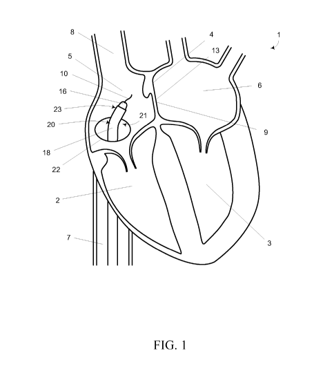
Referring now to FIG. 1, a cross-sectional schematic view of a portion of a
four
chamber heart 1 is illustrated. The outflow tracts of the right and left
ventricles 2, 3 are not
shown in order to better illustrate the septum 4 between the right and left
atria 5, 6. As
shown, the inferior vena cava (IVC) 7 and superior vena cava (SVC) 8
communicate with
the right atrium 5 which is separated from the left atrium 6 by the intra-
atrial septum 4.
While not a limitation of the present disclosure, it is may be advantageous to
make the
transseptal puncture 13 through the fossa ovalis 9 since the fossa ovalis 9 is
thinnest portion
of the intra-atrial septum 4.
According to one embodiment consistent with the present disclosure, a first
guide
wire 10 may be advanced up the IVC 7 and into the right atrium 5. The first
guide wire 10
may include any guide wire configured to be advanced up the IVC 7 and into the
right
atrium 5. Consistent with one embodiment, the first guide wire 10 may be the
same as a
delivery (e.g., second) guide wire discussed herein; however, the first guide
wire 10 may
also be separate and distinct from the delivery guide wire. Without
limitation, access to the
right atrium 5 may be accomplished by way of the Seldinger wire technique. For
example,
the right femoral vein (not shown) may be accessed with a hollow needle (not
shown) and a
first guide wire 10 may be inserted. The needle may be removed and a dilator
16 may be
inserted over the first guide wire 10. The sheath 18 of a catheter 20 (such
as, but not
limited to, a Mullins catheter or the like) having a pre-bent region 21
proximate the distal
tip 23 of the catheter 20 may be inserted over the dilator 16. The sheath 18,
dilator 16,
catheter 20 and first guide wire 10 may then be advanced up the IVC 7 through
the opening
22 into the right atrium 5 as generally illustrated in FIG. 1.
With the sheath 18, dilator 16, catheter 20 and first guide wire 10 in the
right atrium
5, access to the left atrium 6 may be achieved by transseptal puncture 13 from
the right
atrium 5 through the intra-atrial septum 4. For example, at least a portion of
the first guide
wire 10 may be advanced out of the distal tip 23 of the dilator 16, sheath 18
and/or catheter
20 as generally shown in FIG. 2. According to an embodiment, the first guide
wire 10 may
be at least partially advanced into the SVC 8 as generally illustrated in FIG.
2 and the distal
tip 23 of the catheter 20 may then be at least partially advanced along the
first guide wire 10
into the SVC 8 as generally illustrated in FIG. 3. Because the SVC 8 is a thin-
walled vein,
CA 02915073 2015-12-10
WO 2014/201452
PCT/US2014/042465
- 12 -
it may be advantageous to place the first guide wire 10 in the SVC 8 and then
advance the
catheter 20 along the first guide wire 10 since the spring-tipped atraumatic
first guide wire
reduces the potential for damaging the SVC 8 compared to the catheter 20 and
dilator
16.
With the distal tip 23 at least partially received in the SVC 8, the first
guide wire 10
may be retracted into the dilator 16 and the catheter 20 may be retracted
(i.e., pulled
downward) such that the pre-bent portion 21 of the sheath 18 facilitates
guiding the distal
tip 23 to the fossa ovalis 9 as generally illustrated in FIG. 4. For example,
using one or
more visualization techniques (such as, but not limited to, intracardiac echo
(ICE),
fluoroscopy, and the like), the sheath 18 may be retracted proximally,
dragging the distal tip
23 along the intra-atrial septum 4 until the distal tip 23 is positioned
proximate to the fossa
ovalis 9. Optionally, the position of the sheath 18 relative to the fossa
ovalis 9 may be
confirmed by gently pushing the sheath 18 distally against the intra-atrial
septum 4 to "tent"
the fossa ovalis 9 as generally illustrated in FIG. 5. The "tenting" of the
fossa ovalis 9 may
be seen on ICE, fluoroscopy or the like.
With the distal tip 23 proximate and/or contacting the fossa ovalis 9, the
first guide
wire 10 may be removed from the catheter 20 and a transseptal needle 26 may be
advanced
through the catheter 20 towards the distal end 23 of the catheter 20 as
generally shown in
FIG. 6. The position of the catheter 20 may optionally be confirmed (for
example, but not
limited to, by "tenting") and the transseptal needle 26 may be advanced out of
the distal tip
23 to form a puncture 28 through the fossa ovalis 9 and into the left atrium
6. The sheath
18, dilator 16 and catheter 20 may than be advanced through the puncture 28 of
the fossa
ovalis 9 and into the left atrium 6 as generally shown in FIG. 7. Once the
sheath 16, dilator
28 and catheter 20 are through the fossa ovalis 9, the needle 26 may be
removed from the
catheter 20 as generally shown in FIG. 8.
With the catheter 20 in the left atrium 6, a delivery (e.g., second) guide
wire 30 may
be advanced through the catheter 20 until at least a portion of the distal tip
32 of the
delivery guide wire 30 extends from the distal tip 23 of the catheter 20 and
into the left
atrium 6 as generally illustrated in FIG. 9. Once the distal tip 32 of the
delivery guide wire
30 is disposed in the left atrium 6, the dilator 16 and the sheath 18 may be
removed, leaving
just the delivery guide wire 30 in the left atrium 6 as generally illustrated
in FIG. 10.
CA 02915073 2015-12-10
WO 2014/201452
PCT/US2014/042465
- 13 -
The delivery guide wire 30 may be used as a guide for advancing other devices
into
the heart 1, and ultimately, into the left ventricle 3. Accordingly to at
least one
embodiment, the delivery guide wire 30 may be sufficiently stiff to resist
undesirable
bending and/or kinking and to resist undesirable movement of the distal tip
32. For
example, the delivery guide wire 30 may comprise a stiff, 0.018" diameter
guide wire
having a stiffness of approximately 19,900,000 psi. The stiffness of the
delivery guide wire
30 was determined as follows.
When a force is applied to a long thin column, there is no movement of the
column
until a minimum critical buckling force is achieved, Per, then further
buckling occurs,
though the force does not increase. For a long column of uniform cross-section
and length
1, which buckles under a critical force, Per, the following formula applies:
2 E I
P ¨ n 71-
Cr Id2
Where:
n = a constant that is equal to 4 if both ends of the column are
clamped and
cannot move or rotate.
E = Modulus of elasticity of the material (psi)
I = Moment of inertia (in4)
For a circular cross-section the moment of inertia is:
64
Substituting for I in the first equation for Per leads to:
E d 4
Po, = n TC3 _____________________________
64L2
And solving for the modulus leads to:
E = 64L2 ]r
Cr
3d4
n 71"
CA 02915073 2015-12-10
WO 2014/201452
PCT/US2014/042465
- 14 -
Based on the above, an 8 cm section of the delivery guide wire 30 was tested
and a buckling force of 0.41 lbs. was determined. Therefore,
64 (3.15)2 (0.41)
E = _________________________________ =19,900,000psi
47z-3(0.018)4
This stiffness (modulus of elasticity) of the delivery guide wire 30 may
therefore be
approximately 19,900,000 psi. Of course, the delivery guide wire 30 may have a
stiffness
greater than or less than 19,900,000 psi.
According to at least one other embodiment, the delivery guide wire 30 may
include
a typical 0.018" guide wire (for example a 0.018" angled standard exchange
guide wire
made by Merit Medical Systems of South Jordan, Utah, Model H2OSTDA18260EX
which
was determined to have a stiffness of approximately 1,360,000 psi based on the
same
methodology). In either embodiment, the delivery guide wire 30 may have a
diameter
greater than or less than 0.018".
Turning now to FIG. 11, a dilator 34 may be advanced over the delivery guide
wire
30 into the left atrium 6. The dilator 34 may be configured to pass through
the mitral valve
61 into the left ventricle 3 without damaging the mitral valve 61 or becoming
entangled in
the mitral valve 61 (for example, the cusps 66, the chordae and/or papillary
muscles 68 of
the mitral valve 61). According to at least one embodiment, the dilator 34 of
the present
disclosure may be used to eliminate the delivery guide wire as disclosed in
U.S. Patent
Application Serial No. 12/209,686 filed September 12, 2008. However, it may be
appreciated that the system and method disclosed in the present disclosure
(and in particular
the dilator 34) is not inconsistent with the system and method in U.S. Patent
Application
Serial No. 12/209,686, and as such, the system and method disclosed in the
present
disclosure (including the dilator 34) may be used in conjunction with the
system and
method in U.S. Patent Application Serial No. 12/209,686.
One embodiment of a dilator 34a consistent with the present disclosure is
generally
illustrated in FIG. 12. The dilator 34a may include define at least one lumen
94 configured
to receive at least a portion of the delivery guide wire 30. For example, the
lumen 94 may
have an internal diameter of approximately 0.038". The dilator 34a may also
comprise a
shaft 96 including a tapered tip region 98. The shaft 96 may comprise a
plurality of
CA 02915073 2015-12-10
WO 2014/201452
PCT/US2014/042465
- 15 -
segments or portions having different stiffness or hardness to produce the
desired overall
curvature. The shaft 96 may be formed from one or more suitable polymers such
as, but
not limited to, a polyether block amide. The shaft 96 may have a constant
inner and/or
outer diameter and may be made from different materials to provide the various
stiffness or
hardness. Alternatively, or in addition, the shaft 96 may have different inner
and/or outer
diameters and may be made from one or more materials. For example, the various
stiffness
or hardness of the shaft 96 may be provided by varying the thickness of the
shaft 96 at the
different segments or portions. The different hardness of the segments may
provide
differing degrees of bending stiffness to the dilator 34a which may facilitate
advancing the
dilator 34a into and/or out of the left ventricle 3.
As shown, the dilator 34a may comprise four different segments 97a, 97b, 97c
and
97d. The first segment 97a may be disposed proximate the distal end region 98.
The first
segment 97a may optionally include the tapered distal tip 98 and may have a
length of
approximately 6 inches. The tapered distal tip 98 may be provided to
facilitate advancing
the tip 98 into the percutaneous puncture site in the groin as the dilator 34a
is introduced
over the delivery guide wire 30.
According to at least one embodiment, the first segment 97a may be formed of
PEBAXTM 3533 having a durometer of 35 D. The second segment 97b may be
adjacent to
the first segment 97a and may have a length of approximately 1.5 inches.
According to at
least one embodiment, the second segment 97b may be formed of PEBAXTM 2533
having a
durometer of 25 D. The third segment 97c may be adjacent to the second segment
97b and
may have a length of approximately 2 inches. According to at least one
embodiment, the
third segment 97c may be formed of PEBAXTM 3533 having a durometer of 35 D.
The
forth segment 97d may be adjacent to the third segment 97c and may have a
length of
approximately 42.5 inches. According to at least one embodiment, the forth
segment 97d
may be formed of PEBAXTM 7233 having a durometer of 72 D.
It should be understood that the various lengths and hardness described above
for
the segments 97a-97d may be adjusted or changed depending upon the
circumstances of its
intended use. For example, patients with larger and/or smaller hearts may
require one or
more of the segments to be harder or softer. An important aspect of the
segments 97a-97d
is that the softest segment is the second segment 97b. Also, the second
segment 97b is
disposed approximately 6 inches from the tapered distal tip 98. As will be
explained
CA 02915073 2015-12-10
WO 2014/201452
PCT/US2014/042465
- 16 -
herein, the location of the second segment 97b may generally correspond to the
of the
transseptal puncture site 13 where the curvature of the dilator 34a may be
greatest.
Turning now to FIGS. 13A and 13B, another embodiment of a dilator 34b
consistent
with the present disclosure is generally illustrated. The dilator 34b may
include a
deflectable tip 98a configured to allow the user to bend the distal region 109
of the dilator
34b. The deflectable tip 98a may facilitate advancement of the dilator 34b
through the
mitral valve 61 by allowing the user to generally aim the tip 98 towards the
mitral valve 61.
According to at least one embodiment, the dilator 34b may include a handle
assembly 102
coupled to a proximal end 104 of the shaft 96a. The shaft 96a may include a
plurality of
segments, for example, the segments 97a-97d described above. One or more
deflecting
wires 106 may be coupled to the distal end region 109 of the shaft 96a, for
example, as
generally illustrated in FIG. 13B. The deflecting wire 106 may optionally be
disposed in a
second lumen 113 disposed along the length of the shaft 96a. Additional
deflecting wires
106 (not shown) may be provided in one or more additional lumens.
The deflecting wire 106 may be coupled to the handle assembly 102 such that
the
distal tip 98a may be bent as desired. According to one embodiment, the handle
assembly
102 may include at least one knob, slider or the like 115 coupled to the
deflecting wire 106
such that actuation of the knob 115 may result in movement of the distal tip
98a. For
example, the knob 115 may be coupled to the deflecting wire 106 and may pull
the
deflecting wire 106 generally towards the handle assembly 102 causing the
distal tip 98a to
bend to one side.
The handle assembly 102 may also optionally include one or more valves or
fittings.
For example, the handle assembly 102 may include a fitting 111 (such as, but
not limited to,
a Luer lock fitting or the like) configured to allow the lumen 97 to be
flushed. The handle
assembly 102 may also optionally include a valve 112 (such as, but not limited
to, a
hemostasis valve) configured to seal with the delivery guide wire 30 (not
shown).
The lumen 97 may have various diameters along the length of the shaft 96a. For
example, the lumen 97 may have a smaller diameter proximate the distal tip 98a
compared
to the remainder of the shaft 96a. The lumen 97 proximate the tip 98a may be
slightly
larger than the diameter of the delivery guide wire 30 (for example, but not
limited to,
slightly larger than 0.018") such that the dilator 34a tracks well over the
delivery guide wire
30. The remainder of the lumen 97 may have a larger diameter configured to
reduce drag
CA 02915073 2015-12-10
WO 2014/201452
PCT/US2014/042465
- 17 -
as the dilator 34a is advanced over the delivery guide wire 30. Lumen 97 may
also have a
diameter sufficient to accommodate a puncturing (e.g., third) guide wire,
discussed later
below.
Turning now to FIGS. 14A-14C, yet another embodiment of a dilator 34c
consistent
with the present disclosure is generally illustrated. The dilator 34c may
comprise an
expandable device 114 (such as, but not limited to a balloon or the like)
configured to
facilitate advancement of the dilator 34c through the mitral valve 61 without
damaging the
mitral valve 61 or becoming entangled in the mitral valve 61 (for example, the
cusps 66, the
chordae and/or papillary muscles 68 of the mitral valve 61). The expanding
portion 114
may be disposed proximate the distal end region 109 of the shaft 96b, for
example,
substantially adjacent to the tapered tip 98a. The expanding portion 114 may
be fluidly
coupled to an expansion medium (inflation fluid) such as, but not limited to,
a gas and/or
liquid which may expand and/or enlarge the expanding portion 114 from the
deflated or
retracted position as generally illustrated in FIG. 14B to the inflated or
expanded position as
generally illustrated in FIG. 14A. According to at least one embodiment, the
expanding
medium may include carbon dioxide CO2 gas and/or saline. Optionally, contrast
media
may be introduced into the expanding portion 114 to allow the expanding
portion 114 to be
more easily visually located using fluoroscopy or the like. The contrast media
may coat the
inside surface of the expanding portion 114.
The expanding medium may be introduced through a fitting 111. According to at
least one embodiment, the expanding medium may be coupled to the expanding
portion 114
by way of the lumen 116 as generally illustrated in FIG. 14C. As may be
appreciated, the
delivery guide wire 30 and/or a puncturing guide wire may be received in the
lumen 97
when the dilator 34c is expanded or deflated. The expanding medium may be
coupled to
the expanding portion 114 by way of a separate passageway (i.e., a passageway
different
from the lumen 97 configured to receive the delivery guide wire 30). This
passageway may
be the same lumen as the deflecting (e.g., steering) wire 106 is housed in,
provided there is
enough room for the expansion medium to pass around the steering wire.
The expanding portion 114 may include a resiliently expandable/collapsible
material such as, but not limited to, silicone, YulexTM or the like which may
be selectively
collapsed and/or expanded. The expanding portion 114 may be bonded to the
shaft 96b of
the dilator 34c and may include one or more passageways, aperture or lumen 116
fluidly
CA 02915073 2015-12-10
WO 2014/201452
PCT/US2014/042465
- 18 -
coupled to the lumen 97 to allow the expansion medium (inflation fluid) to
expand/collapse
the expanding portion 114. The diameter of the expanding portion 114 should be
small
enough in the first or retracted/collapsed position to be advanced over the
delivery guide
wire 30 to the left atrium 6 and large enough when in the second or
expanded/inflated
position to be advanced through the cusps 66 and chordae 68 of the mitral
valve 61 to
reduce the potential of damaging the heart 1 and/or getting entangled within
the mitral
valve 61. For example, the shaft 97 may have an outer diameter of
approximately 0.062"
(e.g., a 5 Fr) and a length of approximately 110 cm or greater. The expanding
portion 114
may diameter of approximately 0.100" in the first position and a diameter of
approximately
15 mm to approximately 20 mm cm in the second position with a length of
approximately 8
to approximately 10 mm.
The dilator 34c may optionally include a deflectable tip 98a configured to
allow the
user to bend the distal region 109 of the dilator 34b as generally described
herein. The
dilator 34c may also optionally include one or more radiopaque markers 118a-
118n, for
example, disposed about the distal end region 109. The position markers 118a-
118n may
be spaced evenly along the shaft 97 (such as, but not limited to,
approximately 2 cm
intervals from the distal tip 98a) and may be used to verify the position of
the dilator 34c
and/or for sizing the implant to be delivered.
While various embodiments of the dilator 34 consistent with the present
disclosure
have been described herein, it should be understood that one or more features
of any of the
various embodiments may be combined with any other embodiment. The dilator 34
consistent with the present disclosure may have an overall length (i.e., from
the distal tip 98
to the handle assembly 102 of approximately 145 cm or less. However, the
length and/or
the diameter of the dilator 34 may depend upon the introduction site as well
as the intended
patient's physiology.
Turning now to FIG. 15, the dilator 34 may be advanced over the delivery guide
wire 30 proximate to the tip 32 of the delivery guide wire 30. The tip 32 may
still extend
beyond the tip 98 of the dilator 34 to protect the atrial wall from
perforation. According to
one embodiment, the expanding portion 114 may be expanded as generally
illustrated. The
dilator 34 may aimed generally towards the mitral valve 61 as generally
illustrated in FIG.
16. For example, the tip 98 may be bent or curved by actuating one or more
knobs or the
like (not shown) to move one or more deflecting wires as discussed herein. The
tip 32 of
CA 02915073 2015-12-10
WO 2014/201452
PCT/US2014/042465
- 19 -
the delivery guide wire 30 may optionally be retracted into the lumen 97 of
the dilator 34 to
increase the flexibility of the distal tip region 109. The curvature of the
dilator 34 may be
confirmed using fluoroscopic and/or echo guidance techniques or the like. For
example,
the contrast media and/or the radiopaque markers may be used.
Turning now to FIG. 17, with the dilator 34 aimed at the mitral valve 61 and
the
expanding portion 114 inflated, the distal end region 109 of the dilator 34
may be advanced
through the mitral valve 61. It should be understood that the dilator 34 may
be advanced
through the mitral valve without either the deflectable tip 98 and/or the
expandable portion
114; however, the use of one or more of the deflectable tip 98 and/or the
expandable
portion 114 may reduce the potential of damaging the heart 1 and/or getting
entangled
within the mitral valve 61. The second segment 97b of the shaft 96 may
generally
correspond to the location of the bend or curve of the dilator 34 proximate
the transseptal
puncture site 13. As may be appreciated, the necessary curvature of the
dilator 34 between
the transseptal puncture site 13 and the left ventricle 3 is relatively sharp.
The tip 32 of the delivery guide wire 30 may be still located inside the lumen
97 of
the dilator 34 back in the left atrium 6 generally where it was located in
FIG. 16. The
dilator 34 may not yet be aimed or directed at the intended implantation site
(e.g., the apex
36 of the heart) at this point. Instead, it may only be important that the
distal end region
109 of the dilator 34 is through the mitral valve 61 without damaging and/or
entangling the
cusps 66 and the chordae/papillary muscles 68.
Turning now to FIG. 18, dilator 34 may be aimed at and extended to an intended
implantation site (in this case, apex 36) within the heart such that its
distal end 109 is
proximate to the intended implantation site, in this case apex 36. Before or
after dialator 34
is so positioned, delivery guide wire 30 may be retracted and exchanged for a
third (e.g.,
puncturing) guide wire 1801. As will be discussed in detail below, third guide
wire 1801
may generally function to extend through a puncture at an intended
implantation site of a
heart, and may serve as a guide wire for the delivery of a valve implant using
a trans-apical
delivery procedure, e.g., through a thoracotomy or incision in the torso of a
patient.
In this regard, third guide wire 1801 may in some embodiments be configured to
pierce a heart at an intended implantation site, e.g., apex 36 of FIG. 18.
Thus for example
third guide wire 1801 may be configured to include relatively sharp distal tip
(e.g. a trocar
tip) that may enable third guide wire 1801 to pierce the heart when it is
urged against and
CA 02915073 2015-12-10
WO 2014/201452
PCT/US2014/042465
- 20 -
pushed through an intended implantation site such as apex 36. Alternatively or
additionally, the distal tip of third guide wire 1801 may be threaded or
otherwise configured
to enable third guide wire to bore through an intended implantation site when
it is urged and
twisted against said implantation site. In any case, third guide wire 1801 may
have a
stiffness that is sufficient to enable it to be pushed and/or threaded through
an intended
implantation site of a heart, e.g., apex 36.
After third guide wire 1801 has pierced the heart, a distal end thereof may
extend
outside of said heart and into surrounding tissue such as the pericardium, or
even into the
pericardial space. At this point or upon further distal urging, third guide
wire 1801 may be
manipulated (e.g., grasped) and pulled until the distal end thereof may extend
a significant
distance outside of said heart, and potentially outside of the body of a
patient. For example,
through a thoracotomy or other incision, a surgeon may insert one or more
instruments
(e.g., graspers) into the torso of the patient to grab or otherwise manipulate
a distal portion
of the third guide wire 1801 such that it is pulled or otherwise advanced
further outside of
the heart. At that point, a hollow needle 1920 and needle hub 1922 and/or
other elements
may be advanced over the third guide wire 1801, as generally shown in FIG. 19.
Alternatively or additionally, a hollow needle 1920 (which may be coupled to a
needle hub 1922) may be positioned proximal to an apex 36 at an exterior of
the heart and
aligned with a distal tip of third guide wire 1801, e.g., using fluoroscopy or
another imaging
technique. To facilitate alignment of hollow needle 1920 with the distal tip
of third guide
wire 1801, hollow needle 1920 and third guide wire 1801 may be include one or
more
radiopaque or other visualization markers. In embodiments, alignment of hollow
needle
1920 and the distal tip of third guide wire 1801 may be considered achieved if
a lumen of
hollow needle 1920 and the distal tip of third guide wire 1801 are pointed at
generally
opposing sides of an intended implantation site of the heart. For example,
when the
implantation site is apex 36, alignment of hollow needle 1920 and the distal
tip of third
guide wire 1801 may involve aiming the distal tip of third guide wire 1801 at
first portion
of said apex 36 internal to said left ventricle, and aiming a distal tip (not
labeled) of said
hollow needle at a second portion of said apex 36 that is external to the
heart.
Positioning of hollow needle 1920 as discussed above may be achieved for
example
by inserting the hollow needle 1920 through a thoracotomy or other incision,
and
maneuvering hollow needle 1920 to the correct location. For example, hollow
needle 1920
CA 02915073 2015-12-10
WO 2014/201452
PCT/US2014/042465
- 21 -
may be gently maneuvered so that it pierces the pericardial sack (not shown) 1
of the heart.
Using visualization means (e.g., fluoroscopy), hollow needle 1920 may be aimed
at the
second portion of the intended implantation site external to the heart and
advanced
proximate to said second portion. In some embodiments, hollow needle 1920 may
be
exchanged for a biopsy needle (not shown) including a biopsy lumen, wherein
the biopsy
needle may be advanced over a fourth guide wire (not shown) which may be
inserted to
confirm the position of the hollow needle 1920. If used, the biopsy needle may
remove
tissue, e.g., from the pericardium, so as to facilitate the insertion of other
components
through the pericardium and/or other tissues surrounding the heart.
Once the distal tip of third guide wire 1801 and the hollow needle 1920 (or
biopsy
need) are aligned, third guide wire 1801 may be advanced through the intended
implantation site (e.g., apex 36) and into the lumen of hollow needle 1920.
Alternatively or
additionally, hollow needle 1920 may be advanced through the intended
implantation site
(e.g., apex 36) and into left ventricle 3. Simultaneously or subsequently,
third guide wire
1801 may be captured within a lumen of hollow needle 1920, as generally
illustrated in
FIG. 19.
In any case, third guide wire 1801 may be pushed or otherwise advanced through
hollow needle 1820, needle hub 1822, and into the pericardium and/or
pericardial space
external to the heart. A distal portion of the third guide wire 1801 may then
be pulled or
otherwise manipulated until it extends a substantial distance outside the
heart, and
potentially to an exterior of a patient. At that point, hollow needle 1920 may
be removed
from the heart, leaving third guide wire 1801 remaining in the left ventricle
3 and extending
through the intended implantation site (e.g., apex 36) and into a region
external to the heart
(and potentially to a patient). The third guide wire 1801 may then be used as
a pathway for
advancing other instruments and devices into the heart. For example, an
introducer 2026
and/or dilator 2028 may be advanced along third guide wire 1801 into the left
ventricle 3 as
generally illustrated in FIG. 20.
The distal end 2030 of the shaft of the introducer 2026 may be beveled to aid
in
passing the introducer 2026 through the puncture in the apex 36. The
introducer 2026 may
also feature a predefined bend 2027. The predefined bend 2027 may be formed in
the
introducer 2026 during the manufacturing of the introducer 2026 and may be
configured to
facilitate alignment of the distal end 2030 of the introducer 2026 with the
mitral valve 61.
CA 02915073 2015-12-10
WO 2014/201452
PCT/US2014/042465
- 22 -
Without the bend 2027 (e.g., if the introducer was linear), it may be
difficult to align the tip
2030 of the introducer 2026 with the mitral valve 61, between the two
papillary muscles,
and into the outflow tract of the mitral valve 61. While the bend 2027 does
not appear to be
perfectly aligned with the mitral valve 61 in FIG. 20, this is due (in part)
to the three-
dimensional path which is not readily shown in two-dimensional drawings. The
bend 2027
may be disposed at an angle of approximately 20-40 degrees, for example 30
degrees, from
the longitudinal axis of the main portion of the introducer 2026 extending
outwardly from
the incision in the apex 36.
The introducer 2026 may optionally include a splitter (also referred to as the
introducer hub) 2032 configured to longitudinally split the shaft of the
introducer 2026 such
that the introducer 2026 forms a split catheter which can be easily removed
while allowing
an object within the lumen of the introducer 2026 (e.g., the third guide wire
1801 and/or a
portion of an implant loaded in the introducer) to remain within the lumen of
the introducer
2026. The splitter 2032 may include a seal configured to allow another device
and/or
lumen to be selectively and removably sealed and/or advanced through to the
splitter 2032
and into the lumen of the introducer 2026.
For example, the splitter 2032 (introducer hub) may include at least two
parts,
namely, an outer shell made of a polymer that has been molded in such a way as
to provide
a preferential and controlled break-away seam, and the inner seal made of
silicone rubber
also with a molded break-away seam. The outer shell and silicone seal are
mechanically
connected so that the break-away seams are both positioned along the same axis
as the
shaft/lumen of the introducer 2026. The splitter 2032 (introducer hub) is
mechanically
connected to the proximal end of the introducer's tubular shaft. When the
"handles" of the
outer shell of the splitter 2032 (introducer hub) are actuated in opposite
directions, with
sufficient force, rotating away from the axis of the introducer 2026 toward
the distal end of
the introducer 2026, preferential break-away seams of the outer shell and of
the inner seal
of the splitter 2032 (introducer hub) may separate and propagate a tear in the
wall of the
tube of the introducer 2026. Continuing to further separate the handles of the
splitter 2032
(introducer hub) in turn may continue to advance the tear in the tube of the
introducer 2026.
A user may thus continue to separate the handles to tear the tube until the
tear reaches a
distal end of the tube and complete axial separation of the introducer 26
results.
CA 02915073 2015-12-10
WO 2014/201452
PCT/US2014/042465
-23 -
Once the introducer 2026 has been advanced into the left ventricle 3 through
the
puncture in apex 36, one or more (e.g., 2, 3, 4, 5, 6, 7, 8, 9, 10 or more)
purse-string sutures
and/or one or more (e.g., 2, 3, 4, 5, 6, 7, 8, 9, 10 or more) pledgets 2101
may be secured
around the shaft of the introducer 2026 and the puncture as generally
illustrated in FIG 21.
The purse-string sutures and/or pledgets 2101 are configured to apply a
radially
compressive force against the shaft of the introducer 2026 during the
procedures, thereby
minimizing the potential for accidentally tearing the heart tissue proximate
to the incision
and also minimizing blood loss during the procedure. For example, one or more
heavy-
gauge sutures may be passed around the shaft of the introducer 2026 in a
continuous loop,
so that when it is all the way around, the suture can be pulled tight like a
noose or purse-
string to hold the surrounding tissue tightly around the introducer 2026. To
prevent the
suture from tearing through the tissue, each time the suture passes through
tissue, the suture
also passes through a small pledget of woven polyester fabric. For example, 1,
2, 3, 4, 5, 6,
7, 8, 9, 10 or more purse-string sutures, each with 1, 2, 3, 4, 5, 6, 7, 8, 9,
10 or more
pledgets, may be used to secure the introducer to the ventricle wall. In one
embodiment, 2
purse-strings, each purse-string with 2 pledgets is used to secure the
introducer 2026 to the
left ventricle wall. In another embodiment, 2 purse-strings, each purse-string
with 3
pledgets is used to secure the introducer 2026 to the left ventricle wall. In
another
embodiment, 2 purse-strings, each purse-string with 4 pledgets is used to
secure the
introducer 2026 to the left ventricle wall. In one embodiment, 4 purse-
strings, each purse-
string with 2 pledgets is used to secure the introducer 2026 to the left
ventricle wall. One
of skill in the art will readily appreciate the number of purse-strings and
pledgets to use in
the methods described herein.
In one embodiment dilator 2028 may include at least one lumen configured to
receive at least a portion of the third guide wire 1801. For example, the
lumen may have an
internal diameter of approximately 0.038". The dilator 2028 may also comprise
a shaft
including a tapered tip region 2046. The tip 2046 may be provided to
facilitate advancing
the tip 2046 into the puncture site in the apex 36 as the dilator 2028 is
introduced over the
third guide wire 1801. The shaft may comprise a plurality of segments or
portions having
different stiffness or hardness to produce the desired overall curvature. The
shaft may be
formed from one or more suitable polymers such as, but not limited to, a
polyether block
amide. The shaft may have a constant inner and/or outer diameter and may be
made from
CA 02915073 2015-12-10
WO 2014/201452
PCT/US2014/042465
- 24 -
different materials to provide the various stiffness or hardness.
Alternatively, or in
addition, the shaft may have different inner and/or outer diameters and may be
made from
one or more materials. For example, the various stiffness or hardness of the
shaft may be
provided by varying the thickness of the shaft at the different segments or
portions. The
different hardness of the segments may provide differing degrees of bending
stiffness to the
dilator 2028 which may facilitate advancing the dilator 2028 into and/or out
of the left
ventricle 3.
Because of the predetermined bend 2027, the distal end 2030 of the introducer
2026
and/or dilator 2028 is generally aligned with the mitral valve 61. With this
in mind, once
the introducer 2026 is positioned in the left ventricle 3, the introducer 2026
may be
advanced over the third guide wire 1801 until tip 2046 of dilator 2028 is
present in left
atrium 6. To facilitate this movement, dilator 2028 may be configured to
include a
messenger balloon (see FIG. 14A), which may be inflated to ease passage
through the
chordae 68. Because introducer 2026 and/or dilator 2028 may be advanced over
third guide
wire 1801 however, the use of such a messenger balloon is not required.
Once the introducer 2026 has been advanced through the mitral valve 61 into
the
left atrium 6, the dilator 2028 may be withdrawn over through introducer 2026.
This leaves
the distal end of introducer 2026 and third guide wire 1801 present in left
atrium 6, as
generally shown in FIG. 22. Third guide wire 1801 may then be withdrawn by
drawing it
proximally back through transseptal puncture 13 and the vasculature of the
patient, or by
drawing it distally through introducer 2026 and out of the patient through a
thoracotomy or
other incision. Upon withdrawal of third guide wire 1801, a distal end of
introducer 2026
may be left in left atrium 6, as generally shown in FIG. 23.
At this point, an implant 2310 may be loaded into the introducer 2026 (for
example,
through the splitter 2032) as also shown in FIG. 23. Prior to loading the
implant 2310 into
the introducer 2026, the implant 2310 may be de-aired. If entrapped air from
the implant
2310 is allowed to be introduced into the patient's cardiovascular system, the
air may travel
to the patient's brain or other parts of the patient's body where it may cause
serious bodily
harm and/or death (for example, due to blood clotting or the like). As will be
described
later, implant 2310 may include an elongated shaft 2301 that includes at least
one lumen
2303 in fluid communication with an inflatable valve body 2302 comprising a
spacer cavity
2304. Implant 2310 may further include an anchor assembly 2316 To de-air the
implant
CA 02915073 2015-12-10
WO 2014/201452
PCT/US2014/042465
- 25 -
2310, a fluid (such as, but not limited to, a saline solution or the like) may
be injected
through the lumen 2303 into the spacer cavity 2304 to flush away and/or remove
any
entrapped air before the implant 2310 is inserted into the introducer 2026.
Shaft 2301 of implant 2310 may have a length that is substantially longer than
the
length of introducer 2026, and may extend outside the heart, into a thoracic
space, and
potentially out of the body of a patient (e.g., through a thoracotomy or other
incision) even
when implant 2310 is sited within the heart. For example, the shaft 2301 may
be long
enough to allow a surgeon to manipulate the implant 2310 from outside of the
patient's
body while the implant 2310 is disposed within the left atrium 6/mitral valve
61. The shaft
2301 may include generally flexible tubing such as, but not limited to, a
poly(tetrafluoroethylene) (PTFE) tube defining a lumen. Optionally, the
exterior surface of
the shaft 2301 may include a fabric sheath or the like configured to prevent
blood clots
from becoming dislodged off the shaft 2301. The shaft 2301 may also optionally
include
one or more stiffeners (not shown) to provide the necessary amount of rigidity
to the shaft
2301 such that it is able to maintain the position of the implant 2310 with
respect to the
mitral valve 61 when installed. The stiffener may include, for example,
braided mesh or the
like.
According to one embodiment, the shaft 2301 is secured to a handle assembly
2354
and the anchor assembly 2316 may be disposed proximate to the handle assembly
2354, as
shown in FIG. 23. The handle assembly 2354 may be used to advance implant 2310
through the introducer 2026 until at least a portion of the implant 2310
(e.g., a deflated
inflatable valve body 2302) protrudes beyond the distal end 2030 of the
introducer 2026 in
the left atrium 6 as generally illustrated in FIG. 24. Once a portion of the
valve body 2302
of implant 2310 protrudes beyond the distal end 2030 of the introducer 2026,
the introducer
2026 may be retracted slightly to allow the rest of the valve body 2302 to
protrude beyond
the distal end 2030. The valve body 2302 may also be inflated using the handle
assembly
2354 and pulled back from the left atrium 6 and into the annulus of the mitral
valve 3 as
generally illustrated in FIG. 25. The position of the implant 2310 within the
annulus of the
mitral valve 61 may be determined using one or more markers on the implant
2310 (e.g.,
radio-opaque markers) which may be visible under fluoroscopy. The distal end
2030 of the
introducer 2026 is now disposed in the left ventricle 3. Contrast medium can
be injected
into the introducer 2026, to the left ventricle 3 to verify if the mitral
regurgitation has been
CA 02915073 2015-12-10
WO 2014/201452
PCT/US2014/042465
- 26 -
significantly reduced by the action of the valve body 2302 engaging with the
cusps 66 of
the mitral valve 61.
One example of the structure of implant 2310 is shown in FIG. 26. As noted
previously, implant 2310 includes shaft 2301 and an inflatable valve body
2302. Inflatable
valve body 2302 comprises a proximal end and a distal end. A distal end of the
inflatable
valve body 2302 is furthest from an opening 2601. A proximal end of inflatable
valve body
2302 is at or near opening 2601. In some aspects, one or more radiopaque
markers are
positioned at or near the proximal end of the inflatable valve body. In some
aspects, one or
more radiopaque markers are positioned at or near the distal end of the
inflatable valve
body. In yet another aspect, one or more radiopaque markers are positioned at
or near the
proximal and distal ends of the inflatable valve body. As will be appreciated
by one of skill
in the art, one or more radiopaque markers assist a physician to perform the
methods
described herein. Using known techniques (e.g., x-ray, fluoroscopy, etc.), a
physician can
confirm correct placement of the implant 2310 in an individual.
Shaft 2301 includes a lumen 2303 which is in fluid communication with spacer
cavity 2304. In one embodiment, shaft 2301 extends to at least a proximal end
(e.g., at or
near opening 2601) of the inflatable valve body. In another embodiment, shaft
2301
extends through a proximal end of the inflatable valve body 2302. In another
embodiment,
shaft 2302 is attached to a distal end of inflatable valve body 2302 and
extends through a
proximal end of the inflatable valve body. Any or all of the portions of
implant 2310 may
be formed from or biologically acceptable material, for example, Elast-EonTM
material or
the like. In some embodiments, at least the walls of inflatable valve body
2302 are formed
of a resiliently deformable biologically acceptable material.
A first (e.g., proximal) end of the wall of inflatable valve body 2302 may be
coupled, mounted, integrally formed with or otherwise secured to a portion of
the shaft
2301. Implant 2310 may include an opening 2601 proximate to the point of
connection
with shaft 2301, and which may fluidly connect lumen 2303 of shaft 2301 with
spacer
cavity 2304 of inflatable valve body 2302 so as to allow an expansion medium
(such as, but
not limited to, saline or the like) into a spacer cavity 2304 from an
inflation device 2701, as
generally shown in FIG. 27. Inflation device 2701 may for example be handle
assembly
2354 (e.g., as shown in FIGS. 23 and 29A) or an inflation port 2901 (e.g., as
shown in FIG.
CA 02915073 2015-12-10
WO 2014/201452
PCT/US2014/042465
-27 -
29B). In any case, opening 2601 may be a component of the valve body 2302
and/or may
include an extension of the shaft 2301.
The cavity 2304 may be defined by the opening 2601 and the wall of inflatable
valve body 2302. The distal end of the inflatable valve body 2302 may include
an end plug
2602 configured to seal the distal end of valve body 2302. Alternatively, the
distal end of
inflatable valve body 2302 may be formed of a continuous piece of material
such that
spacer cavity is naturally sealed at the distal end of valve body 2302.
As may be appreciated, a surgeon may selectively expand and retract inflatable
valve body 2302 and more specifically spacer cavity 2304 by injecting and
withdrawing an
expansion or inflation medium into and from spacer cavity 2304 (e.g., via
lumen 2303).
Once the spacer cavity 2304 is inflated to a desired degree, the degree of
inflation may be
maintained by inflation device 2701, which may be configured to limit or
prevent the
withdrawal of expansion or inflation medium from spacer cavity 2304 by
plugging or
backstopping lumen 2303 at a proximal end of shaft 2301.
Turning now to FIG. 27, the implant 2310 is illustrated with the inflatable
valve
body 2302 within the heart. The shaft 2301 of the implant 2310 is disposed
within the
introducer 2026 (e.g., a split catheter) and coupled to the inflation device
2701. The anchor
assembly 2316 is also shown disposed proximate to the inflation device 2701.
The inflation
device 2701 may include, comprise or be coupled to a source of an expansion
medium (e.g.,
a plunger, a syringe, an inflation port, etc.) for injecting and withdrawing
expansion
medium (inflation fluid) into/from body 2302 of implant 2310 via lumen 2303 in
shaft
2301. According, a surgeon or physician may control the inflation (e.g.,
injection) and/or
withdrawal of expansion medium by appropriately controlling the influx or
withdrawal of
expansion medium from and to the source of expansion medium.
As noted previously, a surgeon may use the inflation device 2701 (e.g., a
handle
assembly 2354) to manipulate the implant 2310 such that the inflatable valve
body 2302 is
disposed within the mitral valve 61. The inflatable valve body 2302 may also
be expanded
to the desired size using the inflation device 2701 and an associated source
of expansion
medium. The spacer cavity 2304 may be sealed using the inflation device 2701
once the
desired size of the inflatable valve body 2302 is determined.
After the operation of the inflatable valve body 2302 has been verified and
the
spacer cavity 2304 has been sealed, the introducer 2026 may be removed from
the shaft
CA 02915073 2015-12-10
WO 2014/201452
PCT/US2014/042465
-28-
2301, for example, as generally illustrated in FIG. 28. For example, the
splitter 2032 may
be used to split the introducer 2026 into two or more pieces 2806(1), 2806(2)
along its
length, for example, by pulling the two halves 2808(1), 2808(2) generally in
the directions
of arrows 2810(1), 2810(2). As the introducer 2026 is split, the introducer
2026 may be
retracted from the heart through the puncture in the apex 36. The purse string
sutures 2101
(not shown for clarity) may also be tightened as the introducer 2026 is
removed from the
puncture in the apex 36 to minimize blood loss. Once the introducer 2026 has
been
removed from the shaft 2301, the anchor assembly 2316 may be advanced along
the shaft
2301 until the anchor assembly 2316 is adjacent to and/or abuts against the
apex 36 of the
heart, for example as generally illustrated in FIGS. 29A and 29B.
As shown in FIG. 29A and generally described above, inflation device 2701 may
be
in the form of an inflation handle assembly 2990. Inflation handle assembly
2990 may
include an inflation port (not labeled) which may be fluidly coupled to an
expansion
medium source 2910, such as a plunger, syringe, etc., as shown in FIG. 29A. By
appropriate manipulation of inflation handle assembly 2990 and/or expansion
medium
source 2910, a surgeon may inject or withdraw expansion medium (inflation
fluid) into and
from spacer cavity 2304 of implant 2310.
Alternatively or additionally, inflation device 2701 may be in the form of an
inflation (e.g., injection) port 2901, as generally illustrated in FIG. 29B.
In such instances,
inflation (e.g., injection) port 2901 comprises a septum 2902 and at least one
(e.g., one or
more) opening or lumen that is in fluid communication with lumen 2303 of shaft
2301 of
implant 2310. In some embodiments, injection port 2901 is configured to allow
for
introduction of an expansion medium (inflation fluid) to lumen 2303 and
inflatable valve
body 2302. In one embodiment, injection port 2901 is in fluid communication
with lumen
2303 and inflatable valve body 2302.
Inflation (e.g., injection) port 2901 may be configured to seal lumen 2303 of
shaft
2301 when it is not in use, e.g., when a desired size/operation of inflatable
valve body 2302
has been achieved. To facilitate injection and/or withdrawal of an expansion
medium
(inflation fluid) to and/or from implant 2310, port 2901 comprises a septum
2902. In some
embodiments, septum 2902 is a pierceable septum. In some embodiments, septum
2902 is
a self-sealing septum. In some embodiments, septum 2902 is pierceable and self-
sealing.
For example, septum 2902 may be pierced by a needle of a syringe, whereafter
the syringe
CA 02915073 2015-12-10
WO 2014/201452
PCT/US2014/042465
- 29 -
may inject or withdraw an expansion medium into or from port 2901 (and hence
implant
2310). One of skill in the art will readily appreciate that septum 2902
comprises any one of
a number of suitable materials that allow septum 2902 to be pierceable and/or
self-sealing
(e.g., to be pierced by a needle and self-seal after the needle is withdrawn
from the septum).
For example, septum 2902 comprises of any one or any combination of the
following:
silicone, silicone gels, nitrile rubbers, polyurethanes, and thermoplastics.
When the
injection port is not in use (e.g., a needle is not piercing the septum), the
injection port 2901
and septum 2902 are liquid and/or air tight. Expansion medium (e.g., a liquid
and/or gas)
contained within port 2901, lumen 2303, and inflatable valve body 2302 does
not escape
the implant. Any suitable ports may be used as injection port 2901. For
example, in some
embodiments, the Primo Port, commercially available from SyncMedical, is used
as
injection port 2901.
In some embodiments, the expansion medium (inflation fluid) can be any
suitable
fluid, such as, for example, saline. In one embodiments, the expansion medium
is a liquid.
There are a number of suitable liquids that can be used to inflate inflatable
valve body
2302. For example, normal saline, phosphate buffered saline (PBS), Ringer's
solution,
water (e.g. sterilized, deionized, etc.), contrast medium (e.g., iodine,
barium) can be used as
an expansion medium. In one embodiment, the expansion medium comprises water.
In one
embodiment, the expansion medium comprises a contrast medium. In another
embodiment,
the contrast medium comprises an iodine-based contrast medium. In another
embodiment,
the contrast medium comprises a barium-based contrast medium.
In another embodiment, the expansion medium is a gel. In another embodiment,
the
expansion medium is a gas. In one embodiment, the gas comprises air. In
another
embodiment, the gas comprises CO2 (carbon dioxide). In another embodiment, the
gas
comprises N2 (nitrogen).
In some embodiments, inflation (e.g., injection) port 2901 is implanted in a
patient,
e.g., so as to permit long term adjustment capability to implant 2310 (e.g.,
by adding more,
or removing a portion, of the inflation fluid). In such instances, the
injection port 2901
may be formed from biocompatible materials. In some embodiments, the injection
port
comprises materials with other mechanical and physiological properties that
would be
beneficial in the devices and methods described herein. Additional properties,
for example,
may include hypoallergenic, anti-inflammatory, and anti-microbial. One of
skill in art will
CA 02915073 2015-12-10
WO 2014/201452
PCT/US2014/042465
- 30 -
readily appreciate that injection port 2901 may be implanted in a patient in
such a manner
to allow a physician to easily gain access to the injection port 2901 (e.g.,
subdermally or
subcutaneously). In some embodiments, injection port further comprises one or
more
suture holes to allow the injection port to be secured with sutures.
With reference to FIG. 29C, the injection port 2901 can be implanted
subdermally
in an individual. As will be appreciated by one of skill in the art, injection
port can be
secured below the skin line (e.g. subdermally, subcutaneously, etc.) in any
number of
anatomic locations. In one embodiment, the injection port 2901 is at or near
the chest wall.
In another embodiment, the injection port 2901 is implanted subdermally and
positioned at
or near the chest wall, near the apex of the heart of an individual. In an
embodiment,
injection port is at or near the abdomen. In another embodiment, injection
port 2901 is
implanted transdermally in an individual.
With reference again to FIG. 29B, the heart valve implant 2310 is shown
secured to
an exterior surface of the apex 36 of the heart by anchor assembly 2316. Once
the anchor
assembly 2316 is secured to the heart 1, the shaft 2301 may be sealed proximal
to the
anchor assembly 2316 and the shaft 2301 may be cut proximal to the seal.
Alternatively or
additionally, shaft 2301 may remain sealed by an inflation device (e.g.,
inflation port 2901),
in which case subsequent adjustment of the inflation of implant 2310 may be
permitted. As
noted previously, the inflation device 2701 (e.g., inflation port 2901) may
itself be
implanted (e.g., subdermally) within the patient, e.g., in instances where
long term
adjustment of the inflation of implant 2310 may be desired. In some aspects,
when implant
2310 is installed, inflatable valve body 2302 is configured to interact and/or
cooperate with
(e.g., engage) at least a portion of the native mitral valve 61 (e.g., the
cusps 66) to reduce
and/or eliminate regurgitation. As such, the configuration and/or geometries
of the implant
2310 and in particular inflatable valve body 2302 may vary depending upon the
particulars
of the condition of the patient's mitral valve 61 and the damage thereto. In
addition, the
implant 2310 (e.g., the inflatable valve body 2302 and/or the shaft 2301) may
have
sufficient overall rigidity to maintain the inflatable valve body 2302 within
the mitral valve
66 such that the implant 2310 performs its function as intended.
Turning now to FIGS. 30 and 31, one embodiment of an inflation handle assembly
2990 is generally illustrated. A proximal end 2992 of the shaft 2301 may be
secured (either
permanently or releasably) to a portion of the inflation handle assembly 2990.
For
CA 02915073 2015-12-10
WO 2014/201452
PCT/US2014/042465
- 31 -
example, the shaft 2301 may be hermetically sealed and coupled to inflation
handle
assembly 2990 using one or more seals 2994. The body 2996 of the inflation
handle
assembly 2990 includes an inflation port 2998 which is fluidly coupled to the
lumen 2303
of the shaft 2301. The inflation port 2998 is configured to be secured to an
inflation source
(e.g., but not limited to, a plunger/syringe or the like, not shown) for
providing the
expansion medium to the spacer cavity 2304 as described herein.
The plunger wire 2982 extends from the lumen 2303 of the shaft 2301 and passes
through the body 2996 of the inflation handle assembly 2990. One more seals
2999 may be
provided to seal the body 96 to the plunger wire 2982 as the plunger wire 2982
passes
through the body 2996. The proximal end of the plunger wire 2982 is optionally
secured to
a translator 2900. The translator 2900 (which may include a ring, slide, knob,
or the like)
may be configured to move with respect to the body 2996 to push or pull the
plunger wire
2982 within lumen 2303 and thus seal or unseal lumen 2303. For example, when
the
translator 2900 is in the position illustrated in FIG. 30, the plunger wire
2982 may be
disposed within the lumen 2303 of shaft 2301, and thus lumen 2303 may be
sealed. When
the translator 2900 is in the position illustrated in FIG. 31, the plunger
wire 2982 may be at
least partially withdrawn from the proximal end of shaft 2301, thus unsealing
lumen 2303
and permitting the injection/withdrawal of expansion medium (withdrawal of
plunger wire
2882 not shown for clarity).
The inflation handle assembly 2990 may optionally include one or more handle
features 2950 extending from the body 2996 that are configured to facilitate
handling of the
inflation handle assembly 2990 with one hand. For example, the inflation
handle assembly
2990 may include two handle features 2950 disposed on generally opposite sides
of the
body 2996, each of which is configured to receive a different one of a user's
fingers (for
example, the pointer and middle fingers, respectively). The translator 100 may
feature a
ring configured to receive a user's thumb. With this arrangement, the surgeon
may grip the
inflation handle assembly 2990 with a single hand and translate the translator
2900 back
and forth to urge the plunger wire 2982 into and out of lumen 2303 of shaft
2301. This
arrangement allows the surgeon to control the sealing and unsealing of lumen
2303, and
may permit the surgeon to control an expansion medium source with his other
hand.
Turning now to FIGS. 32-35, various views of one embodiment of an anchor
assembly 2316 are generally illustrated. The anchor assembly 2316 (as best
seen in FIG. 33
CA 02915073 2015-12-10
WO 2014/201452
PCT/US2014/042465
-32 -
which is a cross-sectional view taken along line B-B of FIG. 34) includes a
clamp ring
3212, a collar 3214, a nut 3216, an anchor support 3218, and optionally a felt
pad 3220.
The anchor assembly 2316 defines a passageway 3222 extending therethrough
which is
configured to receive and be advanced over the shaft 2301 of the implant 2310.
The clamp
ring 3212, collar 3214, and nut 3216 are configured to define a compression
fitting around a
perimeter of the shaft 2301, thereby securing the anchor assembly 2316 to the
shaft 2301.
In particular, once the anchor assembly 2316 is in place (e.g., abutting
against the tissue
surround the incision site proximate to the apex 36), the surgeon holds the
anchor support
3218 while rotating the nut 3216, thereby compressing the clamp ring 3212 and
the collar
3214 to apply a radially compressive force against the shaft 3214. The
radially
compressive force secures the anchor assembly 2316 to the shaft 2301. For
illustrative
purposes, the anchor support 3218 may have a length L of 0.875 cm and
thickness T of
0.030 cm, and the passageway 3222 may have a diameter D of 0.116 cm.
To secure the anchor assembly 2316 to the heart, the anchor support 3218 may
be
sutured to the heart tissue. The anchor support 3218 may include one or more
openings
3224 and/or arms 3226 over which one or more sutures (not shown for clarity)
may be
passed to stitch the anchor support 3218 to the heart tissue, and secure the
anchor assembly
2316. The mounting surface 3228 of the anchor support 3218 may have a
curvature which
substantially corresponds to the curvature of the heart tissue proximate to
the incision site
about the apex 7. The anchor support 3218 may optionally be
coated/covered/wrapped with
pledget material. The pledget material facilitates tissue to grow over the
anchor support
3218, thereby further enhancing the connection between the anchor assembly
2316 and the
heart.
Other anchor assemblies can be used to secure the implant 2310 to the heart.
For
example, a one or more prongs, barbs, staples, clamps, and/or helical screws
can be used to
secure the implant 2310 to the heart.
As described herein, the method of delivering a heart valve implant within in
a heart
is achieved by a variety of procedures. In one embodiment, the method
comprises trans-
apically delivering a heart valve implant within a heart. With reference to
FIG. 36, the
trans-apical system and method includes gaining access to the left ventricle
5.
One of skill in the art will readily appreciate that one or more purse-string
sutures
are in place before performing the methods described herein. In one
embodiment, one or
CA 02915073 2015-12-10
WO 2014/201452
PCT/US2014/042465
-33 -
more purse-string sutures and/or pledgets are secured at or near the apex 7 of
the heart
before hollow needle 20 is inserted through the apex 7 of the left ventricle 5
(not shown in
FIG. 36). The purse-string sutures and/or pledgets 34 (see FIG. 40) are
configured to
minimize the potential for accidentally tearing the heart tissue proximate to
the incision and
also minimize blood loss during the procedure.
To prevent the suture from tearing through the tissue, each time the suture
passes
through tissue, the suture also passes through a small pledget of woven
polyester fabric.
For example, 1, 2, 3, 4, 5, 6, 7, 8, 9, 10 or more purse-string sutures, each
with 1, 2, 3, 4, 5,
6, 7, 8, 9, 10 or more pledgets, may be used to secure the heart so that
hollow needle 20 is
inserted through apex 7. In one embodiment, 2 purse-strings, each purse-string
with 2
pledgets is used to secure the heart. In another embodiment, 2 purse-strings,
each purse-
string with 3 pledgets is used to secure the heart. In another embodiment, 2
purse-strings,
each purse-string with 4 pledgets is used to secure the heart. In one
embodiment, 4 purse-
strings, each purse-string with 2 pledgets is used to secure the heart. One of
skill in the art
will readily appreciate the number of purse-strings and pledgets to use in the
methods
described herein.
Referring to FIG. 36, once one or more purse-string sutures with one or more
pledgets are in place, a hollow needle 20 (which may be coupled to a needle
hub 22) is
inserted through the apex 7 of the left ventricle 5 and into the left
ventricle 5. Once access
has been achieved to the left ventricle 5, a guide wire 24 is introduced
through the lumen of
the hollow needle 20 into the left ventricle 5 as illustrated in FIG. 37. The
guide wire 24
may include, for example, a 1/32" wire and may optionally form a curved, pig-
tail-like
shape after the guide wire 24 exits the lumen of the hollow needle 20 in the
left ventricle 5.
With the guide wire 24 in the left ventricle 5, the hollow needle 20 is
removed from
heart 1, leaving the guide wire 24 remaining in the left ventricle 5 as
illustrated in FIG. 38.
The guide wire 24 may be used as a pathway for advancing other instruments and
devices
into the heart 1. For example, an introducer 26 and/or dilator 28 may be
advanced along
the guide wire 24 into the left ventricle 5 as generally illustrated in FIG.
39.
The distal end 30 of the shaft of the introducer 26 may be beveled to aid in
passing
the introducer 26 through incision in the apex 7. The introducer 26 may also
feature a
predefined bend 27. The predefined bend 27 is formed in the introducer 26
during the
manufacturing of the introducer 26 and is configured to facilitate alignment
of the distal end
CA 02915073 2015-12-10
WO 2014/201452
PCT/US2014/042465
- 34 -
30 of the introducer 26 with the mitral valve 3. Without the bend 27 (e.g., if
the introducer
was just linear), it would be very difficult to align the tip 30 of the
introducer 26 with the
mitral valve 3 and between the two papillary muscles, and into the outflow
tract of the
mitral valve 3. While the bend/curvature 27 does not appear to be perfectly
aligned with
the mitral valve 3, this is due (in part) to the three-dimensional path which
is not readily
shown in a two-dimensional drawings. The bend 27 may be disposed at an angle
of
approximately 20 to 40 degrees, for example 30 degrees, from the longitudinal
axis of the
main portion of the introducer 26 extending outwardly from the incision in the
apex 7.
The introducer 26 may optionally include a splitter (also referred to as the
introducer hub) 32 configured to longitudinally split the shaft of the
introducer 26 such that
the introducer 26 forms a split catheter which can be easily removed while
allowing an
object within the lumen of the introducer 26 (e.g., the guidewire 24 and/or a
portion of the
implant 10) to remain within the lumen of the introducer 26. The splitter 32
may include a
seal configured to allow another device and/or lumen to be selectively and
removably
sealed and/or advanced through the to the splitter 32 into the lumen of the
introducer 26.
For example, the splitter 32 (introducer hub) may include at least two parts,
namely,
an outer shell made of a polymer that has been molded in such a way as to
provide a
preferential and controlled break-away seam, and the inner seal made of
silicone rubber
also with a molded break-away seam. The outer shell and silicone seal are
mechanically
connected so that the break-away seams are both positioned along the same axis
as the
shaft/lumen of the introducer 26. The splitter 32 (introducer hub) is
mechanically
connected to the proximal end of the introducer's tubular shaft. When the
"handles" of the
outer shell of the splitter 32 (introducer hub) are actuated in opposite
directions, with
sufficient force, rotating away from the axis of the introducer 26 toward the
distal end of
the introducer 26, the preferential break-away seams of the outer shell and of
the inner seal
of the splitter 32 (introducer hub) permanently separate and propagate a tear
in the wall of
the tube of the introducer 26. Continuing to further separate the handles of
the splitter 32
(introducer hub) in turn continues to advance the tear in the tube of the
introducer 26. The
user continues to separate the handles, tearing the tube until the tear
reached the distal end
of the tube and completes the axial separation of the introducer 26.
One embodiment of a dilator 28 may include define at least one lumen
configured to
receive at least a portion of the delivery guide wire 24. For example, the
lumen may have
CA 02915073 2015-12-10
WO 2014/201452
PCT/US2014/042465
-35 -
an internal diameter of approximately 0.038". The dilator 28 may also comprise
a shaft
including a tapered tip region 46. The tapered distal tip 46 may be provided
to facilitate
advancing the tip 46 into the puncture site in the apex 7 as the dilator 28 is
introduced over
the delivery guide wire 24. The shaft may comprise a plurality of segments or
portions
having different stiffness or hardness to produce the desired overall
curvature. The shaft
may be formed from one or more suitable polymers such as, but not limited to,
a polyether
block amide. The shaft may have a constant inner and/or outer diameter and may
be made
from different materials to provide the various stiffness or hardness.
Alternatively, or in
addition, the shaft may have different inner and/or outer diameters and may be
made from
one or more materials. For example, the various stiffness or hardness of the
shaft may be
provided by varying the thickness of the shaft at the different segments or
portions. The
different hardness of the segments may provide differing degrees of bending
stiffness to the
dilator 28 which may facilitate advancing the dilator 28 into and/or out of
the left ventricle
3.
Once the introducer 26 is positioned in the left ventricle 5, the guidewire 24
may be
removed, leaving the introducer 26 and dilator 28 in the left ventricle 5 as
generally
illustrated in FIG. 41. Because of the predetermined bend 27, the distal end
30 of the
introducer 26 and/or dilator 28 is generally aligned with the mitral valve 3.
A deflated
inflatable valve body (balloon) 48 may be advanced through the lumen of the
introducer 26
and/or dilator 28 until at least a portion of the inflatable valve body 48
exits the distal end
30 of the introducer 26 and/or dilator 28 as generally illustrated in FIG. 42
(the dilator 28 is
shown retracted into the introducer 26 for clarity). A shaft 50 of the heart
valve implant
may include indicia 51 for indicating the position of the inflatable valve
body 48 relative to
the introducer 26. For example, when the indicia (which may include the
proximal end of a
fabric covering the shaft 50) is aligned with and/or protrudes a few
millimeters from the
splitter 32, about 1 cm of the inflatable valve body 48 is protruding from the
end 30 of the
introducer 26.
The inflatable valve body 48, when partially expanded, is configured to
facilitate
atraumatic advancement of the introducer 26 and/or dilator 28 through the
mitral valve 3
without damaging the mitral valve 3 or becoming entangled in the mitral valve
3 (for
example, the cusps 4, the chordae and/or papillary muscles 8 of the mitral
valve 3). The
inflatable valve body 48 may be disposed proximate the distal end region of a
shaft 50 and
CA 02915073 2015-12-10
WO 2014/201452
PCT/US2014/042465
- 36 -
may be fluidly coupled through the shaft 50 to an expansion medium such as,
but not
limited to, a gas and/or liquid which may expand and/or enlarge the inflatable
valve body
48 from the deflated or retracted position as generally illustrated in FIG. 42
to the inflated
or expanded position as generally illustrated in FIG. 43 (note, that the
inflatable valve body
48 is only partially extending from the introducer 26). The inflatable valve
body 48 forms a
soft tip which serves as an atraumatic "bumper" tip to minimize the risk of
damaging or
even irritating the delicate lining (endocardium) of the left ventricle 5.
Physical contact
with the left ventricle 5 can cause a dangerous arrhythmia. According to at
least one
embodiment, the expansion medium may include carbon dioxide (CO2) gas, saline,
or
water. Optionally, contrast media may be introduced into the inflatable valve
body 48 to
allow the inflatable valve body 48 to be located using fluoroscopy or the
like. In some
embodiments, the contrast media coats the inside surface of the inflatable
valve body 48, so
that an outline of the entire inflatable valve body is observed.
The inflatable valve body 48 may include a resiliently expandable/collapsible
material such as, but not limited to, silicone, YulexTM or the like which may
be selectively
collapsed and/or expanded. The inflatable valve body 48 may be bonded to the
shaft 50
and may include one or more passageways, apertures or lumens to allow the
expansion
medium to expand/collapse the inflatable valve body 48. The diameter of the
inflatable
valve body 48 should be small enough in the first or retracted/collapsed
position through
the introducer 26 and/or dilator 28 to the left ventricle 5 and large enough
when in the
second or expanded/inflated position to be advanced through the cusps 4 and
chordae 8 of
the mitral valve 3 to reduce the potential of damaging the heart 1 and/or
getting entangled
within the mitral valve 3. For example, the shaft 50 may have an outer
diameter of
approximately 0.062" (e.g., a 5 Fr). In one embodiment, the inflatable valve
body 48 has a
diameter of approximately 0.01 inches to 0.50 inches in the first position. In
another
embodiment, the inflatable valve body has a diameter of approximately 0.05 to
0.25 inches
in the first position. In one embodiment, the inflatable valve body has
approximately a
0.100" in the first position. In another embodiment, the inflatable valve body
has a
diameter of approximately 15 mm to approximately 20 mm in the second position
with a
length of approximately 8 to approximately 10 mm.
The inflatable valve body 48 is advanced towards the mitral valve 3 as
generally
illustrated in FIG. 44. As can be seen, the bend 27 in the introducer 26 helps
to get the
CA 02915073 2015-12-10
WO 2014/201452
PCT/US2014/042465
-37 -
introducer 26 correctly orientated spatially, to find the space between the
two papillary
muscles and avoid the chordae. As noted above, the limitations of the two-
dimensional
figures do not completely convey the advantage of the bend 27. With the
inflatable valve
body 48 proximate to the mitral valve 3, the inflatable valve body 48 may be
advanced
through the mitral valve 3. The backflow from the left ventricle 5 through the
mitral valve
3 into the left atrium 6 (even for a normal mitral valve) helps "pull" the
inflated inflatable
valve body 48 into the mitral space such that the inflatable valve body 48 may
ultimately be
advanced into the left atrium 6 as generally illustrated in FIGS. 24 and 25.
The introducer
26 and the dilator 28 may then be advanced into the left atrium 6.
As previously described, placement of the inflatable valve body 48 is
confirmed
with one or more radiopaque markers and/or contrast media within the
inflatable valve
body using fluoroscopy.
After delivery of the heart valve implant as described herein, the inflatable
valve
body is configured to reduce or restrict the amount of blood flow (i.e.,
regurgitation)
through the valve in a closed position. In some embodiments, about 100% of the
blood
flow (mitral valve regurgitation) through the valve in a closed position is
eliminated,
reduced, or restricted (i.e., treated). In other words, after delivery of the
heart valve
implant, there is little to no mitral valve regurgitation. In other
embodiments, less than
100% (i.e, 99%, 98%, 97%, 96%, 95%, 90%, 85, 80%, 75%, 70%, 65%, 60%, 55%,
50%,
45%, 40%, 35%, 30%, 25%, 20%, 15%, 10%, 5%, 4%, 3%, 2%, /0 ,0,,
1 or less)
of the mitral
valve regurgitation is eliminated, reduced or restricted (i.e., treated). As
will be
appreciated by one of skill in the art, the amount of reduction of mitral
valve regurgitation
is adjusted with the amount of expansion medium (inflation fluid) injected
into injection
port (FIGS. 29B-29C). The amount (volume) of expansion medium within the
inflatable
valve body affects the amount of blood flow through the valve in a closed
position that
occurs.
In some embodiments, the method of trans-apically delivering a heart valve
implant
within a heart further comprises adjusting the amount (e.g., volume) of
inflation fluid
within the inflatable valve body to eliminate, reduce, or restrict (i.e.,
treat) the amount of
blood flow when the heart valves are in a closed position or the amount
regurgitation. The
reduction, restriction, or elimination of an amount of blood flow when the
heart valves are
in a closed position or the amount of regurgitation can be measured as a
percentage (e.g., %
CA 02915073 2015-12-10
WO 2014/201452
PCT/US2014/042465
- 38 -
reduction from a baseline), a volume (e.g., milliliters, liters, etc.), or
another appropriate
unit of measure. It will be apparent to one of skill in the art, that a 100%
reduction of
regurgitation immediately after delivery of the heart valve implant may not be
the most
effective treatment for certain patients or individuals. It will also be
apparent to one of skill
in the art, that a 100% reduction or complete elimination of regurgitation
after all
treatments may not be the most effective as well. In some embodiments, a
gradual
reduction of regurgitation through the heart valve implant occurs over a
period of time and
is performed with one more treatments (e.g., adjustments of the inflatable
valve body).
In one embodiment, the heart valve implant causes a reduction of blood flow
when
the heart valves are in a closed position of at least 1% to about 100%. In
another
embodiment, the reduction of regurgitation is about 5% to about 90%. In
another
embodiment, the reduction is about 10% to about 80%. In another embodiment,
the
reduction is about 15% to about 70%. In another embodiment, the reduction is
about 20%
to about 60%. In another embodiment, the reduction is about 25% to about 50%.
In
another embodiment, the reduction is about 30% to about 60%. In another
embodiment, the
reduction is about 1%, about 5%, about 10%, about 15%, about 20%, about 25%,
about
30%, about 35%, about 40%, about 45%, about 50%, about 55%, about 60%, about
65%,
about 70%, about 75%, about 80%, about 85%, about 90%, or about 95%. In some
embodiments, gradually correcting the blood flow when the heart valves are in
a closed
position allows the cardiopulmonary system and/or other organ systems to
adjust to the
physiological changes (e.g., increased cardiac output and ejection fraction)
as a result of the
reduced regurgitation.
In some embodiments, the methods described herein comprise reducing,
restricting,
or eliminating regurgitation that occurs over one or more (e.g., 2, 3, 4, 5,
6, 7, 8, 9, 10 or
more) treatments. As used herein, a "treatment" comprises any intervention
that affects the
cardiovascular system in an individual. In one embodiment, a treatment
comprises
adjusting the inflatable valve body via the injection (inflation) port with an
expansion
medium. In another embodiment, a treatment comprises adjusting the position of
the heart
valve implant within the heart. In another embodiment, a treatment comprises
adjusting the
inflatable valve body via the injection (inflation) port with an expansion
medium and
adjusting the position of the heart valve implant within the heart.
CA 02915073 2015-12-10
WO 2014/201452
PCT/US2014/042465
- 39 -
In some embodiments, the method further comprises one or more treatments
administered over a period of time. It will be readily apparent to one of
skill in the art any
number of treatments and any period of time between each treatment are
possible. In one
embodiment, each treatment is administered every one or more (e.g., 2, 3, 4,
5, 6, 7, 7, 8, 9,
10, or more) days. In another embodiment, each treatment is administered every
other day
(e.g., every 2 days). In another embodiment, each treatment is administered
every 3 days.
In another embodiment, each treatment is administered every 4 days. In another
embodiment, each treatment is administered every 5 days. In another
embodiment, each
treatment is administered every 6 days. In another embodiment, each treatment
is
administered every 7 days (week). In another embodiment, each treatment is
administered
every one or more (e.g., 2, 3, 4, 5, 6, 7, 8, 9, 10, or more) weeks. In one
embodiment, each
treatment is administered every other week. In one embodiment, each treatment
is
administered every 3 weeks. In one embodiment, each treatment is administered
every 4
weeks. In one embodiment, each treatment is administered every 5 weeks. In one
embodiment, each treatment is administered every 6 weeks. In one embodiment,
each
treatment is administered every 7 weeks. In one embodiment, each treatment is
administered every 8 weeks. In another embodiment, each treatment is
administered every
one or more months (e.g., 2, 3, 4, 5, 6, 7, 8, 9, 10, or more). In one
embodiment, each
treatment is administered every other month. In one embodiment, each treatment
is
administered every 3 months. In one embodiment, each treatment is administered
every 4
months. In one embodiment, each treatment is administered every 5 months. In
one
embodiment, each treatment is administered every 6 months. In one embodiment,
each
treatment is administered every 7 months. In one embodiment, each treatment is
administered every 8 months. In one embodiment, each treatment is administered
every 9
months. In one embodiment, each treatment is administered every 10 months. In
one
embodiment, each treatment is administered every 11 months. In one embodiment,
each
treatment is administered every 12 months. In one embodiment, each treatment
is
administered once every year or more. In another embodiment, the period of
time between
each treatment varies.
By way of example, after delivery of the heart valve implant, the inflatable
valve
body is inflated so that about a 10-30% reduction of regurgitation is
corrected in the first
treatment. After a period of time (e.g., one or more days, one or more weeks,
or one or
CA 02915073 2015-12-10
WO 2014/201452
PCT/US2014/042465
- 40 -
more months), the inflatable valve body is further adjusted (i.e., inflated)
so that an
additional 10-30% of the regurgitation is corrected in a second treatment.
Since the
inflation (injection) port is subdermally located in a patient, access and
inflation of the
inflatable valve body is performed without the need of an additional surgery.
After an
additional period of time (e.g., one or more days, one or more weeks, or one
or more
months), the inflatable valve body is even further expanded (i.e., inflated)
so that an
additional 10-30% of the regurgitation is reduced in a third treatment. In
some
embodiments, individuals with the heart valve implant are routinely monitored
and
treatments are modified or altered as necessary. At the end of the one or more
treatments,
all (100%) or a portion (less than 100%) of the regurgitation is corrected.
One of skill in
the art will readily appreciate that the treatment scheduling and amount of
treatment
administered (i.e., reduction of regurgitation) will vary from individual to
individual.
The methods and implants described herein are used for the treatment of mitral
valve regurgitation. The cause or underlying etiology of the mitral valve
regurgitation may
be known or unknown (idiopathic). For example, mitral valve prolapse, damaged
tissue
cords (cordae tendineae), rheumatic fever, endocarditis, age-related
regurgitation,
myocardial infarction, hypertension, and congenital heart defects can all
cause mitral valve
regurgitation.
According to one aspect, the present disclosure features a trans-apical
implant. The
implant includes an inflatable valve body defining spacer cavity configured to
be expanded
from a retracted position, a shaft extending from the inflatable valve body,
the shaft
defining an inflation lumen fluidly coupled to the spacer cavity and fluidly
coupled to an
inflation device. The inflation device can seal said inflation lumen and can
allow
selectively introducing allow an expansion medium (inflation fluid) to flow
into the spacer
cavity so as to selectively expand the spacer body from a retracted position
to an expanded
position. Alternatively, the inflation device can be used to extract at least
a portion of an
expansion medium from the spacer cavity. Thus, the inflation device allows
adjusting the
degree of expansion of the spacer body.
According to another aspect, the present disclosure features an implant
delivery
system. The implant delivery system includes an introducer having at least one
lumen and
an implant. The implant is configured to be received in the lumen and includes
an
inflatable valve body and a shaft. The inflatable valve body defines spacer
cavity
CA 02915073 2015-12-10
WO 2014/201452
PCT/US2014/042465
-41 -
configured to be expanded from a retracted position while disposed within the
lumen of the
introducer. The shaft is configured to extend from the spacer and defines an
inflation
lumen fluidly coupled to the spacer cavity and fluidly coupled to an inflation
device.
According to yet another aspect, the present disclosure provides a method of
implanting an implant within a heart. The implant includes a shaft and an
inflatable valve
body configured to interact with at least a portion of at least one cusp of a
mitral valve to at
least partially restrict a flow of blood through the heart valve in a closed
position. The
method includes trans-septally advancing a guide wire to left ventricle of the
heart; piercing
the left ventricle at a puncture site corresponding to an implant site;
advancing a distal end
of the guide wire through the puncture and out of an incision in the torso of
a patient; trans-
apically advancing an introducer over the guide wire through the incision,
into the puncture
site of the heart, and into the left ventricle; advancing the introducer over
the guide wire
through the mitral valve into a left atrium of the heart; advancing the
implant through a
lumen, defined by the introducer, into the left atrium, wherein the shaft
extends within the
lumen from the spacer and beyond the puncture site; introducing an expansion
medium
(inflation fluid) through the shaft to expand the inflatable valve body;
locating the inflatable
valve body within a mitral valve of the heart to reduce (and in some cases
eliminating)
mitral regurgitation; removing the introducer from the heart; and securing the
implant to an
external surface of the heart proximate to the puncture site.
As mentioned above, the present disclosure is not intended to be limited to a
system
or method which must satisfy one or more of any stated or implied object or
feature of the
present disclosure and should not be limited to the preferred, exemplary, or
primary
embodiment(s) described herein. The foregoing description of a preferred
embodiment of
the present disclosure has been presented for purposes of illustration and
description. It is
not intended to be exhaustive or to limit the present disclosure to the
precise form disclosed.
Obvious modifications or variations are possible in light of the above
teachings. The
embodiment was chosen and described to provide the best illustration of the
principles of
the present disclosure and its practical application to thereby enable one of
ordinary skill in
the art to utilize the present disclosure in various embodiments and with
various
modifications as is suited to the particular use contemplated. All such
modifications and
variations are within the scope of the present disclosure as determined by the
claims when
CA 02915073 2015-12-10
WO 2014/201452
PCT/US2014/042465
- 42 -
interpreted in accordance with breadth to which they are fairly, legally and
equitably
entitled.