Note: Descriptions are shown in the official language in which they were submitted.
CA 02915224 2015-12-15
Balloon for Ablation around Pulmonary Veins
BACKGROUND OF THE INVENTION
1. Field of the Invention.
[0001] This invention relates to medical devices. More particular-
ly, this invention relates to improvements in cardiac catheterization.
2. Description of the Related Art.
[0002] Cardiac arrhythmias, such as atrial fibrillation, occur
when regions of cardiac tissue abnormally conduct electric signals to
adjacent tissue, thereby disrupting the normal cardiac cycle and causing
asynchronous rhythm.
[0003] Procedures for treating arrhythmia include surgically dis-
rupting the origin of the signals causing the arrhythmia, as well as dis-
rupting the conducting pathway for such signals. By selectively ablating
cardiac tissue by application of energy via a catheter, it is sometimes
possible to cease or modify the propagation of unwanted electrical sig-
nals from one portion of the heart to another. The ablation process de-
stroys the unwanted electrical pathways by formation of non-
conducting lesions.
[0004] Circumferential lesions at or near the ostia of the pulmo-
nary veins have been created to treat atrial arrhythmias. U.S. Patent Nos.
6,012,457 and 6,024,740, both to Lesh, disclose a radially expandable
ablation device, which includes a radiofrequency electrode. Using this
device, it is proposed to deliver radiofrequency energy to the pulmo-
nary veins in order to establish a circumferential conduction block,
thereby electrically isolating the pulmonary veins from the left atrium.
[0005] U.S. Patent No. 6,814,733 to Schwartz et al., which is
commonly assigned herewith and herein incorporated by reference, de-
scribes a catheter introduction apparatus having a radially expandable
helical coil as a radiofrequency emitter. In one application the emitter is
introduced percutaneously, and transseptally advanced to the ostium of
1 of 15
CA 02915224 2015-12-15
a pulmonary vein. The emitter is radially expanded, which can be ac-
complished by inflating an anchoring balloon about which the emitter is
wrapped, in order to cause the emitter to make circumferential contact
with the inner wall of the pulmonary vein. The coil is energized by a ra-
diofrequency generator, and a circumferential ablation lesion is pro-
duced in the myocardial sleeve of the pulmonary vein, which effectively
blocks electrical propagation between the pulmonary vein and the left
atrium.
[0006] Another example is found in U.S. Patent No. 7,340,307 to
Maguire, et al., which proposes a tissue ablation system and method
that treats atrial arrhythmia by ablating a circumferential region of tis-
sue at a location where a pulmonary vein extends from an atrium. The
system includes a circumferential ablation member with an ablation el-
ement and includes a delivery assembly for delivering the ablation
member to the location. The circumferential ablation member is general-
ly adjustable between different configurations to allow both the delivery
through a delivery sheath into the atrium and the ablative coupling be-
tween the ablation element and the circumferential region of tissue.
SUMMARY OF THE INVENTION
[0007] Embodiments of the present invention provide a catheter
that enables delivery of an ablation balloon to the ostium of a pulmo-
nary vein. The balloon and the method of delivery simplify the proce-
dure for the physician.
[0008] There is provided according to embodiments of the inven-
tion a method of ablation, which is carried out by introducing a catheter
into a left atrium of a heart, extending a lasso guide through the lumen
of the catheter to engage an interior wall of a pulmonary vein, deploying
an inflated balloon over a portion of the lasso guide, the balloon having
an electrode assembly disposed on an exterior wall thereof. The elec-
trode assembly includes a plurality of ablation electrodes circumferen-
tially arranged about the longitudinal axis. The method is further car-
ried out by positioning the balloon against the pulmonary vein ostium,
2 of 15
CA 02915224 2015-12-15
so that the ablation electrodes are in galvanic contact with the pulmo-
nary vein, and conducting electrical energy through the ablation elec-
trodes to produce a circumferential lesion that circumscribes the pul-
monary vein.
[0009] One aspect of the method includes injecting a contrast
agent through the catheter into the pulmonary vein after inflating and
positioning the balloon.
[0010] A further aspect of the method includes injecting a con-
trast agent through the catheter into the balloon after positioning the
balloon.
[0011] In
still another aspect of the method, the lasso guide has a
mapping electrode disposed thereon. The method is further carried out
by obtaining a pre-ablation electrogram with the mapping electrode pri-
or to performing conducting electrical energy through the ablation elec-
trodes.
[0012] In another aspect of the method, the lasso guide has a
mapping electrode disposed thereon. The method is further carried out
by obtaining a post-ablation electrogram with the mapping electrode af-
ter performing conducting electrical energy through the ablation elec-
trodes.
[0013] There is further provided according to embodiments of
the invention an ablation apparatus including a probe, a lasso guide that
assumes a collapsed state for delivery through the lumen of the probe
and assumes an expanded state after delivery through the probe. The
lasso guide has a plurality of mapping electrodes that are connectable
to electrocardiographic circuitry. The apparatus further includes an in-
flatable balloon deployable through the lumen over the lasso guide, the
balloon having a plurality of ablation electrodes arranged circumferen-
tially about the longitudinal axis on its exterior wall. The balloon is fe-
nestrated by a plurality of irrigation pores and is connected to a source
of fluid for passage of the fluid through the pores.
3 of 15
CA 02915224 2015-12-15
[0014] In an additional aspect of the apparatus, a subassembly
has a plurality of strips radiating outwardly from the longitudinal axis
of the balloon, wherein the ablation electrodes are disposed on the
strips.
[0015] According to another aspect of the apparatus, the subas-
sembly has apertures formed therethrough that are in fluid communica-
tion with the pores of the balloon.
[0016] In another aspect of the apparatus wires in the distal por-
tion of the probe lead to the ablation electrodes, and the strips of the
subassembly comprise pigtails extending over a surface of the balloon
and overlying respective wires.
BRIEF DESCRIPTION OF THE SEVERAL VIEWS OF THE DRAWINGS
[0017] For
a better understanding of the present invention, ref-
erence is made to the detailed description of the invention, by way of
example, which is to be read in conjunction with the following draw-
ings, wherein like elements are given like reference numerals, and
wherein:
[0018]
Fig. 1 is a pictorial illustration of a system for performing
catheterization procedures on a heart, in accordance with a disclosed
embodiment of the invention;
[0019]
Fig. 2 is a view of the distal portion of the catheter shown
in Fig. 1 in accordance with an embodiment of the invention;
[0020]
Fig. 3 is another view of the distal portion of the catheter
shown in Fig. 1 in accordance with an embodiment of the invention;
[0021] Fig. 4 is a
view of the distal portion of the catheter shown
in Fig. 1 in an operating position for ablation in accordance with an em-
bodiment of the invention;
[0022] Fig. 5 is a bottom plan view of the catheter electrode as-
sembly shown in Fig. 4 in accordance with an embodiment of the inven-
tion;
[0023] Fig. 6 is a top plan view of the catheter electrode assem-
bly shown in Fig. 4 in accordance with an embodiment of the invention;
4 of 15
CA 02915224 2015-12-15
[0024] Fig. 7 is a side elevation of an embodiment of a balloon of
the catheter shown in Fig. 4 in accordance with an embodiment of the
invention;
[0025]
Fig. 8 is a cut-away sectional view through line 8-8 of the
balloon shown in fig. 7 in accordance with an embodiment of the inven-
tion; and
[0026] Fig. 9 is a flow-chart of a method of pulmonary vein isola-
tion in accordance with an embodiment of the invention.
DETAILED DESCRIPTION OF THE INVENTION
[0027] In the
following description, numerous specific details are
set forth in order to provide a thorough understanding of the various
principles of the present invention. It will be apparent to one skilled in
the art, however, that not all these details are necessarily needed for
practicing the present invention. In this instance, well-known circuits,
control logic, and the details of computer program instructions for
conventional algorithms and processes have not been shown in detail in
order not to obscure the general concepts unnecessarily.
[0028] Turning now to the drawings, reference is initially made
to Fig. 1, which is a pictorial illustration of a system 10 for evaluating
electrical activity and performing ablative procedures on a heart 12 of a
living subject, which is constructed and operative in accordance with a
disclosed embodiment of the invention. The system comprises a cathe-
ter 14, which is percutaneously inserted by an operator 16 through the
patient's vascular system into a chamber or vascular structure of the
heart 12. The operator 16, who is typically a physician, brings the cathe-
ter's distal tip 18 into contact with the heart wall, for example, at an ab-
lation target site. Electrical activation maps may be prepared, according
to the methods disclosed in U.S. Patent Nos. 6,226,542, and 6,301,496,
and in commonly assigned U.S. Patent No. 6,892,091, whose disclosures
are herein incorporated by reference. One commercial product embody-
ing elements of the system 10 is available as the CARTO 3 System,
5 of 15
CA 02915224 2015-12-15
available from Biosense Webster, Inc., 3333 Diamond Canyon Road, Di-
amond Bar, CA 91765. This system may be modified by those skilled in
the art to embody the principles of the invention described herein.
[0029] Areas determined to be abnormal, for example by evalua-
tion of the electrical activation maps, can be ablated by application of
thermal energy, e.g., by passage of radiofrequency electrical current
through wires in the catheter to one or more electrodes at the distal
tip 18, which apply the radiofrequency energy to the myocardium. The
energy is absorbed in the tissue, heating it to a point (typically
above 60 C) at which it permanently loses its electrical excitability.
When successful, this procedure creates non-conducting lesions in the
cardiac tissue, which disrupt the abnormal electrical pathway causing
the arrhythmia. The principles of the invention can be applied to differ-
ent heart chambers to diagnose and treat many different cardiac ar-
rhythmias.
[0030] The catheter 14 typically comprises a handle 20, having
suitable controls on the handle to enable the operator 16 to steer, posi-
tion and orient the distal end of the catheter as desired for the ablation.
To aid the operator 16, the distal portion of the catheter 14 contains po-
sition sensors (not shown) that provide signals to a processor 22, locat-
ed in a console 24. The processor 22 may fulfill several processing func-
tions as described below.
[0031] Wire connections 35 link the console 24 with body surface
electrodes 30 and other components of a positioning sub-system for
measuring location and orientation coordinates of the catheter 14. The
processor 22 or another processor (not shown) may be an element of the
positioning subsystem. Catheter electrodes (not shown) and the body
surface electrodes 30 may be used to measure tissue impedance at the
ablation site as taught in U.S. Patent No. 7,536,218, issued to Govari et
al., which is herein incorporated by reference. Temperature sensors (not
shown), typically a thermocouple or thermistor, may be mounted on ab-
6 of 15
CA 02915224 2015-12-15
lation surfaces on the distal portion of the catheter 14 as described be-
low.
[0032] The console 24 typically contains one or more ablation
power generators 25. The catheter 14 may be adapted to conduct abla-
tive energy to the heart using any known ablation technique, e.g., ra-
diofrequency energy, ultrasound energy, and laser-produced light ener-
gy. Such methods are disclosed in commonly assigned U.S. Patent
Nos. 6,814,733, 6,997,924, and 7,156,816, which are
here-
in incorporated by reference.
[0033] In one embodiment, the positioning subsystem comprises
a magnetic position tracking arrangement that determines the position
and orientation of the catheter 14 by generating magnetic fields in a
predefined working volume and sensing these fields at the catheter, us-
ing field generating coils 28. The positioning subsystem is described in
U.S. Patent No. 7,756,576, which is hereby incorporated by reference,
and in the above-noted U.S. Patent No. 7,536,218.
[0034] As noted above, the catheter 14 is coupled to the con-
sole 24, which enables the operator 16 to observe and regulate the func-
tions of the catheter 14. Console 24 includes a processor, preferably a
computer with appropriate signal processing circuits. The processor is
coupled to drive a monitor 29. The signal processing circuits typically
receive, amplify, filter and digitize signals from the catheter 14, includ-
ing signals generated by sensors such as electrical, temperature and
contact force sensors, and a plurality of location sensing electrodes (not
shown) located distally in the catheter 14. The digitized signals are re-
ceived and used by the console 24 and the positioning system to com-
pute the position and orientation of the catheter 14, and to analyze the
electrical signals from the electrodes.
[0035] In order to generate electroanatomic maps, the proces-
sor 22 typically comprises an electroanatomic map generator, an image
registration program, an image or data analysis program and a graphical
7 of 15
CA 02915224 2015-12-15
user interface configured to present graphical information on the moni-
tor 29.
[0036] Typically, the system 10 includes other elements, which
are not shown in the figures for the sake of simplicity. For example, the
system 10 may include an electrocardiogram (ECG) monitor, coupled to
receive signals from one or more body surface electrodes, in order to
provide an ECG synchronization signal to the console 24. As mentioned
above, the system 10 typically also includes a reference position sensor,
either on an externally-applied reference patch attached to the exterior
of the subject's body, or on an internally-placed catheter, which is in-
serted into the heart 12 maintained in a fixed position relative to the
heart 12. Conventional pumps and lines for circulating liquids through
the catheter 14 for cooling the ablation site are provided. The system 10
may receive image data from an external imaging modality, such as an
MRI unit or the like and includes image processors that can be incorpo-
rated in or invoked by the processor 22 for generating and displaying
images.
[0037] Reference is now made to Fig. 2, which is a view of the
distal portion of the catheter 14 (Fig. 1) in accordance with an embodi-
ment of the invention. The distal tip 18 of the catheter is within the left
atrium of the heart 12 (Fig. 1). Pulmonary vein ostia 37, 39 are visible. A
lasso guide 41 has been partially deployed beyond the distal tip 18. The
lasso guide 41 may have a shape memory, and when extended through
the distal tip 18 of the catheter 14, the distal portion of the lasso
guide 41 configures itself into a ring or spiral. Multiple ring elec-
trodes 43 may be disposed on the lasso guide 41. The electrodes 43 are
useful for obtaining electrograms to confirm electrical isolation of the
pulmonary vein following ablation while the lasso guide 41 is still en-
gaged with the wall of the pulmonary vein. Other types of electrodes
and sensors may be mounted on the lasso guide 41, for example contact
force sensors and magnetic location sensors.
8 of 15
CA 02915224 2015-12-15
[0038] Reference is now made to Fig. 3, which is a view of the
distal portion of the catheter 14 (Fig. 1) in accordance with an embodi-
ment of the invention. The lasso guide 41 has been deployed and is en-
gaged with the wall of pulmonary vein 45. A balloon 47 has been inflat-
ed, aided by the stability provided by the lasso guide 41 that is an-
chored against the vessel wall. Correct placement of the balloon 47 can
be verified by injecting a contrast agent through the catheter 14. Addi-
tionally or alternatively the contrast agent may be injected into the bal-
loon 47.
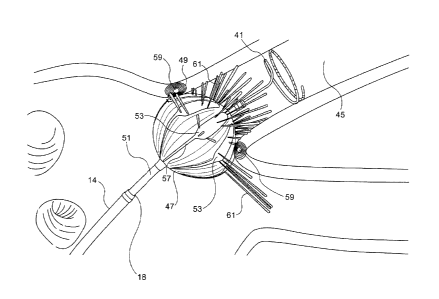
[0039] Reference is now made to Fig. 4, which is a pictorial side
view of distal segment of the catheter 14 (Fig. 1) shown in an operating
position at ostium 49 of pulmonary vein 45 in accordance with an em-
bodiment of the invention. The lasso guide 41 has been fully extended
through the distal tip 18. Once the guide is positioned in the vein, the
balloon 47, which is mounted on a shaft 51, extends beyond the distal
tip 18 of the catheter 14. The balloon 47 is inflated by injection with sa-
line solution, in order to close off the vein at the ostium 49. The bal-
loon 47 is fenestrated. Apertures or pores (best seen in Fig. 6) allow the
saline to irrigate the ostium 49. The balloon 47 has an electrode assem-
bly 53 disposed on its eternal surface. Multiple ablation electrodes are
disposed on the electrode assembly 53, as best seen in Fig. 5. The com-
ponents of the electrode assembly 53 are elongate, and directed longi-
tudinally in respective planes that are normal to the shaft 51 in order to
maximize galvanic contact between its electrodes 55 (Fig. 5) and the
wall of the ostium 49. Pigtails 57 prevent the electrode assembly 53
from delaminating when the balloon 47 is retracted into the shaft of the
catheter 14 and protect wires (not shown) leading to the electrodes of
the electrode assembly 53. Other geometric configurations for the elec-
trode assembly 53 are possible, for example a spiral arrangement, or
concentric rings. Passage of electrical energy through the electrodes 55
(Fig. 5) creates a circumferential lesion 59 at the ostium 49 that blocks
electrical propagation and isolates the pulmonary vein from the heart.
9 of 15
CA 02915224 2015-12-15
The ablation site is cooled by flow of a cooling fluid 61 through pores
formed in the balloon 47 and the electrode assembly 53. Alternatively, a
portion of the electrodes 55 may be configured for electrical mapping.
[0040] Reference is now made to Fig. 5, which is a bottom plan
view of the electrode assembly 53 in accordance with an embodiment of
the invention. The electrode assembly 53 is shown detached from the
balloon 47. The bottom surface of the electrode assembly 53 is adapted
to be adhered to the external surface of the balloon 47 (Fig. 4) The elec-
trode assembly 53 comprises a central aperture 63 through which the
shaft 51 (Fig. 4) extends. This arrangement permits injection of contrast
material or sampling through the shaft 51 as may be required by the
medical procedure. The electrode assembly 53 comprises a substrate of
radiating strips 65 that extend about the balloon 47 and are brought in-
to contact with a pulmonary vein ostium when the balloon is inflated
and navigated to the pulmonary vein. Electrodes 55 are disposed on
each of the strips 65, and come into galvanic contact with the ostium
during an ablation operation, during which electrical current flows
through the electrodes 55 and the ostium. Ten strips 65 are shown in
the example of Fig. 5 and are evenly distributed about of central axis the
aperture 63. Other numbers of strips are possible. However, there
should be a sufficiently small angle between adjacent strips 65 such
that at least one continuous circumferential lesion is produced in the
pulmonary vein when the electrodes 55 are activated for ablation.
[0041] Numerous pores 67 (typically 25-100 microns in diameter)
are formed through each of the strips 65 and perforate the underlying
balloon 47 as well. The pores 67 conduct a flow of cooling irrigation flu-
id from the interior of the balloon 47 onto and near the ablation site.
The flow rate may be varied by a pump control (not shown) from an idle
rate of about 4mL/min to the ablation flow rate of 60mL/min.
[0042] Reference is now made to Fig. 6, which is a top plan view
of the electrode assembly 53 in accordance with an embodiment of the
10 of 15
CA 02915224 2015-12-15
invention. Electrodes 55 are shown. In operation they come into contact
with the wall of the pulmonary vein.
[0043] Reference is now made to Fig. 7, which is a side elevation
of an embodiment of a balloon 69 having a proximal end 71 and a distal
end 73 in accordance with an embodiment of the invention. An elec-
trode assembly 75 is adhered to the exterior of the outer wall 77 of the
balloon 69. At its proximal end 71, the balloon 69 is narrowed and con-
figured to adapt to a connecting tube, which provides mechanical sup-
port and a supply of fluid. The distal end 73 is narrowed to permit fluid
continuity between the interior of the balloon 69 and the lumen of a
vessel.
[0044] Reference is now made to Fig. 8, which is a cut-away sec-
tional view through line 8-8 of the balloon 69 (fig. 7) in accordance with
an embodiment of the invention. A rim 79 seals the balloon 69 to a sup-
port (not shown), and prevents escape of fluid used for inflation of the
balloon and irrigation fluid. An inner passage 81 permits fluid commu-
nication between a vessel and a location outside the body. For example
contrast material may be transmitted through the passage 81.
[0045] Reference is now made to Fig. 9, which is a flow-chart of a
method of pulmonary vein isolation in accordance with an embodiment
of the invention. At initial step 83 a cardiac catheter is conventionally
introduced into the left atrium of a heart.
[0046] Next, at step 85 the lasso guide 41 is deployed and posi-
tioned to engage the interior wall of a pulmonary vein. Pre-ablation elec-
trograms may be acquired once the lasso guide 41 is in position.
[0047] Next, at step 87 the balloon 47 is extended over the lasso
guide 41 and inflated.
[0048] Next, at step 89 the balloon 47 is navigated into circum-
ferential contact with a pulmonary vein ostium in order to occlude the
ostium.
[0049] Next, at step 91 a radio-opaque contrast agent is injected
through the lumen of the catheter, The contrast agent passes through a
11 of 15
CA 02915224 2015-12-15
gap between the lasso guide 41 and the wall of the lumen in order to
confirm that the balloon 47 is in a correct position against the pulmo-
nary vein ostium. The contrast agent does not enter the balloon.
[0050] Control now
proceeds to decision step 93, where it is de-
termined if the balloon 47 is correctly positioned. If the determination
at decision step 93 is negative, then control returns to step 89 and an-
other attempt is made to position the balloon.
[0051] If the
determination at decision step 93 is affirmative,
then control proceeds to step 95 where ablation is performed using the
ablation electrodes of the electrode assembly 53 (Fig. 4). A circumferen-
tial lesion is created in a region of tissue that circumscribes the pulmo-
nary vein. The lesion blocks electrical propagation and effectively elec-
trically isolates the pulmonary vein from the heart. Post-ablation elec-
trograms may be obtained from the electrodes 43 of the lasso guide 41
(Fig. 2) in order to confirm functional isolation of the pulmonary vein.
[0052] After completion of the ablation, the procedure may be it-
erated using another pulmonary vein ostium by withdrawal of the bal-
loon 47 and the lasso guide 41. Control may then return to step 85. Al-
ternatively, the procedure may end by removal of the catheter 14 at fi-
nal step 97.
[0053] It will be
appreciated by persons skilled in the art that the
present invention is not limited to what has been particularly shown
and described hereinabove. Rather, the scope of the present invention
includes both combinations and sub-combinations of the various
features described hereinabove, as well as variations and modifications
thereof that are not in the prior art, which would occur to persons
skilled in the art upon reading the foregoing description.
12 of 15