Note: Descriptions are shown in the official language in which they were submitted.
CA 02915378 2015-12-14
WO 2014/204957
PCT/US2014/042730
- 1 -
VENTED IMPLANTABLE DRUG-DELIVERY DEVICE
CROSS-REFERENCE TO RELATED APPLICATIONS
[0001] The present application claims priority to, and the benefits of,
U.S. Serial No.
61/835,832, filed on June 17, 2013, the entire disclosure of which is hereby
incorporated by
reference.
FIELD OF THE INVENTION
[0002] In various embodiments, the present invention relates generally to
implantable
medical devices and, more specifically, to devices in which a pressure offset
is created within
the device or at the interface between the device and its surroundings.
BACKGROUND
[0003] Implantable drug-delivery devices typically utilize an actuation
mechanism to drive
medicament from a reservoir through a cannula into target areas. The actuation
mechanism
may be pressure-driven or cause pressure changes within the drug-delivery
device or at the
interface between the device and its surroundings. The pressure magnitudes and
gradients in
these regions can make it difficult to precisely control delivery of small
amounts of drug,
especially when the device is refillable or used for repeated dosing over a
relatively long
period. For example, without proper regulation of the pressure in the drug
reservoir, pressure
or vacuum buildup can interfere with smooth, continuous administration of a
liquid
medicament. This problem is particularly acute in devices whose driving
mechanism involves
generation of pressurized gas. In such devices, excess gas can leak to various
device regions.
More generally, when the device is implanted in a patient, the difficulties of
limited physical
space and access to the device, as well as the overall complexity of in vivo
implantation and
operation, can make pressure regulation in the device essential and exacerbate
the problems
arising from inadequate regulation.
[0004] Gas-driven drug-delivery devices may produce excess gas, and
ensuring gas-
tightness along the pressurization route can require significant efforts in
design, manufacture
and quality control. For example, in electrolytic drug-delivery devices,
hydrogen and oxygen
CA 02915378 2015-12-14
WO 2014/204957
PCT/US2014/042730
- 2 -
are generated as an actuating mechanism during dosing. Hydrogen is known to
penetrate thin
walls easily and leak into reservoir chambers and their perimeters, resulting
in inaccurate
pressure-dosing characteristics or even unintended delivery of gas. For some
drug-delivery
regimes, instantaneous bursts of drug may be required (alone or to supplement
steady-state
delivery). The excess gas and its effects on delivery accuracy can be pose
major challenges,
especially in the sub-milliliter scale.
[0005] Excess gas can also adversely affect the refilling of drug-
delivery devices. As
excess gas accumulates in the drug reservoir chambers, refill routes, and/or
other adjacent
interior spaces, it can complicate the refilling process and create
considerable dead volume.
More importantly, some drug-delivery devices have compliant reservoir walls to
minimize dead
volumes and provide ease in handling during refilling. With these devices, the
excess gas
accumulating in the perimeter creates a differential pressure that can
eventually prevent the
refilling operation from proceeding to completion.
[0006] Venting may seem like an obvious solution to unwanted gas buildup,
but can be
difficult to achieve in devices intended for implantation. While valved
passages connecting the
pump to a region outside of the device body have been proposed for managing
excess gas in
drug-delivery devices, such an approach is often unsuitable for biomedical
implants, as the
transport of gases through the human body via a catheter or artificial vehicle
for venting may be
painful and increase risk of infection. In addition, as most biomedical
implants are highly
integrated and miniaturized, the limited physical space and access to the
device further
complicates venting: the venting component in an implantable drug-delivery
device must
generally be compact, easy to integrate and, notably, compatible with the
anatomic
environment in which various body fluids and tissues may interact with the
vent.
[0007] One possible approach to venting an implantable drug-delivery
device is to connect
additional gas-filled space to the region of excess gas accumulation in order
to buffer abrupt
pressure changes inside the device. This may be additional space within the
device itself or a
chamber that is tethered by a fluidic connection but external to the main drug-
delivery device.
This approach, however, requires a relatively large space that may be
impractical for
biomedical applications that demand space efficiency. Additionally, without a
passage through
which excess gas may be expelled from the device, pressure will continue to
build up within,
and potentially overwhelm, the buffer volume. Another possible approach would
employ a gas-
CA 02915378 2015-12-14
WO 2014/204957
PCT/US2014/042730
- 3 -
permeable outer shell to expel excess gas. This approach, however, would pose
challenges of
material choice, fabrication complexity, fabrication cost, and compromised
mechanical strength
of the surface. Furthermore, pores that confer gas permeability can also allow
for tissue
ingrowth that may block a sufficient number of the pores to compromise their
effectiveness.
SUMMARY
[0008] Embodiments of the invention utilize a selectively permeable
membrane structure
integrated in the outer shell and/or in other areas of an implantable drug-
delivery device. In
various embodiments, the device includes an aperture through the housing of
the device and,
spanning the aperture, a membrane structure permeable to gas but not to
liquid. In this way,
excess gas may be vented from the device. The membrane and aperture are
designed to
discourage or even prevent tissue ingrowth.
[0009] Accordingly, in a first aspect, the invention pertains to an
implantable device for
administering a liquid. In various embodiments, the device comprises a housing
including an
aperture therethrough; within the housing, a pump assembly including a
reservoir, a gas-driven
forcing mechanism and a cannula for conducting liquid from the reservoir to an
ejection site
exterior to the housing in response to pressure applied by the forcing
mechanism; and external
to the pumping mechanism but within the housing and spanning the aperture, a
membrane
structure comprising a gas-permeable membrane and at least one support layer
attached thereto.
The membrane structure is permeable to gas but not to liquid at least within
the area thereof
exposed by the aperture.
[0010] In various embodiments, the membrane structure, at least within
the area thereof
exposed by the aperture, has a pore size sufficiently small to prevent tissue
ingrowth and
endotheliazation. Furthermore, the membrane structure may have a pore size
that allows gas to
flow therethrough at a sufficient rate to substantially offset a positive
pressure or vacuum
pressure applied to the device. The membrane structure may be biocompatible.
At least the
surface of the membrane structure exposed by the aperture may comprise an
oleophobic
coating thereover. For example, the membrane structure may comprise or consist
essentially of
ePTFE.
[0011] At least a portion of the membrane structure surface may comprise
(e.g., have
coated thereon) an adhesive material for affixation to an interior surface of
the housing. A
CA 02915378 2015-12-14
WO 2014/204957
PCT/US2014/042730
- 4 -
portion of the membrane structure may be bonded to an interior surface of the
housing with an
epoxy. The membrane structure may have a thickness less than 500 m.
[0012] In some embodiments the support layer(s) is/are perforated. For
example, the
support layer(s) may be perforated with clusters of holes each having a
diameter in the range of
50-400[1m. The support layer(s) may be substantially rigid. In various
embodiments, the
support layer(s) comprise or consist essentially of one or more of
polypropylene, polyethylene,
polyvinylidene fluoride, poly(methyl methacrylate), or polyether ether ketone.
In some
embodiments, the support layer(s) comprise or consist essentially of one or
more of spunbond
fabric, a woven fabric, an extruded film, a cast film, a blown film or an
injection-molded film.
[0013] Reference throughout this specification to "one example," "an
example," "one
embodiment," or "an embodiment" means that a particular feature, structure, or
characteristic
described in connection with the example is included in at least one example
of the present
technology. Thus, the occurrences of the phrases "in one example," "in an
example," "one
embodiment," or "an embodiment" in various places throughout this
specification are not
necessarily all referring to the same example. Furthermore, the particular
features, structures,
routines, steps, or characteristics may be combined in any suitable manner in
one or more
examples of the technology. The headings provided herein are for convenience
only and are not
intended to limit or interpret the scope or meaning of the claimed technology.
The term
"substantially" or "approximately" means 10% (e.g., by weight or by volume),
and in some
embodiments, 5%.
BRIEF DESCRIPTION OF THE DRAWINGS
[0014] In the drawings, like reference characters generally refer to the
same parts
throughout the different views. Also, the drawings are not necessarily to
scale, with an
emphasis instead generally being placed upon illustrating the principles of
the invention. In the
following description, various embodiments of the present invention are
described with
reference to the following drawings, in which:
[0015] FIG. 1A schematically illustrates the outer shell of a device in
accordance with the
present invention, the outer shell including a selectively permeable membrane
structure.
[0016] FIG. 1B is an elevation of an embodiment of the selectively
permeable membrane
structure.
CA 02915378 2015-12-14
WO 2014/204957
PCT/US2014/042730
- 5 -
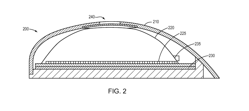
[0017] FIG. 2 is a sectional elevation of a representative drug-delivery
device including the
an embodiment of the invention.
[0018] FIG. 3 is an enlarged portion of the elevation shown in FIG. 2.
[0019] FIG. 4 is an enlarged perspective view of an aperture spanned by
the selectively
permeable membrane structure.
[0020] FIG. 5 is a sectional elevation of a representative drug-delivery
device illustrating
its mode of operation.
DETAILED DESCRIPTION
[0021] Embodiments of the present invention provide a vent solution based
on a selectively
permeable membrane structure integrated into the rigid outer shell of an
implantable drug-
delivery device. Although the ensuing discussion focuses on the integration
into the outer
shell, this vent solution may be deployed in other areas of the implantable
drug-delivery device
that may require venting (e.g., the refill port). Additionally, it should be
understood that the
selectively permeable membrane structure may be placed above or below any
surface of a drug-
delivery device that is perforated or allows for some form of fluid/gas
permeation.
[0022] With reference to FIGS. 1A and 1B, the outer shell 100 of an
implantable device
that generates excess gas is provided with a vent 110, which comprises or
consists of an
aperture through the shell 100 and, coextensive with or (more typically)
extending beyond the
perimeter of the aperture, a selectively permeable membrane structure. The
rigid shell 100 may
be made of a metal such as titanium or may consist of, or include, a
biocompatible plastic
material alternatively or in addition. More generally, the shell 100 may
include or consist
essentially of one or more of a ceramic, an epoxy encapsulation, a metal
(e.g., titanium (Ti),
niobium (Nb), or tantalum (Ta)), polyetherether ketone (PEEK), polypropylene,
polydimethylsiloxane (PDMS), or parylene. For example, the shell may be at
least partially
coated with parylene.
[0023] The membrane structure 115, a representative embodiment of which
is illustrated in
FIG. 1B, may have multiple layers comprising or consisting of a functional
layer (i.e., a gas-
permeable membrane) 120 and one or more support layers 130. For example, the
permeable
layer 120 may be a membrane laminated onto a plastic thin film as a backing
layer 130. The
support layer 130 may have a series of perforations 135 to permit the passage
of gas through
CA 02915378 2015-12-14
WO 2014/204957
PCT/US2014/042730
- 6 -
the functional layer 120. Alternatively, the support layer 130 may have a
single large opening
or multiple large slots beneath a portion of the functional layer 120 through
which gas is
released.
[0024] Additional layers can also be incorporated for improved adhesion
to the device outer
shell 100 and enhanced overall mechanical strength of the vent port 110. Other
suitable
adhesion techniques known in the field of implantable medical devices may also
be used. For
example, a biocompatible epoxy may be used to join the gas-permeable layer 120
to the
backing layer(s) 130, as well as to join the resulting structure 115 to the
outer shell 100 of the
implantable pump device as shown in greater detail below. The layer(s) of the
structure 120
that actually adheres to the shell 100 can undergo surface treatment such as
sandblasting and/or
plasma bombardment to improve adhesion when using biocompatible epoxy.
[0025] FIGS. 2-5 show a representative deployment of the invention in an
implantable
electrolytic drug pump 200. With reference to FIGS. 2-4, the pump 200 includes
a hard outer
shell 210, which may be made of, for example, titanium. Within the shell 210
is a dome-
shaped structure 220, which may be formed from a hard polymer, such as acrylic
or metals
such as titanium, aluminum or other biocompatible material. Alternatively, the
dome-shaped
structure 220 may be made of a shape-retaining but compliant material such as
parylene; at a
thickness of 100 pm, for example, it is found that a parylene structure 220
maintains its shape
but is capable of slight flexure under pressure. A combination of the
foregoing materials may
also be used by coating with parylene any surfaces that may contact drug or
bodily fluids. At
the floor of the dome 220 is a corrugated, expandable membrane 225, which may
be made of
parylene, silicone or other suitably flexible material. Beneath the membrane
225 is a set of
electrolysis electrodes on a floor 230, and an electrolysis liquid is
contained within the space
formed by the floor 230 and the expandable membrane 225. The space between the
expandable
membrane 225 and the dome 220 contains the drug to be dispensed; a cannula 235
is in fluid
communication with this interior space (i.e., drug chamber). As best seen in
FIGS. 3 and 4, an
aperture 240 extends through the shell 210, and beneath the aperture 240 is a
selectively
permeable membrane structure 250 including a gas-permeable, liquid-impermeable
functional
layer 255 and a support layer 260. As noted earlier, the support layer 260 may
have
perforations or an enlarged opening within the aperture 240 to permit gas to
flow through the
functional layer and out the aperture. As shown in FIG. 4, the upper surface
of the membrane
structure 250 is bonded to the interior surface of the dome 220. In some
embodiments, the
CA 02915378 2015-12-14
WO 2014/204957
PCT/US2014/042730
- 7 -
peripheral edge of the support layer 260 extends beyond that of the functional
layer 255, and it
is the exposed annular upper surface of the support layer 260 that is actually
adhered to the
dome 220.
[0026] Various other embodiments may incorporate the one or more of the
functional and
support layers into the shell by various methods. In one embodiment, the
aperture is tiered into
one or more steps, and each layer or subset of layers may be incorporated to
be flush with the
subsequent step in the aperture. Alternative approaches to adhering and
securing the layers
such as the use of pins, screws, or tabs may be employed to bond the layers
and integrate the
membrane structure into the dome.
[0027] The operation of the pump device 200 is illustrated in FIG. 5. Upon
activation of
the electrodes 275, gas is evolved from the liquid in the electrolysis chamber
280 (which is
bounded by the floor 230 and the expandable membrane 225), inflating the
membrane 225 and
thereby reducing the volume of the drug chamber 280, forcing liquid therein
out through the
cannula 235. The cannula 235 may be equipped with a check valve and/or a flow
sensor.
Suitable control circuitry and a battery (not shown) may be mounted on a
circuit board
integrated into the bottom portion of the housing 210; see, e.g., U.S. Patent
Nos. 8,285,328 and
8,231,608, the entire disclosures of which are hereby incorporated by
reference. In some
embodiments, the electrodes 275 are etched, printed, or otherwise deposited
directly onto the
circuit board for cost-savings and ease of manufacturing.
[0028] Gas penetrating the dome structure 220 and accumulating in the dead
space between
that structure and the hard shell 210 is vented through the aperture 240,
which, again, is
spanned by the gas-permeable membrane structure 250 as described above.
[0029] The functional layer 120 of the membrane structure 115 desirably
has a high
permeability to most gases to allow for rapid gas transit but is virtually
impermeable to liquid,
preventing the intrusion of, for example, aqueous fluids. The pore diameter of
the layer 120 is
chosen to be much smaller (e.g., orders of magnitude smaller) than the typical
pore size that
would permit tissue ingrowth and endothelialization, so that the ingrowth of
soft tissues in the
vent can be minimized. Although the minimum pore size permitting tissue
ingrowth depends on
the surrounding tissue, in general it ranges from ¨10 m to a few mm (which is
much greater
than the permeable membrane pore size required to create adequate gas
permeability).
CA 02915378 2015-12-14
WO 2014/204957
PCT/US2014/042730
- 8 -
[0030] Typically, the support layer 130 is one or more layers of solid
thin film with
adequate mechanical strength and a surface bondable to the interior wall of
the shell 210.
Depending on the material used, the support layer 130 can be as-manufactured
(e.g., if porous)
or intentionally perforated, as discussed above, at least in the venting area
for unobstructed gas
passage. As a result, even a small aperture 240 is capable of releasing excess
gas at a
reasonably high rate under a low differential pressure. In addition, the
surface of membrane
structure 115 within the aperture 240 may be treated to impart or enhance
oleophobicity in
order to reject molecules in human body fluids (e.g., proteins, lipids, and
blood cells) that might
interfere with gas exchange. One example surface coating is super-hydrophobic
reagent such
as a monolayer of TEFLON. With these attributes, the venting arrangement of
the present
invention provides rapid pressure equilibration between the internal space of
the device and the
human body environment where the device is implanted for long-term
applications. Its function
does not require direct access to the device 200 for manipulation of the gas-
driving
components.
[0031] The materials of the permeable membrane 120 and the backing layer(s)
130 are
chosen based on both the functionalities and the biomedical compatibility. The
gas-permeable
membrane 120 can be expanded polytetrafluoroethylene (ePTFE) with a pore
diameter on the
scale of submicrons. Alternatively, TEFLON AF or other materials having (or
which can be
manipulated to have) an inter-nodal distance that is permeable to gas but not
liquid may be
employed.
[0032] The gas-permeable membrane 120 may further be altered to enhance
robustness
while maintaining acceptable gas flow rates. One approach is to use a thicker
membrane or to
create a thicker membrane by stacking multiple layers of membranes. In one
embodiment an
ePTFE membrane with a pore diameter between 0.2-0.41m and an intermodal
distance of 10iim
and a thickness of over 600[.tm exhibited a mass flow rate of over 0.5mL/min
under a driving
pressure below 0.05psi.
[0033] This driving pressure has been calculated to be more than
sufficient to drive the gas
flow through the gas-permeable membrane 120. By using ideal gas law, PV = nRT,
the
equation of PVI=P'(V1+AV) shows the pressure change caused by a change in
pressure that
occurs as incremental amounts of drug are pumped from the reservoir. Thus,
AP=AV/(VI-FAV)P, where P = atmospheric pressure (14.7psi), AV = change in mass
of the
CA 02915378 2015-12-14
WO 2014/204957
PCT/US2014/042730
- 9 -
drug reservoir, and V1= space between the dome 220 and the hard shell 110. In
one
embodiment, the drug reservoir is filled to 300 L, leaving V1= 2000¨ A dose
of 50 L
creates a pressure change of 50/(200+50)14.7psi = 2.94psi. Applying this
pressure differential
to the above embodiment of an ePTFE membrane, an adequate gas flow rate is
achieved.
[0034] In embodiments where a vacuum is created in the space between the
dome 220 and
the hard shell 110 with each subsequent dose, the vacuum may be offset by
drawing gas in
through the gas-permeable membrane. In certain implant positions, the vacuum
may not be
offset if adequate gas cannot be drawn in from the environment. However, the
vacuum is
beneficial in that it promotes refilling, which is inhibited by gas
accumulation in the space
between the dome and the hard shell. This gas is vented out through the gas-
permeable
membrane.
[0035] The backing layer(s) 130 can be any one or more of various plastic
thin films
including polypropylene, polyethylene, polyvinylidene fluoride (PVDF),
poly(methyl
methacrylate) (PMMA), and PEEK. The backing layer(s) can take the form of a
spunbond or
woven fabric with intrinsic gas permeability, or extruded, cast, blown, or
injection-molded
solid films perforated with clusters of holes (at least where the layer will
face the hard-shell
aperture) each having a diameter in the range of 50-400 m. The overall
thickness of the
membrane structure 115 can be smaller than 500 m.
[0036] The backing layer 130 may further be altered to enhance robustness
while
maintaining acceptable gas-flow rates. According to principles of material
strength, the
deflection of an edge-clamped plate is highly related to its diameter. By
using a refined
perforation pattern on the support, the membrane deformation under
pressurization/vacuum can
be minimized. The porosity typically reduces with hole diameter, which can be
expressed by a
model featuring an array of uniformly distributed holes:
m(dhole/2)2
porosity =
ldspacing dhole)2
While the hole diameter can be further minimized by using advanced techniques
such as deep-
UV laser drilling, the spacing of holes is primarily limited due to both
technical and economical
CA 02915378 2015-12-14
WO 2014/204957
PCT/US2014/042730
- 10 -
reasons. For example, a hole diameter of 5 gm and typical spacing of 20 gm
results in a
porosity of approximately 3%. Because of the gas permeability of the venting
membrane,
lower porosities within this range may be used while still enabling efficient
venting.
[0037] In some embodiments, the membrane structure 115 is integrated into
the same plane
or applied to the internal or external surface of the implantable drug
delivery device in different
configurations. The membrane structure is not limited in terms of shape, size
or orientation.
For example, it may take the form of strips, circles, ovals, squares, or any
other pattern.
Furthermore, the layers of the membrane can be of different shapes and sizes
to allow for better
adhesion and provide a seamless integration with the surface of the
implantable drug delivery
device.
[0038] Certain embodiments of the present invention have been described
above. It is,
however, expressly noted that the present invention is not limited to those
embodiments,
but rather the intention is that additions and modifications to what was
expressly
described herein are also included within the scope of the invention.
Moreover, it is to be
understood that the features of the various embodiments described herein were
not
mutually exclusive and can exist in various combinations and permutations,
even if such
combinations or permutations were not made express herein, without departing
from the
spirit and scope of the invention. In fact, variations, modifications, and
other
implementations of what was described herein will occur to those of ordinary
skill in the art
without departing from the spirit and the scope of the invention. As such, the
invention is
not to be defined only by the preceding illustrative description.