Note: Descriptions are shown in the official language in which they were submitted.
CA 02915626 2015-12-15
WO 2014/204991 PCT/US2014/042785
METHOD FOR DETERMINING COPY NUMBER VARIATIONS IN SEX
CHROMOSOMES
CROSS REFERENCE TO RELATED APPLICATIONS
This application claims benefit of priority under 35 U.S.C. 119(e) to U.S.
Provisional Patent Application No. 61/836,057, titled "METHOD FOR
DETERMINING COPY NUMBER VARIATIONS IN SEX CHROMOSOMES" and
filed on June 17, 2013 (Attorney Docket No. ARTEPOO8P), which is hereby
incorporated by reference in its entirety.
BACKGROUND
One of the critical endeavors in human medical research is the discovery of
genetic abnormalities that produce adverse health consequences. In many cases,
specific genes and/or critical diagnostic markers have been identified in
portions of
the genome that are present at abnormal copy numbers. For example, in prenatal
diagnosis, extra or missing copies of whole chromosomes are frequently
occurring
genetic lesions. In cancer, deletion or multiplication of copies of whole
chromosomes
or chromosomal segments, and higher level amplifications of specific regions
of the
genome, are common occurrences.
Most information about copy number variation (CNV) has been provided by
cytogenetic resolution that has permitted recognition of structural
abnormalities.
Conventional procedures for genetic screening and biological dosimetry have
utilized
invasive procedures, e.g., amniocentesis, cordocentesis, or chorionic villus
sampling
(CVS), to obtain cells for the analysis of karyotypes. Recognizing the need
for more
rapid testing methods that do not require cell culture, fluorescence in situ
hybridization (FISH), quantitative fluorescence PCR (QF-PCR) and array-
Comparative Genomic Hybridization (array-CGH) have been developed as molecular-
cytogenetic methods for the analysis of copy number variations.
The advent of technologies that allow for sequencing entire genomes in
relatively short time, and the discovery of circulating cell-free DNA (cfDNA)
have
provided the opportunity to compare genetic material originating from one
chromosome to be compared to that of another without the risks associated with
invasive sampling methods, which provides a tool to diagnose various kinds of
copy
number variations of genetic sequences of interest.
1
CA 02915626 2015-12-15
WO 2014/204991 PCT/US2014/042785
Diagnosis of copy number variations of the Y chromosome involves
heightened technical challenges compared to autosomes, because coverage of the
Y
chromosome is lower than that of autosomes, and repeated sequences on the Y
chromosome complicate mapping of reads to their correct location. There are
about
10 Mb of unique Y sequences accessible by current NGS technologies, but gender
detection remains to be a challenging task in fetal diagnostic world where the
amount
of fetal cfDNA in a maternal sample is at least an order of magnitude lower
than that
of maternal DNA, emphasizing the problem of nonspecific mapping. Additionally,
some current sequencing protocols utilize ultra-short reads such as 25mer
reads and
tags, presenting yet another alignment challenge since 25mer tags are shorter
than
typical size of most ubiquitous repeatable elements. Some embodiments
disclosed
herein describe a strategy for filtering out (or masking) non-discriminant
sequence
reads on chromosome Y using representative training set of female samples. In
some
embodiments, this filtering strategy is also applicable to filtering autosomes
for
evaluation of copy number variation of sequences on the autosomes.
Limitations of existing methods in noninvasive prenatal diagnostics, which
include insufficient sensitivity stemming from the limited levels of cfDNA,
and the
sequencing bias of the technology stemming from the inherent nature of genomic
information, underlie the continuing need for noninvasive methods that would
provide
any or all of the specificity, sensitivity, and applicability, to reliably
diagnose copy
number changes in a variety of clinical settings. Embodiments disclosed herein
fulfill
some of the above needs and in particular offers an advantage in providing a
reliable
method that is applicable to the practice of noninvasive prenatal diagnostics.
SUMMARY
In some embodiments, methods are provided for determining copy number of
the Y chromosome, including, but not limited to, methods for gender
determination or
Y chromosome aneuploidy of fetus using maternal samples comprising maternal
and
fetal cell free DNA.
In some embodiments, methods are provided for determining copy number
variation (CNV) of any fetal aneuploidy, and CNVs known or suspected to be
associated with a variety of medical conditions. CNV that can be determined
according to the present method include trisomies and monosomies of any one or
2
CA 02915626 2015-12-15
WO 2014/204991 PCT/US2014/042785
more of chromosomes 1-22, X and Y, other chromosomal polysomies, and deletions
and/or duplications of segments of any one or more of the chromosomes, which
can
be detected by sequencing only once the nucleic acids of a test sample. Any
aneuploidy can be determined from sequencing information that is obtained by
sequencing only once the nucleic acids of a test sample.
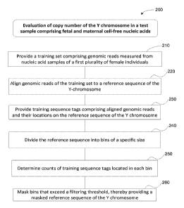
In one embodiments, the method comprises: (a) providing, on the computer
system, a training set comprising genomic reads measured from nucleic acid
samples
of a first plurality of female individuals; (b) aligning, by the computer
system, at least
about 100,000 genomic reads per individual of the training set to a reference
sequence
of the Y-chromosome, thereby providing training sequence tags comprising
aligned
genomic reads and their locations on the reference sequence of the Y
chromosome;
(c) dividing, by the computer system, the reference sequence of the Y
chromosome
into a plurality of bins; (d) determining, by the computer system, counts of
training
sequence tags located in each bin; (e) masking, by the computer system, bins
that
exceed a masking threshold, the masking threshold being based on the counts of
training sequence tags in each bin, thereby providing a masked reference
sequence of
the Y chromosome for evaluation of copy number of the Y chromosome in a test
sample. In some embodiments, the test sample comprises fetal and maternal cell-
free
nucleic acids.
In some embodiments, the method for evaluation of copy number of the Y
chromosome in a test sample further comprises: (f) sequencing the cell free
nucleic
acids from the test sample comprising fetal and maternal cell-free nucleic
acids using
a sequencer, thereby generating genomic reads of the test sample; and (g)
aligning, by
the computer system, the genomic reads of the test sample to the reference
sequence,
thereby providing testing sequence tags comprising aligned genomic reads and
locations thereof.
In some embodiments, the method for evaluation of copy number of the Y
chromosome in a test sample further comprises: (h) measuring, by the computer
system, counts of the testing sequence tags on the masked reference sequence
of the Y
chromosome; and (i) evaluating, by the computer system, copy number of the Y
chromosome in the test sample based on the counts of the testing sequence tags
on the
masked reference sequence of the Y chromosome.
3
CA 02915626 2015-12-15
WO 2014/204991 PCT/US2014/042785
In any one of the embodiments described above, the test sample may be a
maternal sample selected from blood, plasma, serum, urine and saliva samples.
In
any one of the embodiments, the test sample is may be plasma sample. The
nucleic
acid molecules of the maternal sample are a mixture of fetal and maternal cell-
free
DNA molecules. Sequencing of the nucleic acids can be performed using next
generation sequencing (NGS). In some embodiments, sequencing is massively
parallel sequencing using sequencing-by-synthesis with reversible dye
terminators. In
other embodiments, sequencing is sequencing-by-ligation. In yet other
embodiments,
sequencing is single molecule sequencing. Optionally, an amplification step is
performed prior to sequencing.
Another embodiment provides a method for identifying copy number variation
(CNV) of a sequence of interest, e.g., a clinically relevant sequence, in a
test sample.
The method assesses copy number variation of sequences of interest instead of
complete chromosomes or segments of chromosomes.
In certain embodiments embodied on a computer system, the number of
sequence tags identified for each of the one or more chromosomes of interest
or
chromosome segments of interest is at least about 10,000, or at least about
100,000.
The disclosed embodiments also provide a computer program product including a
non-transitory computer readable medium on which is provided program
instructions
for performing the recited operations and other computational operations
described
herein.
In some embodiments, a method additionally includes sequencing at least a
portion of said nucleic acid molecules of said maternal test sample to obtain
said
sequence information for said fetal and maternal nucleic acid molecules of
said test
sample. The sequencing may involve massively parallel sequencing on maternal
and
fetal nucleic acids from the maternal test sample to produce the sequence
reads.
In some embodiments, the masking threshold is determined by operations
performed by or on the computer system: providing two or more masking
threshold
candidates; masking bins that exceed the masking threshold candidates, thereby
providing two or more masked reference sequences; calculating a threshold
evaluation index for evaluation of copy number of the genetic sequence of
interest
4
CA 02915626 2015-12-15
WO 2014/204991 PCT/US2014/042785
based on each of the two or more masked reference sequences; and selecting the
candidate having the highest threshold evaluation index as the masking
threshold.
In some embodiments, calculating the threshold evaluation index includes
evaluating copy number of the Y chromosome for nucleic acid samples of (a)
female
individuals different from the female individuals of the training set and (b)
male
individuals known to have a Y chromosome. In some embodiments, the threshold
evaluation index is calculated as the difference between the means of (a) and
(b),
divided by the standard deviation
In some embodiments, the size of each bin is determined by operations of a
computer system: dividing the reference sequence of the Y chromosome into bins
of a
candidate bin size; calculating a bin evaluation index based on the candidate
bin size;
iteratively repeating the preceding steps of this claim on the computer system
using
different candidate bin sizes, thereby yielding two or more different
evaluation
indices; and electing the candidate bin size yielding the highest bin
evaluation index
as the size of the bins.
In some embodiments, female individuals of a training set have diverse
alignment profiles characterized by different distributions of the genomic
reads on the
reference sequence of the Y chromosome. In some embodiments, providing a
training
set involves dividing a second plurality of female individuals into two or
more
clusters and selecting a number of individuals in each of the two or more
clusters to
form the first plurality of female individuals as members of the training set.
In some
embodiments, an equal number of individuals are selected in each of the two or
more
clusters. In some embodiments, the dividing the plurality of female
individuals into
two or more clusters involves hierarchical ordered partitioning and collapsing
hybrid
(HOPACH) clustering.
In some embodiments, a method further includes automatically recording,
using a processor, the presence or absence of a fetal chromosomal aneuploidy
as
determined as described above in a patient medical record for a human subject
providing the maternal test sample. The
recording may include recording
chromosome doses and/or a diagnosis based said chromosome doses in a computer-
readable medium. In some cases, the patient medical record is maintained by a
5
CA 02915626 2015-12-15
WO 2014/204991 PCT/US2014/042785
laboratory, physician's office, a hospital, a health maintenance organization,
an
insurance company, or a personal medical record website. A method may further
include prescribing, initiating, and/or altering treatment of a human subject
from
whom the maternal test sample was taken. Additionally or alternatively, the
method
may include ordering and/or performing one or more additional tests.
In some embodiments, system and computer program products are provided to
perform the methods for evaluation of copy number of a genetic sequence of
interest
in a test sample.
Although the examples herein concern humans and the language is primarily
directed to human concerns, the concepts described herein are applicable to
genomes
from any plant or animal.
INCORPORATION BY REFERENCE
All patents, patent applications, and other publications, including all
sequences
disclosed within these references, referred to herein are expressly
incorporated herein
by reference, to the same extent as if each individual publication, patent or
patent
application was specifically and individually indicated to be incorporated by
reference. All documents cited are, in relevant part, incorporated herein by
reference
in their entireties for the purposes indicated by the context of their
citation herein.
However, the citation of any document is not to be construed as an admission
that it is
prior art with respect to the present disclosure.
BRIEF DESCRIPTION OF THE DRAWINGS
Figure 1 shows sequence classes, genes, and palindromes on the human Y
chromosome. (a) Schematic representation of the entire human Y chromosome,
with
the male-specific region (MSY) indicated. (b) A more detailed representation
that
focuses on the euchromatic MSY and excludes the major heterochromatic block on
Yq.
Figure 2 shows an example of regions that are masked on the Y chromosome
in one embodiment. The masked Y chromosome can be used as a reference sequence
for evaluation of copy number of the Y chromosome.
Figure 3A-3B show block diagrams of embodiments of a method for
evaluation of copy number of the Y chromosome in a test sample comprising
fetal and
6
CA 02915626 2015-12-15
WO 2014/204991 PCT/US2014/042785
maternal cell-free nucleic acids. In some embodiments, the method is
implemented at
a computer system that includes one or more processors and system memory.
Figure 4 is a flowchart of a method 100 for determining the presence or
absence of a copy number variation in a test sample comprising a mixture of
nucleic
acids.
Figure 5 is a block diagram of a dispersed system for processing a test sample
and ultimately making a diagnosis.
Figure 6 schematically illustrates how different operations in processing test
samples may be grouped to be handled by different elements of a system.
Figures 7A and 7B shows electropherograms of a cfDNA sequencing library
prepared according to the abbreviated protocol described in Example la (Fig.
7A),
and the protocol described in Example lb (Fig. 7B).
Figure 8 illustrates a heatmap of pairwise chrY lkb coverage correlations
across 475 females, sorted by using HOPACH results.
Figure 9 shows the ChrY ratio (i.e. chrY count/chr4 count) in 1 Mb vs. lkb bin
sizes for female (2) and male (3).
Figure 10 shows Male/Female discrimination signal to noise ratio as a
function of fraction of bins masked.
Figure 11 shows the frequency distribution of sequence tags mapped to the Y
chromosome for samples including female (light gray) vs. male (dark gray)
fetal
cfDNAs. The left panel shows the distribution of sequence tags mapped to an
unmasked Y chromosome. The right panel shows the distribution mapped to a
masked Y chromosome according to methods described herein.
Figures 12A and 12B illustrate the distribution of the chromosome dose for
chromosome 21 determined from sequencing cfDNA extracted from a set of 48
blood
samples obtained from human subjects each pregnant with a male or a female
fetus.
Chromosome 21 doses for qualified, i.e., normal for chromosome 21 (0), and
trisomy
21 test samples are shown (A) for chromosomes 1-12 and X (Figure 12A), and for
chromosomes 1-22 and X (Figure 12B).
Figures 13A and 13B illustrate the distribution of the chromosome dose for
chromosome 18 determined from sequencing cfDNA extracted from a set of 48
blood
7
CA 02915626 2015-12-15
WO 2014/204991 PCT/US2014/042785
samples obtained from human subjects each pregnant with a male or a female
fetus.
Chromosome 18 doses for qualified, i.e., normal for chromosome 18 (0), and
trisomy
18 (A) test samples are shown for chromosomes 1-12 and X (Figure 13A), and for
chromosomes 1-22 and X (Figure 13B).
Figures 14A and 14B illustrate the distribution of the chromosome dose for
chromosome 13 determined from sequencing cfDNA extracted from a set of 48
blood
samples obtained from human subjects each pregnant with a male or a female
fetus.
Chromosome 13 doses for qualified, i.e., normal for chromosome 13 (0), and
trisomy
13 (A) test samples are shown for chromosomes 1-12 and X (Figure 14A), and for
chromosomes 1-22 and X (Figure 14B).
Figures 15A and 15B illustrate the distribution of the chromosome doses for
chromosome X determined from sequencing cfDNA extracted from a set of 48 test
blood samples obtained from human subjects each pregnant with a male or a
female
fetus. Chromosome X doses for males (46,XY; (0)), females (46,XX; (A));
monosomy X (45,X; (+)), and complex karyotypes (Cplx (X)) samples are shown
for
chromosomes 1-12 and X (Figure 15A), and for chromosomes 1-22 and X (Figure
15B).
Figures 16A and 16B illustrate the distribution of the chromosome doses for
chromosome Y determined from sequencing cfDNA extracted from a set of 48 test
blood samples obtained from human subjects each pregnant with a male or a
female
fetus. Chromosome Y doses for males (46,XY; (A)), females (46,XX; (0));
monosomy X (45,X; (+)), and complex karyotypes (Cplx (X)) samples are shown
for
chromosomes 1-12 (Figure 16A), and for chromosomes 1-22 (Figure 16B).
Figure 17 shows the coefficient of variation (CV) for chromosomes 21 (N), 18
(D) and 13 (A) that was determined from the doses shown in Figures 12A and
12B,
13A and 13B, and 14A and 14B, respectively.
Figure 18 shows the coefficient of variation (CV) for chromosomes X (N) and
Y (D) that was determined from the doses shown in Figures 15A and 15B and 16A
and 16B, respectively.
Figures 19A-19E illustrate the distribution of normalized chromosome doses
for chromosome 21 (19A), chromosome 18 (19B), chromosome 13 (19C),
8
CA 02915626 2015-12-15
WO 2014/204991 PCT/US2014/042785
chromosome X (19D) and chromosome Y (19E) relative to the standard deviation
of
the mean (Y-axis) for the corresponding chromosomes in the unaffected samples.
Figures 20A and 20B show two flow diagrams of design and sampling plans
for the study described in Example 7. Figure 20A shows a flow diagram of the
design
plan and Figure 20B shows a random sampling plan.
Figures 21A-21F show flow diagrams for the analyses for chromosomes 21,
18, and 13 (Figures 21A-21C, respectively), and gender analyses for female,
male,
and monosomy X (Figures 21D-21F, respectively). Ovals contain results obtained
from sequencing information from the laboratory, rectangles contain karyotype
results, and rectangles with rounded corners show comparative results used to
determine test performance (sensitivity and specificity). The dashed lines in
Figure
21A and 21B denote the relationship between mosaic samples for T21 (n=3) and
T18
(n=1) that were censored from the analysis of chromosome 21 and 18,
respectively,
but were correctly determined as described in Example 7.
Figure 22 shows normalized chromosome values (NCV) versus karyotype
classifications for chromosomes 21 (D), 18 (=), and 13 (A) for the test
samples of the
study described in Example 7. Circled samples denote unclassified samples with
trisomy karyotype.
Figure 23 shows normalized chromosome values for chromosome X (NCV)
versus karyotype classifications for gender classifications of the test
samples of the
study described in Example 7. Samples with female karyotypes (0), samples with
male karyotypes (D), samples with 45,X (o), and samples with other karyotypes,
i.e.,
XXX, XXY, and XYY (N) are shown.
Figure 24 shows a plot of normalized chromosome values for chromosome Y
versus normalized chromosome values for chromosome X for the test samples of
the
clinical study described in Example 7. Euploid male and female samples (0),
XXX
samples (D), 45,X samples (X), XYY samples (N), and XXY samples (A) are shown.
The dashed lines show the threshold values used for classifying samples as
described
in Example 7.
DETAILED DESCRIPTION
The disclosed embodiments concern methods, apparatus, and systems for
evaluation of copy number of the Y chromosome in a test sample comprising
fetal and
9
CA 02915626 2015-12-15
WO 2014/204991 PCT/US2014/042785
maternal cell-free nucleic acids. In some embodiments, sequences of interest
include
genomic segment sequences ranging from, e.g., kilobases (kb) to megabases (Mb)
to
entire chromosomes that are known or are suspected to be associated with a
genetic or
a disease condition. In some embodiments, copy number of the Y chromosome is
used to determine fetal gender. In some embodiments, CNV that can be
determined
according to the present method include monosomies and trisomies of sex
chromosome Y (e.g. 47,XXY and 47,XYY), other polysomies of sex chromosomes
such as tetrasomy and pentasomies (e.g. )000CY and XYYYY), and deletions
and/or
duplications of segments of any one or more of the sex chromosomes. Other
examples of sequences of interest include chromosomes associated with well-
known
aneuploidies, e.g., trisomy XXX, trisomy 21, and segments of chromosomes that
are
multiplied in diseases such as cancer, e.g., partial trisomy 8 in acute
myeloid
leukemia.
Unless otherwise indicated, the practice of the method and system disclosed
herein involves conventional techniques and apparatus commonly used in
molecular
biology, microbiology, protein purification, protein engineering, protein and
DNA
sequencing, and recombinant DNA fields, which are within the skill of the art.
Such
techniques and apparatus are known to those of skill in the art and are
described in
numerous texts and reference works (See e.g., Sambrook et al., "Molecular
Cloning:
A Laboratory Manual," Third Edition (Cold Spring Harbor), [2001]); and Ausubel
et
al., "Current Protocols in Molecular Biology" [1987]).
Numeric ranges are inclusive of the numbers defining the range. It is intended
that every maximum numerical limitation given throughout this specification
includes
every lower numerical limitation, as if such lower numerical limitations were
expressly written herein. Every minimum numerical limitation given throughout
this
specification will include every higher numerical limitation, as if such
higher
numerical limitations were expressly written herein. Every numerical range
given
throughout this specification will include every narrower numerical range that
falls
within such broader numerical range, as if such narrower numerical ranges were
all
expressly written herein.
The headings provided herein are not intended to limit the disclosure.
Unless defined otherwise herein, all technical and scientific terms used
herein have
the same meaning as commonly understood by one of ordinary skill in the art.
CA 02915626 2015-12-15
WO 2014/204991 PCT/US2014/042785
Various scientific dictionaries that include the terms included herein are
well known
and available to those in the art. Although any methods and materials similar
or
equivalent to those described herein find use in the practice or testing of
the
embodiments disclosed herein, some methods and materials are described.
The terms defined immediately below are more fully described by reference to
the Specification as a whole. It is to be understood that this disclosure is
not limited
to the particular methodology, protocols, and reagents described, as these may
vary,
depending upon the context they are used by those of skill in the art.
Definitions
As used herein, the singular terms "a," "an," and "the" include the plural
reference unless the context clearly indicates otherwise.
Unless otherwise indicated, nucleic acids are written left to right in 5' to
3'
orientation and amino acid sequences are written left to right in amino to
carboxy
orientation, respectively.
The term "assessing" when used herein in the context of analyzing a nucleic
acid sample for CNV refers to characterizing the status of a chromosomal or
segment
aneuploidy by one of three types of calls: "normal" or "unaffected,"
"affected," and
"no-call." Thresholds for calling normal and affected are typically set. A
parameter
related to aneuploidy or other copy number variation is measured in a sample
and the
measured value is compared to the thresholds. For duplication type
aneuploidies, a
call of affected is made if a chromosome or segment dose (or other measured
value
sequence content) is above a defined threshold set for affected samples. For
such
aneuploidies, a call of normal is made if the chromosome or segment dose is
below a
threshold set for normal samples. By contrast for deletion type aneuploidies,
a call of
affected is made if a chromosome or segment dose is below a defined threshold
for
affected samples, and a call of normal is made if the chromosome or segment
dose is
above a threshold set for normal samples. For example, in the presence of
trisomy the
"normal" call is determined by the value of a parameter, e.g., a test
chromosome dose
that is below a user-defined threshold of reliability, and the "affected" call
is
determined by a parameter, e.g., a test chromosome dose, that is above a user-
defined
threshold of reliability. A "no-call" result is determined by a parameter,
e.g., a test
chromosome dose, that lies between the thresholds for making a "normal" or an
"affected" call. The term "no-call" is used interchangeably with
"unclassified".
11
CA 02915626 2015-12-15
WO 2014/204991 PCT/US2014/042785
The term "copy number variation" herein refers to variation in the number of
copies of a nucleic acid sequence present in a test sample in comparison with
the copy
number of the nucleic acid sequence present in a reference sample. In certain
embodiments, the nucleic acid sequence is 1 kb or larger. In some cases, the
nucleic
acid sequence is a whole chromosome or significant portion thereof. A "copy
number
variant" refers to the sequence of nucleic acid in which copy-number
differences are
found by comparison of a sequence of interest in test sample with an expected
level of
the sequence of interest. For example, the level of the sequence of interest
in the test
sample is compared to that present in a qualified sample. Copy number
variants/variations include deletions, including microdeletions, insertions,
including
microinsertions, duplications, multiplications, inversions, translocations and
complex
multi-site variants. CNVs encompass chromosomal aneuploidies and partial
aneuploidies.
The term "aneuploidy" herein refers to an imbalance of genetic material
caused by a loss or gain of a whole chromosome, or part of a chromosome.
The terms "chromosomal aneuploidy" and "complete chromosomal
aneuploidy" herein refer to an imbalance of genetic material caused by a loss
or gain
of a whole chromosome, and includes germline aneuploidy and mosaic aneuploidy.
The terms "partial aneuploidy" and "partial chromosomal aneuploidy" herein
refer to an imbalance of genetic material caused by a loss or gain of part of
a
chromosome, e.g., partial monosomy and partial trisomy, and encompasses
imbalances resulting from translocations, deletions and insertions.
The term "plurality" refers to more than one element. For example, the term is
used herein in reference to a number of nucleic acid molecules or sequence
tags that is
sufficient to identify significant differences in copy number variations in
test samples
and qualified samples using the methods disclosed herein. In some embodiments,
at
least about 3 x 10⁶ sequence tags of between about 20 and 40bp are obtained
for each
test sample. In some embodiments, each test sample provides data for at least
about 5
x 106, 8 x 10⁶, 10 x 106, 15 x 106, 20 x 106, 30 x 106, 40 x 106, or 50 x 106
sequence
tags, each sequence tag comprising between about 20 and 40bp.
The terms "polynucleotide," "nucleic acid" and "nucleic acid molecules" are
used interchangeably and refer to a covalently linked sequence of nucleotides
(i.e.,
ribonucleotides for RNA and deoxyribonucleotides for DNA) in which the 3'
position
12
CA 02915626 2015-12-15
WO 2014/204991 PCT/US2014/042785
of the pentose of one nucleotide is joined by a phosphodiester group to the 5'
position
of the pentose of the next. The nucleotides include sequences of any form of
nucleic
acid, including, but not limited to RNA and DNA molecules such as cfDNA
molecules. The term "polynucleotide" includes, without limitation, single- and
double-stranded polynucleotide.
The term "portion" is used herein in reference to the amount of sequence
information of fetal and maternal nucleic acid molecules in a biological
sample that in
sum amount to less than the sequence information of 1 human genome.
The term "test sample" herein refers to a sample, typically derived from a
biological fluid, cell, tissue, organ, or organism, comprising a nucleic acid
or a
mixture of nucleic acids comprising at least one nucleic acid sequence that is
to be
screened for copy number variation. In certain embodiments the sample
comprises at
least one nucleic acid sequence whose copy number is suspected of having
undergone
variation. Such samples include, but are not limited to sputum/oral fluid,
amniotic
fluid, blood, a blood fraction, or fine needle biopsy samples (e.g., surgical
biopsy, fine
needle biopsy, etc.), urine, peritoneal fluid, pleural fluid, and the like.
Although the
sample is often taken from a human subject (e.g., patient), the assays can be
used to
copy number variations (CNVs) in samples from any mammal, including, but not
limited to dogs, cats, horses, goats, sheep, cattle, pigs, etc. The sample may
be used
directly as obtained from the biological source or following a pretreatment to
modify
the character of the sample. For example, such pretreatment may include
preparing
plasma from blood, diluting viscous fluids and so forth. Methods of
pretreatment may
also involve, but are not limited to, filtration, precipitation, dilution,
distillation,
mixing, centrifugation, freezing, lyophilization, concentration,
amplification, nucleic
acid fragmentation, inactivation of interfering components, the addition of
reagents,
lysing, etc. If such methods of pretreatment are employed with respect to the
sample,
such pretreatment methods are typically such that the nucleic acid(s) of
interest
remain in the test sample, sometimes at a concentration proportional to that
in an
untreated test sample (e.g., namely, a sample that is not subjected to any
such
pretreatment method(s)). Such "treated" or "processed" samples are still
considered
to be biological "test" samples with respect to the methods described herein.
The term "qualified sample" herein refers to a sample comprising a mixture of
nucleic acids that are present in a known copy number to which the nucleic
acids in a
13
CA 02915626 2015-12-15
WO 2014/204991 PCT/US2014/042785
test sample are to be compared, and it is a sample that is normal, i.e., not
aneuploid,
for the sequence of interest. In certain embodiments, qualified samples are
used for
identifying one or more normalizing chromosomes or segments for a chromosome
under consideration. For example, qualified samples may be used for
identifying a
normalizing chromosome for chromosome 21. In such case, the qualified sample
is a
sample that is not a trisomy 21 sample. Qualified samples may also be employed
in
determining thresholds for calling affected samples.
The term "training set" herein refers to a set of samples that can comprise
affected and/or unaffected samples and are used to develop a model for
analyzing test
samples. In some embodiments, the training set includes unaffected samples. In
these embodiments, thresholds for determining CNV are established using
training
sets of samples that are unaffected for the copy number variation of interest.
The
unaffected samples in a training set may be used as the qualified samples to
identify
normalizing sequences, e.g., normalizing chromosomes, and the chromosome doses
of
unaffected samples are used to set the thresholds for each of the sequences,
e.g.,
chromosomes, of interest. In some embodiments, the training set includes
affected
samples. The affected samples in a training set can be used to verify that
affected test
samples can be easily differentiated from unaffected samples.
"Training set" is also used herein in reference to a set of individuals of a
statistical sample of a population of interest, data of which individuals are
used to
determine one or more quantitative values of interest generalizable to the
population. The statistical sample is a subset of individuals in the
population of
interest. The individuals may be persons, animals, tissues, cells, other
biological
samples (i.e., a statistical sample may include multiple biological samples),
and other
individual entities providing data points for statistical analysis.
Usually, a training set is used in conjunction with a validation set. The term
"validation set" is used here in reference to a set of individuals in a
statistical sample,
data of which individuals are used to validate or evaluate the quantitative
values of
interest determined using a training set. In some embodiments, for instance, a
training
set provides data for calculating a mask for a reference sequence; a
validation set
provides data to validate or evaluate the mask.
"Evaluation of copy number" is used herein in reference to the statistical
evaluation of the status of a genetic sequence related to the copy number of
the
14
CA 02915626 2015-12-15
WO 2014/204991 PCT/US2014/042785
sequence. For example, in some embodiments, the evaluation comprises the
determination of the presence or absence of a genetic sequence. In some
embodiments the evaluation comprises the determination of the partial or
complete
aneuploidy of a genetic sequence. In other embodiments the evaluation
comprises
discrimination between two or more samples based on the copy number of a
genetic
sequence. In some embodiments, the evaluation comprises statistical analyses,
e.g.,
normalization and comparison, based on the copy number of the genetic
sequence.
The term "qualified nucleic acid" is used interchangeably with "qualified
sequence," which is a sequence against which the amount of a test sequence or
test
nucleic acid is compared. A qualified sequence is one present in a biological
sample
preferably at a known representation, i.e., the amount of a qualified sequence
is
known. Generally, a qualified sequence is the sequence present in a "qualified
sample." A "qualified sequence of interest" is a qualified sequence for which
the
amount is known in a qualified sample, and is a sequence that is associated
with a
difference in sequence representation in an individual with a medical
condition.
The term "sequence of interest" herein refers to a nucleic acid sequence that
is
associated with a difference in sequence representation in healthy versus
diseased
individuals. A sequence of interest can be a sequence on a chromosome that is
misrepresented, i.e., over- or under-represented, in a disease or genetic
condition. A
sequence of interest may be a portion of a chromosome, i.e., chromosome
segment, or
a chromosome. For example, a sequence of interest can be a chromosome that is
over-represented in an aneuploidy condition, or a gene encoding a tumor-
suppressor
that is under-represented in a cancer. Sequences of interest include sequences
that are
over- or under- represented in the total population, or a subpopulation of
cells of a
subject. A "qualified sequence of interest" is a sequence of interest in a
qualified
sample. A "test sequence of interest" is a sequence of interest in a test
sample.
The term "normalizing sequence" herein refers to a sequence that is used to
normalize the number of sequence tags mapped to a sequence of interest
associated
with the normalizing sequence. In some embodiments, the normalizing sequence
displays a variability in the number of sequence tags that are mapped to it
among
samples and sequencing runs that approximates the variability of the sequence
of
interest for which it is used as a normalizing parameter. The normalizing
sequence
can differentiate an affected sample from one or more unaffected samples. In
some
CA 02915626 2015-12-15
WO 2014/204991 PCT/US2014/042785
implementations, the normalizing sequence best or effectively differentiates,
when
compared to other potential normalizing sequences such as other chromosomes,
an
affected sample from one or more unaffected samples. A "normalizing
chromosome"
or "normalizing chromosome sequence" is an example of a "normalizing
sequence."
A "normalizing chromosome sequence" can be composed of a single chromosome or
of a group of chromosomes. A "normalizing segment" is another example of a
"normalizing sequence." A "normalizing segment sequence" can be composed of a
single segment of a chromosome or it can be composed of two or more segments
of
the same or of different chromosomes. In certain embodiments, a normalizing
sequence is intended to normalize for variability such as process-related,
interchromosomal (intra-run), and inter-sequencing (inter-run) variability.
The term "differentiability" herein refers to a characteristic of a
normalizing
chromosome that enables one to distinguish one or more unaffected, i.e.,
normal,
samples from one or more affected, i.e., aneuploid, samples. A normalizing
chromosome displaying the greatest "differentiability" is a chromosome or
group of
chromosomes that provides the greatest statistical difference between the
distribution
of chromosome doses for a chromosome of interest in a set of qualified samples
and
the chromosome dose for the same chromosome of interest in the corresponding
chromosome in the one or more affected samples.
The term "variability" herein refers to another characteristic of a
normalizing
chromosome that enables one to distinguish one or more unaffected, i.e.,
normal,
samples from one or more affected, i.e., aneuploid, samples. The variability
of a
normalizing chromosome, which is measured in a set of qualified samples,
refers to
the variability in the number of sequence tags that are mapped to it that
approximates
the variability in the number of sequence tags that are mapped to a chromosome
of
interest for which it serves as a normalizing parameter.
The term "sequence dose" herein refers to a parameter that relates the number
of sequence tags identified for a sequence of interest and the number of
sequence tags
identified for the normalizing sequence. In some cases, the sequence dose is
the ratio
of the number of sequence tags identified for a sequence of interest to the
number of
sequence tags identified for the normalizing sequence. In some cases, the
sequence
dose refers to a parameter that relates the sequence tag density of a sequence
of
interest to the tag density of a normalizing sequence. A "test sequence dose"
is a
16
CA 02915626 2015-12-15
WO 2014/204991 PCT/US2014/042785
parameter that relates the sequence tag density of a sequence of interest,
e.g.,
chromosome 21, to that of a normalizing sequence, e.g., chromosome 9,
determined in
a test sample. Similarly, a "qualified sequence dose" is a parameter that
relates the
sequence tag density of a sequence of interest to that of a normalizing
sequence
determined in a qualified sample.
The term "sequence tag density" herein refers to the number of sequence reads
that are mapped to a reference genome sequence, e.g., the sequence tag density
for
chromosome 21 is the number of sequence reads generated by the sequencing
method
that are mapped to chromosome 21 of the reference genome. The term "sequence
tag
density ratio" herein refers to the ratio of the number of sequence tags that
are
mapped to a chromosome of the reference genome, e.g., chromosome 21, to the
length
of the reference genome chromosome.
The term "Next Generation Sequencing (NGS)" herein refers to sequencing
methods that allow for massively parallel sequencing of clonally amplified
molecules
and of single nucleic acid molecules. Non-limiting examples of NGS include
sequencing-by-synthesis using reversible dye terminators, and sequencing-by-
ligation.
The term "parameter" herein refers to a numerical value that characterizes a
physical property. Frequently, a parameter numerically characterizes a
quantitative
data set and/or a numerical relationship between quantitative data sets. For
example,
a ratio (or function of a ratio) between the number of sequence tags mapped to
a
chromosome and the length of the chromosome to which the tags are mapped, is a
parameter.
The terms "threshold value" and "qualified threshold value" herein refer to
any number that is used as a cutoff to characterize a sample such as a test
sample
containing a nucleic acid from an organism suspected of having a medical
condition.
The threshold may be compared to a parameter value to determine whether a
sample
giving rise to such parameter value suggests that the organism has the medical
condition. In certain embodiments, a qualified threshold value is calculated
using a
qualifying data set and serves as a limit of diagnosis of a copy number
variation, e.g.,
an aneuploidy, in an organism. If a threshold is exceeded by results obtained
from
methods disclosed herein, a subject can be diagnosed with a copy number
variation,
e.g., trisomy 21. Appropriate threshold values for the methods described
herein can
be identified by analyzing normalizing values (e.g. chromosome doses, NCVs or
17
CA 02915626 2015-12-15
WO 2014/204991 PCT/US2014/042785
NSVs) calculated for a training set of samples. Threshold values can be
identified
using qualified (i.e., unaffected) samples in a training set which comprises
both
qualified (i.e., unaffected) samples and affected samples. The samples in the
training
set known to have chromosomal aneuploidies (i.e., the affected samples) can be
used
to confirm that the chosen thresholds are useful in differentiating affected
from
unaffected samples in a test set (see the Examples herein). The choice of a
threshold
is dependent on the level of confidence that the user wishes to have to make
the
classification. In some embodiments, the training set used to identify
appropriate
threshold values comprises at least 10, at least 20, at least 30, at least 40,
at least 50, at
least 60, at least 70, at least 80, at least 90, at least 100, at least 200,
at least 300, at
least 400, at least 500, at least 600, at least 700, at least 800, at least
900, at least
1000, at least 2000, at least 3000 , at least 4000, or more qualified samples.
It may
advantageous to use larger sets of qualified samples to improve the diagnostic
utility
of the threshold values.
The term "masking threshold" is used herein to refer to a quantity against
which a value based on the number of sequence tags in a sequence bin is
compared,
wherein a bin having a value exceeding the masking threshold is masked. In
some
embodiments, the masking threshold can be a percentile rank, an absolute
count, or
other suitable values. A masking threshold value is different from the
threshold value
as a cutoff to characterize a sample containing a nucleic acid from an
organism
suspected of having a medical condition mentioned above.
The term "normalizing value" herein refers to a numerical value that relates
the number of sequence tags identified for the sequence (e.g. chromosome or
chromosome segment) of interest to the number of sequence tags identified for
the
normalizing sequence (e.g. normalizing chromosome or normalizing chromosome
segment). For example, a "normalizing value" can be a chromosome dose as
described elsewhere herein, or it can be an NCV (Normalized Chromosome Value)
as
described elsewhere herein, or it can be an NSV (Normalized Segment Value) as
described elsewhere herein.
The term "read" refers to a sequence read from a portion of a nucleic acid
sample. Typically, though not necessarily, a read represents a short sequence
of
contiguous base pairs in the sample. The read may be represented symbolically
by
the base pair sequence (in ATCG) of the sample portion. It may be stored in a
18
CA 02915626 2015-12-15
WO 2014/204991 PCT/US2014/042785
memory device and processed as appropriate to determine whether it matches a
reference sequence or meets other criteria. A read may be obtained directly
from a
sequencing apparatus or indirectly from stored sequence information concerning
the
sample. In some cases, a read is a DNA sequence of sufficient length (e.g., at
least
about 30 bp) that can be used to identify a larger sequence or region, e.g.,
that can be
aligned and specifically assigned to a chromosome or genomic region or gene.
The term "genomic read" is used in reference to a read of any segments in the
entire
genome of an individual.
The term "sequence tag" is herein used interchangeably with the term
"mapped sequence tag" to refer to a sequence read that has been specifically
assigned,
i.e., mapped, to a larger sequence, e.g., a reference genome, by alignment.
Mapped
sequence tags are uniquely mapped to a reference genome, i.e., they are
assigned to a
single location to the reference genome. Unless otherwise specified, tags that
map to
the same sequence on a reference sequence are counted once. Tags may be
provided
as data structures or other assemblages of data. In certain embodiments, a tag
contains a read sequence and associated information for that read such as the
location
of the sequence in the genome, e.g., the position on a chromosome. In certain
embodiments, the location is specified for a positive strand orientation. A
tag may be
defined to provide a limit amount of mismatch in aligning to a reference
genome. In
some embodiments, tags that can be mapped to more than one location on a
reference
genome, i.e., tags that do not map uniquely, may not be included in the
analysis.
As used herein, the terms "aligned," "alignment," or "aligning" refer to the
process of comparing a read or tag to a reference sequence and thereby
determining
whether the reference sequence contains the read sequence. If the reference
sequence
contains the read, the read may be mapped to the reference sequence or, in
certain
embodiments, to a particular location in the reference sequence. In some
cases,
alignment simply tells whether or not a read is a member of a particular
reference
sequence (i.e., whether the read is present or absent in the reference
sequence). For
example, the alignment of a read to the reference sequence for human
chromosome 13
will tell whether the read is present in the reference sequence for chromosome
13. A
tool that provides this information may be called a set membership tester. In
some
cases, an alignment additionally indicates a location in the reference
sequence where
the read or tag maps to. For example, if the reference sequence is the whole
human
19
CA 02915626 2015-12-15
WO 2014/204991 PCT/US2014/042785
genome sequence, an alignment may indicate that a read is present on
chromosome
13, and may further indicate that the read is on a particular strand and/or
site of
chromosome 13.
Aligned reads or tags are one or more sequences that are identified as a match
in terms of the order of their nucleic acid molecules to a known sequence from
a
reference genome. Alignment can be done manually, although it is typically
implemented by a computer algorithm, as it would be impossible to align reads
in a
reasonable time period for implementing the methods disclosed herein. One
example
of an algorithm from aligning sequences is the Efficient Local Alignment of
Nucleotide Data (ELAND) computer program distributed as part of the Illumina
Genomics Analysis pipeline. Alternatively, a Bloom filter or similar set
membership
tester may be employed to align reads to reference genomes. See US Patent
Application No. 61/552,374 filed October 27, 2011 which is incorporated herein
by
reference in its entirety. The matching of a sequence read in aligning can be
a 100%
sequence match or less than 100% (non-perfect match).
The term "alignment profile" is used in reference to the distribution of
sequence tags
aligned to locations which may be identified as base pair bins in a reference
sequence
of interest.
The term "mapping" used herein refers to specifically assigning a sequence
read to a larger sequence, e.g., a reference genome, by alignment.
As used herein, the term "reference genome" or "reference sequence" refers to
any particular known genome sequence, whether partial or complete, of any
organism
or virus which may be used to reference identified sequences from a subject.
For
example, a reference genome used for human subjects as well as many other
organisms is found at the National Center for Biotechnology Information at
ncbi.nlm.nih.gov. A "genome" refers to the complete genetic information of an
organism or virus, expressed in nucleic acid sequences.
In various embodiments, the reference sequence is significantly larger than
the
reads that are aligned to it. For example, it may be at least about 100 times
larger, or
at least about 1000 times larger, or at least about 10,000 times larger, or at
least about
105 times larger, or at least about 106 times larger, or at least about 107
times larger.
In one example, the reference sequence is that of a full length human genome.
Such sequences may be referred to as genomic reference sequences. In another
CA 02915626 2015-12-15
WO 2014/204991 PCT/US2014/042785
example, the reference sequence is limited to a specific human chromosome such
as
chromosome 13. In some embodiments, a reference Y chromosome is the Y
chromosome sequence from human genome version hg19. Such sequences may be
referred to as chromosome reference sequences. Other examples of reference
sequences include genomes of other species, as well as chromosomes, sub-
chromosomal regions (such as strands), etc., of any species.
In various embodiments, the reference sequence is a consensus sequence or
other combination derived from multiple individuals.
However, in certain
applications, the reference sequence may be taken from a particular
individual.
The term "clinically-relevant sequence" herein refers to a nucleic acid
sequence that is known or is suspected to be associated or implicated with a
genetic or
disease condition. Determining the absence or presence of a clinically-
relevant
sequence can be useful in determining a diagnosis or confirming a diagnosis of
a
medical condition, or providing a prognosis for the development of a disease.
The term "derived" when used in the context of a nucleic acid or a mixture of
nucleic acids, herein refers to the means whereby the nucleic acid(s) are
obtained
from the source from which they originate. For example, in one embodiment, a
mixture of nucleic acids that is derived from two different genomes means that
the
nucleic acids, e.g., cfDNA, were naturally released by cells through naturally
occurring processes such as necrosis or apoptosis. In another embodiment, a
mixture
of nucleic acids that is derived from two different genomes means that the
nucleic
acids were extracted from two different types of cells from a subject.
The term "based on" when used in the context of obtaining a specific
quantitative value, herein refers to using another quantity as input to
calculate the
specific quantitative value as an output.
The term "patient sample" herein refers to a biological sample obtained from a
patient, i.e., a recipient of medical attention, care or treatment. The
patient sample
can be any of the samples described herein. In certain embodiments, the
patient
sample is obtained by non-invasive procedures, e.g., peripheral blood sample
or a
stool sample. The methods described herein need not be limited to humans.
Thus,
various veterinary applications are contemplated in which case the patient
sample
may be a sample from a non-human mammal (e.g., a feline, a porcine, an equine,
a
bovine, and the like).
21
CA 02915626 2015-12-15
WO 2014/204991 PCT/US2014/042785
The term "mixed sample" herein refers to a sample containing a mixture of
nucleic acids, which are derived from different genomes.
The term "maternal sample" herein refers to a biological sample obtained from
a pregnant subject, e.g., a woman.
The term "biological fluid" herein refers to a liquid taken from a biological
source and includes, for example, blood, serum, plasma, sputum, lavage fluid,
cerebrospinal fluid, urine, semen, sweat, tears, saliva, and the like. As used
herein,
the terms "blood," "plasma" and "serum" expressly encompass fractions or
processed
portions thereof Similarly, where a sample is taken from a biopsy, swab,
smear, etc.,
the "sample" expressly encompasses a processed fraction or portion derived
from the
biopsy, swab, smear, etc.
The terms "maternal nucleic acids" and "fetal nucleic acids" herein refer to
the
nucleic acids of a pregnant female subject and the nucleic acids of the fetus
being
carried by the pregnant female, respectively.
As used herein, the term "corresponding to" sometimes refers to a nucleic acid
sequence, e.g., a gene or a chromosome, that is present in the genome of
different
subjects, and which does not necessarily have the same sequence in all
genomes, but
serves to provide the identity rather than the genetic information of a
sequence of
interest, e.g., a gene or chromosome.
As used herein, the term "substantially cell free" used in connection with a
desired sample encompasses preparations of the desired sample from which cell
components normally associated with the sample are removed. For example, a
plasma sample is rendered substantially cell free by removing blood cells,
e.g., red
cells, which are normally associated with it. In some embodiments,
substantially cell
free samples are processed to remove cells that would otherwise contribute to
the
desired genetic material that is to be tested for a CNV.
As used herein, the term "fetal fraction" refers to the fraction of fetal
nucleic
acids present in a sample comprising fetal and maternal nucleic acid. Fetal
fraction is
often used to characterize the cfDNA in a mother's blood.
As used herein the term "chromosome" refers to the heredity-bearing gene
carrier of a living cell, which is derived from chromatin strands comprising
DNA and
protein components (especially histones). The
conventional internationally
22
CA 02915626 2015-12-15
WO 2014/204991 PCT/US2014/042785
recognized individual human genome chromosome numbering system is employed
herein.
As used herein, the term "polynucleotide length" refers to the absolute number
of nucleic acid molecules (nucleotides) in a sequence or in a region of a
reference
genome. The term "chromosome length" refers to the known length of the
chromosome given in base pairs, e.g., provided in the NCB136/hg18 assembly of
the
human chromosome found at
genome.ucsc. edu/cgi-
bin/hgTracks? hgsid=167155613 & chromInfoP age= on the World Wide Web.
The term "subject" herein refers to a human subject as well as a non-human
subject such as a mammal, an invertebrate, a vertebrate, a fungus, a yeast, a
bacterium, and a virus. Although the examples herein concern humans and the
language is primarily directed to human concerns, the concepts disclosed
herein are
applicable to genomes from any plant or animal, and are useful in the fields
of
veterinary medicine, animal sciences, research laboratories and such.
The term "condition" herein refers to "medical condition" as a broad term that
includes all diseases and disorders, but can include [injuries] and normal
health
situations, such as pregnancy, that might affect a person's health, benefit
from
medical assistance, or have implications for medical treatments.
The term "complete" when used in reference to a chromosomal aneuploidy
herein refers to a gain or loss of an entire chromosome.
The term "partial" when used in reference to a chromosomal aneuploidy
herein refers to a gain or loss of a portion, i.e., segment, of a chromosome.
The term "mosaic" herein refers to denote the presence of two populations of
cells with different karyotypes in one individual who has developed from a
single
fertilized egg. Mosaicism may result from a mutation during development which
is
propagated to only a subset of the adult cells.
The term "non-mosaic" herein refers to an organism, e.g., a human fetus,
composed of cells of one karyotype.
The term "using a chromosome" when used in reference to determining a
chromosome dose, herein refers to using the sequence information obtained for
a
chromosome, i.e., the number of sequence tags obtained for a chromosome.
The term "sensitivity" as used herein is equal to the number of true positives
divided by the sum of true positives and false negatives.
23
CA 02915626 2015-12-15
WO 2014/204991 PCT/US2014/042785
The term "specificity" as used herein is equal to the number of true negatives
divided by the sum of true negatives and false positives.
The term "enrich" herein refers to the process of amplifying polymorphic
target nucleic acids contained in a portion of a maternal sample, and
combining the
amplified product with the remainder of the maternal sample from which the
portion
was removed. For example, the remainder of the maternal sample can be the
original
maternal sample.
The term "original maternal sample" herein refers to a non-enriched biological
sample obtained from a pregnant subject, e.g., a woman, who serves as the
source
from which a portion is removed to amplify polymorphic target nucleic acids.
The
"original sample" can be any sample obtained from a pregnant subject, and the
processed fractions thereof, e.g., a purified cfDNA sample extracted from a
maternal
plasma sample.
The term "primer," as used herein refers to an isolated oligonucleotide that
is
capable of acting as a point of initiation of synthesis when placed under
conditions
inductive to synthesis of an extension product (e.g., the conditions include
nucleotides, an inducing agent such as DNA polymerase, and a suitable
temperature
and pH). The primer is preferably single stranded for maximum efficiency in
amplification, but may alternatively be double stranded. If double stranded,
the
primer is first treated to separate its strands before being used to prepare
extension
products. Preferably, the primer is an oligodeoxyribonucleotide. The primer
must be
sufficiently long to prime the synthesis of extension products in the presence
of the
inducing agent. The exact lengths of the primers will depend on many factors,
including temperature, source of primer, use of the method, and the parameters
used
for primer design.
The phrase "cause to be administered" refers to the actions taken by a medical
professional (e.g., a physician), or a person controlling or directing medical
care of a
subject, that control and/or permit the administration of the
agent(s)/compound(s) at
issue to the subject. Causing to be administered can involve diagnosis and/or
determination of an appropriate therapeutic or prophylactic regimen, and/or
prescribing particular agent(s)/compounds for a subject. Such prescribing can
include, for example, drafting a prescription form, annotating a medical
record, and
the like. Similarly, "cause to be performed," e.g., for a diagnostic procedure
refers to
24
CA 02915626 2015-12-15
WO 2014/204991 PCT/US2014/042785
the actions taken by a medical professional (e.g., a physician), or a person
controlling
or directing medical care of a subject, that control and/or permit the
performance of
one or more diagnostic protocols to or on the subject.
Introduction
Methods, apparatus, and systems are disclosed herein for determining copy
number and copy number variations (CNV) of different sequences of interest in
a test
sample that comprises a mixture of nucleic acids derived from two different
genomes,
and which are known or are suspected to differ in the amount of one or more
sequence
of interest. Copy number variations determined by the methods and apparatus
disclosed herein include gains or losses of entire chromosomes, alterations
involving
very large chromosomal segments that are microscopically visible, and an
abundance
of sub-microscopic copy number variation of DNA segments ranging from single
nucleotide, to kilobases (kb), to megabases (Mb) in size
The method is applicable to determining CNV of any fetal aneuploidy, and
CNVs known or suspected to be associated with a variety of medical conditions.
In
some embodiments involving human subjects, CNV that can be determined
according
to the present method include trisomies and monosomies of any one or more of
chromosomes 1-22, X and Y, other chromosomal polysomies, and deletions and/or
duplications of segments of any one or more of the chromosomes, which can be
detected by sequencing only once the nucleic acids of a test sample. Any
aneuploidy
can be determined from sequencing information that is obtained by sequencing
only
once the nucleic acids of a test sample.
CNV in the human genome significantly influence human diversity and
predisposition to disease (Redon et al., Nature 23:444-454 [2006], Shaikh et
al.
Genome Res 19:1682-1690 [2009]). CNVs have been known to contribute to genetic
disease through different mechanisms, resulting in either imbalance of gene
dosage or
gene disruption in most cases. In addition to their direct correlation with
genetic
disorders, CNVs are known to mediate phenotypic changes that can be
deleterious.
Recently, several studies have reported an increased burden of rare or de novo
CNVs
in complex disorders such as Autism, ADHD, and schizophrenia as compared to
normal controls, highlighting the potential pathogenicity of rare or unique
CNVs
(Sebat et al., 316:445 - 449 [2007]; Walsh et al., Science 320:539 ¨ 543
[2008]).
CA 02915626 2015-12-15
WO 2014/204991 PCT/US2014/042785
CNV arise from genomic rearrangements, primarily owing to deletion,
duplication,
insertion, and unbalanced translocation events.
The methods and apparatus described herein may employ next generation
sequencing technology (NGS), which is massively parallel sequencing. In
certain
embodiments, clonally amplified DNA templates or single DNA molecules are
sequenced in a massively parallel fashion within a flow cell (e.g. as
described in
Volkerding et al. Clin Chem 55:641-658 [2009]; Metzker M Nature Rev 11:31-46
[2010]). In addition to high-throughput sequence information, NGS provides
quantitative information, in that each sequence read is a countable "sequence
tag"
representing an individual clonal DNA template or a single DNA molecule. The
sequencing technologies of NGS include pyrosequencing, sequencing-by-synthesis
with reversible dye terminators, sequencing by oligonucleotide probe ligation
and ion
semiconductor sequencing. DNA from individual samples can be sequenced
individually (i.e., singleplex sequencing) or DNA from multiple samples can be
pooled and sequenced as indexed genomic molecules (i.e., multiplex sequencing)
on a
single sequencing run, to generate up to several hundred million reads of DNA
sequences. Examples of sequencing technologies that can be used to obtain the
sequence information according to the present method are described herein
after.
Various CNV analyses using DNA samples involve aligning or mapping
sequence reads from a sequencer to a reference sequence. A reference sequence
may
be the sequence of whole genome, the sequence of a chromosome, the sequence of
a
sub chromosomal region, etc. Due to the characteristics of the reference
sequence,
diagnosis of CNV of the Y chromosome involves heightened technical challenges
compared to autosomes, because coverage of the Y chromosome is lower than that
of
autosomes, and repeated sequences on the Y chromosome complicate mapping of
reads to their correct location. There are about 10 Mb of unique Y sequence
accessible by current NGS technologies, but gender detection remains to be a
challenging task in fetal diagnostic world where the amount of fetal cfDNA in
a
maternal sample is at least an order of magnitude lower than that of maternal
DNA,
emphasizing the problem of nonspecific mapping.
Additionally, some current sequencing protocols utilize ultra-short reads such
as 25mer reads and tags. Ultra-short sequencing utilized in processes of
sequencing
protocols generate short read lengths that presented technical challenges for
sequence
26
CA 02915626 2015-12-15
WO 2014/204991 PCT/US2014/042785
alignment since nearly half of the human genome is covered by repeats, many of
which have been known about for decades. From a computational perspective,
repeats create ambiguities in alignment, which, in turn, can produce biases
and errors
even at the whole chromosome counting level. A case-study of 15 most common
chromosome Y (chrY) 25mers in samples from pregnant women with female fetuses
showed that they all fall within 1 edit distance away from most abundant
repetitive
sequences in human genome. This illustrates a problem that is inherent in the
process
of aligning reads to a reference genome: the source DNA is virtually never
identical to
the reference and systematic alignment of reads to incorrect positions on
chromosome
Y inevitably leads to false gender inferences. The human genome has millions
of
copies of repeats in the range of 200-500bp, which is longer than the reads
that are
produced by NGS technology, especially currently utilized ultra-short read
sequencing, hence a need for targeted post-filtering of unique and non-
redundant
reads on chromosome Y.
The human Y chromosome is heterogeneous, consisting heterochromatic,
pseudoautosomal, X-transposed, X-degenerate, and ampliconic, see Figure 1.
Specifically,
1. A significant fraction of the male-specific region of the Y chromosome
comprises several discrete blocks of heterochromatic sequence, including a
single
¨40 Mb mass of heterochromatin on the long arm.
2. Pseudoautosomal regions (PAR) are located at the extreme termini of the
Y
and X chromosomes and constitute a small fraction of the total Y-chromosome
sequence.
3. The X-transposed regions, which originated from an X-to-Y transposition
event that span 3.4 Mb.
4. The X-degenerate sequences are a deteriorated version of the X
chromosome.
They are sparsely populated with 16 single-copy genes.
5. Ampliconic sequences are composed entirely of long stretches of
duplicated
sequence.
Accurately mapping reads to a reference sequence is one of the most critical
tasks for next-generation sequencing, which remains to be one of the most
challenging areas in commercial NGS system application, especially in gender
calling
that relies on accurate mapping of chromosome Y reads. Duke 25mer mapability
track
27
CA 02915626 2015-12-15
WO 2014/204991 PCT/US2014/042785
(available within UCSC's Genome Browser) reflects the uniqueness of all 25-
base
sequences and suggests that only 11Mb of chrY is completely unique. That said,
limiting chrY mapped read count to unique sequences does not protect chrY
total
count from gender-indiscriminant hits that represent majority of male and all
of the
female coverage estate. Some conventional filtering methods address non-
uniqueness
of mapped reads: sequence read to sequence tag conversion involves removing
all
reads that map to multiple genomic positions; and tags to site conversion is a
process
of removing duplicated 25-mers mapping to the same genomic position. However,
more efficient filtering methods are desirable to achieve better diagnostic
results.
A study of many of the common chrY tags present in a cohort of de-identified
commercial female samples suggests that the gender-indiscriminant tags
represent
sequencing errors occurring within highly duplicated genomic regions. For
example,
one specific 25mer gives 10,000+ hits across the genome and zero hits on
chromosome Y, yet a similar 25mer with a single mismatch produces zero hits
across
the genome excluding Y and a single hit on chromosome Y. Hence, gender-
indiscriminant tags represent a cohort of 25mers within short edit distances
from
25mers with most frequent genomic duplications/repeats.
Some embodiments disclosed herein describe a strategy for filtering out (or
masking) non-discriminant sequence reads on chromosome Y using a
representative
training set of female samples. In some embodiments, this filtering strategy
is also
applicable to filtering autosomes for evaluation of copy number variation of
sequences on the autosomes.
In some embodiments, the reference sequence contains masked or excluded
regions that are not considered when determining how many reads are mapped to
the
reference sequence. Such regions may have sequences that are identical or
nearly
identical to sequences in other locations. Therefore any of such mapping could
be
problematic. A read mapped to the Y chromosome could actually originate at
another
location in the genome, e.g., in the X chromosome. In such cases, a false
positive
could occur. In some embodiments, the reads identically mapped to the
reference
sequence are excluded during read-to-tag conversion before sequence tags are
counted
to determine the mask. In such embodiments, reads nearly identically mapped to
the
Y chromosome still present the problem stated above. Some embodiments
disclosed
herein concern techniques for determining regions to be excluded or masked on
the Y
28
CA 02915626 2015-12-15
WO 2014/204991 PCT/US2014/042785
chromosome. In some embodiments, the techniques for masking a reference
sequence are applicable to chromosomes other than the Y chromosome.
In some implementations, excluded regions on the reference sequence remain
available for mapping. In such cases, reads are first aligned to excluded
regions to
yield sequence tags, but then sequence tags falling on the masked regions are
not
considered in subsequent calculation and classification. In
alternative
implementations, the excluded regions are simply removed from the reference
sequence so that no read can map to an excluded region. However, this latter
approach may lead to stray hits appearing elsewhere on the genome. For
instance,
some of a male fetus's reads from the Y chromosome of the fetus will be mapped
to
non-Y reference chromosomes. Such stray hits need to be addressed accordingly
in
this approach.
The empirical methods of filtering chromosome Y disclosed herein do not rely
on a pre-defined/pre-calculated notion of gender non-discriminant regions.
However,
there is a fairly pronounced "masking" structure that is conserved between
different
versions of assays and reflects underlying repeat structure of chromosome Y.
Figure
2 shows an example of segments of Y chromosome that are masked in one
embodiment. The masked segments correspond to dark bands indexed by Y
chromosome base pair numbers shown on the Y axis of the plot. In some
embodiments, the masked Y chromosome can be pre-calculated and used as a
reference sequence for evaluation of copy number of the Y chromosome. As can
be
seen, a majority of the mask bins fall below position 2 e7. In some
embodiments, at
least about 80% of the mask bins fall below position 3 e7. In some
embodiments, at
least about 90% of the mask bins fall below position 3 e7 and most or all of
the
remainder of the bins fall in region between positions 5.5 e7 and 6.2 e7.
Maskin2 Reference Sequence
Some embodiments disclosed herein employ a strategy for filtering out (or
masking) non-discriminant sequence reads on chromosome Y using a
representative
training set of female samples. In some embodiments, the filtering strategy is
also
applicable to filtering autosomes for evaluation of copy number variation of
sequences on the autosomes. In some embodiments, the reference Y chromosome is
the Y chromosome sequence from human genome version hg19. Using the masked
reference sequences generated by the methods described herein, one can
reliably
29
CA 02915626 2015-12-15
WO 2014/204991 PCT/US2014/042785
determine gender and/or determine various genetic conditions related to copy
number
and CNV with improved sensitivity, selectivity, and/or efficiency relative to
conventional methods.
In some embodiments, a process is provided for chromosome Y filtering of
uniquely mapped non-redundant reads (e.g., 25mers) based on their empirical
frequency of occurrence in a representative cohort of clinical female samples.
Figure 3A-3B show block diagrams of embodiments of a method for evaluation of
copy number of the Y chromosome in a test sample comprising fetal and maternal
cell-free nucleic acids. In some embodiments, the method is implemented at a
computer system that includes one or more processors and system memory.
Figure 3A shows a block diagram of embodiments of the method of block 200.
According to these embodiments, the method first provides a training set
comprising
genomic reads measured from nucleic acid samples of a first plurality of
female
individuals, block 210. In some embodiments described hereinafter, a training
set
selected by a method that maximizes the representativeness of the training set
relative
to the population to be tested. In some embodiments, the genomic reads
comprise
ultra-short sequences (e.g., 25 bp sequences). In some embodiments, the
evaluation
of copy number of the Y-chromosome is used to determine the gender of the
fetus.
In some embodiments, the method further involves aligning genomic reads of
the training set to a reference sequence of the Y-chromosome, block 220.
Typically,
genomic reads of sequences from the genome of the samples of the training set
are
aligned to a reference genome including the complete or nearly complete Y-
chromosome. The alignment provides training sequence tags comprising aligned
genomic reads and their locations on the reference sequence of the Y
chromosome,
see block 230.
Furthermore, the method involves dividing the reference sequence into bins of
a specific size, see block 240. This division may be performed prior to
aligning
genomic reads. The method then determine the counts of training sequence tags
located in each bin, see block 250. The method further involves masking bins
that
exceed a masking threshold, thereby providing a masked reference sequence of
the Y
chromosome, see block 260. In some embodiments, the method also involves
determining the masking threshold. The masked reference sequence of the Y
CA 02915626 2015-12-15
WO 2014/204991 PCT/US2014/042785
chromosome can be used to analyze copy number of the Y chromosome in test
samples as described further below.
Selectinz a traininz set
Typically, a random sample set of female samples is used for training
purposes for copy number evaluation of the Y chromosome. In an ideal scenario,
a
training set is a large set of genomic reads from females having similar Y
chromosome alignment profiles as the test samples. So a goal of training set
selection
may be to make it as representative as possible, maintaining one or more of
the
following properties. (1) Training set is significantly smaller in size
compared to the
original dataset. (2) It captures the most of information from the original
dataset
compared to any subset of the same size. (3) It has low redundancy among the
representatives it contains. (4) Adequate data must remain to substantiate
validation
results.
The female population has significant heterogeneity in "alignment profiles"
for the Y chromosome. An alignment profile in this context is the distribution
within
the Y chromosome of sequence tags from female samples. Some female samples
have reads that align to particular regions of the Y chromosome, while other
female
samples do not. An effective mask of the Y chromosome should be applicable
across
a wide range of female genotypes. To this end, the locations of the mask on
the Y
chromosome are selected by purposefully considering disparate alignment
profiles
identified from a number of female samples.
Some embodiments provide a method for selecting a training set to generate a
mask for the Y chromosome that reduces the incidence of false positives (male
gender
identification) across many different types of female samples in the
population. A
female sample can be characterized by the distribution of reads from a sample
mapping to a reference Y chromosome. Each female sample will have its own
distribution, which can be referred to as an alignment profile in the Y
chromosome.
To provide an effective masked reference sequence of the Y chromosome,
female samples for a training set are selected to cover a wide range of
alignment
profiles represented in the population at large.
Various techniques can be employed for selecting samples to be used in the
training
set. One technique that can be used requires clustering of samples and
selecting
samples from each cluster. Other techniques may be applied to select a
training set
31
CA 02915626 2015-12-15
WO 2014/204991 PCT/US2014/042785
that is representative of the population to be tested, therefore providing
adequate
information to derive a useful mask of the reference sequence. Other methods
for
training set selection that may be implemented include, but are not limited
to,
intentional samples diversification with respect to vendors, reagents,
instruments,
operators and specific clinical sample parameters, e.g. cfDNA yield, etc.
In some embodiments, the training set selection technique divides female
samples into clusters based upon similarities in alignment profile. The
clustering
technique is implemented to provide a reasonable number of clusters (e.g.,
about 10 to
30). In one embodiment, female DNA samples are separated into 20 clusters.
Thereafter, a number of samples are selected from each cluster to populate the
training set. In certain embodiments, the samples are randomly selected from
each
cluster.
In certain embodiments, the same number of samples is selected from each
cluster (e.g., 15 samples are selected from each cluster). If a cluster has
less than the
required number of samples for selection, all members of the cluster are
selected. In
other embodiments, the number of members selected from each cluster is
determined
by the relative size of the clusters. For example, a cluster having a
relatively large
number of members would contribute a relatively large number of members to the
training set. Conversely, a cluster having a relatively small number of
samples would
contribute a relatively small number of members to the training set. In some
implementations, the contribution of each cluster is a fraction of its number
of
samples.
In some embodiments, clustering of training samples is performed by a hybrid
clustering method, Hierarchical Ordered Partitioning and Collapsing Hybrid
(HOPACH), which is a hierarchical tree of clusters. See, M. van der Laan and
K.
Pollard. A new algorithm for hybrid hierarchical clustering with visualization
and the
bootstrap. Journal of Statistical Planning and Inference, 117:275-303, 2003.
HOPACH methodology combines the strengths of both partitioning and
agglomerative clustering methods and allows a researcher to review clusters at
increasing levels of detail. Further details of an embodiment are illustrated
in
example 2.
32
CA 02915626 2015-12-15
WO 2014/204991 PCT/US2014/042785
Defininz a mask for the Y chromosome
In some embodiments involving CNV analysis of the Y chromosome, the
mask of the Y chromosome is comprised of a plurality of mask segments. Each
segment comprises one or more bins, the segment having a length and a starting
point.
In some embodiments, the starting point may be defined as an offset from a
defined
location on the Y chromosome sequence. In the process of determining the mask
segments, one may assume a particular bin size. In one example the length is 1
Mb
and in another example the length is 1 kb. In principle, the bin size can
extend down
to the length of a single read, e.g., about 20 to 50 base pairs in length. In
some
embodiments, it is shown that methods using 1-kb bin size perform better than
1-Mb
bin size.
In some embodiments, the size of the bins can be adjusted by a discrimination
analysis or other technique. In some embodiments, an arbitrarily small bin
size down
to the size of a sequencer read would be appropriate. On the other hand,
sequencing
protocols and computational efficiencies may require a larger size. In some
embodiments, bin size selection is driven by the most frequent size of the
repeat seen
in human genome. In some implementations, bins in the range of 500-1000 bp
work
well for initial binning that can later be coupled with bin merging to produce
a final
set of masking segments. Treangen TJ, Salzberg SL. Repetitive DNA and next-
generation sequencing: computational challenges and solutions. Nat Rev Genet.
2011
Nov 29;13(1):36-46. doi: 10.1038/nrg3117. However, other technical restriction
may
possibly contribute to increase of bin size, e.g. an upper limit on total
count of
masking segments, etc.
In some embodiments, the sequence of each member of the training set is used
to generate all possible reads. Each of those reads is checked for a match or
alignment
with a reference Y chromosome. In some embodiments, alignment allows up to two
base mismatches in the read. In some embodiments, an alignment algorithm
provides
a match not only when a read exactly matches a portion of a reference
chromosome,
but also when a one or two base variation of the read matches a portion of the
reference chromosome. The clustering of samples and calculation of sequence
tags
are not limited to alignment requiring exact match or allowing mismatches.
Each female sample in the training set is analyzed to produce the alignment
profile of
sequence tags based on how the reads from the female sample align to the
reference Y
33
CA 02915626 2015-12-15
WO 2014/204991 PCT/US2014/042785
chromosome. The reference Y chromosome is divided into bins of, typically,
equal
size. The alignment profile provides the number of sequence tags in each bin
of the
reference Y chromosome. Each of the bins of the reference Y chromosome is
sorted
by counts of reads for the members of the training set; i.e., the most
overrepresented
bins are the top candidates for masking.
In some embodiments, all bins having at least one count are considered for
masking. In some embodiments, the number of such bins that are actually
removed,
or more precisely the fraction of such bins actually removed, can be selected
empirically. The topmost bin ¨ the bin having the greatest number of counts
from the
training set ¨ is the first bin to be removed. The bin with the second largest
number
of counts is the second to be be removed, and so on. Thus, even when the
threshold
fraction for masking is very low, typically the top-ranked bins will
nevertheless be
removed. If the threshold is set at 50%, one half of the bins will be masked.
Those
are the bins having count values at the 50th percentile and higher. In some
embodiments, the masking threshold is set at 90th percentile or higher.
In the embodiment above, the threshold number of bins to be masked is
determined empirically using a discrimination metric such as a male/female or
aneuploidy discrimination metric. In some embodiments, the signal-to-noise
ratio
may be used as such metric as described above. Other discrimination metrics
known
in the art may also be employed.
Detertnininz cm, number of the Y chromosome
In some embodiments, chromosome Y filtering techniques described above
are used to determine the copy number of the Y chromosome. Figure 2B shows a
block diagram of embodiments of the method for evaluation of copy number of
the Y
chromosome, block 200. The method provides a masked reference sequence of the
Y
chromosome determined according to various embodiments described above, see
block 260. The method further involves sequencing cell free nucleic acids from
a test
sample using a sequencer, thereby generating genomic reads of the test sample,
block
262. The sample and sample processing methods are described with further
details
hereinafter. The samples may be sequenced by methods described hereinafter.
The
method further involves aligning the genomic reads of the test sample to a
reference
sequence 264, providing testing sequence tags comprising aligned genomic reads
and
locations on the reference sequence 266. Typically, the test sample reads are
aligned
34
CA 02915626 2015-12-15
WO 2014/204991 PCT/US2014/042785
to the unmasked reference sequence, although it is also possible to align the
reads to
the masked reference sequence. In some embodiments, aligning to unmasked
reference sequence may yield better results. This may be especially true when
the
alignment allows for certain degree of mismatch.
In some embodiments, the method further involves measuring counts of the
testing sequence tags on the masked reference sequence of the Y chromosome,
block
268. The method can then evaluate copy number of the Y chromosome in the test
sample based on the counts of the testing sequence tags on the masked
reference
sequence. See block 270.
Maskinz chromosomes other than the Y chromosome
In some embodiments, chromosome Y filtering techniques described above
may be extended to other chromosomes for evaluation of CNV or other purpose.
In
such embodiments, a filtering method first involves selecting a training set
for whole
genome filtering to represent distinct clusters of normal samples without
known
aberrant genetic condition or aneuploidy of interest. The training set is
selected by,
for instance, maximizing cluster representation as in the above-described
approaches
for chromosome Y. For validation, known affected samples with confirmed
aneuploidies are used along with a set of normal samples not in the training
set.
In some embodiments, the method involves determining the total count of
non-duplicated sequence tags for every non-overlapping genomic bin of pre-
defined
size (not limited to, e.g., chrY) across all samples in the training set. In
some
embodiments, the method involves standardization by subtracting from the bin
sequence tag counts the expected count that can be approximated by median
coverage
across bins (the median calculated, e.g., whole genome-wide, autosome-wide, or
within-chromosome). Alternatively, mean or other values representative of the
training set may be used instead of median.
The value of the deviation from the median/mean is then compared to a
masking threshold. Bins that exceed the threshold are masked from the
reference
sequence. These bins contain relatively large fluctuation of sequence tag
counts,
which occurs within the non-aberrant training set. Therefore, the sequence tag
counts
in these bins tend to be noisy when used to derive a discrimination metric for
discriminating unaffected vs. affected cohorts. By masking or filtering out
these bins
CA 02915626 2015-12-15
WO 2014/204991
PCT/US2014/042785
from the reference sequence, discrimination between the two cohorts is
improved in
some embodiments. In some embodiments, only the positive deviation from the
median is considered for masking, removing bins that have over representation
of
sequence tags due to mis-alignment of reads from non-reference sequences.
Then in a SNR calculation, the method considers discrimination between
affected validation cohort vs. independent un-affected cohort and finds an
optimal
masking threshold value via consensus across all chromosomes of interest
(e.g.,
chromosome 13, 18, and/or 21), the optimal masking threshold value being the
value
that yields the highest SNR of a discrimination metric for differentiating the
affected
vs. unaffected cohorts.
Finally, the method provides a mask including bins having sequence tag
counts exceeding the optimal masking threshold value. The mask is applied to a
reference sequence that is used for evaluation of CNV.
In some embodiments, the process may be characterized by the following
sequence of operations:
1. receive a training set of reads for each of a plurality of samples
unaffected by a CNV in a genomic region of interest.
2. align the reads to a reference genome (or other large genomic
reference sequence).
3. determine the number of tags in each of a plurality of equally sized
bins in the reference genome.
4. standardize the tag counts in the bins of the samples by subtracting a
median (or mean) tag count calculated across much or all of the reference
sequence. Standardization may be conducted for each member of the training
set.
Standardizing is an optional step.
5. rank bins based on their standardized counts. Disregard bins having
negative standardized counts. The bins with the larger values will be masked
first.
6. evaluate different thresholds in the fraction of ranked bins to mask
for the thresholds' ability to discriminate affected and unaffected samples.
The mask
may be defined for the chromosome or chromosomes of interest for testing (or
for
another region of the genome).
36
CA 02915626 2015-12-15
WO 2014/204991 PCT/US2014/042785
7. determine a threshold based on discrimination power and define a
mask by including all high ranked bins above the threshold.
This strategy may target bins that are over-represented due to cross-talk with
repetitive portions of the genome yielding stray hits that increase coverage
compared
to the baseline. In alternative embodiments, the absolute value of the
standardized
bins is used in the filtering strategy.
Determination of CNV
Methods for determination of CNV
Using the masked reference sequences generated by the methods described
above, one can determine various genetic conditions related to copy number and
CNV
of Y chromosome and other chromosomes with improved sensitivity, selectivity,
and/or efficiency relative to conventional methods.
For example, in some embodiments, the masked reference sequences are used
for determining the presence or absence of any two or more different complete
fetal
chromosomal aneuploidies in a maternal test sample comprising fetal and
maternal
nucleic acid molecules. Exemplary methods provided below align reads to
reference
sequences (including reference genomes). The alignment can be performed on an
unmasked or masked reference sequence, thereby yielding sequence tags mapped
to
the reference sequence. In subsequent calculations, only sequence tags falling
on
unmasked segments of the reference sequence are taken into account to
determine
copy number variation.
In some embodiments, the method for determining the presence or absence of
any two or more different complete fetal chromosomal aneuploidies in a
maternal test
sample comprises (a) obtaining sequence information for the fetal and maternal
nucleic acids in the maternal test sample; (b) using the sequence information
and the
masked reference sequence obtained as described above to identify a number of
sequence tags for each of the any two or more chromosomes of interest selected
from
chromosomes 1-22, X and Y and to identify a number of sequence tags for a
normalizing chromosome sequence for each of the any two or more chromosomes of
interest; (c) using the number of sequence tags identified for each of the any
two or
more chromosomes of interest and the number of sequence tags identified for
each
normalizing chromosome to calculate a single chromosome dose for each of the
any
two or more chromosomes of interest; and (d) comparing each of the single
37
CA 02915626 2015-12-15
WO 2014/204991 PCT/US2014/042785
chromosome doses for each of the any two or more chromosomes of interest to a
threshold value for each of the two or more chromosomes of interest, and
thereby
determining the presence or absence of any two or more complete different
fetal
chromosomal aneuploidies in the maternal test sample.
In some embodiments, step (a) described above can comprise sequencing at
least a portion of the nucleic acid molecules of a test sample to obtain said
sequence
information for the fetal and maternal nucleic acid molecules of the test
sample. In
some embodiments, step (c) comprises calculating a single chromosome dose for
each
of the chromosomes of interest as the ratio of the number of sequence tags
identified
for each of the chromosomes of interest and the number of sequence tags
identified
for the normalizing chromosome sequence for each of the chromosomes of
interest.
In some other embodiments, chromosome dose is based on sequence tag density
ratio,
instead of number of sequence tags. A sequence tag density ratio is the number
of
sequence tag standardized by sequence length. In
such embodiments, the
chromosome dose is calculated as the ratio of the sequence tag density ratio
for each
of the chromosomes of interest and the sequence tag density ratio for the
normalizing
chromosome sequence for each of the chromosomes of interest.
In any one of the embodiments above, the different complete chromosomal
aneuploidies are selected from complete chromosomal trisomies, complete
chromosomal monosomies and complete chromosomal polysomies. The different
complete chromosomal aneuploidies are selected from complete aneuploidies of
any
one of chromosome 1-22, X, and Y. For example, the said different complete
fetal
chromosomal aneuploidies are selected from trisomy 2, trisomy 8, trisomy 9,
trisomy
20, trisomy 21, trisomy 13, trisomy 16, trisomy 18, trisomy 22õ 47 ,)00C,
47,XYY,
and monosomy X.
In any one of the embodiments above, steps (a)-(d) are repeated for test
samples from different maternal subjects, and the method comprises determining
the
presence or absence of any two or more different complete fetal chromosomal
aneuploidies in each of the test samples.
In any one of the embodiments above, the method can further comprise
calculating a normalized chromosome value (NCV), wherein the NCV relates the
chromosome dose to the mean of the corresponding chromosome dose in a set of
qualified samples as:
38
CA 02915626 2015-12-15
WO 2014/204991 PCT/US2014/042785
NCV. = xi ' _____________________________ ¨
Qii
where jai and ej are the estimated mean and standard deviation, respectively,
for the j-
th chromosome dose in a set of qualified samples, and xii is the observed j-th
chromosome dose for test sample i.
In another embodiment, a method is provided for determining the presence or
absence of different partial fetal chromosomal aneuploidies in a maternal test
sample
comprising fetal and maternal nucleic acids. The method involves procedures
analogous to the method for detecting complete aneuploidy as outlined above.
However, instead of analyzing a complete chromosome, a segment of a chromosome
is analyzed. See U.S. Patent Application Publication No. 20130029852, which is
incorporated by reference.
Figure 4 shows a method for determining the presence of copy number
variation in accordance with some embodiments. From an over-view perspective,
the
method makes use of normalizing sequences of qualified samples in
determination of
CNV of test samples. Normalizing sequences provide a mechanism to normalize
measurements for intra-run and inter-run variabilities. Normalizing sequences
are
identified using sequence information from a set of qualified samples obtained
from
subjects known to comprise cells having a normal copy number for any one
sequence
of interest, e.g., a chromosome or segment thereof Determination of
normalizing
sequences is outlined in steps 110, 120, 130, 140, and 145 of the embodiment
of the
method depicted in Figure 4. In some embodiments, the normalizing sequences
are
used to calculate sequence dose for test sequences. See step 150. In some
embodiments, normalizing sequences are also used to calculate a threshold
against
which the sequence dose of the test sequences is compared. See step 150. The
sequence information obtained from the normalizing sequence and the test
sequence
is used for determining statistically meaningful identification of chromosomal
aneuploidies in test samples (step 165)
Turning to the details of the method for determining the presence of copy
number variation according to some embodiments, Figure 4 provides a flow
diagram
100 of an embodiment for determining a CNV of a sequence of interest, e.g., a
chromosome or segment thereof, in a biological sample. In some embodiments, a
biological sample is obtained from a subject and comprises a mixture of
nucleic acids
contributed by different genomes. The different genomes can be contributed to
the
39
CA 02915626 2015-12-15
WO 2014/204991 PCT/US2014/042785
sample by two individuals, e.g., the different genomes are contributed by the
fetus and
the mother carrying the fetus. Alternatively, the genomes are contributed to
the
sample by aneuploid cancerous cells and normal euploid cells from the same
subject,
e.g., a plasma sample from a cancer patient.
Apart from analyzing a patient's test sample, one or more normalizing
chromosomes or one or more normalizing chromosome segments are selected for
each possible chromosome of interest. The normalizing chromosomes or segments
are identified asynchronously from the normal testing of patient samples,
which may
take place in a clinical setting. In other words, the normalizing chromosomes
or
segments are identified prior to testing patient samples. The associations
between
normalizing chromosomes or segments and chromosomes or segments of interest
are
stored for use during testing. As explained below, such association is
typically
maintained over periods of time that span testing of many samples. The
following
discussion concerns embodiments for selecting normalizing chromosomes or
chromosome segments for individual chromosomes or segments of interest.
A set of qualified samples is obtained to identify qualified normalizing
sequences and to provide variance values for use in determining statistically
meaningful identification of CNV in test samples. In step 110, a plurality of
biological qualified samples are obtained from a plurality of subjects known
to
comprise cells having a normal copy number for any one sequence of interest.
In one
embodiment, the qualified samples are obtained from mothers pregnant with a
fetus
that has been confirmed using cytogenetic means to have a normal copy number
of
chromosomes. The biological qualified samples may be a biological fluid, e.g.,
plasma, or any suitable sample as described below. In some embodiments, a
qualified
sample contains a mixture of nucleic acid molecules, e.g., cfDNA molecules. In
some
embodiments, the qualified sample is a maternal plasma sample that contains a
mixture of fetal and maternal cfDNA molecules. Sequence information for
normalizing chromosomes and/or segments thereof is obtained by sequencing at
least
a portion of the nucleic acids, e.g., fetal and maternal nucleic acids, using
any known
sequencing method. Preferably, any one of the Next Generation Sequencing (NGS)
methods described elsewhere herein is used to sequence the fetal and maternal
nucleic
acids as single or clonally amplified molecules. In various embodiments, the
CA 02915626 2015-12-15
WO 2014/204991 PCT/US2014/042785
qualified samples are processed as disclosed below prior to and during
sequencing.
They may be processed using apparatus, systems, and kits as disclosed herein.
In step 120, at least a portion of each of all the qualified nucleic acids
contained in the qualified samples are sequenced to generate millions of
sequence
reads, e.g., 36bp reads, which are aligned to a reference genome, e.g., hg18.
In some
embodiments, the sequence reads comprise about 20bp, about 25bp, about 30bp,
about 35bp, about 40bp, about 45bp, about 50bp, about 55bp, about 60bp, about
65bp,
about 70bp, about 75bp, about 80bp, about 85bp, about90bp, about 95bp, about
100bp, about 110bp, about 120bp, about 130, about 140bp, about 150bp, about
200bp,
about 250bp, about 300bp, about 350bp, about 400bp, about 450bp, or about
500bp.
It is expected that technological advances will enable single-end reads of
greater than
500bp enabling for reads of greater than about 1000bp when paired end reads
are
generated. In one embodiment, the mapped sequence reads comprise 36bp. In
another embodiment, the mapped sequence reads comprise 25bp.
Sequence reads are aligned to a reference genome, and the reads that are
uniquely mapped to the reference genome are known as sequence tags. Sequence
tags
falling on mask segments of a masked reference sequence are counted for
analysis of
CNV.
In one embodiment, at least about 3 x 10⁶ qualified sequence tags, at least
about 5 x 10⁶ qualified sequence tags, at least about 8 x 10⁶ qualified
sequence tags, at
least about 10 x 106 qualified sequence tags, at least about 15 x 106
qualified sequence
tags, at least about 20 x 106 qualified sequence tags, at least about 30 x 106
qualified
sequence tags, at least about 40 x 106 qualified sequence tags, or at least
about 50 x
106 qualified sequence tags comprising between 20 and 40bp reads are obtained
from
reads that map uniquely to a reference genome.
In step 130, all the tags obtained from sequencing the nucleic acids in the
qualified samples are counted to determine a qualified sequence tag density.
In one
embodiment the sequence tag density is determined as the number of qualified
sequence tags mapped to the sequence of interest on the reference genome. In
another
embodiment, the qualified sequence tag density is determined as the number of
qualified sequence tags mapped to a sequence of interest normalized to the
length of
the qualified sequence of interest to which they are mapped. Sequence tag
densities
that are determined as a ratio of the tag density relative to the length of
the sequence
41
CA 02915626 2015-12-15
WO 2014/204991 PCT/US2014/042785
of interest are herein referred to as tag density ratios. Normalization to the
length of
the sequence of interest is not required, and may be included as a step to
reduce the
number of digits in a number to simplify it for human interpretation. As all
qualified
sequence tags are mapped and counted in each of the qualified samples, the
sequence
tag density for a sequence of interest, e.g., a clinically-relevant sequence,
in the
qualified samples is determined, as are the sequence tag densities for
additional
sequences from which normalizing sequences are identified subsequently.
In some embodiments, the sequence of interest is a chromosome that is
associated with a complete chromosomal aneuploidy, e.g., chromosome 21, and
the
qualified normalizing sequence is a complete chromosome that is not associated
with
a chromosomal aneuploidy and whose variation in sequence tag density
approximates
that of the sequence (i.e., chromosome) of interest, e.g., chromosome 21. The
selected normalizing chromosome(s) may be the one or group that best
approximates
the variation in sequence tag density of the sequence of interest. Any one or
more of
chromosomes 1-22, X, and Y can be a sequence of interest, and one or more
chromosomes can be identified as the normalizing sequence for each of the any
one
chromosomes 1-22, X and Y in the qualified samples. The normalizing chromosome
can be an individual chromosome or it can be a group of chromosomes as
described
elsewhere herein.
In another embodiment, the sequence of interest is a segment of a
chromosome associated with a partial aneuploidy, e.g., a chromosomal deletion
or
insertion, or unbalanced chromosomal translocation, and the normalizing
sequence is
a chromosomal segment (or group of segments) that is not associated with the
partial
aneuploidy and whose variation in sequence tag density approximates that of
the
chromosome segment associated with the partial aneuploidy. The selected
normalizing chromosome segment(s) may be the one or more that best
approximates
the variation in sequence tag density of the sequence of interest. Any one or
more
segments of any one or more chromosomes 1-22, X, and Y can be a sequence of
interest.
In other embodiments, the sequence of interest is a segment of a chromosome
associated with a partial aneuploidy and the normalizing sequence is a whole
chromosome or chromosomes. In still other embodiments, the sequence of
interest is
42
CA 02915626 2015-12-15
WO 2014/204991 PCT/US2014/042785
a whole chromosome associated with an aneuploidy and the normalizing sequence
is a
chromosomal segment or segments that are not associated with the aneuploidy.
Whether a single sequence or a group of sequences are identified in the
qualified samples as the normalizing sequence(s) for any one or more sequences
of
interest, the qualified normalizing sequence may be chosen to have a variation
in
sequence tag density that best or effectively approximates that of the
sequence of
interest as determined in the qualified samples. For example, a qualified
normalizing
sequence is a sequence that produces the smallest variability across the
qualified
samples when used to normalize the sequence of interest, i.e., the variability
of the
normalizing sequence is closest to that of the sequence of interest determined
in
qualified samples. Stated another way, the qualified normalizing sequence is
the
sequence selected to produce the least variation in sequence dose (for the
sequence of
interest) across the qualified samples. Thus, the process selects a sequence
that when
used as a normalizing chromosome is expected to produce the smallest
variability in
run-to-run chromosome dose for the sequence of interest.
The normalizing sequence identified in the qualified samples for any one or
more sequences of interest remains the normalizing sequence of choice for
determining the presence or absence of aneuploidy in test samples over days,
weeks,
months, and possibly years, provided that procedures needed to generate
sequencing
libraries, and sequencing the samples are essentially unaltered over time. As
described above, normalizing sequences for determining the presence of
aneuploidies
are chosen for (possibly among other reasons as well) the variability in the
number of
sequence tags that are mapped to it among samples, e.g., different samples,
and
sequencing runs, e.g., sequencing runs that occur on the same day and/or
different
days, that best approximates the variability of the sequence of interest for
which it is
used as a normalizing parameter. Substantial alterations in these procedures
will
affect the number of tags that are mapped to all sequences, which in turn will
determine which one or group of sequences will have a variability across
samples in
the same and/or in different sequencing runs, on the same day or on different
days that
most closely approximates that of the sequence(s) of interest, which would
require
that the set of normalizing sequences be re-determined. Substantial
alterations in
procedures include changes in the laboratory protocol used for preparing the
sequencing library, which includes changes related to preparing samples for
multiplex
43
CA 02915626 2015-12-15
WO 2014/204991 PCT/US2014/042785
sequencing instead of singleplex sequencing, and changes in sequencing
platforms,
which include changes in the chemistry used for sequencing.
In some embodiments, the normalizing sequence chosen to normalize a
particular sequence of interest is a sequence that best distinguishes one or
more
qualified, samples from one or more affected samples, which implies that the
normalizing sequence is a sequence that has the greatest differentiability,
i.e., the
differentiability of the normalizing sequence is such that it provides optimal
differentiation to a sequence of interest in an affected test sample to easily
distinguish
the affected test sample from other unaffected samples. In other embodiments,
the
normalizing sequence is a sequence that has a combination of the smallest
variability
and the greatest differentiability.
The level of differentiability can be determined as a statistical difference
between the sequence doses, e.g., chromosome doses or segment doses, in a
population of qualified samples and the chromosome dose(s) in one or more test
samples as described below and shown in the Examples. For
example,
differentiability can be represented numerically as a t-test value, which
represents the
statistical difference between the chromosome doses in a population of
qualified
samples and the chromosome dose(s) in one or more test samples. Similarly,
differentiability can be based on segment doses instead of chromosome doses.
Alternatively, differentiability can be represented numerically as a
Normalized
Chromosome Value (NCV), which is a z-score for chromosome doses as long as the
distribution for the NCV is normal.
Similarly, in the case where chromosome
segments are the sequences of interest, differentiability of segment doses can
be
represented numerically as a Normalized Segment Value (NSV), which is a z-
score
for chromosome segment doses as long as the distribution for the NSV is
normal. In
determining the z-score, the mean and standard deviation of chromosome or
segment
doses in a set of qualified samples can be used. Alternatively, the mean and
standard
deviation of chromosome or segment doses in a training set comprising
qualified
samples and affected samples can be used. In other embodiments, the
normalizing
sequence is a sequence that has the smallest variability and the greatest
differentiability or an optimal combination of small variability and large
differentiability.
44
CA 02915626 2015-12-15
WO 2014/204991 PCT/US2014/042785
The method identifies sequences that inherently have similar characteristics
and that are prone to similar variations among samples and sequencing runs,
and
which are useful for determining sequence doses in test samples.
Determination of sequence doses
In some embodiments, chromosome or segment doses for one or more
chromosomes or segments of interest are determined in all qualified samples as
described in step 140 shown in Figure 4, and a normalizing chromosome or
segment
sequence is identified in step 145. Note, although step 145 is shown as
downstream
of step 140, some normalizing sequences are provided before sequence doses are
calculated. Then one or more normalizing sequences are identified according to
various criteria as further described below, see step 145. In some
embodiments, e.g.,
the identified normalizing sequence results in the smallest variability in
sequence dose
for the sequence of interest across all qualified samples.
In step 140, based on the calculated qualified tag densities, a qualified
sequence dose, i.e., a chromosome dose or a segment dose, for a sequence of
interest
is determined as the ratio of the sequence tag density for the sequence of
interest and
the qualified sequence tag density for additional sequences from which
normalizing
sequences are identified subsequently in step 145. The identified normalizing
sequences are used subsequently to determine sequence doses in test samples.
In one embodiment, the sequence dose in the qualified samples is a
chromosome dose that is calculated as the ratio of the number of sequence tags
for a
chromosome of interest and the number of sequence tags for a normalizing
chromosome sequence in a qualified sample. The normalizing chromosome sequence
can be a single chromosome, a group of chromosomes, a segment of one
chromosome, or a group of segments from different chromosomes. Accordingly, a
chromosome dose for a chromosome of interest is determined in a qualified
sample as
the ratio of the number of tags for a chromosome of interest and the number of
tags
for (i) a normalizing chromosome sequence composed of a single chromosome,
(ii) a
normalizing chromosome sequence composed of two or more chromosomes, (iii) a
normalizing segment sequence composed of a single segment of a chromosome,
(iv) a
normalizing segment sequence composed of two or more segments form one
chromosome, or (v) a normalizing segment sequence composed of two or more
segments of two or more chromosomes. Examples for determining a chromosome
CA 02915626 2015-12-15
WO 2014/204991 PCT/US2014/042785
dose for chromosome of interest 21 according to (i)-(v) are as follows:
chromosome
doses for chromosome of interest, e.g., chromosome 21, are determined as a
ratio of
the sequence tag density of chromosome 21 and one of the following sequence
tag
densities: (i) each of all the remaining chromosomes, i.e., chromosomes 1-20,
chromosome 22, chromosome X, and chromosome Y; (ii) all possible combinations
of two or more remaining chromosomes; (iii) a segment of another chromosome,
e.g.,
chromosome 9; (iv) two segments of one other chromosome, e.g., two segments of
chromosome 9; (v) two segments of two different chromosomes, e.g., a segment
of
chromosome 9 and a segment of chromosome 14.
In another embodiment, the sequence dose in the qualified samples is a
segment dose as opposed to a chromosome dose, which segment dose is calculated
as
the ratio of the number of sequence tags for a segment of interest, that is
not a whole
chromosome, and the number of sequence tags for a normalizing segment sequence
in
a qualified sample. The normalizing segment sequence can be any of the
normalizing
chromosome or segment sequences discussed above.
Identification of nortnalizinz sequences
In step 145, a normalizing sequence is identified for a sequence of interest.
In
some embodiments, e.g., the normalizing sequence is the sequence based on the
calculated sequence doses, e.g., that results in the smallest variability in
sequence
dose for the sequence of interest across all qualified samples. The method
identifies
sequences that inherently have similar characteristics and are prone to
similar
variations among samples and sequencing runs, and which are useful for
determining
sequence doses in test samples.
Normalizing sequences for one or more sequences of interest can be identified
in a set of qualified samples, and the sequences that are identified in the
qualified
samples are used subsequently to calculate sequence doses for one or more
sequences
of interest in each of the test samples (step 150) to determine the presence
or absence
of aneuploidy in each of the test samples. The normalizing sequence identified
for
chromosomes or segments of interest may differ when different sequencing
platforms
are used and/or when differences exist in the purification of the nucleic acid
that is to
be sequenced and/or preparation of the sequencing library. The use of
normalizing
sequences according to the methods described herein provides specific and
sensitive
46
CA 02915626 2015-12-15
WO 2014/204991 PCT/US2014/042785
measure of a variation in copy number of a chromosome or segment thereof
irrespective of sample preparation and/or sequencing platform that is used.
In some embodiments, more than one normalizing sequence is identified, i.e.,
different normalizing sequences can be determined for one sequence of
interest, and
multiple sequence doses can be determined for one sequence of interest. For
example, the variation, e.g., coefficient of variation (CV= standard
deviation/mean),
in chromosome dose for chromosome of interest 21 is least when the sequence
tag
density of chromosome 14 is used. However, two, three, four, five, six, seven,
eight
or more normalizing sequences can be identified for use in determining a
sequence
dose for a sequence of interest in a test sample. As an example, a second dose
for
chromosome 21 in any one test sample can be determined using chromosome 7,
chromosome 9, chromosome 11 or chromosome 12 as the normalizing chromosome
sequence as these chromosomes all have CV close to that for chromosome 14 (see
Example 4, Table 2).
In some embodiments, when a single chromosome is chosen as the
normalizing chromosome sequence for a chromosome of interest, the normalizing
chromosome sequence will be a chromosome that results in chromosome doses for
the
chromosome of interest that has the smallest variability across all samples
tested, e.g.,
qualified samples. In some instances, the best normalizing chromosome may not
have
the least variation, but may have a distribution of qualified doses that best
distinguishes a test sample or samples from the qualified samples, i.e., the
best
normalizing chromosome may not have the lowest variation, but may have the
greatest differentiability.
Determination of aneuploidies in test samples
Based on the identification of the normalizing sequence(s) in qualified
samples, a sequence dose is determined for a sequence of interest in a test
sample
comprising a mixture of nucleic acids derived from genomes that differ in one
or
more sequences of interest.
In step 115, a test sample is obtained from a subject suspected or known to
carry a clinically-relevant CNV of a sequence of interest. The test sample may
be a
biological fluid, e.g., plasma, or any suitable sample as described below. As
explained, the sample may be obtained using a non-invasive procedure such as a
simple blood draw. In some embodiments, a test sample contains a mixture of
nucleic
47
CA 02915626 2015-12-15
WO 2014/204991 PCT/US2014/042785
acid molecules, e.g., cfDNA molecules. In some embodiments, the test sample is
a
maternal plasma sample that contains a mixture of fetal and maternal cfDNA
molecules.
In step 125, at least a portion of the test nucleic acids in the test sample
is
sequenced as described for the qualified samples to generate millions of
sequence
reads, e.g., 36bp reads. As in step 120, the reads generated from sequencing
the
nucleic acids in the test sample are uniquely mapped or aligned to a reference
genome
to produce tags. As described in step 120, at least about 3 x 10⁶ qualified
sequence
tags, at least about 5 x 10⁶ qualified sequence tags, at least about 8 x 10⁶
qualified
sequence tags, at least about 10 x 106 qualified sequence tags, at least about
15 x 106
qualified sequence tags, at least about 20 x 106 qualified sequence tags, at
least about
30 x 106 qualified sequence tags, at least about 40 x 106 qualified sequence
tags, or at
least about 50 x 106 qualified sequence tags comprising between 20 and 40bp
reads
are obtained from reads that map uniquely to a reference genome. In certain
embodiments, the reads produced by sequencing apparatus are provided in an
electronic format. Alignment is accomplished using computational apparatus as
discussed below. Individual reads are compared against the reference genome,
which
is often vast (millions of base pairs) to identify sites where the reads
uniquely
correspond with the reference genome. In some embodiments, the alignment
procedure permits limited mismatch between reads and the reference genome. In
some cases, 1, 2, or 3 base pairs in a read are permitted to mismatch
corresponding
base pairs in a reference genome, and yet a mapping is still made.
In step 135, all or most of the tags obtained from sequencing the nucleic
acids
in the test samples are counted to determine a test sequence tag density using
a
computational apparatus as described below. In some embodiments, each read is
aligned to a particular region of the reference genome (a chromosome or
segment in
most cases), and the read is converted to a tag by appending site information
to the
read. As this process unfolds, the computational apparatus may keep a running
count
of the number of tags/reads mapping to each region of the reference genome
(chromosome or segment in most cases). The counts are stored for each
chromosome
or segment of interest and each corresponding normalizing chromosome or
segment.
In certain embodiments, the reference genome has one or more excluded
regions that are part of a true biological genome but are not included in the
reference
48
CA 02915626 2015-12-15
WO 2014/204991 PCT/US2014/042785
genome. Reads potentially aligning to these excluded regions are not counted.
Examples of excluded regions include regions of long repeated sequences,
regions of
similarity between X and Y chromosomes, etc. Using a masked reference sequence
obtained by masking techniques described above, only tags on unmasked segments
of
the reference sequence are taken into account for analysis of CNV.
In some embodiments, the method determines whether to count a tag more
than once when multiple reads align to the same site on a reference genome or
sequence. There may be occasions when two tags have the same sequence and
therefore align to an identical site on a reference sequence. The method
employed to
count tags may under certain circumstances exclude from the count identical
tags
deriving from the same sequenced sample. If a disproportionate number of tags
are
identical in a given sample, it suggests that there is a strong bias or other
defect in the
procedure. Therefore, in accordance with certain embodiments, the counting
method
does not count tags from a given sample that are identical to tags from the
sample that
were previously counted.
Various criteria may be set for choosing when to disregard an identical tag
from a single sample. In certain embodiments, a defined percentage of the tags
that
are counted must be unique. If more tags than this threshold are not unique,
they are
disregarded. For example, if the defined percentage requires that at least 50%
are
unique, identical tags are not counted until the percentage of unique tags
exceeds 50%
for the sample. In other embodiments, the threshold number of unique tags is
at least
about 60%. In other embodiments, the threshold percentage of unique tags is at
least
about 75%, or at least about 90%, or at least about 95%, or at least about
98%, or at
least about 99%. A threshold may be set at 90% for chromosome 21. If 30M tags
are
aligned to chromosome 21, then at least 27M of them must be unique. If 3M
counted
tags are not unique and the 30 million and first tag is not unique, it is not
counted.
The choice of the particular threshold or other criterion used to determine
when not to
count further identical tags can be selected using appropriate statistical
analysis. One
factor influencing this threshold or other criterion is the relative amount of
sequenced
sample to the size of the genome to which tags can be aligned. Other factors
include
the size of the reads and similar considerations.
In one embodiment, the number of test sequence tags mapped to a sequence of
interest is normalized to the known length of a sequence of interest to which
they are
49
CA 02915626 2015-12-15
WO 2014/204991 PCT/US2014/042785
mapped to provide a test sequence tag density ratio. As described for the
qualified
samples, normalization to the known length of a sequence of interest is not
required,
and may be included as a step to reduce the number of digits in a number to
simplify
it for human interpretation. As all the mapped test sequence tags are counted
in the
test sample, the sequence tag density for a sequence of interest, e.g., a
clinically-
relevant sequence, in the test samples is determined, as are the sequence tag
densities
for additional sequences that correspond to at least one normalizing sequence
identified in the qualified samples.
In step 150, based on the identity of at least one normalizing sequence in the
qualified samples, a test sequence dose is determined for a sequence of
interest in the
test sample. In various embodiments, the test sequence dose is computationally
determined using the sequence tag densities of the sequence of interest and
the
corresponding normalizing sequence as described herein. The computational
apparatus responsible for this undertaking will electronically access the
association
between the sequence of interest and its associated normalizing sequence,
which may
be stored in a database, table, graph, or be included as code in program
instructions.
As described elsewhere herein, the at least one normalizing sequence can be a
single sequence or a group of sequences. The sequence dose for a sequence of
interest in a test sample is a ratio of the sequence tag density determined
for the
sequence of interest in the test sample and the sequence tag density of at
least one
normalizing sequence determined in the test sample, wherein the normalizing
sequence in the test sample corresponds to the normalizing sequence identified
in the
qualified samples for the particular sequence of interest. For example, if the
normalizing sequence identified for chromosome 21 in the qualified samples is
determined to be a chromosome, e.g., chromosome 14, then the test sequence
dose for
chromosome 21 (sequence of interest) is determined as the ratio of the
sequence tag
density for chromosome 21 in and the sequence tag density for chromosome 14
each
determined in the test sample. Similarly, chromosome doses for chromosomes 13,
18,
X, Y, and other chromosomes associated with chromosomal aneuploidies are
determined. A normalizing sequence for a chromosome of interest can be one or
a
group of chromosomes, or one or a group of chromosome segments. As described
previously, a sequence of interest can be part of a chromosome, e.g., a
chromosome
segment. Accordingly, the dose for a chromosome segment can be determined as
the
CA 02915626 2015-12-15
WO 2014/204991 PCT/US2014/042785
ratio of the sequence tag density determined for the segment in the test
sample and the
sequence tag density for the normalizing chromosome segment in the test
sample,
wherein the normalizing segment in the test sample corresponds to the
normalizing
segment (single or a group of segments) identified in the qualified samples
for the
particular segment of interest. Chromosome segments can range from kilobases
(kb)
to megabases (Mb) in size (e.g., about lkb to 10 kb, or about 10 kb to 100 kb,
or
about 100kb to 1 Mb).
In step 155, threshold values are derived from standard deviation values
established for qualified sequence doses determined in a plurality of
qualified samples
and sequence doses determined for samples known to be aneuploid for a sequence
of
interest. Note that this operation is typically performed asynchronously with
analysis
of patient test samples. It may be performed, for example, concurrently with
the
selection of normalizing sequences from qualified samples. Accurate
classification
depends on the differences between probability distributions for the different
classes,
i.e., type of aneuploidy. In some examples, thresholds are chosen from
empirical
distribution for each type of aneuploidy, e.g., trisomy 21. Possible threshold
values
that were established for classifying trisomy 13, trisomy 18, trisomy 21, and
monosomy X aneuploidies as described in the Examples, which describe the use
of
the method for determining chromosomal aneuploidies by sequencing cfDNA
extracted from a maternal sample comprising a mixture of fetal and maternal
nucleic
acids. The threshold value that is determined to distinguish samples affected
for an
aneuploidy of a chromosome can be the same or can be different from the
threshold
for a different aneuploidy. As is shown in the Examples, the threshold value
for each
chromosome of interest is determined from the variability in the dose of the
chromosome of interest across samples and sequencing runs. The less variable
the
chromosome dose for any chromosome of interest, the narrower the spread in the
dose
for the chromosome of interest across all the unaffected samples, which are
used to
set the threshold for determining different aneuploidies.
Returning to the process flow associated with classifying a patient test
sample,
in step 160, the copy number variation of the sequence of interest is
determined in the
test sample by comparing the test sequence dose for the sequence of interest
to at least
one threshold value established from the qualified sequence doses. This
operation
51
CA 02915626 2015-12-15
WO 2014/204991 PCT/US2014/042785
may be performed by the same computational apparatus employed to measure
sequence tag densities and/or calculate segment doses.
In step 165, the calculated dose for a test sequence of interest is compared
to
that set as the threshold values that are chosen according to a user-defined
"threshold
of reliability" to classify the sample as a "normal" an "affected" or a "no
call." The
"no call" samples are samples for which a definitive diagnosis cannot be made
with
reliability. Each type of affected sample (e.g., trisomy 21, partial trisomy
21,
monosomy X) has its own thresholds, one for calling normal (unaffected)
samples and
another for calling affected samples (although in some cases the two
thresholds
coincide). As described elsewhere herein, under some circumstances a no-call
can be
converted to a call (affected or normal) if fetal fraction of nucleic acid in
the test
sample is sufficiently high. The classification of the test sequence may be
reported by
the computational apparatus employed in other operations of this process flow.
In
some cases, the classification is reported in an electronic format and may be
displayed, emailed, texted, etc. to interest persons.
Certain embodiments provide a method for providing prenatal diagnosis of a
fetal chromosomal aneuploidy in a biological sample comprising fetal and
maternal
nucleic acid molecules. The diagnosis is made based on obtaining sequence
information from at least a portion of the mixture of the fetal and maternal
nucleic
acid molecules derived from a biological test sample, e.g., a maternal plasma
sample,
computing from the sequencing data a normalizing chromosome dose for one or
more
chromosomes of interest, and/or a normalizing segment dose for one or more
segments of interest, and determining a statistically significant difference
between the
chromosome dose for the chromosome of interest and/or the segment dose for the
segment of interest, respectively, in the test sample and a threshold value
established
in a plurality of qualified (normal) samples, and providing the prenatal
diagnosis
based on the statistical difference. As described in step 165 of the method, a
diagnosis of normal or affected is made. A "no call" is provided in the event
that the
diagnosis for normal or affected cannot be made with confidence.
Samples and Sample Processing
Samples
Samples that are used for determining a CNV, e.g., chromosomal
aneuploidies, partial aneuploidies, and the like, can include samples taken
from any
52
CA 02915626 2015-12-15
WO 2014/204991 PCT/US2014/042785
cell, tissue, or organ in which copy number variations for one or more
sequences of
interest are to be determined. Desirably, the samples contain nucleic acids
that are
that are present in cells and/or nucleic acids that are "cell-free" (e.g.,
cfDNA).
In some embodiments it is advantageous to obtain cell-free nucleic acids,
e.g.,
cell-free DNA (cfDNA). Cell-free nucleic acids, including cell-free DNA, can
be
obtained by various methods known in the art from biological samples including
but
not limited to plasma, serum, and urine (see, e.g., Fan et al., Proc Natl Acad
Sci
105:16266-16271 [2008]; Koide et al., Prenatal Diagnosis 25:604-607 [2005];
Chen
et al., Nature Med. 2: 1033-1035 [1996]; Lo et al., Lancet 350: 485-487
[1997];
Botezatu et al., Clin Chem. 46: 1078-1084, 2000; and Su et al., J Mol. Diagn.
6: 101-
107 [2004]). To separate cell-free DNA from cells in a sample, various methods
including, but not limited to fractionation, centrifugation (e.g., density
gradient
centrifugation), DNA-specific precipitation, or high-throughput cell sorting
and/or
other separation methods can be used. Commercially available kits for manual
and
automated separation of cfDNA are available (Roche Diagnostics, Indianapolis,
IN,
Qiagen, Valencia, CA, Macherey-Nagel, Duren, DE). Biological samples
comprising
cfDNA have been used in assays to determine the presence or absence of
chromosomal abnormalities, e.g., trisomy 21, by sequencing assays that can
detect
chromosomal aneuploidies and/or various polymorphisms.
In various embodiments the cfDNA present in the sample can be enriched
specifically or non-specifically prior to use (e.g., prior to preparing a
sequencing
library). Non-specific enrichment of sample DNA refers to the whole genome
amplification of the genomic DNA fragments of the sample that can be used to
increase the level of the sample DNA prior to preparing a cfDNA sequencing
library.
Non-specific enrichment can be the selective enrichment of one of the two
genomes
present in a sample that comprises more than one genome. For example, non-
specific
enrichment can be selective of the fetal genome in a maternal sample, which
can be
obtained by known methods to increase the relative proportion of fetal to
maternal
DNA in a sample. Alternatively, non-specific enrichment can be the non-
selective
amplification of both genomes present in the sample. For example, non-specific
amplification can be of fetal and maternal DNA in a sample comprising a
mixture of
DNA from the fetal and maternal genomes. Methods for whole genome
amplification
are known in the art. Degenerate oligonucleotide-primed PCR (DOP), primer
53
CA 02915626 2015-12-15
WO 2014/204991 PCT/US2014/042785
extension PCR technique (PEP) and multiple displacement amplification (MDA)
are
examples of whole genome amplification methods. In some embodiments, the
sample
comprising the mixture of cfDNA from different genomes is un-enriched for
cfDNA
of the genomes present in the mixture. In other embodiments, the sample
comprising
the mixture of cfDNA from different genomes is non-specifically enriched for
any
one of the genomes present in the sample.
The sample comprising the nucleic acid(s) to which the methods described
herein are applied typically comprises a biological sample ("test sample"),
e.g., as
described above. In some embodiments, the nucleic acid(s) to be screened for
one or
more CNVs is purified or isolated by any of a number of well-known methods.
Accordingly, in certain embodiments the sample comprises or consists of a
purified or isolated polynucleotide, or it can comprise samples such as a
tissue
sample, a biological fluid sample, a cell sample, and the like. Suitable
biological fluid
samples include, but are not limited to blood, plasma, serum, sweat, tears,
sputum,
urine, sputum, ear flow, lymph, saliva, cerebrospinal fluid, ravages, bone
marrow
suspension, vaginal flow, trans-cervical lavage, brain fluid, ascites, milk,
secretions of
the respiratory, intestinal and genitourinary tracts, amniotic fluid, milk,
and
leukophoresis samples. In some embodiments, the sample is a sample that is
easily
obtainable by non-invasive procedures, e.g., blood, plasma, serum, sweat,
tears,
sputum, urine, sputum, ear flow, saliva or feces. In certain embodiments the
sample
is a peripheral blood sample, or the plasma and/or serum fractions of a
peripheral
blood sample. In other embodiments, the biological sample is a swab or smear,
a
biopsy specimen, or a cell culture. In another embodiment, the sample is a
mixture of
two or more biological samples, e.g., a biological sample can comprise two or
more of
a biological fluid sample, a tissue sample, and a cell culture sample. As used
herein,
the terms "blood," "plasma" and "serum" expressly encompass fractions or
processed
portions thereof. Similarly, where a sample is taken from a biopsy, swab,
smear, etc.,
the "sample" expressly encompasses a processed fraction or portion derived
from the
biopsy, swab, smear, etc.
In certain embodiments, samples can be obtained from sources, including, but
not limited to, samples from different individuals, samples from different
developmental stages of the same or different individuals, samples from
different
diseased individuals (e.g., individuals with cancer or suspected of having a
genetic
54
CA 02915626 2015-12-15
WO 2014/204991 PCT/US2014/042785
disorder), normal individuals, samples obtained at different stages of a
disease in an
individual, samples obtained from an individual subjected to different
treatments for a
disease, samples from individuals subjected to different environmental
factors,
samples from individuals with predisposition to a pathology, samples
individuals with
exposure to an infectious disease agent (e.g., HIV), and the like.
In one illustrative, but non-limiting embodiment, the sample is a maternal
sample that is obtained from a pregnant female, for example a pregnant woman.
In
this instance, the sample can be analyzed using the methods described herein
to
provide a prenatal diagnosis of potential chromosomal abnormalities in the
fetus. The
maternal sample can be a tissue sample, a biological fluid sample, or a cell
sample. A
biological fluid includes, as non-limiting examples, blood, plasma, serum,
sweat,
tears, sputum, urine, sputum, ear flow, lymph, saliva, cerebrospinal fluid,
ravages,
bone marrow suspension, vaginal flow, transcervical lavage, brain fluid,
ascites, milk,
secretions of the respiratory, intestinal and genitourinary tracts, and
leukophoresis
samples.
In another illustrative, but non-limiting embodiment, the maternal sample is a
mixture of two or more biological samples, e.g., the biological sample can
comprise
two or more of a biological fluid sample, a tissue sample, and a cell culture
sample.
In some embodiments, the sample is a sample that is easily obtainable by non-
invasive procedures, e.g., blood, plasma, serum, sweat, tears, sputum, urine,
milk,
sputum, ear flow, saliva and feces. In some embodiments, the biological sample
is a
peripheral blood sample, and/or the plasma and serum fractions thereof In
other
embodiments, the biological sample is a swab or smear, a biopsy specimen, or a
sample of a cell culture. As disclosed above, the terms "blood," "plasma" and
"serum" expressly encompass fractions or processed portions thereof Similarly,
where a sample is taken from a biopsy, swab, smear, etc., the "sample"
expressly
encompasses a processed fraction or portion derived from the biopsy, swab,
smear,
etc.
In certain embodiments samples can also be obtained from in vitro cultured
tissues, cells, or other polynucleotide-containing sources. The cultured
samples can
be taken from sources including, but not limited to, cultures (e.g., tissue or
cells)
maintained in different media and conditions (e.g., pH, pressure, or
temperature),
cultures (e.g., tissue or cells) maintained for different periods of length,
cultures (e.g.,
CA 02915626 2015-12-15
WO 2014/204991 PCT/US2014/042785
tissue or cells) treated with different factors or reagents (e.g., a drug
candidate, or a
modulator), or cultures of different types of tissue and/or cells.
Methods of isolating nucleic acids from biological sources are well known and
will differ depending upon the nature of the source. One of skill in the art
can readily
isolate nucleic acid(s) from a source as needed for the method described
herein. In
some instances, it can be advantageous to fragment the nucleic acid molecules
in the
nucleic acid sample. Fragmentation can be random, or it can be specific, as
achieved,
for example, using restriction endonuclease digestion. Methods for random
fragmentation are well known in the art, and include, for example, limited
DNAse
digestion, alkali treatment and physical shearing. In one embodiment, sample
nucleic
acids are obtained from as cfDNA, which is not subjected to fragmentation.
In other illustrative embodiments, the sample nucleic acid(s) are obtained as
genomic DNA, which is subjected to fragmentation into fragments of
approximately
300 or more, approximately 400 or more, or approximately 500 or more base
pairs,
and to which NGS methods can be readily applied.
Sequencinz Library Preparation
In one embodiment, the methods described herein can utilize next generation
sequencing technologies (NGS), that allow multiple samples to be sequenced
individually as genomic molecules (i.e., singleplex sequencing) or as pooled
samples
comprising indexed genomic molecules (e.g., multiplex sequencing) on a single
sequencing run. These methods can generate up to several hundred million reads
of
DNA sequences. In various embodiments the sequences of genomic nucleic acids,
and/or of indexed genomic nucleic acids can be determined using, for example,
the
Next Generation Sequencing Technologies (NGS) described herein. In various
embodiments analysis of the massive amount of sequence data obtained using NGS
can be performed using one or more processors as described herein.
In various embodiments the use of such sequencing technologies does not
involve the preparation of sequencing libraries.
However, in certain embodiments the sequencing methods contemplated
herein involve the preparation of sequencing libraries. In one illustrative
approach,
sequencing library preparation involves the production of a random collection
of
adapter-modified DNA fragments (e.g., polynucleotides) that are ready to be
sequenced. Sequencing libraries of polynucleotides can be prepared from DNA or
56
CA 02915626 2015-12-15
WO 2014/204991 PCT/US2014/042785
RNA, including equivalents, analogs of either DNA or cDNA, for example, DNA or
cDNA that is complementary or copy DNA produced from an RNA template, by the
action of reverse transcriptase. The polynucleotides may originate in double-
stranded
form (e.g., dsDNA such as genomic DNA fragments, cDNA, PCR amplification
products, and the like) or, in certain embodiments, the polynucleotides may
originated
in single-stranded form (e.g., ssDNA, RNA, etc.) and have been converted to
dsDNA
form. By way of illustration, in certain embodiments, single stranded mRNA
molecules may be copied into double-stranded cDNAs suitable for use in
preparing a
sequencing library. The precise sequence of the primary polynucleotide
molecules is
generally not material to the method of library preparation, and may be known
or
unknown. In one embodiment, the polynucleotide molecules are DNA molecules.
More particularly, in certain embodiments, the polynucleotide molecules
represent the
entire genetic complement of an organism or substantially the entire genetic
complement of an organism, and are genomic DNA molecules (e.g., cellular DNA,
cell free DNA (cfDNA), etc.), that typically include both intron sequence and
exon
sequence (coding sequence), as well as non-coding regulatory sequences such as
promoter and enhancer sequences. In
certain embodiments, the primary
polynucleotide molecules comprise human genomic DNA molecules, e.g., cfDNA
molecules present in peripheral blood of a pregnant subject.
Preparation of sequencing libraries for some NGS sequencing platforms is
facilitated by the use of polynucleotides comprising a specific range of
fragment
sizes. Preparation of such libraries typically involves the fragmentation of
large
polynucleotides (e.g. cellular genomic DNA) to obtain polynucleotides in the
desired
size range.
Fragmentation can be achieved by any of a number of methods known to those
of skill in the art. For example, fragmentation can be achieved by mechanical
means
including, but not limited to nebulization, sonication and hydroshear. However
mechanical fragmentation typically cleaves the DNA backbone at C-0, P-0 and C-
C
bonds resulting in a heterogeneous mix of blunt and 3'- and 5'-overhanging
ends with
broken C-0, P-0 and/ C-C bonds (see, e.g., Alnemri and Liwack, J Biol. Chem
265:17323-17333 [1990]; Richards and Boyer, J Mol Biol 11:327-240 [1965])
which
may need to be repaired as they may lack the requisite 5'-phosphate for the
57
CA 02915626 2015-12-15
WO 2014/204991 PCT/US2014/042785
subsequent enzymatic reactions, e.g., ligation of sequencing adaptors, that
are
required for preparing DNA for sequencing.
In contrast, cfDNA, typically exists as fragments of less than about 300 base
pairs and consequently, fragmentation is not typically necessary for
generating a
sequencing library using cfDNA samples.
Typically, whether polynucleotides are forcibly fragmented (e.g., fragmented
in vitro), or naturally exist as fragments, they are converted to blunt-ended
DNA
having 5'-phosphates and 3'-hydroxyl. Standard protocols, e.g., protocols for
sequencing using, for example, the Illumina platform as described elsewhere
herein,
instruct users to end-repair sample DNA, to purify the end-repaired products
prior to
dA-tailing, and to purify the dA-tailing products prior to the adaptor-
ligating steps of
the library preparation.
Various embodiments of methods of sequence library preparation described
herein obviate the need to perform one or more of the steps typically mandated
by
standard protocols to obtain a modified DNA product that can be sequenced by
NGS.
An abbreviated method (ABB method), a 1-step method, and a 2-step method are
examples of methods for preparation of a sequencing library, which can be
found in
patent application 13/555,037 filed on July 20, 2012, which is incorporated by
reference by its entirety.
Marker Nucleic Acids for trackinz and verifyinz sample intezritv
In various embodiments verification of the integrity of the samples and sample
tracking can be accomplished by sequencing mixtures of sample genomic nucleic
acids, e.g., cfDNA, and accompanying marker nucleic acids that have been
introduced
into the samples, e.g., prior to processing.
Marker nucleic acids can be combined with the test sample (e.g., biological
source sample) and subjected to processes that include, for example, one or
more of
the steps of fractionating the biological source sample, e.g., obtaining an
essentially
cell-free plasma fraction from a whole blood sample, purifying nucleic acids
from a
fractionated, e.g., plasma, or unfractionated biological source sample, e.g.,
a tissue
sample, and sequencing. In some embodiments, sequencing comprises preparing a
sequencing library. The sequence or combination of sequences of the marker
molecules that are combined with a source sample is chosen to be unique to the
source sample. In some embodiments, the unique marker molecules in a sample
all
58
CA 02915626 2015-12-15
WO 2014/204991 PCT/US2014/042785
have the same sequence. In other embodiments, the unique marker molecules in a
sample are a plurality of sequences, e.g., a combination of two, three, four,
five, six,
seven, eight, nine, ten, fifteen, twenty, or more different sequences.
In one embodiment, the integrity of a sample can be verified using a plurality
of marker nucleic acid molecules having identical sequences. Alternatively,
the
identity of a sample can be verified using a plurality of marker nucleic acid
molecules
that have at least two, at least three, at least four, at least five, at least
six, at least
seven, at least eight, at least nine, at least ten, at least 11, at least 12,
at least 13, at
least 14, at least 15, at least 16, at least 17m, at least 18, at least 19, at
least 20, at least
25, at least 30, at least 35, at least 40, at least 50, or more different
sequences.
Verification of the integrity of the plurality of biological samples, i.e.,
two or more
biological samples, requires that each of the two or more samples be marked
with
marker nucleic acids that have sequences that are unique to each of the
plurality of
test sample that is being marked. For example, a first sample can be marked
with a
marker nucleic acid having sequence A, and a second sample can be marked with
a
marker nucleic acid having sequence B. Alternatively, a first sample can be
marked
with marker nucleic acid molecules all having sequence A, and a second sample
can
be marked with a mixture of sequences B and C, wherein sequences A, B and C
are
marker molecules having different sequences.
The marker nucleic acid(s) can be added to the sample at any stage of sample
preparation that occurs prior to library preparation (if libraries are to be
prepared) and
sequencing. In one embodiment, marker molecules can be combined with an
unprocessed source sample. For example, the marker nucleic acid can be
provided in
a collection tube that is used to collect a blood sample. Alternatively, the
marker
nucleic acids can be added to the blood sample following the blood draw. In
one
embodiment, the marker nucleic acid is added to the vessel that is used to
collect a
biological fluid sample, e.g., the marker nucleic acid(s) are added to a blood
collection tube that is used to collect a blood sample. In another embodiment,
the
marker nucleic acid(s) are added to a fraction of the biological fluid sample.
For
example, the marker nucleic acid is added to the plasma and/or serum fraction
of a
blood sample, e.g., a maternal plasma sample. In yet another embodiment, the
marker
molecules are added to a purified sample, e.g., a sample of nucleic acids that
have
been purified from a biological sample. For example, the marker nucleic acid
is
59
CA 02915626 2015-12-15
WO 2014/204991 PCT/US2014/042785
added to a sample of purified maternal and fetal cfDNA. Similarly, the marker
nucleic acids can be added to a biopsy specimen prior to processing the
specimen. In
some embodiments, the marker nucleic acids can be combined with a carrier that
delivers the marker molecules into the cells of the biological sample. Cell-
delivery
carriers include pH-sensitive and cationic liposomes.
In various embodiments, the marker molecules have antigenomic sequences,
that are sequences that are absent from the genome of the biological source
sample.
In an exemplary embodiment, the marker molecules that are used to verify the
integrity of a human biological source sample have sequences that are absent
from the
human genome. In an alternative embodiment, the marker molecules have
sequences
that are absent from the source sample and from any one or more other known
genomes. For example, the marker molecules that are used to verify the
integrity of a
human biological source sample have sequences that are absent from the human
genome and from the mouse genome. The alternative allows for verifying the
integrity of a test sample that comprises two or more genomes. For example,
the
integrity of a human cell-free DNA sample obtained from a subject affected by
a
pathogen, e.g., a bacterium, can be verified using marker molecules having
sequences
that are absent from both the human genome and the genome of the affecting
bacterium. Sequences of genomes of numerous pathogens, e.g., bacteria,
viruses,
yeasts, fungi, protozoa etc., are publicly available on the World Wide Web at
ncbi.nlm.nih.gov/genomes. In another embodiment, marker molecules are nucleic
acids that have sequences that are absent from any known genome. The sequences
of
marker molecules can be randomly generated algorithmically.
In various embodiments the marker molecules can be naturally-occurring
deoxyribonucleic acids (DNA), ribonucleic acids or artificial nucleic acid
analogs
(nucleic acid mimics) including peptide nucleic acids (PMA), morpholino
nucleic
acid, locked nucleic acids, glycol nucleic acids, and threose nucleic acids,
which are
distinguished from naturally-occurring DNA or RNA by changes to the backbone
of
the molecule or DNA mimics that do not have a phosphodiester backbone. The
deoxyribonucleic acids can be from naturally-occurring genomes or can be
generated
in a laboratory through the use of enzymes or by solid phase chemical
synthesis.
Chemical methods can also be used to generate the DNA mimics that are not
found in
nature. Derivatives of DNA are that are available in which the phosphodiester
linkage
CA 02915626 2015-12-15
WO 2014/204991 PCT/US2014/042785
has been replaced but in which the deoxyribose is retained include but are not
limited
to DNA mimics having backbones formed by thioformacetal or a carboxamide
linkage, which have been shown to be good structural DNA mimics. Other DNA
mimics include morpholino derivatives and the peptide nucleic acids (PNA),
which
contain an N-(2-aminoethyl)glycine-based pseudopeptide backbone (Ann Rev
Biophys Biomol Struct 24:167-183 [1995]). PNA is an extremely good structural
mimic of DNA (or of ribonucleic acid [RNA]), and PNA oligomers are able to
form
very stable duplex structures with Watson-Crick complementary DNA and RNA (or
PNA) oligomers, and they can also bind to targets in duplex DNA by helix
invasion
(Mol Biotechnol 26:233-248 [2004]. Another good structural mimic/analog of DNA
analog that can be used as a marker molecule is phosphorothioate DNA in which
one
of the non-bridging oxygens is replaced by a sulfur. This modification reduces
the
action of endo-and exonucleases2 including 5' to 3' and 3' to 5' DNA POL 1
exonuclease, nucleases S1 and P 1 , RNases, serum nucleases and snake venom
phosphodiesterase.
The length of the marker molecules can be distinct or indistinct from that of
the sample nucleic acids, i.e., the length of the marker molecules can be
similar to that
of the sample genomic molecules, or it can be greater or smaller than that of
the
sample genomic molecules. The length of the marker molecules is measured by
the
number of nucleotide or nucleotide analog bases that constitute the marker
molecule.
Marker molecules having lengths that differ from those of the sample genomic
molecules can be distinguished from source nucleic acids using separation
methods
known in the art. For example, differences in the length of the marker and
sample
nucleic acid molecules can be determined by electrophoretic separation, e.g.,
capillary
electrophoresis. Size differentiation can be advantageous for quantifying and
assessing the quality of the marker and sample nucleic acids. Preferably, the
marker
nucleic acids are shorter than the genomic nucleic acids, and of sufficient
length to
exclude them from being mapped to the genome of the sample. For example, as a
30
base human sequence is needed to uniquely map it to a human genome.
Accordingly
in certain embodiments, marker molecules used in sequencing bioassays of human
samples should be at least 30 bp in length.
The choice of length of the marker molecule is determined primarily by the
sequencing technology that is used to verify the integrity of a source sample.
The
61
CA 02915626 2015-12-15
WO 2014/204991 PCT/US2014/042785
length of the sample genomic nucleic acids being sequenced can also be
considered.
For example, some sequencing technologies employ clonal amplification of
polynucleotides, which can require that the genomic polynucleotides that are
to be
clonally amplified be of a minimum length. For example, sequencing using the
Illumina GAII sequence analyzer includes an in vitro clonal amplification by
bridge
PCR (also known as cluster amplification) of polynucleotides that have a
minimum
length of 110bp, to which adaptors are ligated to provide a nucleic acid of at
least 200
bp and less than 600 bp that can be clonally amplified and sequenced. In some
embodiments, the length of the adaptor-ligated marker molecule is between
about
200bp and about 600bp, between about 250bp and 550bp, between about 300bp and
500bp, or between about 350 and 450. In other embodiments, the length of the
adaptor-ligated marker molecule is about 200bp. For example, when sequencing
fetal
cfDNA that is present in a maternal sample, the length of the marker molecule
can be
chosen to be similar to that of fetal cfDNA molecules. Thus, in one
embodiment, the
length of the marker molecule used in an assay that comprises massively
parallel
sequencing of cfDNA in a maternal sample to determine the presence or absence
of a
fetal chromosomal aneuploidy, can be about 150 bp, about 160bp, 170 bp, about
180bp, about 190bp or about 200bp; preferably, the marker molecule is about
170 pp.
Other sequencing approaches, e.g., SOLiD sequencing, Polony Sequencing and 454
sequencing use emulsion PCR to clonally amplify DNA molecules for sequencing,
and each technology dictates the minimum and the maximum length of the
molecules
that are to be amplified. The length of marker molecules to be sequenced as
clonally
amplified nucleic acids can be up to about 600bp. In some embodiments, the
length
of marker molecules to be sequenced can be greater than 600bp.
Single molecule sequencing technologies, that do not employ clonal
amplification of molecules, and are capable of sequencing nucleic acids over a
very
broad range of template lengths, in most situations do not require that the
molecules
to be sequenced be of any specific length. However, the yield of sequences per
unit
mass is dependent on the number of 3' end hydroxyl groups, and thus having
relatively short templates for sequencing is more efficient than having long
templates.
If starting with nucleic acids longer than 1000 nt, it is generally advisable
to shear the
nucleic acids to an average length of 100 to 200 nt so that more sequence
information
can be generated from the same mass of nucleic acids. Thus, the length of the
marker
62
CA 02915626 2015-12-15
WO 2014/204991 PCT/US2014/042785
molecule can range from tens of bases to thousands of bases. The length of
marker
molecules used for single molecule sequencing can be up to about 25bp, up to
about
50bp, up to about 75bp, up to about 100bp, up to about 200bp, up to about
300bp, up
to about 400bp, up to about 500bp, up to about 600bp, up to about 700bp, up to
about
800 bp, up to about 900bp, up to about 1000bp, or more in length.
The length chosen for a marker molecule is also determined by the length of
the genomic nucleic acid that is being sequenced. For example, cfDNA
circulates in
the human bloodstream as genomic fragments of cellular genomic DNA. Fetal
cfDNA molecules found in the plasma of pregnant women are generally shorter
than
maternal cfDNA molecules (Chan et al., Clin Chem 50:8892 [2004]). Size
fractionation of circulating fetal DNA has confirmed that the average length
of
circulating fetal DNA fragments is <300 bp, while maternal DNA has been
estimated
to be between about 0.5 and 1 Kb (Li et al., Clin Chem, 50: 1002-1011 [2004]).
These findings are consistent with those of Fan et al., who determined using
NGS that
fetal cfDNA is rarely >340bp (Fan et al., Clin Chem 56:1279-1286 [2010]). DNA
isolated from urine with a standard silica-based method consists of two
fractions, high
molecular weight DNA, which originates from shed cells and low molecular
weight
(150-250 base pair) fraction of transrenal DNA (Tr-DNA) (Botezatu et al., Clin
Chem. 46: 1078-1084, 2000; and Su et al., J Mol. Diagn. 6: 101-107, 2004). The
application of newly developed technique for isolation of cell-free nucleic
acids from
body fluids to the isolation of transrenal nucleic acids has revealed the
presence in
urine of DNA and RNA fragments much shorter than 150 base pairs (U.S. Patent
Application Publication No. 20080139801). In embodiments, wherein cfDNA is the
genomic nucleic acid that is sequenced, marker molecules that are chosen can
be up to
about the length of the cfDNA. For example, the length of marker molecules
used in
maternal cfDNA samples to be sequenced as single nucleic acid molecules or as
clonally amplified nucleic acids can be between about 100 bp and 600. In other
embodiments, the sample genomic nucleic acids are fragments of larger
molecules.
For example, a sample genomic nucleic acid that is sequenced is fragmented
cellular
DNA. In embodiments, when fragmented cellular DNA is sequenced, the length of
the marker molecules can be up to the length of the DNA fragments. In some
embodiments, the length of the marker molecules is at least the minimum length
required for mapping the sequence read uniquely to the appropriate reference
genome.
63
CA 02915626 2015-12-15
WO 2014/204991 PCT/US2014/042785
In other embodiments, the length of the marker molecule is the minimum length
that
is required to exclude the marker molecule from being mapped to the sample
reference genome.
In addition, marker molecules can be used to verify samples that are not
assayed by nucleic acid sequencing, and that can be verified by common bio-
techniques other than sequencing, e.g., real-time PCR.
Sample controls (e.2., in process positive controls for sequencinz and/or
analysis).
In various embodiments marker sequences introduced into the samples, e.g., as
described above, can function as positive controls to verity the verify the
accuracy and
efficacy of sequencing and subsequent processing and analysis.
Accordingly, compositions and method for providing an in-process positive
control (IPC) for sequencing DNA in a sample are provided. In certain
embodiments,
positive controls are provided for sequencing cfDNA in a sample comprising a
mixture of genomes are provided. An IPC can be used to relate baseline shifts
in
sequence information obtained from different sets of samples, e.g., samples
that are
sequenced at different times on different sequencing runs. Thus, for example,
an IPC
can relate the sequence information obtained for a maternal test sample to the
sequence information obtained from a set of qualified samples that were
sequenced at
a different time.
Similarly, in the case of segment analysis, an IPC can relate the sequence
information obtained from a subject for particular segment(s) to the sequence
obtained from a set of qualified samples (of similar sequences) that were
sequenced at
a different time. In certain embodiments an IPC can relate the sequence
information
obtained from a subject for particular cancer-related loci to the sequence
information
obtained from a set of qualified samples (e.g., from a known
amplification/deletion,
and the like).
In addition, IPCs can be used as markers to track sample(s) through the
sequencing process. IPCs can also provide a qualitative positive sequence dose
value,
e.g., NCV, for one or more aneuploidies of chromosomes of interest, e.g.,
trisomy 21,
trisomy 13, trisomy 18 to provide proper interpretation, and to ensure the
dependability and accuracy of the data. In certain embodiments IPCs can be
created
to comprise nucleic acids from male and female genomes to provide doses for
chromosomes X and Y in a maternal sample to determine whether the fetus is
male.
64
CA 02915626 2015-12-15
WO 2014/204991 PCT/US2014/042785
The type and the number of in-process controls depends on the type or nature
of the test needed. For example, for a test requiring the sequencing of DNA
from a
sample comprising a mixture of genomes to determine whether a chromosomal
aneuploidy exists, the in-process control can comprise DNA obtained from a
sample
known comprising the same chromosomal aneuploidy that is being tested. In some
embodiments, the IPC includes DNA from a sample known to comprise an
aneuploidy of a chromosome of interest. For example, the IPC for a test to
determine
the presence or absence of a fetal trisomy, e.g., trisomy 21, in a maternal
sample
comprises DNA obtained from an individual with trisomy 21. In some
embodiments,
the IPC comprises a mixture of DNA obtained from two or more individuals with
different aneuploidies. For example, for a test to determine the presence or
absence of
trisomy 13, trisomy 18, trisomy 21, and monosomy X, the IPC comprises a
combination of DNA samples obtained from pregnant women each carrying a fetus
with one of the trisomies being tested. In addition to complete chromosomal
aneuploidies, IPCs can be created to provide positive controls for tests to
determine
the presence or absence of partial aneuploidies.
An IPC that serves as the control for detecting a single aneuploidy can be
created using a mixture of cellular genomic DNA obtained from a two subjects
one
being the contributor of the aneuploid genome. For example, an IPC that is
created as
a control for a test to determine a fetal trisomy, e.g., trisomy 21, can be
created by
combining genomic DNA from a male or female subject carrying the trisomic
chromosome with genomic DNA with a female subject known not to carry the
trisomic chromosome. Genomic DNA can be extracted from cells of both subjects,
and sheared to provide fragments of between about 100 - 400 bp, between about
150-
350 bp, or between about 200-300 bp to simulate the circulating cfDNA
fragments in
maternal samples. The proportion of fragmented DNA from the subject carrying
the
aneuploidy, e.g., trisomy 21, is chosen to simulate the proportion of
circulating fetal
cfDNA found in maternal samples to provide an IPC comprising a mixture of
fragmented DNA comprising about 5%, about 10%, about 15%, about 20%, about
25%, about 30%, of DNA from the subject carrying the aneuploidy. The IPC can
comprise DNA from different subjects each carrying a different aneuploidy. For
example, the IPC can comprise about 80% of the unaffected female DNA, and the
remaining 20% can be DNA from three different subjects each carrying a
trisomic
CA 02915626 2015-12-15
WO 2014/204991 PCT/US2014/042785
chromosome 21, a trisomic chromosome 13, and a trisomic chromosome 18. The
mixture of fragmented DNA is prepared for sequencing. Processing of the
mixture of
fragmented DNA can comprise preparing a sequencing library, which can be
sequenced using any massively parallel methods in singleplex or multiplex
fashion.
Stock solutions of the genomic IPC can be stored and used in multiple
diagnostic
tests.
Alternatively the IPC can be created using cfDNA obtained from a mother
known to carry a fetus with a known chromosomal aneuploidy. For example, cfDNA
can be obtained from a pregnant woman carrying a fetus with trisomy 21. The
cfDNA is extracted from the maternal sample, and cloned into a bacterial
vector and
grown in bacteria to provide an ongoing source of the IPC. The DNA can be
extracted from the bacterial vector using restriction enzymes. Alternatively,
the
cloned cfDNA can be amplified by, e.g., PCR. The IPC DNA can be processed for
sequencing in the same runs as the cfDNA from the test samples that are to be
analyzed for the presence or absence of chromosomal aneuploidies.
While the creation of IPCs is described above with respect to trisomies, it
will
be appreciated that IPCs can be created to reflect other partial aneuploidies
including
for example, various segment amplification and/or deletions. Thus, for
example,
where various cancers are known to be associated with particular
amplifications (e.g.,
breast cancer associated with 20Q13) IPCs can be created that incorporate
those
known amplifications.
Seel uencin2 Methods
As indicated above, the prepared samples (e.g., Sequencing Libraries) are
sequenced as part of the procedure for identifying copy number variation(s).
Any of a
number of sequencing technologies can be utilized.
Some sequencing technologies are available commercially, such as the
sequencing-by-hybridization platform from Affymetrix Inc. (Sunnyvale, CA) and
the
sequencing-by-synthesis platforms from 454 Life Sciences (Bradford, CT),
Illumina/Solexa (Hayward, CA) and Helicos Biosciences (Cambridge, MA), and the
sequencing-by-ligation platform from Applied Biosystems (Foster City, CA), as
described below. In addition to the single molecule sequencing performed using
sequencing-by-synthesis of Helicos Biosciences, other single molecule
sequencing
technologies include, but are not limited to, the SMRTTm technology of Pacific
66
CA 02915626 2015-12-15
WO 2014/204991 PCT/US2014/042785
Biosciences, the ION TORRENTTm technology, and nanopore sequencing developed
for example, by Oxford Nanopore Technologies.
While the automated Sanger method is considered as a 'first generation'
technology, Sanger sequencing including the automated Sanger sequencing, can
also
be employed in the methods described herein. Additional suitable sequencing
methods include, but are not limited to nucleic acid imaging technologies,
e.g., atomic
force microscopy (AFM) or transmission electron microscopy (TEM). Illustrative
sequencing technologies are described in greater detail below.
In one illustrative, but non-limiting, embodiment, the methods described
herein comprise obtaining sequence information for the nucleic acids in a test
sample,
e.g., cfDNA in a maternal sample, cfDNA or cellular DNA in a subject being
screened for a cancer, and the like, using single molecule sequencing
technology of
the Helicos True Single Molecule Sequencing (tSMS) technology (e.g. as
described in
Harris T.D. et al., Science 320:106-109 [2008]). In the tSMS technique, a DNA
sample is cleaved into strands of approximately 100 to 200 nucleotides, and a
polyA
sequence is added to the 3' end of each DNA strand. Each strand is labeled by
the
addition of a fluorescently labeled adenosine nucleotide. The DNA strands are
then
hybridized to a flow cell, which contains millions of oligo-T capture sites
that are
immobilized to the flow cell surface. In certain embodiments the templates can
be at
a density of about 100 million templates/cm2. The flow cell is then loaded
into an
instrument, e.g., HeliScopeTM sequencer, and a laser illuminates the surface
of the
flow cell, revealing the position of each template. A CCD camera can map the
position of the templates on the flow cell surface. The template fluorescent
label is
then cleaved and washed away. The sequencing reaction begins by introducing a
DNA polymerase and a fluorescently labeled nucleotide. The oligo-T nucleic
acid
serves as a primer. The polymerase incorporates the labeled nucleotides to the
primer
in a template directed manner. The polymerase and unincorporated nucleotides
are
removed. The templates that have directed incorporation of the fluorescently
labeled
nucleotide are discerned by imaging the flow cell surface. After imaging, a
cleavage
step removes the fluorescent label, and the process is repeated with other
fluorescently labeled nucleotides until the desired read length is achieved.
Sequence
information is collected with each nucleotide addition step. Whole genome
sequencing by single molecule sequencing technologies excludes or typically
obviates
67
CA 02915626 2015-12-15
WO 2014/204991 PCT/US2014/042785
PCR-based amplification in the preparation of the sequencing libraries, and
the
methods allow for direct measurement of the sample, rather than measurement of
copies of that sample.
In another illustrative, but non-limiting embodiment, the methods described
herein comprise obtaining sequence information for the nucleic acids in the
test
sample, e.g., cfDNA in a maternal test sample, cfDNA or cellular DNA in a
subject
being screened for a cancer, and the like, using the 454 sequencing (Roche)
(e.g. as
described in Margulies, M. et al. Nature 437:376-380 [2005]). 454 sequencing
typically involves two steps. In the first step, DNA is sheared into fragments
of
approximately 300-800 base pairs, and the fragments are blunt-ended.
Oligonucleotide adaptors are then ligated to the ends of the fragments. The
adaptors
serve as primers for amplification and sequencing of the fragments. The
fragments
can be attached to DNA capture beads, e.g., streptavidin-coated beads using,
e.g.,
Adaptor B, which contains 5'-biotin tag. The fragments attached to the beads
are
PCR amplified within droplets of an oil-water emulsion. The result is multiple
copies
of clonally amplified DNA fragments on each bead. In the second step, the
beads are
captured in wells (e.g., picoliter-sized wells). Pyrosequencing is performed
on each
DNA fragment in parallel. Addition of one or more nucleotides generates a
light
signal that is recorded by a CCD camera in a sequencing instrument. The signal
strength is proportional to the number of nucleotides incorporated.
Pyrosequencing
makes use of pyrophosphate (PPi) which is released upon nucleotide addition.
PPi is
converted to ATP by ATP sulfurylase in the presence of adenosine 5'
phosphosulfate.
Luciferase uses ATP to convert luciferin to oxyluciferin, and this reaction
generates
light that is measured and analyzed.
In another illustrative, but non-limiting, embodiment, the methods described
herein comprises obtaining sequence information for the nucleic acids in the
test
sample, e.g., cfDNA in a maternal test sample, cfDNA or cellular DNA in a
subject
being screened for a cancer, and the like, using the SOLiDTM technology
(Applied
Biosystems). In SOLiDTM sequencing-by-ligation, genomic DNA is sheared into
fragments, and adaptors are attached to the 5' and 3' ends of the fragments to
generate
a fragment library. Alternatively, internal adaptors can be introduced by
ligating
adaptors to the 5' and 3' ends of the fragments, circularizing the fragments,
digesting
the circularized fragment to generate an internal adaptor, and attaching
adaptors to the
68
CA 02915626 2015-12-15
WO 2014/204991 PCT/US2014/042785
5' and 3' ends of the resulting fragments to generate a mate-paired library.
Next,
clonal bead populations are prepared in microreactors containing beads,
primers,
template, and PCR components. Following PCR, the templates are denatured and
beads are enriched to separate the beads with extended templates. Templates on
the
selected beads are subjected to a 3' modification that permits bonding to a
glass slide.
The sequence can be determined by sequential hybridization and ligation of
partially
random oligonucleotides with a central determined base (or pair of bases) that
is
identified by a specific fluorophore. After a color is recorded, the ligated
oligonucleotide is cleaved and removed and the process is then repeated.
In another illustrative, but non-limiting, embodiment, the methods described
herein comprise obtaining sequence information for the nucleic acids in the
test
sample, e.g., cfDNA in a maternal test sample, cfDNA or cellular DNA in a
subject
being screened for a cancer, and the like, using the single molecule, real-
time
(SMRTTm) sequencing technology of Pacific Biosciences. In SMRT sequencing, the
continuous incorporation of dye-labeled nucleotides is imaged during DNA
synthesis.
Single DNA polymerase molecules are attached to the bottom surface of
individual
zero-mode wavelength detectors (ZMW detectors) that obtain sequence
information
while phospholinked nucleotides are being incorporated into the growing primer
strand. A ZMW detector comprises a confinement structure that enables
observation
of incorporation of a single nucleotide by DNA polymerase against a background
of
fluorescent nucleotides that rapidly diffuse in an out of the ZMW (e.g., in
microseconds). It typically takes several milliseconds to incorporate a
nucleotide into
a growing strand. During this time, the fluorescent label is excited and
produces a
fluorescent signal, and the fluorescent tag is cleaved off. Measurement of the
corresponding fluorescence of the dye indicates which base was incorporated.
The
process is repeated to provide a sequence.
In another illustrative, but non-limiting embodiment, the methods described
herein comprise obtaining sequence information for the nucleic acids in the
test
sample, e.g., cfDNA in a maternal test sample, cfDNA or cellular DNA in a
subject
being screened for a cancer, and the like, using nanopore sequencing (e.g. as
described in Soni GV and Meller A. Clin Chem 53: 1996-2001 [2007]). Nanopore
sequencing DNA analysis techniques are developed by a number of companies,
including, for example, Oxford Nanopore Technologies (Oxford, United Kingdom),
69
CA 02915626 2015-12-15
WO 2014/204991 PCT/US2014/042785
Sequenom, NABsys, and the like. Nanopore sequencing is a single-molecule
sequencing technology whereby a single molecule of DNA is sequenced directly
as it
passes through a nanopore. A nanopore is a small hole, typically of the order
of 1
nanometer in diameter. Immersion of a nanopore in a conducting fluid and
application of a potential (voltage) across it results in a slight electrical
current due to
conduction of ions through the nanopore. The amount of current that flows is
sensitive to the size and shape of the nanopore. As a DNA molecule passes
through a
nanopore, each nucleotide on the DNA molecule obstructs the nanopore to a
different
degree, changing the magnitude of the current through the nanopore in
different
degrees. Thus, this change in the current as the DNA molecule passes through
the
nanopore provides a read of the DNA sequence.
In another illustrative, but non-limiting, embodiment, the methods described
herein comprises obtaining sequence information for the nucleic acids in the
test
sample, e.g., cfDNA in a maternal test sample, cfDNA or cellular DNA in a
subject
being screened for a cancer, and the like, using the chemical-sensitive field
effect
transistor (chemFET) array (e.g., as described in U.S. Patent Application
Publication
No. 2009/0026082). In one example of this technique, DNA molecules can be
placed
into reaction chambers, and the template molecules can be hybridized to a
sequencing
primer bound to a polymerase. Incorporation of one or more triphosphates into
a new
nucleic acid strand at the 3' end of the sequencing primer can be discerned as
a
change in current by a chemFET. An array can have multiple chemFET sensors. In
another example, single nucleic acids can be attached to beads, and the
nucleic acids
can be amplified on the bead, and the individual beads can be transferred to
individual
reaction chambers on a chemFET array, with each chamber having a chemFET
sensor, and the nucleic acids can be sequenced.
In another embodiment, the present method comprises obtaining sequence
information for the nucleic acids in the test sample, e.g., cfDNA in a
maternal test
sample, using the Halcyon Molecular's technology, which uses transmission
electron
microscopy (TEM). The method, termed Individual Molecule Placement Rapid Nano
Transfer (IMPRNT), comprises utilizing single atom resolution transmission
electron
microscope imaging of high-molecular weight (150kb or greater) DNA selectively
labeled with heavy atom markers and arranging these molecules on ultra-thin
films in
ultra-dense (3nm strand-to-strand) parallel arrays with consistent base-to-
base
CA 02915626 2015-12-15
WO 2014/204991 PCT/US2014/042785
spacing. The electron microscope is used to image the molecules on the films
to
determine the position of the heavy atom markers and to extract base sequence
information from the DNA. The method is further described in PCT patent
publication WO 2009/046445. The method allows for sequencing complete human
genomes in less than ten minutes.
In another embodiment, the DNA sequencing technology is the Ion Torrent
single molecule sequencing, which pairs semiconductor technology with a simple
sequencing chemistry to directly translate chemically encoded information (A,
C, G,
T) into digital information (0, 1) on a semiconductor chip. In nature, when a
nucleotide is incorporated into a strand of DNA by a polymerase, a hydrogen
ion is
released as a byproduct. Ion Torrent uses a high-density array of micro-
machined
wells to perform this biochemical process in a massively parallel way. Each
well
holds a different DNA molecule. Beneath the wells is an ion-sensitive layer
and
beneath that an ion sensor. When a nucleotide, for example a C, is added to a
DNA
template and is then incorporated into a strand of DNA, a hydrogen ion will be
released. The charge from that ion will change the pH of the solution, which
can be
detected by Ion Torrent's ion sensor. The sequencer¨essentially the world's
smallest
solid-state pH meter¨calls the base, going directly from chemical information
to
digital information. The Ion personal Genome Machine (PGMTm) sequencer then
sequentially floods the chip with one nucleotide after another. If the next
nucleotide
that floods the chip is not a match. No voltage change will be recorded and no
base
will be called. If there are two identical bases on the DNA strand, the
voltage will be
double, and the chip will record two identical bases called. Direct detection
allows
recordation of nucleotide incorporation in seconds.
In another embodiment, the present method comprises obtaining sequence
information for the nucleic acids in the test sample, e.g., cfDNA in a
maternal test
sample, using sequencing by hybridization. Sequencing-by-hybridization
comprises
contacting the plurality of polynucleotide sequences with a plurality of
polynucleotide
probes, wherein each of the plurality of polynucleotide probes can be
optionally
tethered to a substrate. The substrate might be flat surface comprising an
array of
known nucleotide sequences. The pattern of hybridization to the array can be
used to
determine the polynucleotide sequences present in the sample. In other
embodiments,
each probe is tethered to a bead, e.g., a magnetic bead or the like.
Hybridization to
71
CA 02915626 2015-12-15
WO 2014/204991 PCT/US2014/042785
the beads can be determined and used to identify the plurality of
polynucleotide
sequences within the sample.
In another embodiment, the present method comprises obtaining sequence
information for the nucleic acids in the test sample, e.g., cfDNA in a
maternal test
sample, by massively parallel sequencing of millions of DNA fragments using
Illumina's sequencing-by-synthesis and reversible terminator-based sequencing
chemistry (e.g. as described in Bentley et al., Nature 6:53-59 [2009]).
Template DNA
can be genomic DNA, e.g., cfDNA. In some embodiments, genomic DNA from
isolated cells is used as the template, and it is fragmented into lengths of
several
hundred base pairs. In other embodiments, cfDNA is used as the template, and
fragmentation is not required as cfDNA exists as short fragments. For example
fetal
cfDNA circulates in the bloodstream as fragments approximately 170 base pairs
(bp)
in length (Fan et al., Clin Chem 56:1279-1286 [2010]), and no fragmentation of
the
DNA is required prior to sequencing. Illumina's sequencing technology relies
on the
attachment of fragmented genomic DNA to a planar, optically transparent
surface on
which oligonucleotide anchors are bound. Template DNA is end-repaired to
generate
5'-phosphorylated blunt ends, and the polymerase activity of Klenow fragment
is used
to add a single A base to the 3' end of the blunt phosphorylated DNA
fragments. This
addition prepares the DNA fragments for ligation to oligonucleotide adapters,
which
have an overhang of a single T base at their 3' end to increase ligation
efficiency. The
adapter oligonucleotides are complementary to the flow-cell anchors. Under
limiting-
dilution conditions, adapter-modified, single-stranded template DNA is added
to the
flow cell and immobilized by hybridization to the anchors. Attached DNA
fragments
are extended and bridge amplified to create an ultra-high density sequencing
flow cell
with hundreds of millions of clusters, each containing ¨1,000 copies of the
same
template. In one embodiment, the randomly fragmented genomic DNA, e.g., cfDNA,
is amplified using PCR before it is subjected to cluster amplification.
Alternatively,
an amplification-free genomic library preparation is used, and the randomly
fragmented genomic DNA, e.g., cfDNA is enriched using the cluster
amplification
alone (Kozarewa et al., Nature Methods 6:291-295 [2009]). The templates are
sequenced using a robust four-color DNA sequencing-by-synthesis technology
that
employs reversible terminators with removable fluorescent dyes. High-
sensitivity
fluorescence detection is achieved using laser excitation and total internal
reflection
72
CA 02915626 2015-12-15
WO 2014/204991 PCT/US2014/042785
optics. Short sequence reads of about 20-40 bp, e.g., 36 bp, are aligned
against a
repeat-masked reference genome and unique mapping of the short sequence reads
to
the reference genome are identified using specially developed data analysis
pipeline
software. Non-repeat-masked reference genomes can also be used. Whether repeat-
masked or non-repeat-masked reference genomes are used, only reads that map
uniquely to the reference genome are counted. After completion of the first
read, the
templates can be regenerated in situ to enable a second read from the opposite
end of
the fragments. Thus, either single-end or paired end sequencing of the DNA
fragments can be used. Partial sequencing of DNA fragments present in the
sample is
performed, and sequence tags comprising reads of predetermined length, e.g.,
36 bp,
are mapped to a known reference genome are counted. In one embodiment, the
reference genome sequence is the NCBI36/hgl 8 sequence, which is available on
the
world wide web at genome.ucsc.edu/cgi-
bin/hgGateway?org=Human&db=hg18&hgsid=166260105). Alternatively, the
reference genome sequence is the GRCh37/hg19, which is available on the world
wide web at genome.ucsc.edu/cgi-bin/hgGateway. Other sources of public
sequence
information include GenBank, dbEST, dbSTS, EMBL (the European Molecular
Biology Laboratory), and the DDBJ (the DNA Databank of Japan). A number of
computer algorithms are available for aligning sequences, including without
limitation
BLAST (Altschul et al., 1990), BLITZ (MPsrch) (Sturrock & Collins, 1993),
FASTA
(Person & Lipman, 1988), BOWTIE (Langmead et al., Genome Biology 10:R25.1-
R25.10 [2009]), or ELAND (IIlumina, Inc., San Diego, CA, USA). In one
embodiment, one end of the clonally expanded copies of the plasma cfDNA
molecules is sequenced and processed by bioinformatic alignment analysis for
the
Illumina Genome Analyzer, which uses the Efficient Large-Scale Alignment of
Nucleotide Databases (ELAND) software.
In some embodiments of the methods described herein, the mapped sequence
tags comprise sequence reads of about 20bp, about 25bp, about 30bp, about
35bp,
about 40bp, about 45bp, about 50bp, about 55bp, about 60bp, about 65bp, about
70bp,
about 75bp, about 80bp, about 85bp, about90bp, about 95bp, about 100bp, about
110bp, about 120bp, about 130, about 140bp, about 150bp, about 200bp, about
250bp,
about 300bp, about 350bp, about 400bp, about 450bp, or about 500bp. It is
expected
that technological advances will enable single-end reads of greater than 500bp
73
CA 02915626 2015-12-15
WO 2014/204991 PCT/US2014/042785
enabling for reads of greater than about 1000bp when paired end reads are
generated.
In one embodiment, the mapped sequence tags comprise sequence reads that are
36bp.
Mapping of the sequence tags is achieved by comparing the sequence of the tag
with
the sequence of the reference to determine the chromosomal origin of the
sequenced
nucleic acid (e.g. cfDNA) molecule, and specific genetic sequence information
is not
needed. A small degree of mismatch (0-2 mismatches per sequence tag) may be
allowed to account for minor polymorphisms that may exist between the
reference
genome and the genomes in the mixed sample.
A plurality of sequence tags are typically obtained per sample. In some
embodiments, at least about 3 x 10⁶ sequence tags, at least about 5 x 10⁶
sequence
tags, at least about 8 x 10⁶ sequence tags, at least about 10 x 106 sequence
tags, at least
about 15 x 106 sequence tags, at least about 20 x 106 sequence tags, at least
about 30 x
106 sequence tags, at least about 40 x 106 sequence tags, or at least about 50
x 106
sequence tags comprising between 20 and 40bp reads, e.g., 36bp, are obtained
from
mapping the reads to the reference genome per sample. In one embodiment, all
the
sequence reads are mapped to all regions of the reference genome. In one
embodiment, the tags that have been mapped to all regions, e.g., all
chromosomes, of
the reference genome are counted, and the CNV, i.e., the over- or under-
representation of a sequence of interest, e.g., a chromosome or portion
thereof, in the
mixed DNA sample is determined. The method does not require differentiation
between the two genomes.
The accuracy required for correctly determining whether a CNV, e.g.,
aneuploidy, is present or absent in a sample, is predicated on the variation
of the
number of sequence tags that map to the reference genome among samples within
a
sequencing run (inter-chromosomal variability), and the variation of the
number of
sequence tags that map to the reference genome in different sequencing runs
(inter-
sequencing variability). For example, the variations can be particularly
pronounced
for tags that map to GC-rich or GC-poor reference sequences. Other variations
can
result from using different protocols for the extraction and purification of
the nucleic
acids, the preparation of the sequencing libraries, and the use of different
sequencing
platforms. The present method uses sequence doses (chromosome doses, or
segment
doses) based on the knowledge of normalizing sequences (normalizing chromosome
sequences or normalizing segment sequences), to intrinsically account for the
accrued
74
CA 02915626 2015-12-15
WO 2014/204991 PCT/US2014/042785
variability stemming from interchromosomal (intra-run), and inter-sequencing
(inter-
run) and platform-dependent variability. Chromosome doses are based on the
knowledge of a normalizing chromosome sequence, which can be composed of a
single chromosome, or of two or more chromosomes selected from chromosomes 1-
22, X, and Y. Alternatively, normalizing chromosome sequences can be composed
of
a single chromosome segment, or of two or more segments of one chromosome or
of
two or more chromosomes. Segment doses are based on the knowledge of a
normalizing segment sequence, which can be composed of a single segment of any
one chromosome, or of two or more segments of any two or more of chromosomes 1-
22, X, and Y.
CNV and Prenatal Dinnoses
Cell-free fetal DNA and RNA circulating in maternal blood can be used for
the early non-invasive prenatal diagnosis (NIPD) of an increasing number of
genetic
conditions, both for pregnancy management and to aid reproductive decision-
making.
The presence of cell-free DNA circulating in the bloodstream has been known
for
over 50 years. More recently, presence of small amounts of circulating fetal
DNA
was discovered in the maternal bloodstream during pregnancy (Lo et al., Lancet
350:485-487 [1997]). Thought to originate from dying placental cells, cell-
free fetal
DNA (cfDNA) has been shown to consists of short fragments typically fewer than
200 bp in length Chan et al., Clin Chem 50:88-92 [2004]), which can be
discerned as
early as 4 weeks gestation (Illanes et al., Early Human Dev 83:563-566
[2007]), and
known to be cleared from the maternal circulation within hours of delivery (Lo
et al.,
Am J Hum Genet 64:218-224 [1999]). In addition to cfDNA, fragments of cell-
free
fetal RNA (cfRNA) can also be discerned in the maternal bloodstream,
originating
from genes that are transcribed in the fetus or placenta. The extraction and
subsequent analysis of these fetal genetic elements from a maternal blood
sample
offers novel opportunities for NIPD.
The present method is a polymorphism-independent method that for use in
NIPD and that does not require that the fetal cfDNA be distinguished from the
maternal cfDNA to enable the determination of a fetal aneuploidy. In some
embodiments, the aneuploidy is a complete chromosomal trisomy or monosomy, or
a
partial trisomy or monosomy. Partial aneuploidies are caused by loss or gain
of part
of a chromosome, and encompass chromosomal imbalances resulting from
CA 02915626 2015-12-15
WO 2014/204991 PCT/US2014/042785
unbalanced translocations, unbalanced inversions, deletions and insertions. By
far,
the most common known aneuploidy compatible with life is trisomy 21, i.e.,
Down
Syndrome (DS), which is caused by the presence of part or all of chromosome
21.
Rarely, DS can be caused by an inherited or sporadic defect whereby an extra
copy of
all or part of chromosome 21 becomes attached to another chromosome (usually
chromosome 14) to form a single aberrant chromosome. DS is associated with
intellectual impairment, severe learning difficulties and excess mortality
caused by
long-term health problems such as heart disease. Other aneuploidies with known
clinical significance include Edward syndrome (trisomy 18) and Patau Syndrome
(trisomy 13), which are frequently fatal within the first few months of life.
Abnormalities associated with the number of sex chromosomes are also known and
include monosomy X, e.g., Turner syndrome (XO), and triple X syndrome (XXX) in
female births and Kleinefelter syndrome (XXY) and XYY syndrome in male births,
which are all associated with various phenotypes including sterility and
reduction in
intellectual skills. Monosomy X [45, X] is a common cause of early pregnancy
loss
accounting for about 7% of spontaneous abortions. Based on the liveborn
frequency
of 45,X (also called Turner syndrome) of 1-2/10,000, it is estimated that less
than 1%
of 45,X conceptions will survive to term. About 30% of Turners syndrome
patients
are mosaic with both a 45,X cell line and either a 46,XX cell line or one
containing a
rearranged X chromosome (Hook and Warburton 1983). The phenotype in a liveborn
infant is relatively mild considering the high embryonic lethality and it has
been
hypothesized that possibly all liveborn females with Turner syndrome carry a
cell line
containing two sex chromosomes. Monosomy X can occur in females as 45,X or as
45,X/46XX, and in males as 45,X/46XY. Autosomal monosomies in human are
generally suggested to be incompatible with life; however, there is quite a
number of
cytogenetic reports describing full monosomy of one chromosome 21 in live born
children (Vosranova let al., Molecular Cytogen. 1:13 [2008]; Joosten et al.,
Prenatal
Diagn. 17:271-5 [1997]. The method described herein can be used to diagnose
these
and other chromosomal abnormalities prenatally.
According to some embodiments the methods disclosed herein can determine
the presence or absence of chromosomal trisomies of any one of chromosomes 1-
22,
X and Y. Examples of chromosomal trisomies that can be detected according to
the
present method include without limitation trisomy 21 (T21; Down Syndrome),
76
CA 02915626 2015-12-15
WO 2014/204991 PCT/US2014/042785
trisomy 18 (T18; Edward's Syndrome), trisomy 16 (T16), trisomy 20 (T20),
trisomy
22 (T22; Cat Eye Syndrome), trisomy 15 (T15; Prader Willi Syndrome), trisomy
13
(T13; Patau Syndrome), trisomy 8 (T8; Warkany Syndrome), trisomy 9, and the
)0CY
(Kleinefelter Syndrome), XYY, or )00( trisomies. Complete trisomies of other
autosomes existing in a non-mosaic state are lethal, but can be compatible
with life
when present in a mosaic state. It will be appreciated that various complete
trisomies,
whether existing in a mosaic or non-mosaic state, and partial trisomies can be
determined in fetal cfDNA according to the teachings provided herein.
Non-limiting examples of partial trisomies that can be determined by the
present method include, but are not limited to, partial trisomy 1q32-44,
trisomy 9 p,
trisomy 4 mosaicism, trisomy 17p, partial trisomy 4q26-qter, partial 2p
trisomy,
partial trisomy lq, and/or partial trisomy 6p/monosomy 6q.
The methods disclosed herein can be also used to determine chromosomal
monosomy X, chromosomal monosomy 21, and partial monosomies such as,
monosomy 13, monosomy 15, monosomy 16, monosomy 21, and monosomy 22,
which are known to be involved in pregnancy miscarriage. Partial monosomy of
chromosomes typically involved in complete aneuploidy can also be determined
by
the method described herein. Non-limiting examples of deletion syndromes that
can
be determined according to the present method include syndromes caused by
partial
deletions of chromosomes. Examples of partial deletions that can be determined
according to the methods described herein include without limitation partial
deletions
of chromosomes 1, 4, 5, 7, 11, 18, 15, 13, 17, 22 and 10, which are described
in the
following.
1q21.1 deletion syndrome or 1q21.1 (recurrent) microdeletion is a rare
aberration of chromosome 1. Next to the deletion syndrome, there is also a
1q21.1
duplication syndrome. While there is a part of the DNA missing with the
deletion
syndrome on a particular spot, there are two or three copies of a similar part
of the
DNA on the same spot with the duplication syndrome. Literature refers to both
the
deletion and the duplication as the 1q21.1 copy-number variations (CNV). The
1q21.1 deletion can be associated with the TAR Syndrome (Thrombocytopenia with
Absent radius).
Wolf-Hirschhorn syndrome (WHS) (OMIN #194190) is a contiguous gene
deletion syndrome associated with a hemizygous deletion of chromosome 4p16.3.
77
CA 02915626 2015-12-15
WO 2014/204991 PCT/US2014/042785
Wolf-Hirschhorn syndrome is a congenital malformation syndrome characterized
by
pre- and postnatal growth deficiency, developmental disability of variable
degree,
characteristic craniofacial features (Greek warrior helmet' appearance of the
nose,
high forehead, prominent glabella, hypertelorism, high-arched eyebrows,
protruding
eyes, epicanthal folds, short philtrum, distinct mouth with downturned
corners, and
micrognathia), and a seizure disorder.
Partial deletion of chromosome 5, also known as 5p- or 5p minus, and named
Cris du Chat syndrome (OMIN#123450), is caused by a deletion of the short arm
(p
arm) of chromosome 5 (5p15.3-p15.2). Infants with this condition often have a
high-
pitched cry that sounds like that of a cat. The disorder is characterized by
intellectual
disability and delayed development, small head size (microcephaly), low birth
weight,
and weak muscle tone (hypotonia) in infancy, distinctive facial features and
possibly
heart defects.
Williams-Beuren Syndrome also known as chromosome 7q11.23 deletion
syndrome (OMIN 194050) is a contiguous gene deletion syndrome resulting in a
multisystem disorder caused by hemizygous deletion of 1.5 to 1.8 Mb on
chromosome
7q11.23, which contains approximately 28 genes.
Jacobsen Syndrome, also known as 1 1 q deletion disorder, is a rare congenital
disorder resulting from deletion of a terminal region of chromosome 11 that
includes
band 11q24.1. It can cause intellectual disabilities, a distinctive facial
appearance,
and a variety of physical problems including heart defects and a bleeding
disorder.
Partial monosomy of chromosome 18, known as monosomy 18p is a rare
chromosomal disorder in which all or part of the short arm (p) of chromosome
18 is
deleted (monosomic). The disorder is typically characterized by short stature,
variable degrees of mental retardation, speech delays, malformations of the
skull and
facial (craniofacial) region, and/or additional physical abnormalities.
Associated
craniofacial defects may vary greatly in range and severity from case to case.
Conditions caused by changes in the structure or number of copies of
chromosome 15 include Angelman Syndrome and Prader-Willi Syndrome, which
involve a loss of gene activity in the same part of chromosome 15, the 15q11-
q13
region. It will be appreciated that several translocations and microdeletions
can be
asymptomatic in the carrier parent, yet can cause a major genetic disease in
the
offspring. For example, a healthy mother who carries the 15q11-q13
microdeletion
78
CA 02915626 2015-12-15
WO 2014/204991 PCT/US2014/042785
can give birth to a child with Angelman syndrome, a severe neurodegenerative
disorder. Thus, the methods, apparatus and systems described herein can be
used to
identify such a partial deletion and other deletions in the fetus.
Partial monosomy 13q is a rare chromosomal disorder that results when a
piece of the long arm (q) of chromosome 13 is missing (monosomic). Infants
born
with partial monosomy 13q may exhibit low birth weight, malformations of the
head
and face (craniofacial region), skeletal abnormalities (especially of the
hands and
feet), and other physical abnormalities. Mental retardation is characteristic
of this
condition. The mortality rate during infancy is high among individuals born
with this
disorder. Almost all cases of partial monosomy 13q occur randomly for no
apparent
reason (sporadic).
Smith-Magenis syndrome (SMS ¨ OMIM #182290) is caused by a deletion, or
loss of genetic material, on one copy of chromosome 17. This well-known
syndrome
is associated with developmental delay, mental retardation, congenital
anomalies such
as heart and kidney defects, and neurobehavioral abnormalities such as severe
sleep
disturbances and self-injurious behavior. Smith-Magenis syndrome (SMS) is
caused
in most cases (90%) by a 3.7-Mb interstitial deletion in chromosome 17p11.2.
22q11.2 deletion syndrome, also known as DiGeorge syndrome, is a syndrome
caused by the deletion of a small piece of chromosome 22. The deletion (22
q11.2)
occurs near the middle of the chromosome on the long arm of one of the pair of
chromosome. The features of this syndrome vary widely, even among members of
the same family, and affect many parts of the body. Characteristic signs and
symptoms may include birth defects such as congenital heart disease, defects
in the
palate, most commonly related to neuromuscular problems with closure (velo-
pharyngeal insufficiency), learning disabilities, mild differences in facial
features, and
recurrent infections. Microdeletions in chromosomal region 22q11.2 are
associated
with a 20 to 30-fold increased risk of schizophrenia.
Deletions on the short arm of chromosome 10 are associated with a DiGeorge
Syndrome like phenotype. Partial monosomy of chromosome 10p is rare but has
been
observed in a portion of patients showing features of the DiGeorge Syndrome.
In one embodiment, the methods, apparatus, and systems described herein is
used to determine partial monosomies including but not limited to partial
monosomy
of chromosomes 1, 4, 5, 7, 11, 18, 15, 13, 17, 22 and 10, e.g., partial
monosomy
79
CA 02915626 2015-12-15
WO 2014/204991 PCT/US2014/042785
1q21.11, partial monosomy 4p16.3, partial monosomy 5p15.3-p15.2, partial
monosomy 7q11.23, partial monosomy 11q24.1, partial monosomy 18p, partial
monosomy of chromosome 15 (15q11-q13), partial monosomy 13q, partial
monosomy 17p11.2, partial monosomy of chromosome 22 (22q11.2), and partial
monosomy 10p can also be determined using the method.
Other partial monosomies that can be determined according to the methods
described herein include unbalanced translocation t(8;11)(p23.2;p15.5); 11q23
microdeletion; 17p11.2 deletion; 22q13.3 deletion; Xp22.3 microdeletion; 10p14
deletion; 20p microdeletion, [del(22)(q11.2q11.23)], 7q11.23 and 7q36
deletions;
1p36 deletion; 2p microdeletion; neurofibromatosis type 1 (17q11.2
microdeletion),
Yq deletion ; 4p 16.3 microdeletion; 1p36.2 microdeletion; 11q14 deletion;
19q13 .2
microdeletion; Rubinstein-Taybi (16 p13.3 microdeletion); 7p21 microdeletion;
Miller-Dieker syndrome (17p13.3); and 2q37 microdeletion. Partial deletions
can be
small deletions of part of a chromosome, or they can be microdeletions of a
chromosome where the deletion of a single gene can occur.
Several duplication syndromes caused by the duplication of part of
chromosome arms have been identified (see OMIN [Online Mendelian Inheritance
in
Man viewed online at ncbi.nlm.nih.gov/omim]). In one embodiment, the present
method can be used to determine the presence or absence of duplications and/or
multiplications of segments of any one of chromosomes 1-22, X and Y. Non-
limiting
examples of duplications syndromes that can be determined according to the
present
method include duplications of part of chromosomes 8, 15, 12, and 17, which
are
described in the following.
8p23.1 duplication syndrome is a rare genetic disorder caused by a duplication
of a region from human chromosome 8. This duplication syndrome has an
estimated
prevalence of 1 in 64,000 births and is the reciprocal of the 8p23.1 deletion
syndrome.
The 8p23.1 duplication is associated with a variable phenotype including one
or more
of speech delay, developmental delay, mild dysmorphism, with prominent
forehead
and arched eyebrows, and congenital heart disease (CHD).
Chromosome 15q Duplication Syndrome (Dup 15q) is a clinically identifiable
syndrome which results from duplications of chromosome 15q11-13.1 Babies with
Dupl5q usually have hypotonia (poor muscle tone), growth retardation; they may
be
born with a cleft lip and/or palate or malformations of the heart, kidneys or
other
CA 02915626 2015-12-15
WO 2014/204991 PCT/US2014/042785
organs; they show some degree of cognitive delay/disability (mental
retardation),
speech and language delays, and sensory processing disorders.
Pallister Killian syndrome is a result of extra #12 chromosome material.
There is usually a mixture of cells (mosaicism), some with extra #12 material,
and
some that are normal (46 chromosomes without the extra #12 material). Babies
with
this syndrome have many problems including severe mental retardation, poor
muscle
tone, "coarse" facial features, and a prominent forehead. They tend to have a
very
thin upper lip with a thicker lower lip and a short nose. Other health
problems include
seizures, poor feeding, stiff joints, cataracts in adulthood, hearing loss,
and heart
defects. Persons with Pallister Killian have a shortened lifespan.
Individuals with the genetic condition designated as dup(17)(p11.2p11.2) or
dup 17p carry extra genetic information (known as a duplication) on the short
arm of
chromosome 17. Duplication of chromosome 17p11.2 underlies Potocki-Lupski
syndrome (PTLS), which is a newly recognized genetic condition with only a few
dozen cases reported in the medical literature. Patients who have this
duplication
often have low muscle tone, poor feeding, and failure to thrive during
infancy, and
also present with delayed development of motor and verbal milestones. Many
individuals who have PTLS have difficulty with articulation and language
processing.
In addition, patients may have behavioral characteristics similar to those
seen in
persons with autism or autism-spectrum disorders. Individuals with PTLS may
have
heart defects and sleep apnea. . A duplication of a large region in chromosome
17p12 that includes the gene PMP22 is known to cause Charcot-Marie Tooth
disease.
CNV have been associated with stillbirths. However, due to inherent
limitations of conventional cytogenetics, the contribution of CNV to
stillbirth is
thought to be underrepresented (Harris et al., Prenatal Diagn 31:932-944
[2011]). As
is shown in the examples and described elsewhere herein, the present method is
capable of determining the presence of partial aneuploidies, e.g., deletions
and
multiplications of chromosome segments, and can be used to identify and
determine
the presence or absence of CNV that are associated with stillbirths.
Apparatus and systems for determinin2 CNV
Analysis of the sequencing data and the diagnosis derived therefrom are
typically performed using various computer executed algorithms and programs.
Therefore, certain embodiments employ processes involving data stored in or
81
CA 02915626 2015-12-15
WO 2014/204991 PCT/US2014/042785
transferred through one or more computer systems or other processing systems.
Embodiments disclosed herein also relate to apparatus for performing these
operations. This apparatus may be specially constructed for the required
purposes, or
it may be a general-purpose computer (or a group of computers) selectively
activated
or reconfigured by a computer program and/or data structure stored in the
computer.
In some embodiments, a group of processors performs some or all of the recited
analytical operations collaboratively (e.g., via a network or cloud computing)
and/or
in parallel. A processor or group of processors for performing the methods
described
herein may be of various types including microcontrollers and microprocessors
such
as programmable devices (e.g., CPLDs and FPGAs) and non-programmable devices
such as gate array ASICs or general purpose microprocessors.
In addition, certain embodiments relate to tangible and/or non-transitory
computer readable media or computer program products that include program
instructions and/or data (including data structures) for performing various
computer-
implemented operations. Examples of computer-readable media include, but are
not
limited to, semiconductor memory devices, magnetic media such as disk drives,
magnetic tape, optical media such as CDs, magneto-optical media, and hardware
devices that are specially configured to store and perform program
instructions, such
as read-only memory devices (ROM) and random access memory (RAM). The
computer readable media may be directly controlled by an end user or the media
may
be indirectly controlled by the end user. Examples of directly controlled
media
include the media located at a user facility and/or media that are not shared
with other
entities. Examples of indirectly controlled media include media that is
indirectly
accessible to the user via an external network and/or via a service providing
shared
resources such as the "cloud." Examples of program instructions include both
machine code, such as produced by a compiler, and files containing higher
level code
that may be executed by the computer using an interpreter.
In various embodiments, the data or information employed in the disclosed
methods and apparatus is provided in an electronic format. Such data or
information
may include reads and tags derived from a nucleic acid sample, counts or
densities of
such tags that align with particular regions of a reference sequence (e.g.,
that align to
a chromosome or chromosome segment), reference sequences (including reference
sequences providing solely or primarily polymorphisms), chromosome and segment
82
CA 02915626 2015-12-15
WO 2014/204991 PCT/US2014/042785
doses, calls such as aneuploidy calls, normalized chromosome and segment
values,
pairs of chromosomes or segments and corresponding normalizing chromosomes or
segments, counseling recommendations, diagnoses, and the like. As used herein,
data
or other information provided in electronic format is available for storage on
a
machine and transmission between machines. Conventionally, data in electronic
format is provided digitally and may be stored as bits and/or bytes in various
data
structures, lists, databases, etc. The data may be embodied electronically,
optically,
etc.
One embodiment provides a computer program product for generating an
output indicating the presence or absence of an aneuploidy, e.g., a fetal
aneuploidy or
cancer, in a test sample. The computer product may contain instructions for
performing any one or more of the above-described methods for determining a
chromosomal anomaly. As explained, the computer product may include a non-
transitory and/or tangible computer readable medium having a computer
executable or
compilable logic (e.g., instructions) recorded thereon for enabling a
processor to
determine chromosome doses and, in some cases, whether a fetal aneuploidy is
present or absent. In one example, the computer product comprises a computer
readable medium having a computer executable or compilable logic (e.g.,
instructions) recorded thereon for enabling a processor to diagnose a fetal
aneuploidy
comprising: a receiving procedure for receiving sequencing data from at least
a
portion of nucleic acid molecules from a maternal biological sample, wherein
said
sequencing data comprises a calculated chromosome and/or segment dose;
computer
assisted logic for analyzing a fetal aneuploidy from said received data; and
an output
procedure for generating an output indicating the presence, absence or kind of
said
fetal aneuploidy.
The sequence information from the sample under consideration may be
mapped to chromosome reference sequences to identify a number of sequence tags
for
each of any one or more chromosomes of interest and to identify a number of
sequence tags for a normalizing segment sequence for each of said any one or
more
chromosomes of interest. In various embodiments, the reference sequences are
stored
in a database such as a relational or object database, for example.
It should be understood that it is not practical, or even possible in most
cases,
for an unaided human being to perform the computational operations of the
methods
83
CA 02915626 2015-12-15
WO 2014/204991 PCT/US2014/042785
disclosed herein. For example, mapping a single 30 bp read from a sample to
any one
of the human chromosomes might require years of effort without the assistance
of a
computational apparatus. Of course, the problem is compounded because reliable
aneuploidy calls generally require mapping thousands (e.g., at least about
10,000) or
even millions of reads to one or more chromosomes.
The methods disclosed herein can be performed using a system for evaluation
of copy number of a genetic sequence of interest in a test sample. The system
comprising: (a) a sequencer for receiving nucleic acids from the test sample
providing
nucleic acid sequence information from the sample; (b) a processor; and (c)
one or
more computer-readable storage media having stored thereon instructions for
execution on said processor to evaluate the copy number of the Y chromosome in
the
test sample using a reference sequence of the Y chromosome filtered by a mask.
The
mask comprises bins of specific size on the reference sequence of the Y
chromosome.
The bins have more than a threshold number of training sequence tags aligned
thereto.
The training sequence tags comprise genomic reads from a first plurality of
female
individuals aligned to the reference sequence of the Y chromosome.
In some embodiments, the methods are instructed by a computer-readable
medium having stored thereon computer-readable instructions for carrying out a
method for identifying any CNV, e.g., chromosomal or partial aneuploidies.
Thus
one embodiment provides a computer program product comprising one or more
computer-readable non-transitory storage media having stored thereon computer-
executable instructions that, when executed by one or more processors of a
computer
system, cause the computer system to implement a method for evaluation of copy
number of the Y chromosome in a test sample comprising fetal and maternal cell-
free
nucleic acids. The method comprises: (a) providing, on the computer system, a
training set comprising genomic reads measured from nucleic acid samples of a
first
plurality of female individuals; (b) aligning, by the computer system, at
least about
100,000 genomic reads per individual of the training set to a reference
sequence of the
Y-chromosome, thereby providing training sequence tags comprising aligned
genomic reads and their locations on the reference sequence of the Y
chromosome;
(c) dividing, by the computer system, the reference sequence of the Y
chromosome
into bins of a specific size; (d) determining, by the computer system, counts
of
training sequence tags located in each bin; (e) masking, by the computer
system, bins
84
CA 02915626 2015-12-15
WO 2014/204991 PCT/US2014/042785
that exceed a masking threshold, the masking threshold being based on the
counts of
training sequence tags in each bin, thereby providing a masked reference
sequence of
the Y chromosome for evaluation of copy number of the Y chromosome in a test
sample comprising fetal and maternal cell-free nucleic acids.
In some embodiments, the instructions may further include automatically
recording information pertinent to the method such as chromosome doses and the
presence or absence of a fetal chromosomal aneuploidy in a patient medical
record for
a human subject providing the maternal test sample. The patient medical record
may
be maintained by, for example, a laboratory, physician's office, a hospital, a
health
maintenance organization, an insurance company, or a personal medical record
website. Further, based on the results of the processor-implemented analysis,
the
method may further involve prescribing, initiating, and/or altering treatment
of a
human subject from whom the maternal test sample was taken. This may involve
performing one or more additional tests or analyses on additional samples
taken from
the subject.
Disclosed methods can also be performed using a computer processing system
which is adapted or configured to perform a method for identifying any CNV,
e.g.,
chromosomal or partial aneuploidies. One embodiment provides a computer
processing system which is adapted or configured to perform a method as
described
herein. In one embodiment, the apparatus comprises a sequencing device adapted
or
configured for sequencing at least a portion of the nucleic acid molecules in
a sample
to obtain the type of sequence information described elsewhere herein. The
apparatus
may also include components for processing the sample. Such components are
described elsewhere herein.
Sequence or other data, can be input into a computer or stored on a computer
readable medium either directly or indirectly. In one embodiment, a computer
system
is directly coupled to a sequencing device that reads and/or analyzes
sequences of
nucleic acids from samples. Sequences or other information from such tools are
provided via interface in the computer system. Alternatively, the sequences
processed
by system are provided from a sequence storage source such as a database or
other
repository. Once available to the processing apparatus, a memory device or
mass
storage device buffers or stores, at least temporarily, sequences of the
nucleic acids.
In addition, the memory device may store tag counts for various chromosomes or
CA 02915626 2015-12-15
WO 2014/204991 PCT/US2014/042785
genomes, etc. The memory may also store various routines and/or programs for
analyzing the presenting the sequence or mapped data. Such programs/routines
may
include programs for performing statistical analyses, etc.
In one example, a user provides a sample into a sequencing apparatus. Data is
collected and/or analyzed by the sequencing apparatus which is connected to a
computer. Software on the computer allows for data collection and/or analysis.
Data
can be stored, displayed (via a monitor or other similar device), and/or sent
to another
location. The computer may be connected to the internet which is used to
transmit
data to a handheld device utilized by a remote user (e.g., a physician,
scientist or
analyst). It is understood that the data can be stored and/or analyzed prior
to
transmittal. In some embodiments, raw data is collected and sent to a remote
user or
apparatus that will analyze and/or store the data. Transmittal can occur via
the
internet, but can also occur via satellite or other connection. Alternately,
data can be
stored on a computer-readable medium and the medium can be shipped to an end
user
(e.g., via mail). The remote user can be in the same or a different
geographical
location including, but not limited to a building, city, state, country or
continent.
In some embodiments, the methods also include collecting data regarding a
plurality of polynucleotide sequences (e.g., reads, tags and/or reference
chromosome
sequences) and sending the data to a computer or other computational system.
For
example, the computer can be connected to laboratory equipment, e.g., a sample
collection apparatus, a nucleotide amplification apparatus, a nucleotide
sequencing
apparatus, or a hybridization apparatus. The computer can then collect
applicable
data gathered by the laboratory device. The data can be stored on a computer
at any
step, e.g., while collected in real time, prior to the sending, during or in
conjunction
with the sending, or following the sending. The data can be stored on a
computer-
readable medium that can be extracted from the computer. The data collected or
stored can be transmitted from the computer to a remote location, e.g., via a
local
network or a wide area network such as the internet. At the remote location
various
operations can be performed on the transmitted data as described below.
Among the types of electronically formatted data that may be stored,
transmitted, analyzed, and/or manipulated in systems, apparatus, and methods
disclosed herein are the following:
Reads obtained by sequencing nucleic acids in a test sample
86
CA 02915626 2015-12-15
WO 2014/204991 PCT/US2014/042785
Tags obtained by aligning reads to a reference genome or other reference
sequence or
sequences
The reference genome or sequence
Sequence tag density - Counts or numbers of tags for each of two or more
regions
(typically chromosomes or chromosome segments) of a reference genome or other
reference sequences
Identities of normalizing chromosomes or chromosome segments for particular
chromosomes or chromosome segments of interest
Doses for chromosomes or chromosome segments (or other regions) obtained from
chromosomes or segments of interest and corresponding normalizing chromosomes
or
segments
Thresholds for calling chromosome doses as either affected, non-affected, or
no call
The actual calls of chromosome doses
Diagnoses (clinical condition associated with the calls)
Recommendations for further tests derived from the calls and/or diagnoses
Treatment and/or monitoring plans derived from the calls and/or diagnoses
These various types of data may be obtained, stored transmitted, analyzed,
and/or manipulated at one or more locations using distinct apparatus. The
processing
options span a wide spectrum. At one end of the spectrum, all or much of this
information is stored and used at the location where the test sample is
processed, e.g.,
a doctor's office or other clinical setting. In other extreme, the sample is
obtained at
one location, it is processed and optionally sequenced at a different
location, reads are
aligned and calls are made at one or more different locations, and diagnoses,
recommendations, and/or plans are prepared at still another location (which
may be a
location where the sample was obtained).
In various embodiments, the reads are generated with the sequencing
apparatus and then transmitted to a remote site where they are processed to
produce
aneuploidy calls. At this remote location, as an example, the reads are
aligned to a
reference sequence to produce tags, which are counted and assigned to
chromosomes
or segments of interest. Also at the remote location, the counts are converted
to doses
using associated normalizing chromosomes or segments. Still further, at the
remote
location, the doses are used to generate aneuploidy calls.
87
CA 02915626 2015-12-15
WO 2014/204991 PCT/US2014/042785
Among the processing operations that may be employed at distinct locations
are the following:
Sample collection
Sample processing preliminary to sequencing
Sequencing
Analyzing sequence data and deriving aneuploidy calls
Diagnosis
Reporting a diagnosis and/or a call to patient or health care provider
Developing a plan for further treatment, testing, and/or monitoring
Executing the plan
Counseling
Any one or more of these operations may be automated as described elsewhere
herein. Typically, the sequencing and the analyzing of sequence data and
deriving
aneuploidy calls will be performed computationally. The other operations may
be
performed manually or automatically.
Examples of locations where sample collection may be performed include
health practitioners' offices, clinics, patients' homes (where a sample
collection tool
or kit is provided), and mobile health care vehicles. Examples of locations
where
sample processing prior to sequencing may be performed include health
practitioners'
offices, clinics, patients' homes (where a sample processing apparatus or kit
is
provided), mobile health care vehicles, and facilities of aneuploidy analysis
providers.
Examples of locations where sequencing may be performed include health
practitioners' offices, clinics, health practitioners' offices, clinics,
patients' homes
(where a sample sequencing apparatus and/or kit is provided), mobile health
care
vehicles, and facilities of aneuploidy analysis providers. The location where
the
sequencing takes place may be provided with a dedicated network connection for
transmitting sequence data (typically reads) in an electronic format. Such
connection
may be wired or wireless and have and may be configured to send the data to a
site
where the data can be processed and/or aggregated prior to transmission to a
processing site. Data aggregators can be maintained by health organizations
such as
Health Maintenance Organizations (HMOs).
The analyzing and/or deriving operations may be performed at any of the
foregoing locations or alternatively at a further remote site dedicated to
computation
88
CA 02915626 2015-12-15
WO 2014/204991 PCT/US2014/042785
and/or the service of analyzing nucleic acid sequence data. Such locations
include for
example, clusters such as general purpose server farms, the facilities of an
aneuploidy
analysis service business, and the like. In some embodiments, the
computational
apparatus employed to perform the analysis is leased or rented. The
computational
resources may be part of an internet accessible collection of processors such
as
processing resources colloquially known as the cloud. In
some cases, the
computations are performed by a parallel or massively parallel group of
processors
that are affiliated or unaffiliated with one another. The processing may be
accomplished using distributed processing such as cluster computing, grid
computing,
and the like. In such embodiments, a cluster or grid of computational
resources
collective form a super virtual computer composed of multiple processors or
computers acting together to perform the analysis and/or derivation described
herein.
These technologies as well as more conventional supercomputers may be employed
to
process sequence data as described herein. Each is a form of parallel
computing that
relies on processors or computers. In the case of grid computing these
processors
(often whole computers) are connected by a network (private, public, or the
Internet)
by a conventional network protocol such as Ethernet. By contrast, a
supercomputer
has many processors connected by a local high-speed computer bus.
In certain embodiments, the diagnosis (e.g., the fetus has Downs syndrome or
the patient has a particular type of cancer) is generated at the same location
as the
analyzing operation. In other embodiments, it is performed at a different
location. In
some examples, reporting the diagnosis is performed at the location where the
sample
was taken, although this need not be the case. Examples of locations where the
diagnosis can be generated or reported and/or where developing a plan is
performed
include health practitioners' offices, clinics, internet sites accessible by
computers,
and handheld devices such as cell phones, tablets, smart phones, etc. having a
wired
or wireless connection to a network. Examples of locations where counseling is
performed include health practitioners' offices, clinics, internet sites
accessible by
computers, handheld devices, etc.
In some embodiments, the sample collection, sample processing, and
sequencing operations are performed at a first location and the analyzing and
deriving
operation is performed at a second location. However, in some cases, the
sample
collection is collected at one location (e.g., a health practitioner's office
or clinic) and
89
CA 02915626 2015-12-15
WO 2014/204991 PCT/US2014/042785
the sample processing and sequencing is performed at a different location that
is
optionally the same location where the analyzing and deriving take place.
In various embodiments, a sequence of the above-listed operations may be
triggered by a user or entity initiating sample collection, sample processing
and/or
sequencing. After one or more these operations have begun execution the other
operations may naturally follow. For example, the sequencing operation may
cause
reads to be automatically collected and sent to a processing apparatus which
then
conducts, often automatically and possibly without further user intervention,
the
sequence analysis and derivation of aneuploidy operation. In some
implementations,
the result of this processing operation is then automatically delivered,
possibly with
reformatting as a diagnosis, to a system component or entity that processes
reports the
information to a health professional and/or patient. As explained such
information
can also be automatically processed to produce a treatment, testing, and/or
monitoring
plan, possibly along with counseling information. Thus, initiating an early
stage
operation can trigger an end to end sequence in which the health professional,
patient
or other concerned party is provided with a diagnosis, a plan, counseling
and/or other
information useful for acting on a physical condition. This is accomplished
even
though parts of the overall system are physically separated and possibly
remote from
the location of, e.g., the sample and sequence apparatus.
Figure 5 shows one implementation of a dispersed system for producing a call
or diagnosis from a test sample. A sample collection location 01 is used for
obtaining
a test sample from a patient such as a pregnant female or a putative cancer
patient.
The samples then provided to a processing and sequencing location 03 where the
test
sample may be processed and sequenced as described above. Location 03 includes
apparatus for processing the sample as well as apparatus for sequencing the
processed
sample. The result of the sequencing, as described elsewhere herein, is a
collection of
reads which are typically provided in an electronic format and provided to a
network
such as the Internet, which is indicated by reference number 05 in Figure 5.
The sequence data is provided to a remote location 07 where analysis and call
generation are performed. This location may include one or more powerful
computational devices such as computers or processors. After the computational
resources at location 07 have completed their analysis and generated a call
from the
sequence information received, the call is relayed back to the network 05. In
some
CA 02915626 2015-12-15
WO 2014/204991 PCT/US2014/042785
implementations, not only is a call generated at location 07 but an associated
diagnosis is also generated. The call and or diagnosis are then transmitted
across the
network and back to the sample collection location 01 as illustrated in Figure
5. As
explained, this is simply one of many variations on how the various operations
associated with generating a call or diagnosis may be divided among various
locations. One common variant involves providing sample collection and
processing
and sequencing in a single location. Another variation involves providing
processing
and sequencing at the same location as analysis and call generation.
Figure 6 elaborates on the options for performing various operations at
distinct
locations. In the most granular sense depicted in Figure 6, each of the
following
operations is performed at a separate location: sample collection, sample
processing,
sequencing, read alignment, calling, diagnosis, and reporting and/or plan
development.
In one embodiment that aggregates some of these operations, sample
processing and sequencing are performed in one location and read alignment,
calling,
and diagnosis are performed at a separate location. See the portion of Figure
6
identified by reference character A. In another implementation, which is
identified by
character B in Figure 6, sample collection, sample processing, and sequencing
are all
performed at the same location. In this implementation, read alignment and
calling
are performed in a second location. Finally, diagnosis and reporting and/or
plan
development are performed in a third location. In the implementation depicted
by
character C in Figure 6, sample collection is performed at a first location,
sample
processing, sequencing, read alignment, callingõ and diagnosis are all
performed
together at a second location, and reporting and/or plan development are
performed at
a third location. Finally, in the implementation labeled D in Figure 6, sample
collection is performed at a first location, sample processing, sequencing,
read
alignment, and calling are all performed at a second location, and diagnosis
and
reporting and/or plan management are performed at a third location.
One embodiment provides a system for use in determining the presence or
absence of any one or more different complete fetal chromosomal aneuploidies
in a
maternal test sample comprising fetal and maternal nucleic acids, the system
including a sequencer for receiving a nucleic acid sample and providing fetal
and
maternal nucleic acid sequence information from the sample; a processor; and a
91
CA 02915626 2015-12-15
WO 2014/204991 PCT/US2014/042785
machine readable storage medium comprising instructions for execution on said
processor, the instructions comprising:
(a) code for obtaining sequence information for said fetal and maternal
nucleic
acids in the sample;
(b) code for using said sequence information to computationally identify a
number of sequence tags from the fetal and maternal nucleic acids for each of
any one
or more chromosomes of interest selected from chromosomes 1-22, X, and Y and
to
identify a number of sequence tags for at least one normalizing chromosome
sequence
or normalizing chromosome segment sequence for each of said any one or more
chromosomes of interest;
(c) code for using said number of sequence tags identified for each of said
any
one or more chromosomes of interest and said number of sequence tags
identified for
each normalizing chromosome sequence or normalizing chromosome segment
sequence to calculate a single chromosome dose for each of the any one or more
chromosomes of interest; and
(d) code for comparing each of the single chromosome doses for each of the
any one or more chromosomes of interest to a corresponding threshold value for
each
of the one or more chromosomes of interest, and thereby determining the
presence or
absence of any one or more complete different fetal chromosomal aneuploidies
in the
sample.
In some embodiments, the code for calculating a single chromosome dose for
each of the any one or more chromosomes of interest comprises code for
calculating a
chromosome dose for a selected one of the chromosomes of interest as the ratio
of the
number of sequence tags identified for the selected chromosome of interest and
the
number of sequence tags identified for a corresponding at least one
normalizing
chromosome sequence or normalizing chromosome segment sequence for the
selected
chromosome of interest.
In some embodiments, the system further comprises code for repeating the
calculating of a chromosome dose for each of any remaining chromosome segments
of the any one or more segments of any one or more chromosomes of interest.
In some embodiments, the one or more chromosomes of interest selected from
chromosomes 1-22, X, and Y comprise at least twenty chromosomes selected from
chromosomes 1-22, X, and Y, and wherein the instructions comprise instructions
for
92
CA 02915626 2015-12-15
WO 2014/204991 PCT/US2014/042785
determining the presence or absence of at least twenty different complete
fetal
chromosomal aneuploidies is determined.
In some embodiments, the at least one normalizing chromosome sequence is a
group of chromosomes selected from chromosomes 1-22, X, and Y. In other
embodiments, the at least one normalizing chromosome sequence is a single
chromosome selected from chromosomes 1-22, X, and Y.
Another embodiment provides a system for use in determining the presence or
absence of any one or more different partial fetal chromosomal aneuploidies in
a
maternal test sample comprising fetal and maternal nucleic acids, the system
comprising: a sequencer for receiving a nucleic acid sample and providing
fetal and
maternal nucleic acid sequence information from the sample; a processor; and a
machine readable storage medium comprising instructions for execution on said
processor, the instructions comprising:
(a) code for obtaining sequence information for said fetal and maternal
nucleic
acids in said sample;
(b) code for using said sequence information to computationally identify a
number of sequence tags from the fetal and maternal nucleic acids for each of
any one
or more segments of any one or more chromosomes of interest selected from
chromosomes 1-22, X, and Y and to identify a number of sequence tags for at
least
one normalizing segment sequence for each of said any one or more segments of
any
one or more chromosomes of interest;
(c) code using said number of sequence tags identified for each of said any
one
or more segments of any one or more chromosomes of interest and said number of
sequence tags identified for said normalizing segment sequence to calculate a
single
chromosome segment dose for each of said any one or more segments of any one
or
more chromosomes of interest; and
(d) code for comparing each of said single chromosome segment doses for
each of said any one or more segments of any one or more chromosomes of
interest to
a corresponding threshold value for each of said any one or more chromosome
segments of any one or more chromosome of interest, and thereby determining
the
presence or absence of one or more different partial fetal chromosomal
aneuploidies
in said sample.
93
CA 02915626 2015-12-15
WO 2014/204991 PCT/US2014/042785
In some embodiments, the code for calculating a single chromosome segment
dose comprises code for calculating a chromosome segment dose for a selected
one of
the chromosome segments as the ratio of the number of sequence tags identified
for
the selected chromosome segment and the number of sequence tags identified for
a
corresponding normalizing segment sequence for the selected chromosome
segment.
In some embodiments, the system further comprises code for repeating the
calculating of a chromosome segment dose for each of any remaining chromosome
segments of the any one or more segments of any one or more chromosomes of
interest.
In some embodiments, the system further comprises (i) code for repeating (a)-
(d) for test samples from different maternal subjects, and (ii) code for
determining the
presence or absence of any one or more different partial fetal chromosomal
aneuploidies in each of said samples.
In other embodiments of any of the systems provided herein, the code further
comprises code for automatically recording the presence or absence of a fetal
chromosomal aneuploidy as determined in (d) in a patient medical record for a
human
subject providing the maternal test sample, wherein the recording is performed
using
the processor.
In some embodiments of any of the systems provided herein, the sequencer is
configured to perform next generation sequencing (NGS). In some embodiments,
the
sequencer is configured to perform massively parallel sequencing using
sequencing-
by-synthesis with reversible dye terminators. In other embodiments, the
sequencer is
configured to perform sequencing-by-ligation. In yet other embodiments, the
sequencer is configured to perform single molecule sequencing.
EXPERIMENTAL
Example 1
Preparation and sequencin2 of primary and enriched sequencin2 libraries
a. Preparation of sequencinz libraries ¨ abbreviated protocol (ABB)
All sequencing libraries, i.e., primary and enriched libraries, were prepared
from approximately 2 ng of purified cfDNA that was extracted from maternal
plasma.
Library preparation was performed using reagents of the NEBNextTM DNA Sample
Prep DNA Reagent Set 1 (Part No. E6000L; New England Biolabs, Ipswich, MA),
94
CA 02915626 2015-12-15
WO 2014/204991 PCT/US2014/042785
for Illumina as follows. Because cell-free plasma DNA is fragmented in
nature, no
further fragmentation by nebulization or sonication was done on the plasma DNA
samples. The overhangs of approximately 2 ng purified cfDNA fragments
contained
in 40 1 were converted into phosphorylated blunt ends according to the
NEBNext0
End Repair Module by incubating in a 1.5 ml microfuge tube the cfDNA with 5 1
10X phosphorylation buffer, 2 1 deoxynucleotide solution mix (10 mM each
dNTP),
1 1 of a 1:5 dilution of DNA Polymerase I, 1 1 T4 DNA Polymerase and 1 1 T4
Polynucleotide Kinase provided in the NEBNextTM DNA Sample Prep DNA Reagent
Set 1 for 15 minutes at 20 C. The enzymes were then heat inactivated by
incubating
the reaction mixture at 75 C for 5 minutes. The mixture was cooled to 4 C, and
dA
tailing of the blunt-ended DNA was accomplished using 10 1 of the dA-tailing
master
mix containing the Klenow fragment (3' to 5' exo minus) (NEBNextTM DNA Sample
Prep DNA Reagent Set 1), and incubating for 15 minutes at 37 C. Subsequently,
the
Klenow fragment was heat inactivated by incubating the reaction mixture at 75
C for
5 minutes. Following the inactivation of the Klenow fragment, 1 1 of a 1:5
dilution
of Illumina Genomic Adaptor Oligo Mix (Part No. 1000521; Illumina Inc.,
Hayward,
CA) was used to ligate the Illumina adaptors (Non-Index Y-Adaptors) to the dA-
tailed
DNA using 4 1 of the T4 DNA ligase provided in the NEBNextTM DNA Sample Prep
DNA Reagent Set 1, by incubating the reaction mixture for 15 minutes at 25 C.
The
mixture was cooled to 4 C, and the adaptor-ligated cfDNA was purified from
unligated adaptors, adaptor dimers, and other reagents using magnetic beads
provided
in the Agencourt AMPure XP PCR purification system (Part No. A63881; Beckman
Coulter Genomics, Danvers, MA). Eighteen cycles of PCR were performed to
selectively enrich adaptor-ligated cfDNA (25 1) using Phusion 0 High-Fidelity
Master Mix (25 1; Finnzymes, Woburn, MA) and Illumina's PCR primers (0.5 ILLM
each) complementary to the adaptors (Part No. 1000537 and 1000537). The
adaptor-
ligated DNA was subjected to PCR (98 C for 30 seconds; 18 cycles of 98 C for
10
seconds, 65 C for 30 seconds, and 72 C for 30; final extension at 72 C for 5
minutes,
and hold at 4 C) using Illumina Genomic PCR Primers (Part Nos. 100537 and
1000538) and the Phusion HF PCR Master Mix provided in the NEBNextTM DNA
Sample Prep DNA Reagent Set 1, according to the manufacturer's instructions.
The
amplified product was purified using the Agencourt AMPure XP PCR purification
system (Agencourt Bioscience Corporation, Beverly, MA) according to the
CA 02915626 2015-12-15
WO 2014/204991 PCT/US2014/042785
manufacturer' s instructions available at
www.beckmangenomics. com/products/AMPureXPProtocol 000387v001.pdf. The
purified amplified product was eluted in 40 1 of Qiagen EB Buffer, and the
concentration and size distribution of the amplified libraries was analyzed
using the
Agilent DNA 1000 Kit for the 2100 Bioanalyzer (Agilent technologies Inc.,
Santa
Clara, CA).
b. Preparation of sequencinz libraries ¨full-lenzth protocol
The full-length protocol described here is essentially the standard protocol
provided by Illumina, and only differs from the Illumina protocol in the
purification
of the amplified library. The Illumina protocol instructs that the amplified
library be
purified using gel electrophoresis, while the protocol described herein uses
magnetic
beads for the same purification step. Approximately 2 ng of purified cfDNA
extracted from maternal plasma was used to prepare a primary sequencing
library
using NEBNextTM DNA Sample Prep DNA Reagent Set 1 (Part No. E6000L; New
England Biolabs, Ipswich, MA) for Illumina essentially according to the
manufacturer's instructions. All steps except for the final purification of
the adaptor-
ligated products, which was performed using Agencourt magnetic beads and
reagents
instead of the purification column, were performed according to the protocol
accompanying the NEBNextTM Reagents for Sample Preparation for a genomic DNA
library that is sequenced using the Illumina GAIL The NEBNextTM protocol
essentially follows that provided by Illumina, which is available at
grc fj hml.edu/hts/protocols/11257047 ChIP S ample Prep .pdf.
The overhangs of approximately 2 ng purified cfDNA fragments contained in
40 1 were converted into phosphorylated blunt ends according to the NEBNext0
End
Repair Module by incubating the 40 1 cfDNA with 5 1 10X phosphorylation
buffer, 2
1 deoxynucleotide solution mix (10 mM each dNTP), 1 1 of a 1:5 dilution of
DNA
Polymerase I, 1 1 T4 DNA Polymerase and 1 1 T4 Polynucleotide Kinase
provided
in the NEBNextTM DNA Sample Prep DNA Reagent Set 1 in a 200 1 microfuge tube
in a thermal cycler for 30 minutes at 20 C. The sample was cooled to 4 C, and
purified using a QIAQuick column provided in the QIAQuick PCR Purification Kit
(QIAGEN Inc., Valencia, CA) as follows. The 50 1 reaction was transferred to
1.5
ml microfuge tube, and 250 1 of Qiagen Buffer PB were added. The resulting
300 1
were transferred to a QIAquick column, which was centrifuged at 13,000 RPM for
1
96
CA 02915626 2015-12-15
WO 2014/204991 PCT/US2014/042785
minute in a microfuge. The column was washed with 750 1 Qiagen Buffer PE, and
re-centrifuged. Residual ethanol was removed by an additional centrifugation
for 5
minutes at 13,000 RPM. The DNA was eluted in 39 1 Qiagen Buffer EB by
centrifugation. dA tailing of 34 1 of the blunt-ended DNA was accomplished
using
16 1 of the dA-tailing master mix containing the Klenow fragment (3' to 5'
exo
minus) (NEBNextTM DNA Sample Prep DNA Reagent Set 1), and incubating for 30
minutes at 37 C according to the manufacturer's NEBNext0 dA-Tailing Module.
The sample was cooled to 4 C, and purified using a column provided in the
MinElute
PCR Purification Kit (QIAGEN Inc., Valencia, CA) as follows. The 50 1
reaction
was transferred to 1.5 ml microfuge tube, and 250 1 of Qiagen Buffer PB were
added. The 300 1 were transferred to the MinElute column, which was
centrifuged at
13,000RPM for 1 minute in a microfuge. The column was washed with 750 1
Qiagen Buffer PE, and re-centrifuged. Residual ethanol was removed by an
additional centrifugation for 5 minutes at 13,000 RPM. The DNA was eluted in
15 1
Qiagen Buffer EB by centrifugation. Ten microliters of the DNA eluate were
incubated with 1 1 of a 1:5 dilution of the Illumina Genomic Adapter Oligo
Mix
(Part No. 1000521), 15 1 of 2X Quick Ligation Reaction Buffer, and 4 1 Quick
T4
DNA Ligase, for 15 minutes at 25 C according to the NEBNext0 Quick Ligation
Module. The sample was cooled to 4 C, and purified using a MinElute column as
follows. One hundred and fifty microliters of Qiagen Buffer PE were added to
the 30
1 reaction, and the entire volume was transferred to a MinElute column were
transferred to a MinElute column, which was centrifuged at 13,000RPM for 1
minute
in a microfuge. The column was washed with 750 1 Qiagen Buffer PE, and re-
centrifuged. Residual ethanol was removed by an additional centrifugation for
5
minutes at 13,000 RPM. The DNA was eluted in 28 1 Qiagen Buffer EB by
centrifugation. Twenty three microliters of the adaptor-ligated DNA eluate
were
subjected to 18 cycles of PCR (98 C for 30 seconds; 18 cycles of 98 C for 10
seconds, 65 C for 30 seconds, and 72 C for 30; final extension at 72 C for 5
minutes,
and hold at 4 C) using Illumina Genomic PCR Primers (Part Nos. 100537 and
1000538) and the Phusion HF PCR Master Mix provided in the NEBNextTM DNA
Sample Prep DNA Reagent Set 1, according to the manufacturer's instructions.
The
amplified product was purified using the Agencourt AMPure XP PCR purification
system (Agencourt Bioscience Corporation, Beverly, MA) according to the
97
CA 02915626 2015-12-15
WO 2014/204991 PCT/US2014/042785
manufacturer' s instructions available at
www.beckmangenomics. com/products/AMPureXPProtocol 000387v001.pdf. The
Agencourt AMPure XP PCR purification system removes unincorporated dNTPs,
primers, primer dimers, salts and other contaminates, and recovers amplicons
greater
than 100 bp. The purified amplified product was eluted from the Agencourt
beads in
40 1 of Qiagen EB Buffer and the size distribution of the libraries was
analyzed
using the Agilent DNA 1000 Kit for the 2100 Bioanalyzer (Agilent technologies
Inc.,
Santa Clara, CA).
c.
Analysis of sequencinz libraries prepared accordinz to the abbreviated (a)
and the full-lenzth (b) protocols
The electropherograms generated by the Bioanalyzer are shown in Figures 7A
and 7B. Figure 7A shows the electropherogram of library DNA prepared from
cfDNA purified from plasma sample M24228 using the full-length protocol
described
in (a), and Figure 7B shows the electropherogram of library DNA prepared from
cfDNA purified from plasma sample M24228 using the full-length protocol
described
in (b). In both figures, peaks 1 and 4 represent the 15 bp Lower Marker, and
the
1,500 Upper Marker, respectively; the numbers above the peaks indicate the
migration times for the library fragments; and the horizontal lines indicate
the set
threshold for integration. The electropherogram in Figure 7A shows a minor
peak of
fragments of 187 bp and a major peak of fragments of 263 bp, while the
electropherogram in Figure 7B shows only one peak at 265 bp. Integration of
the
peak areas resulted in a calculated concentration of 0.40 ng/ 1 for the DNA of
the 187
bp peak in Figure 7A, a concentration of 7.34 ng/ 1 for the DNA of the 263bp
peak in
Figure 7A, and a concentration of 14.72 ng/ 1 for the DNA of the 265 bp peak
in
Figure 7B. The Illumina adaptors that were ligated to the cfDNA are known to
be 92
bp, which when subtracted from the 265 bp, indicate that the peak size of the
cfDNA
is 173 bp. It is possible that the minor peak at 187 bp represents fragments
of two
primers that were ligated end-to-end. The linear two-primer fragments are
eliminated
from the final library product when the abbreviated protocol is used. The
abbreviated
protocol also eliminates other smaller fragments of less than 187 bp. In this
example,
the concentration of purified adaptor-ligated cfDNA is double that of the
adaptor-
ligated cfDNA produced using the full-length protocol. It has been noted that
the
98
CA 02915626 2015-12-15
WO 2014/204991 PCT/US2014/042785
concentration of the adaptor-ligated cfDNA fragments was always greater than
that
obtained using the full-length protocol (data not shown).
Thus, an advantage of preparing the sequencing library using the abbreviated
protocol is that the library obtained consistently comprises only one major
peak in the
262-267 bp range while the quality of the library prepared using the full-
length
protocol varies as reflected by the number and mobility of peaks other than
that
representing the cfDNA. Non-cfDNA products would occupy space on the flow cell
and diminish the quality of the cluster amplification and subsequent imaging
of the
sequencing reactions, which underlies the overall assignment of the aneuploidy
status.
The abbreviated protocol was shown not to affect the sequencing of the
library.
Another advantage of preparing the sequencing library using the abbreviated
protocol is that the three enzymatic steps of blunt-ending, d-A tailing, and
adaptor-
ligation, take less than an hour to complete to support the validation and
implementation of a rapid aneuploid diagnostic service.
Another advantage is that the three enzymatic steps of blunt-ending, d-A
tailing, and adaptor ligation, are performed in the same reaction tube, thus
avoiding
multiple sample transfers that would potentially lead to loss of material, and
more
importantly to possible sample mix-up and sample contamination.
Example 2
Selecting a training set for the Y chromosome using HOPACH clustering
Data reduction has a wide variety of applications, and there exist a variety
of
suggested approaches. This example used a hybrid clustering method to select a
representative training set of female samples for calculation of a mask for
the Y
chromosome. The derived mask filters out gender non-discriminatory segments of
the
Y chromosome, providing a useful tool for non-invasive fetal gender
discrimination.
The clustering method, Hierarchical Ordered Partitioning And Collapsing Hybrid
(HOPACH), is a hierarchical tree of clusters. HOPACH methodology combines the
strengths of both partitioning and agglomerative clustering methods and allows
a
research to review clusters at increasing levels of detail. The example
involved
analyzing samples of 475 normal females known to have no Y chromosomes. A
subset of the 475 samples are selected as the training set that is
representative of
females in the population to be test.
99
CA 02915626 2015-12-15
WO 2014/204991 PCT/US2014/042785
Building a representative training set as performed by the example involves
the
following steps:
1. Providing genomic reads (e.g., 25mer reads) of all available female
samples
for training purposes (N);
2. Aligning genomic reads of all available female samples to a reference
genome,
thereby providing sequence tags relating to sequence reads and their aligned
locations;
3. dividing sequence tag counts in contiguous genomic regions of bins
of pre-
defined size ( e.g. M lkb bins);
4. Calculating a per-sample within-bin coverage as the total count of non-
duplicated sequence tags that have been aligned uniquely to a given region on
chromosome Y;
5. Performing HOPACH on a NxM matrix and optimizing the number of clusters
when Partitioning Around Medoids (PAM) by maximizing average silhouette over a
range of possible values;
6. Selecting samples for training sets, e.g., by randomly selecting an
equal
number of samples for each cluster as described above.
Figure 8 illustrates a correlation heatmap of pairwise chrY lkb coverage
across 475
females. The heatmap shows pairwise coverage correlations across samples in
the
training set. Both X- and Y- axis are samples sorted by HOPACH results, with
each
cell representing the degree of correlation of chrY hit coverage for two given
training
set samples in 1 kb bin. The visible pattern of the correlation map indicates
that the
samples underlying the obtained clusters have diverse distribution profiles on
the Y
chromosome.
For validation of diagnostic efficacy of the masked reference, an independent
set of female samples and a cohort of low fetal fraction males are used to
assess
male/female discrimination of chromosome Y counts obtained using a reference
sequence filtered by a mask obtained using a training described above.
Example 3
Obtainin2 a mask for the Y chromosome
In calculation to obtain a mask for the Y chromosome, bin size selection
should be driven by the most frequent size of the repeat seen in human genome.
Studies of various classes of repeat in the human genome and their pattern of
100
CA 02915626 2015-12-15
WO 2014/204991 PCT/US2014/042785
occurrence suggest that a 500-1000 bp range as the most optimal for initial
binning
that can later be coupled with bin merging to produce a final set of masking
intervals.
However, other technical restrictions may require an analysis to increase bin
size, e.g.,
an upper limit on total count of masking segments, etc.
In this example, a lkb bin size was used to obtain a mask using the training
set
obtained in Example 2. The mask obtained is used to perform initial chrY
filtering,
resulting in significant improvement of chrY performance (SNR 20 vs. 35)
compared
to masking that was based on similar filtering approach with a bin size of 1
Mb, see
Figure 9. Figure 9 shows the chrY count/chr4 count using the mask Y chromosome
obtained with the method obtained by the following greedy approach:
1. Calculate total of non-duplicated 25mer read counts for every non-
overlapping
genomic bin of pre-defined size across all female samples in training dataset.
2. Genomic bins are then sorted by absolute counts in decreasing order with
most
overrepresented bins that correspond to chromosome Y regions being the top
candidates for removal/masking.
3. Next, masking threshold is varied from low (e.g. 10% of the bins being
masked) to
high (e.g. 100% of the bins being masked) and male/female discrimination
metric
(e.g. a signal to noise ratio, or SNR, calculated by the difference between
the samples
divided by the standard deviation of the samples) is calculated in an
independent
validation set. The validation set includes female samples not in the training
set and
male samples having low fetal fraction.
4. Masking threshold is then established at highest SNR achieved.
Figure 9 shows box-whisker charts of the chrY count/chr4 count for 1 Mb bin
size on the left panel and for 1 kb bin size on the right panel. The box on
the left
labeled by the number "2" shows data obtained from validating female samples
that
are independent from the female samples in the training set. The box on the
right
labeled by the number "3" shows data from validating "male samples," which are
maternal samples comprising low fraction of male fetal DNA. The line in the
middle
of a box indicates the mean of the chrY ratio, the upper and lower sides of
the box
indicate the standard deviation around means. The whiskers indicate the 95%
confidence interval. The large SD in males is explained by underlying low
fetal
fraction. As apparent from the difference between the left panel (1 Mb in
size) and
101
CA 02915626 2015-12-15
WO 2014/204991 PCT/US2014/042785
the right panel (one Kb bin size), the Y chromosome mask obtained using
smaller bin
size provides results that further separate male samples from female samples.
Regarding masking threshold, empirical analyses can assist identification of
the most effective threshold value. Figure 10 shows Male/Female discrimination
signal to noise ratio as a function of fraction of bins masked. Consistent
with
theoretical expectations, examination of various thresholds shows that
aggressive
removal of bins with non-zero representation in females leads to highest SNR.
The
discrimination signal increases continuously up to more than 99%. The signal
only
starts to drop when very close to 100% of bins having 1 sequence tag count
from the
female samples were removed. The more aggressive threshold values reduce
observed coverage estate observed in fetal male by about 68%.
Masks of the Y chromosome and other chromosomes may then be used to
calculate the sequence tags that fall on the sequences of interest (including
chromosomes and sub-chromosome regions). Using a masked Y chromosome, some
embodiments can more efficiently differentiate gender of fetus using cfDNA
compared to using an unmasked Y chromosome. Figure 11 shows the frequency
distribution of sequence tags mapped to the Y chromosome for samples including
female (light gray) vs. male (dark gray) fetal cfDNAs. The left panel shows
the
distribution of sequence tags mapped to an unmasked Y chromosome. The right
panel shows the distribution mapped to a masked Y chromosome according to
methods described above. The difference between female (light gray) vs. male
(dark
gray) samples is significantly and obviously larger for the masked Y
chromosome
(right panel) relative to the unmasked Y chromosome (left panel).
The following examples illustrate how one may use masked reference
sequences such as those described above to evaluate copy number and CNVs of
allosomes and autosomes. At least some of the data presented in the examples
below
were obtained without using masked reference sequences obtained as described
above. Nevertheless, the examples provide technical guidance to enable one
skilled in
the art to use in reference sequence in practicing CNV evaluation and genetic
diagnoses.
102
CA 02915626 2015-12-15
WO 2014/204991 PCT/US2014/042785
Example 4
Dose and variance for chromosomes 13, 18, 21, X, and Y
To examine the extent of inter-chromosomal and inter-sequencing variation in
the number of mapped sequence tags for all chromosomes, plasma cfDNA obtained
from peripheral blood of 48 volunteer pregnant subjects was extracted,
sequenced and
analyzed as follows.
The total number of sequence tags that were mapped to each chromosome
(sequence tag density) was determined. Alternatively, the number of mapped
sequence tags may be normalized to the length of the chromosome to generate a
sequence tag density ratio. The normalization to chromosome length is not a
required
step, and can be performed solely to reduce the number of digits in a number
to
simplify it for human interpretation. Chromosome lengths that can be used to
normalize the sequence tags counts can be the lengths provided on the world
wide
web at genome . ucs c . edu/goldenP athistats . html#hg18 .
The resulting sequence tag density for each chromosome was related to the
sequence tag density of each of the remaining chromosomes to derive a
qualified
chromosome dose, which was calculated as the ratio of the sequence tag density
for
the chromosome of interest, e.g., chromosome 21, and the sequence tag density
of
each of the remaining chromosomes, i.e., chromosomes 1-20, 22 and X. Table 1
provides an example of the calculated qualified chromosome dose for
chromosomes
of interest 13, 18, 21, X, and Y, determined in one of the qualified samples.
Chromosomes doses were determined for all chromosomes in all samples, and the
average doses for chromosomes of interest 13, 18, 21, X and Y in the qualified
samples are provided in Tables 2 and 3, and depicted in Figures 12-16. Figures
12-16
also depict the chromosome doses for the test samples. The chromosome doses
for
each of the chromosomes of interest in the qualified samples provides a
measure of
the variation in the total number of mapped sequence tags for each chromosome
of
interest relative to that of each of the remaining chromosomes. Thus,
qualified
chromosome doses can identify the chromosome or a group of chromosomes, i.e.,
normalizing chromosome that has a variation among samples that is closest to
the
variation of the chromosome of interest, and that would serve as ideal
sequences for
normalizing values for further statistical evaluation. Figures 17 and 18
depict the
103
CA 02915626 2015-12-15
WO 2014/204991 PCT/US2014/042785
calculated average chromosome doses determined in a population of qualified
samples for chromosomes 13, 18, and 21, and chromosomes X and Y.
In some instances, the best normalizing chromosome may not have the least
variation, but may have a distribution of qualified doses that best
distinguishes a test
sample or samples from the qualified samples, i.e., the best normalizing
chromosome
may not have the lowest variation, but may have the greatest
differentiability. Thus,
differentiability accounts for the variation in chromosome dose and the
distribution of
the doses in the qualified samples.
Tables 2 and 3 provide the coefficient of variation as the measure of
variability, and p values of Student's t-test as a measure of
differentiability for
chromosomes 18, 21, X and Y, wherein the smaller the t-test p value, the
greater the
differentiability. The differentiability for chromosome 13 was determined as
the ratio
of difference between the mean chromosome dose in the qualified samples and
the
dose for chromosome 13 in the only T13 test sample, and the standard deviation
of
mean of the qualified dose.
The qualified chromosome doses also serve as the basis for determining
threshold values when identifying aneuploidies in test samples as described in
the
following.
TABLE 1. Qualified Chromosome Dose for Chromosomes 13, 18, 21, X and Y
(n=1; sample #11342, 46 XY)
Chromosome chr 21 chr 18 chr 13 chr X chrY
chrl 0.149901 0.306798 0.341832 0.490969 0.003958
chr2 0.15413 0.315452 0.351475 0.504819 0.004069
chr3 0.193331 0.395685 0.44087 0.633214 0.005104
chr4 0.233056 0.476988 0.531457 0.763324 0.006153
chr5 0.219209 0.448649 0.499882 0.717973 0.005787
chr6 0.228548 0.467763 0.521179 0.748561 0.006034
chr7 0.245124 0.501688 0.558978 0.802851 0.006472
chr8 0.256279 0.524519 0.584416 0.839388 0.006766
chr9 0.309871 0.634203 0.706625 1.014915 0.008181
chr 1 0 0.25122 0.514164 0.572879 0.822817 0.006633
chrll 0.257168 0.526338 0.586443 0.8423 0.00679
chr12 0.275192 0.563227 0.627544 0.901332 0.007265
104
CA 02915626 2015-12-15
WO 2014/204991 PCT/US2014/042785
chr13 0.438522 0.897509 1 1.436285
0.011578
chr14 0.405957 0.830858 0.925738 1.329624 0.010718
chr15 0.406855 0.832697 0.927786 1.332566 0.010742
chr16 0.376148 0.769849 0.857762 1.231991 0.009931
chr17 0.383027 0.783928 0.873448 1.254521 0.010112
chr18 0.488599 1 1.114194
1.600301 0.0129
chr19 0.535867 1.096742 1.221984 1.755118 0.014148
chr20 0.467308 0.956424 1.065642 1.530566 0.012338
chr21 1 2.046668
2.280386 3.275285 0.026401
chr22 0.756263 1.547819 1.724572 2.476977 0.019966
chrX 0.305317 0.624882 0.696241 1 0.008061
chrY 37.87675 77.52114 86.37362 124.0572 1
TABLE 2. Qualified Chromosome Dose, Variance and Differentiability for
chromosomes 21, 18 and 13
21 18
(n=35) (n=40)
p value p value
Avg Stdev CV Avg Stdev CV
of t-test of t-test
chrl 0.15335 0.001997 1.30 3.18E-10 0.31941 0.008384 2.62 0.001675
chr2 0.15267 0.001966 1.29 9.87E-07 0.31807 0.001756 0.55 4.39E-05
chr3 0.18936 0.004233 2.24 1.04E-05 0.39475 0.002406 0.61 3.39E-05
chr4 0.21998 0.010668 4.85 0.000501 0.45873 0.014292 3.12 0.001349
chr5 0.21383 0.005058 2.37 1.43E-05 0.44582 0.003288 0.74 3.09E-05
chr6 0.22435 0.005258 2.34 1.48E-05 0.46761 0.003481 0.74 2.32E-05
chr7 0.24348 0.002298 0.94 2.05E-07 0.50765 0.004669 0.92 9.07E-05
chr8 0.25269 0.003497 1.38 1.52E-06 0.52677 0.002046 0.39 4.89E-05
chr9 0.31276 0.003095 0.99 3.83E-09 0.65165 0.013851 2.13 0.000559
chr10 0.25618 0.003112 1.21 2.28E-10 0.53354 0.013431 2.52 0.002137
chrl 1 0.26075 0.00247 0.95 1.08E-09 0.54324 0.012859 2.37 0.000998
chr12 0.27563 0.002316 0.84 2.04E-07 0.57445 0.006495 1.13 0.000125
chr13 0.41828 0.016782 4.01 0.000123 0.87245 0.020942 2.40 0.000164
chr14 0.40671 0.002994 0.74 7.33E-08 0.84731 0.010864 1.28 0.000149
chr15 0.41861 0.007686 1.84 1.85E-10 0.87164 0.027373 3.14 0.003862
105
CA 02915626 2015-12-15
WO 2014/204991 PCT/US2014/042785
chr16 0.39977 0.018882 4.72 7.33E-06 0.83313 0.050781 6.10 0.075458
chr17 0.41394 0.02313 5.59 0.000248 0.86165 0.060048 6.97 0.088579
chr18 0.47236 0.016627 3.52 1.3E-07
chr19 0.59435 0.05064 8.52 0.01494 1.23932 0.12315 9.94 0.231139
chr20 0.49464 0.021839 4.42 2.16E-06 1.03023 0.058995 5.73 0.061101
chr21 2.03419
0.08841 4.35 2.81E-05
chr22 0.84824 0.070613 8.32 0.02209 1.76258 0.169864 9.64 0.181808
chrX 0.27846 0.015546 5.58 0.000213 0.58691 0.026637 4.54 0.064883
TABLE 3. Qualified Chromosome Dose, Variance and Differentiability for
chromosomes 13, X, and Y
13 (n=47) X (n=19)
Avg Stdev CV Diff Avg Stdev CV t-test
chrl 0.36536 0.01775 4.86 1.904 0.56717 0.025988 4.58
0.00101
chr2 0.36400 0.009817 2.70 2.704 0.56753 0.014871 2.62
chr3 0.45168 0.007809 1.73 3.592 0.70524 0.011932 1.69
chr4 0.52541 0.005264 1.00 3.083 0.82491 0.010537 1.28
chr5 0.51010 0.007922 1.55 3.944 0.79690 0.012227 1.53
1.29E-1
chr6 0.53516 0.008575 1.60 3.758 0.83594 0.013719 1.64
2.79E-1
chr7 0.58081 0.017692 3.05 2.445 0.90507 0.026437 2.92
7.41E-C
chr8 0.60261 0.015434 2.56 2.917 0.93990 0.022506 2.39
2.11E-C
chr9 0.74559 0.032065 4.30 2.102 1.15822 0.047092 4.07
0.0002:
chr10 0.61018 0.029139 4.78 2.060 0.94713 0.042866 4.53
0.0009(
chrl 1 0.62133 0.028323 4.56 2.081 0.96544 0.041782
4.33 0.00041
chr12 0.65712 0.021853 3.33 2.380 1.02296 0.032276 3.16
3.95E-C
chr13
1.56771 0.014258 0.91 2.47E-1
chr14 0.96966 0.034017 3.51 2.233 1.50951 0.05009 3.32
8.24E-C
chr15 0.99673 0.053512 5.37 1.888 1.54618 0.077547 5.02
0.0029:
106
CA 02915626 2015-12-15
WO 2014/204991 PCT/US2014/042785
chr16 0.95169 0.080007 8.41 1.613 1.46673 0.117073 7.98
0.1142
chr17 0.98547 0.091918 9.33 1.484 1.51571 0.132775 8.76
0.1882-,
chr18 1.13124 0.040032 3.54 2.312 1.74146 0.072447 4.16
0.0016-,
chr19 1.41624 0.174476 12.32 1.306 2.16586 0.252888 11.68
0.4607.4.
chr20 1.17705 0.094807 8.05 1.695 1.81576 0.137494 7.57
0.08801
chr21 2.33660 0.131317 5.62 1.927 3.63243 0.235392 6.48
0.0067.4.
chr22 2.01678 0.243883 12.09 1.364 3.08943 0.34981 11.32
0.4094z
chrX 0.66679 0.028788 4.32 1.114
chr2-6 0.46751 0.006762 1.45 4.066
chr3-6 0.50332 0.005161 1.03 5.260
Examples of diagnoses of T21, T13, T18 and a case of Turner syndrome
obtained using the normalizing chromosomes, chromosome doses and
differentiability
for each of the chromosomes of interest are described in Example 5. Note
that
although Example 5 shows that the average of the tags on the normalizing
chromosome is used for analysis of aneuploidy, the sum of the tags for the
normalizing chromosome can be used instead in other embodiments.
Example 5
Dinnosis of Fetal Aneuploidy Usin2 Normalizin Chromosomes
To apply the use of chromosome doses for assessing aneuploidy in a
biological test sample, maternal blood test samples were obtained from
pregnant
volunteers and cfDNA was prepared, sequenced and analyzed using method
described
above.
Trisomv 21
Table 4 provides the calculated dose for chromosome 21 in an exemplary test
sample (#11403). The calculated threshold for the positive diagnosis of T21
aneuploidy was set at > 2 standard deviations from the mean of the qualified
(normal)
samples. A diagnosis for T21 was given based on the chromosome dose in the
test
sample being greater than the set threshold. Chromosomes 14 and 15 were used
as
107
CA 02915626 2015-12-15
WO 2014/204991 PCT/US2014/042785
normalizing chromosomes in separate calculations to show that either a
chromosome
having the lowest variability, e.g., chromosome 14, or a chromosome having the
greatest differentiability, e.g., chromosome 15, can be used to identify the
aneuploidy.
Thirteen T21 samples were identified using the calculated chromosome doses,
and the
aneuploidy samples were confirmed to be T21 by karyotype.
TABLE 4. Chromosome Dose for a T21 aneuploidy (sample #11403, 47 XY +21)
Chromosome Sequence Tag Chromosome Threshold
Density Dose for Chr
21
Chr21 333,660
Chr14 795,050 0.419672 0.412696
Chr21 333,660
0.441038 0.433978
Chr15 756,533
Trisomv 18
Table 5 provides the calculated dose for chromosome 18 in a test sample
(#11390). The calculated threshold for the positive diagnosis of T18
aneuploidy was
set at 2 standard deviations from the mean of the qualified (normal) samples.
A
diagnosis for T18 was given based on the chromosome dose in the test sample
being
greater than the set threshold. Chromosome 8 was used as the normalizing
chromosome. In this instance chromosome 8 had the lowest variability and the
greatest differentiability. Eight T18 samples were identified using chromosome
doses, and were confirmed to be T18 by karyotype.
These data show that a normalizing chromosome can have both the lowest
variability and the greatest differentiability.
TABLE 5. Chromosome Dose for a T18 aneuploidy (sample #11390, 47 XY +18)
Chromosome
Sequence Tag
Chromosome Dose for Chr Threshold
Density
18
Chr18 602,506
0.585069 0.530867
Chr8 1,029,803
108
CA 02915626 2015-12-15
WO 2014/204991 PCT/US2014/042785
Trisomy 13
Table 6 provides the calculated dose for chromosome 13 in a test sample
(#51236). The calculated threshold for the positive diagnosis of T13
aneuploidy was
set at 2 standard deviations from the mean of the qualified samples. A
diagnosis for
T13 was given based on the chromosome dose in the test sample being greater
than
the set threshold. The chromosome dose for chromosome 13 was calculated using
either chromosome 5 or the group of chromosomes 3, 4, 5, and 6 as the
normalizing
chromosome. One T13 sample was identified.
TABLE 6. Chromosome Dose for a T13 aneuploidy (sample #51236, 47 XY +13)
Chromosome Sequence Tag Chromosome Threshold
Density Dose for Chr
13
Chr13 692,242
Chr5 1,278,749 0.541343 0.52594
Chr13 692,242
C hr3 -6 1,304,954 0.530472 0.513647
[average]
The sequence tag density for chromosomes 3-6 is the average tag counts for
chromosomes 3-6.
The data show that the combination of chromosomes 3, 4, 5 and 6 provide a
variability that is lower than that of chromosome 5, and the greatest
differentiability
than any of the other chromosomes.
Thus, a group of chromosomes can be used as the normalizing chromosome to
determine chromosome doses and identify aneuploidies.
Turner Syndrome (monosomy X)
Table 7 provides the calculated dose for chromosomes X and Y in a test
sample (#51238). The calculated threshold for the positive diagnosis of Turner
Syndrome (monosomy X) was set for the X chromosome at < -2 standard deviations
from the mean, and for the absence of the Y chromosome at < -2 standard
deviations
from the mean for qualified (normal) samples.
TABLE 7. Chromosome Dose for a Turners (XO) aneuploidy (sample #51238,
45X)
109
CA 02915626 2015-12-15
WO 2014/204991 PCT/US2014/042785
Chromosome Sequence Tag Chromosome Threshold
Density Dose for Chr X
and Chr Y
ChrX 873,631
0.786642 0.803832
Chr4 1,110,582
ChrY 1,321
Chr Total 856,623.6
0.001542101 0.00211208
(1-22, X)
(Average)
A sample having an X chromosome dose less than that of the set threshold was
identified as having less than one X chromosome. The same sample was
determined
to have a Y chromosome dose that was less than the set threshold, indicating
that the
sample did not have a Y chromosome. Thus, the combination of chromosome doses
for X and Y were used to identify the Turner Syndrome (monosomy X) samples.
Thus, the method provided enables for the determination of CNV of
chromosomes. In particular, the method enables for the determination of over-
and
under-representation chromosomal aneuploidies by massively parallel sequencing
of
maternal plasma cfDNA and identification of normalizing chromosomes for the
statistical analysis of the sequencing data. The sensitivity and reliability
of the
method allow for accurate first and second trimester aneuploidy testing.
Example 6
Demonstration of Detection of Aneuploidy
Sequencing data obtained for the samples described in Examples 2 and 3, and
shown in Figures 12-16 were further analyzed to illustrate the sensitivity of
the
method in successfully identifying aneuploidies in maternal samples.
Normalized
chromosome doses for chromosomes 21, 18, 13 X and Y were analyzed as a
distribution relative to the standard deviation of the mean (Y-axis) and shown
in
Figures 19A-19E. The normalizing chromosome used is shown as the denominator
(X-axis).
Figure 19A shows the distribution of chromosome doses relative to the
standard deviation from the mean for chromosome 21 dose in the unaffected
samples
(o) and the trisomy 21 samples (T21; A) when using chromosome 14 as the
normalizing chromosome for chromosome 21. Figure 19B shows the distribution of
chromosome doses relative to the standard deviation from the mean for
chromosome
110
CA 02915626 2015-12-15
WO 2014/204991 PCT/US2014/042785
18 dose in the unaffected samples (o) and the trisomy 18 samples (T18; A) when
using chromosome 8 as the normalizing chromosome for chromosome 18. Figure
19C shows the distribution of chromosome doses relative to the standard
deviation
from the mean for chromosome 13 dose in the unaffected samples (o) and the
trisomy
13 samples (T13; A), using the average sequence tag density of the group of
chromosomes 3, 4, 5, and 6 as the normalizing chromosome to determine the
chromosome dose for chromosome 13. Figure 19D shows the distribution of
chromosome doses relative to the standard deviation from the mean for
chromosome
X dose in the unaffected female samples (o), the unaffected male samples (A),
and
the monosomy X samples (XO; +) when using chromosome 4 as the normalizing
chromosome for chromosome X. Figure 19E shows the distribution of chromosome
doses relative to the standard deviation from the mean for chromosome Y dose
in the
unaffected male samples (o the unaffected female sample s (A), and the
monosomy X
samples (+), when using the average sequence tag density of the group of
chromosomes 1-22 and X as the normalizing chromosome to determine the
chromosome dose for chromosome Y.
The data show that trisomy 21, trisomy 18, trisomy 13 were clearly
distinguishable from the unaffected (normal) samples. The monosomy X samples
were easily identifiable as having chromosome X dose that were clearly lower
than
those of unaffected female samples (Figure 19D), and as having chromosome Y
doses
that were clearly lower than that of the unaffected male samples (Figure 19E).
Therefore the method provided is sensitive and specific for determining the
presence or absence of chromosomal aneuploidies in a maternal blood sample.
Example 7
Genome Wide Fetal Aneuploidy Detection by Sequencin2 of Maternal Plasma
DNA: Dinnostic Accuracy in a Prospective, Blinded, Multicenter Study
The method for determining the presence or absence of aneuploidies in
maternal test samples was used in a prospective study, and its diagnostic
accuracy was
shown as described below. The prospective study further demonstrates the
efficacy of
the method to detect fetal aneuploidy for multiple chromosomes across the
genome.
The blinded study emulates an actual population of pregnant women in which the
fetal karyotype is unknown, and all samples with any abnormal karyotypes were
selected for sequencing. Determinations of the classifications made according
to the
111
CA 02915626 2015-12-15
WO 2014/204991 PCT/US2014/042785
method of the disclosure were compared to fetal karyotypes from invasive
procedures
to determine the diagnostic performance of the method for multiple chromosomal
aneuploidies.
Summary of this example.
Blood samples were collected in a prospective, blinded study from 2,882
women undergoing prenatal diagnostic procedures at 60 United States sites
(clinicaltrials.gov NCT01122524).
An independent biostatistician selected all singleton pregnancies with any
abnormal karyotype, and a balanced number of randomly selected pregnancies
with
euploid karyotypes. Chromosome classifications were made for each sample
according the method disclosed herein and compared to fetal karyotype.
Within an analysis cohort of 532 samples, 89/89 trisomy 21 cases, (sensitivity
100% (95% CI 95.9 ¨ 100)), 35/36 trisomy 18 cases (sensitivity 97.2%, (95% CI
85.5
¨ 99.9)), 11/14 trisomy 13 cases (sensitivity 78.6%, (95% CI 49.2 ¨ 99.9)),
232/233
females (sensitivity 99.6%, (95% CI 97.6 ¨ >99.9)), 184/184 males (sensitivity
100%,
95% CI 98.0 ¨ 100)), and 15/16 monosomy X cases (sensitivity 93.8%, 95% CI
69.8 ¨
99.8)) were classified. There were no false positives for autosomal
aneuploidies in
unaffected subjects (100% specificity, (95% CI >98.5 ¨ 100)). In addition,
fetuses
with mosaicism for trisomy 21 (3/3), trisomy 18 (1/1), and monosomy X (2/7),
three
cases of translocation trisomy, two cases of other autosomal trisomies (20 and
16) and
other sex chromosome aneuploidies (XXX, XXY and XYY) were correctly
classified.
The results further demonstrate the efficacy of the present method to detect
fetal aneuploidy for multiple chromosomes across the genome using maternal
plasma
DNA. The high sensitivity and specificity for the detection of trisomies 21,
18, 13
and monosomy X suggest that the present method can be incorporated into
existing
aneuploidy screening algorithms to reduce unnecessary invasive procedures.
Materials and Methods
The MELISSA (MatErnal BLood IS Source to Accurately diagnose fetal
aneuploidy) study was conducted as a prospective, multi-center observational
study
with blinded nested case: control analyses. Pregnant women, 18 years and older
undergoing an invasive prenatal procedure to determine fetal karyotype were
recruited
112
CA 02915626 2015-12-15
WO 2014/204991 PCT/US2014/042785
(Clinicaltrials.gov NCT01122524). Eligibility criteria included pregnant women
between 8 weeks, 0 days and 22 weeks, 0 days gestation who met at least one of
the
following additional criteria: age > 38 years, positive screening test result
(serum
analytes and/or nuchal translucency (NT) measurement), presence of ultrasound
markers associated with increased risk for fetal aneuploidy, or prior
aneuploid fetus.
Written informed consent was obtained from all women who agreed to
participate.
Enrollment occurred at 60 geographically dispersed medical centers in 25
states per protocol approved by institutional review boards (IRB) at each
institution.
Two clinical research organizations (CROs) (Quintiles, Durham, NC and
Emphusion,
San Francisco, CA) were retained to maintain study blinding and provide
clinical data
management, data monitoring, biostatistics, and data analysis services.
Before any invasive procedure, a peripheral venous blood sample (17 mL) was
collected in two acid citrate dextrose (ACD) tubes (Becton Dickinson) that
were de-
identified and labeled with a unique study number. Site research personnel
entered
study number, date, and time of blood draw into a secure electronic case
report form
(eCRF). Whole blood samples were shipped overnight in temperature-controlled
containers from sites to the laboratory (Verinata Health, Inc., CA). Upon
receipt and
sample inspection, cell-free plasma was prepared and stored frozen at -80 C in
2 to 4
aliquots until time of sequencing. Date and time of sample receipt at the
laboratory
were recorded. A sample was determined to be eligible for analysis if it was
received
overnight, was cool to touch, and contained at least 7 mL blood. Samples that
were
eligible at receipt were reported to the CRO weekly and used for selection on
a
random sampling list (see below and Figure 20). Clinical data from the woman's
current pregnancy and fetal karyotype were entered into the eCRF by site
research
personnel and verified by CRO monitors through source document review.
Sample size determination was based on the precision of the estimates for a
targeted range of performance characteristics (sensitivity and specificity)
for the index
test. Specifically, the number of affected (T21, T18, T13, male, female, or
monosomy
X) cases and unaffected (non-T21, non-T18, non-T13, not male, not female, or
not
monosomy X) controls were determined to estimate the sensitivity and
specificity,
respectively, to within a pre-specified small margin of error based on the
normal
approximation (N=(1.96 ip(1-p)/margin of error)2, where p=the estimate of the
sensitivity or specificity). Assuming a true sensitivity of 95% or greater, a
sample
113
CA 02915626 2015-12-15
WO 2014/204991 PCT/US2014/042785
size between 73 to 114 cases ensured that the precision of the estimate of
sensitivity
would be such that the lower bound of the 95% confidence interval (CI) would
be
90% or greater (margin of error <5%). For smaller sample sizes, a larger
estimated
margin of error of the 95% CI for sensitivity was projected (from 6% to
13.5%). To
estimate the specificity with greater precision a larger number of unaffected
controls
(-4:1 ratio to cases) were planned at the sampling stage. This ensured the
precision of
the estimate of specificity to at least 3%. Accordingly, as the sensitivity
and/or
specificity increased, the precision of the confidence interval would also
increase.
Based on sample size determination, a random sampling plan was devised for
the CRO to generate lists of selected samples to sequence (minimum of 110
cases
affected by T21, T18, or T13 and 400 non-affected for trisomy, allowing up to
half of
these to have karyotypes other than 46,XX or 46,XY). Subjects with a singleton
pregnancy and an eligible blood sample were eligible for selection. Subjects
with
ineligible samples, no karyotype recorded, or a multiple gestation were
excluded
(Figure 20). Lists were generated on a regular basis throughout the study and
sent to
the Verinata Health laboratory.
Each eligible blood sample was analyzed for six independent categories. The
categories were aneuploidy status for chromosomes 21, 18 and 13, and gender
status
for male, female and monosomy X. While still blinded, one of three
classifications
(affected, unaffected, or unclassified) were generated prospectively for each
of the six
independent categories for each plasma DNA sample. Using this scenario, the
same
sample could be classified as affected in one analysis (e.g., aneuploidy for
chromosome 21) and unaffected for another analysis (e.g., euploid for
chromosome
18).
Conventional metaphase cytogenetic analysis of cells obtained by chorionic
villus sampling (CVS) or amniocentesis was used as the reference standard in
this
study. Fetal karyotyping was performed in diagnostic laboratories routinely
used by
the participating sites. If after enrollment a patient underwent both CVS and
amniocentesis, karyotype results from amniocentesis were used for study
analysis.
Fluorescence in situ hybridization (FISH) results for targeting chromosomes
21, 18,
13, X, and Y was allowed if a metaphase karyotype was not available (Table 9).
All
abnormal karyotype reports (i.e., other than 46, XX and 46, XY) were reviewed
by a
114
CA 02915626 2015-12-15
WO 2014/204991 PCT/US2014/042785
board-certified cytogeneticist and classified as affected or unaffected with
respect to
chromosomes 21, 18, and 13 and gender status for XX, XY and monosomy X.
Pre-specified protocol conventions defined the following abnormal
karyotypes to be assigned a status of 'censored' for karyotype by the
cytogeneticist:
triploidy, tetraploidy, complex karyotypes other than trisomy (e.g.,
mosaicism) that
involved chromosomes 21, 18, or 13, mosaics with mixed sex chromosomes, sex
chromosome aneuploidy or karyotypes that could not be fully interpreted by the
source document (e.g. marker chromosomes of unknown origin). Since the
cytogenetic diagnosis was not known to the sequencing laboratory, all
cytogenetically
censored samples were independently analyzed and assigned a classification
determined using sequencing information according to the method disclosure
herein
(Sequencing Classification), but were not included in the statistical
analysis.
Censored status pertained only to the relevant one or more of the six analyses
(e.g., a
mosaic T18 would be censored from chromosome 18 analysis, but considered
'unaffected' for other analyses, such as chromosomes 21, 13, X, and Y) (Table
10).
Other abnormal and rare complex karyotypes, which could not be fully
anticipated at
the time of protocol design, were not censored from analysis (Table 11).
The data contained in the eCRF and clinical database were restricted to
authorized users only (at the study sites, CROs, and contract clinical
personnel). It
was not accessible to any employees at Verinata Health until the time of
unblinding.
After receiving random sample lists from the CRO, total cell-free DNA (a
mixture of maternal and fetal) was extracted from thawed selected plasma
samples.
Sequencing libraries were prepared utilizing the Illumina TruSeq kit v2.5.
Sequencing was carried out (6-plex ¨, i.e., 6 samples/lane) was performed on
an
Illumina HiSeq 2000 instrument in the Verinata Health laboratory¨Single-end
reads
of 36 base pairs were obtained. The reads were mapped across the genome, and
the
sequence tags on each chromosome of interest were counted and used to classify
the
sample for independent categories as described above.
The clinical protocol required evidence of fetal DNA presence in order to
report a classification result. A classification of male or aneuploid was
considered
sufficient evidence of fetal DNA. In addition, each sample was also tested for
the
presence of fetal DNA using two allele specific methods. In the first method,
the
115
CA 02915626 2015-12-15
WO 2014/204991 PCT/US2014/042785
AmpflSTR Minifiler kit (Life Technologies, San Diego, CA) was used to
interrogate
the presence of a fetal component in the cell free DNA. Electrophoresis of
short
tandem repeat (STR) amplicons was carried out on the ABI 3130 Genetic Analyzer
following manufacturer's protocols. All nine STR loci in this kit were
analyzed by
comparing the intensity of each peak reported as a percentage of the sum of
the
intensities of all peaks, and the presence of minor peaks was used to provide
evidence
of fetal DNA. In cases in which no minor STR could be identified, an aliquot
of the
sample was examined with a single nucleotide polymorphism (SNP) panel of 15
SNPs
with average heterozygosity > 0.4 selected from the Kidd et al. panel (Kidd et
al.,
Forensic Sci Int 164(1):20-32 [2006]). Allele specific methods that can be
used to
detect and/or quantify fetal DNA in maternal samples are described in U.S.
Patent
Publications 20120010085, 20110224087, and 20110201507, which are herein
incorporated by reference.
Normalized chromosome values (NCVs) were determined by calculating all
possible permutations of denominators for all autosomes and sex chromosomes as
described above, however, because the sequencing is this study was carried out
on a
different instrument than our previous work with multiple samples/lane, new
normalizing chromosome denominators had to be determined. The normalizing
chromosome denominators in the current study were determined based on a
training
set of 110 independent (i.e., not from MELISSA eligible samples) unaffected
samples
(i.e., qualified samples) sequenced prior to analysis of the study samples.
The new
normalizing chromosomes denominators were determined by calculating all
possible
permutations of denominators for all autosomes and sex chromosomes that
minimized
the variation for the unaffected training set for all chromosomes across the
genome
(Table 8).
The NCV rules that were applied to provide the autosome classification of
each test sample were those described above. For classification of
aneuploidies of
autosomes, a NCV > 4.0 was required to classify the chromosome as affected
(i.e.,
aneuploid for that chromosome) and a NCV < 2.5 to classify a chromosome as
unaffected. Samples with autosomes that have an NCV between 2.5 and 4.0 were
named "unclassified".
Sex chromosome classification in the present test was performed by sequential
application of NCVs for both X and Y as follows:
116
CA 02915626 2015-12-15
WO 2014/204991 PCT/US2014/042785
1. If NCV X < -4.0 AND NCV Y < 2.5, then the sample was classified as
monosomy X.
2. If NCV X> -2.5 AND NCV X < 2.5 AND NCV Y < 2.5, then the
sample was classified as female (XX).
3. If NCV X > 4.0 AND NCV Y < 2.5, then the sample was classified as
XXX.
4. If NCV X> -2.5 AND NCV X < 2.5 AND NCV Y> 33, then the
sample was classified as XXY.
5. If NCV X < -4.0 AND NCV Y> 4.0, then the sample was classified as
male (XY).
6. If condition 5 was met, but NCV Y was approximately 2 times greater
than expected for the measured NCV X value, then the sample was classified as
XYY.
7. If the chromosome X and Y NCVs did not fit into any of the above
criteria, then the sample was classified as unclassified for sex.
Because the laboratory was blinded to the clinical information, the sequencing
results were not adjusted for any of the following demographic variables:
maternal
body mass index, smoking status, presence of diabetes, types of conception
(spontaneous or assisted), prior pregnancies, prior aneuploidy, or gestational
age.
Neither maternal nor paternal samples were utilized for classification, and
the
classifications according to the present method did not depend on the
measurement of
specific loci or alleles.
The sequencing results were returned to an independent contract
biostatistician
prior to unblinding and analysis. Personnel at the study sites, CROs
(including the
biostatistician generating random sampling lists) and the contract
cytogeneticist were
blinded to sequencing results.
TABLE 8. Systematically Determined Normalizing Chromosome Sequences for
All Chromosomes
Chromoso Systematically Chromosome Systematically
me Determined of determined
of Normalizing Sequence Interest Normalizing
Interest Sequence
1 6+10+14+15+17+22 13 4+6
117
CA 02915626 2015-12-15
WO 2014/204991 PCT/US2014/042785
2 1+3+4+6+8+9+10 14 1+3+4+5+9+11+15
+17
3 +5+6+10+12 15 1+10+20
4 5 16 20
3+4+8+12 17 15+19+22
6 2+3+4+14 18 5+8
7 3+4+6+8+14+16+19 19 22
8 5+6+10 20 15+16+17+22
9 1+2+5+7+8+11+14+15 21 4+17+22
+16+17+22
2+9+15+16+20 22 19
11 2+8+9+14+16+19+20 X 4+5+8
12 1+3+5+6+8+15+19 Y 4
Statistical methods were documented in a detailed statistical analysis plan
for
the study. Point estimates for sensitivity and specificity along with exact
95%
confidence intervals using the Clopper-Pearson method were computed for each
of
the six analysis categories. For all statistical estimation procedures
performed,
5 samples with no fetal DNA detected, 'censored' for complex karyotype (per
protocol-
defined conventions), or 'unclassified' by the sequencing test were removed.
Results
Between June 2010 and August 2011, 2,882 pregnant women were enrolled in
the study. The characteristics of the eligible subjects and the selected
cohort are given
10 in Table 9. Subjects that enrolled and provided blood, but were later
found during
data monitoring to exceed inclusion criteria and have an actual gestational
age at
enrollment beyond 22 weeks, 0 days were allowed to remain in the study (n=22)
Three of these samples were in the selected set. Figure 20 shows the flow of
samples
between enrollment and analysis. There were 2,625 samples eligible for
selection.
TABLE 9. Patient Demographics
Eligible Analyzed Affected
Patients Patients Patients
( n=2882) (n=534) (n=221)
Maternal Age, yrs
Mean (SD) 35.8 (5.93) 35.2 (6.40) 34.4 (6.73)
Min/Max 18/49 18/46 18/46
Multiparous, N (%) 2348 (81.5) 425 (79.5) 176
(79.6)
Pregnancy by Assisted
Reproductive Techniques, 247 (8.6) 38 (7.1) 17 (7.7)
N(%)
118
CA 02915626 2015-12-15
WO 2014/204991
PCT/US2014/042785
Race, N (%)
White 2078 (72.1) 388 (72.7) 161 (72.9)
African American 338 (11.7) 58 (10.9) 28 (12.7)
Asian 271 (9.4) 53 (9.9) 18 (8.1)
American Indian or Alaska 22 (0.8) 5 (0.9) 2 (0.9)
Native 173 (6.0) 30 (5.6) 12 (5.4)
Multi-racial
BMI(kg/m2)
Mean (SD) 26.6 (5.89) 26.2 (5.73) 26.2 (5.64)
Min/Max 15/76 17/59 18/56
Current Smoker, N (%) 165 (5.7) 29 (5.4) 6 (2.7)
Maternal Diabetes Mellitus, 61 (2.1) 11 (2.1) 6 (2.7)
N(%)
Trimester
First 832 (28.9) 165 (30.9) 126 (57.0)
Second 2050 (71.1) 369 (69.1) 95 (43.0)
Gestational Age (GA)*, wks,
days 15.5 (3.27) 15.1 (3.16) 14.8 (3.18)
Mean 8/31 10/23 10/23
Min/Max
Karyotype Source, N (%)
CVS 1044 (36.8) 228 (42.7) 121 (54.8)
Amniocentesis 1783 (62.8) 301 (56.4) 95 (43.0)
Products of Conception 10 (0.4) 5 (0.9) 5 (2.2)
Amniocentesis after CVS, N 7 (0.2) 1 (0.2) 0 (0.0)
(%)
Karyotype by FISH-only, N 105 (3.6) 18 (3.4) 13 (5.9)
(%)
Number of Fetuses
1 2797 (97.1) 534 (100.0) 221 (100.0)
2 76 (2.6) 0 (0.0) 0 (0.0)
3 7(0.2) 0(0.0) 0(0.0)
4 2(0.2) 0(0.0) 0(0.0)
Prenatal Risk, N (%)
AMA only (>38 years) 1061 (36.8) 152 (28.5) 21 (9.5)
Positive screen risk 622 (21.6) 91 (17.0) 14 (6.3)
Ultrasound abnormality 477 (6.6) 122 (22.8) 81 (36.7)**
Prior aneuploidy pregnancy 82 (2.8) 15 (2.8) 4 (1.8)
More than 1 risk 640 (22.2) 154 (28.9) 101 (45.7)**
Screening Risk Estimated 1749 310 125
By, N (%) 179 (10.2) 53 (17.1) 36 (28.8)
Nuchal Translucency 677 (38.7) 117 (37.7) 47 (37.6)
measure alone 414 (23.7) 72 (23.3) 16 (12.8)
First Trimester Combined 137 (7.8) 14 (4.5) 3 (2.4)
Second Trimester Triple or 218 (12.5) 32 (10.3) 15 (12.0)
Quadruple 124 (7.1) 22(7.1) 8(6.4)
Fully Integrated (1st and 2nd
Trimester)
Sequential
119
CA 02915626 2015-12-15
WO 2014/204991 PCT/US2014/042785
Other
**
Abnormal Fetal 837 (29.0) 242 (45.3) 166 (75.1)
Ultrasound, N (%) 719 (24.9) 212 (39.7) 143 (64.7)
One or more Soft Marker 228 (7.9) 79 (15.8) 65 (29.4)
One or more Major Marker 26(0.9) 11 (2.1) 11 (5.0)
IUGR (<10th percentile) 24(0.8) 7(1.3) 4(1.8)
Amniotic Fluid Volume
Abnormality
*GA at time of invasive procedure.
**Higher penetrance of ultrasound abnormalities in fetuses with abnormal
karyotypes
Abbreviations: BMI ¨ Body Mass Index, IUGR ¨ Intrauterine growth retardation
Per the random sampling plan, all eligible subjects with an abnormal
karyotype were selected for analysis (Figure 20B) as well as a set of subjects
carrying
euploid fetuses so that the total sequenced study population resulted in an
approximately 4:1 ratio of unaffected to affected subjects for trisomies 21.
From this
process, 534 subjects were selected. Two samples were subsequently removed
from
analysis due to sample tracking issues in which a full chain of custody
between
sample tube and data acquisition did not pass quality audit (Figure 20). This
resulted
in 532 subjects for analysis contributed by 53 of the 60 study sites. The
demographics
of the selected cohort were similar to the overall cohort.
Test Performance
Figures 21A-21C show the flow diagram for aneuploidy analysis of
chromosomes 21, 18 and 13 and Figures 21D-21F show gender analysis flow. Table
12 shows the sensitivity, specificity and confidence interval for each of the
six
analyses, and Figures 22, 23, and 24, show the graphical distribution of
samples
according to the NCVs following sequencing. In all 6 categories of analysis,
16
samples (3.0%) were removed due to no fetal DNA detected. After unblinding,
there
were no distinguishing clinical features for these samples. The number of
censored
karyotypes for each category was dependent on the condition being analyzed
(fully
detailed in Figure 22).
Sensitivity and specificity of the method to detect T21 in the analysis
population (n=493) were 100% (95% CI = 95.9, 100.0) and 100% (95% CI = 99.1,
100.0), respectively (Table 12 and Figure 21A). This included correct
classification
for one complex T21 karyotype, 47, XX, inv(7)(p22q32),+21, and two
translocation
120
CA 02915626 2015-12-15
WO 2014/204991 PCT/US2014/042785
T21 arising from Robertsonian translocations one of which was also mosaic for
monosomy X (45, X,+21,der(14;21)q10;q10)[4]/46, XY,+21,der(14;21)q10;q10)[17]
and 46, XY,+21,der(21;21)q10;q10).
Sensitivity and specificity to detect T18 in the analysis population (n=496)
were 97.2% (85.5, 99.9) and 100% (99.2, 100.0) (Table 12 and Figure 21B).
Although censored (as per protocol) from the primary analysis, four samples
with
mosaic karyotype for T21 and T18 were all correctly classified by the method
disclosure here as 'affected' for aneuploidy (Table 10). Because they were
correctly
detected they are indicated on the left side of Figures 21A and 21B. All
remaining
censored samples were correctly classified as unaffected for trisomies 21, 18,
and 13
(Table 10). Sensitivity and specificity to detect T13 in the analysis
population were
78.6% (49.2, 99.9) and 100% (99.2, 100.0) (Figure 21C). One T13 case detected
arose from a Robertsonian translocation (46, XY,+13,der(13;13)q10;q10). There
were seven unclassified samples in the chromosome 21 analysis (1.4%), five in
the
chromosome 18 analysis (1.0%), and two in the chromosome 13 analysis (0.4%)
(Figure 21A-21C). In all categories there was an overlap of three samples that
had
both a censored karyotype (69,XXX) and no fetal DNA detected. One unclassified
sample in the chromosome 21 analysis was correctly identified as T13 in the
chromosome 13 analysis and one unclassified sample in the chromosome 18
analysis
was correctly identified as T21 in the chromosome 21 analysis.
TABLE 10. Censored Karyotypes
Karyotype Censore Sequencing Sequencing
d Classificatio Classificatio
Category n n
Aneuploidy Gender
Mosaic Trisomy 21 and 18 (n=4)
47,XY,+21[5]/46,XY[ 1 2] 21 Affected Male
(T21)
47,XX,+21[4]/46,XX [5] 21 Affected Unclassified
(T21)
47,XY,+21[21]/48,XY,+21+mar[4]* 21, 18, Affected Male
13, (T21)
gender
121
CA 02915626 2015-12-15
WO 2014/204991
PCT/US2014/042785
47,XX,+18 [42] /46, XX [8] 18 Affected Female
(T18)
Other Complex Mosaicism (n=2)
45,XY,-13[5]/46, XY,r(13) 13 Unaffected Male
(p11.1q22)[15] (21,18, 13)
92,XXXX[20]/46,XX[61] 21, 18, Unaffected Unclassified
13, (21,18, 13)
gender
Added material of uncertain origin
(n=5)
46,XX, add (X)(p22.1) 21, 18, Unaffected
Female
13, (21,18, 13)
gender
46,XY, add(10)(q26) 21, 18, Unaffected
Male
13, (21,18, 13)
gender
46,XY,add(15)(p11.2) 21, 18, Unaffected Male
13, (21,18, 13)
gender
47,XY,+mar/46,XY 21, 18, Unaffected Male
13, (21,18, 13)
gender
47,XX+mar [12]/46,XX[8] 21, 18, Unaffected
Female
13, (21,18, 13)
gender
Triploidy (n=10)
69,XXY 21, 18, Unaffected Unclassified
13, (21,18, 13) sex
gender
69,XXX (n=9) 21, 18, Unaffected
Female (n=5)
13, (21,18, 13)
Unclassified
gender (n=6)
(n=4)
Unclassified
(n=3)
Sex Chromosome Aneuploidy (n=10)
122
CA 02915626 2015-12-15
WO 2014/204991 PCT/US2014/042785
47 ,XXX (n=4) gender Unaffected XXX (n=3)
(21,18, 13)
Monosomy X
(n=4)
(n=1)
47,XXY (n=3) gender Unaffected XXY (n=2)
(21, 18, 13)
Unclassified
(n=2)
(n=1)**
Unclassified
(18) and
Unaffected
(21, 13) (n=1)
47,XYY (n=3) gender Unaffected XYY (n=3)
(21,18, 13)
(n=3)
Mosaic Monosomy X (n=7)
45,X/46,XX (n=3) gender Unaffected Female (n=2)
(21,18, 13)
Monosomy X
(n=3)
(n=1)
45,X/47,XXX gender Unaffected Monosomy X
(21,18, 13)
45,X/46,XY (n=2) gender Unaffected Male (n=2)
(21,18, 13)
(n=2)
45 ,X,+21,der(14;21)(q10;q10) [4]/46,XY gender Affected Male
(T21) and
,
+21, der(14;21)(q10;q10)[17] Unaffected
Other Reasons (n=3)
Gender not disclosed in report (n=2) gender Unaffected Female (n=2)
(21,18, 13)
46,XY with maternal cell contamination gender Unaffected Male
(n=1) (21,18, 13)
*Subject excluded from all analysis categories due to marker chromosome in one
cell
line.
**Subject with karyotype 48,XXY,+18 was unclassified in chromosome 18 analysis
and sex aneuploidy was not detected.
123
CA 02915626 2015-12-15
WO 2014/204991 PCT/US2014/042785
TABLE 11. Abnormal and complex karyotypes that were not censored
Karyotype Sequencing Sequencing
Classification Classification
Aneuploidy Gender
Monosomy X (n=20)
45,X (n=15) Unaffected (21, 18, 13) Monosomy X
45,X (n=4) Unaffected (21, 18, 13) Unclassified
45,X (n=1) Unaffected (21, 18, 13) Female
Other Autosomal Trisomy or
Partial Trisomy (n=5)
47,XX,+16 Chromosome 16 Unclassified
aneuploidy
47,XX,+20 Chromosome 20 Unclassified
aneuploidy
Partial trisomy 6q12q16.3 and Unaffected (21, 18, Female
6q16.3, no gender 13)*
47,XY,+22 Unaffected (21, 18, 13) Male
47,XX,+22 Unclassified (21, 18, Unclassified
13)
Translocations (n=7)
Balanced (n=6) Unaffected (21, 18, 13) correct class (Male
or
Female)
Unbalanced (n=1) Unaffected (21, 18, 13) Female
Other Complex Mosaicism (n=4) Unaffected (21, 18, 13) correct class (Male
or
Female)
Other Complex Variants (n=4) Unaffected (21, 18, 13) correct class (Male
or
Female)
*An increased normalized chromosome value (NCV) of 3.6 was noticed from
sequencing tags in chromosome 6 after unblinding.
The sex chromosome analysis population for determining performance of the
method (female, male, or monosomy X) was 433. Our refined algorithm for
classifying the gender status, which allowed for accurate determination of sex
chromosome aneuploidies, resulted in a higher number of unclassified results.
Sensitivity and specificity for detecting diploid female state POO were 99.6%
(95%
CI = 97.6, >99.9) and 99.5% (95% CI = 97.2, >99.9), respectively; sensitivity
and
specificity to detect male (XY) were both 100% (95% CI = 98.0, 100.0); and
sensitivity and specificity for detecting monosomy X (45,X) were 93.8% (95% CI
=
69.8, 99.8) and 99.8% (95% CI = 98.7, >99.9). Although censored from the
analysis
(as per protocol), the sequencing classifications of mosaic monosomy X
karyotypes
were as follows (Table 10): 2/7 classified as monosomy X, 3/7 classified with
a Y
chromosome component classified as XY and 2/7 with )0( chromosome component
classified as female. Two samples that were classified as monosomy X had
124
CA 02915626 2015-12-15
WO 2014/204991 PCT/US2014/042785
karyotypes of 47, XXX and 46, XX. Eight of ten sex chromosome aneuploidies for
karyotypes 47, XXX, 47,XXY and 47,XYY were correctly classified (Table 10). If
the sex chromosome classifications had been limited to monosomy X, XY and XX,
most of the unclassified samples would have been correctly classified as male,
but the
XXY and XYY sex aneuploidies would not have been identified.
In addition to accurately classifying trisomies 21, 18, 13 and gender, the
sequencing results also correctly classified aneuploidy for chromosomes 16 and
20 in
two samples (47,XX,+16 and 47,XX,+20) (Table 11). Interestingly, one sample
with
a clinically complex alteration of the long arm of chromosome 6 (6q) and two
duplications, one of which was 37.5Mb in size, showed an increased NCV from
sequencing tags in chromosome 6 (NCV=3.6). In another sample, aneuploidy of
chromosome 2 was detected according to the method disclosed herein but not
observed in the fetal karyotype at amniocentesis (46,XX). Other complex
karyotype
variants shown in Tables 10 and 11 include samples from fetuses with
chromosome
inversions, deletions, translocations, triploidy and other abnormalities that
were not
detected here, but could potentially be classified at higher sequencing
density and/or
with further algorithm optimization using the method of the disclosure. In
these
cases, the method correctly classified the samples as unaffected for trisomy
21, 18, or
13 and as male or female.
In this study, 38/532 analyzed samples were from women who underwent
assisted reproduction. Of these, 17/38 samples had chromosomal abnormalities;
no
false positives or false negatives were detected in this sub-population.
TABLE 12. Sensitivity and Specificity of the Method
Performance Sensitivity 95% CI Specificity 95% CI
(%) (%)
Trisomy 21 100.0 95.9 - 100.0 100.0 99.1 - 100.0
(n=493) (89/89) (404/404)
Trisomy 18 97.2 85.5 - 99.9 100 99.2 - 100.0
(n=496) (35/36) (460/460)
Trisomy 13 78.6 49.2 - 99.9 100.0 99.2 - 100.0
(n=499) (11/14) (485/485)
Female 99.6 97.6 - >99.9 99.5 97.2 - >99.9
(n=433) (232/233) (199/200)
Male 100.0 98.0 - 100.0 100.0 98.5 - 100.0
(n=433) (184/184) (249/249)
Monosomy X 93.8 69.8 - 99.8 99.8 98.7 ->99.9
125
CA 02915626 2015-12-15
WO 2014/204991 PCT/US2014/042785
(n=433) (15/16) i (416/417)
Discussion
This prospective study to determine whole chromosome fetal aneuploidy from
maternal plasma was designed to emulate the real world scenario of sample
collection, processing and analysis. Whole blood samples were obtained at the
enrollment sites, did not require immediate processing, and were shipped
overnight to
the sequencing laboratory. In contrast to a prior prospective study that only
involved
chromosome 21 (Palomaki et al., Genetics in Medicine 2011:1), in this study,
all
eligible samples with any abnormal karyotype were sequenced and analyzed. The
sequencing laboratory did not have prior knowledge of which fetal chromosomes
might be affected nor the ratio of aneuploid to euploid samples. The study
design
recruited a high-risk study population of pregnant women to assure a
statistically
significant prevalence of aneuploidy, and Tables 10 and 11 indicate the
complexity of
the karyotypes that were analyzed. The results demonstrate that: i) fetal
aneuploidies
(including those resulting from translocation trisomy, mosaicism, and complex
variations) can be detected with high sensitivity and specificity and ii)
aneuploidy in
one chromosome does not affect the ability of the method disclosed herein to
correctly identify the euploid status of other chromosomes. The algorithms
utilized in
the previous studies appear to be unable to effectively determine other
aneuploidies
that inevitably would be present in a general clinical population (Erich et
al., Am J
Obstet Gynecol 2011 Mar;204(3):205 el-11, Chiu et al., BMJ 2011;342:c7401).
With regard to mosaicism, the analysis of sequencing information in this study
was able to correctly classify samples that had mosaic karyotypes for
chromosomes
21 and 18 in 4/4 affected samples. These results demonstrate the sensitivity
of the
analysis for detecting specific characteristics of cell free DNA in a complex
mixture.
In one case, the sequencing data for chromosome 2 indicated a whole or partial
chromosome aneuploidy while the amniocentesis karyotype result for chromosome
2
was diploid. In two other examples, one sample with 47)00( karyotype and
another
with a 46,XX karyotype, the method classified these samples as monosomy X. It
is
possible these are mosaic cases, or that the pregnant woman herself is mosaic.
(It is
important to remember that the sequencing is performed on total DNA, which is
a
combination of maternal and fetal DNA.) While cytogenetic analysis of
amniocytes
126
CA 02915626 2015-12-15
WO 2014/204991 PCT/US2014/042785
or villi from invasive procedures is currently the reference standard for
aneuploidy
classification, a karyotype performed on a limited number of cells cannot rule
out
low-level mosaicism. The current clinical study design did not include long
term
infant follow-up or access to placental tissue at delivery, so we are unable
to
determine if these were true or false positive results. We speculate that the
specificity
of the sequencing process, coupled with optimized algorithms according to the
method to detect genome wide variation, may ultimately provide more sensitive
identification of fetal DNA abnormalities, particularly in cases of mosaicism,
than
standard karyotyping.
The International Society for Prenatal Diagnosis has issued a Rapid Response
Statement commenting on the commercial availability of massively parallel
sequencing (MPS) for prenatal detection of Down syndrome (Benn et al., Prenat
Diagn 2012 doi : 10.1002/p d.2919). They state that before routine MPS-based
population screening for fetal Down syndrome is introduced, evidence is needed
that
the test performs in some sub-populations, such as in women who conceive by in
vitro
fertilization. The results reported here suggest that the present method is
accurate in
this group of pregnant women, many of whom are at high risk for aneuploidy.
Although these results demonstrate the excellent performance of the present
method with optimized algorithms for aneuploidy detection across the genome in
singleton pregnancies from women at increased risk for aneuploidy, more
experience,
particularly in low-risk populations, is needed to build confidence in the
diagnostic
performance of the method when the prevalence is low and in multiple
gestation. In
the early stages of clinical implementation, classification of chromosomes 21,
18 and
13 using sequencing information according to the present method should be
utilized
after a positive first or second trimester screening result. This will reduce
unnecessary invasive procedures caused by the false positive screening
results, with a
concomitant reduction in procedure related adverse events. Invasive procedures
could
be limited to confirmation of a positive result from sequencing. However, that
there
are clinical scenarios (e.g., advanced maternal age and infertility) in which
pregnant
women will want to avoid an invasive procedure; they may request this test as
an
alternative to the primary screen and/or invasive procedure. All patients
should
receive thorough pre-test counseling to ensure that they understand the
limitations of
the test and the implications of the results. As experience accumulates with
more
127
CA 02915626 2015-12-15
WO 2014/204991 PCT/US2014/042785
samples, it is possible that this test will replace current screening
protocols and
become a primary screening and ultimately a noninvasive diagnostic test for
fetal
aneuploidy.
128