Note: Descriptions are shown in the official language in which they were submitted.
CA 02916080 2015-12-18
WO 2014/149270 PCMJS2014/016421
TREATMENT OF PAIN USING PROTEIN SOLUTIONS
INTRODUCTION
[0001] The present
technology relates to methods of treating pain, including
pain associated with injury and inflammatory disorders. In particular, methods
comprise
use of solutions comprising cytokines, including such solutions derived from
blood and
other tissues.
[0002] Pain can be described
as an unpleasant sensation associated with
actual or potential tissue damage or disease, and is the most common reason
for
1 0
consultations with physicians in the United States. On a basic level, pain
serves as a
warning mechanism to the body, to avoid or minimize exposure to potentially
harmful
environmental or physiological stimuli. However. pain ¨ particularly chronic
pain ¨ can
significantly interfere with quality of life, including emotional well-being
and ability to
work.
1 5 [0003] Pain can
be caused by injury or any of a variety of underlying
physiological disorders. For example, nociceptive pain is caused by
stimulation of
peripheral nerves to a stimulus that may cause injury to tissue, such as heat,
cold,
mechanical action, and chemicals. Neuropathic pain is caused by damage to the
nervous
system, itself.
20 [0004] Pain can
be characterized as being either acute or chronic. Acute pain
generally results from disease, inflammation or tissue injury, subsiding after
the
underlying cause is removed or treated. Thus acute pain is typically
transient, and self-
limiting. Chronic pain, on the other hand, may be a disorder in and of itself,
and can be
associated with chronic underlying conditions such as arthritis, and
neuropathy.
25 [0005] There are
a variety of treatments of pain. Many treatments focus on
removing the underlying pain stimulus, while others block the perception of
pain.
Treatments that focus on the perception of pain include anesthetics and
analgesics.
Analgesics include opiates (such as morphine and codeine), acetaminophen. non-
steroidal anti-inflammatories (such as aspirin, ibuprofen and naproxen).
30 [0006] However,
many such treatments may present side effects, and may
have limited long term utility as underlying conditions become worse.
Accordingly,
there remains a need to develop novel therapies for the treatment of pain,
particularly
therapies that improve efficacy and have reduced side effects.
1
CA 02916080 2015-12-18
WO 2014/149270 PCMJS2014/016421
SUMMARY
[0007] The present technology provides methods and therapeutic
compositions for the treatment of pain. Methods include the treatment of pain
disorders
associated with osteoarthritis, pain associated with synovitis, sacroiliac
joint pain, back
pain, pain associated with post-surgical injections, pain associated with
tendon
injections, pain associated with a sports medicine procedure, pain associated
with
contusions, pain associated with muscle strains, pain associated with
emphysema, or pain
associated with post traumatic osteoarthritis. In various embodiments, methods
comprise
1 0 administering a blood-derived composition to the site of the pain, such
as by direct
injection of the composition to tissue at the site of the pain. The
composition may
comprise at least two proteins selected from the group consisting of IL- lra,
sTNF-RI,
sTNF-RII, IGF-I, EGF, HGF, PDGF-AB, PDGF-BB, VEGF, TGF-I31, and sIL-1RII,
wherein the concentration of each protein in the composition is greater than
the
1 5 concentration of the protein in normal blood. For example, compositions
may comprise
(a) at least about 10,000 pg/ml ILl-ra;
(b) at least about 1,200 pg/ml sTNF-RI; and
(c) a protein selected from the group consisting of sTNF-RII, IGF-I, EGF,
HGF, PDGF-AB, PDGF-BB, VEGF, TGF-f31, and sIL-1RII. and mixtures
20 thereof, wherein the protein has a concentration higher than the
protein's
baseline concentration in normal blood.
In some embodiments, the compositions further comprises a protein selected
from the
group consisting of sTNF-RII. IGF-I, EGF, HGF, PDGF-AB, PDGF-BB, VEGF, TGF-
131, and sIL-1RII, and mixtures thereof, wherein the concentration of the
protein in the
25 composition is greater than the concentration of the protein in normal
blood. The
compositions may comprise white blood cells, platelets and combinations
thereof.
[0008] The present technology also provides methods for making
compositions for treating a pain disorder in a mammalian subject, comprising:
(a) obtaining a liquid comprising cytokine-producing cells, such as white
30 blood cells, from the subject;
(b) fractionating the liquid to produce a protein solution comprising
interleukin-1 receptor antagonist;
2
CA 02916080 2015-12-18
WO 2014/149270 PCT/US2014/016421
(c) administering the autologous protein solution to the site of
the pain in the
subject.
The liquid comprising cytokine-producing cells may comprise whole blood, bone
marrow aspirate, adipose tissue, urine, fractions thereof, and mixtures
thereof. For
example, fractionating may comprise placing blood in a container a separator
operable to
separate the blood into two or more fractions; and centrifuging the separator
to create a
platelet-rich plasma fraction. The platelet-rich plasma may be contacted with
a solid
extraction material, such as polyacrylamide beads, to form the autologous
protein
solution.
BRIEF DESCRIPTION OF THE DRAWINGS
[0009] Figure 1 is a block diagram illustrating a method for
producing an
anti-inflammatory cytokine composition;
[0010] Figure 2 is a diagram of a fractionation device;
[0011] Figures 3 shows a device for activating a sample to generate
anti-
inflammatory cytokines, before (Fig. 3A) and after (Fig. 3B) centrifugation;
[0012] Figure 4 is a diagram of a device for generating a blood
clot;
[0013] Figure 5 is a diagram of a single device capable of
generating an anti-
inflammatory cytokine composition;
[0014] Corresponding reference numerals indicate corresponding
parts
throughout the several views of the drawings. It should be noted that the
figures set forth
herein are intended to exemplify the general characteristics of materials,
compositions,
devices, and methods among those of the present technology, for the purpose of
the
description of certain embodiments. These figures may not precisely reflect
the
characteristics of any given embodiment, and are not necessarily intended to
fully define
or limit specific embodiments within the scope of this technology.
DETAILED DESCRIPTION
[0015] The following description of technology is merely exemplary in
nature of the composition, manufacture and use of one or more inventions, and
is not
intended to limit the scope, application, or uses of any specific invention
claimed in this
application or in such other applications as may be filed claiming priority to
this
3
CA 02916080 2015-12-18
WO 2014/149270 PCMJS2014/016421
application, or patents issuing therefrom. A non-limiting discussion of terms
and phrases
intended to aid understanding of the present technology is provided at the end
of this
Detailed Description.
[0016] The present
technology relates to treating pain, using compositions
comprising proteins, including interleukin-1 receptor antagonist protein and
other
cytokines. In various embodiments, methods for treating a pain disorder in a
human or
other mammalian subject, comprise:
(a) obtaining a liquid comprising cytokine-producing cells (a "cytokine
cell
suspension," as discussed further below) from one or more mammalian
subjects;
(b) fractionating the liquid to produce protein solution comprising one or
more proteins, such as interleukin-1 receptor antagonist; and
(c) administering the protein solution to the site of the pain in the
subject.
.. Protein Compositions
[0017] The present
technology provides methods for treating pain in humans
or other mammalian subjects using compositions (herein referred to as "Protein
Solutions") comprising proteins dissolved, suspended or otherwise carried for
delivery to
a mammalian subject in a physiologically-acceptable medium. In various
embodiments,
such compositions comprise proteins (e.g., cytokines) that are native to whole
blood in
normal mammal subjects. Such compositions may also contain viable cells,
including
platelets, white blood cells, and combinations thereof.
[0018] In various
embodiments, the Protein Solution comprises at least two
proteins selected from the group consisting of IL- lra (interleukin-1 receptor
antagonist),
sTNF-RI, sTNF-RII (soluble tumor necrosis factor-receptor 2), IGF-I (insulin-
like
growth factor 1), EGF (epidermal growth factor), HGF (hepatocyte growth
factor),
PDGF-AB (platelet-derived growth factor AB), PDGF-BB (platelet-derived growth
factor BB), VEGF (vascular endothelial growth factor), TGF-I31 (transforming
growth
factor- 131, and sIL-1RII (soluble interleukin receptor II), wherein the
concentration of
each protein in the composition is greater than the concentration of the
protein in normal
blood. For the sake of clarity, the Protein Solution may contain three or more
of the
proteins from the recited group. While the concentration of every such protein
in the
composition may be greater than its respective concentrations in in normal
blood, it is
4
CA 02916080 2015-12-18
WO 2014/149270 PCMJS2014/016421
not necessary that the concentration of more than two of the proteins be
greater than their
respective concentrations in normal blood.
[0019] In various embodiments, a Protein Solution comprises the following
components.
Table 1. Protein Solution Exemplary Protein Components
Component Composition Concentration Normal
Whole Blood
Concentration
plasma proteins about 80 mg/ml or greater about 67 mg/ml
(total) about 100 mg/m1 or greater
about 200 mg/ml or greater
about 250 mg/ml or greater
albumin about 60 mg/ml or greater about 56 mg/ml
about 100 mg/ml of greater
fibrinogen about 3.2 mg/ml or greater about 2.9
mg/ml
about 4 mg/ml or greater
IL-lra about 10,000 pg/ml or greater about 4200 pg/ml
about 25,000 pg/ml or greater
about 30,000 pg/ml or greater
from about 25,000 to about 110,000
pg/ml
from about 25,000 to about 40,000
pg/ml
sTNF-RI about 1,200 pg/ml or greater about 630 pg/ml
about 1,800 pa/m1 or greater
about 3,000 pg/ml or greater
sTNF-RII about 3,000 pg/ml or greater about 1200 pg/ml
5
CA 02916080 2015-12-18
WO 2014/149270
PCMJS2014/016421
about 5,000 pa/m1 or greater
about 7,000 pg/ml or greater
about 9,000 pg/ml or greater
sIL-1RII about 15,000 pg/ml or greater about 11,800 pg/ml
about 20,000 pg/ml or greater
about 25,000 pg/ml or greater
Growth factors
EGF about 800 pg/m1 or greater about 250 pg/ml
about 1,000 pg/ml or greater
about 1,200 pg/ml or greater
HGF about 1,000 pg/ml or greater about 500 pg/ml
about 2,500 pg/ml or greater
about 2,800 pg/ml or greater
about 3,000 pa/m1 or greater
PDGF-AB about 35,000 pg/ml or greater about 6,000 pg/ml
about 50,000 pg/ml or greater
about 70,000 pg/ml or greater
PDGF-BB about 10,000 pg/ml or greater about 1,500 pg/ml
about 15,000 pg/ml or greater
about 20,000 pg/ml or greater
TGF-131 about 100,000 pg/ml or greater about 10,000 pg/ml
about 150,000 pg/ml or greater
about 190,000 pg/ml or greater
IGF-1 about 130,000 pg/ml or greater about 70,000 pg/ml
about 150,000 pg/ml or greater
6
CA 02916080 2015-12-18
WO 2014/149270 PCMJS2014/016421
about 160,000 pg/ml or greater
VEGF about 500 pg/ml or greater about 150 pg/ml
about 600 pg/ml or greater
about 800 pg/ml or greater
Protein concentrations can be measured using the methods set forth in Example
4.
[0020] The composition further preferably comprises viable white blood
cells, lysed white blood cells, or both. In a preferred composition, the
Protein Solution
comprises monocytes, granulocytes, and platelets. In various embodiments, a
Protein
Solution comprises the following components.
Table 2. Protein Solution Exemplary Cellular Components
Component Composition Concentration Normal
Whole Blood
Concentration
white blood cells at least about 15 k/ tl 6.5 k/u1
at least about 30 k/ ul
from about 30 to about 60 k/ il
from about 40 to about 50 k/ il
red blood cells less than about 3 M/ pl 4.5 M/ ul
less than about 2 M/ tl
less than about 2.5 M/ pl
platelets at least about 400 k/ il 240 k/ ul
at least about 800 k/ il
at least about 1,000 k/ pl
neutrophils at least about 5 k/ ul 3.7 k/ ul
at least about 10 k/ tl
at least about 12 k/ ul
7
CA 02916080 2015-12-18
WO 2014/149270 PCMJS2014/016421
monocytes at least about 1 k/ .1 0.5 k/
at least about 2 k/ iLt1
at least about 3 k/
lymphocytes at least about 5 k/ pl 2 k/ pl
at least about 10 k/ pl
at least about 20 k/ pl
eosinophiles at least about 0.15 k/ pi 0.1 k/ pl
at least about 0.18 k/ pl
basophils at least about 0.2k! tl 0.1 k/ .1
at least about 0.4 k/ [1.1
at least about 0.6 k/ tl
[0021] It will be understood that this concentration is species specific.
Further, it is understood that concentrations may vary among individual
subjects. Thus,
in methods comprising production of a Protein Solution from the blood or other
tissue
containing cytokine-producing cells, the concentration of proteins and cells
in the Protein
Solution may vary from those recited above; the values recited above are mean
values
for concentrations as may be seen in a population of subjects.
[0022] In various embodiments, the concentration of one or more of the
proteins or other components in the Protein Solution is greater than the
concentration of
the component in normal blood. (Compositions with such higher concentrations
of
components are said to be "rich" in such components.) As referred to herein,
the
concentration of a component in "normal" blood or other tissue is the
concentration
found in the general population of mammalian subjects from which the tissue is
obtained, e.g., in normal whole blood. It will be understood that this
concentration is
species specific. In methods wherein the anti-inflammatory cytokine
composition is
derived from tissue from a specific subject to whom the composition is to be
8
CA 02916080 2015-12-18
WO 2014/149270 PCMJS2014/016421
administered (i.e., in an autologous procedure, as further described below),
the "normal"
concentration of a protein or cell may be the concentration in the blood of
that individual
before processing is performed to derive the protein or cell.
[0023] Thus, in various
embodiments, the concentration of one or more
components of the Protein Solution is greater than about 1.5 times, about 2
times, or
about 3 times, greater than the concentration of the component in normal
blood. For
example, components may have greater concentrations in the compositions,
relative to
normal (whole) blood, as follows:
= IL-Ira, at a concentration that is at least about 2.5, or at least about
3 or at
1 0 least about 5, times greater;
= sTNF-RI, at a concentration that is at least about 2, or at least about
2.5 or at
least about 3, times greater;
= sTNF-RII, at a concentration that is at least about 2, or at least about
2.5 or at
least about 3, times greater;
= sIL-1RII, at a concentration that is at least about 1.5, or at least about
1.8 or at
least about 2, times greater;
= EGF, at a concentration that is at least about 2, or at least about 3 or
at least
about 5, times greater;
= HGF, at a concentration that is at least about 2, or at least about 3 or
at least
about 4, times greater;
= PDGF-AB, at a concentration that is at least about 2, or at least about 3
or at
least about 5, times greater;
= PDGF-BB, at a concentration that is at least about 2, or at least about 3
or at
least about 5, times greater;
= TGF-I31, at a concentration that is at least about 3, or at least about 4 or
at
least about 6, times greater;
= IGF-1, at a concentration that is at least about 1.2, or at least about
1.4 or at
least about 1.5, times greater;
= VEGF, at a concentration that is at least about 2, or at least about 2.5
or at
least about 3, times greater
= white blood cells, at a concentration that is at least about 2, or at
least about 3
or at least about 4, times greater;
9
CA 02916080 2015-12-18
WO 2014/149270 PCMJS2014/016421
= platelets, at a concentration that is at least about 2, or at least about
3 or at
least 4. times greater;
= neutrophils, at a concentration that is at least 1.5, or at least 2 or at
least 3,
times greater;
= monocytes, at a
concentration that is at least 3, or at least 4 or at least 6, times
greater;
= lymphocytes, at a concentration that is at least 5, or at least 8 or at
least 10,
times greater; and
= basophils, at a concentration that is at least 2, or at least 4 or at
least 6, times
greater.
Also, the concentration of erythrocytes in the Protein Solution is preferably
at least half,
or at least a third, of the concentration of erythrocytes in normal blood.
[0024] For example, a Protein Solution may comprise:
(a) at least about 10,000 pg/ml IL1 -ra;
(b) at least about 1,200 pg/ml sTNF-RI; and
(c) a protein selected from the group consisting of sTNF-RII, IGF-
I, EGF,
HGF, PDGF-AB, PDGF-BB, VEGF, TGF-I31, and sIL-1RII, and mixtures thereof,
wherein the protein has a concentration higher than the protein's baseline
concentration
in normal blood. In another example, a Protein Solution comprises:
(a) platelets, at a concentration of from about to about ;
(b) interleukin-1 receptor antagonist (IL- lra), at a concentration of from
at
least 3 times greater than the concentration of IL- lra in normal blood;
(c) soluble tissue necrosis factor-rl (sTNF-r1), at a concentration at
least 2
times greater than the concentration of IL- lra in normal blood;
(c) white blood cells at a concentration at least 2 times greater than the
concentration of white blood cells in normal blood; and
(d) platelets, at a concentration at least 2 times greater than the
concentration
of platelets in normal blood.
[0025] In
some embodiments, the concentration of IL-1ra in the Protein
Solution is preferably at least 5,000, or at least 10,000, times greater than
the
concentration of interleukin- la in the Protein Solution. The ratio of IL-
lra:interleukin-
113 (IL-1 {3) concentrations is preferably at least 100. In some embodiments,
the
CA 02916080 2015-12-18
WO 2014/149270 PCMJS2014/016421
concentration of IL- lra in the Protein Solution is preferably at least 1500,
or at least
8000, times greater than the concentration of IL-1 13 in the Protein Solution.
The ratio of
sIL-1R11:interleukin-113 (IL-1 13) concentrations is preferably greater than
1. In some
embodiments, the sIL-1RII in the Protein Solution is preferably at least 2000,
or at least
45000, times greater the concentration of interleukin-113 in the Protein
Solution.
[0026] In various
embodiments, the Protein Solution comprises one or more
components (e.g., platelets) derived from the subject to whom the solution is
to be
administered in a treatment methods according to this technology. Such
components are,
accordingly. "autologous." In
some embodiments, the Protein Solutions (e.g.,
Autologous Protein Solutions) consisting essentially of such autologous
components. In
other embodiments, one or more components of the solution may be obtained from
non-
autologous sources, such as through recombinant or synthetic methods, or by
isolation
from allogeneic sources (i.e., from subjects of the same species as the
subject to whom
the solution is to be administered) or xenogeneic sources (i.e., from animal
sources other
than the species to whom the solution is to be administered).
Methods of Making Protein Solutions
[0027] Protein Solutions may
be made by any of a variety of methods,
including admixture of individual components and processes wherein one or more
components are derived from a source material. In various embodiments, the
Protein
Solution is made by fractionating a cytokine cell suspension, to produce a
protein
solution comprising ILl-ra.
Obtaining Protein Solutions by Contacting Cytokine-Producing Cells with an
Extraction
Material
[0028] In various
embodiments, Protein Solutions are made by derivation of
one or more components from tissue comprising cytokine-producing cells. As
referred
to herein, a "cytokine producing tissue" is a tissue obtained from a mammalian
subject,
comprising cells that are capable of producing cytokines. Such cells include
white blood
cells, adipose stromal cells, bone marrow stromal cells, and combinations
thereof. It is
understood that white blood cells include monocytes, lymphocytes, and
granulocytes
such as neutrophils, eosinophils, and basophils. White blood cell useful in
the methods
of this technology preferably include monocytes and neutrophils. Cytokine
producing
11
CA 02916080 2015-12-18
WO 2014/149270 PCMJS2014/016421
tissues among those useful herein include blood, adipose tissue, bone marrow,
and
fractions thereof, as further discussed below.
[0029] Blood
useful herein includes whole blood, plasma, platelet-rich
plasma, platelet-poor plasma, and blot clots. In a preferred embodiment,
methods of the
present technology use platelet-rich plasma (PRP), containing white blood
cells and
platelets, comprising the buffy coat layer created by sedimentation of whole
blood.
Adipose tissue useful herein includes any fat tissue, including white and
brown adipose
tissue, which may be derived from subcutaneous, omental/visceral, mammary,
gonadal,
or other adipose tissue sites. Bone marrow useful herein includes red marrow
and yellow
marrow. In a preferred embodiment, bone marrow is bone marrow concentrate,
obtained
from the red marrow of long bones, comprising hematopoietic and mesenchymal
stems
cells. As discussed above, blood, adipose, and bone marrow tissue useful
herein may be
from either autologous or allogeneic sources, relative to the subject to be
treated
according to methods of this technology.
[0030] In some
embodiments, methods comprise fractionating a liquid (a
"cytokine cell suspension.") comprising cells capable of producing cytokines,
such as
ILl-ra and sTNF-R1. As discussed above, such cells include white blood cells,
adipose
stromal cells, bone marrow stromal cells, and combinations thereof. In some
embodiments, the cytokine cell suspension is a liquid comprising white blood
cells. It
should be understood that the cytokine cell suspension comprises cells and an
extra-
cellular liquid, regardless of the relative proportions of the cells and
liquid. In some
embodiments, the suspension may comprise primarily cells, with liquid being
present as
only a minor component, essentially wetting the cells. In some embodiments,
the liquid
may comprise two phases, consisting of a phase primarily consisting of liquid
and a
phase primarily consisting of cells, forming a suspension of cells in the
liquid only upon
agitation or other mixing.
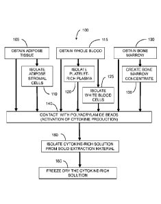
[0031] As exemplified in Figure 1, such processes comprise:
(a)
obtaining a cytokine cell suspension, such as a liquid comprising
white blood cells (steps 105, 115 or 135, or combinations thereof);
(b) contacting the tissue with a solid extraction material (step 140); and
(c)
isolating a protein-containing liquid from the solid extraction material
(step 150).
12
CA 02916080 2015-12-18
WO 2014/149270 PCMJS2014/016421
Obtaining the suspension 105, 115, 135 can comprise any of a variety of
methods for
creating a liquid containing cells among those known in the art. Such methods
include
isolation from tissue and culturing. Obtaining may be performed directly in
the method,
whereby a health care practitioner or other individual performs isolation,
processing,
culturing or other processes for creating the suspension, in a procedure that
includes the
contacting and isolating steps. In some embodiments, the processes for
creating the
suspension are performed contemporaneously with the contacting and isolating
steps, as
part of a point-of-care procedure, as discussed further herein. Alternatively,
obtaining
the suspension may be indirect, involving only the acquisition of the
suspension for use
in the contacting and isolating steps, wherein the processing to create the
suspension has
previously been performed by another party.
[0032] In various embodiments, obtaining comprises isolating a cytokine cell
suspension, comprising white blood cells or other cytokine-producing cells,
from blood,
adipose tissue, bone marrow aspirate or other tissue comprising cytokine-
producing
cells, as exemplified in Steps 110, 120 and 125 of Figure 1. Methods may
comprise
obtaining a cytokine cell suspension from two, three or more tissue sources.
Obtaining a Cytokine Cell Suspension from Blood
[0033] In embodiments comprising the use of blood, the blood may be used
directly in contacting the solid extraction material, as exemplified in step
140 of Figure
1, or may be processed to provide a blood fraction, such as PRP, in a
preferred
embodiment. Many devices and methods for creating blood fractions are known in
the
art, using such means as centrifugation and filtering.
[0034] In various embodiments, methods of the present technology comprise
creating PRP as the cytokine cell suspension, using centrifugation. Such
methods
generally comprise placing blood in a container a separator operable to
separate the
blood into two or more fractions, and centrifuging the separator to create a
platelet-rich
plasma fraction. Such devices may include a tube and a buoy disposed in the
tube,
wherein the buoy has a density such that the buoy reaches an equilibrium
position upon
centrifugation of the tissue in the tube, the equilibrium position being
between a first
fraction and a second fraction comprising white blood cells, the second
fraction having a
concentration of white blood cells greater than the concentration of white
blood cells in
the first fraction. Such methods further comprise centrifuging the tube so
that the buoy
13
CA 02916080 2015-12-18
WO 2014/149270 PCMJS2014/016421
defines an interface between the first fraction and the second fraction
comprising white
blood cells. The second fraction is then collected for further use in the
methods of this
technology.
[0035] One such device useful herein is described in U.S. Patent No.
7,992,725,
Leach et al., issued August 9, 2011. Such a device is commercially available
as GPS III
Platelet Concentrate and Separation System, from Biomet Biologics, LLC
(Warsaw,
Indiana. USA). The device can be used in a clinical or laboratory environment
to isolate
fractions from a suspension or multi-component tissue material obtained from a
subject,
such as blood, bone marrow aspirate, cerebrospinal fluid, adipose tissue,
Isolated
fractions can include platelets, platelet poor plasma, platelet rich plasma
and stromal
cells. The isolated fractions can each have equilibrium point or positions
within the
separation container that are achieved when separation has occurred. For
example, a
buffy coat (PRP) of whole blood may have an equilibrium position above that of
the red
blood cells when a sample of whole blood is separated.
[0036] The fractionation device 200 is exemplified in Figure 2. The
fractionation
device 200 comprises a buoy 210 and a container wall 215. When the separation
container 205 is centrifuged, the buoy perimeter 210a and the container wall
215 have
clearance allowing the buoy 210 to move within the separation container 205
and a
material to pass between the buoy perimeter 210a and the container wall 215.
Alternatively, the buoy 210 could have an opening, such as a centrally or
internally
located opening or a peripheral channel running the height of the buoy, which
would
allow a material to move through the buoy.
[0037] The buoy 210 is carried in the separation container 205 and has a tuned
density that is configured to reach a selected equilibrium position in a
suspension. The
buoy can have its density tuned in the range from about 1.0 g/cc to about 1.10
g/cc. such
as about 1.06 g/cc. The buoy 210, according to various embodiments, can be
formed to
include the tuned density and can be formed of one or more materials to
achieve the
tuned density.
[0038] Referring to Figure 2, a collection area 220 is positioned within the
device 200 after a separation procedure has occurred. The collection area 220,
defined
relative to the buoy 210, is positioned at an equilibrium position of a
separated or
isolated middle fraction 225 in the container. The equilibrium position of a
selected
fraction can be defined as its position within the container relative to other
fractions in
14
CA 02916080 2015-12-18
WO 2014/149270 PCMJS2014/016421
the container of a separated sample or material. The equilibrium position can
also be
defined relative to the axis X of the buoy 210 or the container 12. The
equilibrium
position, however, may depend upon the amount of the sample of the amount of a
selected fraction within a sample. According to the illustration in Figure 2,
the
equilibrium position of the fraction 230 is above or nearer a top 235 of the
device 200
than the equilibrium position of the fraction 225. Thus, the buoy 210 can be
tuned, such
as including a selected density or specific gravity, to position the
collection area 220
relative to an equilibrium position of any selected fraction.
[0039] In some embodiments, the buoy 210 can comprise a collection port 240.
The collection port 240 communicates with access port 245 and communicates
with a
collection space 220 above buoy upper surface 250 and can be located near the
buoy
perimeter 210a. In some embodiments, the collection port 240 is not carried on
the
buoy, but rather the collection port is a withdraw device such as a syringe
that is inserted
through an access port or top of the device 200.
[0040] According to various
embodiments, an isolator 255, is coupled to the
buoy 210. The combination of the isolator and buoy, according to various
embodiments,
can also be referred to as a separation assembly member. The isolator 255, for
example,
provides a means for creating the collection compartment 220 and comprises one
or
more spacers 260, 265 to position the isolator 255 apart from the buoy 210 to
create the
collection compartment 220. A withdraw port 270 can be carried on the isolator
255
communicating with the withdraw port 245 and the collection port 240. The
spacer 260,
265 can also serve as a conduit 275 between the collection port 50 and a
withdraw or
withdraw port 245. The withdraw port 245 serves as a structure for withdrawing
the
isolated or second fraction 310 from the collection compartment 220.
[0041] After centrifuging
the device 200 containing whole blood, the first
fraction or top fraction 230, can be platelet-poor-plasma, the middle fraction
225 can be
platelet-rich plasma or platelet concentrate. and a bottom fraction 278 can be
red blood
cells. Therefore, the fractionation method further comprises withdrawing a
desired
fraction from the device 200. Various ports 205, 245 and 280 can be provided
to allow
access to any appropriate compartment of the device 200. The access ports 205.
245,
280 can be any means that allow communication from outside the separation
device 200
to the device's interior, such as a Luer lock port, a septum, a valve, or
other opening.
Additionally, collection vent tube 285 allows removal of a fractionated
suspension in the
CA 02916080 2015-12-18
WO 2014/149270 PCMJS2014/016421
collection area 220 through opening 290 without the need to remove the
fraction, such as
plasma, above the isolator 255. Although, without a collection vent tube 285,
the
fraction above the isolator could be removed and the collection area could be
vented to
the area above the isolator.
[0042] A method for using the fractionation device 200 can begin by inputting
whole blood via an access port 205. The fractionation device 200 is placed
into a
centrifuge and spun for a period that is appropriate for fractionating whole
blood. An
exemplary period can be for about five minutes to about twenty minutes at a
rate of
about 320 rpm to about 5000 rpm. This speed may produce a selected gravity
that may
be approximately 7.17 xg to about 1750 xg (times greater than the normal force
of
gravity).
[0043] Other devices that
may be used to isolate platelet-rich plasma
described, for example, in U.S. Patent 5,585,007, Antanavich, issued December
17,
1996; U.S. Patent No. 6,398,972, Blasetti et al., issued June 4, 2002; U.S.
Patent No.
6,649,072, Brandt et al., issued November 18, 2003; U.S. Patent No. 6,790,371,
Dolocek,
issued September 14, 2004; U.S. Patent No. 7,011.852, Sukavaneshvar et al.,
issued
March 14, 2006; U.S. Patent No. 7,179,391, Leach et al., issued February 20,
2007; U.S.
Patent No. 7,374,678, Leach et al., issued May 20, 2008; U.S. Patent No.
7,223,346,
Dorian et al., issued May 29, 2007; and U.S. Patent No. 7,708,152, Dorian et
al., issued
May 4, 2010.
[0044] In addition to the
GPS Platelet Concentrate and Separation Systems,
a variety of other commercially available devices may be used to isolate
platelet-rich
plasma, including the MagellanTM Autologous Platelet Separator System,
commercially
available from Medtronic, Inc. (Minneapolis, Minnesota, USA); SmartPRePTM,
commercially available from Harvest Technologies Corporation (Plymouth,
Massachusetts, USA); DePuy (Warsaw, Indiana, USA); the AutoloGelTM Process,
commercially available from Cytomedix, Inc. (Rockville, Maryland, USA); the
GenesisCS System, commercially available from EmCyte Corporation (Fort Myers,
Florida, USA); and the PCCS System, commercially available from Biomet 3i,
Inc.
(Palm Beach Gardens, Florida, USA).
[0045] Referring again to
Figure 1, blood drawn from the patient may be
mixed with an anticoagulant in one or more of Steps 115, 120. 125, and 130, so
as to
facilitate processing. Suitable anticoagulants include heparin, citrate
phosphate dextrose
16
CA 02916080 2015-12-18
WO 2014/149270 PCMJS2014/016421
(CPD), ethylenediaminetetraacetic acid (EDTA), anticoagulant citrate dextrose
solution
(ACD), and mixtures thereof. For example, the anticoagulant may be placed in
the
syringe used for drawing blood from the subject, or may be mixed with the
blood after it
is drawn.
[0046] A liquid containing
white blood cells may be prepared by admixing
cells with a suitable liquid, as shown in step 125, using methods known in the
art. For
example, white blood cells may be isolated from whole blood by lysing red
blood cells
or by centrifugation of whole blood utilizing a density gradient where the
white blood
cells sediment to the bottom of a centrifuge tube. An example of density
centrifugation
includes the Ficoll-PaqueTM Plus (GE Healthcare Bio-Sciences, Piscataway, New
Jersey,
USA). In some cases, a density gradient may be used to further separate
mononuclear
and polymorphonuclear cells. White blood cells may also be prepared from whole
blood
using filtration; an example includes the AcelereTM MNC Harvest System (Pall
Life
Sciences, Ann Arbor, Michigan, USA). White blood cells can also be obtained
from
bone marrow. The white blood cells may be then suspended in a suitable medium,
such
as plasma, so as to maintain their viability.
[0047] Other methods may be
used to create platelet-rich plasma or other
liquid containing white blood cells. For example, whole blood can be
centrifuged
without using a buoy system, whole blood may be centrifuged in multiple
stages,
continuous-flow centrifugation can be used, and filtration can also be used.
In addition,
a blood component including platelet-rich plasma can be produced by separating
plasma
from red blood cells using a slow speed centrifugation step to prevent
pelleting of the
platelets. In other embodiments, the buffy coat fraction formed from
centrifuged blood
can be separated from remaining plasma and re-suspended to form platelet-rich
plasma.
[0048] Obtaining a Cytokine Cell Suspension from Adipose Tissue
[0049] In embodiments
comprising the use of adipose tissue, the adipose
tissue may be used directly in contacting the solid extraction material, as
exemplified in
step 140 of Figure 1, or the adipose tissue may be processed to provide
isolated
adipocytes in step 110. Cell fractions comprising adipose-derived stem cells
are also
useful in this method. In some embodiments, adipose tissue is derived from
human
subcutaneous fat isolated by suction assisted lipectomy or liposuction.
Stromal cells may
be isolated from the adipose tissue and/or tissue portions using any suitable
method,
including methods known in the art such as mechanical and breakdown
centrifugation.
17
CA 02916080 2015-12-18
WO 2014/149270 PCMJS2014/016421
Stromal cells can also be isolated using enzymatic digestion. For example,
stromal cells
can be isolated from lipoaspirate, treated by sonication and/or enzymatic
digestion, and
enriched by centrifugation. Stromal cells isolated from adipose tissue may be
washed
and pelleted.
[0050] For example, adipose
tissue can be collected by suction-assisted
tumescent liposuction inside a specialized collection container attached to
suction hoses
and to a liposuction cannula. The collection container can have a gauze-type
grid filter
that allows the tumescent fluid to pass through and retains the solid adipose
tissue. After
collecting the adipose tissue, the collection container is removed from the
suction device
and reattached to a centrifugation device. The filter unit may further contain
a filter
having approximately a 100 micrometer pore size. Once the collection container
containing the adipose tissue is attached to the centrifugation device, the
tissue is
sonicated. After sonication, the entire apparatus is inserted into a
centrifuge bucket and
centrifuged at, for example, 300xg for 5 minutes. After centrifugation, the
collection
container together with the filter unit is detached and can be discarded. The
pellet
containing the stromal cells can then be re-suspended in biocompatible
solutions, such as
plasma, plasma concentrate and platelet-rich plasma.
[0051] Various methods and
devices for isolating and/or fractionating
adipose tissue and adipocytes include those as described by U.S. Patent No.
7,374,678,
Leach, issued May 20, 2008; U.S. Patent No. 7,179,391 to Leach et al., issued
February
20, 2007; U.S. Patent No. 7,992,725, Leach et al., issued August 9, 2011; U.S.
Patent
No. 7,806,276, Leach et al.. issued October 5, 2010; and U.S. Patent No.
8.048,297,
Leach et al., issued November 1, 2011. A device, such as the GPSTM Platelet
Concentrate System, commercially available from Biomet Biologics, LLC (Warsaw,
Indiana, USA), may be used to isolate adipocytes.
Obtaining a Liquid Containing White Blood Cells from Bone Marrow
[0052] In embodiments
comprising the use of bone marrow, the marrow may
be used directly in contacting the solid extraction material, as exemplified
in step 140 of
Figure 1, or may be processed to provide a bone marrow concentrate, as in step
135.
Many devices and methods for obtaining and concentrating bone marrow are known
in
the art.
18
CA 02916080 2015-12-18
WO 2014/149270 PCMJS2014/016421
[0053] An exemplary process
for isolating and creating a bone marrow
concentrate (cBMA) is diagrammed in Figure 6. Generally, the method 600 may
start in
step 605 with obtaining a bone marrow aspirate volume. The bone marrow
aspirate
(BMA) may be obtained in any selected or generally known manner. For example,
a
selected region of bone, such as a portion near an operative procedure, may be
used to
obtain the bone marrow aspirate. Generally, an accessing device, such as a
syringe and
needle, may be used to access an intramedullary area of a selected bone. A
small volume
of the selected portion may be drawn from a plurality of locations to obtain
an
appropriate volume of BMA or selected fraction of the BMA.
[0054] Once a selected
volume of the BMA is obtained in step 605, it may be
separated and concentrated using a gravimetric separator. Separators among
those useful
herein are operable to separate a multi-component fluid that generally
includes various
components or constituents of varying densities that are commingled or mixed
together,
including those described above for separation of fractions from blood and
adipose
tissue. The separator may include a buoy that is of a selected density
relative to BMA.
Such separators include those described above for use in concentrating and
isolating
fractions from blood and adipose tissue, including those described in U.S.
Patent No.
7,374,678. Leach, issued May 20, 2008; U.S. Patent No. 7,179,391 to Leach et
al.,
issued February 20, 2007; U.S. Patent No. 7,992,725, Leach et al., issued
August 9,
2011; U.S. Patent No. 7,806,276, Leach et al.. issued October 5, 2010; and
U.S. Patent
No. 8,048,297, Leach et al., issued November 1, 2011. A device, such as the
GPSTM
Platelet Concentrate System, commercially available from Biomet Biologics, LLC
(Warsaw, Indiana, USA), may be used to isolate adipocytes. Separators and
methods
that may be used to fractionate BMA at steps 610 and 615 are also described,
for
example, in U.S. Application Publication No. 2006/0278588, Woodell-May,
published
December 14, 2006. The BMA may be positioned in a separator according to
various
embodiments in step 610. Once the BMA is positioned in the separator, a
selected
fraction of the BMA may be separated from the BMA in step 615.
[0055] Once the BMA is
placed in the separator, separator is spun in a
centrifuge in a range between about 1,000 and about 8,000 RPM. This produces a
force
between about 65 and about 4500 times greater than the force of normal
gravity, as
generally calculated in the art, on the separator and the BMA. At this force,
the more
dense material in a BMA sample is forced toward the bottom end of the tube.
The
19
CA 02916080 2015-12-18
WO 2014/149270 PCMJS2014/016421
separator can thus be used to remove nucleated cells from the bone marrow
sample. In
various embodiments, concentrated BMA has a concentration of nucleated cells
that is at
least 2, at least 3. at least 4, or at least 5 times the concentration of
nucleated cells in
BMA.
Obtaining a Liquid Containing White Blood Cells from Blood Clots
[0056] In other embodiments
comprising the use of blood, a liquid
comprising cytokine-producing cells trapped in a blood clot. Cell releasate
can be
generated from the blood clot by either compression ("squeezing"), clot
disruption, or
centrifugation. The blood clot can be made with or without anticoagulant and
with or
without exogenous thrombin by combining blood or a blood fraction with a
clotting
agent. Suitable clotting agents include thrombin (e.g., bovine, recombinant
human,
pooled human, or autologous), autologous clotting protein, and polyethylene
glycol.
Calcium may be in the form of a calcium salt, such as calcium chloride.
[0057] In some embodiments,
the clotting agent comprises a clotting protein,
which may be a clotting fraction derived from a blood obtained from the
patient to be
treated. A suitable clotting fraction can be obtained by a process of: loading
whole
blood or plasma with a calcium solution (e.g., calcium chloride in ethanol)
into a blood
isolation device; optionally heating the whole blood or plasma for at least
about 20
minutes, at a temperature of at least about 20 C; and isolating the clotting
fraction. The
isolating may be performed by centrifuging the heated whole blood or plasma. A
suitable isolation device is commercially available as the ClotalystTm
Autologous
Thrombin Collection System (hereinafter "Clotalyst System"), sold by Biomet
Biologics
LLC, Warsaw, Indiana, USA.
[0058] An exemplary
procedure for producing a clotting agent using a device
400 of Figure 4 begins with injecting a reagent comprising calcium chloride
and ethanol
into the main chamber 405 through the first port 410. Glass beads are also
placed in the
main chamber 405. After the reagent has been injected, the first port 410 is
closed using
the first replacement cap 415. Blood with anticoagulant is injected into the
main
chamber 405 through the second port 420. After the blood has been injected,
the second
port 420 is closed using the second replacement cap 425. Optionally, the
syringes and
blood separation device 400 are pre-heated to a temperature of about 25 C.
CA 02916080 2015-12-18
WO 2014/149270 PCMJS2014/016421
[0059] The contents of the
blood component separation device 400 are mixed
by repeatedly inverting the device 400, e.g. about twelve times, so as to
contact the blood
with the glass beads. After mixing, the device is incubated The incubation
process can
be at a temperature and for a duration that will permit the contents of the
device 400 to
be heated at about 25 C for about 15 minutes. Upon completion of the
incubation
period, a clotted mass of red blood cells, blood plasma, and glass beads forms
at a second
end 406 of the main chamber 405. After incubation is complete, the device 400
is
shaken enough to dislodge and break-up any gel that may be present.
Obtaining a Liquid Containing White Blood Cells Using Non-Centrifugal Methods
[0060] As noted above, the
liquid containing white blood cells can be
obtained by non-centrifugal means, such as by culturing. As referred to
herein, a -non-
centrifugal method" comprises a process for obtaining tissue fractions
comprising
cytokine-producing cells from tissue without use of a centrifuge. In some
embodiments,
methods are "non-gravimetric," wherein , based on physical, chemical or
physicochemical properties of the cells other than density, wherein the
concentration of
white blood cells in the fraction are higher than the concentration of white
blood cells in
the tissue. Such non-gravimetric methods are, in particular, distinguished
from methods
wherein a white blood cell fraction is created by centrifugation of whole
blood or other
tissue. In some embodiments, the non-centrifugal method comprises a process
solely
based on such properties of white blood cells other than density. Non-
centrifugal
methods include filtration, antibody binding, and electrophoretic methods.
[0061] For example, as
discussed above, white blood cells may be prepared
from whole blood, bone marrow aspirate or other tissue, using filtration.
White blood
cells and other cytokine-producing cells obtained from blood, bone marrow,
adipose
tissue or other sources may also be cultured, using methods among those known
in the
art. The cells may be then suspended in a suitable medium, such as plasma, so
as to
maintain their viability and facilitate mixing or other contact with a solid
extraction
material. A liquid containing the cells may also be produced by compression or
disruption of blood clots, as described above.
21
CA 02916080 2015-12-18
WO 2014/149270 PCMJS2014/016421
Contacting a Liquid Containing White Blood Cells With an Extraction Material
and
Isolating a Protein Solution
[0062] In further reference
to the exemplified process of Figure 1, the
cytokine cell suspension is incubated or otherwise contacted with a solid
extraction
material (step 140) to produce a protein-containing liquid. This liquid is
then isolated
(step 150) from the solid extraction material, as a Protein Solution of the
present
technology. Without limiting the scope, mechanism or function of the present
technology, solid extraction materials useful herein concentrate cytokines or
other
proteins in the liquid volume of white blood cells and may, in some
embodiments,
activate, stimulate or otherwise increase production of cytokines, including
IL-1ra.
Thus, in some embodiments, methods comprising activating a cytokine cell
suspension
with a solid extraction material.
[0063] The solid extraction
material can include various materials that
provide a particular surface area to contact the cells. The solid extraction
material may
be a continuous material or may be discontinuous and comprise a plurality of
separate
particles. For example, the solid extraction material may be in the form of a
plurality of
beads, fibers, powder, a porous material, or a surface of a container
comprising the liquid
containing the cells. The solid extraction material may comprise geometric
forms having
various cross-sectional shapes, such as spherical, oval, or polygonal, among
others. The
solid extraction material can also comprise a continuous porous network,
similar to a
sponge, or can include a plurality of individual porous particles. The solid
extraction
material may also provide a larger surface area by being porous in comparison
to a non-
porous material.
[0064] In some embodiments,
the solid extraction material includes particles
having a large aspect ratio, for example, where the particles are needle-like
in shape.
The solid extraction material may also be formed as long fibers and may be or
take a
form similar to glass wool.
[0065] In some cases, the
solid extraction material can comprise the internal
walls of a container holding the cytokine cell suspension. For example, the
solid
extraction material may comprise the lumen of a syringe that contains the
cytokine cell
suspension. Other containers include tubes, such as centrifuge tubes, or a
blood
fractionation device or concentrator assembly as described elsewhere herein.
22
CA 02916080 2015-12-18
WO 2014/149270 PCMJS2014/016421
[0066] Where the solid
extraction material is a continuous material, such as a
porous sponge-like material, the solid extraction material can be used in an
amount
sufficient to absorb or adsorb or include substantially the entire liquid
volume of white
blood cells within the pores or interstices of the solid extraction material.
Where the
solid extraction material is a discontinuous material, such as a plurality of
particles, the
solid extraction material can be combined with the liquid containing the cells
to form a
slurry-like composition. The slurry can vary in consistency from paste-like,
having a
high-solids fraction, to a readily flowable slurry having a low-solids
fraction.
[0067] The solid extraction
material can provide a large surface area with
which to contact the cells. However, in some cases, the solid extraction
material can be
further treated to increase its surface area, for example, by physically or
chemically
etching or eroding the surface of the solid extraction material. With respect
to chemical
etching, a corrosive agent can be used to modify the surface of the solid
extraction
material depending on the nature of the material. The modified surface may be
produced
by employing an alkali or an acid, for example chromosulphonic acid, in
particular about
20% to about 80% in strength, preferably about 50% chromosulphonic acid. The
solid
extraction material can be incubated with the corrosive agent for about 5 min
to about 30
min in order to chemically etch the surface and increase the surface area. The
solid
extraction material can then be washed to remove the corrosive agent. For
example, the
solid extraction material can include the internal walls of a container for
holding the
cytokine cell suspension where the internal walls are etched to subsequently
increase the
surface area in contact with the liquid.
[0068] Various polymers,
metals, ceramics, and glasses can be used as the
solid extraction material. In some embodiments, the solid extraction material
comprises
a hygroscopic material. Examples of suitable solid extraction material
materials include
glasses, minerals, polymers, metals, and polysaccharides. Minerals include
corundum
and quartz.
Polymers include polystyrene, polyethylene, polyvinyl chloride,
polypropylene, and polyacrylamide. Metals include titanium. Polysaccharides
include
dextran and agarose. A preferred solid extraction material comprises, or
consists
essentially of, polyacrylamide, as further described below.
[0069] The solid extraction
material may comprise, for example, continuous
solid extraction material of glass or a plurality of glass particles, glass
wool, a continuous
solid extraction material of metal such as titanium, a plurality of metal
beads, metal
23
CA 02916080 2015-12-18
WO 2014/149270 PCMJS2014/016421
powder, and combinations thereof. A continuous solid extraction material of
metal can
include a block or other three-dimensional shape formed of porous metal or
metal alloys
with an open cell structure. The solid extraction material may include various
beads or
particles of various sizes including substantially spherical beads. Beads
include
polystyrene beads, polyacrylamide beads, glass beads, metal (e.g., titanium)
beads, or
any other appropriate beads. Beads may be any size appropriate for the
container and the
amount of cytokine cell suspension being used. In some instances, bead sizes
can range
from about 0.001 millimeters to about 3 millimeters in diameter. Where the
bead size is
sufficiently small, the beads can appear more like a powder.
[0070] Polyacrylamide beads
used as the solid extraction material can be
formed by polymerizing acrylamide monomer using controlled and standardized
protocols as known in the art to produce relatively uniform beads formed of
polyacrylamide gel. In general, polyacrylamide is formed by polymerizing
acrylamide
with a suitable bifunctional crosslinking agent, most commonly N,N'-
methylenebisacrylamide (bisacrylamide). Gel polymerization is usually
initiated with
ammonium persulfate and the reaction rate is accelerated by the addition of a
catalyst,
such as N,N,N',IV-tetramethylethylenediamine (TEMED). In various embodiments,
polyacrylamide beads comprise 0.5 micromole of carboxyl groups per milliliter
of beads,
imparting a slight anionic character (negative charge). The beads are also
typically
resistant to changes in pH, and are stable in many aqueous and organic
solutions. By
adjusting the total acrylamide concentration, the polyacrylamide gel can be
formed in a
wide range of pore sizes. Moreover, the polyacrylamide beads can be formed in
many
sizes and can have relatively uniform size distributions. Bead size may range
from
several micrometers in diameter to several millimeters in diameter. For
example, various
types of BioGelTM P polyacrylamide gel beads (Bio-Rad Laboratories, Hercules,
California, USA) have particle sizes ranging from less than about 45 1..tm up
to about 180
[nu. Polyacrylamide beads are also available from SNF Floerger (Riceboro,
Georgia,
USA), Pierce Biotechnology, Inc. (Rockford, Illinois, USA), and Polymers, Inc.
(Fayetteville, Arkansas, USA).
[0071] Once polymerized,
polyacrylamide beads can be dried and stored in a
powder-like form. The dry beads are insoluble in water but can swell
considerably upon
being rehydrated. Rehydration returns the polyacrylamide beads to a gel
consistency that
can be from about two to about three times the dry state size. Thus, dry
polyacrylamide
24
CA 02916080 2015-12-18
WO 2014/149270 PCMJS2014/016421
beads (i.e., desiccating polyacrylamide beads) may be used to absorb a portion
of a liquid
volume, including solutes smaller than the bead pore size, and can serve to
concentrate
IL-ira and other proteins produced by the white blood cells. For example,
combining
dry polyacrylamide beads with the blood and/or platelet-rich plasma in step
230 activates
production of IL- lra by the white blood cells and also reduces the total
liquid volume as
the dry beads rehydrate and swell.
[0072] Without limiting the
scope, mechanism or function of the present
technology, it has been discovered that surface contact with the solid
extraction material
can activate the cells and the solid extraction material can, in some cases,
assist in the
separation and concentration of the resulting Protein Solution rich in
cytokines, including
IL-ira. For example, in the case of a porous solid extraction material, a
portion of the
liquid comprising the cells can enter the pores and remain therein. Cells in
the liquid
may contact this additional surface area. In some embodiments, the pores are
too small
for the cells to enter, but a portion of the liquid can enter the pores.
Liquid can be
removed from the solid extraction material and pores by centrifuging, for
example.
[0073] The solid extraction
material is preferably sterilized, using techniques
among known in the art, in order to prevent contamination of the cytokine cell
suspension. For example, heat and pressure sterilization methods, such as
autoclaving,
may be used depending on the particular composition of the solid extraction
material.
Alternative methods, such as chemical sterilization or irradiation, can be
used where the
solid extraction material may be adversely affected by the autoclaving
process.
[0074] In some embodiments,
the cytokine cell suspension is incubated with
solid extraction material for a time effective to remove a portion of the
liquid. The
incubation may be carried out over a period from about 30 seconds to about 72
hours and
may be carried out at a temperature from about 20 C to about 41 C. For
example, the
incubation may be 24 hours or less, 10 hours or less, 5 hours or less, 2 hours
or less, 1
hour or less, 30 minutes or less, 15 minutes or less 10 minutes or less, 5
minutes or less,
4 minutes or less. 3, minutes or less, or 2 minutes or less. Incubation may be
at least
about 15 seconds, at least about 30 seconds, at least about 1 minutes, at
least about 90
seconds, at least about 2 minutes, at least about 10 minutes, or at least
about 30 minutes.
In some embodiments, incubation s from about 1 minute to about 3 minutes. In
some
embodiments, the incubation is conducted at about 37 C. In some embodiments
the
liquid is not incubated, but is contacted with the solid extraction material
for only so long
WO 2014/149270 PCT/US2014/016421
as necessary to perform subsequent processing. The contacting may occur at
ambient
conditions, e.g., at a temperature of about 20-25 C.
[00751 In some embodiments, the
cytokine cell suspension and the solid
extraction material are agitated to more thoroughly mix these components
during
contact. The agitation may be accomplished by inverting, shaking, rocking,
stirring, or
vortexing the liquid and solid extraction material. Agitation may increase
contact of the
cells within the liquid with the solid extraction material. Agitation may be
performed
once, repeated multiple times, repeated periodically, or may be continuous.
The liquid
comprising the cells and the solid extraction material may also be agitated
while the
liquid is stimulated with the electromagnetic field. Additional aspects and
features
relating to producing protein-rich solutions using polyacrylamide beads and
other solid
extraction materials are described in: U.S. Patent Application Publication No.
2009/0220482, Higgins et al., published September 3, 2009; U.S. Patent
Application
Publication No. 2010/0055087, Higgins et al., published March 4, 2010; U.S.
Patent
Application Publication 2011/0052561, Hoeppner, published March 3, 2011;
International Application Publication 2012/030593, Higgins et al., published
March 8,
2012; and U.S. Patent Application Publication 2012/0172836, Higgins et al.,
published
July 5, 2012. U.S. Patent Application Serial Number 13/840562, Binder et al.,
Methods
and Non-Immunogenic Compositions for Treating Inflammatory Diseases; U.S.
Patent
Application Serial Number 13/841083, Landrigan, et al., Treatment of
Inflammatory
Respiratory Disease Using Protein Solutions; U.S. Patent Application Serial
Number
13/837005 Woodell-May et al., Methods and Acellular Compositions for Treating
Inflammatory Disorders; U.S. Patent Application Serial Number 13/839280, Leach
et
al., Methods for Making Cytokine Compositions from Tissue Using Non-
Centrifugal
Methods; U.S. Patent Application Serial Number 13/840129, Matusuka, et al.,
Treatment
of Collagen Defects Using Protein Solutions; and U.S. Patent Application
Serial Number
13/841103, Landrigan, et al., Treatment of Peripheral Vascular Disease Using
Protein
Solutions,
[0076] Contacting of the liquid
containing white blood cells with the solid
extraction material may be performed using a suitable container or other
apparatus to
affect the contact. Contacting may be performed in a continuous process
wherein a flow
of the liquid is passed over or through the solid extraction material, or the
liquid and
solid extraction material may be contained in a vessel. As discussed above,
the vessel
26
CA 2916080 2018-08-15
CA 02916080 2015-12-18
WO 2014/149270 PCMJS2014/016421
may comprise the solid extraction material, or may merely serve as a container
holding
the beads or other forms of the material. Containers useful in the present
technology
include those known in the art, such as the PlasmaxTM Plus Plasma
Concentrator,
commercially available from Biomet Biologics, LLC (Warsaw, Indiana, USA) and
may
include those devices and methods of use as described in U.S. Patent No.
7.553,413,
Dorian et al., issued June 30, 2009; and U.S. Patent No. 7,694,828, Swift et
al., issued
April 13, 2010.
[0077] Such a device is
shown in Figures 3A and 3B, for exemplary use with
a polyacrylamide gel bead solid extraction material. The device 300 has an
upper
chamber 305 and a lower chamber 310. The upper chamber 305 has an end wall 315
through which the agitator stem 320 of a gel bead agitator 325 extends. The
device 300
also has an inlet port 330 that extends through the end wall 315 and into the
upper
chamber 305. The device 300 also includes an outlet port 335 that communicates
with a
plasma concentrate conduit 340. The floor of upper chamber 305 includes a
filter 345,
the upper surface of which supports desiccated concentrating polyacrylamide
beads 350.
[0078] During use, a fluid
355 containing white blood cells and, optionally,
platelets is injected to the upper chamber 305 via the inlet port 330 and
mixed with the
polyacrylamide beads 350. The fluid 355 and polyacrylamide beads 350 may be
mixed
by rotating the agitator stem 320 and the gel bead agitator 325, to help mix
the fluid 355
and beads 350. The mixed fluid 355 and polyacrylamide beads 350 are then
incubated
for the desired time at the desired temperature. The device 300 is then
centrifuged so
that liquid passes to the lower chamber 310 while the polyacrylamide beads 350
are
retained by a filter 345, thereby separating the polyacrylamide beads 350 from
the
resulting solution 360 of IL- lra and other proteins that collects in the
lower chamber
310. The solution 360 may be removed from the device via outlet port 335.
[0079] In some embodiments,
a Protein Solution can be made in a process
wherein a liquid containing white blood cells is isolated from a tissue and
then contacted
with a solid extraction material in a continuous process. Referring again to
Figure 1, in
some embodiments the isolating 110, 120, 135 and contacting 140 are performed
using a
single apparatus, referred to herein as a single separation and concentration
device ("S/C
device"). One such device is described in U.S. Patent Application Serial
Number
13/434,245, O'Connell, filed March 29, 2012.
27
CA 02916080 2015-12-18
WO 2014/149270 PCMJS2014/016421
[0080] The S/C device
comprises a separation region, a first concentration
region, a second concentration region, a buoy system, an inlet port, a check
valve, a first
withdrawal port and a second withdrawal port. Figure 5 shows an S/C device 500
capable of generating an anti-inflammatory cytokine composition from whole
blood. For
example, the method may start with obtaining a volume of whole blood, which is
filled
into a separation region 505 of the S/C device 500 by injecting through the
inlet port 510.
A buoy system 515 is located within the separation region 505. The buoy system
comprises a first buoy member 520, a second buoy member 525 , and a third buoy
member 530 that couples the first buoy member 520 to the second buoy member
525. A
space between the first and second buoy members 520, 525 defines a buoy
separation
region 535. A density of each buoy member can be selected depending on what
blood
fraction is desired as a result of a separation. The buoy system 515 can
include a selected
buoy system, such as the buoy system generally used in the GPS@ II or GPS III
gravity
platelet separation system sold by Biomet Biologics, LLC. (Warsaw, Indiana,
USA).
Buoy systems are disclosed in U.S. Pat. No. 7,845,499 and U.S. Pat. No.
7,806,276, and
U.S. Pat. No. 7,992,725.
[0081] A method for
obtaining a Protein Solution comprises spinning the S/C
device 500 by centrifugation. Centrifugal forces allow the buoy system 515 to
move
through the whole blood, resulting in a fraction of the whole blood to be
located in the
buoy separation region 535. For example, this fraction may comprise platelet-
rich
plasma. With a use of a withdrawal syringe, the selected fraction can be
removed from
the collection volume 535 through the third buoy member 530 that defines a
removal
passage 540 that is connected with collection face passages 545. A connection
elbow
550 can interconnect with the removal passage 540 to allow a vacuum to be
formed
through the connection elbow 550, the collection passage 540, and the buoy
collection
passages 545. A collection tube 555 can interconnect the connection elbow 550
with a
withdrawal elbow 560 that extends from a wall 565 that can be a bottom wall of
concentration region 570. A second withdrawal tube 575 can be first connected
with a
check valve assembly 580 and a first withdrawal port 585. The first withdrawal
port 585
can be connected with the withdrawal syringe with a Luer lock type connection
or other
appropriate connection.
[0082] The check valve
assembly 580 ensures the fraction being removed
flows in one direction and prevents the fraction being removed from reentering
the
28
CA 02916080 2015-12-18
WO 2014/149270 PCT/1JS2014/016421
second withdrawal tube 575. Furthermore, when material is pushed back into the
check
valve assembly 580 from the first withdrawal port 585, such that material will
enter the
concentration region 570, a disc within the check valve 580 can flex down
towards the
second withdrawal tube 575 and close an opening and thereby open a second
opening
within the check valve assembly 580. The second opening allows the fraction to
be
pushed into the concentration region 570.
[0083] Therefore, the blood
fraction is then re-injected through the first
withdrawal port 285, through the check valve assembly 580, and into an upper
volume
588 of the concentration region 570. Polyacrylamide beads 590 are added to the
blood
fraction in the upper volume 588 and the blood fraction and the polyacrylamide
beads
590 can be mixed by shaking. Optionally, the blood fraction and the beads 590
can be
incubated for a selected period of time before proceeding with the method.
[0084] The method comprises
a second step of spinning by centrifugation.
During the second centrifugation, the anti-inflammatory cytokine composition
is
separated from the beads 590 by being forced through a filter 592 and into a
lower
concentration region 595 of the concentration region 570. The Protein Solution
can be
withdrawn through a third withdrawal tube 596 and out a second withdrawal port
598 by
use of a second withdrawal syringe. Again, the syringe can be connected to the
second
withdrawal port by a LuerO lock type connection.
[0085] Referring again to
Figure 1, following contacting the liquid with the
solid extraction materials, a Protein Solution is isolated, as indicated at
step 150.
Isolation may be accomplished by drawing off at least a portion of the liquid
volume and
leaving the beads. In some cases, the extraction material may be sedimented by
centrifugation prior to drawing off the Protein Solution. Isolation may also
be performed
by filtration, where the material is retained by a filter and the Protein
Solution passes
through the filter using centrifugal force or by using vacuum, for example. If
the
incubation with extraction material utilizes dry polyacrylamide beads, the
liquid volume
may be reduced as the beads swell upon rehydration, thereby concentrating the
resulting
Protein Solution. To maintain the increased concentration, care should be
taken in the
isolation step so as to avoid compressing the beads or drawing liquid out from
the
swollen beads. For example, high centrifugal force or high vacuum may collapse
the
beads and/or draw liquid out of the internal volume of the beads.
29
CA 02916080 2015-12-18
WO 2014/149270 PCMJS2014/016421
Optional Electromagnetic Stimulation
[0086] The cytokine cell suspension can be stimulated with an
electromagnetic field, before or during the contacting of the liquid with a
solid extraction
material. Thus, in some embodiments, stimulation of the liquid comprising the
cells can
be performed prior to contacting the liquid and the solid extraction material.
However, it
is preferred that at least a portion of the contacting step and at least a
portion of the
stimulating step overlap in time such that the liquid comprising the cells is
concurrently
in contact with the solid extraction material and stimulated with the
electromagnetic
field.
[0087] Stimulating the
cytokine cell suspension with an electromagnetic field
may involve various forms of electromagnetic stimulation, such as a pulsed
electromagnetic field or a capacitively coupled electromagnetic field. In some
embodiments, the liquid is stimulated using a power source coupled to a
stimulation coil.
The current passing through the coil produces a pulsing magnetic field which
induces in
the liquid a pulsing electric field. The coil may partially surround the
liquid as it is held
within a container, such as a tube or syringe. The coil may be integrated into
to the
container holding the cytokine cell suspension or may be removable. For
example, a
plastic tube can be formed with an integrated coil or the coil can be
temporarily coupled
to the container or placed within the container; for example, the tube can be
configured
so that the coil can be snapped onto the container. The power source can be
coupled to
the coil as needed to perform the stimulating step.
[0088] Stimulation of the
liquid with an electromagnetic field may also
include placing at least two electrodes across the liquid. Electrical energy
may then be
applied to the electrodes so as to capacitively couple the electrodes and
generate the
electromagnetic field there between. The electromagnetic field is therefore
able to pass
through the liquid so as to increase the rate and/or amount of cytokine
production. In
other embodiments, electrodes can be used to produce a direct current or one
or more
coils can be used to produce a pulsed electromagnetic field.
[0089] The strength of the
electromagnetic field during stimulation can be at
least about 0.5 microvolts per centimeter, whether produced by direct current,
capacitively coupled current, or pulsed electromagnetic field. In the case of
a direct
current electrode, the amplitude of the current can be from about 1 to about
200
microamperes, and in some embodiments, the amplitude may be from about 20 to
about
CA 02916080 2015-12-18
WO 2014/149270 PCMJS2014/016421
100 microamperes. In still further embodiments, the current may be about 20,
about 60,
or about 100 microamperes. It should be understood, however, that the
amplitude of the
current may be of other suitable magnitudes.
[0090] The electromagnetic
field applied during the stimulating step may be
constant or vary over time. For example, a sinusoidal time varying
electromagnetic field
can be applied using the electrodes placed across the liquid. Such a
sinusoidal time
varying electromagnetic field can have a peak voltage across the electrodes
from about 1
volt to about 10 volts, and in some embodiments, the peak voltage can be about
5 volts.
The corresponding electric field produced can have an amplitude of from about
0.1
millivolt per centimeter (mV/cm) to about 100 mV/cm, and in some embodiments
can be
about 20 mV/cm. The sinusoidal time varying electric field may have a
frequency of
from about 1,000 Hz to about 200,000 Hz, and in some embodiments the frequency
may
be about 60,000 Hz.
[0091] The electromagnetic
field applied to the liquid may also be a pulsed
electromagnetic field. The pulsed electromagnetic field can be induced using
an external
coil and a pulse generator. In this regard, a pulsed electromagnetic field may
have a
pulse duration of from about 10 microseconds per pulse to about 2000
microseconds per
pulse. The pulse duration in one embodiment can be about 225 microseconds. The
pulses may include electromagnetic bursts, in which a burst can comprise from
1 pulse to
about 200 pulses. Alternatively, the electromagnetic field may have bursts
that comprise
from about 10 pulses to about 30 pulses. In this regard, in one embodiment
each burst
may comprise about 20 pulses.
[0092] The frequency at
which bursts in the pulsed electromagnetic are
applied may vary. In this regard, bursts can be repeated at a frequency of
from about 1
Hz to about 100 Hz in some embodiments, and can be repeated at a frequency of
about
10 Hz to about 20 Hz in other embodiments. Furthermore, bursts can repeat at a
frequency of about 1.5 Hz, about 15 Hz or about 76 Hz. A burst can have a
duration
from about 10 microseconds up to about 40,000 microseconds. In this regard, a
burst
can have a duration of about 4.5 milliseconds.
[0093] Suitable devices for
generating a capacitively coupled electromagnetic
field include SpinalPak spinal stimulator (EBI, L.P., Parsippany, New Jersey)
or a DC
stimulation device such as an SpF0 XL _Lib spinal fusion stimulator (EBI,
L.P.,
Parsippany, New Jersey). Pulsed electromagnetic fields can be produced using
various
31
CA 02916080 2015-12-18
WO 2014/149270 PCMJS2014/016421
known methods and apparatuses, such as using a single coil or a pair of
Helmholtz coils.
For example, a suitable apparatus includes the EBI Bone Healing System Model
2001
(EBI, L.P., Parsippany, New Jersey) and the BTBS stimulation coil. With
respect to
direct current, an electric field may be generated using any known device for
generating
a direct current electric field, such as for example, the OsteogenTM
implantable bone
growth stimulator (EBI. L.P., Parsippany, New Jersey). Other suitable devices
for
generating electromagnetic fields may be used.
[0094] Electromagnetic
stimulation of the cytokine cell suspension can be
continued and/or repeated as desired with respect to contacting the liquid and
the solid
extraction material. It should be understood, however, that the step of
stimulating the
liquid with an electromagnetic field includes fields other than, or in
addition to, electric
or electromagnetic fields associated with ambient conditions (such the
electromagnetic
fields generated by casual exposure to radios, telephones, desktop computers
or similar
devices).
[0095] In some embodiments,
both the contacting and stimulating steps as
shown in Figure 1 are performed in less than about 1 hour. The contacting and
stimulating steps can also be performed at temperatures ranging from about 20
C to
about 37 C. In a preferred embodiment, the temperature of the cytokine cell
suspension
is kept at about 37 C during the contacting and stimulating steps. One or both
of the
contacting and stimulating steps are typically performed ex vivo.
Other Methods for Forming Protein Solutions
[0096] The present
technology provides other methods for forming Protein
Solutions, such as the admixture of proteins and other components and the
isolation and
concentration of proteins and components without using solid extraction
materials.
Protein Solutions of the present technology can be made entirely comprising
proteins
made by such methods, or by addition of proteins made by such methods with
components or solutions made by tissue isolation and processing with solid
extraction
materials, as described above.
[0097] For example, various
methods provide acellular or substantially
acellular Protein Solutions, comprising one or more proteins as described
above.
Without limiting the scope, mechanism or function of the present technology,
such
acellular anti-inflammatory cytokine compositions may offer advantages in
certain
32
CA 02916080 2015-12-18
WO 2014/149270 PCMJS2014/016421
applications, insofar as they may not create an immunogenic response in
subjects to
whom they are administered.
[0098] In particular, by way
of example, a Protein Solution may comprise
interleukin-1 receptor antagonist (IL- lra) that is synthetic or recombinant,
or isolated
from autologous, allogeneic or xenogeneic blood or other biologic sources,
aside from
the methods described above. For example, KineretTM (anakinra) is a
recombinant, non-
glycosylated form of IL- lra, sold by Amgen Manufacturing, Ltd. (Thousand
Oaks,
California). Various recombinant interleukin-1 inhibitors and methods of
treatment are
described in U.S. Patent No. 6,599,873, Sommer et al., issued July 29, 2003;
U.S. Patent
No. 5,075,222. Hannum et al.. issued December 24, 1991; and U.S. Application
Publication No. 2005/0197293, Mellis et al., published September 8, 2005. In
addition,
methods for producing IL- lra from body fluids, including the use of
autologous fluids,
are described in U.S. Patent No. 6,623,472, Reinecke et al., issued September
23, 2003;
U.S. Patent No. 6,713,246, Reinecke et al., issued March 30. 2004; and U.S.
Patent No.
6,759,188, Reinecke et al., issued July 6, 2004. When an allogeneic anti-
inflammatory
cytokine composition is to be generated, multiple sources of IL- lra from
multiple
subjects may be pooled together.
[0099] More generally,
methods for making acellular Protein Solutions can
comprise culturing cells in a cell culture that either naturally produce anti-
inflammatory
cytokines, such as IL- lra, or cells that are engineered to produce such
cytokines. Non-
limiting examples of cells that naturally produce anti-inflammatory cytokines
include
adipose tissue cells, adipocytes, adipose-derived stem cells, stromal cells,
bone marrow
cells, mesenchymal stem cells, and blood cells.
[0100] In various embodiments, cell lines can be engineered to overproduce an
anti-inflammatory cytokine. Non-limiting examples of anti-inflammatory
cytokines
include VEGF, TNF-a, IL- lra, sTNF-RI, sTNF-RII, PGDF-AB, PDGF-BB, IGF-I, EGF,
TGF-I31, sIL-1RII, and HGF. Stable eukaryotic cell lines can be generated that
overexpress an anti-inflammatory cytokine by transfecting eukaryotic cells,
such as
mammalian cells, with recombinant DNA comprising a gene encoding an anti-
inflammatory cytokine and a selectable marker. Alternatively, prokaryotes and
yeast can
be engineered to overexpress an anti-inflammatory cytokine by transformation
with
recombinant DNA comprising a gene encoding an anti-inflammatory cytokine and a
selectable marker.
Transformations and transfections can be performed with
33
CA 02916080 2015-12-18
WO 2014/149270 PCMJS2014/016421
recombinant DNA molecules comprising a DNA sequencing encoding an anti-
inflammatory cytokine, such as IL- lra, and a selectable marker. Eukaryotic
and
prokaryotic cells can be engineered to overexpress the anti-inflammatory
cytokine
constitutively or by induction. Methods for expressing anti-inflammatory
cytokines,
such as IL- lra, sTNF-RI, and sTNF-RII, and sILl-RII in eukaryotic and
prokaryotic cells
are described in U.S. Patent No. 6,337,072, Ford et al., issued January 8,
2002; and U.S.
Application Publication No. 2001/0053764, Sims et al., published December 20,
2001.
[0101] When a IL- Ira gene is transcribed in humans, the mRNA can be spliced
into four variants, resulting in four isoforms of translated IL- lra. SEQ ID
NOs: 1, 3, 5,
and 7 are the cDNAs for IL- lra isoforms 1-4 respectively, and SEQ ID NOs: 2,
4, 6, and
8 are the amino acid sequences of IL- lra isoforms 1-4 respectively.
Collectively, the IL-
lra isoforms are referred to as -IL- lra." SEQ ID NO: 9 is the cDNA sequence
for
sTNF-RI and SEQ ID NO:10 is the amino acid sequence for sTNF-RI. SEQ ID NO:11
is
the cDNA sequence for sTNF-RII and SEQ ID NO:12 is the amino acid sequence for
sTNF-RII. SEQ ID NO:13 is the cDNA sequence for sIL-1RI and SEQ ID NO:14 is
the
amino acid sequence for sIL-1RI. SEQ ID NOs 15 and 17 are the cDNAs for sIL-
1RIIv land sIL-1RIIv3 respectively, and SEQ ID NOs:16 and 18 are the amino
acid
sequences for sIL-1RIIv land sIL-1RIIv3 respectively. The cDNA sequence for IL-
1RIIv2 is a non-coding sequence; therefore, it is not included.
[0102] To express either IL- lra, sTNF-RI, or sTNF-RII (generically referred
to
as a "protein of interest") in a prokaryotic culture, for example in a
particular bacteria, a
cDNA sequence (SEQ ID NOs: 1, 3, 5, 7, 9, 11, 13, 15, or 17) is cloned into an
expression vector suitable for the bacteria. The expression vector should
comprise a
strong promoter, and a selectable marker, such as antibiotic resistance. Non-
limiting
examples of antibiotics capable of killing bacteria cells include ampicillin,
tetracycline,
kanamycin, and chloramphenicol. The expression vector should further comprise
elements that result in constitutive or inducible expression of the protein of
interest.
Optionally, a DNA sequence corresponding to a tag functionally coupled to the
protein
of interest that allows for identification and purification of the protein can
be present in
the vector adjacent to the gene for the protein of interest. For example, an N
or C-
terminal His tag can be used to detect proteins with anti-His antibodies, and
they allow
for purification on nickel columns. When the expression vector comprising a
gene
expressing a protein of interest is prepared, a bacteria cell, for example E.
coli, can be
34
CA 02916080 2015-12-18
WO 2014/149270 PCMJS2014/016421
transformed with the expression vector. The selectable marker ensures that
only cells
transformed with the vector will survive in LB broth supplemented with an
antibiotic
corresponding to the selectable marker. The bacteria can then be grown in LB
broth
supplemented with the antibiotic for expression and purification. Expression
vectors,
methods for cloning a protein of interest into an expression vector, methods
for
transforming prokaryotic cells, methods for expressing protein from
transformed
prokaryotic cells, and protein purification methods are commonly known by
those with
ordinary skill in the art.
[0103] To express a protein of interest in a eukaryotic culture, for example
in
mammalian cells, a cDNA sequence (SEQ ID NOs: 1, 3, 5, 7, 9, 11, 13, 15, or
17) is
cloned into an expression vector suitable for a particular mammalian cell. The
expression vector should comprise a strong promoter, and a selectable marker,
such as
antibiotic resistance. Non-limiting examples of antibiotics capable of killing
mammalian
cells include geneticin and gentamicin. The expression vector should further
comprise
elements that result in constitutive or inducible expression of the protein of
interest.
Optionally, a DNA sequence corresponding to a tag functionally coupled to the
protein
of interest that allows for identification and purification of the protein can
be present in
the vector adjacent to the gene for the protein of interest. When the
expression vector
comprising a gene expressing a protein of interest is prepared, a mammalian
cell, such as
a human cell, can be transfected with the expression vector. Transfected cells
can be
grown in a cell culture medium supplemented with an antibiotic corresponding
to the
selectable marker. The presence of the antibiotic allows for the isolation of
stable cell
lines. Stable cell lines can then be grown in cell culture medium supplemented
with
antibiotic for expression and purification. Expression vectors, methods for
cloning a
protein of interest into an expression vector, methods for transfecting
eukaryotic cells
and developing stable cell lines, methods for expressing protein from
transfected
eukaryotic cells, and protein purification methods are commonly known by those
with
ordinary skill in the art.
[0104] Alternatively, eukaryotic cells that have not been genetically altered
by
DNA transfection can be cultured. The eukaryotic cells can be primary
cultures, i.e.
cells grown directly from a eukaryotic donor, such as a human, or the
eukaryotic cells
can be established cell lines. Many established cell lines are available
commercially
from American Type Culture Collection, Inc. (Manassas, VA, USA). The cells can
be
CA 02916080 2015-12-18
WO 2014/149270 PCT/1JS2014/016421
grown with or an exogenous signal, such as a recombinant protein. Eukaryotic
cells are
often cultured in culture flasks with cell culture medium. The cell culture
medium can
be recovered from the flasks, and centrifuged to remove any non-adherent
cells.
[0105] A cell culture can be a monolayer culture, a non-adherent culture, or a
bioreactor. A monolayer culture comprises anchorage-dependent cells that are
cultured
on a suitable substrate that allows cell adhesion and spreading, such as cell
culture flasks
and cell culture dishes. A non-adherent culture comprises cells that are
maintained in a
suspension. Suitable cells are either not anchorage-dependent, or they are
anchorage-
dependent cells that have been adapted for culture in a suspension. Many cell
lines, for
example many insect cells, can be grown in either a monolayer or a suspension.
A
bioreactor is a device that can support a biologically active environment in
which
chemical processes are carried out and/or biochemically active substances are
derived.
Bioreactors can include suspended or immobilized cells. Monolayer cultures,
non-
adherent cultures, and bioreactors can be maintained by methods commonly used
in the
art.
[0106] In some embodiments. the cell culture is subjected to an
electromagnetic
field, so as to stimulate the production of one or more proteins. Stimulating
the culture
with an electromagnetic field may involve various forms of electromagnetic
stimulation,
such as a pulsed electromagnetic field or a capacitively coupled
electromagnetic field.
Methods and conditions for stimulation include those discussed above.
[0107] Cell cultures can either release anti-inflammatory cytokines into
culture
medium naturally, or the cultures can be induced to release the anti-
inflammatory
cytokines into the culture medium. The culture medium can be isolated by
aspiration,
centrifugation or filtration to form the acellular anti-inflammatory cytokine
composition.
[0108] In some embodiments, an anti-inflammatory cytokine is isolated from
urine, for use in producing a Protein Solution of the present technology.
Proteins can be
isolated from urine by methods among those known in the art. One such method
is
employed in the ProteoSpinTM Urine Protein Concentration Maxi Kit sold by
Norgen
Biotek Corp. (Thorold, Ontario, Canada). This kit utilizes an ion exchange
resin
integrated into a spin column. Briefly, a urine sample is obtained and its pH
adjusted to
3.5. The urine is then transferred to a spin column containing the ion
exchange resin,
which is placed in a collection tube. The column is then centrifuged, wherein
the
proteins attach to the resin, and the remaining fluids and salts flow into the
collection
36
CA 02916080 2015-12-18
WO 2014/149270 PCMJS2014/016421
tube and are discarded. The proteins are then washed by applying supplied
column
activation and wash buffer followed by centrifugation. The flow through is
discarded
and the wash procedure is repeated. An elution buffer (10 mM sodium phosphate,
pH
12.5) is added to the column and neutralizer is added to an elution tube. The
spin
column containing the elution buffer is placed in the elution tube and
centrifuged,
whereby the proteins are eluted and captured in the elution tube containing
neutralizer.
Therapeutic Compositions
[0109] The present technology also provides compositions comprising a Protein
Solution and a second component comprising active materials, physiological
carriers,
and combinations thereof. In some embodiments, compositions comprise a safe
and
effective amount of the Protein Solution and a safe and effective amount of a
second
active. A "safe and effective" amount of a component is an amount that is
sufficient to
have the desired therapeutic effect in the human or other mammalian subject,
without
undue adverse side effects (such as toxicity, irritation, or allergic
response),
commensurate with a reasonable benefit/risk ratio when used in the manner of
this
technology. The specific safe and effective amount of the component will,
obviously,
vary with such factors as the particular condition being treated, the physical
condition of
the patient, the nature of concurrent therapy (if any), the specific
components used, the
specific route of administration and dosage form, the carrier (if any)
employed, and the
desired dosage regimen.
[0110] Active materials among those useful herein include biologics and
pharmaceutical actives. Biologics include blood fractions, such as PRP, blood
products,
and concentrated bone marrow aspirate (cBMA).
[0111] Accordingly, in some embodiments, the present technology provides
compositions comprising a safe and effective amount of a Protein Solution and
a safe
and effective amount of cBMA. cBMA can include hematopoietic, stem cells,
stromal
stem cells, mesenchymal stem cells, endothelial progenitor cells, red blood
cells, white
blood cells, fibroblasts, reticulocytes, adipose cells, or endothelial cells.
As described
above, the Protein Solution may be made using bone marrow aspirate as a
cytokine
containing tissue. However, a therapeutic composition may additionally
comprise
cBMA with Protein Solution. In one embodiment, a therapeutic composition
comprises
a Protein Solution and cBMA in an Protein Solution:cBMA ratio of about 1:1,
about 1:2,
37
CA 02916080 2015-12-18
WO 2014/149270 PCMJS2014/016421
about 1:3, about 1:4, about 1:5, about 1:6, about 1:7, about 1:8, about 1:9 or
about 1:10.
Alternatively, the Protein Solution:cBMA ratio can be about 2:1, about 3:1,
about 4:1,
about 5:1, about 6:1, about 7:1, about 8:1, about 9:1 or about 10:1. The cBMA
and
Protein Solution may also be produced simultaneously. Thus, in reference to
Figure 1
and the processes described above, bone marrow aspirate may be added to the
whole
blood obtained in step 115, prior to or during the contacting with a solid
extraction
material in step 140; such a process involves operation of both steps 115 and
130. For
example, bone marrow aspirate may be added to whole blood prior or during
isolation of
platelet-rich plasma in step 120. Such methods include those described in U.S.
Application Publication No. 2006/0278588, Woodell-May, published December 14,
2006.
[0112] In some embodiments, the cBMA and Protein Solution may be may
produced simultaneously. Thus, in reference to Figure 1 and the processes
described
above, bone marrow aspirate may be added to the whole blood obtained in step
115,
prior to or during the contacting with a solid extraction material in step
140; such a
process involves operation of both steps 115 and 130. For example, bone marrow
aspirate may be added to whole blood prior or during isolation of platelet-
rich plasma in
step 120. Such methods include those described in U.S. Application Publication
No.
2006/0278588, Woodell-May, published December 14, 2006.
[0113] Pharmaceutical actives among those useful herein include herein include
organic molecules, proteins, peptides, peptidomimetics, nucleic acids,
nucleoproteins,
antisense molecules, polysaccharides, glycoproteins, lipoproteins,
carbohydrates and
polysaccharides, botanical extracts, and synthetic and biologically engineered
analogs
thereof, living cells (other than white blood cells stromal cells) such as
chondrocytes,
bone marrow cells, viruses and virus particles, natural extracts, and
combinations
thereof. Specific non-limiting examples of bioactive materials include
hormones,
antibiotics and other anti-infective agents, hematopoietics, thrombopoietics,
antiviral
agents, antitumor agents (chemotherapeutic agents), antipyretics, analgesics,
anti-
inflammatory agents, antiallergy agents, vasodilators, cytokines, growth
factors, gene
regulators, vitamins, minerals and other nutritionals, nutraceuticals and
combinations
thereof. In some embodiments, compositions may comprise growth factors in
addition to
those present in the Protein Solution, such Platelet-Derived Growth Factor
(PDGF),
Transforming Growth Factor Beta (TGF-(3), Insulin-Like Growth Factor (IGF),
38
CA 02916080 2015-12-18
WO 2014/149270 PCMJS2014/016421
Fibroblast Growth Factor (FGF), Epidermal Growth Factor (EGF), Vascular
Endothelial
Growth Factor (VEGF), and Bone Morphogenetic Proteins (BMPs).
[0114] The compositions may comprise a carrier material, in addition to any
liquid comprising the Protein Solution. It should be understood that in
various
embodiments of the present technology, methods of treatment employ the Protein
Solution as comprised and made above, without further carrier, by direct
injection or
other application to the site of treatment. However, in other embodiments, an
additional
carrier material may be used for such reasons as for ease of administration,
to facilitate
administration using a particular delivery device, enhancing activity, an
increasing the
length of time the Protein Solution remains at the site of administration.
Carriers among
those useful herein include saline, hyaluronic acid, collagen, buffers (such
as Hank's
Buffer), cell culture media, blood products (such as PRP and platelet poor
plasma), and
mixtures thereof.
[0115] Protein Solutions, and compositions comprising Protein Solutions may be
sterilized prior to administration, by any suitable method. For example, a
Protein
Solution may be sterilized by including a sterile filter to process the
product made by the
processes described above. In some embodiments, an antibiotic may be included
in the
solid extraction material during the contacting step described above, or may
be added at
one or more of the various steps in the methods and treatments described
herein.
Alternatively, or in addition, the Protein Solution may be produced
aseptically.
[0116] Protein Solutions and compositions comprising Protein Solutions may
also be lyophilized (freeze drying, or cryodesiccation) after production,
using methods
among those known in the art. Thus, as depicted in Figure 1, the Protein
Solution can be
lyophilized after it is isolated from the solid extraction material. When
freeze dried, the
anti-inflammatory cytokine composition can be hydrated with saline at a time
before
administration or at a time of administration. When freeze dried, the anti-
inflammatory
cytokine composition can be hydrated with a suitable media, at a time before
administration or at a time of administration. Hydration may be accomplished
by mixing
the composition with a solution including saline, buffers. blood, blood
fractions, bone
marrow aspirate, concentrated bone marrow aspirate, and combinations thereof.
[0117] The present technology also provides compositions comprising
components derived from blood or other tissue that are suitable for allogeneic
administration. In particular, such compositions may comprise proteins and
other
39
CA 02916080 2015-12-18
WO 2014/149270 PCMJS2014/016421
components isolated from a mammalian subject, or a plurality of mammalian
subjects,
other than the subject to whom the composition is to be administered in a
method of this
technology. In further reference to Figure 1, compositions made by contacting
a liquid
containing white blood cells with a solid extraction material may be made
suitable for
.. allogeneic administration by freeze drying, as depicted in step 160, after
isolation of the
Protein Solution from the solid extraction material. In some embodiments, the
composition can be processed to remove white blood cells present in the
Protein Solution
composition after contacting step 140. Methods for removing white blood cells
include
those known in the art, including filtering, clotting and gravimetric methods.
In some
embodiments, isolating the blood fraction comprising plasma and removing white
blood
cells are performed essentially simultaneously. Thus, the present technology
provides
methods for making a non-immunogenic anti-inflammatory cytokine composition,
comprising:
(a) obtaining a cytokine cell suspension from a mammalian donor;
(b) contacting the liquid with solid extraction material to generate a
composition rich in interleukin-1 receptor antagonist;
(c) performing one or both of:
(i) removing cells from the composition; and
(ii) freezing (such as by freeze drying) the composition to produce the
non-immunogenic anti-inflammatory cytokine composition.
[0118] In some embodiments, a crypreservative storage solution is added to the
Protein Solution, to provide stability for subsequent storage at reduced
temperatures.
Suitable storage solutions include those in the art, such as glycerol and
dimethylsulfoxide
(DMSO). The composition may be stored at reduced temperatures, such as from
about 1
C to about 6 C. In some embodiments, the composition is stored under liquid
nitrogen,
at about ¨ 80 C. Preferably, the cryopreservative storage solution is removed
from the
Protein Solution prior to administration to a mammalian subject. Removal of
the storage
solution may be performed by methods including those known in the art for
processing
stored blood comprising cryopreservatives. Washing may be performed using a
wash
solution, such as saline. In such embodiments, the blood type of the subject
to be treated
may be matched to the blood type of the donor from whom the cytokine cell
suspension
was obtained.
CA 02916080 2015-12-18
WO 2014/149270 PCMJS2014/016421
Methods of Treatment
[0119] The present technology provides methods for the treatment of a pain
disorder in a human or other mammalian subject, comprising administration of a
Protein
Solution of the present technology to site of the pain in the subject. As
referred to
herein, "treatment" includes one or more of preventing, reducing, and
eliminating pain.
Pain disorders may be acute or chronic, and may be associated with an
underlying injury,
trauma, disease, or other physiologic insufficiency of bone, muscle,
cartilage, vascular
tissue, or other tissue which causes pain. In various embodiments, the pain
disorder is
associated with an inflammatory disorder, including inflammation mediated by
IL1-ra.
Such inflammatory disorders include rheumatoid arthritis, osteoarthritis,
osteolytis,
tendonitis, synovitis, peripheral vascular disease, and inflammatory
respiratory diseases
(such as chronic obstructive pulmonary disease, fibrosis, emphysema, acute
respiratory
distress syndrome, and pneumonia). Specific pain disorders include pain
associated with
traumatic injury, muscle strain, arthritis (rheumatoid arthritis and
osteoarthritis),
synovitis, sacroiliac joint disorders, back disorders, post-surgical
injections, tendon
injections, sports medicine procedure (for example, ACL repair, MCL repair,
BTB
repair, patella repair, or cartilage repair), contusions, muscle strains, post
traumatic
osteoarthritis
[0120] In various embodiments, methods are for the treatment of pain in a
human. In other embodiments, treatment is for non-human mammals, such as
companion, working, and sports animals. For example, such methods of this
technology
may be used for the treatment of pain associated with a joint injury in
horses.
[0121] In various embodiments, methods of the present technology comprise a
point-of-care method for making a Protein Solution. As referred to herein, a
"point-of-
care method" wherein the processes of the present technology are performed at
a time
proximate to the administration of the Protein Solution to the subject being
treated. Such
methods may be performed at a location proximate, such as in the same room
(for
example, bed side) or otherwise immediately adjacent, to the mammalian subject
to be
treated with the Protein Solution. In various embodiments, a "proximate time"
may be,
for example, within 12 hours, within 8 hours, within 2 hours. within 1 hour or
within 30
minutes of administration of the Protein Solution to the subject.
[0122] ln some embodiments, the Protein Solution is administered with a
concomitant therapy. Such therapies include, for example, the administration
of
41
CA 02916080 2015-12-18
WO 2014/149270 PCMJS2014/016421
pharmaceutical actives or biologics, as described above. In some embodiments,
concomitant therapies are administered concurrently with a Protein Solution.
For
example, methods may comprise administration of a Protein Solution with a safe
and
effective amount of an active selected from the group consisting of analgesics
and
glucocorticosteroids .
[0123] In some embodiments, methods comprise administration of a Protein
Solution with concentrated bone marrow aspirate, as described above. For
example,
cBMA and a Protein Solution may be administered concomitantly.
[0124] Methods of the present technology generally comprise administration of
a
Protein Solution to the site of pain in a mammalian subject. Administration of
the
Protein Solution can be performed with any suitable device, including such
devices
known in the art for topical delivery of compositions to the muscle, joint,
vascular, lung
or other tissue. For example, topical delivery for treatment of pain
associated with joint
disorders may comprise injection of a Protein Solution at or near the joint.
Treatment for
pain associated with inflammatory respiratory diseases may comprise delivery
of a
Protein Solution by endotracheal tubes, inhalers and nebulizers.
[0125] Embodiments of the present technology are further illustrated through
the
following non-limiting examples.
Example 1
Preparing and Characterizing a Protein Solution
[0126] A Protein Solution rich in interleukin-I receptor antagonist is
prepared
from seven consented human providers. Blood (55 mL) is drawn into a 60 cc
syringe
with 5 mL of anticoagulant citrate dextrose solution A (ACD-A, Citra Labs,
Braintree,
MA). Platelet-rich plasma (PRP) is created using the GPS III platelet
concentration
system (800-1 003A, Biomet Biologics, Warsaw, Indiana) according to the
instructions
for use. The solution is generated by adding 6 mL of PRP to a modified Plasmax
device
containing 1 gram of polyacrylamide beads (Biomet Biologics, Warsaw, IN). The
IL-Ira
solution is removed from the Plasmax devices and frozen at minus 50 C for the
assay.
Cytokine content is assayed on a 16-plex ELISA (Searchlight Protein Array,
Aushon
Biosystems, Billerica, MA). The analytes included IL-4, IL-10, IL-11, IL-13,
IL-Ira, IFN-
y, sTNF-RI, sTNF-RII, IL- 1 a, IL-113, TNF-a, IL-17, IL-18, bFGF, TBF-I31, and
TBF-(32.
42
CA 02916080 2015-12-18
WO 2014/149270 PCMJS2014/016421
[0127] The solution contains both anabolic (bFGF, TGF-131, TGF-132 (see Table
2)) and anti-inflammatory (IL- lra, sTNF-RI, sTNF-RII, IL-4, IL-10, IL-11, IL-
13, IFN-
y, (see Table 3)) cytokines without expressing large doses of catabolic
cytokines (IL-la,
TNF-a, IL-17, IL-18 (see Table 4)). The anti-inflammatory cytokines IL-Ira and
sTNF-R are all detected in ng/mL quantities, while all of the catabolic
analytes were in
pg/mL quantities. However, donor-to-donor variability is detected.
Correlations between
the catabolic cytokines IL-1 and TNF-a and anti-inflammatory analytes IL-lra
and sTNF-
R are compared, but no large correlations detected (Table 5). On average,
there is about
13,260 times more IL-Ira than IL-la and about 7,561 times more than IL-113.
Table 2. Anabolic cytokines in the solution.
Donor bFGF TGF-f31 IC F-132
18.5 1,458,008 153,833
2 10.7 1,137,404 119,545
3 L1.9 585,298 70,544
4 4.9 1,342,442 162,707
5 20,0 1,579,361
204,670
6 7.7 1,393,746 170,345
7 13.9 1,474,155 174,502
Average 12.5 1,281,488 150,878
SD 5.5 . 336,345 43,617
Table 3. Anti-inflammatory cytokines in the solution.
Donor 1FN-y 1L-4 IL-H1 1L-13 IL-1 ra TN
F-RI TNF-1111 1L-11
1 <0.4 2.1 0.5 3.5 9,660
2.728 2,249 <2.0
2 <0.4 1.3 0.3 2.8 17,477
5,120 2,900 <2.0
3 <0.4 <0.8 0.3 0.1 23,126 6,247 2,446 <2.0
4 40.4 59.9 8.9 19.9 10,458
4,374 2,612 <2.0
5 30.2 33.9 23.3 15.8 13,462 2,763 1,394 <2.0
6 2.6 23.3 1.4 25.6 8,813
2,992 2,716 <2.0
7 0.7 1.2 0.6 1.8 11,277 3,330
1,915 <2.0
Average 10.7 17.5 5.0 9.9 13,468 3,936 2,319 <2.0
SD 17.0 22.9 8.7 110.3 5,154 1,356 520
0
43
CA 02916080 2015-12-18
WO 2014/149270 PCMJS2014/016421
Table 4. Catabolic cytokines in the solution.
Donor HAT TN.F.a. IL-k LIAO IL-IS
1 3,1. 16:0 <0.8 1.5 239
2 1.7 <2,3 2.5 3.3 559
3 0.7 <2.3 1.8 5'11
4 28.9 195 0.8 1.3 29
13.8 661 0,8 2.0 450
6 22Ø 105 0,3 1.7 .333
7 6,7 K2,3 1.9 1.0 787
Average 13.8 141 1.3 1.9 458
SD 14.1 241 0.8 .. 183
Table 5. Correlation analysis.
Analytes compared Ratio
IL-1.ra and 11,-1a, 046. 132260X
IL-Ira and 11,4f3 0:45.. 7,561X
TNF-RI and TNF- a. 0,17 945X
5 TN.F-Rll and. T.NF, a 0.47 477X
Example 2
Generation of IL-Ira from Platelet-Rich Plasma.
[0128] An IL- lra-rich solution is created as follows. Whole blood (70 mL)
anticoagulated (10%) with ACD-A (Braintree, Massachusetts, USA) is drawn from
5
healthy volunteers. A portion (10 mL) is reserved for a whole blood
measurement.
Platelet-rich plasma (PRP) (6 mL) is produced using the GPSO II System (Biomet
Biologics, LLC, Warsaw, Indiana, USA). Complete blood counts are collected for
the
whole blood and PRP samples following a validated procedure, as described in
Woodell-
May JE, Ridderman DN, Swift MJ, Higgins J. "Producing Accurate Platelet Counts
for
Platelet Rich Plasma: Validation of a Hematology Analyzer and Preparation
Techniques
for Counting" J. Craniofac. Surg. (2005) Sep. 16(5):749-56.
[0129] Following the PRP production, 5 mL of the PRP is added to a modified
plasma concentration device (PlasmaxTm, Biomet Biologics LLC, Warsaw, Indiana,
USA) and incubated with polyacrylamide desiccating beads in the device for 24
hours at
room temperature. Following the contact with polyacrylamide beads the
electromagnetic
field, the plasma concentration device is centrifuged to separate the serum
fraction.
44
CA 02916080 2015-12-18
WO 2014/149270 PCMJS2014/016421
[0130] To analyze baseline IL- lra levels at time zero, the whole blood and
PRP
samples are activated with 50 ILIL of thrombin and 10 % CaCl2 (1,000
units/mL). A
blood clot is formed and incubated for 30 minutes at room temperature.
Following
incubation, the clot is centrifuged for 5 minutes at 3,000 rpm. Serum is
collected from
the clots and retained for ELISA analysis. The serum fraction from the plasma
concentrator does not require activation by thrombin, and is tested directly.
All samples
are analyzed for IL- lra using an ELISA kit (IL- lra QuantikineTM Kit, R&D
Systems,
Minneapolis, Minnesota, USA).
[0131] The PRP samples result in about an eight-fold increase in platelets,
about
five-fold increase in total white blood cells (WBCs), about nine-fold increase
in the
monocyte fraction of the WBCs, and about a three-fold increase in the PMN
fraction of
the WBCs. The IL- lra production in the whole blood and PRP samples is
correlated
most closely to the WBC concentration. The five-fold increase in the PRP is
likely due
to the increase in WBCs, and both the whole blood and PRP IL-Ira values can be
considered baseline IL- lra content. This is in contrast to the 195-fold
increase in IL-lra
following incubation in the plasma concentrator. This plasma concentration
device
typically results in a 3-fold increase in plasma protein concentration due to
a volume
reduction caused by the desiccation process. This 3-fold decrease in volume
does not
account for the levels of increase seen in the amount of IL-lra. Therefore,
this level of
increase indicates stimulation of WBCs to produce IL- lra during the contact
with the
solid extraction material (e.g., polyacrylamide beads) and electromagnetic
field
stimulation.
[0132] Correlation analysis demonstrates that IL- lra production is more
closely
correlated with the increase in WBCs than the platelet content. The IL- lra
levels do not
.. correlate as closely with the monocytes population in the PRP. This is not
surprising
since the monocytes are not activated, and the serum is collected by thrombin
activation
of the plasma. However, it is possible that the monocytes, once activated in
the plasma
concentration device, participate in the significant production of IL-lra
seen.
45
CA 02916080 2015-12-18
WO 2014/149270
PCMJS2014/016421
Example 3
Production of Protein Solution from PRP
[0133] Anticoagulated blood (120 cc) is collected from 5 human donors.
Platelet-rich plasma (PRP) is prepared using GPS III disposables (Biomet
Biologics
LLC, Warsaw, Indiana, USA). PRP is loaded into modified plasma concentration
devices (PlasmaxTM, Biomet Biologics LLC, Warsaw, Indiana, USA) and processed.
The output is divided into 4 groups: IL- lra in concentrated plasma with and
without
thrombin activation (1000 U/mL in 1M CaC12), or cell-free IL-Ira with and
without
thrombin activation. IL-1ra is measured using ELISA (R&D Systems) over time.
[0134] The PRP contacts polyacrylamide beads in the PlasmaxTM device while
electromagnetic field stimulation is provided using a capacitively coupled
electromagnetic field.
[0135] Unclotted PRP produces an average of about 50 ng over 24 hrs. The cell-
free samples produce about 34 ng without changing over 24 hrs. Once clotted,
the
elution of IL- lra is slowed, with only about 30% being eluted after 10 hours.
Release in
the cell-free samples is also delayed, but eluted 100% of available IL- lra
after 10 hours.
Example 4
Generation of Protein Solution and
Characterization of Cytokine Levels In Healthy Subjects and Osteoarthritis
Subjects
[0136] An Autologous Protein Solution (APS) from healthy patients are prepared
as follows for the measurement of growth factors. 72 ml of anticoagulated
whole blood
are drawn by venipuncture from each of six donors. 3 ml of each donor's
anticoagulated
whole blood are aliquoted into microcentrifuge tubes and frozen at -50 C. 60
ml of the
anticoagulated whole blood is loaded into GPS III disposable devices (Biomet
Biologics
LLC, Warsaw, Indiana, USA), which is processed according to the manufacturer's
instructions to produce PRP. The PRP is removed from the GPS III devices and
added
to PlasmaxTM devices (Biomet Biologics LLC, Warsaw, Indiana, USA), which is
processed according to the manufacturer's instructions to produce APS. APS is
extracted from each device, aliquoted into microcentrifuge tubes, and frozen
at -50 C.
Each sample, whole blood and PRP, is subjected to three freeze-thaw cycles.
Quantikine
Human Immunoassays (R&D Systems, Inc., Minneapolis, MN) for VEGF, PDGF-BB,
46
CA 02916080 2015-12-18
WO 2014/149270
PCMJS2014/016421
PDGF-AB, EGF, TGF-I31, TGF-I32, and IGF-1 are run in duplicate according to
the
manufacturer's instructions for each APS and whole blood sample.
[0137] APS from healthy patients is prepared as above for the measurement of
anti-inflammatory cytokines. Quantikine Human Immunoassays (R&D Systems, Inc.,
Minneapolis, MN) for IL- lra, IL-10, IL-8, sTNF-RI, TNF-a, IL-6, sTNF-R11, IL-
10, IL-
13, and IL-4 are run in duplicate according to the manufacturer's instructions
for each
APS and whole blood sample. Immunoassays are also performed to detect
hepatocyte
growth factor (HGF) and soluble IL-1RII.
[0138] APS from 105 osteoarthritis patients is prepared as above for the
1 0 measurement of growth factors anti-inflammatory cytokines. The APS is
stored at -50 C
or in dry ice.
[0139] Cytokine concentrations are compared between healthy donors and OA
patients in baseline blood and APS. IL-113 is concentrated at a higher level
in OA
patients, but the fold increase is still much lower than that of IL- lra.
Other cytokines
1 5 and growth factors that are concentrated at least to the level of that
observed in healthy
donors include sTNF-RI, IGF-I. IL-8, VEGF, and IL-6. The soluble cytokines
sTNF-RII
and sIL-1RII are concentrated to a level not quite as high but very similar to
the healthy
concentration level. The results are displayed in Table 6.
20 Table 6.
Concentration of growth factors and anti-inflammatory cytokines from APS
derived from healthy patients and patients with osteoarthritis (in pg/ml).
Fold
Cytokine Baseline APS
Increase
Average StDev Average StDev Average
Healthy 276 109 742 494 2.7
VEGF
OA 484 201 1710 1025 3.8
Healthy 3.4 2 3.8 0.8 1.1
IL-1p
OA 3.3 1.1 8.9 7.3 2.8
Healthy 74 16 315 198 4.3
IL-8
OA 73.5 29.6 287.9 192.7 4.2
Healthy 3.1 0.4 3.4 0.7 1.1
IL-6
OA 1.8 1.3 3 3.5 1.6
47
CA 02916080 2015-12-18
WO 2014/149270 PCT/US2014/016421
Healthy ND ND 3.4 0.7 ND
TNF-a
OA 2.4 2 4.3 3 5.3
Healthy 8092 2536 30853 16737 3.8
1L-1ra
OA 7576 2469 41896 19669 5.9
Healthy 2485 338 9491 1387 3.8
sTNF-R11 ________________________________________________________
OA 1491 492 5060 1946 3.5
Healthy 13400 3400 91700 24100 6.8
PDGF-AB _________________________________________________________
OA 16799 5731 37889 24922 2.5
Healthy 4702 1027 23810 6126 5.1
PDGF-BB _________________________________________________________
OA 5306 2422 11936 8655 2.5
_
Healthy 114000 30000 155000 34000 1.4
IGF-1
OA 79072 22137 118060 42827 1.5
Healthy 240 71 1227 300 5.1
EGF
OA 374 199 707 489 2.2
Healthy 629 76 2408 338 3.8
sTNF-RI OA 808 275 3011 964 3.9
Healthy 25717 11131 181245 56420 7.1
TGF-[31 _________________________________________________________
OA 56594 56940 153567 145973 4.2
Healthy 11,786 ND 26,000 ND 2.2
sIL-1R11 ________________________________________________________
OA ND ND ND ND ND
Healthy 782 ND 3244 ND 4.1
HGF
OA ND ND ND ND ND
Example 5
Generation of a Protein Solution from Adipose Tissue.
[0140] Adipose stromal cells are prepared as follows. Adipose tissue is minced
into small pieces (about 1 cm3) and digested in 2 mg/mL type I collagenase
(Worthington Biochemical Corp., Lakewood, N.J.) under intermittent mechanical
agitation in a water bath at 37 C for 180 minutes. Digestion can be
neutralized by the
addition of medium or a blood-derived solution. The cell suspension is
centrifuged
(300xg for 7 minutes at 25 C) followed by removal of the supernatant from the
cell
pellet. The pellet is then re-suspended in a compatible solution to provide a
liquid
volume comprising adipose stromal cells.
48
CA 02916080 2015-12-18
WO 2014/149270 PCMJS2014/016421
[0141] Alternatively, the pellet is suspended with whole blood obtained from
the
subject, and added to a GPSTM Platelet Concentrate System, from Biomet
Biologics, Inc.
(Warsaw, Ind.). Following centrifugation, the platelet-rich plasma layer,
which also
contains the adipose stromal cells, is extracted from the system.
[0142] The adipose stromal cells, optionally including platelet-rich plasma,
are
then combined with polyacrylamide beads and subjected to a pulsed
electromagnetic
field by using a pair of Helmholtz coils to stimulate production of IL- lra.
The adipose
stromal cells and polyacrylamide beads are separated from the liquid solution
to obtain a
solution rich in IL-1ra.
Example 6
Generation of Protein Solution From Lipoaspirate.
[0143] A therapeutic composition of IL- lra is generated from stromal cells
isolated from adipose tissue. Isolation of human stromal cells is performed by
obtaining
human subcutaneous adipose tissue from lipoaspiration/liposuction procedures
and
digesting the tissue in collagenase type I solution (Worthington Biochemical
Corp.,
Lakewood, N.J.) under gentle agitation for 1 hour at 37 C. The dissociated
cells are
filtered with 500 lam and 2501..tm Nitex filters. The fraction is centrifuged
at 300xg for 5
minutes. The supernatant is discarded and the cell pellet is re-suspended in a
compatible
liquid solution, such as a blood-derived solution.
Non-limiting Discussion of Terminology
[0144] The headings (such as "Introduction" and "Summary") and sub-headings
used herein are intended only for general organization of topics within the
present
disclosure, and are not intended to limit the disclosure of the technology or
any aspect
thereof. In particular, subject matter disclosed in the "Introduction" may
include novel
technology and may not constitute a recitation of prior art. Subject matter
disclosed in
the "Summary" is not an exhaustive or complete disclosure of the entire scope
of the
technology or any embodiments thereof. Classification or discussion of a
material within
a section of this specification as having a particular utility is made for
convenience, and
no inference should be drawn that the material must necessarily or solely
function in
accordance with its classification herein when it is used in any given
composition.
49
WO 2(114/149270 PCT/US2014/016421
[0145]
[0146] The description and specific examples, while indicating embodiments of
the technology, are intended for purposes of illustration only and are not
intended to limit
the scope of the technology. Equivalent changes, modifications and variations
of
specific embodiments, materials, compositions and methods may be made within
the
scope of the present technology, with substantially similar results. Moreover,
recitation
of multiple embodiments having stated features is not intended to exclude
other
embodiments having additional features, or other embodiments incorporating
different
combinations of the stated features. Specific examples are provided for
illustrative
purposes of how to make and use the compositions and methods of this
technology and,
unless explicitly stated otherwise, are not intended to be a representation
that given
embodiments of this technology have, or have not, been made or tested.
[0147] As used herein, the words "prefer" or "preferable" refer to embodiments
of the technology that afford certain benefits, under certain circumstances.
However,
other embodiments may also be preferred, under the same or other
circumstances.
Furthermore, the recitation of one or more preferred embodiments does not
imply that
other embodiments are not useful, and is not intended to exclude other
embodiments
from the scope of the technology.
[0148] As used herein, the word "include," and its variants, is intended to be
non-
limiting, such that recitation of items in a list is not to the exclusion of
other like items
that may also be useful in the materials, compositions, devices, and methods
of this
technology. Similarly, the terms "can" and "may" and their variants are
intended to be
non-limiting, such that recitation that an embodiment can or may comprise
certain
elements or features does not exclude other embodiments of the present
technology that
do not contain those elements or features.
[0149] Although the open-ended term "comprising," as a synonym of non-
restrictive terms such as including, containing, or having, is used herein to
describe and
claim embodiments of the present technology, embodiments may alternatively be
described using more limiting terms such as "consisting or or "consisting
essentially
of." Thus, for any given embodiment reciting materials, components or process
steps,
the present technology also specifically includes embodiments consisting of,
or
consisting essentially of, such materials, components or processes excluding
additional
CA 2916080 2018-08-15
CA 02916080 2015-12-18
WO 2014/149270 PCMJS2014/016421
materials, components or processes (for consisting of) and excluding
additional
materials, components or processes affecting the significant properties of the
embodiment (for consisting essentially of), even though such additional
materials,
components or processes are not explicitly recited in this application. For
example,
recitation of a composition or process reciting elements A, B and C
specifically
envisions embodiments consisting of, and consisting essentially of, A. B and
C,
excluding an element D that may be recited in the art, even though element D
is not
explicitly described as being excluded herein. Further, as used herein the
term
"consisting essentially of' recited materials or components envisions
embodiments
-consisting of' the recited materials or components.
[0150] A" and "an" as used herein indicate "at least one" of the item is
present; a
plurality of such items may be present, when possible. "About" when applied to
values
indicates that the calculation or the measurement allows some slight
imprecision in the
value (with some approach to exactness in the value; approximately or
reasonably close
to the value; nearly). If, for some reason, the imprecision provided by
"about" is not
otherwise understood in the art with this ordinary meaning, then "about" as
used herein
indicates at least variations that may arise from ordinary methods of
measuring or using
such parameters.
[0151] As referred to herein, ranges are, unless specified otherwise,
inclusive of
endpoints and include disclosure of all distinct values and further divided
ranges within
the entire range. Thus, for example, a range of "from A to B" or "from about A
to about
B" is inclusive of A and of B. Disclosure of values and ranges of values for
specific
parameters (such as temperatures, molecular weights, weight percentages, etc.)
are not
exclusive of other values and ranges of values useful herein. It is envisioned
that two or
more specific exemplified values for a given parameter may define endpoints
for a range
of values that may be claimed for the parameter. For example, if Parameter X
is
exemplified herein to have value A and also exemplified to have value Z, it is
envisioned
that Parameter X may have a range of values from about A to about Z.
Similarly, it is
envisioned that disclosure of two or more ranges of values for a parameter
(whether such
ranges are nested, overlapping or distinct) subsume all possible combination
of ranges
for the value that might be claimed using endpoints of the disclosed ranges.
For
example, if Parameter X is exemplified herein to have values in the range of 1-
10, or 2-
51
CA 02916080 2015-12-18
WO 2014/149270 PCT/US2014/016421
9, or 3-8, it is also envisioned that Parameter X may have other ranges of
values
including 1-9,1-8,1-3,1-2,2-10,2-8,2-3,3-10, and 3-9.
52