Note: Descriptions are shown in the official language in which they were submitted.
NUCLEIC ACID AMPLIFICATION SIGNAL ACQUISITION AND SIGNAL ANALYSIS
RELATED APPLICATIONS
100011
BACKGROUND
100021 Estimation of the amount of a substance in a sample, including
failure to detect that
substance, has utility in a variety of fields. For example, if the substance
is a nucleic acid sequence that is
diagnostic for a pathogenic organism, it may be useful to know that that
particular nucleic acid sequence
is undetectable in a sample, or that the quantity of those sequences is
decreasing in response to
medication. In genetic diagnostics, the estimation of the copy number of a
genomic DNA sequence can
be diagnostic for the duplication or deletion of a chromosome segment. For
example, in fetal ploidy,
determination for diagnosis of Downs syndrome can be performed by comparing
the copy number of
chromosome 21 diagnostic sequences to the copy number of sequences that are
diagnostic for another
somatic chromosome. Because accurate diagnosis requires distinguishing two
copies from three copies of
chromosome 21 sequences, accurate quantification of copy number differences is
essential. hurther
examples may be drawn from epidemiology, ecology, or research applications in
gene expression, as well
as other fields.
100031 In many cases, the substance of interest may be present in the
sample in an amount that is
too small to be measured directly. Improved methods for nucleic acid
amplification signal acquisition and
signal analysis, for example for determining small amounts of substances in a
sample with higher
confidence and/or greater efficiency as compared to existing methods, are
needed.
SUMMARY
100041 The present invention provides, in various embodiments, systems and
methods for analyzing
data obtained from a biochemical amplification reaction. In some embodiments,
the amplification
reaction is a continuous (i.e., non-cyclic) reaction, from which data (e.g.,
signal proportional to product
accumulation rate) are periodically sampled. In preferred embodiments, the
invention uses an
amplification reaction for which there exists an explicit mathematical model
of the amplification. In
alternative embodiments, the invention provides for empirical analysis of
amplification reactions for
which no explicit rate model exists.
100051 In one aspect, the invention comprises a computer-implemented
method for determining a
value representative of an initial condition of a sample, comprising
receiving, by a computer having at
least one central processing unit including at least one microprocessor
configured to execute instructions
stored on at least one non-transitory computer-readable medium, (signal, time)
data from a biochemical
amplification procedure amplifying the nucleic acid sequence, a sub-sequence
of the nucleic acid
sequence, or a complementary sequence of nucleic acid sequence, wherein the
(signal, time) data
uate mecue/uate meceivea zuz I-uz-ue
CA 02916236 2015-12-18
WO 2014/186526 PCT/US2014/038103
comprises signals proportional to accumulated amplification products produced
in the biochemical
amplification procedure, repeatedly recorded at a frequency sufficient for
statistically significant
biochemical reaction rate determination.
[0006] in some embodiments, the method includes performing first
processing, by the computer, of
the (signal, time) data to determine whether amplification has occurred; when
amplification is determined
not to have occurred, ending the method; and when amplification is determined
to have occurred,
performing second processing, by the computer, of the (signal, time) data,
identifying an initial point.
[0007] In some embodiments, the method includes performing third
processing, by the computer, of
the (signal, time) data, selecting a current region around the initial point,
and fitting a model to the current
region.
[0008] In some embodiments, the method includes performing fourth
processing, by the computer,
of the (signal, time) data, determining a first correlation coefficient for
the current region; extending the
current region by a predetermined step size; determining a second correlation
coefficient for the extended
region; and comparing the first correlation coefficient and the second
correlation coefficient. In some
embodiments, when a difference between the first correlation coefficient and
the second correlation
coefficient is within predetermined fit criteria, the method includes
defining, by the computer, the
extended region as the current region and repeating said extending. In some
embodiments, when the
difference between the first correlation coefficient and the second
correlation coefficient is not within the
predetermined fit criteria, the method includes defining, by the computer, the
extended region as a
selected data subset.
[0009] In some embodiments, the method includes determining, by the
computer, values of
predefined statistical measures for the data subset, including initial and
final time points delimiting the
data subset, number of data points contained in the data subset, and values
for predefined parameters of
the model. In some embodiments, the method includes storing, by the computer,
the data subset and the
statistical measures in a database comprising at least one non-transitory
computer-readable medium.
[0010] In some embodiments, the method includes determining, by the
computer, the value
representative of the initial condition of the sample based on the values for
the parameters of the model;
and displaying to a user, by the computer, the value representative of the
initial condition of the sample.
[0011] In some embodiments, the second processing comprises fitting a
smoothing spline to the
(signal, time) data, and the initial point is a first derivative maximum
obtained from the fitted spline.
[0012] In some embodiments, the model is a linear model, and the
statistical measures include a
slope and a Y-intercept of the line.
[0013] In some embodiments, the first processing comprises dividing, by
the computer, the (signal,
time) data into multiple sub-segments of predetermined size; dividing, by the
computer, each sub-
segment equally into proximal signal data comprising earlier data points and
distal signal data comprising
later data points; and determining, by the computer, a segment ratio for each
sub-segment by determining
a sum of the distal signal data and a sum of the proximal signal data, and
dividing the sum of the distal
signal data by the sum of the proximal signal data.
2
CA 02916236 2015-12-18
WO 2014/186526 PCT/US2014/038103
[0014] In some embodiments, the first processing further comprises
determining, by the computer,
a maximum segment ratio, and dividing the (signal, time) data into a baseline
segment and a plateau
segment separated at the position of the maximum segment ratio; dividing, by
the computer, the baseline
segment and the plateau segment into proximal and distal halves, and
determining a first mode
.. comprising the statistical mode of the proximal half of the baseline
segment and a second mode
comprising the statistical mode of the distal half of the plateau segment; and
determining reaction
amplitude, comprising the difference between the first mode and the second
mode. In some embodiments,
when the amplitude is determined not to be greater than a predetermined
threshold, the first processing
further comprises determining, by the computer, that no amplification has
occurred. In some
embodiments, when the amplitude is determined to be greater than the
predetermined threshold, the first
processing further comprises determining, by the computer, that amplification
has occurred.
[0015] In some embodiments, the predetermined segment size is based on at
least one of the
number of data points and data variance.
[0016] In some embodiments, the value representative of the initial
condition of the sample is a
value representative of an amount of the nucleic acid sequence in the sample.
In some embodiments, the
method is performed in replicate on a plurality of aliquots of the sample.
[0017] In some embodiments, the method performed in replicate further
comprises: when
amplification is determined to have occurred in at least one aliquot, and not
to have occurred in at least
one other aliquot, determining, by the computer, a ratio of number of aliquots
with no amplification to
number of aliquots with amplification; and determining, by the computer, the
value representative of the
amount of the nucleic acid sequence in the sample based on statistical
analysis of the ratio.
[0018] In some embodiments, the method performed in replicate further
comprises: when
amplification is determined to have occurred in at least two aliquots,
generating, by the computer, a
tabular compilation of the values for the parameters of the model for each of
the at least two aliquots, and
.. determining, by the computer, the value representative of the amount of the
nucleic acid sequence in each
of the at least two aliquots, based on statistical analysis of values in the
tabular compilation.
[0019] In some embodiments, the method performed in replicate further
comprises: determining, by
the computer, the value representative of the amount of the nucleic acid
sequence in the sample using
values determined from all the replicates.
[0020] In some embodiments, the nucleic acid sequence is bound to a non-
nucleic acid molecule,
and the method further comprises determining, by the computer, a value
representative of an amount of
the non-nucleic acid molecule in the sample based on the value representative
of the amount of the
nucleic acid sequence in the sample.
[0021] In some embodiments, the biochemical amplification procedure
comprises a polymerase
chain reaction (PCR) having denature, anneal, and extend steps, and wherein
the signals are recorded at
the extension step. In some embodiments, the biochemical amplification
procedure comprises two-primer
ramified rolling circle amplification (RAM). In some embodiments, the
biochemical amplification
procedure comprises helix-destabilized amplification (HDA), recombinase-
mediated amplification
3
CA 02916236 2015-12-18
WO 2014/186526 PCT/US2014/038103
(RPA), rolling circle amplification (RCA), loop-mediated isothermal
amplification (LAMP), nicking
enzyme amplification reaction (NEAR), or self-sustained sequence replication
(3SSR).
[0022] In some embodiments, the biochemical amplification procedure has
not been mathematically
characterized, and the third processing comprises empirical analysis of the
signals.
[0023] In some embodiments, the method is performed on each of a plurality
of samples wherein
the biochemical amplification procedure is the same and one and or more
experimental factors of the
biochemical amplification procedure are systematically varied for process
optimization.
[0024] In some embodiments, the invention comprises means for performing
any of the methods
described above.
[0025] Additional features and advantages of the present invention are
described further below.
This summary section is meant merely to illustrate certain features of the
invention, and is not meant to
limit the scope of the invention in any way. The failure to discuss a specific
feature or embodiment of the
invention, or the inclusion of one or more features in this summary section,
should not be construed to
limit the invention as claimed.
BRIEF DESCRIPTION OF THE DRAWINGS
[0026] The foregoing summary, as well as the following detailed
description of the preferred
embodiments of the application, will be better understood when read in
conjunction with the appended
drawings. For the purposes of illustrating the methods of the present
application, there are shown in the
drawings preferred embodiments. It should be understood, however, that the
application is not limited to
the precise arrangements and instrumentalities shown. In the drawings:
[0027] FIGS. 1A-D show graphic displays of (cycle, signal) data pairs for
a polymerase chain
reaction (PCR) and displays of (time, signal) data pairs for a two-primer
ramified rolling circle (RAM)
reaction.
[0028] FIG. lA shows exemplary signal vs. cycle data from a PCR reaction.
The horizontal axis
records the count of thermal cycles; the vertical axis records a signal that
is proportional to the reaction's
cumulative amplification. One data point (shown as solid circles) was obtained
for each thermal cycle.
The graphic data points are connected by a line generated by a smoothing
spline fitted to the data. The
baseline region and the plateau are labeled.
[0029] FIG. 1B shows exemplary signal vs. time data from a RAM reaction.
The horizontal axis
records the elapsed reaction time in minutes; the vertical axis records a
signal that is proportional to the
reaction's cumulative amplification. One data point (shown as open circles)
was obtained at three to five-
second intervals. The graphic data points are connected by a line generated by
a smoothing spline fitted to
the data. The baseline region and the plateau are labeled.
[0030] FIG. 1C shows the data of FIG. 1A, transformed such that the
vertical axis records a signal
that is proportional to the logarithm of the reaction's cumulative
amplification. An arrowed point is the
maximum first derivative of the smoothing spline.
4
CA 02916236 2015-12-18
WO 2014/186526 PCT/US2014/038103
[0031] FIG. 11) shows the data of FIG. 1B, transformed such that the
vertical axis records a signal
that is proportional to the logarithm of the reaction's cumulative
amplification. An an-owed point is the
maximum first derivative of the smoothing spline.
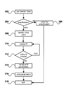
[0032] FIG. 2 shows an exemplary flowchart for a computer-implemented
assessment of a single
amplification reaction, according to some embodiments.
[0033] FIG. 3 shows an exemplary flowchart for a computer-implemented
assessment of whether
amplification has occurred, according to some embodiments.
[0034] FIG. 4A shows a detailed view of the data of FIG. 1B, showing
local data variance among
individual data points at the time in the amplification reaction when the
amplification signal begins to
exceed the background signal level.
[0035] FIG. 4B illustrates exemplary aspects of the method that is used
to determine whether an
amplification reaction has occurred. Open circles represent the data points of
FIG. 1B; grey-filled
diamonds are the segment-ratios. A vertical line indicates the maximum segment-
ratio. The position of
said maximum segment ratio defines larger segments (double-headed arrows): a
proximal segment
containing the baseline, and a distal segment containing the plateau. Said
larger segments are divided into
proximal and distal halves, with sub-segments containing the baseline signal
region and the plateau signal
region marked by grey boxes labeled 'mode box' where modal values are
determined from the data. The
right side of FIG. 4B shows a schematic representation of the mode-boxes with
a graphic circle
representing the modes, and the reaction amplitude defined as the difference
between the modes.
[0036] FIGS. 5A-H show a conceptual explanation and rationale for
evaluating results from a set of
replicate reactions. FIG. 5A shows three higher-concentration samples, and
FIG. 5B shows those three
samples after ideal dilution. FIG. 5C illustrates ideal exponential
amplification product accumulation over
time, and FIG. 5D shows a logarithmic transformation of FIG. 5C. FIG. 5E
superimposes idealized real-
time amplification data on the amplification traces of FIG. 51), and FIG. 5F
represents the slopes and
intercepts of FIG. 5E on a slope, intercept plot. FIG. 5G illustrates how
statistical error would alter the
slopes and intercepts of data from FIG. 5E, and FIG. 5H illustrates how the
statistical error illustrated in
FIG. 5G would appear if plotted as in FIG. 5F.
[0037] FIGS. 6A-D illustrate an exemplary application of the described
methods to the
amplification data shown in FIG. ID and in replicate reactions to said
amplification data.
[0038] FIG. 6A shows a collection of 44 points, chosen by the procedure
shown in FIG. 2, from the
amplification data shown in FIG. 1D. Filled circles represent [time,
log(signal)] data points. The line
intersecting the data points was fitted to the collected points by linear
regression.
[0039] FIG. 6B shows a subset of the data points from FIG. 1D, without
initial points from the
baseline and without terminal points from the plateau. The line through the
data points and extending to
the horizontal axis was plotted using the slope and intercept of the
regression line shown in FIG. 6A.
[0040] FIG. 6C shows the data from FIG. ID, without terminal points from
the plateau, and with
the vertical axis extended to form a figure similar to a single data-trace of
FIG. 5E. The line through the
5
CA 02916236 2015-12-18
WO 2014/186526 PCT/US2014/038103
data points and extending to the vertical axis, like the line shown in FIG.
6B, was plotted using the slope
and intercept of the regression line shown in FIG. 6A.
[0041] FIG. 6D shows the data from FIG. 6C, adding three similarly-
determined regression lines
determined from reactions that are replicate reactions to the reaction that
was the source of the data
shown in FIG. 1D. The resulting figure resembles the outcome anticipated as
shown in FIG. 5G.
[0042] FIGS. 7A-D show data from two sets of replicate reactions
initiated from two different
RAM template dilutions. For both sets of results, an initial panel and a post-
data-quality-control panel is
shown.
[0043] FIG. 7A shows slope, intercept pairs from the most dilute template
sample. Points identified
by quality control standards are lightly circled. FIG. 7B shows data points
from the same dilution that
pass a data-quality metric. Dashed lines in FIG. 7B are aligned parallel to
the apparent negative-sloped
cluster of data-points.
[0044] FIG. 7C shows slope, intercept pairs from a less dilute template
sample that was still
expected to include some failures to amplify due to absence of template
(Poisson failure). Points
identified by quality control standards are lightly circled. FIG. 7D shows
data points from the same
dilution that pass a data-quality metric. Dashed lines in FIG. 7D are aligned
parallel to the apparent
negative-sloped cluster of data-points.
DETAILED DESCRIPTION
[0045] The present invention relates to molecular quantification of nucleic
acid sequences, and
provides systems and methods for improvements in real-time data collection and
in real-time signal
analysis. In some embodiments, said improvements are applied to limiting
dilution methods for nucleic
acid quantification, hitherto analyzed only by endpoint methods.
[0046] As used herein, "sequence" means the order, number, and identity
of nucleotide bases that
compose a DNA or RNA molecule, or the order, number, and identity of
nucleotide bases that compose a
subset of said molecule. As used herein, "quantification" means a
determination of the number of
specified sequences in a given sample. In some embodiments, said sample may be
in liquid form and the
units of quantification may be expressed in number of sequences per volume of
liquid. The abbreviation
"log" is used for logarithmic transformation (e.g., "log(signal)").
[0047] As used herein, "aliquoting" means the dispensing of an initial
volume to multiple equal
smaller volumes, and "aliquots" refers to said multiple equal smaller volumes.
As used herein, "reaction
core mixture" means the components of a biochemical amplification mixture,
typically containing but not
limited to: one or more enzymes; ionic components that establish a desired
ionic strength and ionic
composition; and a buffer for control of reaction pH. A reaction core mixture
may be assembled at an
initial concentration that is greater than the final reaction concentration,
so that said reaction core mixture
may be brought to final reaction concentration by addition of a liquid volume,
said liquid volume possibly
being a sample that is to be assayed. As used herein, "reaction mixture"
comprises a reaction core mixture
at final reaction concentration, where said reaction mixture may contain
amplification templates. As used
6
CA 02916236 2015-12-18
WO 2014/186526 PCT/US2014/038103
herein, "replicate reactions" means aliquoting a reaction mixture to nominally
identical reaction
chambers; in contrast, "replicate assays" would refer to aliquots of a
reaction core mixture in nominally
identical reaction chambers, wherein sample volumes are individually added to
said aliquots.
[0048] Substances of interest are frequently present in small amounts in
a sample collected for
.. analysis. A biochemical method for estimation of the amount of a substance
is the specific amplification
of said substance, or the specific amplification of a marker that is attached
to said substance. A signal
proportional to the amount of amplification is measured during or after the
amplification process. If the
amount and rate of amplification are known, said signal can be used to
estimate the amount of starting
material. Estimation of an initial amount given the nature and degree of
amplification can be illustrated
by analogy to a bank account. If the present amount of money in a bank account
is known, as well as the
continuously-compounded interest rate and the time of accrual, then the
initial amount can be calculated.
Both the interest rate and time on deposit must be known to make the
calculation.
[0049] The accuracy of methods that calculate initial quantities from
amplified quantities is
dependent on knowledge of the nature of the amplification; for calculation of
initial quantities, a
mathematical model of the amplification process is prefen-ed. For biochemical
amplification,
mathematical models are derived from the biochemical reaction mechanism. Two
amplification methods
that are useful in molecular diagnostics are the polymerase chain reaction
(PCR) and two-primer ramified
rolling circle amplification (RAM). Models of the PCR and RAM reactions
predict that each reaction
ideally should exhibit exponential amplification. Other amplification methods
may share a model but
achieve amplification by different biochemical methods; for example, helicase-
dependent amplification
(HDA) and recombinase polymerase amplification (RPA) implement a PCR-like
mechanism without
thermal cycling. Still other amplification methods have been described with
mechanistic diagrams, but
have not yet been reduced to formal mathematical models (e.g., loop-mediated
isothermal amplification;
LAMP).
[0050] Measurement of nucleic acids by amplification was initially an
endpoint procedure that was
interpreted as revealing the presence or absence of a target sequence, and
possibly a tentative assessment
of target quantity. Estimation of target quantity was greatly improved by the
advent of instruments that
repeatedly detect and record a signal that is proportional to the amount of an
amplification product. Said
instruments are called real-time amplification detection systems; said systems
provide a signal that is
proportional to the progress of the amplification reaction. Amplification
reactions that are performed in
real-time amplification detection systems are called real-time reactions.
[0051] FIGS. IA-D provide examples of plotted data from real-time
reactions generated from PCR
reactions and RAM reactions. In real-time RAM or PCR reactions the early
amplification products are
not directly observed, because the signal due to those early amplification
products does not rise above a
basal, or baseline, level (see, e.g., FIG. lA and FIG. 1B); the pre-
amplification signal is attributable to
instrument noise or other factors. The initial basal signal is followed by a
transition period where the
observed signal is the sum of basal noise and amplification signal; this
transition period occurs as
amplification products accumulate to a point such that the signal due to those
products becomes greater
7
CA 02916236 2015-12-18
WO 2014/186526 PCT/US2014/038103
than the basal noise level. Exponential change of signal with respect to time,
ideally, follows the
transition out of the basal level. Following a period of exponential change,
the amplification rate slows as
expected of an exponential reaction in a limited volume (said amplification
rate change is due at least in
part to reactant limitation, changed reaction chemistry, or product
inhibition). In both RAM (FIG. 1B)
and PCR (FIG. 1A) the exponential growth rate decreases to a plateau, after
which signal does not change
significantly with respect to time.
[0052] RAM and PCR produce similar fluorescence-signal curves in real-
time reactions, as shown
in FIG. I. However, there are substantive differences between the isothermal
RAM reaction and the
thermocycling inherent to PCR. The thermal cycle is a necessary component of
the PCR process. Real-
time measurements of PCR reaction progress provide a single observation per
thermal cycle or provide a
single statistical summary per thermal cycle. PCR product yield at fractional
cycles can be estimated, but
is not, at the present state of the art, measured or analyzed. A common
analysis method for PCR is to
compute a fractional cycle at a signal threshold; the fractional cycle is
reported as a cycle threshold (Ct)
or quantification cycle (Cq).
[0053] There are few PCR cycles in which the amplification is in
exponential phase. Identifying
cycles that exhibit characteristics of exponential signal change is important
because it is assumed that it is
only in the exponential phase that the fractional cycle reflects the input
template level. Ideally, the
fractional cycle at which the Ct/Cq is estimated should be in the exponential
change region of the
amplification. However, due to the paucity of exponential phase PCR cycles,
distinguishing those cycles
is not a simple or straightforward process.
[0054] The RAM reaction can be analyzed as a continuous time-dependent
process rather than a
discrete cycle-dependent process; the time to reach a threshold fluorescence
is designated the response
time (RI) corresponding to the cycle threshold of real-time PCR. The natural
unit of isothermal
amplification is product yield per time unit, just as product yield per
thermal cycle is the natural unit of
PCR. Unlike cycle number, time is continuous and can be infinitely subdivided;
therefore, product yield
measurement frequency of isothermal amplification is limited only by
instrument capacity.
[0055] The utility of finding the exponential phase PCR cycles has
motivated a series of
publications that continue on after more than two decades, describing a
variety of methods for PCR
analysis. These methods can generally be divided into two groups: first,
parametric methods that seek to
fit mathematical models by parameter fitting to the data; and second, non-
parametric methods that extract
statistical measures directly from the data. Non-parametric approaches to
exponential phase signal
determination include mathematical operations on the signal such as computing
the fractional cycle at
which the second derivative of the signal vs. cycle data is at a maximum.
Parametric, or model-fitting,
methods are a more computationally intensive process. Some reported procedures
fit a mathematical
object having similarity to the whole reaction signal (baseline,
amplification, signal plateau). Others have
extensively refined model-fitting techniques; their computer applications find
parameters of the fitted
model, and analyze points or regions of the fitted object.
WO 2014/186526 PCT/US2014/038103
[0056] Identification of the PCR exponential amplification phase has at
least two motivations:
determination of Ct/Cq, and determination of the PCR reaction efficiency.
Accurate comparison of
multiple PCR reactions requires an estimate of each reaction's efficiency.
Ideally, each PCR cycle should
yield twice as much product as the previous cycle. However, various factors
including sub-optimum
primers, amplification inhibitors in the sample, or sub-optimum reaction
chemistry can result in less than
ideal exponential growth; therefore, comparison of two PCR reactions with
different efficiencies can lead
to inaccurate estimates of initial amounts.
[0057] A substantial effort has been devoted to defining PCR efficiency;
for example, the online
bibliography provided by the Gene Quantification platform lists more than 50
publications devoted to
various aspects of defining and applying measures of PCR efficiency. These and
other publications
provide examples of definition and analysis of regions of PCR data. In
parametric modeling the model
that is fitted to the data is not, however, the exponential growth ideally
observed in PCR; instead, the
fitted models are used to find the exponential region.
[0058] An essential input to some model-based methods of finding the
exponential region in current
PCR methods is finding the signal baseline, defined as the early signals that
do not change much over
dine. Various methods for finding a PCR baseline have been described.
[0059] Practical applications of specific nucleic acid sequence
detection require comparison among
multiple samples; said samples might include, for example, positive controls
for amplification and/or a
set of standard concentrations of known targets. Absolute quantification,
expressed in numerical units of
target molecules, has not yet been accomplished via real-time amplification
monitoring, as evidenced by
the adoption of digital PCR, which abandons real-time methods in favor of
endpoint detection and
statistical analysis. However, accurate quantification via PCR has encountered
both practical and
theoretical limits. Practically, the difficulty of per-reaction efficiency
measurement has limited PCR
quantification, and Poisson variance at limiting dilutions has made
quantification at less than 5 to 10
template copies per reaction theoretically challenging. Some researchers have
turned Poisson limiting
dilution effects from a problem into an advantage by abandoning any use of
real-time signal analysis,
instead using endpoint detection and statistical inference of template copy
number after sample dilution.
[0060] Poisson statistics in this context describes the distribution of
small numbers of events. For
example, if ten marbles are tossed randomly into ten cups, one cup may get two
marbles and another cup
may get no marbles. If the number of marbles tossed is less than the number of
cups, some cups are
guaranteed to be empty; and the ratio of empty to non-empty cups could be used
to estimate the original
number of marbles, given the number of cups. The Poisson distribution is a
statistical tool that could be
used for predicting how many cups get some number of marbles. After the
tossing, the Poisson
distribution could be used to predict, from a count of marbles from a sample
of cups, how many marbles
were tossed. The Poisson distribution is an appropriate model for the
partitioning by dilution of samples
for molecular diagnostics.
[0061] The present invention provides, in various embodiments,
improvements in molecular
measurement methods using amplification. The description herein is illustrated
with data from an
9
Date Recue/Date Received 2021-02-09
CA 02916236 2015-12-18
WO 2014/186526 PCT/US2014/038103
amplification using RAM (FIG. IB), but the invention is not limited to that
reaction mechanism. For
example, in alternative embodiments, other isothermal systems may be used for
high-frequency sampling.
The methods described herein have utility with any amplification method where
sufficient samples that fit
a mathematically defined region can be obtained. Examples of suitable
amplification methods include,
but are not limited to, RAM, primer generation-rolling circle amplification,
amplification via
recombination proteins (RPA), loop-mediated amplification on circle-substrate,
real-time NASBA
(Nucleic Acid Sequence Based Amplification) with beacons, and helicase-
mediated isothermal PCR
(HDA).
[0062] The PCR is not sampled uniformly in the sense described here
because a single data point
per PCR cycle is observed (or computed). Although the sampling is regular, the
reaction is not sampled
uniformly in the exponential signal change region of interest. In alternative
embodiments, the methods
provided herein could be applied to uniform sampling of thermocycling PCR with
appropriate product
accumulation or product accumulation marker detection systems; however,
correction for differential rate
of product accumulation vs. time in non-uniform reaction environments (e.g.,
thermal denaturation
cycles, thermal ramping) would preferably be made. A reaction temperature
datum would preferably be
recorded for each tune point and product measurement.
[0063] Previous methods used the intersection of a line generated by an
exponential model fit to the
amplification region of a RAM reaction and the baseline of that RAM reaction
to define a response time.
Such computations require a baseline definition (as noted above, not a trivial
operation) and, as described,
do not compute or consider efficiency.
[0064] FIG. 2 shows an exemplary flowchart for computer-implemented
assessment of a single
amplification reaction, according to some embodiments. At step 202,
amplification data are read into a
table; then at step 204 a decision is made as to whether a significant
amplification has occurred (see, e.g.,
FIG. 3). If no amplification has occurred, this result is noted at step 206
and the evaluation is ended at
step 218. If an amplification has occurred then at step 208 an initial point
for evaluation is chosen. From
the initial point, additional points are added at step 210 and evaluated at
step 212 until a next point causes
the collection of points to fail predetermined evaluation criteria. The chosen
collection of points is
evaluated to compute fit parameters at step 214 and statistical measures of
the chosen collection of points
are recorded (e.g., written to a table) at step 216, completing the evaluation
of the current reaction at step
218.
[0065] FIG. 3 shows an exemplary flowchart for a computer-implemented
assessment of whether
amplification has occurred (step 204), according to some embodiments. In some
embodiments, the input
data to the algorithm at step 302 is a set of (time, signal) data, obtained,
for example, at FIG. 2 step 202.
Said data is evenly sub-divided into multiple segments at step 304. Segments
are divided into proximal
and distal halves, segment ratios are computed at step 306, and a maximum sub-
segment ratio is
determined at step 308. At step 310, the modes of the signal data are
determined, and the reaction
amplitude is defined as the difference between the baseline mode and the
plateau mode. The amplitude
difference is evaluated at step 312. If the amplitude is greater than a preset
threshold value ("YES- at step
CA 02916236 2015-12-18
WO 2014/186526 PCT/US2014/038103
312) then amplification is deemed to have occurred at step 316, the analysis
of FIG. 3 ends at step 318,
and the analysis of FIG. 2 proceeds to step 208; otherwise ("NO" at step 312)
no amplification is deemed
to have occurred at step 314, the analysis of FIG. 3 ends at step 318, and the
analysis of FIG. 2 proceeds
to step 206.
[0066] Based on the values for the parameters of the model (e.g., as
recorded at step 216), the
present invention further comprises determining a value representative of an
initial condition of the
sample. In some embodiments, the value is a value representative of an amount
of a nucleic acid sequence
in the sample. For example, the system may display to the user an estimate of
copy number (e.g., for
Downs syndrome fetal diagnostics: "2 copies of Chromosome 21" for the normal
euploid complement).
In another embodiment, for process optimization, the system may display to the
user a message such as:
"Condition A results in an 0.1x increase in slope compared to condition B."
The systems and methods of
the present invention are adaptable, multi-purpose tools, and the preceding
examples represent only two
out of many possible output displays from two exemplary uses.
[0067] FIGS. 1B and 1D show data points collected at high frequency from
a typical isothermal
amplification reaction (a RAM reaction). These data were analyzed by the steps
shown in the flowcharts
of FIGS. 2 and 3, as described below. The data density provided by the methods
described here
necessitated statistical methods to distinguish local data variance from
larger trends. FIG. 4A illustrates
the variance in the (time, signal) data points in the region where the
amplification signal initially rises
above the baseline.
[0068] A set of (time, signal) data was accessed (step 202), and a decision
as to whether an
amplification occurred (step 204) was made as follows, according to the
process shown in FIG. 3.
Reaction data input at step 302 was subdivided into multiple segments (step
304); the segment size may
vary depending on the number of data points and the data variance. Each
segment was equally divided
into an earlier set of signal data points and a later set of signal data
points. A segment ratio was computed
for each segment by dividing the sum of the distal signal data by the sum of
the proximal signal data (step
306). The set of segment ratios are indicated graphically by diamonds in FIG.
4B. The maximum segment
ratio was determined (step 308), and used to define two larger data segments:
a baseline segment
extending from the start of data to the maximum segment ratio, and a plateau
segment from the maximum
segment ratio to the last data point (said larger segments are illustrated in
FIG. 4B). Said larger segments
were subdivided by halves into a proximal baseline segment and a distal
plateau segment, shown by
graphic boxes in FIG. 4B. The statistical mode of each said sub-segment was
determined, and the
reaction's amplitude was defined as the difference between the mode of the
proximal baseline segment
and the mode of the distal plateau segment (step 310). When the amplitude was
greater than a predefined
value ("YES" at step 312), the reaction was scored as having an amplification
signal (step 316); otherwise
("NO" at step 312) the reaction was scored as no amplification (step 314).
[0069] In alternative embodiments, other processes for computer-
implemented assessment of
whether amplification has occurred may be used for the conditional of step
204, which may vary in
complexity and computation time per data set; the method is not limited to the
process described and
11
CA 02916236 2015-12-18
WO 2014/186526 PCT/US2014/038103
shown in FIG. 3 and FIG. 4B. For example, in other embodiments,
amplification/no amplification could
be detected by analysis of spline fit parameters, or by parametric fitting of
a sigmoid model (as is done in
some PCR analysis methods).
[0070] Identification of an initial point of interest (step 208) was
accomplished by fitting a
smoothing spline to the data (the smoothing spline is represented by a gray
line in FIG. 1D). A first
derivative maximum was obtained from the fitted spline, completing step 208.
In alternative
embodiments, other methods for selecting an initial point may be used (e.g., a
segment-wise evaluation
could be used, wherein the whole data set is segmented, R^2 correlation
coefficients are determined for
each segment, and an optimum segment is chosen; or a second derivative from a
spline fit could be used).
A fixed region around said first derivative maximum was chosen, then a linear
model was fitted to said
region around said first derivative maximum. A correlation coefficient (RA2)
was computed for the
current region. Then the region was extended for a defined step size (step
210), and a second correlation
coefficient (12^2) was computed for the extended region. When the difference
between the first and
second correlation coefficients did not exceed a set maximum ("YES" at step
212), the extended region
was redefined as the current region. Otherwise ("NO" at step 212), the current
region was taken as the
preferred data subset for the current reaction, and statistical measures
describing said subset (step 214)
were written (step 216) to a data table. In this example, said measures of
said subset included: a least-
squares linear regression fit to the [log(signal), time] data; the slope and Y-
intercept; the correlation
coefficient (RA2); the variance; and the time points defining the start and
end of said subset. In alternative
embodiments, other statistical measures or other combinations thereof may be
used. FIG. 6A illustrates
the data points of a subset extracted as described from the data of FIG. 1D,
with a fitted line generated by
the slope and Y- intercept derived from a linear regression model fitted to
the log-linear data as described.
[0071] The Y-intercept is the point at which said fitted line crosses the
Y-axis. Said Y-intercept is a
hypothetical fluorescence signal at time 0 (zero); a greater initial template
at time 0 will have a greater Y-
intercept, if slopes are equal. The Y-axis is demarcated in units of signal,
not copy number, so Y-axis
intercepts may be below a nominal 0 (zero) level. Slope, in isothermal
reactions, is product increase over
time; the PCR equivalent is efficiency, product increase per thermal cycle. A
per-reaction efficiency
measure is a long-sought but not-yet achieved goal for PCR; high-frequency
data sampling and analysis
as presented here accomplish that goal for isothermal amplifications.
[0072] The per-reaction slope and Y-intercept scores would, a priori, both
be significant in the
interpretation of isothermal reactions. The slope and intercept significance
is illustrated conceptually in
FIGS. 5A-H, before further presentation of experimental data. Consider three
sample solutions containing
different concentrations of a DNA sequence, and suppose those concentrations
are sufficient for DNA
concentration measurement (e.g., by measuring fluorescence emission after
addition of a suitable dye).
These measurements can be represented as points on a vertical axis, as shown
in FIG. 5A, where the
sample amounts are represented as doublings in the ratio 1:2:4.
[0073] Suppose samples from said three solutions each to be ideally
diluted, said dilution
maintaining the copy number ratio, such that direct measurement of DNA
sequence is no longer possible,
12
CA 02916236 2015-12-18
WO 2014/186526 PCT/US2014/038103
and with a maximum dilution such that the lowest DNA copy number is as low as
one (1) copy per
sample. FIG. 5B represents the (now not directly measurable) DNA
concentrations of the diluted samples
on a vertical axis. Suppose the diluted samples are amplified by an ideal
exponential amplification
system, so that FIG. 5C represents the amplification process as product
accumulation over time. FIG. 5D
depicts a logarithmic transformation of the product yield plotted against
time, resulting in a linear relation
of log(product) vs. time.
[0074] FIG. 5E illustrates what is seen in real-time monitoring of
exponential amplification by
overlaying the idealized amplification time course with a representation of a
real-time signal trace (e.g.,
FIG. ID). In this idealized system, the slopes of the amplification time-
course are all equal; in isothermal
amplifications this would indicate equal rates of product synthesis over time,
and in PCR equal slopes
would indicate identical reaction efficiencies. Therefore, FIG. 5F shows that
if the time-zero intercept in
FIG. 5E is plotted on a vertical axis, and the (equal) slopes corresponding to
those intercepts are plotted
on a horizontal axis, the result is a vertically aligned set of points that is
proportional to the logarithms of
the initial substrate amounts that were amplified. The vertical data of FIG.
5F are an idealized
experimental determination of the ratios of the starting material of FIG. 5B.
[0075] In actual amplification systems, experimental noise introduces
statistical error into estimates
of slopes; and replication provides a means of estimation of the magnitude of
said statistical error. FIG.
5G depicts replicate amplification reactions, wherein each said replicate
reaction provides a slope and an
intercept. FIG. 5G shows that slope variation results in intercept variation,
wherein greater slopes result in
lower intercepts. Therefore, when slope is plotted on an increasing scale, the
corresponding intercepts
decrease in magnitude as shown in FIG. 5H.
[0076] FIGS. 6A-D show experimental data illustrating some aspects of the
theoretical treatment of
FIGS. 5A-H. FIG. 6A shows data points identified by the procedure shown in
FIG. 2, from the data of
FIG. 1D, as well as a line defined by a linear model that was fit to those
data. FIG. 6B illustrates said
fitted line over a larger subset of the FIG. 1D data from baseline to plateau.
FIG. 6C shows all the
baseline data on an extended vertical axis to illustrate the Y-intercept of
the fitted line, and FIG. 6D
shows data from three additional replicate reactions, illustrating with
experimental data the
conceptualization of FIG. 5G.
[0077] The experimental data from which the graphics of FIGS. 1A-D and 6A-
D are derived
included two different dilutions that were made from a single source stock of
single-stranded DNA
circles. The amount of dilution was calculated to be such that some aliquots
of each dilution would be
predicted from Poisson statistics to include some aliquots without any
template molecules. An aliquot of
each dilution was added to a reaction core mixture to create two reaction
mixtures, and said reaction
mixtures were aliquoted to multiple reaction chambers. 45 replicate RAM
reactions were performed in a
real-time instrument for each reaction mixture; said instrument was programmed
to obtain real-time data
at the shortest possible time-intervals. Said data was processed by a set of
scripts implemented in the "R"
system for statistics such that each reaction was evaluated according to the
processes shown in the
flowcharts of FIGS. 2 and 3. A table of statistical measures was constructed
for reactions in which an
13
CA 02916236 2015-12-18
WO 2014/186526 PCT/US2014/038103
amplification was detected, and the number of reactions in which no
amplification was detected was
likewise recorded.
[0078] FIGS. 7A and 7B show plots of data from the more dilute of the two
dilutions. The 17 data
points of FIG. 7A show the slope and intercept of each positive reaction. The
¨62% failure rate implies a
Poisson estimated mean of 0.47 template molecules per reaction. Initial
statistical evaluation showed that
three of the 17 data points of FIG. 7A had lower RA2 correlation coefficients
and later response times;
those data were eliminated from consideration and the remaining points appear
in FIG. 7B. These points
fall along a negative slope as depicted in FIG. 5H. Applying Poisson
statistics further indicates that for 45
samples at a Poisson mean of 0.47, about 13 samples at one template per sample
are expected, about three
samples at two templates per sample are expected, and there is about a 50%
chance of seeing one sample
with three templates. Dashed lines plotted in FIG. 7B show that the multiple
diagonals anticipated from
FIG. 5H separate consistent groups of data points.
[0079] The second dilution prepared as described above was designed to
have a four-fold greater
number of template molecules per sample. FIGS. 7C and 7D show plots similar to
FIGS. 7A and 7B, with
FIG. 7C showing all data points from the less dilute sample, and FIG. 7D
showing data points excluded
as outliers. Three failures out of 45 replicate reactions implies a Poisson
mean of ¨2.7 template molecules
per reaction tube. Arranging diagonal separator lines as for FIG. 7D reveals a
distribution of data points
consistent with Poisson expectations.
[0080] The results shown in FIGS. 7A-D demonstrate that the methods of
the present invention can
facilitate the identification of amplification reactions that contain one and
only one template molecule.
The present invention's use of slope, intercept plots for estimating small
copy numbers is in the general
field that is now served by digital PCR. As currently practiced, only
product/no-product ratios obtained
from replicate reactions are used in statistical analysis of digital PCR
assays, and real-time data is not
collected. The results shown here illustrate the utility of collecting real-
time data from isothermal
reactions on samples made by appropriate dilutions, or of assays on undiluted
samples of appropriate
concentrations. The utility of the identification of amplification reactions
that contain a single template
molecule includes the potential greater efficiency of target concentration
estimation when a count of
single molecule reactions is combined with a proportion of reactions that fail
to amplify and are
interpreted as containing Zero template molecules. Further efficiency of
target concentration estimation
may result if the number of two-template molecule amplifications can be
estimated, and so on for greater
template molecule numbers.
[0081] In alternative embodiments, the methods of the present invention
may be applied to digital
PCR. Digital PCR requires some zeros, and thus requires a large number of
wells when the average copy
number per reaction is high. A system based on the methods disclosed herein,
optionally with some added
wells for standards, can advantageously provide accurate concentration
estimates without any zero wells.
[0082] The methods described herein may also be used for assay
optimization. The present
invention recognizes that distinguishing one-template amplifications from 2-,
3-, or more-template
amplifications can be more reliably done if the target-number groupings (as
shown in FIGS. 7B and 7D)
14
CA 02916236 2015-12-18
WO 2014/186526 PCT/US2014/038103
are well-separated. In addition, experimental manipulation of reaction
conditions may allow a greater
separation of the target-number groups that are shown separated by dashed
lines in FIGS. 7B and 7D.
Further, the methods of the present invention provide improved accuracy in
reaction rate determination.
[0083] In cases where no mathematical model for reaction rate has been
defined but densely
sampled data can be obtained, data can be sampled as described above. In some
embodiments, the present
invention contemplates finding inflection points using derivatives of such
data, regions where the
amplification rate changes; said points may, for example, be correlated with
experimentally defined
factors and used for process optimization.
[0084] As new isothermal amplification methods continue to be described
there is an ongoing need
to elucidate reaction mechanisms and to optimize reaction conditions. Process
optimization using
statistical design of experiments (DOE) will also benefit from the methods
disclosed here. In DOE,
multiple experimental factors (such as reagent concentrations, or reaction
conditions such as temperature)
are systematically changed and the effects of those changes are measured; a
strength of DOE is the
detection of interacting factors where a combination of factors has a greater
effect than the sum of the
effects of the separate factors. Analysis of such experiments is facilitated
by variety and detail of
descriptive data; therefore, DOE methods applied to biochemical amplification
reactions will benefit
greatly from the methods disclosed here due to the detailed recording of the
reaction process.
[0085] Some amplification methods may not produce data subsets to which
linear models can be
fitted, either to raw data or to data after the logarithmic transformations
used for RAM or PCR. Where no
linear model can be fitted the data density yielded by the present methods can
still prove advantageous by
allowing the fitting of well-defined nonlinear models, and those models may be
useful in elucidating
reaction mechanisms. In cases where no well-defined model can be fit, the data
density revealed by the
methods disclosed here may still be used to advantage, for example in the
empirical piece-wise or
segment-wise fitting of polynomial splines.
[0086] The applications of the methods described herein represent only a
few examples of the
utility of high-frequency biochemical amplification data collection and
analysis. Automated detection of
noise signals and other quality-control applications are contemplated in
further embodiments. The
methods of the invention disclosed herein provide improved estimates of
significant parameters,
applicable in many areas of analysis.
[0087] While there have been shown and described fundamental novel features
of the invention as
applied to the preferred and exemplary embodiments thereof, it will be
understood that omissions and
substitutions and changes in the form and details of the disclosed invention
may be made by those skilled
in the art without departing from the spirit of the invention. Moreover, as is
readily apparent, numerous
modifications and changes may readily occur to those skilled in the art.
Hence, it is not desired to limit
the invention to the exact construction and operation shown and described and,
accordingly, all suitable
modification equivalents may be resorted to falling within the scope of the
invention as claimed. It is the
intention, therefore, to be limited only as indicated by the scope of the
claims appended hereto.
EXAMPLE 1
100881 This example describes amplification of a single-stranded, nucleic
acid target, according to
some embodiments of the invention. A C-probe with gene-specific termini that
are specific for the wild-
type locus of the human Factor V gene was 5 phosphorylated, then circularized
and amplified as
described below to provide an amplification substrate for 2-primer ramified
rolling circle (RAM)
amplification and analysis.
100891 A ligation reaction was made by combining synthetic ligation
target TgtEctVWT+
(5'pAGAGACATCGCCTCTGGGCTAATA GGACTACTTCTAATCTGTAA GAGCAGATCCCTG)
(SEQ ID NO: 1) to 9 M; C-probe Cpr8FVWt I
(5'GCCTGICCAGGGATCTGCTCTTACAATACGAGAACACCCGAT
TGAGAGAGTTTGGAAG TGTAGGCGTGAAGTCCATAACACATACCTGTATTCCTC) (SEQ ID
NO: 2) to 6 M, Taq DNA Ligase buffer (New England Biolabs, Ipswich, MA) to
lx, Taq DNA ligase
(New England Biolabs, Ipswich, MA) to 0.8 units/ 1, and water to 100 p1
volume. The reaction mixture
was incubated at 95 C, 30 sec for initial denaturation then cycled five times
through the following
regimen: 30 C, 1 min for hybridization; 65 'C, 30 sec for ligation. then 95
C, 10 sec for denaturation.
100901 An estimate of circular, single-stranded DNA template
concentration was made, bearing in
mind possible sources of error: random error with mean zero effect and error
based on 100% optima
assumption where real percentages less than 100% result in lower template
concentration. Examples of
the former error types include the algorithmic assignment of an extinction
coefficient to the C-probe,
error in spectrophotometric measurement, and liquid handling error. Idealized
100% assumptions include:
the assumption that 100% of the C-probes are full length and capable of
ligation after hybridization on the
target; that 100% of the C-probes are phosphorylated after exposure to kinase
enzyme; and that 100% of
the ligatable, phosphorylated C-probes are circularized after the
aforementioned ligation regimen.
Assuming zero-mean random error and 100% C-probe competence, ligation mix was
diluted to two
calculated concentrations, differing by four-fold, and such that each dilution
should provide at least one
amplification reaction without any template molecules, when replicate
reactions were performed at the
designed replicate number and reaction volume.
100911 A 2x RAM core reaction mix was created containing: 2x isothermal
reaction buffer and 0.26
units/al Bst Pol2 WS (New England Biolabs, Ipswich, MA), 0.4 mM each dNTP, and
0.5x Eva Green
(Biotiurn Inc., Hayward, CA). Two, 2x primer-template mixtures each containing
RAM Forward primer
Cpr8FVEwd76_18 (5'ATGGAC1TCACGCCTACA) (SEQ ID NO: 3) at 2.4 FM; RAM reverse
primer
Cpr8FVRvs87_18 (5'TGTATTCCTCGCCTGTCC) (SEQ ID NO: 4) at 2 PM; 2 M fluorescein
(BioRad,
Hercules, CA) solution in DMSO were prepared, and each primer-template mixture
was brought to final
volume by addition of a template dilution prepared as described. Reaction
mixtures were made by
combining equal volumes of 2x core reaction mix and of one of the two 2x
primer-template mixes; then,
20 al reaction mix was aliquoted into the wells of a 96 well plate. Real-time
reactions were done in an
iCycler real-time instrument (BioRad, Hercules, CA) set to collect continuous
real-time data.
16
NY 75044593
2969023
CA 2916236 2019-04-17