Note: Descriptions are shown in the official language in which they were submitted.
CA 02916394 2015-12-18
WO 2015/013432
PCT/US2014/047860
MODEL OF COLORECTAL CANCER
Field of the Invention
The present invention relates generally to a clinically relevant model of
colorectal cancer (CRC)
and methods of using the model to screen for compounds that inhibit
tumorigenesis.
Background of the Invention
Colorectal cancer (CRC) is the third most common cancer worldwide and the
fourth most
common cause of death. CRC accounts for over 9% of all cancer incidences. In
2013, it is estimated
that 142,820 new CRC cases will be diagnosed in the United States and that
50,830 people will succumb
to the disease (American Cancer Society: Cancer Facts and Figures 2013,
Atlanta, GA, 2013). The poor
ratio of survival to incidence of CRC is due, at least in part, to the high
percentage of cases that are
diagnosed at an advanced stage. The overall five-year relative survival of
patients with advanced stage
metastatic CRC stands at 12,5% (Howlader et al. SEEF? Cancer Statistics
Review, 1975-2010. NCI.
Bethesda, MD, 2013),
Although xenograft, chemical-induced, and genetically-engineered models (e.g.,
mouse
models) of CRC have been developed to study CRC, tumors in these models fail
to reproducibly
metastasize to the regional intestinal lymph nodes and liver, the target
organs relevant to human
CRC. Thus, there is an unmet need in the field for the development of a model
of CRC that is
capable of recapitulating the pathogenesis of the human disease. Such a model
of CRC would be
a valuable tool for testing therapeutics and developing novel treatment
strategies.
Summary of the Invention
The present invention provides a model of colorectal cancer (CRC) that
recapitulates the
pathogenesis of the human disease, as well as methods for generating and using
the model.
In a first aspect, the invention features a non-human mammal including a donor
tumorigenic cell
implant on the colonic mucosal surface, wherein implantation does not result
in breach (e.g.,
opening, tear, rupture, or puncture) of the colon wall (i.e., the integrity of
the deeper colon wall layers is
maintained). In one embodiment, the donor tumorigenic cell implant is capable
of invasive growth
through the colon wall to the colonic serosal surface (e.g., growth resulting
in penetration through the
collagen IV-rich basement membrane of the muscularis externa to the serosal
surface). In another
embodiment, the invasive growth of the donor tumorigenic cell implant is
characterized by metastases in
common target organs (i.e., target metastatic organs or metastatic tissues) of
CRC (e.g., human CRC),
such as the intestinal lymph nodes, liver, or lungs. In another embodiment,
the non-human mammal does
not exhibit detectable tumor formation in the peritoneal cavity (e.g.,
peritoneal carcinomatosis) post-
implantation. In another embodiment, the donor tumorigenic cell implant
includes cells of a cancer cell
line. The cancer cell line, in one embodiment, is a CRC cell line (e.g.,
HCT116). In another embodiment,
the cancer cell line is a non-CRC cell line (e.g., a lung cancer cell line, a
liver cancer cell line, a brain
cancer cell line, a lymph node cancer cell line, a kidney cancer cell line, a
stomach cancer cell line, a
ovarian cancer cell line, a skin cancer cell line, a pancreatic cancer cell
line, a thyroid cancer cell line, a
1
CA 02916394 2015-12-18
WO 2015/013432
PCT/US2014/047860
prostate cancer cell line, or a breast cancer cell line, e.g., MDA-231). In
another embodiment, the donor
tumorigenic cell implant is an intact tumor, or fragment thereof. The intact
tumor, or fragment thereof, in
one embodiment, may be malignant (e.g., metastatic, regionally invasive,
and/or distantly invasive). In
another embodiment, the intact tumor, or fragment thereof, may be benign
(e.g., non-metastatic and/or
locally invasive). In some embodiments, the intact tumor, or fragment thereof,
is an intact CRC tumor, or
fragment thereof. In other embodiments, the intact tumor, or fragment thereof,
is an intact non-CRC
tumor, or fragment thereof (e.g., a breast cancer tumor, a lung cancer tumor,
a liver cancer tumor, a brain
cancer tumor, a lymph node cancer tumor, a kidney cancer tumor, a stomach
cancer tumor, a ovarian
cancer tumor, a skin cancer tumor, a pancreatic cancer tumor, a thyroid cancer
tumor, or a prostate
cancer tumor, or fragment thereof). In another embodiment, a subset (e.g., 5%,
10%, 11%, 12%, 13%,
14%, 15%, 16%, 17%, 18%, 19%, 20%, 21%, 22%, 23%, 24%, 25%, 26%, 27%, 28%,
29%, 30%, 35%,
40%, 45%, 50%, 55%, 60%, 65%, 70%, 75%, 80%, 85%, 90%, 95%, 96%, 97%, 98%, or
99% or more) of
cells of the donor tumorigenic cell implant is capable of invasive growth. In
other embodiments, growth of
every (i.e., 100 /o) cell of the donor tumorigenic cell implant may be
characterized as invasive growth. In
another embodiment, the non-human mammal is a rodent, such as a mouse or a
rat. In some
embodiments, the rodent (e.g., mouse or rat) may be immunodeficient or
immunocompromised. An
immunodeficient mouse, in certain embodiments, may be a NOD/SCID mouse or a
NOD/SCID
interleukin-2 receptor gamma chain null (NSG) mouse. In other embodiments, the
non-human mammal
is wild-type andlor immune-competent (e.g., a wild-type or immune-competent
rodent, e.g., a wild-type or
immune-competent mouse or rat).
In a second aspect, the invention features a method for generating a non-human
mammal (e.g.,
rodent, e.g., mouse or rat) of the first aspect (i.e., a non-human mammal
(e.g., rodent) model for CRC),
the method including exteriorizing the colonic mucosal surface of a host non-
human mammal, implanting
one or more tumorigenic cells onto the colonic mucosal surface, and re-
inserting the exteriorized colon
comprising the one or more implanted tumorigenic cells into the host non-human
mammal.
In a third aspect, the invention features a method of screening for a compound
that inhibits
growth of tumorigenic cells (e.g., inhibits growth of tumorigenic cells by at
least 20%, 25%, 30%, 35%,
40%, 45%, 50%, 55%, 60%, 65%, 70%, 75%, 80%, 85%, 90%, 95%, 96%, 97%, 98%, or
99% or greater,
e.g., compared to an untreated or control-treated group), the method including
contacting the donor
tumorigenic cell implant of a non-human mammal of the invention with a
candidate compound and
determining whether the candidate compound inhibits growth of the tumorigenic
cells, thereby identifying
the candidate compound as a compound that inhibits growth of tumorigenic
cells.
In a fourth aspect, the invention features a method of screening for an
adjuvant that inhibits
growth of tumorigenic cells (e.g., inhibits growth of tumorigenic cells by at
least 20%, 25%, 30%, 35%,
40%, 45%, 50%, 55%, 60%, 65%, 70%, 75%, 80%, 85%, 90%, 95%, 96%, 97%, 98%, or
99% or greater,
e.g., compared to an untreated or control-treated group), the method
including: removing the donor
tumorigenic cell implant from the colonic mucosal surface of a non-human
mammal of the first aspect,
administering to the non-human mammal a candidate compound, and determining
whether the candidate
compound inhibits growth of tumorigenic cells, thereby identifying the
candidate compound as an
adjuvant that inhibits growth of tumorigenic cells.
In one embodiment of the third or fourth aspect of the invention, the step of
determining whether
the candidate compound inhibits growth of tumorigenic cells includes
evaluating the ability of the
2
CA 02916394 2015-12-18
WO 2015/013432
PCT/US2014/047860
candidate compound to evoke at least one response (e.g., 1, 2, 3, 4, or 5
responses) selected from the
group consisting of: reduction or stabilization in the number of tumorigenic
cells; reduction or
stabilization of tumor size; reduction or stabilization of tumor load;
reduction or stabilization of tumorigenic
cell invasiveness; and reduction or stabilization of tumor metastasis. In
another embodiment of the third
or fourth aspect, the candidate compound may be a small molecule, a peptide, a
polypeptide, an
antibody, an antibody fragment, or an immunoconjugate. In another embodiment
of the third or fourth
aspect, the donor tumorigenic cell implant may be capable of invasive growth
through the colon wall to
the colonic serosal surface (e.g., growth resulting in penetration through the
collagen IV-rich basement
membrane of the muscularis externa to the serosal surface). In other
embodiments of the third or fourth
aspect, invasive growth of the tumorigenic cells may be characterized by
metastases in one or more (e.g.,
1, 2, or 3 or more) common target organs (i.e., target metastatic organs or
metastatic tissues) of CRC
(e.g., human CRC), such as the intestinal lymph nodes, liver, or lungs. In
other embodiments of the third
or fourth aspect, the non-human mammal does not exhibit detectable tumor
formation in the peritoneal
cavity (e.g., peritoneal carcinomatosis) post-implantation. In another
embodiment of the third or fourth
aspect, the non-human mammal is a rodent, such as a mouse or rat.
Brief Description of the Drawings
The application file contains at least one drawing executed in color. Copies
of this patent or
patent application with color drawings will be provided by the Office upon
request and payment of the
necessary fee.
FIGURE 1A shows images of the colon (top and middle panels) and liver (bottom
panel) from a
donor Apen1+; Villin-Cre control mouse at 9 weeks of age. Colons were opened
longitudinally and
mucosal (top) and serosal (middle) views were imaged, with the anus positioned
to the left. Arrows
indicate colon polyps. Boxed areas of the liver have been enlarged.
FIGURE 1B depicts images of the colon (top and middle panels) and liver
(bottom panel) from a
donor Apcmml* ; KrasLSLG1 2 D/+ ; Villin-Cre mouse at 9 weeks of age. Colons
were opened longitudinally and
mucosal (top) and serosal (middle) views were imaged, with the anus positioned
to the left. Arrows
indicate colon polyps. Boxed areas of the liver have been enlarged.
FIGURE 1C is a graph showing endogenous colon tumor burden in Apen1+; Villin-
Cre (n = 5) and
Ape' ni+ ; KrasLSLG1 2 D/+ ; Villin-Cre (n = 4) mice at 6 weeks of age. Each
point represents data from an
individual mouse. Means s.e.m. are also shown. *P < 0.05.
FIGURE 1D is a graph showing Kaplan-Meier survival curves for ApcmIni+; Villin-
Cre (n = 49) and
ApcmIni+; KrasLSLG1 2D/+ ; Villin-Cre (n = 14) mice. HR = hazard ratio.
FIGURE lE is a set of gross colon images (top panel: mucosal view; bottom
panel: serosal view)
from a host wild-type C57BL/6 mouse that has received a lumen implant of a
single intact
Apen1+; KrasLSLG1 2 D/+ ; Villin-Cre colon tumor fragment from a donor
subcutaneous allograft, harvested at 9
wpi. The boxed areas of the colon have been enlarged to the right of the
respective image. Scale bars
represent 3 mm.
FIGURES 1F and 1G are images of colons (top and middle panels) and livers
(bottom panels)
from two host wild-type C57BL/6 mice that have received lumen implants of a
single intact
Apen1+; KrasLSLG1 2 D/+ ; Villin-Cre donor tumor, harvested at 63 wpi (F; host
mouse #359; benign) or at 79
wpi (G; host mouse #590; malignant). Colon mucosal and/or serosal views were
imaged with the anus
3
CA 02916394 2015-12-18
WO 2015/013432
PCT/US2014/047860
positioned to the left. Arrows indicate implanted donor tumors. Boxed areas of
the liver have been
enlarged. In Figure 1G, the boxed area of the colon has been enlarged to
highlight a lymph node
metastasis.
FIGURE 1H is a table showing the incidence of benign versus malignant
progression following
lumen implantation of single intact ApeIni+; KrasLSLG 1 2 D/+ ; Villin-Cre
donor tumors into wild-type C57BL/6
host mouse colons.
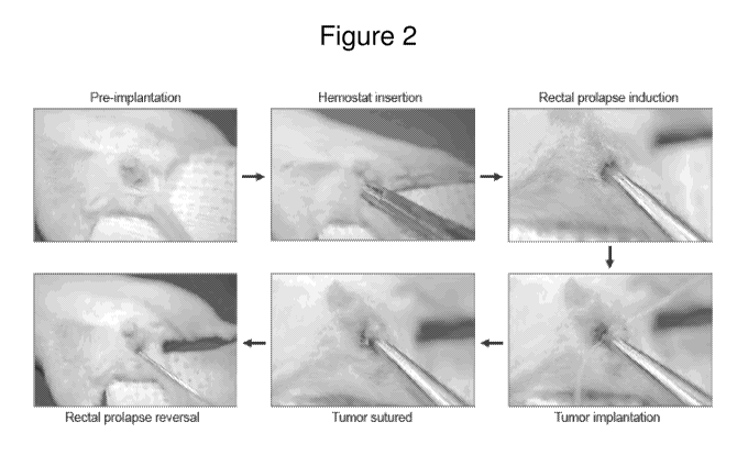
FIGURE 2 is a schematic diagram with representative images of the lumen
implantation
technique. Pre-implantation: the mouse is anesthetized by isofluorane
inhalation and placed in a supine
position, and the extremities are secured to a gauze-covered platform with
adhesive tape. Hemostat
insertion: a blunt hemostat is inserted into the anus, and the mucosa is
gently clasped. Rectal prolapse
induction: the hemostat is retracted from the anus, thus exteriorizing the
mucosa. Tumor implantation: a
donor tumor of approximately 10 mm3 is sutured onto the mucosal surface of the
exteriorized colon.
Tumor sutured: the suture ends are cut, leaving a donor tumor securely
attached to the mucosa. Rectal
prolapse reversal: a blunt gavage needle is used to re-insert the exteriorized
colon together with the
sutured donor tumor, thus reversing the rectal prolapse.
FIGURE 3A is a series of endoscopy images following lumen implantation of a
single ApeIni+;
KrasLSLG 1 2 D/+ ; Villin-Cre colon polyp from donor mouse #4700-260 into the
colon of wildtype C57BL/6 host
mouse #344, showing that the lumen-implanted ApeIni+; KrasLSLG 1 2 D/+ ;
Villin-Cre colon polyps remain
benign. Serial images were captured from the host at the indicated times.
FIGURE 3B is a series of endoscopy images following lumen implantation of a
single ApeIni+;
KrasLSLG 1 2 D/+ ; Villin-Cre colon polyp from donor mouse #4700-260 into the
colon of wildtype C57BL/6 host
mouse #346, showing that the lumen-implanted ApeIni+; KrasLSLG 1 2 D/+ ;
Villin-Cre colon polyps remain
benign. Serial images were captured from the host at the indicated times.
FIGURE 4A is a series of images showing the time course of gross colorectal
tumor development
following lumen implantation. Colons from NOD/SCID mice bearing HCT116-DsRed
lumen tumors were
harvested at weekly intervals from 0 to 7 weeks post-implantation (wpi),
opened longitudinally and
imaged. Top and bottom panels show mucosal and serosal views of the colon,
respectively, with the
anus positioned to the left of every panel.
FIGURE 4B is a series of endoscopy images following HCT116-DsRed lumen
implantation. Serial
images were captured from the same host from 0 to 4 wpi. Dotted line at 2 wpi
indicates the perimeter of
the implanted tumor.
FIGURE 4C is a graph showing primary tumor volume following lumen
implantation. Number of
mice per time point: 0 wpi (n = 3), 1 wpi (n = 3), 2 wpi (n = 6), 3 wpi (n =
6), 4 wpi (n = 11), 5 wpi (n = 15),
6 wpi (n = 11), 7 wpi (n = 18). Data are represented by the mean and s.e.m.
FIGURE 4D is a histological image of a host colon implanted with an HCT116-
DsRed donor
tumor at 1 wpi, showing haematoxylin and eosin (H&E) staining.
FIGURE 4E is a histological image of a host colon implanted with an HCT116-
DsRed donor
tumor at 1 wpi, showing collagen IV (Col IV; green), DsRed (red), and 4,6-
diamidino-2-phenylindole
(DAPI; blue) staining.
FIGURE 4F is a histological image of a host colon implanted with an HCT116-
DsRed donor
tumor at 3 wpi, showing H&E staining.
4
CA 02916394 2015-12-18
WO 2015/013432
PCT/US2014/047860
FIGURES 4G-4I are enlarged histological images of the indicated locations in
Figure 4F, each
showing Col IV (green), DsRed (red) and DAPI (blue) staining.
FIGURE 5A is a histological image of a colon from a NOD/SCID mouse stained
with
haematoxylin and eosin (H&E) immediately following lumen implantation of an
HCT116-DsRed tumor
fragment.
FIGURES 5B and 5C are enlarged histological images of the indicated locations
in Figure 5A,
each showing H&E staining.
FIGURE 5D is a histological image of a colon from a NOD/SCID mouse stained
with Col IV
(green), DsRed (red) and DAPI (blue) mouse immediately following lumen
implantation of an HCT116-
DsRed tumor fragment.
FIGURES 5E and 5F are enlarged histological images of the indicated locations
in Figure 5D,
each showing Col IV (green), DsRed (red), and DAPI (blue) staining.
FIGURE 5G is a set of images and graphs showing that tumor cell dissemination
was not
detectable at Day 1 post-transplantation. On Day 0, HCT116-DsRed donor tumor
fragments were
implanted onto the mucosal surface of host mouse colons (n = 2). On Day 1 post-
transplant, endoscopy
was performed and images of the transplanted tumors were captured (top). Mice
were then sacrificed
and the liver, lungs, intestinal vascular tract, and blood were harvested and
assessed by flow cytometry
for DsRed + cells. Negative controls consisted of tissue samples harvested
from a wild-type mouse.
Positive controls consisted of tissue samples containing HCT116-DsRed+ tumor
cells. An average of
5x10⁶ viable cells were assessed by flow cytometry. Gates were established
such that no DsRed + cells
within the respective negative control samples were detectable. Numbers within
the gates denote the
percentage of DsRed + cells.
FIGURE 6 is a series of images of colons from NOD/SCID mice bearing HCT116-
DsRed lumen
tumors showing the time course of colorectal tumor development following lumen
implantation. The
colons were harvested at weekly intervals from 0 to 7 wpi, opened
longitudinally, fixed and sectioned, and
stained by H&E (left column) or for Col IV (green), DsRed (red), and DAPI
(blue) (right column). In all
panels, the anus is positioned to the left with the lumen of the colon towards
the top.
FIGURE 7A is a set of images of a gross colon of a NOD/SCID mouse bearing an
HCT116-
DsRed lumen tumor at 7 wpi, with arrows indicating regional lymph node
metastases; the bottom left
panel showing histological staining by haematoxylin and eosin (H&E); and the
blue- and red-boxed areas
shown enlarged in the bottom middle and right panels, respectively, and
stained for Col IV (green),
DsRed (red), and DAPI (blue).
FIGURE 7B is a set of images of a liver of a NOD/SCID mouse bearing an HCT116-
DsRed
lumen tumor at 7 wpi, with arrows indicating metastases; the middle panel
showing histological staining
by H&E, the dotted line indicating the perimeter of a metastatic nodule; and
the boxed area shown
enlarged in the right panel and stained for Col IV (green), DsRed (red), and
DAPI (blue).
FIGURE 7C is a set of images of lungs of a NOD/SCID mouse bearing an HCT116-
DsRed lumen
tumor at 7 wpi, with arrows indicating metastases; the middle panel showing
histological staining by H&E,
arrows indicating metastatic nodules; and the boxed area shown enlarged in the
right panel and stained
for Col IV (green), DsRed (red), and DAPI (blue).
FIGURE 7D is a graph showing the number of macroscopic metastases within the
intestinal
lymph nodes, liver, and lungs following lumen implantation of an HCT116-DsRed
tumor in NOD/SCID
5
CA 02916394 2015-12-18
WO 2015/013432
PCT/US2014/047860
mice. Number of mice per time point: 0 wpi (n = 14), 1 wpi (n = 3), 2 wpi (n =
6), 3 wpi (n = 6), 4 wpi (n =
11), 5 wpi (n = 15), 6 wpi (n = 20), 7 wpi (n =18). Each point represents data
from an individual mouse.
Means s.e.m. are also shown.
FIGURE 7E is a graph showing the DsRed-positive tumor cell burden within the
intestinal lymph
nodes, liver, and lungs following lumen implantation of an HCT116-DsRed tumor
in NOD/SCID mice.
Data are expressed as the number of DsRed-positive tumor cells per lx 106
viable events. Number of
mice per time point: 0 wpi (n=14), 1 wpi (n =3), 2 wpi (n =3), 3 wpi (n =3), 4
wpi (n = 8), 5 wpi (n = 8), 6
wpi (n= 13),7 wpi (n = 14). Each point represents data from an individual
mouse. Means s.e.m. are
also shown.
FIGURE 7F is a set of images of a liver of a NODISCID mouse bearing an HCT116-
DsRed lumen
tumor at 3 wpi, with the middle panel showing histological staining by H&E,
the right panel showing
staining for Col IV (green), DsRed (red), and DAPI (blue), and arrows
indicating DsRed-positive
disseminated tumor cells.
FIGURE 7G is a set of images of lungs of a NODiSCID mouse bearing an HCT116-
DsRed lumen
tumor at 3 wpi, with the middle panel showing histological staining by H&E,
the right panel showing
staining for Col IV (green), DsRed (red), and DAPI (blue), arrows indicating
DsRed-positive disseminated
tumor cells, and arrowheads indicating autofluorescent macrophages.
FIGURE 8A is a histological image of a colon stained with H&E from a NOD/SCID
mouse bearing
a lumen-implanted HCT116-DsRed tumor at 6 wpi, showing that lumen-implanted
colorectal tumors
exhibit locoregional spread as well as hematogenous/lymphatic/perineural
invasion, and generate
macroscopic lymph node metastases.
FIGURE 8B is an enlarged histological image of the indicated location in
Figure 8A, stained with
H&E, demonstrating locoregional spread distal to the primary tumor indicated
by arrows.
FIGURE 8C is an enlarged histological image of the indicated location in
Figure 8A, stained with
H&E, demonstrating locoregional spread of a tumor migratory front into the
normal mucosa outlined by a
dotted line.
FIGURE 8D is an enlarged histological image of the indicated location in
Figure 8A, stained with
H&E, demonstrating muscularis externa penetration.
FIGURE 8E is an enlarged histological image of the indicated location in
Figure 8A, stained with
H&E, demonstrating primary tumor viability.
FIGURE 8F is an enlarged histological image of the indicated location in
Figure 8A, stained with
H&E, demonstrating locoregional spread proximal to the primary tumor indicated
by arrows.
FIGURE 8G is an enlarged histological image of the indicated location in
Figure 8A, stained with
H&E, demonstrating hematogenous invasion indicated by an arrow.
FIGURE 8H is an enlarged histological image of the indicated location in
Figure 8A, stained with
H&E, demonstrating perineural invasion indicated by arrows.
FIGURE 81 is an enlarged histological image of the indicated location in
Figure 8A, stained with
H&E, demonstrating regional lymph node colonization.
FIGURE 8J is an enlarged histological image of the indicated location in
Figure 8A, stained with
H&E, demonstrating lymphatic invasion indicated by an arrow.
FIGURE 9A is a series of images showing the time course of gross colorectal
tumor development
following lumen implantation. Colons from NOD/SCID mice bearing LS174T-DsRed
lumen tumors were
6
CA 02916394 2015-12-18
WO 2015/013432
PCT/US2014/047860
harvested from 0 to 8 wpi, opened longitudinally and imaged. Top and bottom
panels show mucosal and
serosal views of the colon, respectively, with the anus positioned to the left
of every panel. Arrows
indicate intestinal lymph node metastases.
FIGURE 9B is a graph showing primary tumor volume following lumen implantation
of LS174T-
DsRed tumors in NOD/SCID mice. Number of mice per time point: 0 wpi (n = 9), 1
wpi (n = 3), 2 wpi (n =
3), 3 wpi (n = 3), 4 wpi (n = 3), 5 wpi (n = 3), 6 wpi (n = 3), 7 wpi (n = 4),
8 (n = 12). Data are represented
by the mean and s.e.m.
FIGURE 9C is an image of a liver from a mouse bearing a lumen-implanted LS174T-
DsRed
tumor at 7 wpi. Arrows indicate metastases.
FIGURE 9D is an image of lungs from a mouse bearing a lumen-implanted L5174T-
DsRed tumor
at 8 wpi. Arrow indicates a metastatic outgrowth.
FIGURE 9E is a graph showing the number of macroscopic metastases within the
intestinal
lymph nodes, liver and lungs following lumen implantation of LS174T-DsRed
tumors in NOD/SCID mice.
Number of mice per time point: 0 wpi (n = 9), 1 wpi (n = 3), 2 wpi (n = 3), 3
wpi (n = 3), 4 wpi (n = 3), 5 wpi
(n = 3), 6 wpi (n = 3), 7 wpi (n = 4), 8 wpi (n = 12). Each point represents
data from an individual mouse.
Means s.e.m. are also shown.
FIGURE 10A is an image of a colon from a mouse bearing a lumen-implanted human
stage II
patient colorectal tumor, showing that lumen-implanted stage II patient tumors
remain non-metastatic and
benign.
FIGURE 10B is an image of a colon from a mouse bearing a lumen-implanted human
stage III
patient colorectal tumor, showing that lumen-implanted stage III patient
tumors give rise to lymph node
metastases.
FIGURE 10C is a graph showing the number of macroscopic metastases that result
from stage II
and stage III donor tumors. Each point represents data from an individual
mouse. Means s.e.m.
are also shown. ****P < 0.0001.
FIGURE 11A is a graph showing the number of macroscopic metastases within
various organs
following lumen or s.c. implantation of HCT116-DsRed tumors into NOD/SCID or
NSG mice.
Number of mice: NOD/SCID lumen (n = 22 for liver, lungs, lymph nodes; n = 4
for adrenal gland,
kidney, spleen, brain); NOD/SCID s.c. (n= 10); NSG lumen (n= 16); NSG s.c. (n=
10). Each point
represents data from an individual mouse. Means s.e.m. are also shown. *P <
0.05; **p < 0.01;
**P < 0.001; ****P < 0.0001. n.s. = not significant. n.d. = not determined.
FIGURE 11B is a graph showing the DsRed-positive tumor cell burden within
various organs
following lumen or s.c. implantation of HCT116-DsRed tumors into NOD/SCID or
NSG mice. Data
are expressed as the number of DsRed-positive tumor cells per 1 x 10⁶ viable
events. Number of
mice: NOD/SCID lumen (n = 13 for liver, lungs, lymph nodes; n = 4 for adrenal
gland, kidney, spleen,
brain, bone marrow); NOD/SCID s.c. (n = 10); NSG lumen (n = 7 for liver,
lungs, lymph nodes; n = 3
for adrenal gland, kidney, spleen, brain, bone marrow); NSG s.c. (n = 10).
Each point represents
data from an individual mouse. Means s.e.m. are also shown. *P 0.05; *P <
0.01; ***P 0.001;
**** P 0.0001. n.s. = not significant. n.d. = not determined.
FIGURE 11C is an image of a gross colon of an NSG mouse bearing an HCT116-
DsRed
lumen tumor at 7 wpi, with arrows indicating the primary tumor and regional
lymph node metastases.
7
CA 02916394 2015-12-18
WO 2015/013432
PCT/US2014/047860
FIGURE 11D is a histological image of a colon of an NSG mouse bearing an
HCT116-DsRed
lumen tumor at 7 wpi, the colon stained with H&E, and boxed area shown
enlarged in the right panel
and stained for Col IV (green), DsRed (red), and DAPI (blue). Arrows indicate
regional lymph node
metastases.
FIGURE 11E are images of various organs from an NSG mouse bearing an HCT116-
DsRed
lumen tumor at 7 wpi, with liver metastases clearly evident and lung
metastases indicated by arrows.
FIGURE 11F is a histological image of a liver of an NSG mouse bearing an
HCT116-DsRed
lumen tumor at 7 wpi, the liver stained with H&E (left panel) and the for Col
IV (green), DsRed (red),
and DAPI (blue) (right panel). Dotted lines indicate the perimeter of liver
metastatic nodules.
FIGURE 11G is a histological image of lungs from an NSG mouse bearing an
HCT116-
DsRed lumen tumor at 7 wpi, the lungs stained with H&E (left panel) and the
for Col IV (green),
DsRed (red) and DAPI (blue) (right panel).
FIGURE 11 H are images of the liver and lungs of an NSG mouse bearing an
HCT116-DsRed
s.c. tumor at 7 wpi.
FIGURE 111 is an image of the associated primary s.c. tumor of the NSG mouse
of Figure
11H.
FIGURE 11J is a graph showing the circulating tumor cell number at 6-7 wpi
following lumen
or s.c. implantation of HCT116-DsRed tumors into NOD/SCID or NSG mice. Data
are represented
by the mean and s.e.m., and are expressed as the number of DsRed-positive
tumor cells in the
blood per 1 x 10⁶ viable events. Number of mice: NOD/SCID lumen (n = 12);
NOD/SCID s.c. (n = 9);
NSG lumen (n= 9); NSG s.c. (n= 10). **P < 0.01; ***P,-, 0.001.
FIGURE 12A are images of the indicated organs from a NOD/SCID mouse bearing a
lumen-
implanted HCT116-DsRed tumor at 7 wpi.
FIGURE 12B are images of the liver and lungs from a NOD/SCID mouse bearing an
s.c.-
implanted HCT116-DsRed tumor at 7 wpi.
FIGURE 12C is a graph showing the primary tumor volume following s.c.
implantation of
HCT116-DsRed tumor cells (n = 10). Data are represented by the mean s.e.m.
FIGURE 13A is a histological image of a lumen-implanted HCT116-DsRed tumor at
6 wpi,
stained with H&E.
FIGURE 13B is a histological image of an s.c.-implanted HCT116-DsRed tumor at
6 wpi, stained
with H&E.
FIGURE 13C is a histological image of a lumen-implanted HCT116-DsRed tumor at
6 wpi,
stained with MECA-32 (green) and DAPI (blue).
FIGURE 13D is a histological image of an s.c.-implanted HCT116-DsRed tumor at
6 wpi, stained
with MECA-32 (green) and DAPI (blue).
FIGURE 13E is a graph showing vascular density of HCT116-DsRed tumors
implanted into the
lumen (n = 7) or s.c. (n = 7) at 6 wpi. Data are represented by the mean and
s.e.m., and are expressed
as a ratio of the MECA-32-positive vascular area over the total DAPI-positive
viable tumor area x 100. *P
< 0.05.
FIGURE 14A are graphs showing correlations between lymph node metastatic
burden and
liver metastatic burden in NOD/SCID mice bearing lumen-implanted HCT116-DsRed
tumors from 1-
7 wpi. Data are the total number of macroscopic liver metastases versus either
the total number of
8
CA 02916394 2015-12-18
WO 2015/013432
PCT/US2014/047860
macroscopic lymph node metastases (top) or the total number of DsRed+ tumor
cells within the
lymph nodes (bottom).
FIGURE 14B are graphs showing correlations between lymph node metastatic
burden and
liver metastatic burden in NSG mice bearing lumen-implanted HCT116-DsRed
tumors from 5-7 wpi.
Data are the total number of macroscopic liver metastases versus either the
total number of
macroscopic lymph node metastases (top) or the total number of DsRed+ tumor
cells within the
lymph nodes (bottom).
FIGURE 14C are gross images of colons of NOD/SCID mice bearing lumen-implanted
HCT116-DsRed tumors at 6-7 wpi following the indicated antibody treatments.
Arrowheads indicate
intestinal lymph node metastases.
FIGURE 14D is a graph showing the primary tumor volumes of NOD/SCID mice
bearing
lumen-implanted HCT116-DsRed tumors at 6-7 wpi following the indicated
antibody treatments or no
antibody treatment. Number of mice per treatment condition: untreated (n =
22), anti-VEGF-A (n =
9), anti-VEGF-C (n = 8), anti-VEGF-A/C (n = 10). Each point represents data
from an individual
mouse. Means s.e.m. are also shown. *P 0.05; **P 0.01; ***P< 0.001:
****P 0.0001. n.s. =
not significant.
FIGURE 14E is a graph showing the number of lymph node macroscopic metastases
of
NOD/SCID mice bearing lumen-implanted HCT116-DsRed tumors at 6-7 wpi following
the indicated
antibody treatments or no antibody treatment. Number of mice per treatment
condition: untreated
(n = 22), anti-VEGF-A (n = 9), anti-VEGF-C (n = 8), anti-VEGF-A/C (n = 10).
Each point represents
data from an individual mouse. Means s.e.m. are also shown. *P 0.05; **P <
0.01; ***P < 0.001;
****P < 0.0001. n.s. = not significant.
FIGURE 14F is a graph showing the number of liver macroscopic metastases of
NOD;SCI D
mice bearing lumen-implanted HCT116-DsRed tumors at 6-7 wpi following the
indicated antibody
treatments or no antibody treatment. Number of mice per treatment condition:
untreated (n = 22),
anti-VEGF-A (n = 9), anti-VEGF-C (n = 8), anti-VEGF-A/C (n = 10). Each point
represents data from
an individual mouse. Means s.e.m. are also shown. *P < 0.05; **P < 0.01;
***P 0.001; ****P
0.0001. n.s. = not significant.
FIGURE 14G is a set of gross images of the livers of NOD/SCID mice bearing
lumen-
implanted HCT116-DsRed tumors at 6-7 wpi following the indicated antibody
treatments.
Arrowheads indicate liver metastases.
FIGURE 14H is a graph showing DsRed-positive tumor cell burden within the
liver alongside
control analyses from non-tumor-bearing mice (n = 8), with data expressed as
the number of DsRed-
positive tumor cells per 1 x 10⁶ viable events, for NOD/SCID mice bearing
lumen-implanted
HCT116-DsRed tumors at 6-7 wpi following the indicated antibody treatments or
no antibody
treatment. Number of mice per treatment condition: untreated (n = 13), anti-
VEGF-A (n = 9), anti-
VEGF-C (n = 8), anti-VEGF-A/C (n = 9). Each point represents data from an
individual mouse.
Means s.e.m. are also shown. *P 0.05; **P 0.01; ***P < 0.001; ****P
0.0001. n.s. = not
significant.
FIGURE 141 is a contingency analysis comparing the number of NOD/SCID mice
bearing
lumen-implanted HCT116-DsRed tumors with or without liver macrometastases at 6-
7 wpi, with data
expressed as the percentage of mice in each category. Number of mice per
treatment condition:
9
CA 02916394 2015-12-18
WO 2015/013432
PCT/US2014/047860
untreated (n = 13), anti-VEGF-A (n = 9), anti-VEGF-C (n = 8), anti-VEGF-A/C (n
= 9). Means
s.e.m. are also shown. *P < 0.05; **P 0.01; ***P 0.001; ****P < 0.0001.
n.s. = not significant.
FIGURE 14J is a contingency analysis comparing the number of NOD/SCID mice
bearing
lumen-implanted HCT116-DsRed tumors with or without liver micrometastatic
DsRed+ cells at 3 wpi,
prior to macroscopic metastasis manifestation, with data expressed as the
percentage of mice in
each category. Means s.e.m. are also shown. *P 0.05; **P < 0.01; ***P
0.001; ****P 0.0001.
n.s. = not significant.
FIGURE 14K is a graph showing the number of lymph node macroscopic metastases
of
NOD/SCID mice bearing lumen-implanted LS174T-DsRed tumors at 8 wpi following
the indicated
antibody treatments or no antibody treatment. Number of mice per treatment
condition: untreated
(n = 7), anti-VEGF-A (n = 3), anti-VEGF-C (n = 13), anti-VEGF-A/C (n = 5).
Each point represents
data from an individual mouse. Means s.e.m. are also shown. *P < 0.05. n.s.
= not significant.
FIGURE 14L is a contingency analysis comparing the number of NOD/SCID mice
bearing
lumen-implanted LS174T-DsRed tumors with or without liver macrometastases at 8
wpi, with data
expressed as the percentage of mice in each category. Number of mice per
treatment condition:
untreated (n = 7), anti-VEGF-A (n = 3), anti-VEGF-C (n = 13), anti-VEGF-A/C (n
= 5). Each point
represents data from an individual mouse. Means s.e.m. are also shown. **P
: 0.01. n.s. = not
significant.
FIGURE 15A is a graph showing the effect of targeting angiogenesis and/or
lymphangiogenesis
on the formation of regional lymph node metastases in NOD/SCID mice bearing
lumen-implanted
HCT116-DsRed tumors at 6-7 wpi, with the number of lymph node macroscopic
metastases normalized
to primary tumor volume. Data are expressed as the number of macroscopic
metastases per 1 mm3 of
primary tumor. Number of mice per treatment condition: untreated (n = 22),
anti-VEGF-A (n = 9), anti-
VEGF-C (n = 8), anti-VEGF-A/C ((n = 10). Each point represents data from an
individual mouse. Means
s.e.m. are also shown. * P < 0.05; *** < 0.001; **** < 0.0001. n.s. = not
significant.
FIGURE 15B is a graph showing the effect of targeting angiogenesis and/or
lymphangiogenesis
on the formation of distant liver metastases in NOD/SCID mice bearing lumen-
implanted HCT116-DsRed
tumors at 6-7 wpi, with the number of liver macroscopic metastases normalized
to primary tumor volume.
Number of mice per treatment condition: untreated (n = 22), anti-VEGF-A (n =
9), anti-VEGF-C (n = 8),
anti- VEGF-A/C ((n = 10). Each point represents data from an individual mouse.
Means s.e.m. are also
shown. *P < 0.05; *** < 0.001; ****P < 0.0001. n.s. = not significant.
Detailed Description of the Invention
The present invention is based in part on the generation of a model of
colorectal cancer that
exhibits metastasis to clinically relevant sites.
Unless defined otherwise, technical and scientific terms used herein have the
same meaning as
commonly understood by one of ordinary skill in the art to which this
invention belongs. Singleton et al.
Dictionary of Microbiology and Molecular Biology. 2nd Ed, J. Wiley & Sons (New
York, N.Y. 1994). For
purposes of the present invention, the following terms are defined below.
10
CA 02916394 2015-12-18
WO 2015/013432
PCT/US2014/047860
Definitions
The term "antibody" herein is used in the broadest sense and refers to any
immunoglobulin (Ig)
molecule comprising two heavy chains and two light chains, and any fragment,
mutant, variant or
derivation thereof so long as they exhibit the desired biological activity
(e.g., epitope binding activity).
Examples of antibodies include monoclonal antibodies, polyclonal antibodies,
multispecific antibodies,
and antibody fragments.
The term "monoclonal antibody" as used herein refers to an antibody obtained
from a population
of substantially homogeneous antibodies, i.e., the individual antibodies
comprising the population are
identical except for possible naturally occurring mutations that may be
present in minor amounts.
Monoclonal antibodies are highly specific, being directed against a single
antigenic site. Furthermore, in
contrast to polyclonal antibody preparations which include different
antibodies directed against different
determinants (epitopes), each monoclonal antibody is directed against a single
determinant on the
antigen. In addition to their specificity, the monoclonal antibodies are
advantageous in that they may be
synthesized uncontaminated by other antibodies. The modifier "monoclonal"
indicates the character of
the antibody as being obtained from a substantially homogeneous population of
antibodies, and is not to
be construed as requiring production of the antibody by any particular method.
For example, the
monoclonal antibodies to be used in accordance, with the present invention may
be made by the
hybridoma method first described by Ohler et al., Nature. 256:495 (1975), or
may be made by
recombinant DNA methods (see, e.g,, U.S, at. No. 4,816,567). The "monoclonal
antibodies" may also
be isolated from phage antibody libraries using the techniques described in
Clackson et al., Nature.
352:624-628 (1991) and Marks et al., J. Moi. Biol. 222:581-597 (1991), for
example.
An "antibody fragment" refers to a molecule other than an intact antibody that
comprises a portion
of an intact antibody, such as the antigen-binding or variable region thereof.
Examples of antibody
fragments include Fab, Fab', F(ab),, and Ev fragments; diabodies; linear
antibodies; single-chain
antibody molecules, and multispecific antibodies formed from antibody
fragment(s). In certain
embodiments, the antibody .fragment binds the same antigen to which the intact
antibody binds.
The terms "cancer" and "cancerous" refer to or describe the physiological
condition in mammals
that is typically characterized by unregulated cell growth. Examples of cancer
include but are not limited
to, carcinoma, lymphoma, blastoma, sarcoma, and leukemia. More particular
examples of such cancers
include squamous cell cancer, small-cell lung cancer, non-small cell lung
cancer, gastrointestinal cancer,
pancreatic cancer, glioblastoma, cervical cancer, ovarian cancer, liver
cancer, bladder cancer, hepatoma,
breast cancer, colon cancer, colorectal cancer, endometrial carcinoma,
salivary gland carcinoma, kidney
cancer, renal cancer, prostate cancer, vulval cancer, thyroid cancer, hepatic
carcinoma and various types
of head and neck cancer.
By "metastasis" is meant the spread of cancer from its primary site to other
places in the body.
cancer cells can break away from a primary tumor, penetrate into lymphatic and
blood vessels, circulate
through the bloodstream, and grow in a distant focus (metastasize) in normal
tissues elsewhere in the
body. Metastasis can be local or distant. Metastasis can be characterized as a
sequential process,
contingent on tumor cells breaking off from the primary tumor, traveling
through the bloodstream or
lymphatics, and stopping at a distant site, After the tumor cells come to rest
at another site, they can re-
penetrate through the blood vessels or lymphatic walls, continue to multiply,
and eventually another tumor
is formed. At the new site, the cells establish a blood supply and can grow to
form a life-threatening
11
CA 02916394 2015-12-18
WO 2015/013432
PCT/US2014/047860
mass. In certain embodiments, this new tumor is referred to as a metastatic
(or secondary) tumor. In
certain embodiments, the term metastatic tumor refers to a tumor that is
capable of metastasizing, but
has not yet metastasized to tissues or organs elsewhere in the body. In
certain embodiments, the term
metastatic tumor refers to a tumor that has metastasized to tissues or organs
elsewhere in the body. in
certain embodiments, metastatic tumors are comprised of metastatic tumor
cells.
The "metastatic organ" or "metastatic tissue" is used in the broadest sense,
refers to an organ or
a tissue in which the cancer cells from a primary tumor or the cancer cells
from another part of the body
have spread. Examples of metastatic organ and metastatic tissue include, but
are not limited to, lung,
liver, brain, ovary, bone, bone marrow, and lymph node. With respect to
colorectal cancer (CRC),
predominant metastatic organ and metastatic tissue are the regional intestinal
lymph nodes, liver, and
lU ngs.
By "micrornetastasis" is meant a small number of cells that have spread from
the primary tumor to
other parts of the body, Micrometastasis may or may not be detected in a
screening or diagnostic test.
By "macrometastasis" is meant a number of cells that are detectable and have
spread from the
primary tumor site to other parts of the body.
By "non-metastatic" is meant a cancer that is benign or that remains at the
primary site (e.g., a
locally invasive cancer) and has not penetrated into the lymphatic or blood
vessel system or to tissues
other than the primary site. In certain embodiments, a non-metastatic cancer
is any cancer that is a
Stage 0, I, or 11 cancer,
"Invasiveness" or "invasive growth," as used herein, refers to the ability of
a cancer or tumor to
leave the tissue site at which it originated and proceed to proliferate at a
different site (e.g., nearby or
distant site) of the body. In some embodiments, a cancer can be "locally
invasive" and proceed to
proliferate at a nearby site of the body, such as surrounding tissue. In other
embodiments, a cancer can
be "regionally invasive" or "distantly invasive" and proceed to proliferate at
a regional or distant site of the
body, respectively.
Reference to a cancer or tumor as a "Stage 0," "Stage I," "Stage 11," "Stage
Ill," or "Stage IV"
indicates classification of the tumor or cancer using the Overall Stage
Grouping or Roman Numeral
Staging methods known in the art. Although the actual stage of the cancer is
dependent on the type of
cancer, in general, a Stage 0 cancer is an in situ lesion, a Stage l cancer is
small localized tumor, a Stage
i s a local advanced tumor, a Stage il cancer is a local advanced tumor that
exhibits involvement of the
local lymph nodes, and a Stage IV cancer represents metastatic cancer. The
specific stage for each type
of tumor is known to the skilled clinician.
"Tumor," as used herein, refers to any neoplastic cell growth, whether
malignant or benign, and
all pre-cancerous and cancerous cells and tissues. The terms "cancer",
"cancerous", "cell proliferative
disorder", "proliferative disorder" and "tumor" are not mutually exclusive as
referred to herein. Tumors
may be solid tumors, such as tumors of the colon (CRC tumor), or non-solid or
soft tumors, such as
leukemia. Examples of soft tissue tumors include leukemia (e.g., chronic
myelogenous leukemia, acute
myelogenous leukemia, adult acute lymphoblastic leukemia, acute myelogenous
leukemia, mature B-cell
acute lymphoblastic leukemia, chronic lymphocytic leukemia, lymphocytic
leukemia, or hairy cell
leukemia), or lymphoma (e,g., non-Hodgkin's lymphoma, cutaneous T-cell
lymphoma, or Hodgkin's
disease), A solid tumor includes any cancer of body tissues other than blood,
bone marrow, or the
lymphatic system. Solid tumors can be further separated into those of
epithelial cell origin and those of
12
CA 02916394 2015-12-18
WO 2015/013432
PCT/US2014/047860
non-epithelial cell origin. Examples of solid tumors include tumors of colon,
breast, prostate, lung, kidney,
liver, pancreas, ovary, head and neck, oral cavity, stomach, duodenum, small
intestine, large intestine,
gastrointestinal tract, anus, gall bladder, labium, nasopharynx, skin, uterus,
male genital organ, urinary
organs, bladder, and skin. Solid tumors of non-epithelial origin include
sarcomas, brain tumors, and bone
tumors. The term "tumor," as used herein, is also meant to be inclusive of
"polyps."
By "primary tumor" or "primary cancer" is meant the original cancer and not a
metastatic lesion
located in another tissue, organ, or location in the subject's body. In
certain embodiments, primary tumor
is comprised of primary tumor cells,
By "benign tumor " or "benign cancer" is meant a tumor that remains localized
at the site of origin
and does not have the capacity to infiltrate, invade, or metastasize to a
distant site.
"Tumorigenic cells," as used herein, refer to any cells (e.g., cancer cells,
e.g., human cancer cells
or non-human cancer cells) that exhibit an abnormal growth state or are
capable of chanaina their normal
growth state to an abnormal growth state in which they eventually form tumors.
Tumorigenic cells are
capable of forming tumors, which are generally the result of uncontrolled
growth of the cells. Tumorigenic
cells can be distinguished from non-tumorigenic cells on the basis of their
tumor-forming phenotype (see,
e.g,, Al-Hajrj, et al. Proc Nati Aced Sci U S A.100: 3983-8, 2003; U.S. Pub.
No, 2002/0119565; U.S. Pub,
No. 2004/0037815; U.S. Pub. No. 2005/0232927; WO 05/005601; U.S,Pub. No.
2005/0089518; U.S.
WI. No. 10/864,207; Al- Hajj eta. Oncogene. 23: 7274, 2004; and Clarke eta.
Ann Ny Mad, Sci. 1044:
90, 2005, all of which are herein incorporated by reference in their
entireties for all purposes).
Tumorigenic cells include, without limitation, tumor cells, embryonic cells,
cells e,ngineered to have
abnormal growth, cancer cell lines, as well as cell masses of any of these
cell types.
The term "implant," and variations thereof., refers to transplanted cells, for
example, tumorigenic
cells (e.g., an intact tumor, or fragment thereof) which are introduced into a
recipient host and which
remain substantially stably established at the site of transplantation in the
recipient.
By "donor" cell, tumor, or tumorigenic cell is meant a cell, tumor, or
tumorigenic cell that is not
derived from the recipient host organism, but may be syngeneic (where the
donor and recipient are
genetically identical), allogeneic (where the donor and recipient are of
different genetic origins but of the
same species), or xenogeneic (where the donor and recipient are from different
species). For example,
the donor cell, tumor, or tumorigenic cell may be derived from a human. A
"donor tumorigenic cell
implant" refers to transplanted tumorigenic cells, as used herein, which are
derived from a source other
than the recipient organism.
By "tumor load" is meant the amount of cancer in the body. Tumor load is also
referred to as
tumor burden, and may be a function of tumor number and tumor size.
"Adjuvant therapy" herein refers to therapy given after surgery, where no
evidence of residual
disease can be detected, so as to reduce the risk of disease recurrence. The
goal of adjuvant therapy is
to prevent recurrence of the cancer, and therefore to reduce the chance of
cancer-related death.
A "small molecule" is defined herein to have a molecular weight below about
500 Daltons.
By "immunoconjugate" is meant an antibody conjugated to one or more
heterologous molecule(s)
(e.g., an antibody-drug conjugate (ADC)), including but not limited to a
cytotoxic agent.
By "reduce" or "inhibit" is meant the ability to cause an overall decrease,
for example, of 20% or
greater, of 50% or greater, or of 75%, 85%, 90%, 95%, or greater. In certain
embodiments, reduce or
inhibit can refer to the growth of tumorigenic cells or a tumor, which can be
measured by a reduction or
13
CA 02916394 2015-12-18
WO 2015/013432
PCT/US2014/047860
inhibition in the number of tumorigenic cells, size of tumors, tumor load,
tumorigenic cell or tumor
invasiveness, and/or tumor metastasis.
The term "non-human animal" refers to all animals, except humans, and
includes, without
limitation, birds, farm animals (e.g., cows), sport animals (e.g., horses),
fish, reptiles, and non-human
mammals (e.g., cats, dogs, and rodents).
The term "non-human mammal" refers to all members of the class Mammalia,
except humans.
The term "rodent" refers to all members of the order Rodentia, including rats,
mice, rabbits,
hamsters, and guinea pigs.
Detailed Description
Colorectal cancer (CRC) initially manifests as benign polyps on the mucosal
surface of the
large intestine. If left unresected, these polyps can progress to invasive
adenocarcinomas that
penetrate through the submucosal and muscularis externa layers of the
colorectal wall to reach the
serosal side. Eventual regional spread to the intestinal lymph nodes and
distant spread to the liver
results in the outgrowth of gross metastases that are the major cause of CRC
mortality.
Deciphering the routes of CRC metastasis to these sites therefore has the
potential to uncover
therapeutic opportunities that may impact mortality rates. Investigations into
metastatic routes,
however, have been hampered by the lack of availability of relevant in vivo
metastatic models of
CRC. Indeed, despite the wide availability of xenograft, chemical-induced, and
genetically-
engineered models (e.g., mouse models) of CRC (Heijstek et al. Dig. Surg. 22:
16-25, 2005. Epub
2005 Apr 14; Kobaek-Larsen et al. Comp. Med. 50(1): 16-26, 2000; Rosenberg et
al. Carcinogenesis.
30(2): 183-196, 2009. Epub 2008 Nov 26; Taketo et al. Gastroenterology.
136(3): 780-798, 2009),
tumors in these models fail to reproducibly metastasize to the regional
intestinal lymph nodes and
liver, the target organs relevant to human CRC.
Lumen Implantation Model of Colorectal Cancer
The present invention is based, at least in part, on the development of a
clinically relevant
model of colorectal cancer (CRC). In contrast to known models of CRC, the
model of CRC of the
invention is generated by a novel lumen implantation technique, and,
importantly, is capable of
recapitulating the etiology of human CRC.
A non-human animal (e.g., a non-human mammal) of any species, subspecies,
genetic
variant, tissue variant, or combination thereof, can be used in the generation
of the lumen implantation
model (LIM) of CRC. The non-human mammal may, for example, be a rodent.
E:xamples of rodent
species include, without limitation, rat, mouse, hamster, rabbit, guinea pig,
and gerbil. The non-human
mammal can be male or female. The non-human mammal can be any age, provided
that the lumen
implantation technique can be successfully executed. Accordingly, the non-
human mammal can be, for
example, less than one week old, from about one week to about five years old,
from about one week to
about three years old, from about two weeks to about two years old, from about
three weeks to about one
year old, from about four weeks to about six months old, from about six weeks
to about three months old,
frorn about eight weeks to about twelve weeks old, older than three years old,
or older than five years old.
The non-human mammal can be wild-type (e.g., immune-competent) or
imrnunodeficient. For
example, when the lumen of the recipient host non-human mammal is implanted
with a donor cell, tumor,
14
CA 02916394 2015-12-18
WO 2015/013432
PCT/US2014/047860
or tumorigenic cell that is xenogeneic (e.g., human), the host non-human
mammal is immunodeficient
When the lumen of the recipient host non-human mammal is implanted with a
donor cell, tumor, or
tumorigenic cell that is syngeneic, however, the host non-human mammal can be
non-immunodeficient
(e.g., wild-type).
In certain instances, the non-human mammal is a mouse. The mouse can be a nude
mouse.
The mouse can be a severely combined immunodeficient (SCID) mouse, for
example, a NOD/SCID
interleukin-2 receptor gamma chain null (NSG) mouse. The NSG mouse is
described in Pearson et al.
Cum Top. Microbiol. 11711111,11101. 324:25-51, 2008; Shultz et al. Curt- Top
Microbiol IMITILMOL 324:25-51
2005; Strom et al. Methods Mat, Biol. 640:491-509, 2010; McDermott et al.
Blood.116(2): 193- 200, 2010;
Lepus et al. HL1177. IMITILMOL 7000)190-802, 2009; Brehm et al. Clin
iiiir171.11101, 135W:84-98, 2010. Any
suitable immunodeficient non-human mammal can be used. Suitable non-human
mammals include
rodents, which can be obtained from such sources as The Jackson Laboratory of
Bar Harbor, fvlaine,
Charles River Laboratories International, Inc. of Wilrninaton, Massachusetts,
and Harlan Laboratories of
Indianapolis, Indiana.
The lumen implantation technique involves the implantation of one or more
(e.g., 1, 2, 3, 4,
5, 10, 20, 30, 40, 50, 102, 103, 104, 105, 106, 107, 108, 109, or 101 or
more) donor tumorigenic cells
that are capable of being implanted on the mucosa! surface (luminal side) of
the colon without
breaching the colon wall. The donor tumorigenic cell(s) can be syngeneic
(where the donor and
recipient are genetically identical), allogeneic (where the donor and
recipient are of different genetic
origins but of the same species), or xenogeneic (where the donor and recipient
are from different species,
e.g,, human) with respect to the recipient non-human mammal host. The donor
tumorigenic cell(s) may
be invasive or non-invasive, benign or malignant, metastatic or non-
metastatic. The tumorigenic cells
may be tumor cells, or alternatively, may be, for example, embryonic cells,
cells engineered to have
abnormal growth, cancer cell lines, as well as cell masses of any of these
cell types.
In instances of implantation of more than one donor tumorigenic cell, the
implanted cells may be
an intact tumor, or fragment thereof. The intact tumor or fragment thereof,
can be an intact malignant
tumor, or fragment thereof, such as a Stage III CRC tumor, which has given
rise to regional metastases
(e.g., in the intestinal lymph node) in the donor organism, or a Stage IV CRC
tumor, which has given rise
to distant metastases (e.g., in the liver or lungs) in the donor organism.
Alternatively, the intact tumor or
fragment thereof, can be an intact benign or locally invasive tumor, or
fragment thereof, such as a Stage
0, Stage I, or Stage II CRC tumor, or fragment thereof, which is confined to
the site or tissue of primary
origin.
The intact tumor, or fragment thereof, is not limited to a CRC tumor, or
fragment thereof. For
example, the intact tumor, or fragment thereof, can be an intact a non-CRC
tumor, or fragment thereof,
such as, without limitation, a breast cancer tumor, lung cancer tumor, liver
cancer tumor, brain cancer
tumor, lymph node cancer tumor, kidney cancer tumor, stomach cancer tumor,
ovarian cancer tumor, skin
cancer tumor, pancreatic cancer tumor, thyroid cancer tumor, or prostate
cancer tumor, or fragment
thereof.
The intact tumor, or fragment thereof, implanted on the colonic mucosal
surface of the recipient
non-human mammal host can be a solid tumor (e.g., a colon/CRC tumor, breast
cancer tumor, lung
cancer tumor, or liver cancer tumor), or fragment thereof. The intact tumor,
or fragment thereof, can be
derived from any suitable donor organism, such as a human or mouse. The intact
tumor, or fragment
CA 02916394 2015-12-18
WO 2015/013432
PCT/US2014/047860
thereof, can be from a particular cell line, such as a CRC cell line (e.g.,
HCT116,LS174T, or LoVo primary
human CRC-derived cell line) or a breast cancer cell line (e.g., MDA-231 human
breast cancer cell line).
The implanted one or more donor tumorigenic cells can be of any collective
size. In instances
when an intact tumor, or fragment thereof, is implanted, the intact tumor, or
fragment thereof, is
around 0.1-100 mm3 in size, e.g., around 1-100 mm3 in size, e.g., around 10
mm3 in size.
The implantation site can be along any region of the mucosal surface of the
colon of the
non-human mammal. In certain embodiments, the implantation site is located
nearby the anus of the
recipient non-human mammal in order to allow for the option of removal of the
implanted tumor from
the implantation site. In mice, for example, a tumor implantation distance of
about 1-20 mm (e.g.,
about 5-15 mm, e.g., about 11-12.5 mm) away from the anus of the host mouse is
preferable.
In general, a mouse LIM of CRC can be created by anesthetizing the mouse
(e.g., by isoflurane
inhalation), placing the mouse in a supine position with extremities secured
and inserting a blunt-ended
hemostat (Micro-Mosquito, No. 13010-12, Fine Science Tools) or other suitable
tool around 1 cm into the
anus, clasping a single mucosal fold (e.g., by closing the hemostat to the
first notch), retracting and
cleaning exteriorized mucosa (e.g, with povidone/iodine), rinsing (e.g., with
lactated ringers solution), and
blotting dry. One or more donor tumorigenic cells (e.g., a donor tumor
fragment or intact polyp of -10
mm3) can be then be sutured onto the mucosa (e.g., using absorbable 4-0 vicryl
sutures (Ethicon)),
ensuring that the suture only penetrates the superficial mucosal layer. After
rehydrating the mucosa with
PBS, the exteriorized colon can be re-inserted together with the sutured
tumor, thus reversing the rectal
prolapsed. To minimize tumor dislodgement during defecation, mice can be
housed on cage floor inserts
and fed a 100% rodent liquid diet (AIN-76A, Casein Hydrolysate without Fiber;
BioServe) from around 3
days pre-surgery to around 7 days post-surgery.
The generated non-human mammal LIMs of CRC have numerous advantages over
established
CRC models, as demonstrated in the Examples section below. These advantages of
the LIM include,
without limitation, the implantation of tumors onto the mucosal surface,
compared to tumor implantation
onto the serosal surface in the existing cecum implantation model, resulting
in: (i) the potential to give
rise to distant metastases in clinically relevant sites, compared to the
widespread tumor dissemination
throughout the peritoneal cavity due to tumor cell shedding rather than actual
metastasis in the existing
cecum implantation model; (ii) the ability to implant intact tumor fragments
into a host mouse instead of
cell suspensions that are unable to maintain tumor structure as in existing
cell suspension injection
models; and (iii) the maintained integrity of the colon wall compared to the
likelihood of puncturing the
colon wall as in existing cell suspension injection models.
Screening of Candidate Compounds that Inhibit Tumorigenesis
The LIM finds utility, for example, in the screening of candidate compounds
that possess anti-
cancer activity (e.g., compounds that inhibit growth of tumorigenic cells).
Anti-cancer activity can include
activity in directly or indirectly mediating any effect in preventing,
delaying, reducing or inhibiting tumor
growth and/or development, which may provide for a beneficial effect to the
host. Anti-cancer activity of a
candidate compound could therefore be reflected by, without limitation, the
ability of the candidate
compound to, directly or indirectly, reduce or stabilize: the number of
tumorigenic cells (e.g., reduce the
number of tumorigenic cells by at least 20%, 30%, 40%, 50%, 60%, 70%, 80%,
85%, 90%, 95%, 96%,
97%, 98%, or 99% or more), tumor size (e.g., reduce the size of a tumor by at
least 20%, 30%, 40%,
16
CA 02916394 2015-12-18
WO 2015/013432
PCT/US2014/047860
50%, 60%, 70%, 80%, 85%, 90%, 95%, 96%, 97%, 98%, or 99% or more), tumor load
or burden (e.g.,
reduce tumor load or burden by at least 20%, 30%, 40%, 50%, 60%, 70%, 80%,
85%, 90%, 95%, 96%,
97%, 98%, or 99% or more), tumorigenic cell invasiveness (e.g., reduce
invasiveness nearby tissue by at
least 20%, 30%, 40%, 50%, 60%, 70%, 80%, 85%, 90%, 95%, 96%, 97%, 98%, or 99%
or more), and/or
tumor metastasis (e.g., reduce the number of metastases and/or metastatic
organs or tissues by at least
20%, 30%, 40%, 50%, 60%, 70%, 80%, 85%, 90%, 95%, 96%, 97%, 98%, or 99% or
more). Accordingly,
the anti-cancer activity of a candidate compound can be assessed by
determining the presence or
absence of, for example, one or more of the above effects related to the
inhibition of growth of
tumorigenic cells in the LIM, wherein the presence of one or more effects on
tumorigenic cell growth is
indicative of the candidate compound possessing anti-cancer activity. For
example, with respect to
determining an effect on tumor size and/or number, the determining step can
include measuring tumor
size and/or number at a first time point and a second time point, comparing
tumor size and/or number
measured at the first time point relative to that measured at the first time
point. in some instances, with
respect to tumor invasiveness and metastasis, the determining step can include
detecting the presence or
absence of tumor invasion (e.g., invasive tumor growth through the colon wall
to the colonic serosal
surface) or metastases (e.g., metastases in the intestinal lymph nodes, liver,
and/or lungs) by gross visual
analysis (e.g,, when detecting rnacrometastases) and/or by histological or
cell counting analyses.
The candidate compounds that can be screened for anti-cancer activity using a
LIM of the
present invention include, without limitation, synthetic, naturally occurring,
or recombinantly produced
molecules, including small molecules, polynucleotides, peptides, polypeptides,
antibodies, and
immunoconjugates. Candidate compounds can be obtained from a wide variety of
sources including
libraries of synthetic or natural compounds. For example, numerous means are
available for random and
directed synthesis of a wide variety of organic compounds and biornolecule.s,
including expression of
randomized oligonucleotides and oligopeptides. Alternatively, libraries of
natural compounds in the form
of bacterial, fungal, plant, and animal extracts are available or readily
produced. Additionally, natural or
synthetically produced libraries and compounds are readily modified through
conventional chemical,
physical, and biochemical means, and may be used to produce combinatorial
libraries. Known
pharmacological agents may be subjected to directed or random chemical
modifications, such as
acylation, alkylation, esterification, and amidification, to produce
structural analogs.
The candidate compounds may be formulated, dosed, and administered in any
mariner desired
and/or appropriate in a fashion consistent with good medical practice and in
order to examine anti-cancer
activity. The candidate compounds may be prepared in therapeutic formulations
using standard methods
known in the art by mixing the active ingredient having the desired degree of
purity with optional
physiologically acceptable carriers, excipients or stabilizers (Remington's
Pharmaceutical Sciences (20th
edition), ed. A. Gennaro, 2000, Lippincott, Williams & Wilkins, Philadelphia,
PA). Acceptable carriers,
include saline, or buffers such as phosphate, citrate and other organic acids;
antioxidants including
ascorbic acid; low molecular weight (less than about 10 residues)
polypeptides; proteins, such as serum
albumin, gelatin or immunoglobulins; hydrophilic polymers such as
polyvinylpyrrolidone, amino acids such
as glycine, glutamine, asparagines, arginine or lysine; monosaccharides,
disaccharides, and other
carbohydrates including glucose, mannose, or dextrins; chelating agents such
as EDTA; sugar alcohols
such as mannitol or sorbitol; salt-forming counterions such as sodium; and/or
nonionic surfactants such
as TWEENTm, PLURONICSTM, or PEG.
17
CA 02916394 2015-12-18
WO 2015/013432
PCT/US2014/047860
Optionally, the formulation contains a pharmaceutically acceptable salt (e.g.,
sodium chloride) at
about physiological concentrations. Optionally, the formulations of the
invention can contain a
pharmaceutically acceptable preservative. In some embodiments the preservative
concentration ranges
from 0.1 to 2.0%, typically v/v. Suitable preservatives include those known in
the pharmaceutical arts.
Benzyl alcohol, phenol, m-cresol, methylparaben, and propylparaben are
preferred preservatives.
Optionally, the formulations of the invention can include a pharmaceutically
acceptable surfactant at a
concentration of 0.005 to 0.02%.
The candidate compounds can be administered singly or can be combined in
combinations of two
or more (e.g., 3, 4, or 5 or more candidate compounds), especially where
administration of a combination
of compounds may result in a synergistic effect,
Adjuvant Model of Colorectal Cancer and Screening of Adjuvants
The LIM can also be utilized to generate an adjuvant model of CRC to be
subsequently used, for
example, in the screening of adjuvants that possess anti-cancer activity
(e.g., compounds that inhibit of
growth of tumorigenic cells). To this end, the implanted primary tumor is
surgically removed after
implantation, and adjuvant screening with candidate compounds can then be
performed on the non-
human mammal (e.g., rodent, e.g., mouse or rat) adjuvant model in a manner
analogous to the screening
of compounds that inhibit growth of tumorigenesis, discussed above.
In this adjuvant model of colorectal cancer, the surgical removal of the
implanted primary tumor
can be performed at various time points post-implantation, corresponding to
different stages of CRC
disease progression (e.g., Stage 0, I, II, 11 1, or IV). The same or different
candidate compounds can then
be tested for efficacy as an adjuvant in the treatment of different stages of
CRC.
A candidate compound that inhibits growth/re-growth of tumorigenic cells in an
adjuvant setting
compared to a counterpart untreated or control-treated adjuvant model
identifies a candidate compound
as an adjuvant.
The duration of adjuvant therapy trials, as well as the formulation, dosage,
and administration
route of an adjuvant candidate or identified adjuvant can be altered as
necessary in any manner desired
and/or appropriate in a fashion consistent with good medical practice, similar
to candidate compounds for
primary therapy, as described above,
Examples
The present invention is illustrated by the following Examples, which are in
no way intended to be
limiting of the invention.
Example 1. Materials and Methods
One skilled in the art will recognize many materials and methods similar or
equivalent to those
described herein, which could be used in the practice of the present
invention. Indeed, the present
invention is in no way limited to the materials and methods described below.
Mice
Wild-type NOD/SCID female mice (8-1 2 weeks old) were purchased from Charles
River
Laboratories. Wild-type NSG female mice (8-1 2 weeks old; stock number 00 55
57), Apcminl+ mice
18
CA 02916394 2015-12-18
WO 2015/013432
PCT/US2014/047860
(stock number 00202C)), and 12.4KbVilere mice (stock number 004586; referred
to as ViIfin-Cre)
+
were purchased from the Jackson Laboratory. KrasLSLG12D/ mice were licensed
from Tyler Jacks
from the Massachusetts Institute of Technology. Apc/Kras compound mutant mice
from colony
number 4028 were bred with CAG-mRFP1 mice (stock number 005884) purchased from
the
Jackson Laboratory. Apc/Kras compound mutant mice from colony number 4700 were
bred with
Rosa26-CAG-LSL-tdTomato mice (stock number 007909) purchased from the Jackson
Laboratory.
All experiments were approved by the Animal Research Ethics and Protocol
Review Committee of
Genentech.
Cell culture and gene transfer
HCT116,LS174T, and LoVo primary human colorectal cancer-derived cell lines
were
purchased from ATCC and maintained in complete RPM! medium (RPM! 1640
supplemented with
10% fetal bovine serum, 2 mM glutamine, 100 Wml penicillin, and 100 mg/ml
streptomycin) at 37 C
and 5% CO2. Cells were transduced with a TZV-CMV-Discosoma red fluorescent
protein (DsRed)
lentiviral vector (Open Biosystems) at a multiplicity-of-infection (M01) of 10
in complete RPM!
medium supplemented with 8 mg m1-1 polybrene for 6 hr at 37 C and 5% CO2.
After 4 passages in
culture, DsRed-positive cells were isolated by fluorescence-activated cell
sorting on a FACSAria
(BO Biosciences). Sorted DsRed-positive cells were expanded for 2-3 passages,
and then stored in
liquid nitrogen. Early passage cells were used for all in vivo experiments.
Lumen implantation technique
Mice were anesthetized by isoflurane inhalation, placed in a supine position,
and the extremities
secured to a gauze-covered platform with tape. A blunt-ended hemostat (Micro-
Mosquito. No. 13010-12,
Fine Science Tools) was inserted -1cm into the anus, and the hemostat angled
towards the mucosa and
opened slightly such that a single mucosal fold could be clasped by closing
the hemostat to the first
notch. The hemostat was retracted from the anus, and the clasped exteriorized
mucosa cleansed with
povidonasiodine, rinsed with lactated ringers solution and blotted dry. A
donor tumor fragment or intact
polyp of -10 mm3was sutured onto the mucosa using absorbable 4-0 vicryl
sutures (Ethicon), ensuring
that the suture only penetrated the superficial mucosal layer. After
rehydrating the mucosa with PBS, the
hemostat was released and a blunt gavage needle used to re-insert the
exteriorized colon together with
the sutured tumor, thus reversing the rectal prolapse. The average tumor
implantation distance was 11.8
0.5 mm away from the anus (n = 18). Mortality post-surgery was less than 1 %,
with morbidity at 2-4
wpi attributable to reversible rectal prolapse in less than 5% of mice and
morbidity at 7 wpi attributable to
weight loss due to increased tumor burden. To minimize tumor dislodgement
during defecation, mice
were housed on cage floor inserts and fed a 100% rodent liquid diet (AIN-76A,
Casein Hydrolysate
without Fiber; BioServe) from 3 days pre-surgery until 7 days post-surgery.
Subcutaneous tumor generation
Cell lines were harvested via trypsinization, counted with trypan blue to
assess viability, and
resuspended in cold complete RPMI medium at a concentration of 100 x 106
cells/ml. Cold Matrigel (BD
Biosciences) was added to the cell suspension at a 1:1 ratio to achieve a
final cell concentration of 50 x
106 cells/ml. NODISCID mice were injected with 5 x 10⁶ cells in a volume of
100 pl subcutaneously
19
CA 02916394 2015-12-18
WO 2015/013432
PCT/US2014/047860
in the left flank. Tumor dimensions were measured using calipers and tumor
volume was
calculated as 0.523 x length x width x width. For subcutaneous tumors used as
donors for the
lumen implantation technique, tumors were harvested between 1000-2000 mms,
necrotic tissue
grossly dissected away under a microscope, and the remaining viable tissue
divided into 10 mms
fragments and placed on ice in complete RPM! medium.
Endoscopy
Prior to endoscopic imaging, mice were anesthetized by isoflurane inhalation,
placed in a
supine position, and their colons evacuated of stool using a gavage needle.
Endoscopic imaging
equipment consisted of a Hopkins II 00 straight forward 1.9 mm outer diameter
telescope
encompassed by an examination and protection sheath, an Image-I high
definition three-chip digital
camera attached to a Mikata Point Setter telescope holding system, a fiber
optic light guide cable
connected to a D Light System xenon light source, an electronic CO2
insufflator to maintain colon
insufflation during imaging, and an AIDA Connect high definition documentation
system connected
to a high definition color monitor (Karl Storz). Endoscopic videos were
reviewed using VLC Media
Player (VideoLAN Team) and still images were captured from these videos.
Whole organ imaging and macrometastasis evaluation
Colons were harvested intact, flushed with PBS, opened longitudinally, pinned
down on thin
cardboard pieces, and imaged both mucosally and serosally. Livers and lungs
were harvested,
washed in PBS, and imaged. All organs were imaged using a DFC295 color digital
camera (Leica)
attached to a M80 stereomicroscope (Leica). Macroscopic metastasis formation
was assessed
visually using a S4 stereomicroscope (Leica). For the intestinal lymph nodes,
the entire intestinal
tract from the anus to the stomach was examined for evidence of lymph node
involvement, and the
number of macrometastases quantified. For the liver and lungs, the entire
external surface of whole
organs was examined and the number of macrometastases quantified. Following
macro metastasis
quantification, organs were fixed in 4% paraformaldehyde in PBS overnight.
Prior to overnight
fixation in 4% paraformaldehyde, lungs were perfused with 4% paraformaldehyde
in PBS. Primary
colorectal tumor dimensions were determined using a reference measurement
scale and tumor
volume was calculated as 0.523 x length x width x width.
Tissue digestion and flow cytometry
Entire tissues were processed on a GentieMACS dissociator (Miltenyi Biotec),
digested in
complete RPM! medium supplemented with 1mg/m1 collagenase/dispase for 30 min
at 37 C with
agitation at 210 rpm, and filtered through a 70-pm strainer. Following red
blood cell lysis and
centrifugation, cells were resuspended in PBS supplemented with 2% fetal
bovine serum, 20 mM
HEPES and 5 pg/ml propidium iodide, filtered into FACS tubes and analyzed on a
FACSAria flow
cytometer (BO Biosystems). For FACS controls, normal tissues from non-tumor-
bearing mice were
used, and DsRed-positive analysis gates were established such that zero DsRed-
positive events
were detectable in control tissue specimens. An average of 5 x 10⁶ viable
events were analyzed
per specimen. Data were expressed as the number of DsRed-positive cells per lx
106 viable
events.
CA 02916394 2015-12-18
WO 2015/013432
PCT/US2014/047860
Circulating tumor cells
Mice were euthanized by CO2 inhalation. Immediately after breathing subsided,
the rib
cage was splayed open to expose the heart. A syringe fitted with a 27 gauge
needle was inserted
into the right chamber of the heart, and =-50 pl of blood was withdrawn. Blood
was immediately
transferred to EOTA-coated Microtainer tubes (BD Biosciences). Following red
blood cell lysis,
blood samples were resuspended in PBS supplemented with 2% fetal bovine serum,
20 mM
HEPES, and 5 pg/mIpropidium iodide, and analyzed by flow cytometry. For FACS
controls, blood
from non-tumor-bearing mice was used, and DsRed-positive analysis gates were
established such
that zero DsRed-positive events were detectable in control blood specimens. An
average of 5 x 10⁶
viable events were analyzed per specimen. Data were expressed as the number of
DsRed-positive
cells per lx 106 viable events.
Human colorectal cancer clinical specimens
Freshly resected human colorectal cancer specimens were obtained from Bio-
options Inc.,
from consenting patients in accordance with federal and state guidelines.
Specimens were shipped
overnight at 4 C in DMEM high glucose medium supplemented with 10% fetal
bovine serum,
glutamine, vancomycin, metronidazole, cefotaxime, amphotericin B, penicillin,
streptomycin, and
protease inhibitor cocktail. Specimens were cut into -2 mm3 tumor fragments,
and individual
fragments implanted under the kidney capsule of athymic nu/nu male mice (6-8
weeks old)
purchased from Harlan Sprague Dawley. Six months post-implantation, tumors
that grew under the
kidney capsule were used as donor tumors, and -2 mm3 donor tumor fragments
were implanted
into the colonic lumens of NOD/SCID mice.
Anti-VEGF-A and anti-VEGF-C antibodies
The anti-VEGF-A monoclonal antibody G6-31 has been described previously (U.S.
Pat. No.
7,758,859; Liang et al. J. Biol. Chem. 281(2): 951-961, 2006. Epub 2005 Nov
7). The anti-VEGF-C
monoclonal antibody VC4.5 was isolated from synthetic phage antibody libraries
built on a single
framework (Lee et al. J. WI. Biol. 340: 1073-1093, 2004) by selection against
a matured form of
human VEGF-C (R&D Systems). One positive clone VC4 as full-length IgG was
verified to block
the interaction between human VEGF-C and human VEGFR3, inhibit VEGF-C induced
cell activity
and cross-bind murine VEGF-C. VC4 was further affinity improved to VC4.5 with
phage display
selection, as previously described (Lee et al. Blood. 108: 3103-3111, 2006.
Epub 2006 Jul 13) and
shown to improve the potency of blocking VEGF-C from receptor binding and cell
signaling. VC4.5
exhibits similar affinity towards human and murine VEGF-C (Kd= 0.3-1 nM) as
determined by
surface plasmon resonance measurement using BlAcore instruments by
immobilizing either VC4.5
IgG or VEGF-C on the chip. If desired, other anti-VEGF-A or anti-VEGF-C
antibodies may be
utilized.
Vascular targeting
One day prior to lumen implantation, NOD/SCID mice were treated with the
function-blocking
monoclonal antibodies anti-VEGF-A (G6-31; 5 mg/kg in PBS) and/or anti-VEGF-C
(VC4.5; 40 mg/kg
21
CA 02916394 2015-12-18
WO 2015/013432
PCT/US2014/047860
in PBS) by intraperitoneal injection. On day 0, HCT116-DsRed tumor fragments
were implanted
onto the colonic mucosa. Antibodies were administered once per week.
Histopathology and immunostaining
Tissues were fixed in 4% paraformaldehyde in PBS overnight, rinsed in PBS,
cryoprotected
in 30% sucrose in PBS overnight at 4 C, embedded in Optimal cutting
temperature (OCT)
compound and frozen at -80 C, and sectioned at 8 pm. For histopathological
analyses, tissue
sections were stained with haematoxylin and eosin (H&E) using a Jung
Autostainer XL (Leica), and
whole tissue section scans were acquired using a NanoZoomer (Hamamatsu). For
immunohistochemical analyses, tissue sections were incubated with primary
antibody overnight at
4 C and secondary antibody for 30 min at room temperature. Primary antibodies
used were rabbit
anti-collagen IV (polyclonal ab6586; Abcam; 1:100 dilution), goat anti-DsRed
(polyclonal sc-33354;
Santa Cruz Biotechnology; 1:100 dilution), and rat anti-pan endothelial cell
marker (clone MECA-32;
Pharmingen; 2 pg/nnl). Secondary antibodies used were conjugated to Alexa
Fluor 488 or 594
(Invitrogen). Images were acquired on an Axioplan 2 imaging microscope (Zeiss)
with an ORCA-ER
digital camera (Hamamatsu). Vascular density was expressed as a ratio of the
MECA-32-positive
vascular area over the total DAPI-positive viable tumor area multiplied by
100. Histology specimens
were reviewed by a trained pathologist with CRC disease expertise.
Statistical analyses
Group differences were evaluated by two-tailed Student's t test. Correlations
were evaluated by
Pearson correlation coefficients. Contingency analyses were evaluated by two-
sided chi-squared test,
using actual mouse numbers as input data. For Kaplan- Meier survival analyses,
P values were
computed using the Log-rank test, and hazard ratios were computed using
Apcmin/ ; Villin-Cre mice as the
comparator. P values less than 0.05 were considered significant.
Example 2. Development of a Clinically Relevant Lumen Implantation Model of
Colorectal Cancer
The most widely utilized genetically-engineered mouse model of intestinal
cancer is the
ApcmIni+ mouse, which harbors a dominant nonsense mutation in one Apc allele
(Su et al. Science.
256(5057): 668-670, 1992). ApcmIni+ mice develop numerous adenomas within the
intestinal tract;
however, these adenomas rarely, if ever, progress to invasive or metastatic
adenocarcinomas
(Moser et al. Science. 247(4940): 322-324, 1990). Moreover, these adenomas
primarily localize to
the small intestine, with relatively few adenomas manifesting in the colon
(Moser et al. Science.
247(4940): 322-324, 1990). Introduction of oncogenic Kras to the mutant Apc
background
promotes intestinal adenoma multiplicity (Janssen et al. Gastroenterology.
131(4): 1096-1109, 2006.
Epub 2006 Aug 16; Luo et al. Int. J. Exp. PathoL 90(5): 558-574, 2009) and
accelerates progression
to invasiveness (Janssen et al. Gastroenterology. 131(4): 1096-1109, 2006.
Epub 2006 Aug 16;
Haigis et al. Nat. Genet. 40(5): 600-608, 2008. Epub 2008 Mar 30; Sansom et
al. Proc. Natl. Acad.
Sci. USA. 103(38): 14122-14127, 2006. Epub 2006 Sep 7), with a marked
enhancement of tumor
development in the relevant anatomical location of the colon (Luo et al. Int.
J. Exp. PathoL 90(5):
558-574, 2009). Development of intestinal lymph node metastases has not been
observed in
Apc/Kras compound mutant mice, with distant liver metastases only detected in
20-27% of
22
CA 02916394 2015-12-18
WO 2015/013432
PCT/US2014/047860
Apc/Kras compound mutant mice by transgene RT-PCR (Janssen et al.
Gastroenterology. 131(4):
1096-1109, 2006. Epub 2006 Aug 16) or gross observation (Hung et al. Proc.
Natl. Acad. Sci. USA.
107: 1565-1570, 2010). We generated Apc
Min/+; KrasLSLG12D/+; Villin-Cre compound mutant mice
(mice carrying a Cre-dependent activated allele of Kras (KrasLSLG12D) on the
ApeIni+ background,
crossed with mice carrying a Villin-Cre transgene that directs expression of
Cre recombinase
throughout the intestine), and confirmed an enhancement of tumor development
in the colon
compared to Apcmml+; Villin-Cre control mice (Figures 1A-1C). As a consequence
of accelerated
intestinal tumorigenesis, however, Apc
Min/+; KrasLSLG12D/+; Villin-Cre mice exhibited a dramatically
reduced lifespan (Figure 1D), consistent with previous reports (Luo et al.
Int. J. Exp. Pathol. 90(5):
558-574, 2009; Haigis et al. Nat. Genet. 40(5): 600-608, 2008. Epub 2008 Mar
30). Of note, disease
status at the onset of morbidity at approximately 9 weeks of age remained
benign, as evidenced by
primary colon tumors that failed to invade through to the serosal side of the
colon wall and a lack of
metastasis formation in the liver (Figure 1B).
Given that Apc/Kras compound mutant tumors exhibit features of early stage
malignant
progression (Janssen et aL Gastroenterology. 131(4): 1096-1109, 2006. Epub
2006 Aug 16; Haigis
et al. Nat. Genet. 40(5): 600-608, 2008. Epub 2008 Mar 30; Sansom et aL Proc.
Natl. Acad. Sci. USA.
103(38): 14122-14127, 2006. Epub 2006 Sep 7), we postulated that maintaining a
compound
mutant tumor in vivo beyond the shortened lifespan of an Apc/Kras mutant mouse
might enable a
realization of metastatic potential. We therefore aimed to transplant a single
intact ApeIni+;
KrasLSLG12D1+. =
, Cre donor tumor within the mucosal layer of a host wild-
type C57BU6 mouse
colon, as this would faithfully represent an orthotopic primary colorectal
tumor at clinical stage 0
with the potential to progress to stage IV disease. Several colon orthotopic
transplantation
techniques have been described to date, including the injection of cancer cell
suspensions directly
into the rectal mucosa (Donigan et al. Surg. Endosc. 24(3): 642-647, 2010.
Epub 2009 Aug 18) or
serosal wall of the cecum (Cespedes et al. Am. J. Pathol. 170(3): 1077-1085,
2007), the instillation
of tumor cell suspensions into the colonic lumen following electrocoagulation
of the mucosa to
promote tumor cell uptake (Bhullar et al. J. Am. Call. Surg. 213(1): 54-60;
discussion 60-61, 2011.
Epub 2011 Mar 31), and the surgical implantation of intact tumor fragments
onto the serosal side of
the cecal wall (Fu et al. Natl. Acad. Sci. USA. 88(20): 9345-9349, 1991; Jin
et al. Tumour. Biol. 32(2):
391-397, 2011. Epub 2010 Nov 19). One drawback of the cecum implant procedure
is the fact that
tumors are implanted on the serosal side of the cecal wall, thus bypassing the
requirement of
primary tumor invasion through the mucosa to the serosa for metastasis to
occur. Importantly, a
major caveat of all of these established techniques is the potential for
inadvertent seeding of tumor
cells into the peritoneal space, whether from a breach of the colon wall
during tumor cell injection
into the mucosa, or from the shedding of tumor cells from the cecal implant
following return to the
peritoneal cavity. Indeed, although these techniques have been utilized to
generate mouse models
of colorectal cancer (CRC) that reportedly develop metastases in the
intestinal lymph nodes, liver
and lungs, these techniques also give rise to widespread peritoneal
carcinomatosis (Bhullar et al. J.
Am. Call. Surg. 213(1): 54-60; discussion 60-61, 2011. Epub 2011 Mar 31;
Cespedes et al. Am. J.
Pathol. 170(3): 1077-1085, 2007; Fu et al. Natl. Acad. Sci. USA. 88(20): 9345-
9349, 1991; Jin et al.
Tumour. Biol. 32(2): 391-397, 2011. Epub 201 0 Nov 19). It is thus plausible
that tumor formation in
these secondary sites may not be true metastases, but rather a result of
peritoneal seeding. That
23
CA 02916394 2015-12-18
WO 2015/013432
PCT/US2014/047860
peritoneal carcinomatosis is not a prominent feature of human metastatic CRC
(Klaver et aL World.
J. GastroenterL 18(39): 5489-5494, 2012) further suggests a limited utility of
these models for
investigating routes of metastatic dissemination.
To circumvent these issues, we have developed a rectal prolapse induction
technique that
exteriorizes the lumen of the host colon, thus rendering the colonic mucosal
surface amenable to
surgical manipulation (Figure 2). Using 6-9 week old Apc
Mini+ ; KrasLSLG12D/+; Villin-Cre mice as
donors, we surgically implanted single intact donor colon tumors into wild-
type C5761/6 host colons.
Of note, donor tumors were not dissociated to single cells prior to
implantation, but rather were
maintained as intact tumor tissues as this would preserve tumor cell-stromal
cell interactions, as
well as extracellular matrix interactions. These intact donor tumors became
established within the
mucosal layer and exhibited long-term persistence in vivo as evidenced by
serial endoscopy
(Figures 3A and 3B) and gross colon biopsy at 9 wpi (Figure 1E). At host mouse
sacrifice due to
age-related morbidity, whereas the vast majority of implanted donor tumors
remained benign
(Figures 1F, 3A, and 3B), a subset of mice exhibited malignant progression
with donor tumor
invasion through the colon wall and concomitant intestinal lymph node and
liver metastasis
formation (Figure 1G). Given that malignant progression was observed in only 3
of 17 (17.6%) host
mice at implantation times of 51-92 weeks post- implantation (wpi; Figure 1H),
our data support a
requirement for additional genetic, epigenetic, and/or microenvironmental
changes over a
prolonged time period for tumor progression and metastasis to occur.
Example 3. Routes of Metastatic Dissemination Using the Lumen Implantation
Model of CRC
In an effort to shorten the time frame of malignant progression in our model
so that routes of
metastasis could be interrogated, we applied our lumen implantation technique
to the poorly-
differentiated HCT116 human CRC-derived cell line. HCT116 cells were
transduced with the gene
encoding the red fluorescent protein, DsRed, and implanted subcutaneously in a
mouse to generate
donor xenograft tumors. Following surgical implantation of donor tumor
fragments of -10 mm3 onto the
mucosal surface of host NODISCID mouse colons, ex vivo gross imaging (Figure
4A) and in vivo
endoscopy (Figure 4B) were used to monitor tumor take rate and growth over
time (Figure 4C).
Implanted tumors initially established and grew within the luminal space of
the colon (Figures 4A and 4B),
with single tumor foci detectable as intramucosal carcinomas 1 wpi (Figures 4D
and 4E). Histological
assessment immediately post-implantation confirmed that the implantation
procedure did not breach the
thickness of the colon wall, as primary tumors were localized exclusively on
the mucosal surface of the
colon (Figures 5A, 5B, 5D, and 5E) with the integrity of the deeper wall
layers maintained (Figures 5C
and 5F). There was also no evidence of tumor cell dissemination at Day 1 post-
transplantation, as
assessed by flow cytometry (Figure 5G).
Stage 0 polyps invariably progressed to stage 1 tumors, which breached the
submucosallmuscularis externa layers, such that by 2-3 wpi invasive stage 11
adenocarcinomas that
penetrated through the collagen IV -rich basement membrane of the muscularis
externa (Vreemann et
Biol. Chem. 390: 481-492, 2009) to reach the serosal side of the colon wall
were evident (Figures 4A
and 6). Of note, at 3 wpi individual clusters of invasive tumor cells were
detectable within the colon wall
both adjacent to (Figures 4F-4H) and distant from (Figures 4F and 41) the
primary adenocarcinoma.
Continued expansion of the primary adenocarcinoma occurred predominantly on
the serosal side of the
24
CA 02916394 2015-12-18
WO 2015/013432
PCT/US2014/047860
colon wall (Figures 4A and 6), to the extent that luminal obstruction was not
evident even at the harvest
endpoint of 7 wpi. Rather, animal morbidity at 7 wpi was attributable
primarily to weight loss due to
primary tumor burden, a common symptom also observed in CRC patients with
later stage disease
(Jellema et al. BMJ. 340: 1269, 2010).
Having developed lumen implantation as a viable technique for generating stage
0 colorectal
tumors that progressed to stage 11 adenocarcinomas, we next assessed tumor-
bearing mice for evidence
of regional andlor distant metastatic progression corresponding to stage
III/11V disease. At 6-7 wpi,
regional intestinal lymph node metastases were detectable as macroscopic tumor
nodules located
adjacent to the serosa within the draining lymphatic network that ran parallel
to the colon wall (Figures
7A, 8A, and 81). Locoregional spread of tumor cells within the colon wall was
also prominent, both
proximal (Figures 8A and 8F) and distal (Figures 7A and 8A-8C) to the primary
tumor site (Figures 8A,
8D, and 8E). Importantly, mice also exhibited hematogenous (Figure 8G).
lymphatic (Figure 8J) and
perineural (Figure 8H) tumor cell invasion and presented with distant
macroscopic, DsRed-positive liver
(Figure 7B) and lung (Figure 7C) metastases. To better characterize the timing
of macroscopic
metastasis manifestation, we performed a temporal assessment of metastatic
burden via gross
examination and determined that macrometastases primarily presented at -4 wpi
(Figure 7D). However,
macrometastasis presentation does not yield information regarding the precise
timing of microscopic
disease dissemination. Therefore, we dissociated entire livers, lungs, and
intestinal marginal vascular
tracts (encompassing the paracolic, intermediate, and principal intestinal
lymph nodes) to single cells and
monitored for DsRed-positive tumor cells by flow cytometry. Disseminated tumor
cells were primarily
detectable at -3 wpi, one week prior to the manifestation of gross
macrometastases (Figure 7E). We
confirmed the presence of disseminated tumor cells within the livers and lungs
at 3 wpi by DsRed
immunofluorescence, despite the absence of detectable disease at either the
gross or histopathological
level (Figures 7F and 7G). These findings demonstrate lumen implantation of
HCT116 colorectal tumors
to be a viable in vivo model that metastasizes in a temporal fashion to
regional and distant sites relevant
to the human disease.
We next determined if the lumen implantation procedure would yield similar
progression and
metastasis profiles if colorectal tumors of different origin were used as
donors. To this end, we used the
well-differentiated primary human CRC-derived LS174T cell line. We transduced
LS174T cells with a
lentivirus encoding DsRed, generated LS174T-DsRed subcutaneous tumors in a
donor mouse, and
transplanted donor tumor fragments onto the mucosal surface of host mouse
colons. Similar to lumen-
implanted HCT116 tumors, lumen-implanted LS174T tumors initially grew within
the mucosal layer and
eventually invaded through the colon wall, such that the bulk of the primary
tumor burden was situated on
the serosal side of the wall at the 8 wpi harvest endpoint (Figures 9A and
9B). Notably, metastatic
outgrowths in the liver, lungs, and regional lymph nodes were also apparent in
this model, albeit with a
longer latency than in the HCT116 tumor-bearing mice (Figures 9A and 9C-9E).
Using primary human
patient CRC tumors as donors, we further demonstrated the utility of this
model by showing that lumen-
implanted tumors from a stage 11 patient remained non-metastatic, whereas
lumen-implanted tumors from
a stage III patient gave rise to intestinal lymph node metastases (Figures 10A-
10C).
Table 1 summarizes the tumor take rates following lumen implantation of
colorectal donor tumors
of various types and sources into host mice of various strains using the lumen
implantation model (LIM)
of CRC. Take rate is defined as the total number of host mice that have
undergone successful
CA 02916394 2015-12-18
WO 2015/013432
PCT/US2014/047860
transplantation of a donor tumor divided by the total number of host mice in
which surgical transplantation
was attempted, expressed as a percentage. These findings highlight the
clinical relevance of disease
progression in the LIM.
Table 1. Tumor Take Rates Following Lumen Implantation of Colorectal
Donor Tumor Donor Source Host Mouse Strain Take
Rate
Apcm; Krasl_SLU12U/+; Villin-Cre Primary polyp C57BL/6J 39%
Apcm; Krasl_SLU12U1+; Villin-Cre Subcutaneous allograft
C57BL/6J 60%
HCT116 Subcutaneous xenograft NOD/SCID 44%
HCT116 Subcutaneous xenograft NSG 70%
L5174T Subcutaneous xenograft NOD/SCID 55%
LoVo Subcutaneous xenograft NSG 93%
Patient Stage II tumor Subcutaneous xenograft NSG 28%
Patient Stage III tumor Subcutaneous xenograft NSG 40%
Example 4. Robust Metastasis to Clinically Relevant Sites Using Lumen
Implantation Model of
CRC
In the clinic, certain tumor types specifically metastasize to certain organs
(Fidler et al. Nat. Rev.
Cancer. 3(6): 453-458, 2003). Indeed, CRC predominantly metastasizes to the
regional intestinal lymph
nodes, liver, and lungs, whereas other organs are largely spared (Chambers et
al. Nat. Rev. Cancer.
2(8): 563-572, 2002). To determine whether the LIM of CRC would accurately
recapitulate the
preferential target organ specificity observed in humans, we assessed
metastatic tumor burden at 6-
7 wpi in various internal organs by both macroscopic examination and DsRed-
positive tumor cell
flow cytometry. NOD/SCID mice bearing lumen-implanted HCT116-DsRed tumors
preferentially
developed metastases in the liver, lungs, and intestinal lymph nodes, with
minimal metastatic burden
detectable in the adrenal gland, kidney, spleen, brain, and bone marrow
(Figures 11A, 11B, and
12A). This preferential target organ homing was maintained when HCT116 tumors
were implanted
into the colons of the more highly immunocompromised NOD/SCID interleukin-2
receptor gamma
chain null (NSG) mouse strain (Figures 11C-11G), despite a significant
increase in metastatic tumor
burden within the liver, lungs, and intestinal lymph nodes in NSG (Figures 11A
and 11B) compared
to NOD/SCID (Figures 7B, 7C, 11A, and 11B) mice. This enhancement of primary
tumor metastatic
potential can be attributed in part to the lack of natural-killer cell
activity in NSG mice, consistent with
previous reports (lkoma et al. OncoL Rep. 14(3): 633-637, 2005; Quintana et
al. Nature. 456: 593-
598, 2008). Importantly, we never observed peritoneal carcinomatosis in any of
our transplanted
NOD/SCID or NSG host mice, a feature that is widespread in the previously
reported CRC models to
date (Bhullar et al. J. Am. Call. Surg. 213(1): 54-60; discussion 60-61, 2011.
Epub 2011 Mar 31;
Cespedes et al. Am. J. Pathol . 170(3): 1077-1085, 2007; Fu et al. Natl. Acad.
Sci. USA. 88(20):
9345-9349, 1991; Jin et al. Tumour. Biol. 32(2): 391-397, 2011. Epub 2010 Nov
19). Given that
peritoneal carcinomatosis is not a common manifestation in human CRC (Klaver
et al. World. J.
GastroenterL 18(39): 5489-5494, 2012), our findings further highlight the
relevance and advantage of
our metastatic CRC model over those that have been reported in the literature.
To determine if HCT116 cells were capable of metastasis regardless of
implantation site, we
implanted HCT116-DsRed cells subcutaneously in both NODISCID and NSG mice and
assessed
metastatic burden. Subcutaneously-implanted tumors did not readily metastasize
compared to their
26
CA 02916394 2015-12-18
WO 2015/013432
PCT/US2014/047860
lumen-implanted counterparts, in both NOD/SCID (Figures 11A, 11B, 12B, and
12C) and NSG
(Figures 11A, 11B, 11H, and 111) mouse strains. Similar findings were observed
with primary patient
colorectal tumor specimens implanted into the subcutaneous and lumen sites.
Histological assessment
of size-matched and time-matched 6 wpi HCT116 tumors in NODISCID mice revealed
that whereas
subcutaneously-implanted tumors were highly necrotic, lumen-implanted tumors
were almost completely
devoid of necrosis (Figures 13A and 13B). This lack of necrosis may be
attributed to enhanced
vascularization in lumen implanted tumors, as evidenced by MECA-32
immunostaining for endothelial
cells (Figures 13C-13E), which in turn may be attributed to enhanced vascular
density at the mucosal
versus subcutaneous implantation sites. Given that lumen-implanted tumors
exhibited increased vascular
density concomitant with increased metastasis relative to subcutaneously-
implanted tumors, we sought to
determine whether circulating tumor cell (CTC) number could be reflective of
metastatic potential, as has
been reported in the clinic (Cohen et al. J. Clin. Oncol. 26(19): 3213-3221,
2008). Consistently, lumen-
implanted tumors gave rise -100-times more CTCs than subcutaneously-implanted
tumors in both
NOIISCID and NSG mice, with lumen-implanted NSG mice exhibiting an
approximately 2.5-fold greater
CTC number than lumen-implanted NODISCID mice (Figure 11J). Hence robust
metastasis formation
following lumen implantation correlated with increased primary tumor
vascularization, which in turn
correlated with enhanced tumor cell entry into the circulation. Taken
together, these findings do not
support an inherent metastatic phenotype in HCT 116 tumors, but rather
highlight the fact that the same
tumor cells can exhibit drastically different metastatic capacity dependent
upon their primary tumor
microenvironment.
Example 5. Colorectal Cancer Cell Metastasis to the Liver Can Occur
Independently of a Lymph
Node Metastatic Intermediary
To date, no in vivo model of CRC has demonstrated predictable and reproducible
distant
metastatic outgrowth within relevant target organs from an orthotopically-
established primary
colorectal tumor (Heijstek et al. Dig. Surg. 22: 16-25, 2005. Epub 2005 Apr
14; Kobaek-Larsen et al.
Comp. Med. 50(1): 16-26, 2000; Rosenberg et al. Carcinogenesis. 30(2); 183-
196, 2009. Epub 2008
Nov 26; Taketo et al. Gastroenterology. 136(3): 780-798, 2009). The lack of
available models has
precluded investigations into the route(s) of metastatic spread to distant
organs. In the clinic, it is
unknown whether colorectal metastases in the liver arise secondary to an
initial colonization of the
regional intestinal lymph nodes, or whether these liver metastases arise via
direct hematogenous
spread from the primary tumor, independent of lymph node metastatic growth
(Bacac et al. Annu.
Rev. Pathot 3: 221-247, 2008). The first hypothesis is supported by the
observations that (i) staging
criteria are based on the degree to which the cancer has spread; clinical
presentation of intestinal
lymph node metastases alone is indicative of stage III disease while the
presence of liver
metastases are cause for the more advanced stage IV diagnosis (Schwartz et al.
Am. J. Health. Syst
Pharm. 65(11): S8-14, S22-24, 2008), (ii) high co-incidence has been reported
for distant liver
metastases and intestinal lymph node metastases (Derwinger et al. World. J.
Surg. Oncol. 6: 127,
2008), (iii) primary tumor lymphatic vessel density correlates with metastasis
to both the lymph
nodes and liver (Saad et al. Mod. Pathot 19(10): 1317-1323. Epub 2006 Jun 23),
and (iv) tumor cell
entry into the lymphatics is presumably easier than entry into the
hematogenous vasculature due to
a discontinuous basement membrane and a lack of pericyte coverage (Saharinen
et al. Trends.
27
CA 02916394 2015-12-18
WO 2015/013432
PCT/US2014/047860
ImmunoL 25(7): 387-395, 2004). Several observations support the second
hypothesis, including (i)
that some CRC patients present with liver metastases in the absence of lymph
node involvement
(Derwinger et al. World. J. Surg. OncoL 6: 127, 2008), (ii) that surgical
removal of an increased
number of draining intestinal lymph nodes in stage III CRC patients may not
improve overall survival
(Prandi et al. Ann. Surg. 235(4): 458-463, 2002; Tsikitis et al. J. Am. Coll.
Surg. 208(): 42-47, 2009;
Wong et al. JAMA. 298(18): 2149-2154, 2007), and (iii) that venous invasion of
the primary tumor is
an independent prognostic indicator of distant liver metastasis development in
CRC (Suzuki et al.
Am. J. Surg. PathoL 33(11): 1601-1607, 2009). Having developed a LIM as a
clinically-relevant
model that recapitulates the metastatic tropism of human CRC, we were uniquely
positioned to
interrogate dissemination routes. In patients a correlation exists between the
presence/absence of
lymph node metastases and the presence/absence of liver metastases (Derwinger
et al. World. J.
Surg. OncoL 6: 127, 2008). In both our NOD/SCID and NSG lumen implantation
models, lymph
node metastatic burden (both total number of involved lymph nodes and total
DsRed-positive tumor
cell burden within the lymph nodes) did not correlate with liver metastatic
burden (Figures 14A and
14B), suggesting that dissemination to the lymph nodes and liver might occur
via distinct routes.
Given that vascular endothelial growth factors (VEGFs) play critical roles in
primary tumor
vascularization (Carmeliet et al. Nature, 473(7347): 298-307, 2011), with VEGF-
A and VEGF-C
primarily functioning to promote hematogenous and lymphatic vascularization,
respectively (Adams
et al, Nat. Rev. MoL Cell. Biol. 8: 464-478, 2007; Oh et al. Dev. Biol. 188:
96-109, 1997), we assessed
the effects of function-blocking antibodies against these factors on
metastatic dissemination in our
model. To this end, we utilized a neutralizing anti-VEGF-A antibody (Liang et
al. J. Biol. Chem.
281(2): 951-961, 2006. Epub 2005 Nov 7) and generated an anti-VEGF-C antibody.
NOD/SCID
host mice were treated with antibodies once per week beginning one day prior
to HCT116-DsRed or
L5174T-DsRed tumor implantation, and metastasis formation was assessed at 6-7
wpi or 8 wpi,
respectively. Anti-VEGF-A inhibited macroscopic metastasis formation in the
liver (Figures 14F and
14G), which was confirmed by reductions in both DsRed-positive tumor cell
burden (Figure 14H) and
the percentage of mice that presented with liver involvement (Figure 141).
Anti-VEGF-A also
attenuated (Figures 14E and 14K) but did not eliminate (Figure 14C) the growth
of gross lymph node
metastases, consistent with its role in supporting the growth of metastatic
lymph node tumors by
promoting angiogenesis (Niki et al. Clin. Cancer. Res. 6: 2431-2439, 2000). In
contrast, despite a
near complete abrogation of lymph node metastases (Figures 14E and 14K), anti-
VEGF-C did not
significantly inhibit liver metastasis formation (Figures 14F and 14G) and
accordingly did not reduce
DsRed-positive tumor cell burden within the liver (Figure 14H). Anti-VEGF-C
also had no effect on
decreasing the percentage of mice that presented with liver macrometastases in
either HCT116 or
L5174T LIM (Figures 141 and 14L). Combination treatment with anti-VEGF-A and
anti-VEGF-C
antibodies inhibited both lymph node and liver metastasis formation (Figures
14E-141 and 14L). To
account for differences in primary tumor volume following antibody treatment
(Figures 14C and
14D), we normalized liver metastatic burden to primary tumor volume in all
treatment arms and
confirmed that anti-VEGF-C-mediated blockade of lymph node metastasis had no
impact on liver
metastasis formation (Figures 15A and 15B). Although our data support a role
for VEGF-A but not
VEGF-C in CRC liver metastasis formation, whether anti-VEGF-A inhibited the
outgrowth of tumor
cells that had already seeded the liver or whether anti-VEGF-A directly
inhibited primary tumor cell
28
CA 02916394 2015-12-18
WO 2015/013432
PCT/US2014/047860
dissemination to the liver remained uncertain. To answer this question, we
treated tumor-bearing
mice with anti-VEGF-A and assessed livers at 3 wpi, prior to the manifestation
of macroscopic liver
metastases (Figures 7D and 7E), for micrometastatic DsRed-positive tumor cells
by flow cytometry.
Anti-VEGF-A significantly reduced the number of mice with detectable
disseminated tumor cells
within the liver (Figure 14J). Taken together, these findings demonstrate that
CRC cell metastasis
from the primary tumor to the liver can occur via direct hematogenous spread,
independent of a
lymph node metastatic intermediary.
Other Embodiments
While the invention has been described in connection with specific embodiments
thereof, it will be
understood that it is capable of further modifications and this application is
intended to cover any
variations, uses, or adaptations of the invention following, in general, the
principles of the invention and
including such departures from the present disclosure that come within known
or customary practice within
the art to which the invention pertains and may be applied to the essential
features hereinbefore set forth.
All patents, patent applications, patent application publications, and other
publications cited or
referred to in this specification are herein incorporated by reference to the
same extent as if each
independent patent, patent application, patent application publication or
publication was specifically and
individually indicated to be incorporated by reference. Such patent
applications specifically include U.S.
Provisional Patent Application Nos. 61/857,638, filed July 23, 2013, and
61/954,788, filed March 18,
2014, from which this application claims benefit.
29