Note: Descriptions are shown in the official language in which they were submitted.
METHODS AND SYSTEMS FOR DETERMINING SPATIAL PATTERNS OF
BIOLOGICAL TARGETS IN A SAMPLE
[0001] Statement Regarding Federally Sponsored Research or Development
[0002] This invention was made with the support by the Department of Health
and
Human Services, National Institute of General Medical Sciences Grant Number
R43GM096706, and National Human Genome Research Institute Grant Number
R43HG006223. The U.S. government may have certain rights in this invention.
Technical Field
[0003] The present disclosure generally relates to assays of biological
molecules, and in
particular, to methods, compositions, and assay systems for determining
spatial patterns of
abundance, expression, and/or activity of one or more biological targets
across multiple sites
in a sample.
1
Date Recue/Date Received 2020-06-18
CA 02916662 2015-12-22
WO 2014/210225 PCT/US2014/044196
Background
[0004] In the following discussion, certain articles and methods are described
for background
and introductory purposes. Nothing contained herein is to be construed as an
"admission" of prior
art. Applicant expressly reserves the right to demonstrate, where appropriate,
that the articles and
methods referenced herein do not constitute prior art under the applicable
statutory provisions.
[0005] Comprehensive gene expression analysis and protein analysis have been
useful tools in
understanding mechanisms of biology. Use of these tools has allowed the
identification of genes
and proteins involved in development and in various diseases such as cancer
and autoimmune
disease. Conventional methods such as in situ hybridization and other
multiplexed detection of
different transcripts have revealed spatial patterns of gene expression and
have helped shed light on
the molecular basis of development and disease. Other technologies that have
enabled the
quantitative analysis of many RNA sequences per sample include microarrays
(see Shi et al., Nature
Biotechnology, 24(9):1151-61 (2006); and Slonim and Yanai, Plos Computational
Biology,
5(10):e1000543 (2009)); serial analysis of gene expression (SAGE) (see
Velculescu etal., Science,
270(5235):484-87 (1995)); high-throughput implementations of qPCR (see
Spurgeon etal., Plos
ONE, 3(2):e1662 (2008)); in situ PCR (see Nuovo, Genome Res., 4:151-67
(1995)); and RNA-Seq
(see Mortazavi et al., Nature Methods, 5(7):621-8 (2008)). As useful as these
methods are,
however, they do not enable simultaneous measurement of the expression of many
genes or the
presence and/or activity of multiple proteins at many spatial locations in a
sample.
[0006] Laser capture microdissection has permitted the analysis of many genes
at a small
number of locations, but it is very expensive, laborious, and does not scale
well. Certain PCR
assays in a 2D format preserve spatial information (see Armani etal., Lab on a
Chip, 9(24):3526-34
(2009)), but these methods have low spatial resolution because they rely on
physically transferring
tissues into wells, which also prevents random access to tissue samples and
high levels of
multiplexing.
[0007] At present, there is a need to analyze at high resolution the spatial
expression patterns of
large numbers of genes, proteins, or other biologically active molecules
simultaneously. There is
2
CA 02916662 2015-12-22
WO 2014/210225 PCT/US2014/044196
also a need for reproducible, high-resolution spatial maps of biological
molecules in tissues. The
present disclosure addresses these needs.
Summary
[0008] In one aspect, disclosed herein is a method of determining a spatial
pattern of
abundance, expression, and/or activity of one or more biological targets
across multiple sites in a
sample, comprising:
[0009] delivering a probe for each of one or more biological targets to
multiple sites in a
sample, wherein each probe comprises: (1) a target-binding moiety capable of
binding to the
probe's corresponding biological target; (2) an address tag that identifies
each of the multiple sites
to which the probe is delivered; and (3) an identity tag that identifies the
probe's corresponding
biological target or target-binding moiety;
[0010] allowing each probe to bind to its corresponding biological target in
the sample;
[0011] analyzing the probe bound to the one or more biological targets, the
analysis comprising:
(1) determining abundance, expression, and/or activity of each of the one or
more biological targets
by assessing the amount of the probe bound to the biological target; and (2)
determining the
identities of the identity tag and the address tag of the probe; and
[0012] determining a spatial pattern of abundance, expression, and/or activity
of the one or
more biological targets across the multiple sites in the sample based on the
analysis. In some
embodiments, the method does not depend on an imaging technique for
determining spatial
information of the one or more biological targets in the sample. In one
embodiment, analysis of the
probe bound to the one or more biological targets can be done by sequencing,
wherein the amount
of a sequencing product indicates abundance, expression, and/or activity of
each of the one or more
biological targets, and the sequencing product may comprise all or a portion
of the address tag
sequence and all or a portion of the identity tag sequence.
[0013] In another aspect, disclosed herein is a method of determining a
spatial pattern of
abundance, expression, and/or activity of one or more biological targets
across multiple sites in a
sample, comprising:
3
CA 02916662 2015-12-22
WO 2014/210225 PCT/US2014/044196
[0014] delivering a probe for each of one or more biological targets to
multiple sites in a
sample, wherein each probe comprises: (1) a target-binding moiety capable of
binding to the
probe's corresponding biological target; and (2) an identity tag that
identifies the probe's
corresponding biological target or target-binding moiety;
[0015] allowing each probe to bind to its corresponding biological target in
the sample;
[0016] delivering an address tag to each of the multiple sites in the sample,
wherein the address
tag is to be coupled to the probe bound to the biological target and
identifies the site to which the
address tag is delivered;
[0017] analyzing the probe/address tag conjugate bound to the one or more
biological targets,
the analysis comprising: (1) determining abundance, expression, and/or
activity of each of the one
or more biological targets by assessing the amount of the probe/address tag
conjugate bound to the
biological target; and (2) determining the identities of the identity tag and
the address tag of the
probe/address tag conjugate; and
[0018] determining a spatial pattern of abundance, expression, and/or activity
of the one or
more biological targets across the multiple sites in the sample based on the
analysis. In some
embodiments, the method does not depend on an imaging technique for
determining spatial
information of the one or more biological targets in the sample. In one
embodiment, the
probe/address tag conjugate bound to the one or more biological targets may be
analyzed by
sequencing, wherein the amount of a sequencing product indicates abundance,
expression, and/or
activity of each of the one or more biological targets, and the sequencing
product may comprise all
or a portion of the address tag sequence and all or a portion of the identity
tag sequence.
[0019] In any of the preceding embodiments or combinations thereof, the one or
more
biological targets can be non-nucleic acid molecules. In any of the preceding
embodiments, the one
or more biological targets may comprise a protein, a lipid, a carbohydrate, or
any combination
thereof. In any of the preceding embodiments, there can be at least two
address tags that identify
each of the multiple sites in the sample.
[0020] In any of the preceding embodiments, the spatial patterns of abundance,
expression,
and/or activity of multiple biological targets can be determined in parallel,
and the address tag or
4
CA 02916662 2015-12-22
WO 2014/210225 PCT/US2014/044196
combination of address tags may be the same for each of the multiple
biological targets at a given
site of the multiple sites in the sample. In any of the preceding embodiments,
the analyzing step
may be performed in parallel in the same reaction run.
[0021] In any of the preceding embodiments or combinations thereof, the one or
more
biological targets may include at least one known marker for the sample, for
example, a tissue-
specific marker, a cell type marker, a cell lineage marker, a cell morphology
marker, a cell cycle
marker, a cell death marker, a developmental stage marker, a stem cell or
progenitor cell marker, a
marker for a differentiated state, an epigenetic marker, a physiological or
pathophysiological
marker, a marker for a transformed state, a cancer marker, or any combination
thereof.
[0022] In yet another embodiment, provided herein is a method of determining a
spatial pattern
of abundance, expression, and/or activity of a target protein across multiple
sites in a sample,
comprising:
[0023] delivering a probe for a target protein to multiple sites in a sample,
wherein the probe
comprises: (1) a target-binding moiety capable of binding to the target
protein; (2) a first address tag
that identifies each of the multiple sites to which the probe is delivered;
and (3) an identity tag that
identifies the target protein or the target-binding moiety;
[0024] allowing the probe to bind to the target protein in the sample;
[0025] analyzing the probe bound to the target protein, the analysis
comprising: (1) determining
abundance, expression, and/or activity of the target protein by assessing the
amount of the probe
bound to the target protein; and (2) determining the identities of the
identity tag and the first address
tag of the probe for the target protein; and
[0026] determining a spatial pattern of abundance, expression, and/or activity
of the target
protein across the multiple sites in the sample based on the analysis.
[0027] In any of the preceding embodiments, the method may further comprise:
[0028] delivering a probe for a target polynucleotide to each of the multiple
sites in the sample,
wherein the probe for the target polynucleotide comprises: (1) a sequence that
hybridizes to and
identifies the target polynucleotide; (2) a second address tag that identifies
each of the multiple sites
to which the probe for the target polynucleotide is delivered;
CA 02916662 2015-12-22
WO 2014/210225 PCT/US2014/044196
[0029] allowing the probe for the target polynucleotide to bind to the target
polynucleotide in
the sample;
[0030] analyzing the probe bound to the target polynucleotide, the analysis
comprising: (1)
determining abundance, expression, and/or activity of the target
polynucleotide by assessing the
amount of the probe bound to the target polynucleotide; and (2) determining
the identities of the
sequence that hybridizes to and identifies the target polynucleotide and the
second address tag of the
probe for the target polynucleotide; and
[0031] determining a spatial pattern of abundance, expression, and/or activity
of the target
polynucleotide across the multiple sites in the sample based on the analysis
of the probe bound to
the target polynucleotide at each of the multiple sites in the sample.
[0032] In another aspect, disclosed herein is a method of determining a
spatial pattern of
abundance, expression, and/or activity of a target protein across multiple
sites in a sample,
comprising:
[0033] delivering a probe for a target protein to multiple sites in the
sample, wherein the probe
comprises: (1) a target-binding moiety capable of binding to the target
protein; and (2) an identity
tag that identifies the target protein or the protein-binding moiety;
[0034] allowing the probe to bind to the target protein in the sample;
[0035] delivering a first address tag to each of the multiple sites in the
sample, wherein the first
address tag is to be coupled to the probe bound to the target protein and
identifies the site to which
it is delivered;
[0036] analyzing the probe/first address tag conjugate bound to the target
protein, the analysis
comprising: (1) determining abundance, expression, and/or activity of the
target protein by
assessing the amount of the probe/first address tag conjugate bound to the
target protein; and (2)
determining the identities of the identity tag and the first address tag of
the probe/first address tag
conjugate; and
[0037] determining a spatial pattern of abundance, expression, and/or activity
of the target
protein across the multiple sites in the sample based on the analysis.
[0038] In any of the preceding embodiment, the method may further comprise:
6
CA 02916662 2015-12-22
WO 2014/210225 PCT/US2014/044196
[0039] delivering a probe for a target polynucleotide to each of the multiple
sites in the sample,
wherein the probe for the target polynucleotide comprises a sequence that
hybridizes to and
identifies the target polynucleotide;
[0040] allowing the probe for the target polynucleotide to bind to the target
polynucleotide in
the sample;
[0041] delivering a second address tag to each of the multiple sites in the
sample, wherein the
second address tag is to be coupled to the probe bound to the target
polynucleotide and identifies the
site to which it is delivered;
[0042] analyzing the probe/second address tag conjugate bound to the target
polynucleotide, the
analysis comprising: (1) determining abundance, expression, and/or activity of
the target
polynucleotide by assessing the amount of the probe/second address tag
conjugate bound to the
target polynucleotide; and (2) determining the identities of the sequence that
hybridizes to and
identifies the target polynucleotide and the second address tag of the
probe/second address tag
conjugate; and
[0043] determining a spatial pattern of abundance, expression, and/or activity
of the target
polynucleotide across the multiple sites in the sample based on the analysis
of the probe/second
address tag conjugate bound to the target polynucleotide at each of the
multiple sites in the sample.
[0044] In one embodiment, the target polynucleotide or the complement thereof
may encode all
or a portion of the target protein. In some embodiments, the step of analyzing
the probe or
probe/first address tag conjugate bound to the target protein and the step of
analyzing the probe or
probe/second address tag conjugate bound to the target polynucleotide may be
performed in parallel
in the same reaction run. In other aspects, the first address tag and the
second address tag may be
the same for a given site of the multiple sites in the sample. In yet other
aspects, the first address
tag and the second address tag can be different for a given site of the
multiple sites in the sample.
In any of the preceding embodiments, the method may further comprise
associating abundance,
expression, and/or activity of the target protein to abundance, expression,
and/or activity of the
target polynucleotide at each of the multiple sites in the sample.
7
CA 02916662 2015-12-22
WO 2014/210225 PCT/US2014/044196
[0045] In any of the preceding embodiments or any combinations thereof, the
biological target
or the target protein may comprise an enzyme activity. In certain aspects, the
target-binding moiety
of the probe in any of the preceding embodiments may comprise an antibody or
an antigen binding
fragment thereof, an aptamer, a small molecule, an enzyme substrate, a
putative enzyme substrate,
an affinity capture agent, or a combination thereof.
[0046] In any of the preceding embodiments or any combinations thereof, the
target-binding
moiety is conjugated to a polynucleotide comprising the identity tag. In any
of the preceding
embodiments, the target-binding moiety may be conjugated to a polynucleotide
capable of
specifically hybridizing to a polynucleotide comprising the identity tag. In
certain aspects, the
probe may comprise a multiplicity of target-binding moieties capable of
binding to the same domain
or different domains of the target, or capable of binding to different
targets.
[0047] In any of the preceding embodiments or any combinations thereof, the
sample can be a
biological sample selected from the group consisting of a freshly isolated
sample, a fixed sample, a
frozen sample, an embedded sample, a processed sample, or a combination
thereof.
[0048] In any of the preceding embodiments or any combinations thereof, there
can be two
address tags that identify each of the multiple sites in the sample. In
certain aspects, two probes for
each target can be delivered to the sample.
[0049] In any of the preceding embodiments or any combinations thereof, the
address tag may
comprise an oligonucleotide. In another aspect, the identity tag of any of the
preceding
embodiments may comprise an oligonucleotide.
[0050] In any of the preceding embodiments or any combinations thereof, the
analyzing step
may be performed by nucleic acid sequencing. In one aspect, the analyzing step
can be performed
by high-throughput digital nucleic acid sequencing.
[0051] In any of the preceding embodiments or any combinations thereof, the
product of the
number of the target(s) being assayed and the number of the multiple sites
being assayed in the
sample can be greater than 20, greater than 50, greater than 75, greater than
100, greater than 1,000,
greater than 10,000, greater than 100.000, or greater than 1,000,000.
8
CA 02916662 2015-12-22
WO 2014/210225 PCT/US2014/044196
[0052] In any of the preceding embodiments or any combinations thereof, at
least one hundred
thousand, at least five hundred thousand, or at least one million probes or
probe/address tag
conjugates bound to the target(s) may be analyzed in parallel.
[0053] In any of the preceding embodiments or any combinations thereof,
software
programmed hardware may perform at least two steps of the delivering step(s),
the analyzing step(s)
and the determining step(s). In any of the preceding embodiments or any
combinations thereof, one
or more microfluidic devices may be used to perform the delivering step(s).
[0054] In any of the preceding embodiments or any combinations thereof, a
known percentage
of the probe for the biological target, the probe for the target protein, or
the probe for the target
polynucleotide can be an attenuator probe. In one aspect, the attenuator probe
may limit production
of an amplifiable product. For example, an attenuator probe may compete with
an active probe for
binding to the target. While an active probe can lead to the generation of an
amplifiable product
from the target, an attenuator probe does not or has reduced ability in
generating an amplifiable
product. In one embodiment where a nucleic acid probe is used, the attenuator
probe can lack a 5'
phosphate.
[0055] In any of the preceding embodiments or any combinations thereof, the
address tag may
be coupled to the probe by ligation, by extension, by ligation following
extension, or any
combination thereof.
[0056] In any of the preceding embodiments or any combinations thereof, the
method may
further comprise constructing a 3-dimensional pattern of abundance,
expression, and/or activity of
each target from spatial patterns of abundance, expression, and/or activity of
each target of multiple
samples. In one aspect, the multiple samples can be consecutive tissue
sections of a 3-dimensional
tissue sample.
[0057] In yet another aspect, provided herein is a system for determining a
spatial pattern of
abundance, expression, and/or activity of one or more biological targets
across multiple sites in a
sample, comprising:
[0058] a first module for delivering a probe for each of one or more
biological targets to
multiple sites in a sample, wherein each probe comprises: (1) a target-binding
moiety capable of
9
CA 02916662 2015-12-22
WO 2014/210225 PCT/US2014/044196
binding to the probe's corresponding biological target; and (2) an identity
tag that identifies the
probe's corresponding biological target or target-binding moiety;
[0059] a second module for delivering an address tag to each of the multiple
sites in the sample,
wherein the address tag is to be coupled to the probe bound to the biological
target and identifies the
site to which the address tag is delivered;
[0060] a third module for analyzing the probe/address tag conjugate bound to
the one or more
biological targets, the analysis comprising: (1) determining abundance,
expression, and/or activity
of the one or more biological targets by assessing the amount of the
probe/address tag conjugate
bound to the biological target; and (2) determining the identities of the
identity tag and the address
tag of the probe/address tag conjugate; and
[0061] a fourth module for determining a spatial pattern of abundance,
expression, and/or
activity of the one or more biological targets across the multiple sites in
the sample based on the
analysis. In one aspect, the system does not depend on an imaging technique
for determining
spatial information of the one or more biological targets in the sample.
[0062] In one embodiment, the second module may comprise one or more
microfluidic devices
for delivering the address tags. In one aspect, the one or more microfluidic
devices may comprise a
first set of multiple addressing channels, each delivering a different first
address tag to the sample.
In one embodiment, the one or more microfluidic devices may further comprise a
second set of
multiple addressing channels, each delivering a different second address tag
to the sample. In one
aspect, the multiple sites in the sample can be chosen by the first and second
set of multiple
addressing channels cooperatively delivering the first address tags and the
second address tags,
respectively, to each of the multiple sites, each site identified by a
different combination of first and
second address tags.
[0063] In another embodiment, disclosed herein is a method comprising:
delivering a probe for
each of one or more biological targets to multiple sites in a sample, wherein
each probe comprises a
target-binding moiety capable of binding to the probe's corresponding
biological target; allowing
each probe to bind to its corresponding biological target in the sample;
delivering at least one
adaptor to the multiple sites in the sample, wherein the at least one adaptor
specifically binds to the
CA 02916662 2015-12-22
WO 2014/210225 PCT/US2014/044196
probe and comprises an address tag that identifies each of the multiple sites
to which the at least one
adaptor is delivered, wherein the probe and/or the adaptor comprises an
identity tag that identifies
the probe's and/or adaptor's corresponding biological target or target-binding
moiety; analyzing the
at least one adaptor and the probe bound to the one or more biological
targets, the analysis
comprising: (1) determining abundance, expression, and/or activity of each of
the one or more
biological targets by assessing the amount of at least one adaptor bound to
the probe bound to the
biological target; and (2) determining the identities of the identity tag, and
the address tag of the at
least one adaptor; and determining a spatial pattern of abundance and/or
activity of the one or more
biological targets across the multiple sites in the sample based on the
analysis. In one aspect, the
method does not depend on an imaging technique for determining spatial
information of the one or
more biological targets in the sample.
[0064] In another embodiment, disclosed herein is a method comprising:
delivering a probe for
each of one or more biological targets to multiple sites in a sample, wherein
each probe comprises a
target-binding moiety capable of binding to the probe's corresponding
biological target; allowing
each probe to bind to its corresponding biological target in the sample;
delivering at least one
adaptor to the multiple sites in the sample, wherein the at least one adaptor
specifically binds to the
probe and comprises an address tag that identifies each of the multiple sites
to which the at least one
adaptor is delivered, wherein the probe and/or the adaptor comprises an
identity tag that identifies
the probe's and/or adaptor's corresponding biological target or target-binding
moiety; analyzing the
at least one adaptor and the probe bound to the one or more biological targets
by sequencing,
wherein the amount of a sequencing product indicates abundance, expression,
and/or activity of
each of the one or more biological targets, the sequencing product comprising
all or a portion of the
address tag sequence and all or a portion of the identity tag sequence; and
determining a spatial
pattern of abundance, expression, and/or activity of the one or more
biological targets across the
multiple sites in the sample based on the analysis. In one aspect, the method
does not depend on an
imaging technique for determining spatial information of the one or more
biological targets in the
sample.
11
CA 02916662 2015-12-22
WO 2014/210225 PCT/US2014/044196
[0065] In yet another embodiment, provided herein is a method comprising:
delivering a probe
for each of one or more biological targets to multiple sites in a sample,
wherein each probe
comprises a target-binding moiety capable of binding to the probe's
corresponding biological target;
allowing each probe to bind to its corresponding biological target in the
sample; delivering at least
one adaptor to the multiple sites in the sample, wherein the at least one
adaptor specifically binds to
the probe, wherein the probe and/or the adaptor comprises an identity tag that
identifies the probe's
and/or adaptor's corresponding biological target or target-binding moiety;
delivering an address tag
to each of the multiple sites in the sample, wherein the address tag is to be
coupled to the at least
one adaptor bound to the probe bound to the biological target and identifies
the site to which the
address tag is delivered; analyzing the adaptor/address tag conjugate, the
analysis comprising: (1)
determining abundance, expression, and/or activity of each of the one or more
biological targets by
assessing the amount of the adaptor/address tag conjugate bound to the probe
bound to the
biological target; and (2) determining the identities of the identity tag, and
the address tag of the
adaptor/address tag conjugate; and determining a spatial pattern of abundance,
expression, and/or
activity of the one or more biological targets across the multiple sites in
the sample based on the
analysis. In one embodiment, the method does not depend on an imaging
technique for determining
spatial information of the one or more biological targets in the sample.
[0066] In still another embodiment, provided herein is a method comprising:
delivering a probe
for each of one or more biological targets to multiple sites in a sample,
wherein each probe
comprises a target-binding moiety capable of binding to the probe's
corresponding biological target;
allowing each probe to bind to its corresponding biological target in the
sample; delivering at least
one adaptor to the multiple sites in the sample, wherein the at least one
adaptor specifically binds to
the probe, wherein the probe and/or the adaptor comprises an identity tag that
identifies the probe's
and/or adaptor's corresponding biological target or target-binding moiety;
delivering an address tag
to each of the multiple sites in the sample, wherein the address tag is to be
coupled to the at least
one adaptor bound to the probe bound to the biological target and identifies
the site to which the
address tag is delivered; analyzing the adaptor/address tag conjugate by
sequencing, wherein the
amount of a sequencing product indicates abundance, expression, and/or
activity of each of the one
12
CA 02916662 2015-12-22
WO 2014/210225 PCT/US2014/044196
or more biological targets, the sequencing product comprising all or a portion
of the address tag
sequence and all or a portion of the identity tag sequence; and determining a
spatial pattern of
abundance, expression, and/or activity of the one or more biological targets
across the multiple sites
in the sample based on the analysis. In one aspect, the method does not depend
on an imaging
technique for determining spatial information of the one or more biological
targets in the sample.
[0067] In any of the preceding embodiments, at least two adaptors can be
delivered to each of
the multiple sites in the sample, wherein the at least two adaptors each
specifically binds to one
probe that specifically binds to the biological target. In one aspect, the at
least two adaptors are
joined, for example, by ligation using a portion of the probe sequence as a
splint.
[0068] In any of the preceding embodiments, the probe for each of the one or
more biological
targets can comprise an affinity agent for the biological target and an
oligonucleotide, and the
adaptor can comprise an oligonucleotide.
Brief Description of the Drawings
[0069] Figure 1 is a flow chart illustrating exemplary steps of a method of
determining a spatial
pattern of abundance, expression, and/or activity of one or more biological
targets across multiple
sites in a sample, according to an embodiment of the present disclosure.
[0070] Figure 2 is a flow chart illustrating exemplary steps of a method of
determining a spatial
pattern of abundance, expression, and/or activity of one or more biological
targets across multiple
sites in a sample, according to an embodiment of the present disclosure.
[0071] Figure 3 illustrates a combinatorial addressing scheme, according to
one embodiment of
the present disclosure.
[0072] Figure 4 illustrates combinatorial addressing schemes applied to a
sample, according to
embodiments of the present disclosure.
[0073] Figure 5 illustrates a combinatorial addressing scheme applied to a
sample, according to
one embodiment of the present disclosure.
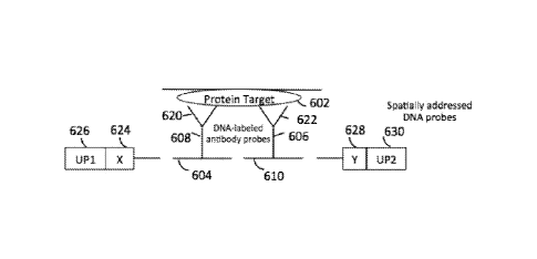
[0074] Figure 6 illustrates multiplexable protein detection assays with
combinatorial addressing
schemes applied to a sample, according to embodiments of the present
disclosure.
13
CA 02916662 2015-12-22
WO 2014/210225 PCT/US2014/044196
[0075] Figure 7 illustrates exemplary antibody-DNA conjugate configurations,
according to
certain embodiments of the present disclosure.
[0076] Figure 8 illustrates sequential address tagging schemes, according to
embodiments of the
present disclosure.
[0077] Figure 9 illustrates a microfluidic addressing device, according to one
embodiment of
the present disclosure.
[0078] Figure 10 provides an immunofluorescence image (Figure 10A) and
representative
expression maps (Figure 10B-C) generated according to some embodiments of the
present
disclosure.
[0079] Figure 11 illustrates a method for reducing random errors during the
sequencing step
(Figure 11A), and exemplary configurations of probes with integrated X and Y
address tags and
variable tag region z (Figure 11B), according to some embodiments of the
present disclosure.
Detailed Description
[0080] A detailed description of one or more embodiments of the claimed
subject matter is
provided below along with accompanying figures that illustrate the principles
of the claimed subject
matter. The claimed subject matter is described in connection with such
embodiments, but is not
limited to any embodiment. It is to be understood that the claimed subject
matter may be embodied
in various forms, and encompasses numerous alternatives, modifications and
equivalents.
Therefore, specific details disclosed herein are not to be interpreted as
limiting, but rather as a basis
for the claims and as a representative basis for teaching one skilled in the
art to employ the claimed
subject matter in virtually any appropriately detailed system, structure or
manner. Numerous
specific details are set forth in the following description in order to
provide a thorough
understanding of the present disclosure. These details are provided for the
purpose of example and
the claimed subject matter may be practiced according to the claims without
some or all of these
specific details. It is to be understood that other embodiments can be used
and structural changes
can be made without departing from the scope of the claimed subject matter.
For the purpose of
clarity, technical material that is known in the technical fields related to
the claimed subject matter
has not been described in detail so that the claimed subject matter is not
unnecessarily obscured.
14
[0081] Unless defined otherwise, all terms of art, notations and other
technical and
scientific terms or terminology used herein are intended to have the same
meaning as is
commonly understood by one of ordinary skill in the art to which the claimed
subject matter
pertains. In some cases, terms with commonly understood meanings are defined
herein for
clarity and/or for ready reference, and the inclusion of such definitions
herein should not
necessarily be construed to represent a substantial difference over what is
generally
understood in the art. Many of the techniques and procedures described or
referenced herein
are well understood and commonly employed using conventional methodology by
those
skilled in the art.
[0082] If a definition set forth herein is contrary to or otherwise
inconsistent with a
definition set forth in the patents, applications, published applications and
other publications
discussed herein, the definition set forth herein prevails.
[0083] The practice of the provided embodiments will employ, unless otherwise
indicated, conventional techniques and descriptions of organic chemistry,
polymer
technology, molecular biology (including recombinant techniques), cell
biology,
biochemistry, and sequencing technology, which are within the skill of those
who practice in
the art. Such conventional techniques include polymer array synthesis,
hybridization and
ligation of polynucleotides, and detection of hybridization using a label.
Specific illustrations
of suitable techniques can be had by reference to the examples herein.
However, other
equivalent conventional procedures can, of course, also be used. Such
conventional
techniques and descriptions can be found in standard laboratory manuals such
as Green, et at.,
Eds., Genome Analysis: A Laboratory Manual Series (Vols. I-IV) (1999); Weiner,
Gabriel,
Stephens, Eds., Genetic Variation: A Laboratory Manual (2007); Dieffenbach,
Dveksler,
Eds., PCR Primer: A Laboratory Manual (2003); Bowtell and Sambrook, DNA
Microarrays:
A Molecular Cloning Manual (2003); Mount, Bioinformatics: Sequence and Genome
Anazysis (2004); Sambrook and Russell, Condensed Protocols from Molecular
Cloning: A
Laboratory Manual (2006); and Sambrook and Russell, Molecular Cloning: A
Laboratory
Manual (2002) (all from Cold Spring Harbor Laboratory Press); Ausubel et al.
eds., Current
Protocols in Molecular Biology (1987); T. Brown ed., Essential Molecular
Biology (1991),
IRL Press; Goeddel ed., Gene Expression Technology (1991), Academic Press; A.
Bothwell et
Date Recue/Date Received 2020-06-18
al. eds., Methods for Cloning and Analysis of Eukaryotic Genes (1990),
Bartlett Publ.; M.
Kriegler, Gene Transfer and Expression (1990), Stockton Press; R. Wu et al.
eds.,
Recombinant DNA Methodology (1989), Academic Press; M. McPherson et al., PCR:
A
Practical Approach (1991), IRL Press at Oxford University Press; Stryer,
Biochemistry (4th
Ed.) (1995), W. H. Freeman, New York N.Y.; Gait, Oligonucleotide Synthesis: A
Practical
Approach (2002), IRL Press, London; Nelson and Cox, Lehninger, Principles of
Biochemistry
(2000) 3rd Ed., W. H. Freeman Pub., New York, N.Y.; Berg, et al., Biochemistry
(2002) 5th
Ed., W. H. Freeman Pub., New York, N.Y.; D. Weir & C. Blackwell, eds.,
Handbook of
Experimental Immunology (1996), Wiley-Blackwell; A. Abbas et al., Cellular and
Molecular
Immunology (1991, 1994), W.B. Saunders Co.; and J. Coligan et al. eds.,
Current Protocols
in Immunology (1991).
[0084] As used herein and in the appended claims, the singular forms "a,"
"an," and "the"
include plural referents unless the context clearly dictates otherwise. For
example, "a" or
"an" means "at least one" or "one or more." Thus, reference to "a biological
target" refers to
one or more biological targets, and reference to "the method" includes
reference to equivalent
steps and methods disclosed herein and/or known to those skilled in the art,
and so forth.
[0085] Throughout this disclosure, various aspects of the claimed subject
matter are
presented in a range format. It should be understood that the description in
range format is
merely for convenience and brevity and should not be construed as an
inflexible limitation on
the scope of the claimed subject matter. Accordingly, the description of a
range should be
considered to have specifically disclosed all the possible sub-ranges as well
as individual
numerical values within that range. For example, where a range of values is
provided, it is
understood that each intervening value, between the upper and lower limit of
16
Date Recue/Date Received 2020-06-18
CA 02916662 2015-12-22
WO 2014/210225 PCT/US2014/044196
these smaller ranges may independently be included in the smaller ranges, and
are also
encompassed within the claimed subject matter, subject to any specifically
excluded limit in the
stated range. Where the stated range includes one or both of the limits,
ranges excluding either or
both of those included limits are also included in the claimed subject matter.
This applies regardless
of the breadth of the range.
[0086] As used herein, an "individual" can be any living organism, including
humans and other
mammals. A "subject" as used herein can be an organism to which the provided
compositions,
methods, kits, devices, and systems can be administered or applied. In one
embodiment, the subject
can be a mammal or a cell, a tissue, an organ or a part of the mammal. Mammals
include, but are
not limited to, humans, and non-human animals, including farm animals, sport
animals, rodents and
pets.
[0087] As used herein, a "biological sample" can refer to any sample obtained
from a living or
viral source or other source of macromolecules and biomolecules, and includes
any cell type or
tissue of a subject from which nucleic acid or protein or other macromolecule
can be obtained. The
biological sample can be a sample obtained directly from a biological source
or a sample that is
processed. For example, isolated nucleic acids that are amplified constitute a
biological sample.
Biological samples include, but are not limited to, body fluids, such as
blood, plasma, serum,
cerebrospinal fluid, synovial fluid, urine and sweat, tissue and organ samples
from animals and
plants and processed samples derived therefrom.
[0088] As used herein, a "composition" can be any mixture of two or more
products or
compounds. It may be a solution, a suspension, liquid, powder, a paste,
aqueous, non-aqueous or
any combination thereof.
[0089] The terms "polynucleotide," "oligonucleotide," "nucleic acid" and
"nucleic acid
molecule" are used interchangeably herein to refer to a polymeric form of
nucleotides of any length,
and may comprise ribonucleotides, deoxyribonucleotides, analogs thereof, or
mixtures thereof. This
term refers only to the primary structure of the molecule. Thus, the term
includes triple-, double-
and single-stranded deoxyribonucleic acid ("DNA"). as well as triple-, double-
and single-stranded
ribonucleic acid ("RNA"). It also includes modified, for example by
alkylation, and/or by capping,
17
CA 02916662 2015-12-22
WO 2014/210225 PCT/US2014/044196
and unmodified forms of the polynucleotide. More particularly, the terms
"polynucleotide,"
"oligonucleotide," "nucleic acid" and "nucleic acid molecule" include
polydeoxyribonucleotides
(containing 2-deoxy-D-ribose), polyribonucleotides (containing D-ribose),
including tRNA, rRNA,
hRNA, and mRNA, whether spliced or unspliced, any other type of polynucleotide
which is an N-
or C-glycoside of a purine or pyrimidine base, and other polymers containing
normucleotidic
backbones, for example, polyamide (e.g., peptide nucleic acids ("PNAs")) and
polymorpholino
(commercially available from the Anti-Virals. Inc.. Corvallis, OR., as
Neugene) polymers, and other
synthetic sequence-specific nucleic acid polymers providing that the polymers
contain nucleobases
in a configuration which allows for base pairing and base stacking, such as is
found in DNA and
RNA. Thus, these terms include, for example, 3'-deoxy-2',5'-DNA,
oligodeoxyribonucleotide N3'
to P5' phosphoramidates, 2'-0-alkyl-substituted RNA, hybrids between DNA and
RNA or between
PNAs and DNA or RNA, and also include known types of modifications, for
example, labels,
alkylation, "caps," substitution of one or more of the nucleotides with an
analog, intemucleotide
modifications such as, for example, those with uncharged linkages (e.g.,
methyl phosphonates,
phosphotriesters, phosphoramidates, carbamates, etc.), with negatively charged
linkages (e.g.,
phosphorothioates, phosphorodithioates, etc.), and with positively charged
linkages (e.g.,
aminoalkylphosphoramidates, aminoalkylphosphotriesters), those containing
pendant moieties, such
as, for example, proteins (including enzymes (e.g. nucleases), toxins,
antibodies, signal peptides,
poly-L-lysine, etc.), those with intercalators (e.g., acridine, psoralen,
etc.), those containing chelates
(of, e.g., metals, radioactive metals, boron, oxidative metals, etc.), those
containing alkylators, those
with modified linkages (e.g., alpha anomeric nucleic acids, etc.), as well as
unmodified forms of the
polynucleotide or oligonucleotide. A nucleic acid generally will contain
phosphodiester bonds,
although in some cases nucleic acid analogs may be included that have
alternative backbones such
as phosphoramidite, phosphorodithioate, or methylphophoroamidite linkages; or
peptide nucleic
acid backbones and linkages. Other analog nucleic acids include those with
bicyclic structures
including locked nucleic acids, positive backbones, non-ionic backbones and
non-ribose backbones.
Modifications of the ribose-phosphate backbone may be done to increase the
stability of the
molecules; for example, PNA:DNA hybrids can exhibit higher stability in some
environments. The
18
CA 02916662 2015-12-22
WO 2014/210225 PCT/US2014/044196
terms "polynucleotide," "oligonucleotide," "nucleic acid" and "nucleic acid
molecule" can
comprise any suitable length, such as at least 5, 6, 7, 8, 9, 10, 20, 30, 40,
50, 100. 200, 300, 400, 500,
1,000 or more nucleotides.
[0090] It will be appreciated that, as used herein, the terms "nucleoside" and
"nucleotide" will
include those moieties which contain not only the known purine and pyrimidine
bases, but also
other heterocyclic bases which have been modified. Such modifications include
methylated purines
or pyrimidines, acylated purines or pyrimidines, or other heterocycles.
Modified nucleosides or
nucleotides can also include modifications on the sugar moiety, e.g., wherein
one or more of the
hydroxyl groups are replaced with halogen, aliphatic groups, or are
functionalized as ethers, amines,
or the like. The term "nucleotidic unit" is intended to encompass nucleosides
and nucleotides.
[0091] "Nucleic acid probe" refers to a structure comprising a polynucleotide,
as defined above,
that contains a nucleic acid sequence that can bind to a corresponding target.
The polynucleotide
regions of probes may be composed of DNA, and/or RNA, and/or synthetic
nucleotide analogs.
[0092] The terms "polypeptide", "oligopeptide", -peptide" and "protein" are
used
interchangeably herein to refer to polymers of amino acids of any length,
e.g., at least 5, 6, 7, 8, 9,
10, 20, 30, 40, 50, 100, 200, 300, 400, 500, 1,000 or more amino acids. The
polymer may be linear
or branched, it may comprise modified amino acids, and it may be interrupted
by non-amino acids.
The terms also encompass an amino acid polymer that has been modified
naturally or by
intervention; for example, disulfide bond formation, glycosylation,
lipidation, acetylation,
phosphorylation, or any other manipulation or modification, such as
conjugation with a labeling
component. Also included within the definition are, for example, polypeptides
containing one or
more analogs of an amino acid (including, for example, unnatural amino acids,
etc.), as well as
other modifications known in the art.
[0093] The terms "binding agent" and "target-binding moiety" as used herein
may refer to any
agent or any moiety thereof that specifically binds to a biological molecule
of interest.
[0094] The biological targets or molecules to be detected can be any
biological molecules
including but not limited to proteins, nucleic acids, lipids, carbohydrates,
ions, or multicomponent
complexes containing any of the above. Examples of subcellular targets include
organelles, e.g.,
19
CA 02916662 2015-12-22
WO 2014/210225 PCT/US2014/044196
mitochondria, Golgi apparatus, endoplasmic reticulum, chloroplasts, endocytic
vesicles, exocytic
vesicles, vacuoles, lysosomes, etc. Exemplary nucleic acid targets can include
genomic DNA of
various conformations (e.g., A-DNA, B-DNA, Z-DNA), mitochondria DNA (mtDNA),
mRNA,
tRNA, rRNA, hRNA, miRNA, and piRNA.
[0095] As used herein, "biological activity" may include the in vivo
activities of a compound or
physiological responses that result upon in vivo administration of a compound,
composition or other
mixture. Biological activity, thus, may encompass therapeutic effects and
pharmaceutical activity
of such compounds, compositions and mixtures. Biological activities may be
observed in vitro
systems designed to test or use such activities.
[0096] The term "binding" can refer to an attractive interaction between two
molecules which
results in a stable association in which the molecules are in close proximity
to each other.
Molecular binding can be classified into the following types: non-covalent,
reversible covalent and
irreversible covalent. Molecules that can participate in molecular binding
include proteins, nucleic
acids, carbohydrates, lipids, and small organic molecules such as
pharmaceutical compounds.
Proteins that form stable complexes with other molecules are often referred to
as receptors while
their binding partners are called ligands. Nucleic acids can also form stable
complex with
themselves or others, for example, DNA-protein complex, DNA-DNA complex, DNA-
RNA
complex.
[0097] As used herein, the term "specific binding" refers to the specificity
of a binder, e.g., an
antibody, such that it preferentially binds to a target, such as a polypeptide
antigen. When referring
to a binding partner (e.g., protein, nucleic acid, antibody or other affinity
capture agent, etc.).
"specific binding" can include a binding reaction of two or more binding
partners with high affinity
and/or complementarity to ensure selective hybridization under designated
assay conditions.
Typically, specific binding will be at least three times the standard
deviation of the background
signal. Thus, under designated conditions the binding partner binds to its
particular target molecule
and does not bind in a significant amount to other molecules present in the
sample. Recognition by
a binder or an antibody of a particular target in the presence of other
potential interfering substances
is one characteristic of such binding. Preferably, binders, antibodies or
antibody fragments that are
CA 02916662 2015-12-22
WO 2014/210225 PCT/US2014/044196
specific for or bind specifically to a target bind to the target with higher
affinity than binding to
other non-target substances. Also preferably, binders, antibodies or antibody
fragments that are
specific for or bind specifically to a target avoid binding to a significant
percentage of non-target
substances, e.g., non-target substances present in a testing sample. In some
embodiments, binders,
antibodies or antibody fragments of the present disclosure avoid binding
greater than about 90% of
non-target substances, although higher percentages are clearly contemplated
and preferred. For
example, binders, antibodies or antibody fragments of the present disclosure
avoid binding about
91%, about 92%, about 93%, about 94%, about 95%, about 96%, about 97%, about
98%, about
99%, and about 99% or more of non-target substances. In other embodiments,
binders, antibodies
or antibody fragments of the present disclosure avoid binding greater than
about 10%, 20%, 30%,
40%, 50%, 60%, or 70%, or greater than about 75%, or greater than about 80%,
or greater than
about 85% of non-target substances.
[0098] The term "antibody" as used herein may include an entire immunoglobulin
or antibody
or any functional fragment of an immunoglobulin molecule which is capable of
specific binding to
an antigen, such as a carbohydrate, polynucleotide, lipid, polypeptide, or a
small molecule, etc.,
through at least one antigen recognition site, located in the variable region
of the immunoglobulin
molecule, and can be an immunoglobulin of any class, e.g., IgG, IgM, IgA. IgD
and IgE. IgY,
which is the major antibody type in avian species such as chicken, is also
included. An antibody
may include the entire antibody as well as any antibody fragments capable of
binding the antigen or
antigenic fragment of interest. Examples include complete antibody molecules,
antibody fragments,
such as Fab, F(ab')2, CDRs, VL, VH, and any other portion of an antibody which
is capable of
specifically binding to an antigen. Antibodies used herein are immunoreactive
or immunospecific
for, and therefore specifically and selectively bind to, for example, proteins
either detected (i.e.,
biological targets) or used for detection (i.e., probes) in the assays of the
invention. An antibody as
used herein can be specific for any of the biological targets disclosed herein
or any combinations
thereof. In certain embodiments, a biological target itself of the present
disclosure can be an
antibody or fragments thereof.
21
CA 02916662 2015-12-22
WO 2014/210225 PCT/US2014/044196
[0099] As used herein, a "fragment thereof" "region thereof' and "portion
thereof" can refer to
fragments, regions and portions that substantially retain at least one
function of the full length
polypeptide.
[00100] As used herein, the term "antigen" may refer to a target molecule that
is specifically
bound by an antibody through its antigen recognition site. The antigen may be
monovalent or
polyvalent, i.e., it may have one or more epitopes recognized by one or more
antibodies. Examples
of kinds of antigens that can be recognized by antibodies include
polypeptides, oligosaccharides,
glycoproteins, polynucleotides, lipids, or small molecules, etc.
[00101] As used herein, the term "epitope" can refer to a peptide sequence of
at least about 3 to
5, preferably about 5 to 10 or 15, and not more than about 1,000 amino acids
(or any integer there
between), which define a sequence that by itself or as part of a larger
sequence, binds to an antibody
generated in response to such sequence. There is no critical upper limit to
the length of the
fragment, which may, for example, comprise nearly the full-length of the
antigen sequence, or even
a fusion protein comprising two or more epitopes from the target antigen. An
epitope for use in the
subject invention is not limited to a peptide having the exact sequence of the
portion of the parent
protein from which it is derived, but also encompasses sequences identical to
the native sequence,
as well as modifications to the native sequence, such as deletions, additions
and substitutions
(conservative in nature).
[00102] The terms "complementary" and "substantially complementary" may
include the
hybridization or base pairing or the formation of a duplex between nucleotides
or nucleic acids, for
instance, between the two strands of a double-stranded DNA molecule or between
an
oligonucleotide primer and a primer binding site on a single-stranded nucleic
acid. Complementary
nucleotides are, generally, A and T (or A and U), or C and G. Two single-
stranded RNA or DNA
molecules are said to be substantially complementary when the nucleotides of
one strand, optimally
aligned and compared and with appropriate nucleotide insertions or deletions,
pair with at least
about 80% of the other strand, usually at least about 90% to about 95%, and
even about 98% to
about 100%. In one aspect, two complementary sequences of nucleotides are
capable of
hybridizing, preferably with less than 25%, more preferably with less than
15%, even more
22
CA 02916662 2015-12-22
WO 2014/210225 PCT/US2014/044196
preferably with less than 5%, most preferably with no mismatches between
opposed nucleotides.
Preferably the two molecules will hybridize under conditions of high
stringency.
[00103] "Hybridization" as used herein may refer to the process in which two
single-stranded
polynucleotides bind non-covalently to form a stable double-stranded
polynucleotide. In one
aspect, the resulting double-stranded polynucleotide can be a "hybrid" or
"duplex." "Hybridization
conditions" typically include salt concentrations of approximately less than 1
M, often less than
about 500 mM and may be less than about 200 mM. A "hybridization buffer"
includes a buffered
salt solution such as 5% SSPE, or other such buffers known in the art.
Hybridization temperatures
can be as low as 5 C, but are typically greater than 22 C, and more typically
greater than about
30 C, and typically in excess of 37 C. Hybridizations are often performed
under stringent
conditions, i.e., conditions under which a sequence will hybridize to its
target sequence but will not
hybridize to other, non-complementary sequences. Stringent conditions are
sequence-dependent
and are different in different circumstances. For example, longer fragments
may require higher
hybridization temperatures for specific hybridization than short fragments. As
other factors may
affect the stringency of hybridization, including base composition and length
of the complementary
strands, presence of organic solvents, and the extent of base mismatching, the
combination of
parameters is more important than the absolute measure of any one parameter
alone. Generally
stringent conditions are selected to be about 5 C lower than the Tn, for the
specific sequence at a
defined ionic strength and pH. The melting temperature T. can be the
temperature at which a
population of double-stranded nucleic acid molecules becomes half dissociated
into single strands.
Several equations for calculating the Tõ, of nucleic acids are well known in
the art. As indicated by
standard references, a simple estimate of the Tõ, value may be calculated by
the equation, T,õ =81.5
+ 0.41 (% G + C), when a nucleic acid is in aqueous solution at 1 M NaCl (see
e.g., Anderson and
Young, Quantitative Filter Hybridization, in Nucleic Acid Hybridization
(1985)). Other references
(e.g., Allawi and SantaLucia, Jr., Biochemistry, 36:10581-94 (1997)) include
alternative methods of
computation which take structural and environmental, as well as sequence
characteristics into
account for the calculation of T..
23
CA 02916662 2015-12-22
WO 2014/210225 PCT/US2014/044196
[00104] In general, the stability of a hybrid is a function of the ion
concentration and
temperature. Typically, a hybridization reaction is performed under conditions
of lower stringency,
followed by washes of varying, but higher, stringency. Exemplary stringent
conditions include a
salt concentration of at least 0.01 M to no more than 1 M sodium ion
concentration (or other salt) at
a pH of about 7.0 to about 8.3 and a temperature of at least 25 C. For
example, conditions of 5 x
SSPE (750 mM NaCl, 50 mM sodium phosphate, 5 mM EDTA at pH 7.4) and a
temperature of
approximately 30 C are suitable for allele-specific hybridizations, though a
suitable temperature
depends on the length and/or GC content of the region hybridized. In one
aspect, "stringency of
hybridization" in determining percentage mismatch can be as follows: 1) high
stringency: 0.1 x
SSPE, 0.1% SDS, 65 C; 2) medium stringency: 0.2 x SSPE, 0.1% SDS, 50 C (also
referred to as
moderate stringency); and 3) low stringency: 1.0 x SSPE, 0.1% SDS, 50 C. It is
understood that
equivalent stringencies may be achieved using alternative buffers, salts and
temperatures. For
example, moderately stringent hybridization can refer to conditions that
permit a nucleic acid
molecule such as a probe to bind a complementary nucleic acid molecule. The
hybridized nucleic
acid molecules generally have at least 60% identity, including for example at
least any of 70%,
75%, 80%, 85%, 90%, or 95% identity. Moderately stringent conditions can be
conditions
equivalent to hybridization in 50% formamide, 5 x Denhardt's solution, 5x
SSPE, 0.2% SDS at
42 C, followed by washing in 0.2 x SSPE, 0.2% SDS, at 42 C. High stringency
conditions can be
provided, for example, by hybridization in 50% formamide, 5 x Denhardt's
solution, 5 x SSPE,
0.2% SDS at 42 C, followed by washing in 0.1 x SSPE, and 0.1% SDS at 65 C. Low
stringency
hybridization can refer to conditions equivalent to hybridization in 10%
formamide, 5 x Denhardt's
solution, 6 x SSPE, 0.2% SDS at 22 C, followed by washing in lx SSPE, 0.2%
SDS, at 37 C.
Denhardt's solution contains 1% Ficoll, 1% polyvinylpyrolidone, and 1% bovine
serum albumin
(BSA). 20 x SSPE (sodium chloride, sodium phosphate, ethylene diamide
tetraacetic acid (EDTA))
contains 3M sodium chloride, 0.2M sodium phosphate, and 0.025 M EDTA. Other
suitable
moderate stringency and high stringency hybridization buffers and conditions
are well known to
those of skill in the art and are described, for example, in Sambrook et al..
Molecular Cloning: A
24
CA 02916662 2015-12-22
WO 2014/210225 PCT/US2014/044196
Laboratory Manual, 2nd ed., Cold Spring Harbor Press, Plainview, N.Y. (1989);
and Ausubel et al.,
Short Protocols in Molecular Biology, 4th ed., John Wiley & Sons (1999).
[00105] Alternatively, substantial complementarity exists when an RNA or DNA
strand will
hybridize under selective hybridization conditions to its complement.
Typically, selective
hybridization will occur when there is at least about 65% complementary over a
stretch of at least
14 to 25 nucleotides, preferably at least about 75%, more preferably at least
about 90%
complementary. See M. Kanehisa, Nucleic Acids Res. 12:203 (1984).
[00106] A "primer" used herein can be an oligonucleotide, either natural or
synthetic, that is
capable, upon forming a duplex with a polynucleotide template, of acting as a
point of initiation of
nucleic acid synthesis and being extended from its 3' end along the template
so that an extended
duplex is formed. The sequence of nucleotides added during the extension
process is determined by
the sequence of the template polynucleotide. Primers usually are extended by a
DNA polymerase.
[00107] "Ligation" may refer to the formation of a covalent bond or linkage
between the termini
of two or more nucleic acids, e.g., oligonucleotides and/or polynucleotides,
in a template-driven
reaction. The nature of the bond or linkage may vary widely and the ligation
may be carried out
enzymatically or chemically. As used herein, ligations are usually carried out
enzymatically to
form a phosphodiester linkage between a 5' carbon terminal nucleotide of one
oligonucleotide with
a 3' carbon of another nucleotide.
[00108] "Sequencing." "sequence determination" and the like means
determination of
information relating to the nucleotide base sequence of a nucleic acid. Such
information may
include the identification or determination of partial as well as full
sequence information of the
nucleic acid. Sequence information may be determined with varying degrees of
statistical reliability
or confidence. In one aspect, the term includes the determination of the
identity and ordering of a
plurality of contiguous nucleotides in a nucleic acid. "High throughput
digital sequencing" or "next
generation sequencing" means sequence determination using methods that
determine many
(typically thousands to billions) of nucleic acid sequences in an
intrinsically parallel manner, i.e.
where DNA templates are prepared for sequencing not one at a time, but in a
bulk process, and
where many sequences are read out preferably in parallel, or alternatively
using an ultra-high
throughput serial process that itself may be parallelized. Such methods
include but are not
limited to pyrosequencing (for example, as commercialized by 454 Life
Sciences, Inc.,
Branford, Conn.); sequencing by ligation (for example, as commercialized in
the SOLiDTM
technology, Life Technologies, Inc., Carlsbad, Calif.); sequencing by
synthesis using
modified nucleotides (such as commercialized in TruSeqTm and HiSeqTM
technology by
Illumina, Inc., San Diego, Calif; HeliScopeTM by Helicos Biosciences
Corporation,
Cambridge, Ma.; and PacBio RS by Pacific Biosciences of California, Inc.,
Menlo Park,
Calif), sequencing by ion detection technologies (such as Ion Ton-entTm
technology, Life
Technologies, Carlsbad, Calif.); sequencing of DNA nanoballs (Complete
Genomics, Inc.,
Mountain View, Calif.); nanopore-based sequencing technologies (for example,
as developed
by Oxford Nanopore Technologies, LTD, Oxford, UK), and like highly
parallelized
sequencing methods.
[00109] "SNP" or "single nucleotide polymorphism" may include a genetic
variation
between individuals; e.g., a single nitrogenous base position in the DNA of
organisms that is
variable. SNPs are found across the genome; much of the genetic variation
between individuals
is due to variation at SNP loci, and often this genetic variation results in
phenotypic variation
between individuals. SNPs for use in the present invention and their
respective alleles may be
derived from any number of sources, such as public databases (U.C. Santa Cruz
Human
Genome Browser Gateway) or the NCBI dbSNP website, or may be experimentally
determined
as described in U.S. Pat. No. 6,969,589; and US Pub. No. 2006/0188875 entitled
"Human
Genomic Polymorphisms." Although the use of SNPs is described in some of the
embodiments
presented herein, it will be understood that other biallelic or multi-allelic
genetic markers may
also be used. A biallelic genetic marker is one that has two polymorphic
forms, or alleles. As
mentioned above, for a biallelic genetic marker that is associated with a
trait, the allele that is
more abundant in the genetic composition of a case group as compared to a
control group is
termed the "associated allele," and the other allele may be referred to as the
"unassociatcd
allele." Thus, for each biallelic polymorphism that is associated with a given
trait (e.g., a
disease or drug response), there is a corresponding associated allele. Other
biallelic
polymorphisms that may be used with the methods presented herein include, but
are not limited
26
Date Recue/Date Received 2020-06-18
CA 02916662 2015-12-22
WO 2014/210225 PCT/US2014/044196
multinucleotide changes, insertions, deletions, and translocations. It will be
further appreciated that
references to DNA herein may include genomic DNA, mitochondrial DNA, episomal
DNA, and/or
derivatives of DNA such as amplicons. RNA transcripts, cDNA, DNA analogs, etc.
The
polymorphic loci that are screened in an association study may be in a diploid
or a haploid state and,
ideally, would be from sites across the genome.
[00110] As used herein, the term "microfluidic device may generally refer to a
device through
which materials, particularly fluid borne materials, such as liquids, can be
transported, in some
embodiments on a micro-scale, and in some embodiments on a nanoscale. Thus,
the microfluidic
devices described by the presently disclosed subject matter can comprise
microscale features,
nanoscale features, and combinations thereof.
[00111] Accordingly, an exemplary microfluidic device typically comprises
structural or
functional features dimensioned on the order of a millimeter-scale or less,
which are capable of
manipulating a fluid at a flow rate on the order of a [tUmin or less.
Typically, such features
include, but are not limited to channels, fluid reservoirs, reaction chambers,
mixing chambers, and
separation regions. In some examples, the channels include at least one cross-
sectional dimension
that is in a range of from about 0.1 vim to about 500 pm. The use of
dimensions on this order allows
the incorporation of a greater number of channels in a smaller area, and
utilizes smaller volumes of
fluids.
[00112] A microfluidic device can exist alone or can be a part of a
microfluidic system which,
for example and without limitation, can include: pumps for introducing fluids,
e.g., samples,
reagents, buffers and the like, into the system and/or through the system;
detection equipment or
systems; data storage systems; and control systems for controlling fluid
transport and/or direction
within the device, monitoring and controlling environmental conditions to
which fluids in the
device are subjected, e.g., temperature, current, and the like.
[00113] As used herein, the terms "channel," "micro-channel," "fluidic
channel," and
"microfluidic channel" are used interchangeably and can mean a recess or
cavity formed in a
material by imparting a pattern from a patterned substrate into a material or
by any suitable material
removing technique, or can mean a recess or cavity in combination with any
suitable fluid-
27
CA 02916662 2015-12-22
WO 2014/210225 PCT/US2014/044196
conducting structure mounted in the recess or cavity, such as a tube,
capillary, or the like. In the
present invention, channel size means the cross-sectional area of the
microfluidic channel.
[00114] As used herein, the terms "flow channel" and "control channel" are
used interchangeably
and can mean a channel in a microfluidic device in which a material, such as a
fluid, e.g., a gas or a
liquid, can flow through. More particularly, the term "flow channel" refers to
a channel in which a
material of interest, e.g., a solvent or a chemical reagent, can flow through.
Further, the term
"control channel" refers to a flow channel in which a material, such as a
fluid, e.g., a gas or a liquid,
can flow through in such a way to actuate a valve or pump.
[00115] As used herein, "chip" may refer to a solid substrate with a plurality
of one-, two- or
three-dimensional micro structures or micro-scale structures on which certain
processes, such as
physical, chemical, biological, biophysical or biochemical processes, etc.,
can be carried out. The
micro structures or micro-scale structures such as, channels and wells,
electrode elements,
electromagnetic elements, are incorporated into, fabricated on or otherwise
attached to the substrate
for facilitating physical, biophysical, biological, biochemical, chemical
reactions or processes on the
chip. The chip may be thin in one dimension and may have various shapes in
other dimensions, for
example, a rectangle, a circle, an ellipse, or other irregular shapes. The
size of the major surface of
chips of the present invention can vary considerably, e.g., from about 1 mm2
to about 0.25 m2.
Preferably, the size of the chips is from about 4 mm2 to about 25 cm2 with a
characteristic
dimension from about 1 mm to about 5 cm. The chip surfaces may be flat, or not
flat. The chips
with non-flat surfaces may include channels or wells fabricated on the
surfaces.
[00116] A microfluidic chip can be used for the methods and assay systems
disclosed herein. A
microfluidic chip can be made from any suitable materials, such as PDMS
(Polydimethylsiloxane),
glass, PMMA (polymethylmethacrylate), PET (polyethylene terephthalate), PC
(Polycarbonate),
etc.. or a combination thereof.
[00117] "Multiplexing" or "multiplex assay" herein may refer to an assay or
other analytical
method in which the presence and/or amount of multiple targets, e.g., multiple
nucleic acid target
sequences, can be assayed simultaneously by using more than one capture probe
conjugate, each of
which has at least one different detection characteristic, e.g., fluorescence
characteristic (for
28
example excitation wavelength, emission wavelength, emission intensity, FWHM
(full width at
half maximum peak height), or fluorescence lifetime) or a unique nucleic acid
or protein
sequence characteristic.
Assays for Determining Spatial Patterns of Biological Targets
[00118] Disclosed herein are spatially-encoded, multiplexed methods and assay
systems
capable of high levels of multiplexing with an efficient spatial encoding
scheme. In one
embodiment, provided herein is instrumentation capable of delivering reagents
to a sample and
thereby spatially encoding multiple sites to which the reagents are delivered.
In one aspect,
reagents can be delivered to a sample according to a known spatial pattern,
for example, a
spatial pattern determined by histological features of the sample. In another
aspect, reagents
are delivered by random-access methods, such as inkjet and pin-spotting. In
another aspect,
microfluidic devices with addressing channels and the like are used to deliver
reagents to a
sample, and to spatially encode multiple sites in the sample to which the
reagents are delivered.
In some embodiments, the spatially-encoded ("addressed," or "address tagged"),
multiplexed
methods and assay systems comprise a decoding feature determined by a readout
that is digital
in nature. In one aspect, the methods and assay systems disclosed herein
detect the presence or
absence of a biological target or a biological activity indicative of a
biological target. In
another aspect, provided herein are methods and assay systems that can detect
the amount or
abundance of a biological target or biological activity indicative of a
biological target at
multiple sites in a sample, as well as the location of each of the multiple
sites in the sample.
Based on the analysis of the amount or abundance and the location information
of one or more
biological targets or activities, spatial patterns across the multiple sites
in the sample can be
generated. In any of the preceding embodiments, the method or assay system may
not depend
on an imaging technique for determining spatial or location information of the
one or more
biological targets in the sample, although the method or assay system may
optionally comprise
using an imaging technique for other purposes. Imaging techniques may include
but are not
limited to conventional immunohistochemical (IHC) imaging and
immunofluorescence (IF)
imaging. Methods and assays systems to determine a spatial pattern of
abundance and/or
29
Date Recue/Date Received 2020-06-18
activity of a biological target in a sample are disclosed in detail in U.S.
Application Serial No.
13/080,616, entitled "Spatially encoded biological assays" (Pub. No.: US
2011/0245111).
[00119] The present disclosure further provides instrumentation with an
ability to deliver
reagents to multiple sites in a sample, wherein each of the multiple sites can
be identified by the
reagents delivered thereto. In one embodiment, reagents are delivered in a
spatially-defined
pattern. The instrumentation, together with software, reagents and protocols,
provides a key
component of the methods and assay systems of the present disclosure, allowing
for
measurement of numerous biological targets or activities, including DNA, RNA
and/or protein
expression, and spatial localization of such biological targets or activities
in a sample. In one
embodiment, the abundance, expression, and/or activity and the location of
biological targets in
the biological samples are determined after the assay products of the
multiplexed assay are
removed from the biological sample and pooled for analysis. Determination of
the abundance,
expression, and/or activity and the location of biological targets can be
performed by, e.g.,
next-generation sequencing, which easily provides millions to trillions of
data points at low
cost. The assay results such as the amount or activity of biological targets
can then be mapped
back to a specific location in the biological sample. The methods and assay
systems provide
tools to analyze the complex spatial patterns of cellular function and
regulation in biological
samples.
[00120] In one aspect, a method of determining a spatial pattern of abundance,
expression,
and/or activity of one or more biological targets across multiple sites in a
sample is provided in
Figure 1. At Step 110, a probe for each of one or more biological targets is
delivered to
multiple sites in a sample, each probe comprising a target-binding moiety, an
address tag that
identifies each site to which the probe is delivered, and an identity tag.
[00121] In any of the embodiments of the present disclosure, the sample can be
any
biological sample or samples that can be affixed to a support or provided
essentially in a
two-dimensional manner, such that an assayed biological target or activity can
be tied back to
the location within the biological sample. In certain embodiments, the sample
can be a freshly
isolated sample, a fixed sample, a frozen sample, an embedded sample, a
processed sample, or
a combination thereof. Exemplary samples of the present disclosure include
tissue sections
(e.g., including whole animal
Date Recue/Date Received 2020-06-18
CA 02916662 2015-12-22
WO 2014/210225 PCT/US2014/044196
sectioning and tissue biopsies), cell populations, or other biological
structure disposed upon a
support, such as on a slide (e.g., a microscope slide) or culture dish, and
the like. In preferred
embodiments, the methods and assay systems of the present disclosure are
compatible with
numerous biological sample types, including fresh samples, such as primary
tissue sections, and
preserved samples including but not limited to frozen samples and formalin-
fixed, paraffin-
embedded (FFPE) samples. In certain embodiments, the sample can be fixed with
a suitable
concentration of formaldehyde or paraformaldehyde, for example, 4% of
formaldehyde or
paraformaldehyde in phosphate buffered saline (PBS). In certain embodiments,
the biological
samples are immobilized on a substrate surface having discrete, independently
measureable areas.
[00122] In one embodiment, the biological sample may contain one or more
biological targets of
interest. In any of the embodiment of the present disclosure, the one or more
biological targets can
be any biological molecules including but not limited to proteins, nucleic
acids, lipids,
carbohydrates, ions, or multicomponent complexes containing any of the above.
Examples of
subcellular targets include organelles, e.g., mitochondria, Golgi apparatus,
endoplasmic reticulum,
chloroplasts, endocytic vesicles, exocytic vesicles, vacuoles, lysosomes, etc.
In some embodiments,
the one or more biological targets can be nucleic acids, including RNA
transcripts, genomic DNA
sequences, cDNAs, amplicons, or other nucleic acid sequences. In other
embodiments, the one or
more biological targets can be proteins, enzymes (protein enzymes or
ribozymes) and the like.
[00123] At Step 110, the probe for each of the multiple biological targets
comprise: (1) a target-
binding moiety capable of binding to the probe's corresponding biological
target; (2) an address tag
that identifies each site to which the probe is delivered; and (3) an identity
tag that identifies the
probe's corresponding biological target or target-binding moiety. Depending on
the nature of the
biological target. the target-binding moiety can be a target-specific
nucleotide sequence (for
example, a sequence complementary to a sequence of a nucleic acid target),
small molecule,
aptamer, antibody. lipid, carbohydrate, ion, affinity capture agent, or
multicomponent complexes
containing any of the above. The address tag identifies the position in the
sample to which the
probe is delivered, and the identity tag identifies the probe's corresponding
biological target being
assayed or the target-binding moiety. Thus, the identities of the address tag
and the identity tag can
31
CA 02916662 2015-12-22
WO 2014/210225 PCT/US2014/044196
be used to link assay results to biological targets and locations in the
sample. In preferred
embodiments, there can be at least two address tags for a biological target at
each of multiple sites
in a sample, each address tag identifying a parameter of each of the multiple
sites. For example,
there can be an X-axis address tag and a Y-axis address tag for each site in a
sample placed on an
X-Y coordinate plane. Thus, each site can be uniquely identified by its
corresponding (X, Y)
coordinates. In preferred embodiments of the present disclosure, the address
tags and/or the identity
tags can be oligonucleotides. In other embodiments, the address tags and/or
the identity tags can be
mass tags, fluorescent labels, or other moieties.
[00124] In some embodiments, the target-binding moiety. address tag, and/or
identity tag of the
probe are pre-coupled before being delivered to the biological sample. In the
case where the probes
are oligonucleotides, the target-binding sequence, address tag sequence,
and/or identity tag
sequence can be synthesized as a single oligonucleotide. Alternatively, the
target-binding moiety,
address tag, and/or identity tag of the probe can be synthesized or obtained
separately and combined
before delivery to the biological sample. For example, two separate
oligonucleotides can be
synthesized and coupled by, e.g., ligation; or an antibody and an
oligonucleotide can be prepared
separately and conjugated before delivery to the biological sample. In other
embodiments, the
probes and the address tags can be synthesized separately, and delivered to
the biological sample at
different steps (e.g., probes first and address tags thereafter, or vice
versa) in the assay.
[00125] At Step 120, the probe is allowed to bind to its corresponding
biological target in the
sample and thereby to react or interact with the biological target. For
example, conditions are
provided to allow oligonucleotides to hybridize to nucleic acid targets,
enzymes to catalyze
reactions with protein targets, antibodies to bind epitopes within a target,
etc. In the case where the
biological targets are nucleic acids, the probes are typically
oligonucleotides and hybridize to the
target nucleic acids. In the case that the biological targets are proteins,
the probes typically are
aptamers, small molecules, or oligonucleotide-conjugated proteins that
interact with target proteins
by binding to them or by reacting with them (that is, one of the proteins is a
substrate for the other).
Oligonucleotides may be coupled to the probes or proteins by conjugation,
chemical or photo-
crosslinking via suitable groups and the like.
32
[00126] In some embodiments, after allowing the probes to bind to or interact
with the
one or more biological targets in the sample, probes bound to the biological
targets may be
separated from probes delivered to the sample but not bound to the biological
targets. In
one aspect, in the case where the biological targets are nucleic acids and the
probes are
oligonucleotides, the separation can be accomplished by, e.g., washing the
unhybridized
probes from the sample. Similarly, for other assays that are based on affinity
binding,
including those using aptamer, small molecule, and protein probes, washing
steps can be
used to remove low affinity binders. In the case where the probe is
transformed via
interaction with the target, e.g., in the case of a peptide, e.g., via
cleavage by a protease or
phosphorylation by a kinase, it is convenient to collect all probes, including
both probes that
have interacted with the biological targets and thus transformed and probes
not transformed.
After collection or pooling, an antibody or other affinity capture agent can
be used to
capture probes transformed by addition of a moiety (e.g., a phosphate group in
cases of
phosphorylation by a kinase). In cases where probes have been transformed via
cleavage,
the transformed probes can be separated, e.g., by capturing the non-
transformed probes via a
tag that is removed from the transformed probes during the transformation
(e.g., by
cleavage), or by adding a new tag at the site of cleavage.
[00127] In certain other embodiments, probes bound to the biological targets
may not
need to be separated from probes not bound to the biological targets for
determining a
spatial pattern of abundance, expression, and/or activity of the biological
targets. At Step
130, probes bound to the one or more biological targets are analyzed. In
certain
embodiments, the analysis comprises determining abundance, expression, and/or
activity of
each biological target and the identities of the identity tag and the address
tag for each
biological target at each site. Numerous methods can be used to identify the
address tags,
identity tags and/or target-binding moieties of the probes of the methods and
assay systems
disclosed herein. The address tags can be detected using techniques such as
mass
spectroscopy (e.g., matrix-assisted laser desorption/ionization-time of flight
mass
spectrometry (MALDI-TOF), LC-MS/MS, and TOE/TOFTm LC/MS/MS), nuclear magnetic
resonance imaging, or, preferably, nucleic acid sequencing. Examples of
techniques for
decoding the probes of the present invention can be found, for example, in US
Pub. No.
33
Date Recue/Date Received 2020-06-18
20080220434. For example, the address tags may be oligonucleotide mass tags
(0MTs or
massTags). Such tags are described, e.g., in US Pub. No. 20090305237. In yet
another
aspect, the probes can be amplified and hybridized to a microarray. This would
require
separate amplification reactions to be carried out, in which each
amplification is specific to
a particular address tag or subset of tags, accomplished by using tag-specific
primers. Each
amplification would also incorporate a different resolvable label (e.g.
fluorophor).
Following hybridization, the relative amounts of a particular target mapping
to different
spatial locations in the sample can be determined by the relative abundances
of the
resolvable labels. At Step 140, based on the analysis of probes bound to the
one or more
biological targets, a spatial pattern of abundance, expression, and/or
activity of the one or
more biological targets across the multiple sites in the sample is determined.
[00128] In a preferred aspect, the probes according to the present disclosure
are
substrates for high-throughput, next-generation sequencing, and highly
parallel next-
generation sequencing methods are used to confirm the sequence of the probes
(including,
for example, the sequence of the target-binding moiety, the address tag,
and/or the identity
tag). Suitable sequencing technologies include but are not limited to SOLiDTM
technology
(Life Technologies, Inc.) or Genome Ananlyzer (Illumina, Inc.). Such next-
generation
sequencing methods can be carried out, for example, using a one pass
sequencing method or
using paired-end sequencing. Next generation sequencing methods include, but
are not
limited to, hybridization-based methods, such as disclosed in e.g., Drmanac,
U.S. Pat. Nos.
6,864,052; 6,309,824; and 6,401,267; and Drmanac et al., U.S. patent
publication
2005/0191656; sequencing-by-synthesis methods, e.g., U.S. Pat. Nos. 6,210,891;
6,828,100;
6,969,488; 6,897,023; 6,833,246; 6,911,345; 6,787,308; 7,297,518; 7,462,449
and
7,501,245; US Publication Application Nos. 20110059436; 20040106110;
20030064398;
and 20030022207; Ronaghi, etal., Science, 281:363-365 (1998); and Li, et al.,
Proc. Natl.
Acad. Sci., 100:414-419 (2003); ligation-based methods, e.g., U.S. Pat. Nos.
5,912,148 and
6,130,073; and U.S. Pat. Appin Nos. 20100105052, 20070207482 and 20090018024;
nanopore sequencing, e.g., U.S. Pat. Appin Nos. 20070036511; 20080032301;
20080128627; 20090082212; and Soni and Meller, Clin Chem 53:1996-2001 (2007),
as
well as other methods, e.g., U.S. Pat. Appin Nos. 20110033854; 20090264299;
34
Date Recue/Date Received 2020-06-18
20090155781; and 20090005252; also, see, McKernan, etal., Genome Res. 19:1527-
41
(2009) and Bentley, et al., Nature 456:53-59 (2008).
[00129] In preferred embodiments, probes bound to the one or more biological
targets are
analyzed by sequencing. Such analysis by sequencing comprises determining the
amount of
a sequencing product, which indicates abundance, expression, and/or activity
of each
biological target, the sequencing product comprising all or a portion of the
address tag
sequence and all or a portion of the identity tag sequence identifying each
biological target
at each site. In one embodiment, the address tag sequence of the sequencing
product allows
the mapping of the assay results back to the multiple sites in the sample.
[00130] In certain embodiments, two probes that bind to the same target
molecule (for
example, two polynucleotide probes that hybridize to adjacent sites on a
nucleic acid target)
may be assayed by extension followed by ligation (the extension-ligation
assay). The
extension-ligation assay allows certain target sequence to be determined de
novo. For
example, if the primer and the downstream oligo are separated by 20 bases and
reverse
transcriptase is used to fill this 20-base gap, 20 bases of sequence of the
RNA target can be
obtained. In certain embodiments, by using the extension-ligation assay in the
present
methods or assay systems, regions of sequence that are of particular interest
may be
characterized. For example, these regions may comprise mutations or
variations, for
example, with implications in cancer, MHC variations, and RNA editing.
[00131] In any of the embodiments disclosed herein, an extension assay may
also be used
and may allow certain target sequences, for example, nucleotide sequences, to
be
determined de novo. In one embodiment, an extension assay of the present
disclosure may
be performed as follows. A first primer may be used to make cDNA from a target
sequence. In certain embodiments, the first primer can be a random primer
(e.g., random
hexamer) or a sequence-specific primer. A random primer can be used to make
cDNA from
the entire transcriptomc, while a sequence-specific primer may be used to make
cDNA from
a specific target sequence. In certain aspects, the first primer may comprise
a universal
priming site for amplification of the assay products, an adaptor to enable
sequence
identification by sequencing techniques, and/or an adaptor for attaching
address tags. In
Date Recue/Date Received 2020-06-18
CA 02916662 2015-12-22
WO 2014/210225 PCT/US2014/044196
other embodiments, the first primer may be conjugated to an adaptor for
attaching address tags.
The X and Y address tags as described infra can be coupled to the first primer
via the adaptor. Note
that the X and Y address tags can be coupled to the same or different side
relative to the target
sequence, and the configuration may be used in any of embodiments disclosed
herein. The Y
address tag can be further linked to a universal priming site or an adaptor
for sequencing coupled to
biotin. The cDNA with conjugated X and Y address tags are then eluted and
captured on a
streptavidin bead, and a second primer can then be installed to the cDNA on
the opposite side
relative to the first primer. In some embodiments, capture of the
polynucleotide sequence can be
based on other hapten-binder combinations other than biotin-streptavidin, or
be sequence-based. In
certain embodiments, the second primer can be a random primer (e.g., random
hexamer) or a
sequence-specific primer. In certain aspects, the second primer may comprise a
universal priming
site for amplification of the assay products, an adaptor to enable sequence
identification by
sequencing techniques, and/or an adaptor for attaching address tags. In other
embodiments, the
second primer may be conjugated to a universal priming site or an adaptor for
sequencing.
Together with the priming site or adaptor coupled to biotin, the sequence can
be extended from the
second primer, amplified, and sequenced.
[00132] The methods and assay systems disclosed herein may comprise an
amplification step,
and in particular, a nucleic acid amplification step. In certain aspects, the
amplification step is
performed by PCR. In some embodiments, linear amplification (e.g., by T7 RNA
polymerase) may
be used instead of PCR or as a partial replacement for PCR. In one aspect,
linear amplification of
polynucleotides may cause less distortion of the relative abundances of
different sequences. This
can be accomplished by including a T7 RNA pol promoter in one of the universal
portions of the
sequence. In this case, the promoter itself and regions upstream of the
promoter are not copied. In
yet other embodiments, other amplification methods may be used in the methods
and assay systems
disclosed herein. For some sequencing methods (e.g., nanopore sequencing),
amplification may be
optional.
[00133] The T7 RNA polymerase based amplification is a commonly used protocol
for mRNA
amplification originally described by van Gelder et al., Proc. Natl. Acad.
Sci. USA 87, 1663-1667
36
CA 02916662 2015-12-22
WO 2014/210225 PCT/US2014/044196
(1990). The protocol consists of the synthesis of a cDNA complementary to the
mRNA ("first
strand synthesis"), effected by reverse transcription, followed by second
strand synthesis to yield
double-stranded cDNA, and in vitro transcription using the double-stranded
cDNA as template
effected with T7 RNA polymerase. The last step provides single-stranded
antisense RNA (aRNA),
which may be labeled in case labeled nucleotides are provided. The nucleotides
may be labeled by
radioactive labeling or non-radioactive labeling methods. Eberwine et al.
(Proc. Natl. Acad, Sci.
USA 89, 3010-3014 (1992)) extended van Gelder's method by adding a second
round of
amplification using the RNA obtained in the first round as template. Wang et
al. (Nature
Biotechnol. 18, 457-459 (2000)) provided a variant of the original T7 method,
characterized by a
modified second strand synthesis method. The second strand synthesis method of
Wang et al. is
known in the art as the SMARTTm technology (Clontech) for cDNA synthesis.
Baugh et al.
(Nucleic Acids Res. 29, E29 (2001)) describe an optimized variant of the
method according to van
Gelder et al. and analyze the performance on Affymetrix DNA chips (GeneChip ).
Affymetrix
GeneChips are designed to probe the anti-sense strand. Any other DNA chip or
microarray probing
the anti-sense strand may be envisaged when performing a T7 RNA amplification,
wherein labeling
occurs during the in vitro transcription step.
[00134] In other embodiments, amplification techniques such as rolling circle
amplification
(RCA) and circle to circle amplification (C2CA) may be used for probe, target,
tag, and/or signal
amplification of the present disclosure. RCA is a linear-isothermal process in
the presence of
certain DNA polymerases, using an ssDNA mini-circle as a template (Fire and
Xu, Proc. Natl.
Acad. Sci., 92: 4641-4645 (1995); Daubendiek etal., J. Am. Chem. Soc.
117:77818-7819 (1995)).
In certain aspects, a polynucleotide sequence can be replicated about 500 to
1000 times, depending
on the amplification time. For example, in a dual targeting assay for a target
protein as discussed
supra, a linear ligated product is formed (e.g., when two antibodies bind to
adjacent domains on a
target protein, the antibodies' oligonucleotide tags can be ligated), and can
be cut by restriction
enzymes and then re-ligated to form a DNA circle by a DNA ligase and a
template. In certain
embodiments, phi29 DNA polymerase can be used to extend the primer, which is
also the template,
to form a long ssDNA containing a number of sequences complementary to the
initial DNA circle.
37
CA 02916662 2015-12-22
WO 2014/210225 PCT/US2014/044196
C2CA is based on RCA, and may include three steps: replication, monomerization
and ligation
(Dahl et al., Proc. Natl. Acad. Sci.. 101: 4548-4553 (2004)). The original
circular DNA is
considered as the positive polarity. After one step of replication (RCA
reaction), the product is
converted into the opposite polarity. Restriction oligos with the positive
polarity (R0+) can form
duplex regions with the RCA product, and the duplex regions can be cleaved by
restriction enzymes
to generate monomers. Then the monomers can be guided into a ligation step and
circularized.
These circles serve as the templates for the next round of RCA, primed by the
R0+. The process
can be further repeated to produce around 100-fold higher concentration of
target sequences than
conventional PCR.
[00135] In another aspect, as shown in Figure 2, a method of determining a
spatial pattern of
abundance, expression, and/or activity of one or more biological targets
across multiple sites in a
sample is provided, featuring an efficient implementation of an address
tagging scheme for the one
or more biological targets across the multiple sites. In one embodiment, the
address tagging scheme
is a combinatorial scheme using at least two address tags for the biological
targets for each of the
multiple sites in the sample. At Step 210, a probe for each of one or more
biological targets to
multiple sites in a sample is delivered, each probe comprising (1) a target-
binding moiety capable of
binding to the probe's corresponding biological target; and (2) an identity
tag that identifies the
probe's corresponding biological target or target-binding moiety. Depending on
the nature of the
biological target, the target-binding moiety can be a target-specific
nucleotide sequence (for
example, a sequence complementary to a sequence of a nucleic acid target),
small molecule,
aptamer, antibody. lipid, carbohydrate, ion, affinity capture agent, or
multicomponent complexes
containing any of the above. At Step 220, each probe is allowed to interact
with or bind to its
corresponding biological target in the sample, under appropriate conditions.
[00136] In certain embodiments, probes not bound to the biological targets may
be removed, and
thereby separated from probes bound to the biological targets. Such separation
can be performed
essentially as discussed above, for example, by washing the sample to remove
unhybridized
oligonucleotide probes. In certain other embodiments, probes bound to the
biological targets may
38
CA 02916662 2015-12-22
WO 2014/210225 PCT/US2014/044196
not need to be separated from probes not bound to the biological targets for
determining a spatial
pattern of abundance, expression, and/or activity of the biological targets.
[00137] Next, at Step 230, an address tag is delivered to each of the multiple
sites in the sample,
and the address tag is to be coupled to the probe for each biological target
and identifies each site to
which the address tag is delivered. Note that in this aspect, the probe and
address tag are delivered
in separate steps. In certain embodiments where the probes are
oligonucleotides, the address tags
may be coupled to the oligonucleotide probes by various means known to the
skilled artisan, for
example, by extension, ligation, ligation followed by extension, or any
combination thereof. For
instance, the information in the address tags can be transferred by using a
DNA polymerase to
extend a probe oligonucleotide that acts as a primer, and thereby copy and
incorporate the sequence
of the address tags.
[00138] At Step 240, probe/address tag conjugates bound to the one or more
biological targets
are analyzed. In certain embodiments, the analysis comprises determining
abundance, expression,
and/or activity of each biological target and the identities of the identity
tag and the address tag for
each biological target at each site. In one aspect, the abundance, expression,
and/or activity of each
biological target can be assessed by determining the amount of the probe or
the probe/address tag
conjugate bound to the target. Numerous methods can be used to identify the
address tags, identity
tags and/or target-binding moieties of the probes, as discussed above. In
preferred embodiments,
probes or probe/address tag conjugates bound to the one or more biological
targets are analyzed by
sequencing. Any suitable sequence techniques and methods as discussed above
can be used,
including high-throughput, next-generation sequencing, and highly parallel
next-generation
sequencing methods. Preferably, in any of embodiments of the present
disclosure, all or a portion
of the address tag sequence and all or a portion of the identity tag sequence
are determined from the
same sequencing product. Preferably, also determined at the same time is the
abundance of the
sequencing product, for example, the "copy number" or "hits" of the sequencing
product. The
abundance of the sequencing product may correlate with the amount of the probe
or probe/address
tag conjugate bound to the target, which in turn can correlate with the
abundance, expression,
39
CA 02916662 2015-12-22
WO 2014/210225 PCT/US2014/044196
and/or activity of each biological target. In some embodiments, the abundance
of sequence
products reveals the relative quantity of biological targets at the location.
[00139] Based on the analysis of probe/address tag conjugates bound to the one
or more
biological targets at Step 240, a spatial pattern of abundance, expression,
and/or activity of the one
or more biological targets across the multiple sites in the sample is
determined at Step 250, for
example, by mapping the assayed abundance, expression, and/or activity of each
biological target
back to each site of the sample.
[00140] Although individual steps are discussed in a particular order in
certain embodiments to
better explain the claimed subject matter, the precise order of the steps can
be varied. For example,
Steps 210 and 230 can be combined, so that a mixture of the probes and address
tags is delivered.
Coupling of the address tag to the probe may be carried out immediately after
the combined steps
210 and 230, or concomitantly with them. It can therefore be appreciated that
the address tagging
of probe molecules and the separation of probes based on their ability to
interact with their
corresponding targets can be accomplished with flexibility. Similarly, there
is considerable
flexibility in the address tagging scheme. As described infra, the methods and
assay systems
disclosed herein are particularly amenable to combinatorial methods.
Spatially Encoded Genomic Assay
[00141] In particular embodiments, the methods and assay systems can be used
for nucleic acid
analysis, for example, for genomic analysis, genotyping, detecting single
nucleotide polymorphisms
(SNPs), quantitation of DNA copy number or RNA transcripts, localization of
particular transcripts
within samples, and the like. Figure 3 illustrates an exemplary assay and
address tagging scheme.
For illustrative purposes, the target is a nucleic acid sequence, and two
oligonucleotide probes are
provided, and it should be understood that the disclosed methods and assay
systems can be used for
any suitable target employing one or more suitable probes. Each
oligonucleotide probe comprises a
target-specific region seen at 305 and 307, respectively. In certain
embodiments, for example for
detecting SNPs, the two target-specific regions are located on either side of
the SNP to be analyzed.
Each oligonucleotide probe also comprises a ligation region, seen at 301 and
303, respectively. The
CA 02916662 2015-12-22
WO 2014/210225 PCT/US2014/044196
oligonucleotide probes are allowed to hybridize to a target nucleic acid (not
shown) in the biological
sample. At Step 302, one or both of the oligonucleotide probes may be extended
and ligated to the
other probe to form an extended probe comprising target nucleic acid region
309 and ligation
regions 301 and 303. In some embodiments, the two probes are immediately
adjacent to each other,
and only ligation is needed to form an extended probe. In some embodiments.
Step 302 may be
used to incorporate an SNP sequence or other target sequences to be assayed.
[00142] Two address tags, both comprising an address tag region (seen at 315
and 317), a
ligation region (seen at 311 and 313), and a primer region (seen at 319 and
321) are combined with
and ligated to the extended probe at step 304 to form a target-specific
oligonucleotide. In contrast
with Figure 1, the probes and address tags are delivered at separate steps. In
some embodiments, a
pair of address tags ligate specifically to one side of the target sequence or
the other (i.e., 5 or 3' of
the target sequence), respectively. In certain embodiments, the ligation and
primer regions of the
address tags and probes are universal; that is, the set of ligation and primer
regions used in
constructing the probes and address tags are constant, and only the target-
specific regions of the
probes and the address tag region of the address tag differ. In alternative
embodiments, the ligation
and primer regions are not universal and each probe and/or address tag may
comprise a different
ligation and/or primer region.
[00143] Following ligation, the probe/address tag conjugates are eluted,
pooled, and, optionally,
sequencing adaptors are added to the probe/address tag conjugates via PCR. In
alternative
embodiments, sequencing primers may be ligated to the address tags, or
sequencing primer
sequences can be included as part of the address tags. As seen in Figure 3,
each sequencing adaptor
comprises primer region 319 or 321, compatible with the primer regions 319 and
321 on the address
tags. The final construct comprising first adaptor 327, first primer region
319, first coding tag 315,
ligation regions 311 and 301, target region 309, ligation regions 313 and 303,
second coding tag
317, second primer region 325 and second adaptor 329 can be subject to
sequencing, for example,
by input into a digital high-throughput sequencing process.
[00144] A combination of extension and ligation reactions are exemplified in
Figure 3, but it
should be appreciated that a variety of reactions may be used to couple the
address tags to the
41
CA 02916662 2015-12-22
WO 2014/210225 PCT/US2014/044196
target-specific probes, including ligation only (e.g., for oligonucleotides
that hybridize to
contiguous portions of the target nucleic acid sequence). Alternatively, an
assay utilizing an
additional oligonucleotide, such as in the GOLDENGATE0 assay (IIlumina, Inc.,
San Diego,
Calif.) (see Fan, et al., Cold Spring Symp. Quant. Biol., 68:69-78 (2003)),
may be employed.
[00145] To maximize the efficiency of address tagging, a combinatorial
approach using pairs of
address tags can be used. By de-coupling the target-specific information and
the spatial information
in the address tags, the number of oligonucleotides required for determining a
spatial pattern of one
or more biological targets across multiple sites in a sample is dramatically
reduced, with a
concomitant decrease in cost.
[00146] Figure 4 illustrates one embodiment of a combinatorial address tagging
scheme, where
nucleic acids in a representative tissue section (shown at 416) are assayed.
Figure 4A shows two
probe/address tag conjugates 420 and 422 specifically bound to a biological
target 402 of interest.
The first probe/address tag conjugate 420 comprises address tag 408,
associated with tag 404. Tag
404 can be a universal priming site for amplification of the assay products or
an adaptor to enable
identification of the address tag 408 and/or other regions of probe/address
tag conjugates 420, for
example, using sequencing technologies. The second probe/address tag conjugate
422 comprises
address tag 406, associated with tag 410. Tag 410 can be a universal priming
site for amplification
of the assay products or an adaptor to enable identification of the address
tag 406 and/or other
regions of probe/address tag conjugates 422, for example, using sequencing
technologies.
[00147] In other embodiments, a biological target 424 is assayed according to
the combinatorial
address tagging scheme shown in Figure 4D. Two probes 426 and 428 specifically
bind to the
biological target 424 of interest. In some embodiments, a portion of each of
probes 426 and 428
specifically binds to the target, while each probe also has a portion that
specifically binds to an
adaptor 438, for example, by specific nucleic acid hybridization. In one
embodiment, the probe or
probes specifically hybridize to the adaptor. In cases where the biological
target is a nucleic acid
and the probes are oligonucleotides, the adaptor can specifically bind to the
following
combinations: 1) the 5' portion of probe 426 and the 3' portion of probe 428;
2) the 3' portion of
probe 426 and the 5' portion of probe 428; 3) the 5' portion of probe 426 and
the 5' portion of probe
42
CA 02916662 2015-12-22
WO 2014/210225 PCT/US2014/044196
428; or 4) the 3' portion of probe 426 and the 3' portion of probe 428. In
certain embodiments,
probe 426 or 428 is a linear molecule, a branched molecule, a circular
molecule, or a combination
thereof. After binding of the two probes to the biological target and the
adaptor to the two probes,
address tags can be delivered to the sample and coupled to the adaptor. For
example, the adaptor
can be tagged with address tag 430, associated with tag 434, and/or with
address tag 432, associated
with tag 436. Tags 434 and 436 can be universal priming sites for
amplification of the assay
products or sequences to enable identification of the address tags and/or
other regions of
adaptor/address tag conjugates, for example, using sequencing technologies. In
certain
embodiments, the address tags are tagged at the same end of the adaptor, or at
different ends of the
adaptor. In other embodiments, an address tag and/or tag 434 or 436 can be pre-
coupled to the
adaptor, and the adaptor/address tag or adaptor/tag conjugate or complex is
then delivered to the
sample in order to bind to the probe bound to the biological target. In
certain aspects, the adaptor is
a linear molecule, a branched molecule, a circular molecule, or a combination
thereof. In some
embodiments, after an address tag is attached to each end of the adaptor, the
ends can be joined.
For example, in Figure 4D, address tags 434 and/or 436 can comprise structures
and/or sequences
that allow the two ends of the tagged adaptor 438 to be joined to form a
circular construct, to
facilitate amplification and/or sequencing of the construct.
[00148] In certain embodiments, all or a portion of the adaptor/address tag
conjugate sequence is
determined, for example, by nucleic acid sequencing. In other embodiments, all
or a portion of the
probe sequence, and/or all or a portion of the adaptor/address tag conjugate
sequence, is determined.
For example, a first address tag can be coupled to probe 426, and a second
address tag can be
coupled to adaptor 438. The duplex formed between probe 426 and adaptor 438
can be subjected to
extension and sequencing, to generate a conjugate that comprises sequences of
the first address tag,
all or a portion of probe 426, all or a portion of adaptor438, and the second
address tag.
[00149] The tagging scheme is not limited to the use of two or more probes for
the same
biological target. For example, in cases where one probe is used, a tag (e.g.,
an address tag, an
adaptor for ligation, or a universal sequencing primer or amplification primer
sequence) can be
coupled to an adaptor that specifically binds to the probe, rather than to the
probe itself.
43
CA 02916662 2015-12-22
WO 2014/210225 PCT/US2014/044196
[00150] In some embodiments, at least two adaptors are used. In one aspect,
more than one
probes are delivered to the sample, and at least one adaptor is provided for
each probe that
specifically binds to the probe. In one aspect, one or more adaptors are
provided for specifically
binding to each probe. For example, a pair of adaptors is used to specifically
bind to the probe 426
and 428, respectively. In certain embodiments, the adaptors of the pair are
DNA molecules that: 1)
hybridize or otherwise bind to probe 426 or 428: 2) have free 3' and/or 5'
ends that enable the
encoding sequences (e.g., address tags 430 and 432) to be attached in a
subsequent step or steps, for
example, by ligation; 3) are in a form where they can be joined if they are co-
localized or in
proximity to each other. In some embodiments, part of probe 426 or 428 acts as
a splint to enable
ligation, or extension and ligation, of the adaptors in the adaptor pair.
Additional tags (e.g., an
address tag, an adaptor for ligation, or a universal sequencing primer or
amplification primer
sequence) can be coupled to the adaptor generated by joining the adaptor pair.
[00151] Figure 4B shows an address tagging scheme that may be used for 100
unique sites in a
sample. For example, twenty probe/address tag conjugates al through a10 and bl
through b10 can
be used, with each of al through a10 corresponding to a probe/address tag
conjugate 420
(comprising an address tag 408) and each of bl through b10 corresponding to a
probe/address tag
conjugate 422 (comprising an address tag 406). The address tag comprised in
each of al through
a10 and bl through b 0 may be uniquely identified. Probe/address tag conjugate
al, for example, is
delivered to the biological sample through an addressing channel shown as the
first horizontal
channel in 412. Probe/address tag conjugate a2 is delivered to the biological
sample through the
second horizontal channel in 412. Probe/address tag conjugate a3 is delivered
to the biological
sample through the third horizontal channel in 412, and so on. Whereas the "a"
probe/address tag
conjugates are delivered in ten horizontal channels, the "b" probe/address tag
conjugates are
delivered in ten vertical channels as shown in 414. For example, probe/address
tag conjugate bl is
delivered to the biological sample through the first horizontal channel of
414, probe/address tag
conjugate b2 is delivered to the biological sample through the second
horizontal channel of 414, and
so on. In other embodiments, the "a" tags may be referred to as the "X" tags
and the "b" tags as
"Y" tags. The intersections or junctions between the horizontal and vertical
channels are shown as
44
CA 02916662 2015-12-22
WO 2014/210225 PCT/US2014/044196
solid squares. Each intersection or junction can be uniquely identified by the
combination of the
"a" probe/address tag conjugate and the "b" probe/address tag conjugate
delivered to the area in the
sample conesponding to the intersection or junction.
[00152] Figure 4C shows a representative tissue section 416 coincident with
grid 418. The
arrows show how the "a" probe/address tag conjugates and the "b" probe/address
tag conjugates are
delivered on grid 418 that is coincident with tissue section 416. If, once
analyzed, probe/address tag
conjugates al and b4, e.g., are associated with a target, then that target is
present in the tissue
section at location (al. b4).
[00153] The methods and assay systems disclosed herein are capable of
multiplexing. For
example, Figure 5 provides an address tagging (or "address coding") scheme
used in a multiplexed
assay. For clarity, two probes TS01 and TS02, specific for target 1 and target
2, respectively, are
shown at 520. Figure 5 shows address tags 510, comprising al, a2. a3, a4 and
bl, b2, b3 and b4. A
delivery or dispensing scheme is shown at 530. Like the grid exemplified in
Figure 4, al through
a4 are delivered to the sample through horizontal channels, and bl through b4
are delivered to the
sample through vertical channels. The intersections between the horizontal and
vertical channels
are shown as solid squares. Each intersection can be uniquely identified by
the combination of the
"a" probe/address tag conjugate and the "b" probe/address tag conjugate
delivered to the area in the
sample corresponding to the intersection.
[00154] Probes TS01 and TS02 are delivered to the biological sample and
allowed to interact
with the entire sample. Probes TS01 and TS02 specifically bind to their
corresponding targets if the
targets are present in the sample. Unbound probes are then removed, for
example, by washing.
Address tags 510 are then delivered to the biological sample according to the
spatial pattern shown
at 530. The address tags are coupled, for example, by ligation (or by
extension followed by
ligation), to probes that specifically bind to the biological target 1 or
biological target 2 in the
sample. The coupled constructs (or "probe/address tag conjugates") are then
eluted from the
biological sample and pooled. In certain embodiments, sequencing adaptors may
be added through,
e.g., PCR or ligation, if the sequencing adaptors are not already included in
the address tags or
CA 02916662 2015-12-22
WO 2014/210225 PCT/US2014/044196
probe/address tag conjugates. The probe/address tag conjugates are sequenced
by, e.g., high
throughput or next generation sequencing.
[00155] The pool of resulting assay products is shown at 540. For example,
presence of the
"alT2b1" product in the pool indicates readout is obtained for TS02 at
position (al, hi) and
therefore target 2 is detected at position (al, bl). Thus, a sequence readout
is obtained for only
TS01 at positions (a4, hi), (a4, b2), (al. b3), (a2, b3), (a3, b3), (a4, b3)
and (a4, b4) (positions
shown with horizontal lines in spatial pattern 550), and a sequence readout is
obtained for TS02
only at position (al, hi) (position shown with vertical lines in spatial
pattern 550). A sequence
readout is obtained for both TS01 and TS02 at positions (a2, hi), (a3, bl),
(al. b2), (a2, b2), and
(a3, b2) (positions shown with cross-hatching in spatial pattern 550). No
sequence readout is
obtained for either TS01 or TS02 at (al, b4), (a2, b4) or (a3, b4) (positions
shown without shading
in spatial pattern 550). Thus, in the biological sample, target 1 is detected
in a large portion of the
left side and at the bottom of the sample, while target 2 is detected only in
the upper left portion of
the sample, and neither target is detected in the upper right portion of the
biological sample. The
differential expression of the two biological targets may be mapped back to
the biological sample
and to the biological structures or cell types in these locations in the
biological sample.
[00156] In addition to location information, relative abundance of the
biological targets across
the multiple sites in the sample can be obtained. For example, if it is found
that there are ten times
as many a4T lbl sequences occurring in the data set as compared to a4T1b2
sequences, this would
indicate that target 1 is ten times more abundant at location (a4, hi) than at
location (a4, b2).
[00157] In the case of nucleotide analysis as shown in Figure 3, by ligating
the address tags
directly to the probes, only 2n probes are needed for n targets. For example,
assaying 100 different
targets at 10,000 sites in a sample would require 2 x 100 probes and 2 x 100
address tags which are
to be coupled to the probes. The total count of assay oligonucleotides would
be only 400 (200
probes and 200 address tags), not counting universal primers. In contrast, if
the address tags are not
decoupled from the probes, the total count of assay oligonucleotides would be
(n x X positions)+(n
x Y positions), or in the above example, 20,000 oligonucleotides, not counting
universal primer
sequences. In other embodiments, for each site in the sample, three, four or
more address tags may
46
CA 02916662 2015-12-22
WO 2014/210225 PCT/US2014/044196
be used, and attached to the probes or one another by varying means and in
varying combinations of
steps.
[00158] The methods and assay systems disclosed herein are particularly
suitable for generating
a large amount of information with even a modest number of assays. For
example, five or more
biological targets assayed at five or more positions in the sample generates
25 or more
combinations. Using digital sequencing as a readout, the optimum number of
sequence reads per
combination depends on the sensitivity and dynamic range required, and can be
adjusted. For
example, if for each combination on average 100 reads are sampled, the total
for 25 combination is
2,500 reads. If 1,000 targets are assayed at 1,000 locations with an average
sampling depth of
1,000, then 109 reads are required. These numbers, although large, are within
the capacity of
intrinsically parallel digital sequencing methods, which can generate datasets
of billions or even
trillions of reads in a reasonable timeframe and at a very low cost per read.
Therefore, by varying
the numbers of positions or biological targets assayed, or both, and using
digital sequencing, large
amounts of information can be obtained. In specific aspects, multiple
locations are assayed for two
or more biological molecules.
[00159] Thus, provided herein is an ability to look at many different
biological targets in many
locations of a sample at the same time, for example, in the same reaction run.
In some
embodiments, the product of the multiple biological targets being assayed and
the multiple sites in
the biological sample is greater than about 20. In other embodiments, the
product of the multiple
biological targets being assayed and the multiple sites in the biological
sample is greater than about
50. In other embodiments, the product of the multiple biological targets being
assayed and the
multiple sites in the biological sample is greater than about 100, greater
than about 500, greater than
about 1,000, greater than about 10.000, greater than about 25,000, greater
than about 100,000,
greater than about 500,000, or greater than about 1,000,000. It will be
appreciated that even much
larger numbers can be contemplated. For example, assaying 10,000 targets per
location for 10,000
locations in a sample would generate 108 different assays. In some
embodiments, sufficient
numbers of sites in a sample can be assayed to reach a resolution on the order
of that of single cells.
Further, in embodiments where high-throughput digital sequencing is employed,
the sequences of at
47
CA 02916662 2015-12-22
WO 2014/210225 PCT/US2014/044196
least 1,000 probes or probe/address tag conjugates are typically determined in
parallel. More
typically, using a digital readout, it is desirable to obtain multiple
sequence reads for each assay
(defined by a target and a location, i.e., by the identities of an identity
tag and an address tag of a
target). It is desirable to obtain an average of at least 3 copies per assay,
and more typically at least
or at least 30 copies per assay, depending on the design of the experiment and
requirements of
the assay. For a quantitative readout with suitable dynamic range, it may be
desirable to obtain at
least 1,000 reads per assay. Therefore, if 1,000,000 assays are carried out,
the number of sequence
reads may be 1 billion or more. With high-throughput digital sequencing, and
allowing for
redundancy, the sequence of at least 10,000 probes or probe/address tag
conjugates can be
determined in parallel, or the sequence of at least 100,000, 500,000,
1,000,000, 10,000,000,
100,000,000, 1,000,000,000 or more probes or probe/address tag conjugates can
be determined in
parallel.
[00160] In certain aspects, disclosed herein are methods and assay systems for
evaluating
differences in the amount and/or activity of biological targets between
different locations in a
sample and/or between samples. In one embodiment, the method comprises
evaluating the
differences in quantity of the biological targets at each location in the
biological sample. In another
embodiment, the method comprises comparing spatial patterns of abundance,
expression, and/or
activity of one or more biological targets among multiple samples.
Spatially Encoded Protein In Situ Assay
[00161] In certain embodiments, it is desirable to correlate spatial patterns
of a target
polynucleotide expression, for example, mRNA expression patterns within a 2D
sample, with
histological features of the sample. In certain aspects, the histological
features may include the
expression pattern of a known marker for the sample, for example, a tissue-
specific marker, a cell
type marker, a cell lineage marker, a cell morphology marker, a cell cycle
marker, a cell death
marker, a developmental stage marker, a stem cell or progenitor cell marker, a
marker for a
differentiated state, an epigenetic marker, a physiological or
pathophysiological marker, a marker
for a transformed state, a cancer marker, or any combination thereof. In
certain aspects, the
48
CA 02916662 2015-12-22
WO 2014/210225 PCT/US2014/044196
histological feature comprises tissue morphology, for example, as indicate by
the expression pattern
of a protein marker. In certain embodiments, in order to obtain spatial
information of the sample,
e.g., histological features of the sample, expression pattern of a protein
marker, and/or tissue
morphology, imaging techniques have to be used. For instance,
immunohistochemical (IHC) and/or
immunofluorescent (IF) imaging may need to be used.
[00162] In certain aspects, provided herein are methods called Spatially
Encoded Protein In Situ
Assays (SEPIA) for multiplexed in situ analysis of proteins. In some
embodiments, SEPIA and
related assay systems can obtain spatial information on the relative abundance
of many proteins in
tissue sections. In certain embodiments, the methods and assay systems of the
present disclosure
are based on the use of antibodies (or other affinity capture agents capable
of specifically binding to
a target, other than by nucleotide sequence complementarity) that are labeled
with an identity tag
that identifies the target protein or the antibody, and one or more address
tags that identify the
location of each of multiple sites in a sample. In one embodiment, there are
provided at least two
address tags for each site, one of the at least two address tags identifying
the location in the tissue
section in one dimension (for example, an X coordinate) and the other
identifying the location in
another dimension (for example, a Y coordinate).
[00163] In any of the embodiments disclosed herein, the biological target can
be a peptide or a
protein, and the methods or assay systems can be used to analyze the presence
of antibodies,
enzymatic and other protein activities, posttranslational modifications,
active and non-active forms
of peptides, as well as peptide isoforms in a biological sample. Accordingly,
the probes may
comprise an active region of an enzyme, a binding domain of an immunoglobulin,
defined domains
of proteins, whole proteins, synthetic peptides, peptides with introduced
mutations, aptamers and
the like.
[00164] In any of the embodiments disclosed herein, the probes can comprise
substrates for
enzymes or proenzymes, e.g., kinases, phosphatases, zymogens, proteases, or
fragments thereof. In
certain aspects, the probes may comprise phosphorylation substrates used to
detect proteins
involved in one or more signal transduction pathways. In other aspects, the
probes can comprise
specific protease substrates that associate with specific individual proteases
or specific classes of
49
CA 02916662 2015-12-22
WO 2014/210225 PCT/US2014/044196
proteases. In other aspects, the probes can comprise different processed
forms, isoforms and/or
domains of an enzyme. In certain embodiments, a protein-based probe can be
conjugated or
otherwise linked to an oligonucleotide address tag. In preferred embodiments,
the oligonucleotide
address tag may comprise a nucleotide sequence component that allows for
identification of the
protein probe.
[00165] In preferred embodiments, antibodies that are conjugated to
oligonucleotide tags are
compatible with the address tagging scheme disclosed herein. In certain
aspects, provided herein
are methods and assay systems that are highly multiplexed, scalable, and high-
throughput for
determining a spatial pattern of abundance, expression, and/or activity of a
target protein across
multiple sites in a sample, using a nucleic acid readout and independent of
imaging for the target
protein. In preferred embodiments, provided herein are methods and assay
systems to correlate
nucleic acid expression patterns (e.g., DNA or RNA expression patterns) with
cell type-specific
protein marker abundance without the need for imaging for the protein marker,
for example, by
immunohistochemical or immunofluorescent imaging. In preferred embodiments,
spatial resolution
of the present methods and assay systems may approach the scale of individual
cells. In certain
aspects, correlated 2D and 3D maps of RNA and protein abundance can be
generated using the
present methods and assay systems.
[00166] As shown in Figure 6, in one aspect, a highly multiplex able protein
detection assay is
carried out on a sample 616 (shown in Figure 6C). In preferred embodiments,
sample 616 preserves
the spatial organization of cells in a tissue. For example, sample 616 can be
a paraffin-embedded or
fresh-frozen tissue section fixed to a glass slide. Figure 6A shows two probes
620 and 622
specifically bound to a protein target 602 of interest. The first probe 620
may comprise target-
binding moiety 608, associated with oligonucleotide tag 604. Target-binding
moiety 608 and
oligonucleotide tag 604 can be conjugated or covalently linked. Target-binding
moiety 608 can
comprise any affinity capture agents, e.g., antibodies, that specifically bind
to protein target 602.
Probe 620 may further comprise address tag 624 and tag 626. Tag 626 can be a
universal priming
site for amplification of the assay products and/or an adaptor to enable
identification of the address
tag 624 and/or oligonucleotide tag 604 and/or other regions of probe 620, for
example, using
CA 02916662 2015-12-22
WO 2014/210225 PCT/US2014/044196
sequencing technologies. In certain embodiments, tag 626 is conjugated or
linked to or otherwise
associated with address tag 624, for example, by ligation, extension, ligation
followed by extension,
or any combination thereof. In one aspect, conjugated, linked or otherwise
associated tag 626 and
address tag 624 as a whole are conjugated or linked to or otherwise associated
with oligonucleotide
tag 604. In alternative embodiments, tag 626 and address tag 624 may be
separately conjugated or
linked to or otherwise associated with probe 620, for example, at target-
binding moiety 608 and/or
oligonucleotide tag 604.
[00167] Similarly, the second probe 622 may comprise target-binding moiety
606, associated
with oligonucleotide tag 610. Target-binding moiety 606 and oligonucleotide
tag 610 can be
conjugated or covalently linked. Target-binding moiety 606 can comprise any
affinity capture
agents, e.g., antibodies, that specifically bind to protein target 602. Probe
622 may further comprise
address tag 628 and tag 630. Tag 630 can be a universal priming site for
amplification of the assay
products and/or an adaptor to enable identification of the address tag 628
and/or oligonucleotide tag
610 and/or other regions of probe 622, for example, using sequencing
technologies. In certain
embodiments, tag 630 is conjugated or linked to or otherwise associated with
address tag 628, for
example, by ligation, extension, ligation followed by extension, or any
combination thereof. In one
aspect, conjugated, linked or otherwise associated tag 630 and address tag 628
as a whole are
conjugated or linked to or otherwise associated with oligonucleotide tag 610.
In alternative
embodiments, tag 630 and address tag 628 may be separately conjugated or
linked to or otherwise
associated with probe 622, for example, at target-binding moiety 606 and/or
oligonucleotide tag
610.
[00168] In certain embodiments, target-binding moiety 606 and target-binding
moiety 608 bind
to adjacent sites on target 602, so that two free ends of oligonucleotide tags
604 and 610 are brought
close to each other. In one embodiment, oligonucleotide tags 604 and 610 may
be ligated and the
ligation product assayed. In other embodiments, one or both of oligonucleotide
tags 604 and 610
may be extended and then ligated to the other probe to form an extended probe
comprising target
target-binding moiety 606 and target-binding moiety 608. For example, a DNA
ligase may be
added together with a splint to join two free ends of oligonucleotide tags 604
and 610, and the DNA
51
CA 02916662 2015-12-22
WO 2014/210225 PCT/US2014/044196
ligated product can serve as the template detectable by real-time PCR and/or
various sequencing
technologies. Such a dual targeting approach may be used to increase assay
specificity. Other
aspects and embodiments of the dual targeting approach that converts specific
protein detection into
nucleic acid analysis, including the proximity ligation assay described in
Fredriksson el al.. 2002,
Nat Biotechnol 20, 473-7, may be used in the methods and assay systems of the
present disclosure.
It is also within the present disclosure that in certain embodiments, target-
binding moiety 606 and
target-binding moiety 608 may bind to different protein targets. When the
protein targets are in
close proximity, for example, when the two are in the same complex or brought
into contact with
each other in a reaction, a ligation product may be formed between
oligonucleotide tags 604 and
610 and detected.
[00169] In certain embodiments, a primary antibody and a secondary antibody
may be used. For
example, target-binding moiety 606 and/or target-binding moiety 608, instead
of specifically
binding to target 602 directly, may specifically bind to a primary antibody
that specifically
recognizes target 602. In this case, target-binding moiety 606 and/or target-
binding moiety 608
may be comprised in a secondary antibody. In certain aspects, the approach
involving a primary
antibody and a secondary antibody may be suitable when target expression in
low in a sample,
because one molecule of target 602 may be able to bind multiple molecules of a
primary antibody,
thereby amplifying the signal.
[00170] In other embodiments, a biological target 632 is assayed according to
the combinatorial
address tagging scheme shown in Figure 6D. Two probes 650 and 652 specifically
bind to the
biological target 632 of interest. In one embodiment, the first probe 650
comprises target-binding
moiety 638, associated with oligonucleotide tag 634, and the second probe 652
comprises target-
binding moiety 636, associated with oligonucleotide tag 640. Target-binding
moiety 638 and
oligonucleotide tag 634 (or target-binding moiety 636 and oligonucleotide tag
640) can be
conjugated or covalently linked. In particular embodiments, target-binding
moiety 638 or 636
comprises an affinity capture agent, e.g., an antibody, that specifically
binds to target 632. In
certain embodiments, target 632 comprises a protein moiety, an oligosaccharide
or polysaccharide
moiety, a fatty acid moiety, and/or a nucleic acid moiety. In some
embodiments. each probe has a
52
CA 02916662 2015-12-22
WO 2014/210225 PCT/US2014/044196
portion that specifically binds to an adaptor 662, for example, by specific
nucleic acid hybridization.
In one embodiment, oligonucleotide tag 634 or 640 (or a portion thereof)
specifically hybridizes to
the adaptor. The adaptor can specifically bind to the following combinations:
1) the 5' portion of
oligonucleotide tag 634 and the 3' portion of oligonucleotide tag 640; 2) the
3' portion of
oligonucleotide tag 634 and the 5' portion of oligonucleotide tag 640; 3) the
5' portion of
oligonucleotide tag 634 and the 5' portion of oligonucleotide tag 640; or 4)
the 3' portion of
oligonucleotide tag 634 and the 3' portion of oligonucleotide tag 640. In
certain embodiments,
oligonucleotide tag 634 or 640 is a linear molecule, a branched molecule, a
circular molecule, or a
combination thereof. After binding of the two probes to the biological target
and the adaptor to the
two probes, address tags can be delivered to the sample and coupled to the
adaptor. For example,
the adaptor can be tagged with address tag 654, associated with tag 656,
and/or with address tag
658, associated with tag 660. Tags 656 and 660 can be universal priming sites
for amplification of
the assay products or sequences to enable identification of the address tags
and/or other regions of
adaptor/address tag conjugates, for example, using sequencing technologies. In
certain
embodiments, the address tags are tagged at the same end of the adaptor, or at
different ends of the
adaptor. In other embodiments, an address tag and/or tag 656 or 660 can be pre-
coupled to the
adaptor, and the adaptor/address tag or adaptor/tag conjugate or complex is
then delivered to the
sample in order to bind to the probe bound to the biological target.
[00171] In certain embodiments, all or a portion of the adaptor/address tag
conjugate sequence is
determined. for example, by nucleic acid sequencing. In other embodiments, all
or a portion of the
oligonucleotide tag sequence, and/or all or a portion of the adaptor/address
tag conjugate sequence,
is determined. For example, a first address tag can be coupled to
oligonucleotide tag 634, and a
second address tag can be coupled to adaptor 662. The duplex formed between
oligonucleotide tag
634 and adaptor 662 can be subjected to extension and sequencing, to generate
a conjugate that
comprises sequences of the first address tag. all or a portion of
oligonucleotide tag 634, all or a
portion of adaptor 662, and the second address tag.
[00172] The tagging scheme is not limited to the use of two or more probes for
the same
biological target. For example, in cases where one probe is used, a tag (e.g.,
an address tag, an
53
adaptor for ligation, or a universal sequencing primer or amplification primer
sequence) can be
coupled to an adaptor that specifically binds to the probe, rather than to the
probe itself
[00173] Additional details of the polynucleotide-protein conjugates used in
the present
disclosure are disclosed in United States Patent No. 10,288,608, entitled
"Polynucleotide
conjugates and methods for analyte detection".
[00174] In some embodiments, more than one adaptor is used. For example, a
pair of adaptors
is used to specifically bind the oligonucleotide tag 634 and 640,
respectively. In certain
embodiments, the adaptors of the pair are DNA molecules that: 1) hybridize or
otherwise bind to
the protein-DNA conjugates, for example, probe 650 or 652; 2) have free 3'
and/or 5' ends that
enable the encoding sequences (e.g., address tags 654 and 658) to be attached
in a subsequent step
or steps, for example, by ligation; 3) are in a form where they can be joined
if they are
co-localized or in proximity to each other. In some embodiments, part of the
oligonucleotide
portion of probe 650 or 652 acts as a splint to enable ligation, or extension
and ligation, of the
adaptors in the adaptor pair. Additional tags (e.g., an address tag, an
adaptor for ligation, or a
universal sequencing or amplification primer sequence) can be coupled to the
adaptor generated
by joining the adaptor pair.
[00175] In another embodiment, a method disclosed herein makes it easier to
carry out
protein-based assays at the same time as nucleic-acid based assays. For
example, the adaptors can
be designed so that they are compatible with the same encoding
oligonucleotides used for the
nucleic-acid based assays, e.g., RNA-based assays. Thus, two types of binding
assays (i.e.,
detecting a protein target using a protein-polynucleotide conjugate, and
detecting a nucleic acid
target using a nucleic acid probe) can be carried out in the same reaction
volume or in the same
experimental run, and the spatial addressing can be performed on both types of
probes
simultaneously.
[00176] In yet another embodiment, the present disclosure provides a control
for assays detecting
a protein target or a biological target comprising a protein moiety. For
example, the nucleic acid
portion of the protein-nucleic acid conjugate is used to hybridize to a
nucleic acid in the sample.
This anchors an "artificial" protein (known composition and abundance based on
the abundance of
the hybridizing sequence) in the sample. The "artificial" protein can then be
detected using a
54
Date Recue/Date Received 2020-06-18
CA 02916662 2015-12-22
WO 2014/210225 PCT/US2014/044196
number of means, including protein-binding spatially-addressed assays
disclosed herein. The
approach is not limited to proteins. For example, small molecules, such as
haptens, can also be
used. In one aspect, Figure 6E illustrates the general concept of a method of
detecting an RNA with
known composition and abundance in the sample, thereby providing a control for
the detection of
other targets (e.g., protein targets) in the sample. In Figure 6E, conjugates
662 and 664 each
comprises a nucleic acid portion and an antibody-binding portion (circle
indicates the antibody-
binding portion of conjugate 662, and triangle indicates the antibody-binding
portion of conjugate
664). In certain aspects, RNA 666 with known composition and abundance in the
sample is
specifically bound by the nucleic acid portions of conjugates 662 and 664. In
some embodiments,
the composition and/or abundance of RNA 666 are determined experimentally, for
example, using a
method of the present disclosure, and in specific embodiments, simultaneously
with the detection of
the protein target. In other embodiments, the composition and/or abundance of
RNA 666 is derived
from prior knowledge or knowledge in the art. In particular embodiments, the
antibody-binding
portions can be HA or FLAG, and the antibody portions of probes 650 and 652
can be an anti-HA
antibody or an anti-FLAG antibody, for example, polyclonal or monoclonal
antibodies. Other
protein-antibody binding pairs are known in the art and can be used in the
present disclosure.
[00177] Figure 6B shows an address tagging scheme that may be used for 100
unique sites in a
sample. For example, twenty probes/address tag conjugates X1 through X10 and
Y1 through Y10
can be used, with each of X1 through X10 comprising an address tag 624 and
each of Y1 through
Y10 comprising an address tag 628. The address tag comprised in each of X1
through X10 and Y1
through Y10 may be uniquely identified. Probe/address tag conjugate X9, for
example, is delivered
to the biological sample in the ninth vertical channel in 612. Whereas the "X"
probe/address tag
conjugates are delivered in ten vertical channels, the "Y" probe/address tag
conjugates are delivered
in ten horizontal channels as shown in 614. For example, probe/address tag
conjugate Y1 is
delivered to the biological sample in the first horizontal channel of 614. In
other embodiments, the
"X" tags may be referred to as the "a" tags and the "Y" tags as "b" tags.
[00178] Figure 6C shows a representative tissue section 616 coincident with
grid 618. The
arrows show how the "X" probe/address tag conjugates and the "Y" probe/address
tag conjugates
CA 02916662 2015-12-22
WO 2014/210225 PCT/US2014/044196
are delivered on grid 618 that is coincident with tissue section 616. If, once
analyzed, probe/address
tag conjugates X9 and Yl, e.g., are associated with a target, then that target
is present in the tissue
section at location (X9, Y1).
[00179] Any suitable configuration of the oligonucleotide/antibody (or other
target-specific
binder) conjugate may be used to convert specific protein detection into
nucleic acid analysis. In
certain embodiments, for example, as shown in Figure 7A, probe 708
specifically binds to protein
target 702. Probe 708 may comprise target-binding moiety 704, associated with
oligonucleotide tag
706. Target-binding moiety 704 and oligonucleotide tag 706 can be conjugated
or covalently
linked. Target-binding moiety 704 can comprise any affinity capture agents,
e.g., antibodies, that
specifically bind to protein target 702. Probe 708 may further comprise "X"
address tag 710 and
"Y" address tag 712. Both address tags 710 and 712 may be conjugated to a
universal priming site
for amplification of the assay products and/or an adaptor (not shown in Figure
7) to enable
identification of the address tags 710 and 712 and/or oligonucleotide tag 706
and/or other regions of
probe 708, for example, using sequencing technologies. Conjugation of the
various tags may be
accomplished by ligation, extension, ligation followed by extension, or any
combination thereof. In
some embodiments, address tags 710 and 712 are conjugated to one side of
oligonucleotide tag 706
or the other (i.e., 5' or 3' of the sequence), respectively. In alternative
embodiments, both address
tags 710 and 712 may be conjugated to either 5' or 3' of oligonucleotide tag
706. For example,
address tags 710 and 712 may be directly or indirectly conjugated, and address
tag 710 or 712 may
be directly or indirectly conjugated to either 5' or 3' of oligonucleotide tag
706.
[00180] In other embodiments, for example, as shown in Figure 7B, probe 720
specifically binds
to protein target 714. Probe 720 may comprise target-binding moiety 716,
conjugated, linked, or
otherwise associated with oligonucleotide tag 718. Target-binding moiety 716
can comprise any
affinity capture agents, e.g., antibodies, that specifically bind to protein
target 714. Probe 720 may
further comprise oligonucleotide sequence 722 that specifically hybridizes to
oligonucleotide tag
718. In one embodiment, sequence 722 is complementary to oligonucleotide tag
718. Sequence
722 may be conjugated to "X" address tag 724 and "Y" address tag 726. Both
address tags 724 and
726 may be conjugated to a universal priming site for amplification of the
assay products and/or an
56
CA 02916662 2015-12-22
WO 2014/210225 PCT/US2014/044196
adaptor (not shown in Figure 7) to enable identification of the address tags
724 and 726 and/or
sequence 722 and/or other regions of probe 720, for example, using sequencing
technologies.
Conjugation of the various tags may be accomplished by ligation, extension,
ligation followed by
extension, or any combination thereof. Similar to Figure 7A, address tags 724
and/or 726 can be
conjugated to either side of oligonucleotide sequence 722 (i.e., 5' or 3' of
the sequence), either
directly or indirectly.
[00181] In further embodiments, for example, as shown in Figure 7C, a "2-
antibody" format may
be used. The "2-antibody" format is similar to the dual targeting approach
discussed above, for
example, in Figure 6. In this embodiment, two antibodies specific for a
protein target are
conjugated to an oligonucleotide, which can be directly or indirectly
conjugated to the "X" and "Y"
address tags and a universal priming site for amplification of assay products
and/or an adaptor for
sequencing. In some embodiments, the two antibodies may bind to different
epitopes or sites on the
protein target. In preferred embodiments, binding of both antibodies to the
target is required to
generate a signal, thus providing higher specificity than using only one
antibody. It is also
contemplated that more than two antibodies may be conjugated to an
oligonucleotide and used in
the methods and assay systems of the present disclosure.
[00182] As disclosed herein, the methods and assay systems permit high levels
of multiplexing.
In one embodiment, the probes can be delivered over the entire surface of a 2D
sample in a bulk
process, and then address tagged by delivering the address tags in a spatially
defined pattern. For
example, two sets of address tags (the "X" and "Y" tags) can be used in a
combinatorial fashion as
discussed supra. Once the in situ assay is completed, the assay products are
eluted and sequenced.
The address tag sequence information identifies the location at which the
assay is performed, and
the probe sequence information (the identity tag) identifies the protein that
is targeted. In one
aspect, the frequency of a particular assay product (for example, a sequencing
product) in the digital
readout can be used to infer the relative abundance of its target in the
sample. This information can
then be associated with other information, including conventional histological
information, and/or
transcript abundance obtained via the related Spatially Encoded Genomic Assays
(SEGA). In
preferred embodiments, the methods and assay systems do not depend on imaging
techniques for
57
CA 02916662 2015-12-22
WO 2014/210225 PCT/US2014/044196
the spatial information of the target protein. Instead, in preferred
embodiments, the spatial pattern
of the target protein abundance, expression, and/or activity can be determined
by sequencing.
[00183] In one embodiment, in order to integrate the protein and gene
expression assays, the
same address tagging scheme is compatible with and can be used for both assay
types. For
example, for each of multiple sites in a sample, the same combination of "X"
and "Y" address tags
can be tagged to an antibody-DNA conjugate for a target protein, and to a
probe for a target
polynucleotide sequence. In one embodiment, the target polynucleotide or the
complement thereof
encodes all or a portion of the target protein. Therefore, for each site in
the sample, the abundance,
expression, and/or activity of the target protein and its corresponding
polynucleotide can be
detected by assaying for sequencing products with the same set of address
tags. In preferred
embodiments, the step of analyzing probes or probe/address tag conjugates
bound to the target
protein and the step of analyzing probes or probe/address tag conjugates bound
to the target
polynucleotide can be performed in parallel in the same reaction run. In
alternative embodiments,
different address tags may be coupled to an antibody-DNA conjugate for a
target protein, and to a
probe for a target polynucleotide sequence, to determine the abundance,
expression, and/or activity
of the target protein and the target polynucleotide at a given site. Assay
results for the target protein
and the target polynucleotide can then be integrated for each site in the
sample.
[00184] Various methods can be used to form an amplifiable construct, for
example, by using
ligation of proximal probes followed by sequential ligation of a pair of
spatial encoding adaptors
(address tags) as shown in Figure 8A. In one embodiment, two DNA probes are
hybridized
proximal to one another on an RNA target (or template). The probes are
subsequently ligated to
one another and the quantity of the ligated pair is taken as a measure of the
amount of the target
present in the sample. In certain cases, however, the efficiency of T4 DNA
ligase is reduced when
the ligation reaction occurs on an RNA template as compared to a DNA template.
In other cases,
the ligation efficiency is dependent on the sequence of the DNA probes that
are being joined, the
particularly on the identity of the first few bases on either side of the
junction. In some
embodiments, a method disclosed herein mitigates both problems. Figure 8B
shows the general
principle of the method. In this case, instead of using probes that hybridize
in direct proximity on
58
CA 02916662 2015-12-22
WO 2014/210225 PCT[US2014/044196
an RNA target, the probes are separated by some distance with non-hybridizing
overhanging
sequences on their proximal ends. These overhanging sequences are designed to
be complementary
to a short DNA splint. This splint can be universal for all the probe pairs in
a multiplexed assay or
can be specific for a given probe pair or subset of probe pairs. The distance
between the two probes
in a pair can be adjusted to optimize ligation efficiency. There is
flexibility in this distance, which
provides an additional degree of freedom when designing probes versus the use
of proximal probes.
Once the probes are hybridized to the RNA target, excess probes are washed
away. The splint is
hybridized to the overhanging regions at the proximal ends of the probes, and
the probes are joined
by enzymatic ligation. After ligation, the remaining steps of the assay are
performed, for example,
ligating the spatial encoding adaptors to each end of the ligated probe pair.
In certain aspects. since
DNA splinted ligation is more efficient than RNA splinted ligation, a method
disclosed herein
improves the efficiency at which the two probes are joined. In addition, using
a universal splint
eliminates the sequence-dependent variation in ligation efficiency between the
multiple probe sets
in a multiplexed in situ assay. In another aspect, probes can be more easily
designed, and more
suitable probe sets can be designed, due to increased freedom of varying the
distance between
probes.
Reagent Delivery Systems
[00185] The reagent delivery system of the present disclosure includes
instrumentation that
allows the delivery of reagents to discrete portions of the biological sample,
maintaining the
integrity of the spatial patterns of the addressing scheme. Reagent delivery
systems of the present
assay systems comprise optional imaging means, reagent delivery hardware and
control software.
Reagent delivery can be achieved in a number of different ways. It should be
noted that reagents
may be delivered to many different biological samples at one time. A single
tissue section has been
exemplified herein; however, multiple biological samples may be manipulated
and analyzed
simultaneously. For example, serial sections of a tissue sample can be
analyzed in parallel and the
data combined to build a 3D map.
59
CA 02916662 2015-12-22
WO 2014/210225 PCT/US2014/044196
[00186] Integral to the assay system of the present disclosure is
instrumentation that allows for
spatial patterning of reagents onto the biological sample. Technologies for
formulating and
delivering both biological molecules (e.g., oligonucleotides or antibodies)
and chemical reagents
(e.g., small molecules or dNTPs) are known in the art, and uses of these
instrument systems are
known to one skilled in the art and easily adaptable to the assay systems of
the present disclosure.
One example of a suitable reagent delivery system is the LabcyteTM Echo
acoustic liquid handler,
which can be used to deliver nanoliter scale droplets containing biological
molecules with high
precision and reproducibility. One skilled in the art could incorporate this
reagent delivery device
into the overall system using software to specify the locations to which
reagents should be
delivered.
[00187] Other instruments that can be used for the delivery of agents and/or
coding identifiers
onto biological samples include, but are not limited to, ink jet spotting;
mechanical spotting by
means of pin, pen or capillary; micro contact printing; photochemical or
photolithographic methods;
and the like. For several applications, it may be preferred to segment or
sequester certain areas of
the biological samples into one or more assay areas for different reagent
distributions and/or
biological target determination. The assay areas may be physically separated
using barriers or
channels.
[00188] In one exemplary aspect, the reagent delivery system may be a flow-
based system. The
flow-based systems for reagent delivery in the present invention can include
instrumentation such as
one or more pumps, valves, fluid reservoirs, channels, and/or reagent storage
cells. Reagent
delivery systems are configured to move fluid to contact a discrete section of
the biological sample.
Movement of the reagents can be driven by a pump disposed, for example,
downstream of the fluid
reagents. The pump can drive each fluid reagent to (and past) the reaction
compartment.
Alternatively, reagents may be driven through the fluid by gravity. US Pub.
Nos. 20070166725 and
20050239192 disclose certain general purpose fluidics tools that can be used
with the assay systems
of the present disclosure, allowing for the precise manipulation of gases,
liquids and solids to
accomplish very complex analytical manipulations with relatively simple
hardware.
CA 02916662 2015-12-22
WO 2014/210225 PCT/US2014/044196
[00189] In a more specific example, one or more flow-cells can be attached to
the substrate-
affixed biological sample from above. The flow-cell can include inlet and
outlet tubes connected
thereto and optionally an external pump is used to deliver reagents to the
flow-cell and across the
biological sample. The flow cells are configured to deliver reagents only to
certain portions of the
biological sample, restricting the amount and type of reagent delivered to any
specific section of the
biological sample.
[00190] In another aspect, a microfluidic system can be integrated into the
substrate upon which
the biological sample is disposed or externally attached on top of the
substrate. Microfluidic
passages for holding and carrying fluid may be formed on and/or above the
planar substrate by a
fluidics layer abutted to the substrate. Fluid reagents can be selected and
delivered according to
selective opening and closing of valves disposed between reagent reservoirs.
[00191] Pumps generally include any mechanism for moving fluid and/or reagents
disposed in
fluid. In some examples, the pump can be configured to move fluid and/or
reagents through
passages with small volumes (i.e., microfluidic structures). The pump can
operate mechanically by
exerting a positive or negative pressure on fluid and/or on a structure
carrying fluid, electrically by
appropriate application of an electric field(s), or both, among other means.
Exemplary mechanical
pumps may include syringe pumps, peristaltic pumps, rotary pumps, pressurized
gas, pipettors, etc.
Mechanical pumps may be micromachined, molded, etc. Exemplary electrical pumps
may include
electrodes and may operate by electrophoresis, electroendosmosis,
electrocapillarity,
dielectrophoresis (including traveling wave forms thereof), and/or the like.
[00192] Valves generally include any mechanism for regulating the passage of
fluid through a
chatmel. Valves can include, for example, deformable members that can be
selectively deformed to
partially or completely close a channel, a movable projection that can be
selectively extended into a
channel to partially or completely block a channel, an electrocapillary
structure, and/or the like.
[00193] An open gasket can be attached to the top of the biological sample and
the sample and
reagents can be injected into the gasket. Suitable gasket materials include,
but are not limited to,
neoprene, nitrile, and silicone rubber. Alternatively, a watertight reaction
chamber may be formed
by a gasket sandwiched between the biological sample on the substrate and a
chemically inert,
61
water resistant material such as, but not limited to, black-anodized aluminum,
thermoplastics
(e.g., polystyrene, polycarbonate, etc.), glass, etc.
[00194] Microfluidic devices that can be used in the methods and systems of
the present
disclosure are disclosed in detail in International Application Publication
No. WO
2014/210223, entitled "Spatially encoded biological assays using a
microfluidic device".
[00195] In an optional embodiment, the assay system comprises imaging means to
determine
features and organization of the biological sample of interest. The images
obtained, e.g., may
be used to design the delivery pattern of the reagents. Imaging means are
optional, as an
individual can instead view the biological sample using, e.g., a microscope,
analyze the
organization of the biological sample, and specify a spatial pattern for
delivery assay reagents.
If included, the delivery system can comprise a microcircuit arrangement
including an imager,
such as a CCD or IGFET-based (e.g., CMOS-based) imager and an ultrasonic
sprayer for
reagent delivery such as described in US Pub. No. 20090197326. Also, it should
be noted that
although an X-Y grid configuration is illustrated herein, other configurations
can be used, such
as, e.g., following the topology of a tissue sample; targeting certain groups
of cells, cell layers
and/or cell types in a tissue, and the like.
[00196] In yet another alternative, the reagent delivery system controls the
delivery of
reagents to specific patterns on a biological sample surface using
semiconductor techniques
such as masking and spraying. Specific areas of a biological sample can be
protected from
exposure to reagents through use of a mask to protect specific areas from
exposure. The
reagents may be introduced to the biological sample using conventional
techniques such as
spraying or fluid flow. The use of masked delivery results in a patterned
delivery scheme on
the substrate surface.
[00197] In one aspect, the reagent delivery instrumentation is based on inkjet
printing
technology. There are a variety of different ink jetting mechanisms (e.g.,
thermal, piezoelectric)
and compatibility has boon shown with aqueous and organic ink formulations.
Sets of
62
Date Recue/Date Received 2020-06-18
CA 02916662 2015-12-22
WO 2014/210225 PCT/US2014/044196
independently actuated nozzles can be used to deliver multiple reagents at the
same time, and very
high resolutions are be achieved.
[00198] In order to target specific sites of interest, an informative image of
the biological sample
to be assayed may be used to assist in the reagent delivery methods and
associated encoding
scheme. Sample regions of the biological sample can be identified using image
processing (e.g.,
images of cell types differentiated by immunohistochemistry or other staining
chemistries)
integrated with other features of the assay system. In some aspects, software
is used to
automatically translate image information into a reagent delivery pattern. In
some embodiments, a
mechanism to register and align very precisely the biological sample for
reagent delivery is an
important component of the assay systems. Mechanisms such as the use of
fiducial markers on
slides and/or other very accurate physical positioning systems can be adapted
to this purpose.
[00199] The present methods and assay systems may comprise a complete suite of
software
tailored to the methods or assay systems. Optionally, oligonucleotide design
software is used to
design the encoding nucleotides (and in embodiments where nucleic acids are
assayed, the target-
specific oligonucleotides) for the specific assay to be run, and may be
integrated as a part of the
system. Also optionally, algorithms and software for reagent delivery and data
analysis (i.e.,
sequence analysis) may be integrated to determine assay results. Integrated
data analysis is
particularly useful, as the type of dataset that is generated may be massive
as a consequence of
scale. Algorithms and software tools that are specifically designed for
analysis of the spatially-
associated data generated by the assay systems, including pattern-analysis
software and
visualization tools, enhance the value of the data generated by the assay
systems.
[00200] In certain aspects, the assay system comprises processes for making
and carrying out the
quality control of reagents, e.g., the integrity and sequence fidelity of
oligonucleotide pools. In
particular, reagents are formulated according to factors such as volatility,
stability at key
temperatures, and chemical compatibility for compatibility with the reagent
delivery
instrumentation and may be analyzed by instrumentation integrated within the
assay system.
Applications of Assay System
63
CA 02916662 2015-12-22
WO 2014/210225 PCT/US2014/044196
[00201] It will be apparent to one skilled in the art upon reading the present
disclosure that there
are numerous important areas of biological research, diagnostics, and drug
development that will
benefit from a high throughput multiplexed assay system that can measure
simultaneously the
amount and spatial location of a biological target in a biological sample. For
example, combining
the ability to estimate the relative abundance of different RNA transcripts
with the ability to
reconstruct an image of spatial patterns of abundance across many locations,
which may be as small
as or even smaller than individual cells, in a tissue enables many different
areas of basic research.
The following are exemplary uses and are by no means meant to be limiting in
scope.
[00202] In one embodiment, the assay systems and devices disclosed herein can
discriminate
different tissue types on the basis of tissue-specific differences in gene
expression. In one aspect,
the assay systems and devices disclosed herein can be used to assay and
discriminate mRNA and
hnRNA, and therefore can be used for parallel analysis of RNA processing in
situ. In one aspect,
probes are designed to target introns and/or exons. In one aspect, intronic
probes give signal from
hnRNA, but not from mRNA. The gDNA background signal can be measured using
selective
pretreatments, with DNase and/or RNase. In one aspect, splice-site specific
probes that are
selective for spliced RNAs may be designed and used. In certain embodiments, a
combination of
intronic probes, exonic probes, and/or splice-site specific probes may be used
to identify the relative
level of processing intermediates and their differences between different
cells in a tissue section. In
general, RNA may be bound to proteins of various types, and hnRNA, in
particular, is complexed
with proteins to form hnRNP (heterogeneous nuclear ribonucleoprotein). In one
embodiment, the
devices and assay systems disclosed herein can be used to perform highly
parallel in situ
footprinting experiments. In certain aspects, instead of targeting 1,000
different RNAs, probes can
be tiled densely through a smaller number of RNAs in order to generate a
signal profile along the
molecule. Relative changes in this profile between cell types would then
indicate differences in
availability of the RNA, at the specific locations assayed.
[00203] In one example, 3-dimensional patterns of gene expression are
determined by analyzing
a series of tissue sections, in a manner analogous to image reconstruction in
CT scanning. Such a
method can be used to measure changes in gene expression in disease pathology,
e.g., in cancerous
64
CA 02916662 2015-12-22
WO 2014/210225 PCT/US2014/044196
tissue and/or a tissue upon injury, inflammation, or infection. With the assay
systems of the
invention, more detailed information on gene expression and protein
localization in complex tissues
is obtained, leading to new insights into the function and regulation both in
normal and diseased
states, and provides new hypotheses that can be tested. For example, an assay
system of the
invention may enable some of the insights gained from many individual studies
and larger programs
like ENCODE (Birney, et al., Nature, 447:799-816 (2007)) and modENCODE to be
integrated at
the tissue level. The assay systems also aid computational efforts to model
interacting networks of
gene expression in the field of systems biology.
[00204] The assay systems also provide a novel approach to analysis of somatic
variation, e.g.,
somatic mutations in cancer or variability in response to infectious
organisms. For example, tumors
are typically highly heterogeneous, containing cancer cells as well as
genetically normal cells in an
abnormal local environment. Cancer cells undergo mutation and selection, and
in this process it is
not unusual for local clones to develop. Identifying relatively rare somatic
mutations in the context
of tumors may enable the study of the role of key mutations in the selection
of clonal variants.
Transcriptional patterns associated with angiogenesis, inflammation, or other
cancer-related
processes in both cancer and genetically normal cells can be analyzed for
insights into cancer
biology and assist in the development of new therapeutic agents for the
treatment of cancers. In
another example, individuals have varying susceptibility to infectious
organisms, and the assay
systems of the invention can be used to study the interaction between microbes
and tissues or the
various cell types within the tissue.
[00205] Importantly, in addition to providing spatially-associated
information, the invention
allows a great increase in the sensitivity of detecting rare mutations, as
signal to noise can be
dramatically increased since only a small location is assayed in any given
reaction. In a typical
assay for rare mutations in a mixed sample, the sample is treated in bulk,
i.e., nucleic acids are
extracted from many cells into a single pool. Thus, if a mutation is present
in one cell in 10,000, it
must be detected against a background of normal DNA from ¨10,000 cells. In
contrast, with the
assay systems of the invention many cells can be analyzed, but individual
cells or small groups of
cells would be identified by the spatial coding system. Therefore, in the
assay systems of the
CA 02916662 2015-12-22
WO 2014/210225 PCT/US2014/044196
present invention, background is reduced by orders of magnitude, greatly
increasing sensitivity.
Furthermore, the spatial organization of mutant cells can be observed, which
may be particularly
important in detecting key mutations in tissue sections in cancer. Already
molecular histological
analyses are yielding insights into cancer biology and may have potential for
use in diagnostics.
The technology of the invention promises to greatly increase the power of such
approaches.
[00206] The following exemplary embodiments and examples are intended to
further describe
and illustrate various aspects of the invention, but not to limit, the scope
of the invention in any
manner, shape, or form, either explicitly or implicitly.
66
CA 02916662 2015-12-22
WO 2014/210225 PCT/US2014/044196
Example 1 Proof of concept of the addressing scheme and scalability
[00207] A model system was developed using a microarray to demonstrate a
working multi-
plexed spatially encoded abundance assays for polynucleotide targets. The
basic design validates
the concept of the assay, and the addressing scheme, and establishes a working
assay prior to
addressing issues related to the analysis of a more complicated biological
sample.
[00208] A microarray was used as a proxy for a tissue section. The target
sequences of the
microarray were fully specified, so that the composition of the targets was
known and was varied
systematically, simplifying analysis by next-generation sequencing. One of
skill in the art would
appreciate that similar assays can be performed on various samples including
tissue sections, and
for various targets including polynucleotide or protein targets, as well as
other biological targets,
according to the present disclosure.
[00209] A 16-plex x 8-site assay using 8-section microarray as artificial
sample
[00210] This 16-plex x 8-site assay was performed using a custom DNA
microarray (Agilent) as
an artificial sample. Eight sites were used because of the commercial
availability of 8-section
microarrays. Sixteen different target sequences were each assayed over a 128-
fold range in DNA
amount. Differences in DNA amount were obtained by varying the surface area
over which each
sequence was synthesized. Differences in DNA amount were detected over the
entire range for all
sixteen targets, using next-generation sequencing as the readout. This example
demonstrated a
working multiplex assay using a microarray as an artificial sample, and the
spatial encoding
accuracy for the model system.
Example 2 A demonstration of spatial encoding using a spotted microarray
[00211] Scalability of both the spatial addressing and assay systems is
demonstrated by carrying
out a 24-plex x 24-site assay using a microarray model system.
[00212] The amount of biological target, here a DNA target sequence, at each
assay location is
systematically varied on the microarray substrate. For example, in a
microarray with 50 micron
67
CA 02916662 2015-12-22
WO 2014/210225 PCT/US2014/044196
spot size (center to center), a 1 mm2 area contains ¨400 spots. The region
around each site is
optionally occupied by a region that is devoid of these spots to allow
individual resolvability of the
target sequences. Alternatively, the spots may be clustered, with two or more
directly adjacent
spots surrounded by or adjacent to a region that is devoid of target
sequences.
[00213] In order to demonstrate that spatial addressing or encoding is
accurate, the sites comprise
different target compositions to show that the assay readout matches the
expected composition of
each site. With 24 target sequences, a simple digital pattern is made with
each site having a
different set of 12 targets present and 12 targets absent, to make a binary
code (0 = absent, 1 =
present). The assay readout is then determined to show that the detected
regions match the expected
signal after spatial decoding. In this particular example, the code (address
tag) space is large
enough (2^24) so that even a few errors would not result in different codes
being mixed up.
Moreover, this design allows identification of errors and allows estimation
not only of accuracy of
spatial encoding but also of accuracy calling the presence or absence of
target sequences.
[00214] The ability to detect quantitative differences is evaluated by
generating dose-response
curves for each of the 24 assays that are carried out at each site in a 24-
site assay. This allows
estimation of the limit of detection, dynamic range, and power to detect a
given fold-change across
the range.
[00215] In one aspect, a latin square design is used to represent individual
targets at different
ratios by varying the number of features for each target. In other words, with
multiple spots in a
site, the number of spots allocated to each of the 24 target sequences can be
varied and each of the
24 sites can have a different composition. A 1 x 3 inch microarray is
sufficiently large to permit
multiple replicates. This larger set of 24 sequences will require
deconvolution, and this is
accomplished by using high throughput techniques such as next-generation
sequencing technologies
(e.g., SOLiDTM technology (Life Technologies, Inc., Carlsbad, Calif.) or
Genome Analyzer
(illumina, Inc., San Diego, Calif.)). The use of the 24-plex assay
demonstrates both the accuracy of
spatial encoding and decoding, and the quantitative response of the assay
system.
68
CA 02916662 2015-12-22
WO 2014/210225 PCT/US2014/044196
Example 3 Assays for preserved samples and biological samples
[00216] Genomic DNA is assayed in order to characterize variation in coding
and regulatory
sequences, such as single nucleotide polymorphisms (SNPs) or mutations, small
insertions and
deletions (indels), copy number variants such as gene deletions or
amplifications, and genetic
rearrangements such as translocations, all of which may be functionally
significant in cancer and
other diseases. Genomic sequence variation as a function of position in the
sample may indicate
somatic mosaicism in the sample. In cancer samples, mutations may provide
prognostic or
diagnostic markers that may be useful in determining the best course of
treatment. Mutations may
identify regions of the sample that contain cancer cells and assist in
distinguishing them from
normal cells, or cells in the tumor microenvironment that are genetically
normal at the sequence
level but perturbed in other ways as a result of the influence of cancer
cells. In order to distinguish
signal generated from DNA targets from those generated by RNA targets, probes
can be designed to
hybridize to non-coding sequences that are not transcribed. Alternatively, or
in order to confirm the
specificity of DNA targeting, RNA may be degraded by treatment with RNase.
Genomic DNA is
also assayed in order to obtain information about its organization and to
provide information on the
state of activation of certain genes. For example, the ability of probes to
bind to DNA may be used
as an indicator of whether DNA is condensed or otherwise inaccessible, or
whether DNA is in an
open conformation for transcription. This type of determination can benefit
from comparative
analysis of samples in which genes are differentially active. Similarly it may
be useful to relate
information about RNA and/or protein abundance to information about the
activation state of genes.
Other types of information are obtained from analysis of epigenetic markers
associated with
genomic DNA, such as methylation state and the presence of histones and other
proteins and
modifications.
[00217] The handling of small absolute numbers of product molecules generated
from very small
or compromised samples are enhanced to counter the issue of low recovery
efficiency; that is,
elution is efficient and losses resulting from adsorption of molecules to
surfaces are prevented. An
approach to addressing the latter issue is to include a carrier material, such
as glycogen or carrier
nucleic acids.
69
CA 02916662 2015-12-22
WO 2014/210225 PCT/US2014/044196
[00218] In order to adapt the assay to a biological sample and make the tissue
section RNA
assays as informative as possible, pre-existing information on expression
levels in specific tissues to
target transcripts across a range of abundances are used in the assay design.
Both high abundance
transcripts, as well as some medium and low abundance transcripts, are
targeted to enable an initial
assessment of the quantitative performance characteristics of the assay. In
this assay, a control
RNA template is immobilized to a solid support in order to create an
artificial system. The assay is
performed using T4 DNA ligase, which can repair nicks in DNA/RNA hybrids.
Assays are carried
out on matched slides, or different sections of the same slide, where in one
case gDNA is assayed
and in the other RNA is assayed. When assaying gDNA the slide can be
pretreated with RNase,
and when assaying RNA the slide is pretreated with DNase. Results of the assay
are confirmed by
extracting gDNA or RNA and quantitating the relative amounts by qPCR or RT-
qPCR,
respectively.
Example 4
Multiplex spatially encoded polynucleotide abundance assays
[00219] This example describes representative multiplex spatially encoded
abundance assays for
polynucleotide targets. One of skill in the art would appreciate that similar
assays can be performed
for protein targets, as well as other biological targets, according to the
present disclosures.
[00220] A 57-plex assay using formalin-fixed, paraffin-embedded (FFPE) samples
[00221] A scheme using ligation of proximal probes followed by sequential
ligation of a pair of
spatial encoding adaptors (address tags) was used to form an amplifiable
construct. For example, as
shown in Figure 8A, two target-specific probe oligos were ligated together
following in situ
hybridization. A unique adaptor or address tag encoding the X position was
introduced via a
microfluidic channel and ligated to the 5' end of the probes. A second address
tag encoding the Y
position was similarly installed to the 3" end of the probes. The address tags
contained universal
priming sites that allowed installation of additional sequencing adaptors via
PCR. The final
construct is a substrate for next-generation sequencing.
[00222] A 57-plex assay was performed using a pool of probes for 57 targets on
commercially sourced FFPE sections of normal human liver and pancreas
(Pantomics). The
pool included probes for 18 liver specific targets, 19 pancreas specific
targets, 4 housekeeping
targets, 6 custom-generated negative controls sequences, and 10 pluripotency
markers. All
liver-specific probes were strongly enriched in liver and all but 3 of the
pancreas-specific
probes were strongly enriched in pancreas. These 3 probes had very few total
counts so it is
likely that they were sequences that hybridized or ligated inefficiently and
thus were not
reporting accurately. The results of this assay were consistent with published
data for
expression in normal liver and pancreas (BioGPS).
[00223] A number of different reagent delivery technologies, including random-
access
methods such as inkjet and pin-spotting, can be used for the multiplex assays.
A system using
microfluidic flow-channel devices was chosen for several reasons. First, soft-
lithographic
techniques allow rapid development of such devices at a fraction of the cost
and time needed to
develop or buy a suitable instrument for printing or spotting reagents.
Second, the size of the
sampling area can be strictly defined using microfluidic devices, whereas
printed droplets of
reagent would likely spread non-uniformly on the surface of an FFPE sample and
yield
sampling areas of varying size and shape. Third, the reagent delivery system
using
microfluidic devices does not require precise alignment of the sample. This
feature allows
sequential ligation of the two encoding positional adaptors (i.e., the two
address tags).
Compared to simultaneous ligation of the two address tags, sequential ligation
minimizes the
formation of undesired products. For reagent delivery technologies using
inkjet or pin-spotting,
the location of each droplet or spot of the first address tag must coincide
with a droplet or spot
of the second address tag in order to form the full construct during
sequential ligation. This
would require that precise registration of the sample be preserved throughout
both printing
steps. In contrast, the microfluidic device based method and system uses a
pair of microfluidic
devices each having a sot of parallel channels, where the first and second
devices have their
channels oriented perpendicular to one another as shown in Figure 9A.
[00224] A microfluidic addressing device is shown with overlayed layout for a
pair of
addressing devices in Figure 9A, a poly(dimethylsiloxane) (PDMS) elastomer
device with 16
71
Date Recue/Date Received 2020-06-18
16 channels and 100 um channel width in Figure 9B, and an assembled device
with the clamp
and peristaltic pump mechanism in Figure 9C.
[00225] The geometry of the devices defines a rectangular array of junctions,
each having an
area that is defined by the width of the two channels. If each channel
receives a different
address tag, the result is a unique pair of identifying address tags for each
junction or
intersection in the array. Fluid flow in microfluidic devices is usually
driven by external
syringe pumps or vacuum and often requires a complex plumbing setup including
connections
between the microscopic channels and the macroscopic components of the system.
The reagent
delivery system used in the example is a self-contained system for loading
reagents into the
channels. The device is cast out of a PDMS elastomer and includes reagent
reservoirs, and
microscopic addressing channels, each of which is connected to a larger
peristaltic pump
channel. The device is applied to the surface of an FFPE sample and clamped in
place. A
thumb-wheel is applied across all the pump channels and the rolling action
draws the liquid
from each reservoir through the addressing channel where it contacts the
tissue sample and the
address tag is ligated onto the hybridized probes. After the first ligation,
the device is removed
and the sample washed. The second, perpendicular device is used to install the
second set of
address tags. Only the probes under the area at the intersection of two
channels receive both
address tags. The devices can be cleaned and reused.
[00226] A set of microfluidic devices with a 5-site x 5-site layout was
fabricated, to match a
set of custom-designed tissue microarrays (TMAs) that contained a
corresponding 5 x 5
checkerboard pattern was produced. The TMAs contained the same commercially
sourced
FFPE sections of normal human liver and pancreas (Pantomics) used above in
this example,
arranged in a checkerboard pattern. This known pattern of tissue spots on the
array was used to
verify the accuracy of the spatial encoding system. Figure 10 shows an
immunofluorescence
image of a TMA as well as expression maps generated by the assay system using
the microfluidic
reagent delivery system. Figure 10A shows immunofluorcsccncc (IF) image of a
custom TMA
stained with two liver specific antibodies: PYGL, specific to hepatocytes and
Annexin A2,
specific to bile-duct cells. The reference was Protein Atlas. The brightly
stained spots are liver
tissue and dim spots are pancreas. Figure 10B shows a map of the sum of the 22
most
72
Date Recue/Date Received 2020-06-18
CA 02916662 2015-12-22
WO 2014/210225 PCT/US2014/044196
abundant liver-specific genes by abundance, normalized to housekeeping genes
(GAPDH and
ActB). Each square corresponds to the signal mapped to one junction, a 500 pm
x 500 pm area
centered on one of the tissue cores of the TMA. Figure 10C shows a map of the
sum of the 22 most
abundant pancreas genes by abundance, normalized by housekeeping genes. The
addressing
channels of the microfluidic devices used are 500iLtm wide at a 2 mm pitch
with a depth of 50 iu m,
which corresponds to a "virtual-volume" of 12.5 nL that encompasses the
intersection of the
perpendicular channels.
[00227] These results demonstrate that mapped sequencing data using the
multiplex system
reproduced the expected expression pattern of the tissue sample, and that the
multiplex assay is
compatible with immunofluorescence imaging, allowing the determination of cell
types based on
protein markers and correlation with gene expression data.
[00228] A 134-plex assay using formalin-fixed, paraffin-embedded (FFPE)
samples
[00229] A probe pool and two device layouts were developed. The probe pool
consisted of 134
targets representing 69 unique genes shown in Table 1. When reading out
expression by
sequencing, a few highly expressed genes can account for the majority of the
reads, limiting
dynamic range of the assay. This issue was mitigated by attenuating some of
the most highly
expressed genes in the pool. This was accomplished by adding in attenuator
probes in known ratios
with the active probes. An attenuator probe lacks a 5' phosphate necessary for
ligation, preventing
production of an amplifiable product and thus decreasing the signal from that
target. Table 2 shows
the results of attenuation of the top 5 genes. Before attenuation they
accounted for 73% of the reads
whereas afterwards they accounted for less than 18%. This strategy can be used
to achieve very
high levels of multiplexing with current sequencing technology while still
achieving high dynamic
range.
[00230] Table 1: List of genes and number of unique targets per gene in 134-
plex probe pool.
73
CA 02916662 2015-12-22
WO 2014/210225 PCT/1JS2014/044196
Nuripotency Liver Uver Pancreas Pancreas
AURKB 3 AGXT 2 KRT19 2 AQP8 2 DPEP1 2
HMGB3 2 ALDO 2 KRT7 2 CARS 2 GP2 2
JARID2 3 APOB 2 MCAM 2 CEL 2 PRS51 2
U N28A 1 BH MT 2 MYH9 2 CLPS 2 SOX9 2
SOX2 1 CPB2 2 POGZ 2 CPA1 2 WDR38 2
Housekeeping CYP2A6 2 SMARCA4 2 GCG 2 A589 2
ACTB 2 CYP2C8 2 ALB 2 INS 1 CHGA 2
GAPDH 2 HPX 2 ARG1 2 PNUP 2 GAD2 2
H2AFX 2 5AA4 2 C014 2 PNUPRP2 2 NSM1 2
Controls SERPIND1 2 tvIBL2 2 PPP4C 2 NCAM1 2
185 3 VTN 2 PYGL 2 REG1B 2 PAX6 2
Rand Neg 3 CA9 2 5LC27A5 2 SEL1L 2 PPY 2
Other EP841L2 2 5TOM 2 CA12 2 5V2A 2
FXR1 2 HNF18 2 CPA2 2 UCHL1 2
[00231] Table 2: Attenuation of top 5 assay targets
Probe Fraction of Reads Atten.
Name w/o Atten. w/Atten. __________________________ Fa:ctor
PNLIP 2 0.253 0.048 5.268
PNLIP 1 0.203 0.035 5.871
PRSS1 2 0.114 0.034 3.343
CPA1 ................. 2 0.111 0.023 4.841
CLPS 2 0.051 1 0.037 1.373
Sum 0.732 1 0.177
Example 5 Elution and preparation of spatially encoded probes for next
generation sequencing
[00232] Using the methods described supra, a 134-plex pool of probe pairs was
hybridized to an
FFPE sample, ligated and spatially encoded with X-positional and Y-positional
adaptors. In
preparation for elution, a hybridization chamber (Agilent) was applied to the
slide and clamped in
place to form a leakproof chamber containing the FFPE tissue sample. Using
syringes, this
74
CA 02916662 2015-12-22
WO 2014/210225 PCT/US2014/044196
chamber was filled with deionized water and the assembly was heated to 80 C
for 30 minutes after
which time the eluate was removed using a syringe and transferred to a tube.
[00233] The spatially encoded constructs were purified by two rounds of
positive selection using
magnetic beads to isolate them from any un-encoded probes, leftover positional-
encoding adaptors,
or malformed constructs. In the first round of purification, the eluate was
hybridized to a
biotinylated capture probe comprising a sequence that was complementary to a
sequence spanning
the junction of the X positional adaptor (address tag) and the 5' end of the
joined probe pair. This
capture probe was then captured on streptavidin functionalized magnetic beads,
which were then
washed extensively to remove unbound material. Constructs hybridized to
capture probes were
then eluted by heating in an elution buffer containing a blocking
oligonucleotide that was
complementary to the capture probe. The eluate was separated from the magnetic
beads using a
magnet, transferred to a new container, and hybridized with a biotinylated
capture probe comprising
a sequence that was complementary to a sequence spanning the junction of the
3' end of the joined
probe pair and the Y positional adaptor (address tag). This capture probe,
together with hybridized
constructs, was subsequently captured on streptavidin functionalized magnetic
beads and washed.
[00234] The beads were transferred directly into a PCR mix that included
primers comprising
sequences that enable sequencing of the PCR products on an Illumina MiSeq
instrument. The
primers also comprised TruSeq barcodes to allow demultiplexing of multiple
samples in a single
sequencing run. A fraction of the PCR product was analyzed by gel
electrophoresis to verify the
presence of the amplified spatially encoded constructs. The remaining product
was purified using a
Qiagen PCR purification Kit. Finally, the spatially encoded constructs were
purified by size
selection using either a conventional gel electrophoresis device or the Pippen
Prep System (Sage
Science).
[00235] The purified encoded construct was sequenced using an IIlumina MiSeq
instrument and
the data were used to generate expression maps.
CA 02916662 2015-12-22
WO 2014/210225 PCT/US2014/044196
Example 6 Spatially encoded protein in situ assays
[00236] This example describes a spatially encoded protein in situ assay. A
highly multiplexable
protein detection assay was carried out on a tissue microarray like the ones
described supra
containing a checkerboard pattern of liver and pancreas tissue cores. In this
case a two-plex assay
was encoded using a 5-site x 5-site addressing scheme. The assay was performed
by first applying
a typical immunostaining procedure with two different primary antibodies, one
specific to exocrine
cells in the pancreas and one specific to hepatocytes in the liver. Two
antibody-DNA conjugates
were used as secondary antibodies and were applied to the entire tissue
microarray. The conjugates
included an oligonucleotide comprising an identity tag as well as an upstream
and downstream
splint region to allow ligation of X and Y address tags. After applying the
primary antibody and
secondary antibody conjugate to the entire sample and washing sufficiently, a
pair of microfluidic
channel devices was used to deliver sequentially the X and Y address tags,
which were ligated to
the oligonucleotide on the conjugate. The conjugates were eluted from the
sample and the
combined X and Y tags plus the intervening identity tag formed an amplifiable
construct which was
amplified, purified and subjected to next generation sequencing to identify
the abundance of the
antibody targets at each spatially encoded location.
Example 7 Spatially encoded protein in situ assays
[00237] This example describes a spatially encoded protein in situ assay. As
shown in Figure
6A, a highly multiplexable protein detection assay can be carried out on a
sample that preserves the
spatial organization of cells in a tissue, e.g., a paraffin-embedded or fresh-
frozen tissue section fixed
to a glass slide. Assay reagents are protein binders (e.g. antibodies) that
are identified via linked
DNA tags that can be further encoded with tag sequences that encode positional
or address
information (in this example, indicated as "X" dimension and "Y" dimension).
The address tags X
and Y are flanked by universal sequences (UPI and UP2) that can be used as PCR
priming sites,
adaptors for next-generation sequencing, or both.
[00238] As shown in Figure 6B, the binders, for example, the DNA-labeled
antibody probes in
this example, are delivered over the entire sample surface in a bulk process.
The X and Y address
76
CA 02916662 2015-12-22
WO 2014/210225 PCT/US2014/044196
tags are then delivered to the sample and coupled to the probes, so that the
probes are encoded by
the X and Y address tags in a spatially defined pattern. In this example, two
sets of tags (i.e., a set
of 10 X address tags, namely Xl, X2, X3, . . X10, and a set of 10 Y address
tags, namely Yl, Y2,
Y3, . . Y10) are used in a combinatorial fashion, and 100 sites in the sample
can be uniquely
identified by the combinations of X and Y address tags. For example, a site in
the sample shown in
Figure XB is uniquely identified as (X9, Y1).
[00239] Once the in situ assay is completed, the assay products are eluted and
sequenced. The
address tag sequence information identifies the site at which the assay is
performed, and the probe
sequence information identifies the protein that is targeted. The frequency of
a particular assay
product in the digital readout can be used to infer the relative abundance of
its target sequence in the
sample. This information can then be associated with other information,
including conventional
histological information, and/or transcript abundance obtained via the related
spatially encoded
genomic assay.
Example 8 A method to reduce background and increase signal-to-noise ratio
[00240] This example describes a method of detecting rare variant sequences in
a mixed
population of nucleic acids. The method can be integrated into the methods and
assay systems
disclosed herein, for example, to reduce the background contributed by random
errors and thus to
increase the signal to noise ratio (S/N).
[00241] Parallel clonal amplification methods in combination with digital
sequencing have
permitted large-scale analysis of variation at resolutions in the range of 1%
(Druley et al., 2009,
Nat. Methods 6: 263-65), but not much below. Although next-generation
sequencing enables de
novo discovery and holds great promise for deep analysis of variation across
the genome, a
combination of factors at various steps in the sequencing process have made it
difficult to obtain
very low error rates at readout. These factors include cross-talk between
detection channels,
incomplete reactions leading to signal loss, increased background as a result
of loss in synchronicity
of nucleotide addition, and noise and errors in image processing and signal
extraction, which
worsen significantly at higher sequencing densities. Thus the sequencing
readout error rate is far
77
above intrinsic rates exhibited by the high fidelity polymerases used in
sequencing reactions.
For example, an error rate of 4.4 10-7 is estimated for PfusionTM polymerase
(New England
Biolabs, Ipswich, MA). The method described in this example addresses the
above technical
issues, by using tags to identify target sequences that are "identical by
descent." As illustrated
in Figure 11A, sequence reads can be partitioned into related groups on this
basis.
[00242] Figure 11A shows the concept of the rare variant assay, and Figure 11B
provides
exemplary configurations of probes that can be used to integrate the rare
variant assay in the
spatially encoded assays of the present disclosure. The top panel of Figure
11A shows a target
sequence of interest flanked by adaptors that contain Illumina adaptor
sequences for surface
PCR (labeled a and b). The target can be obtained from a variety of sources,
for example, a
PCR amplicon. The adaptors contain a variable tag region (labeled z). Both
strands are shown
to illustrate that the Illumina adaptors are asymmetric. The tagged adaptors
are used to
construct libraries for sequencing. Single molecules are amplified to form
"clusters" on the
surface of a flowcell. Sequences are determined for each target region and its
associated tag
regions. In the final step shown, reads are grouped according to their tag
regions, based on the
assumption that reads with the same tag sequences are identical by descent,
given that z is
sufficiently long. The groupings are then analyzed to identify rare variant
sequences (e.g.
targets in the last set numbered 4 are shown in darker color compared to those
in sets 1-3 to
indicate that the target sequence differs from those in sets 1-3). Similar
methods for rare
variant sequence detection have been described in Fu et al., 2011, Proc. Natl.
Acad. Sci., 108:
9026-9031, and in Schmitt etal., 2012, Proc. Natl. Acad. Sci., 109: 14508-
14513.
[00243] With this strategy, the contribution of random sequencing errors can
be virtually
eliminated. Therefore, barring contamination, the ability to detect a rare
variant will be limited
in theory by the sample size. Note that although the design shown in Figure
11A references the
Illumina adaptors and surface amplification methodology, the method is general
and can be
used with other sequencing platforms such as the SOLiD platform (Life
Technologies), the 454
platform (Roche), and the Pacific Biosciences and Ion Torrent library
constructions methods.
78
Date Recue/Date Received 2020-06-18
CA 02916662 2015-12-22
WO 2014/210225
PCT/US2014/044196
[00244] A model system was established to quantitate the improvements in the
limit of detection
over standard sequencing with the Illumina GAIIx instrument. The model system
consisted of a
wild-type 100-mer oligo and a mutant sequence containing a unique, single
point mutation in the
wild-type sequence. Synthetic oligos were cloned into an E. coil plasmid
vector and individual
clones were picked and sequence verified in order to obtain constructs that
contained the desired
sequences, providing pure, well defined sequence constructs free from
oligonucleotide chemical
synthesis errors (typically in the range of 0.3-1%). The 100-mer of interest
was then excised from
the plasmid clone by restriction digestion. Mutant and wild-type oligos were
quantitated and mixed
at ratios of 1:0, 1:20, 1:1000, 1:10,000, 1:100.000 and 1:1,000,000, and 0:1
to simulate the presence
of a rare variant in a wild-type DNA background.
[00245] Next, custom adaptors containing random 10-mer tags were designed and
synthesized.
Libraries were prepared from the defined oligo mixtures, and sequenced on an
Illumina GAIN
instrument according to the constructs and steps outlined in Figure 11A. The
data were first
analyzed without utilizing the tag information (tag z as shown in Figure 11A).
This resulted in
detection of the point mutation only in the 1:20 sample. A second round of
analysis utilizing the
tags was done using only high quality reads in which taglitag2, pairs were
retained if the tags were
grouped with each other >99% of the time and had? 2 replicates. In order for a
tag group to be
scored as a mutation, at least 90% of reads in the group had to agree.
[00246] The mutant allele was also successfully detected in the 1:10.000,
1:100,000, and
1:1,000,000 samples as shown in Table 3. Mutant allele frequencies within a
factor of 2 of the
expected value were observed, and this difference was accounted for in
dilution and pipetting error.
The power to observe a mutation in the wild-type (negative control) sample
with ¨7.5 M tag groups
is greater than 0.999. Therefore, the difference between the 1:1,000,000
spiked sample and the
negative control was highly significant.
[00247] Table 3: Demonstration of ability to detect a mutant allele over ¨ 6
orders of magnitude
Number of Tag Number of Mutant Estimated Allele
Mutant:WT
Groups Assayed Alleles Observed
Frequency
1:20 3,433 273 0.08
79
CA 02916662 2015-12-22
WO 2014/210225
PCT/US2014/044196
Number of Tag Number of Mutant Estimated Allele
Mutant:WT
Groups Assayed Alleles Observed
Frequency
1:1,000 2,539 6 0.0024
1:10.000 157,431 26 1.65E-04
1:100,000 1,752,922 33 1.88E-05
1:1,000,000 4,186,545 5 1.19E-06
(Negative Ctrl) 1:0 7,488,853 0 0
[00248] The power to observe a mutant with frequency f is 1-(1-frittags, so
additional
sequencing depth can increase the detection power. The limit of detection in
this model system is
determined only by sample size and any background contamination that might be
present.
[00249] This method can be used to distinguish in vitro amplification errors
from rare variants
present in the original sample. For example, a simple threshold that the
mutation frequency within
a tag group must be >0.9 can be used to exclude PCR amplification errors from
the analysis. This is
based on the observation that the expected fraction of copies containing an
error at that particular
location equals 0.5, conditional on the error occurring in the very first
cycle and neglecting the
chance of consecutive PCR errors at the same position. No tags in the negative
control pass this
criterion.
[00250] This method can be integrated into the methods and assay systems for
determining a
spatial pattern of a target abundance, expression, or activity, in order to
reduce the background
contributed by random errors and thus to increase the signal to noise ratio
(S/N). Non-limiting
exemplary configurations of probes that integrate the X and Y address tags and
the variable tag
region z are shown in Figure 11B.
Example 9 Analysis of brain tissue
[00251] This example describes production of an at least 24-plex protein assay
panel and
confirmation of its tissue/cell-type specificity by correlation with
fluorescent labeling and by
analysis of tissue microarrays.
[00252] A set of 26 antigens is selected. These antigens are expressed in
neurons, astrocytes,
oligodendrocytes, microglia or proliferating cells, and antibodies that have
been raised against the
antigens are commercially available (Table 4). These antibodies have been
successfully used, in
conjunction with well-established staining techniques, to mark different cell
types and regions
within brain sections (Lyck, et al., 2008, J Histochem Cytochem 56, 201-21).
For the purpose of
the assay, it is necessary to avoid procedures for antibody binding that
damage RNA.
[00253] Antigen accessibility is addressed by exploring systematically a range
of "antigen
retrieval" protocols and testing their compatibility with RNA. See, MacIntyre,
2001, Br J
Biomed Sci. 58,190-6; Kap et al., 2011, PLoS One 6, e27704; Inoue and
Wittbrodt, 2011, PLoS
One 6, e19713. A panel of antibody assays rather than any individual assay are
explored to
identify a suitable subset for use in a multiplexed panel.
[00254] The assay system is also validated by using spatially-encoded and
conventional IHC
fluorescence data and spatially encoded RNA data, applied to brain tissue.
High-dimensional
protein and mRNA data from 32 >< 32 sites in sections of human brain tissue
are generated and
compared with published data and brain atlas data.
[00255] The Allen Brain Atlas can be used to select target genes for
production of a panel of
gene expression assays with high information content, using the methods and
assay systems of
the present disclosure. The "Differential Search" tool is used to interrogate
the rich spatial
expression dataset (generated by in situ hybridization), it is identified that
¨200 genes are present
at a range of abundances in at least one structure/compartment of the brain,
and/or are strongly
differentially expressed between the different structures/compartments. The
selection is reviewed
to incorporate any new information or criteria. Probes against the set of ¨200
mRNAs are
designed and tested for their performance in the multiplexed assay, using the
online gene
expression data as a reference.
[00256] Protein panels and RNA assay panels are applied simultaneously to
analyze sections of
normal human brain. For example, the abundance of at least 24 proteins and 192
mRNA analytes
over a 32 >< 32 grid of 50iiim pixels from sections of healthy human brain is
analyzed. The results
81
Date Recue/Date Received 2020-06-18
CA 02916662 2015-12-22
WO 2014/210225 PCT/US2014/044196
are used to generate a rich map of the brain's spatially-organized molecular
terrain, and are
amenable to analysis in various ways, including those that reveal:
[00257] 1. The organization of brain into distinct sub-structures: both at the
anatomical scale, and
at the lower-level multicellular level;
[00258] 2. Spatial variation in the representation of different cell types
across the tissue (e.g.
using sets of proteins/mRNAs known to be specific to particular cell types);
and
[00259] 3. The relation between mRNA and protein expression from the same gene
at different
tissue locations.
[00260] Table 4: Candidate proteins to differentiate brain tissues, which have
been used in
immunohistochemistry and have commercially available antibodies.
Protein Observed Specificity {Lyck, 2008}
b-tubulin ill Neuropil and neuronal bodies
CD1lb None
CD14 Perivascular macro ha es
CD34 lEndothelium and white blood cells
CD39 .................... !Endothelium, astroglia, and macrophages
0D45Nlicrogila, macrophages, and lymphocytes ___________
CD68 Microgfia and macrophages
CD169 Endothelial and perivascuiar macrophages
CNPase Myelinated fibers and round cell bodies
GFAP Astroglia in white matter and neocortex
HLA-DR 1Microglialmacrophages and lymphocytes
Ki-67 IPerivascular space and sub-ventricular zone
MAP-2 Neurons and proximal part of apical dendrites
MBP Myelinated fibers
Nestin Endothelial cells/vessel wall
NeuN Neuronal cell bodies
Neurofilament Neuronal cell processes
NG2 None
Nloc-2.2 1None ___________________________
NSE [Neuropil
04 sulfatide Myelinated fibers
PDGFa-R ,None
p25a ____________________ iNeuropil and round cell bodies
S100b Astroglia in white matter and neocortex
T0AD--64-1tNeuropii
Vimentin Astrodlia and endothelial cells/vessel wall
82
CA 02916662 2015-12-22
WO 2014/210225 PCT/US2014/044196
[00261] All headings are for the convenience of the reader and should not be
used to limit the
meaning of the text that follows the heading, unless so specified.
[00262] Citation of the above publications or documents is not intended as an
admission that any
of them is pertinent prior art, nor does it constitute any admission as to the
contents or date of these
publications or documents.
[00263] While various embodiments of the invention have been described above,
it should be
understood that they have been presented by way of example only, and not by
way of limitation.
Likewise, the various diagrams may depict an example architectural or other
configuration for the
disclosure, which is done to aid in understanding the features and
functionality that can be included
in the disclosure. The disclosure is not restricted to the illustrated example
architectures or
configurations, but can be implemented using a variety of alternative
architectures and
configurations. Additionally, although the disclosure is described above in
terms of various
exemplary embodiments and implementations, it should be understood that the
various features and
functionality described in one or more of the individual embodiments are not
limited in their
applicability to the particular embodiment with which they are described. They
instead can, be
applied, alone or in some combination, to one or more of the other embodiments
of the disclosure,
whether or not such embodiments are described, and whether or not such
features are presented as
being a part of a described embodiment. Thus the breadth and scope of the
present disclosure
should not be limited by any of the above-described exemplary embodiments.
83