Note: Descriptions are shown in the official language in which they were submitted.
SENSING FOLEY CATHETER
TECHNICAL FIELD OF THE INVENTION
[0001] The disclosed technology relates to the field of medical
devices, in particular
devices capable of sensing physiologic data based on sensors incorporated into
a catheter
or implant adapted to reside in any of a urinary tract, gastrointestinal
tract, rectal
location, pre-peritoneal or other implanted site.
BACKGROUND OF THE INVENTION
[0002] The Foley catheter, named for Dr. Frederick Foley who first
described a self-
retaining balloon catheter in 1929, has been in use since the 1930's, in a
form nearly identical
to its early models. In its most basic form, a Foley catheter has proximal
portion that remains
outside the body, a length that traverses the urethra, and a distal end that
resides in the urinary
bladder. The Foley catheter is held in place by an inflatable balloon that
stabilizes the device
in place, and prevents inadvertent withdrawal from the bladder. A typical
Foley catheter
includes at least two lumens along its length; one lumen serves as a conduit
that drains the
bladder, and the second lumen serves as a fluid conduit that allows the
balloon to be
controllably inflated and deflated.
[0003] Various developments have added diagnostic functionality to
Foley type
catheters, including the ability to measure pressure and temperature. For
example, U.S. Patent
No. 5,389,217 of Singer discloses a catheter with oxygen sensing capability.
U.S. Patent No.
5,916,153 of Rhea and U.S. Patent No. 6,434,418 of Neal both disclose a
pressure sensor
associated with a Foley type catheter. U.S. Patent 6,602,243 to Noda discloses
a temperature
sensor associated with a Foley type catheter.
[0004] The Foley catheter, widespread in use, having a low cost, and easily
put in place
by health care professionals may offer still further opportunity as a vehicle
for deriving
critical diagnostic information. The technology disclosed herein provides for
the delivery of
highly resolved and previously unavailable diagnostic information, as may be
derived from a
Foley catheter with pressure sensing capability.
1
Date Recue/Date Received 2021-02-01
SUMMARY OF THE INVENTION
[0005] The disclosed technology relates to a Foley type catheter for
sensing physiologic
data from the urinary tract of a patient, the physiologic data particularly
including those
gathered by high fidelity pressure sensing and transduction into signals
suitable for
processing. In some embodiments, the pressure-sensing Foley type catheter may
further be
enabled to sense temperature and analytes of clinical significance.
[0006] Generally, one variation of a fluid pressure sensing assembly
may comprise a
catheter (such as a Foley type catheter) having a length and an expandable
retention member
located near or at a distal end of the catheter, the catheter defining a
drainage lumen at least
partially through the catheter length such that a distal end of the drainage
lumen terminates at
a drainage opening defined near or at the distal end of the catheter, a fluid
chamber defining a
receiving channel and a port fluidly coupled to the drainage lumen such that
the receiving
channel is in fluid communication with the drainage opening, and a pressure
sensing
mechanism located within the fluid chamber, wherein a fluid introduced into
the drainage
opening is received within the receiving channel and impinges upon the
pressure sensing
mechanism.
[0007] In use, the catheter may be positioned within a body lumen (as
further described
here) and a fluid from the body lumen may be introduced through the drainage
opening and
into the drainage lumen. The fluid may be received through a port fluidly
coupled to the
drainage lumen and into a receiving channel of a fluid chamber which is
positioned external
to the body lumen and the fluid pressure may be detected from the fluid
impinging upon a
pressure sensing mechanism located within the fluid chamber.
[0008] In another embodiment of the pressure sensing apparatus, a
pressure sensing
catheter having a pressure sensing mechanism may be located near or at a
distal end of the
pressure sensing catheter, wherein the pressure sensing catheter has a
diameter sized for
insertion within the drainage lumen. In this variation, the pressure sensing
catheter may be
positioned within the drainage lumen and detect the fluid pressure when the
fluid from the
body lumen is introduced through the drainage opening and into the drainage
lumen.
[0009] Embodiments of the disclosed technology include an air
handling system. Such
embodiments may be configured for autopriming of the balloon. Embodiments may
further
include features that prevent clogging by an air bubble and/or water droplet
prevention.
Water droplet prevention feature may include a hydrophilic fiber. Embodiments
may further
include a detection and warning system to alert for the presence of a clog,
air bubble or water.
2
Date Recue/Date Received 2021-02-01
[0010] Embodiments of the Foley type catheter include a pressure
sensor having a
pressure interface disposed at a distal end of the catheter, a pressure
transducer at a proximal
end of the catheter, and a fluid column disposed between the pressure
interface and the
pressure transducer. When an embodiment of catheter is appropriately or
functionally
inserted into the urinary tract of a patient and the distal end is residing in
the bladder, the
pressure transducer can transduce pressure impinging on it from the pressure
interface into a
chronological pressure profile. The pressure profile has sufficient resolution
to be processed
into one or more distinct physiologic pressure profiles, including peritoneal
pressure,
respiratory rate, and cardiac rate.
[0011] In some particular embodiments of the Foley type catheter, the
pressure profile
generated by the pressure sensor has sufficient resolution such that, when
sampled by a
transducer at a frequency of at least about 1 Hz, it can be processed to yield
a relative
pulmonary tidal volume profile. In still further embodiments of the Foley type
catheter, the
pressure profile generated by the pressure sensor has sufficient resolution
such that, when
sampled by a transducer at a frequency of at least about 5 Hz, it can be
processed to yield
physiologic pressure profiles selected from a group consisting of cardiac
output, relative
cardiac output, and absolute cardiac stroke volume.
[0012] In various embodiments of the catheter, the fluid within the
fluid column may
include a gas, such as air or carbon dioxide, or it may include a liquid. In
some embodiments
wherein the fluid column includes a liquid, such liquid may include urine, as
sourced from
the bladder.
[0013] In various embodiments of the catheter, the pressure interface
may include an
elastic membrane or a substantially inelastic membrane. In some embodiments,
the pressure
interface is substantially homogeneous across its surface area. In other
embodiments, the
pressure interface can be heterogeneous, having regions that vary in
composition or
thickness, or having features that provide an elasticity bias.
[0014] In particular embodiments of the catheter, the pressure
interface includes an
expandable balloon. Such an expandable balloon may include either an elastic
membrane or a
substantially inelastic membrane. Embodiments of the balloon, particularly
those having an
inelastic membrane, upon expansion, the balloon has a volume in the range of
about 0.1cc to
about 2 cc. Other embodiments of the balloon, upon expansion, may have larger
volumes, for
example, in a range of about 2 cc to about 5 cc, or in a range of about 5cc to
about 250 cc, a
volume that is greater than 250 cc. In another aspect, upon inflation,
embodiments of the
balloon may have a diameter that ranges between about 6mm and 8mm.
3
Date Recue/Date Received 2021-02-01
[0015] In various embodiments of the catheter, the pressure interface
includes a
membrane arranged across an opening. In such embodiments, the membrane is
sufficiently
elastic to respond to an internal-external pressure differential across its
surface.
[0016] In some embodiments, the Foley type catheter further includes
a temperature
sensor to monitor a body core temperature of the patient. In these
embodiments, the
physiologic data from the temperature sensor in the system may be used to
monitor body
temperature and to feedback control delivery of a hypothermic treatment
regimen.
Temperatures sensors appropriate for the Foley type catheter may be of any
conventional
type, including by way of example, a thermistor, a thermocouple, or an optical
temperature
sensor.
[0017] In some embodiments, the Foley type catheter further includes
one or more
analyte sensors. Analyte sensors included in the scope of the disclosed
technology include
sensors for analytes of any clinical significance. For broad examples, such
analytes may
include any analyte selected from a group including pH, a gas, an electrolyte,
a metabolic
substrate, a metabolite, an enzyme, or a hormone. By way of particular
examples, such
analyte sensor may be able to sense any of a metabolic substrate or a
metabolite, the analytes
may include glucose or lactic acid. By way of example of a hormone, the
analyte may include
cortisol.
[0018] In some embodiments, the Foley type catheter further includes
one or more
electrodes arranged as electrical activity sensors. Such electrical activity
sensors may deliver
physiologic data that can be transformed to yield an electrocardiogram (EKG)
or an
electrogastrogram (EGG).
[0019] In some embodiments, the Foley type catheter further includes
a light source and
a light sensor, the sensor configured to capture light emitted from the light
source. In some
embodiments, by way of example, the light source and the light sensor may be
configured to
operate as a pulse oximeter, the light sensor being able to deliver a signal
that can be
transduced into a pulse rate. In another example, the light source and the
light sensor may be
configured to operate as an analyte sensor.
[0020] Some embodiments of the Foley type catheter may further
include an expandable
pressure-delivery balloon disposed on the catheter so as, upon expansion, to
contact a wall of
the bladder or the urethra; and a light source and a light sensor disposed
proximate the tissue-
compressing balloon. The pressure delivery balloon, the light source, and the
light sensor
may be arranged such that when the expandable pressure balloon is expanded so
as to blanche
4
Date Recue/Date Received 2021-02-01
a tissue surrounding it as detected by the light sensor, a light-based signal
from the light
sensor may be processed to yield a perfusion pressure on a urinary bladder
wall or a urethra.
[0021] Some embodiments of the disclosed technology relate to a Foley
type catheter for
sensing pressure-based physiologic data from the urinary tract of a patient
having a pressure
sensor that includes a pressure interface and a transducer, the sensor not
including a pressure-
transmitting column. These embodiments typically have a pressure sensing
mechanism or
transducer proximate the pressure interface. Such pressure sensors may
include, by way of
example, any of a piezoelectric electric mechanism, an optical sensing
mechanism, a
microelectricalmechanical (MEMS) mechanism, or an acoustic wave sensing
mechanism.
When the catheter is appropriately or functionally inserted into the urinary
tract and the distal
end is residing in the bladder, the pressure sensor can transduce pressure
impinging on it from
the pressure interface into a chronological pressure profile, the pressure
profile having
sufficient resolution to allow differentiation into one or more physiologic
pressure profiles
selected from the group consisting of peritoneal pressure, respiratory rate,
and cardiac rate.
[0022] The disclosed technology relates to a Foley type catheter for
sensing pressure-
based physiologic data from the urinary tract of a patient, as summarized
above, but further
being enabled to sense a physiologic response to the delivery of pressure, and
thereby to
determine tissue perfusion pressures. Embodiments of the Foley type catheter
include a
pressure sensor having a pressure interface disposed at a distal end of the
catheter, a pressure
transducer at a proximal end of the catheter, and a fluid column disposed
between the
pressure interface and the pressure transducer. Embodiments of this type
further include an
expandable pressure-delivery balloon disposed on the catheter so as, upon
expansion, to
contact a wall of the bladder or the urethra, and a light source and a light
sensor disposed
proximate the tissue-compressing balloon. When an embodiment of catheter is
appropriately
or functionally inserted into the urinary tract with the distal end residing
in the bladder, the
pressure transducer can transduce pressure impinging on it from the pressure
interface into a
chronological pressure profile. The pressure profile has sufficient resolution
to be processed
into one or more distinct physiologic pressure profiles, including peritoneal
pressure,
respiratory rate, and cardiac rate. And when the expandable pressure balloon
is expanded so
as to blanche a tissue surrounding it (as detected by the light sensor), a
light-based signal
emanating from the light sensor may be processed to yield a perfusion pressure
on a urinary
bladder wall or a urethra.
[0023] The disclosed technology further relates to a system for
sensing and processing
physiologic data from the urinary tract of a patient, the physiologic data
particularly including
5
Date Recue/Date Received 2021-02-01
those gathered by high fidelity pressure sensing and transduction into signals
suitable for
processing; these embodiments will now be summarized. In some embodiments, the
pressure-
sensing Foley type system may further be enabled to sense and process
temperature data
and/or analyte data of clinical significance; these features and embodiments
will be
summarized further, below.
[0024] Thus, particular embodiments of the disclosed technology
relate to a system for
sensing pressure-based physiologic data from the urinary tract of a patient.
Embodiments of
the system include a Foley type catheter with a pressure sensor having a
pressure interface
disposed at a distal end of the catheter, a pressure transducer at a proximal
end of the catheter,
and a fluid column disposed between the pressure interface and the pressure
transducer.
When the catheter is appropriately or functionally inserted into the urinary
tract and the distal
end is residing in the bladder, the pressure transducer can transduce pressure
impinging on it
from the pressure interface into a chronological pressure profile. Embodiments
of the system
further include a data processing apparatus in communication with the pressure
transducer so
as to be able to acquire the physiological data. Embodiments of the data
processing apparatus
are configured to process the chronological pressure profile into one or more
physiologic
pressure profiles from the group including peritoneal pressure, respiratory
rate, and cardiac
rate.
[0025] In particular embodiments of the system, the pressure
transducer is operable to
sample pressure impinging on it at a rate of at least about 1 Hz. In
embodiments such as
these, the data processing apparatus may be configured to determine relative
pulmonary tidal
volume. In other particular embodiments of the system, the pressure transducer
is operable to
sample pressure impinging on it at a rate of at least about 5 Hz. In
embodiments such as
these, the data processing apparatus may be configured to determine any of
cardiac output,
relative cardiac output, or absolute cardiac stroke volume.
[0026] In particular embodiments of the system, the Foley type
catheter may further
include a temperature sensor to monitor body temperature. In embodiments such
as these, the
data processing apparatus may be further configured to acquire and process
signals from
temperature sensor.
[0027] In other embodiments of the system, the Foley type catheter may
further include
one or more analyte sensors. In embodiments such as these, the data processing
apparatus is
further configured to acquire and process signals from the one or more analyte
sensors.
[0028] In some embodiments of the system, the data processing
apparatus includes a
stand-alone console. In some embodiments, the stand-alone console includes a
bedside unit
6
Date Recue/Date Received 2021-02-01
that is dedicated to monitoring a single patient. In some of these types of
embodiments, the
communication between the pressure transducer and the data processing
apparatus is
wireless.
[0029] In some embodiments of the system, the data processing
apparatus includes a
networked computer. In some of these types of embodiments, the networked
computer is able
to track data from a plurality of patients.
[0030] In particular embodiments of the system, the data processing
apparatus may
include both a stand-alone console and a networked computer. In some of these
types of
embodiments of this type, the stand-alone console and the networked computer
are in
communication with each other. In particular embodiments, the in communication
between
the stand-alone console and the networked computer is wireless.
[0031] In some embodiments of the system, the data processing
apparatus may include a
memory into which a normal range of values for the physiologic data may be
entered, and the
data processing apparatus may be configured to initiate an alarm when
physiologic data of the
patient are outside such range of normal values.
[0032] In some embodiments of the system, the data processing
apparatus may include a
memory configured to receive patient-specific clinical data from a source
external to the
Foley type catheter, and the data processing apparatus may be configured to
integrate such
external data and the Foley type catheter-derived physiologic data.
[0033] Some embodiments of the system may include a controller in
communication
with the data processing apparatus. In such embodiments, the controller may be
configured to
tune a level of pressure being applied through the fluid column against the
proximal side of
the pressure interface. Aspects of tuning the pressure level being applied
distally against the
pressure interface are expanded on below, in the context of summarizing
methods provided
by the disclosure. Further, in embodiments of the catheter that include a
pressure delivery
balloon that may be used in a method to measure tissue perfusion pressure, the
controller may
be configured to controllably expand such pressure delivery balloon.
[0034] In some embodiments of the system, the physiologic data from
the pressure
sensor may be used to track clinical parameters relevant to monitoring
intraabdominal
hypertension (IAH) or abdominal compaitment syndrome (ACS). In other
embodiments of
the system, the physiologic data from the pressure sensor may be used to track
clinical
parameters relevant any of monitoring cardiac status, respiratory status, the
onset and
progression of hemorrhage or shock, patient bodily movement, or intestinal
peristalsis.
7
Date Recue/Date Received 2021-02-01
[0035] As noted above, some embodiments of the disclosed technology
relate to a
system for sensing pressure-based and temperature-based physiologic data from
the urinary
tract of a patient, such system including a Foley type catheter with a
pressure sensor and a
temperature sensor. Embodiments of the pressure sensor have a pressure
interface disposed at
a distal end of the catheter, a pressure transducer at a proximal end of the
catheter, and a fluid
column disposed between the pressure interface and the pressure transducer.
When the
catheter is appropriately or functionally inserted into the urinary tract and
the distal end is
residing in the bladder, the pressure transducer transduces pressure impinging
on it from the
fluid column into physiological data comprising a chronological pressure
profile.
Embodiments of the system further include a data processing apparatus in
communication
with the pressure transducer so as to be able to acquire the physiological
data. Embodiments
of the data processing apparatus are configured to process the chronological
pressure profile
into one or more physiologic pressure profiles from the group including
peritoneal pressure,
respiratory rate, and cardiac rate. Embodiments of the data processing
apparatus are further
configured to acquire and process signals from the temperature sensor, such
signals reporting
the core body temperature of the patient.
[0036] Some embodiments of the disclosed technology relate to a
system for sensing
pressure-based and analyte-based physiologic data from the urinary tract of a
patient, such
system including a Foley type catheter with a pressure sensor and one or more
analyte
sensors. Embodiments of the pressure sensor have a pressure interface disposed
at a distal end
of the catheter, a pressure transducer at a proximal end of the catheter, and
a fluid column
disposed between the pressure interface and the pressure transducer. When the
catheter is
appropriately or functionally inserted into the urinary tract and the distal
end is residing in the
bladder, the pressure transducer transduces pressure impinging on it from the
fluid column
into physiological data comprising a chronological pressure profile.
Embodiments of the
system further include a data processing apparatus in communication with the
pressure
transducer so as to be able to acquire the physiological data. Embodiments of
the data
processing apparatus are configured to process the chronological pressure
profile into one or
more physiologic pressure profiles from the group including peritoneal
pressure, respiratory
rate, and cardiac rate. Embodiments of the data processing apparatus are
further configured to
acquire and process analyte signals from the one or more analyte sensors, such
signals
reporting the level of one or more analytes within the urinary tract.
[0037] As noted above, some embodiments of the disclosed technology
relate to a
system for sensing pressure-based, temperature-based, and analyte-based
physiologic data
8
Date Recue/Date Received 2021-02-01
from the urinary tract of a patient, such system including a Foley type
catheter with a pressure
sensor, a temperature sensor, and one or more analyte sensors. Embodiments of
the pressure
sensor have a pressure interface disposed at a distal end of the catheter, a
pressure transducer
at a proximal end of the catheter, and a fluid column disposed between the
pressure interface
and the pressure transducer. When the catheter is appropriately or
functionally inserted into
the urinary tract and the distal end is residing in the bladder, the pressure
transducer
transduces pressure impinging on it from the fluid column into physiological
data comprising
a chronological pressure profile. Embodiments of the system further include a
data processing
apparatus in communication with the pressure transducer so as to be able to
acquire the
physiological data. Embodiments of the data processing apparatus are
configured to process
the chronological pressure profile into one or more physiologic pressure
profiles from the
group including peritoneal pressure, respiratory rate, and cardiac rate.
Embodiments of the
data processing apparatus are further configured to acquire and process
signals from the
temperature sensor, such signals reporting the core body temperature of the
patient.
Embodiments of the data processing apparatus are further configured to acquire
and process
analyte signals from the one or more analyte sensors, such signals reporting
the level of one
or more analytes within the urinary tract.
[0038] In some embodiments of the system, the physiologic data from
the any one or
more of the sensors (pressure sensor, temperature sensor, and/ or analyte
sensor) may be used
to track clinical parameters particularly relevant to monitoring clinical
conditions brought
about by metabolic diseases or diseases with pathophysiologic metabolic
symptoms. For
example, embodiments of the system may be used to monitor clinical parameters
relevant to
kidney function or diabetes. In other embodiments of the method, the
physiologic data from
the sensors, the pressure sensor in particular, may be used to monitor body
movement.
[0039] Some embodiments of the system include a fluid-collecting receptacle
to collect
urine drained from the bladder, and the receptacle may include a fluid volume
measuring
system. In some of such embodiments, the fluid volume measuring system is
configured to
deliver data from which a urine output rate may be determined. Embodiments of
the fluid
volume measuring systems may include any of a weight-sensitive system, a fluid
height
sensing system, a mechanical mechanism, or an optically-sensitive system.
[0040] Some embodiments of the fluid-collecting receptacle may
include a chemical
analyte measuring system to identify and/or quantitate analytes such as those
summarized for
the Foley type catheter itself. More specifically, as example, analyte sensors
may be sensitive
9
Date Recue/Date Received 2021-02-01
to any one or more analytes selected from a group consisting of bacteria,
blood, hemoglobin,
leukocyte esterase, glucose, and particulate matter.
[0041] Some embodiments of the fluid-collecting receptacle may
include an RFID chip
for identification of the receptacle in communications with a data processing
apparatus, or for
conveying sensed data to the data processing apparatus.
[0042] Some embodiments of the system may include a docking station
to accommodate
the collecting receptacle, wherein the docking station and the collecting
receptacle are in
electrical communication with each other. Communication between the docking
station and
the collecting receptacle may occur by way of a data transmission line
connecting the
docking station to the console, or it may occur by way of a wireless
communication system.
[0043] Some embodiments of the system may include a fluid infusion
apparatus, with
the data processing apparatus being configured to control the activity of the
fluid infusion
apparatus in response to physiologic data processed by the data processing
apparatus.
[0044] Some embodiments of the disclosed technology relate to a
method for monitoring
physiologic data from the urinary tract of a patient. These physiologic data
particularly
include pressure-based data, but may further include temperature-based data
and analyte-
based data. In still further embodiments, delivery of pressure in combination
with light-based
data to yield tissue perfusion pressure values.
[0045] Embodiments of the method include providing a physiologic data
monitoring
system that includes a Foley type catheter and a data processing apparatus.
Embodiments of
the Foley type catheter have a pressure sensor, the pressure sensor having a
pressure interface
disposed at a distal end of the catheter, a pressure transducer at a proximal
end of the catheter,
and a fluid column disposed between the pressure interface and the pressure
transducer, the
pressure transducer being able to transduce pressure impinging on it from the
fluid column
into physiological data comprising a chronological pressure profile. The
method may further
include inserting the Foley type catheter in the urinary tract such that the
pressure interface is
residing within the patient's bladder; transferring pressure sensed in the
bladder into a
transducible chronological pressure profile; and processing the chronological
pressure profile
into one or more physiologic pressure profiles selected from the group
consisting of
peritoneal pressure, respiratory rate, and cardiac rate.
[0046] Some embodiments of the method include tuning or priming a
level of pressure
being applied from a proximal side of the pressure interface of a Foley type
catheter toward
equivalence with a baseline physiologic pressure being applied to a distal
side of the pressure
interface. Tuning pressure refers generally to either increasing or decreasing
pressure applied
Date Recue/Date Received 2021-02-01
to the proximal side of the pressure interface. Proximal, in this context,
refers to the side of
the pressure interface facing outward from the body (within the communicating
fluid
column), and toward the main body of the catheter or an operator handling the
catheter. In
one aspect, tuning the pressure level may refer to priming the fluid column
from the proximal
end of the column, directing pressure toward the distal end of the column. In
another aspect,
tuning the pressure level may refer to releasing or bleeding pressure from the
proximal end of
the column, as may be appropriate, for example, if pressure in the column
overshoots a
desired pressure level, or if pressure from within the bladder were to
decrease. Embodiments
of the method may further include repeating the tuning step, as needed, to
maintain
equivalence between the level of pressure being applied from the proximal side
of the
pressure interface and the baseline physiologic pressure being applied to a
distal side of the
pressure interface.
[0047] Embodiments of the tuning step of the method may include
monitoring a
physiologic pressure profile, and adjusting the pressure being applied from a
proximal side of
the pressure interface to a level such that a quality of a physiologic
pressure profile being
processed by the system is optimized. By way of example, the amplitude of
pressure waves
associated with the respiratory rate may be monitored. A high amplitude
pressure profile may
be considered optimal in that it is generally associated with conditions of
equivalence
between baseline pressure on either side of the pressure interface. In another
aspect, a high
amplitude pressure profile may be considered optimal because, other factors
being equal, a
high amplitude signal permits a higher level of resolution of real differences
that may appear
in signal level. In some embodiments, the monitoring step may be performed
automatically
by the data processor, and the adjusting step may be performed by an automatic
controller in
communication with the data processor.
[0048] The desire to prime the catheter is driven, at least in part, by
leakage of gas from
the fluid column. It has been observed, for example, that a Foley type
catheter, per
embodiments of the disclosed technology, that comprises a thin silicone
membrane (e.g., a
membrane with a thickness of 0.003 inch) leak about 2cc of air per hour when
under 15mm
Hg of pressure.
[0049] Some embodiments of the method may include applying pressure to the
proximal
side of the pressure interface by delivering gas under pressure a space
proximal to the
pressure interface. Delivering gas to the space proximal the pressure
interface may be
considered priming the space or tuning the space so as to equilibrate or
substantially
equilibrate pressure on either side of the pressure interface. The source of
the gas, per
11
Date Recue/Date Received 2021-02-01
embodiments of the technology, may be a compressed gas cylinder, or may be a
pump using
atmospheric air or other fluid. Any suitable biologically compatible gas may
be used,
including, by way of example, air or carbon dioxide.
[0050] In some embodiments of the method, appropriate for those in
which the pressure
interface includes a balloon formed from an inelastic membrane, the method
further includes
priming the fluid column from the proximal end of the catheter to maintain the
balloon at a
size that places no substantial strain on the inelastic membrane.
[0051] In some embodiments of the method, appropriate for those in
which the pressure
interface includes a balloon formed from an inelastic membrane having a total
surface area,
the method further include inflating the balloon to a level such that the
total surface area of
the membrane is substantially taut.
[0052] Some embodiments of the method include sampling the pressure
profile
impinging on the transducer at a frequency of at least 1 Hz, the method
further comprising
quantifying respiratory excursions relative to a baseline magnitude of
excursions proximate
the time of catheter insertion. These embodiments may particularly include
monitoring the
relative amplitude of respiratory pressure wave excursions, and relating such
relative
amplitude to relative respiratory tidal volumes.
[0053] Some embodiments of the method include sampling the pressure
profile
impinging on the transducer at a frequency of at least 5 Hz, the method
further including
quantifying peaks on the respiratory pressure wave that are associated with
the cardiac rate.
In particular embodiments of this type, against a background of a
substantially stable
peritoneal pressure, the method may further include determining any of cardiac
output,
relative cardiac output, respiratory tidal volume, or absolute cardiac stroke
volume.
[0054] In some embodiments of the method, the one or more physiologic
pressure
profiles yielded by processing the chronological pressure profile may provide
for monitoring
of body movement. Monitoring body movement may be of particular benefit for
bed-ridden
patients, for example, who have a decubitis ulcer, or are at risk of
developing such an ulcer
when a portion of the body, such as a bony prominence, rests too long in a
pressured position
without movement that would relieve such pressure. Accordingly, monitoring
body
movement may include notifying a health care provider of the level of movement
of a patient
who is at risk of developing a decubitis ulcer, or at risk of exacerbating an
existing decubitis
ulcer. In addition, monitoring of patient activity may also affirmatively
report the presence of
movement. In this case, a patient that is a fall risk can be monitored for
activity that may
12
Date Recue/Date Received 2021-02-01
indicate an attempt to rise from their bed. This may signal an alert and
prevent their mobility
without assistance.
[0055] In some embodiments of the method, wherein the Foley type
catheter has an
expandable pressure delivery balloon, a light source and a light sensor
proximate the
expandable pressure balloon (the light sensor configured to capture light from
the light
source) the method may further include inflating the pressure delivery balloon
to a desired
pressure, and monitoring the pressure within the expandable balloon to
determine the
pressure level required to blanche the tissue, said blanching pressure being
reflective of a
tissue perfusion pressure.
[0056] In some embodiments of the method, wherein the Foley type catheter
has a
temperature sensor, the method may further include monitoring the body
temperature of the
patient. In some embodiments of the method, wherein the Foley type catheter
further
comprises an analyte sensor, the method further may further include monitoring
a level of the
analyte within the urine of the patient.
[0057] Embodiments of the disclosed technology include a method of mining
data from
pressure/acoustic signal. Such data may include values for parameters such as
intraabdominal
pressure, heart rate and stroke volume/cardiac output, respiratory rate and
tidal volume,
bowel activity, patient movement detection, behavioral compliance (periodic
movement
and/or immobility), seizure or shivering detection, cough frequency and
severity, speech
detection, and sleep duration and sleep quality. Dehydration may also be
determined by
monitoring respiratory rate, heart rate, blood pressure, temperature etc.
Internal bleeding may
also be determined by detecting increases in intraabdominal pressure. Blood
volume changes
as low as 50cc or lower may be able to be detected.
[0058] Embodiments of the disclosed technology can determine the
effectiveness of
chest compressions during CPR or other lifesaving activities.
[0059] Embodiments of the disclosed technology may include product
expiration
technologies so that the products are not used for too long a period or re-
used if disposable.
For example, products may include a mechanical or electrical kill switch,
which may be
based on time frame, time frame from initial use, number of uses etc. Products
may also be
labeled with Radio-frequency identification (RFID) to prevent re-use. In some
embodiments
the controller reports and/or displays how long the catheter has been in use.
[0060] Embodiments of the disclosed technology may be configured for
automation of
feedback to control another device. Such automated aspects may include
ventilator settings
based on intraabdominal pressure (TAP), IV fluid infusion based on based on
IAP, pressure-
13
Date Recue/Date Received 2021-02-01
based diagnostics, drug delivery i.e., shivering prevention, paralytics, etc.,
temperature
control as may be applied to fever prevention or therapeutic hypothermia,
triggering urine
flow with increased bladder pressure (which may be advantageous for allowing
for natural
downstream sweeping of bacteria and for reducing risk of infection), base
station alerts with
centralized reporting and data collection and synchronization with mobile
alerts, and signal
analysis and/or predictive algorithms to provide useful clinical data from
sensors.
[0061] Embodiments of the disclosed technology may be configured for
sensing in urine
or on urinary tissues such as the urethral mucosa. Sensing capabilities to be
applied to the
urethral mucosa may include pH, microdialysis, pyruvate, lactate, p02, pCO2,
perfusion
index, near- infrared spectroscopy, laser Doppler flowmetry, urethral
capnography, and
orthogonal polarization spectroscopy, temperature, pulse oximetry, perfusion
pressure,
detection and prevention of infection, and detection of analytes that are
informative regarding
health status of the patient such as (merely by way of example) procalcitonin,
lactoferrin,
leukocyte esterase, specific gravity, pH, protein, glucose, ketones, blood,
leukocyte esterase,
nitrite, bilirubin, urobilinogen, ascorbic acid.
[0062] Embodiments of the disclosed technology include a device for
sensing in the
bladder or urethra, wherein the device may sense any one or more of
temperature, acoustic
detection of body sounds and sound transmission (such as those that may occur
during
speech, apnea, sleep apnea, respiratory wheezes/rhonchi, pneumonia, asthma,
ARDS, cardiac
tamponade, murmur), pulse oximetry, perfusion pressure, electrocardiogram,
electromyogram, or pressure.
[0063] Various embodiments may be applied to any cavity or lumen (GI,
urinary,
gynecologic). Embodiments may further include implantable sensors (pre-
peritoneal, bladder
wall, etc.) and free floating sensors (GI tract, bladder, etc.). Pressure
sensors included within
the scope of the disclosed technology may be of any conventional type, such as
those
configured for air, fluid, or solid state transmission. Embodiments of the
technology may
include a battery backup that allows travel with patient. Embodiments may
include a
controller with its own display and alerts.
[0064] Embodiments of the disclosed technology include embodiments
where the
retention balloon is only slightly inflated in order to increase balloon
sensitivity to small
changes in pressure. This may allow for finer measurements of micro
parameters, such as
heart rate, relative stroke volume, relative cardiac output, respiratory rate,
and relative tidal
volume.
14
Date Recue/Date Received 2021-02-01
[0065] Embodiments of the disclosed technology include a fully
implantable device or a
device fully enclosed in a luminal site (temporary or long-term) and may be
used to sense any
of the parameters disclosed above, and report these parameters externally to
provide
diagnostic information to the healthcare provider. Implantable embodiments may
be enabled
with pressure sensing capability as well as one or more analyte sensing
capabilities, and
further may be enabled with data processing capabilities to yield values for
various
physiologic parameters, as has been described herein, in the context of the
sensing Foley
catheter embodiments.
[0066] Implantable embodiments may employ a balloon positioned in the
pre- peritoneal
space. The balloon may be in fluid communication with a pressure sensor within
the device
and the pressure reported, intermittently or continuously, externally. The
implantable device
may also be rechargeable and may report any parameters mentioned herein. In
particular, the
implantable device, or an external controller, may be capable of extracting
information from
the pressure signal to give an indicator of respiratory rate, cardiac rate
and/or relative cardiac
output or relative stroke volume. The implantable device may be placed fully
within the
preperitoneal space or may be partially or fully placed within the
subcutaneous space. The
device may be recharged transdermally, possibly in its preperitoneal site or
via a tethered
antenna implanted closer to the skin. The device may have its battery changed
once every
few years or may be inductively powered or recharged by a custom belt that may
be worn
over the device for all or part of the day. The device may have therapeutic
abilities and be
able to perform an action based on sensed parameters. In addition to calling
help, the device
may be able to deliver a shock in response to changes in cardiac output,
stroke volume,
and/or heart rate sensed by the device or deliver a drug in response to any
changes in the
sensed parameters. The device may also communicate with the patient through a
receiver or
smart phone which may allow for automatic uploading of data to a healthcare
provider. The
device can be implanted anywhere in the body. In a preferred embodiment, for
optimal
acoustic and pressure data, the device may be placed in the pre-peritoneal
space superior to
the umbilicus just below the xiphoid. This embodiment may measure respiratory
rate, cardiac
rate, relative cardiac output, relative stroke volume, patient activity level,
or peristaltic
activity and data processing by way of algorithms may be applied to yield
clinically
applicable information. By applying the algorithms of this present technology
(for example,
by selectively filtering the noise, extracting frequencies, or reporting
certain frequencies as
physiologic signals), each of these parameters may be obtained from the
peritoneal pressure
signal.
Date Recue/Date Received 2021-02-01
[0067] Other body sounds, such as bowel sounds, heart sounds, and
respiratory sounds
may also be transmitted and detected in order to detect pathology related to
changes in these
sounds (for example, bowel obstruction, pneumonia, or decreased cardiac
output). In some
embodiments, the device has adequate hoop strength to support an
acoustic/pressure sensing
membrane to ensure that capsular contracture does not occur. In these
embodiments the hoop
may be constructed of nitinol to allow for its compression into a small
delivery package. The
preperitoneal space may be dissected using a blunt dissection tool at an angle
to the peritoneal
lining and the device deployed into this space by expansion into a larger
configuration. In
some embodiments, this design may also include a small catheter for accessing
the peritoneal
cavity to sense analytes within the peritoneal fluid and/or deliver compounds
to this space.
Implantable embodiments may be used as long-term implants monitoring chronic
conditions
(ie monitoring for fluid on the lungs, cardiac output, etc. for congestive
heart failure,
monitoring heart rate and respiratory rate for any condition that can cause
acute
decompensation, etc.) while allowing the patient to remain ambulatory. The
implantable
device may be positioned close to any organ of interest (i.e. over lower
quadrants for
monitoring of bowel sounds).
[0068] Embodiments of the disclosed technology include embodiments
where
temperature is measured and tracked over time. Also, acceleration data may be
recorded and
used to measure patient activity levels. Acceleration data may also be
combined with other
data, such as pressure and acoustic data, to more accurately identify events
such as coughs or
sneezes and filter out external artifacts. In other embodiments, the device
may have offset
electrodes to measure electrical cardiac activity. In other embodiments, the
device may also
have a glucose sensor that can continuously track the patient's blood glucose
levels.
[0069] Embodiments of the disclosed technology include acoustic
detection of body
sounds and sound transmission through the use of a microphone and/or an
acoustic signal
generator and/or other technologies disposed within the sensing catheter or
implant. Acoustic
sound detection may also allow for the detection of speech, sleep apnea, sleep
stage
characterization, respiratory wheezes/rhonchi, pneumonia, asthma, acute
respiratory distress,
or other abnormal respiratory sounds, intestinal sounds, or cardiac sounds.
Acoustic sound
detection may also be used to detect changes in heart sounds that may occur
with progression
or onset of an illness (ie the third heart sound) or changes in bowel sounds
that may indicate
progression or onset of an illness (ie high pitched bowel sounds with bowel
obstruction in
high risk candidates).
16
Date Recue/Date Received 2021-02-01
[0070] Embodiments of the disclosed technology include embodiments
which are able to
detect indicators or markers of infection, such as, by way of example, urine
nitrates, urine pH,
glucose, leukocyte esterase, etc. These markers may be continuously or
intermittently
monitored. In these embodiments, a change in such infection markers in the
urine may be
detected and reported to prompt further investigation of a potential urinary
tract infection
and/or removal or replacement of the catheter. A catheter with this sensing
capability may be
able to be left in place for a longer duration for some patients, such as
those considered at risk
but who have not yet shown signs of infection. A shorter implantation period
may be
appropriate for patients who have already been diagnosed with an infection, in
which case the
catheter may be useful for monitoring resolution of an infection while the
patient is being
treated.
[0071] These embodiments allow infections to be prevented and/or
treated early and
have the potential to allow optimal residence time for each individual
catheter versus the
relatively arbitrary recommendation to remove and replace all Foley catheters
after 7 days of
dwell time. Urinary tract infections may also be rapidly detected and treated,
thus resulting in
a shorter overall hospital stay for these patients. Sensors within the
catheter or within the
collection reservoir may also detect urine flow rate (catheter or reservoir
based), bacteria
presence, procalcitonin, lactoferrin, leukocyte esterase, specific gravity,
pH, protein, glucose,
ketones, blood, leukocyte esterase, nitrite, bilirubin, urobilinogen, ascorbic
acid. The pressure
sensor may also allow for triggering of urine flow with increased bladder
pressure, which
mimics the natural flow of urine and sweeps bacteria downstream (and may
reduce infection).
In this scenario, a valve may be incorporated into the urine outflow line that
may be
intermittently opened and closed based on bladder pressure.
[0072] These embodiments may allow rinsing lavage of the bladder, so
as to treat
infection or other insult or injury to the bladder. A lavage may serve, for
example, to cleanse
the bladder interior of bacteria or blood clots. Further, anti-infective
agents may be delivered
through embodiments of the disclosed catheter.
[0073] A balloon or an infusion catheter that slowly infuses fluid
may also be used to
sense peritoneal or intraabdominal or other pressure through placement in
peritoneal sites
other than the bladder, such as the rectum or stomach. Regardless of where the
sensing occurs
(bladder, rectum, stomach, etc.) or whether the pressure transmission medium
is liquid or air,
the method of determining parameters such as respiratory rate, cardiac rate,
relative cardiac
output, relative stroke volume, patient activity level, or peristaltic
activity, data processing by
way of algorithms may be applied to yield clinically applicable information.
By applying the
17
Date Recue/Date Received 2021-02-01
algorithms of this present technology (for example, by selectively filtering
the noise,
extracting frequencies, or reporting certain frequencies as physiologic
signals), each of these
parameters can be obtained from this peritoneal pressure signal. Other body
sounds, such as
bowel sounds, heart sounds, and respiratory sounds may also be transmitted and
detected in
order to detect pathology related to changes in these sounds (for example,
bowel obstruction,
pneumonia, or decreased cardiac output).
[0074] In some embodiments, noise filtering may have requirements
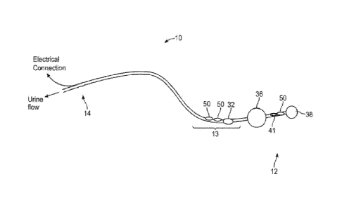
particular
physiological pressure measurements. For example, noise in this situation may
include patient
coughing, moving, or other types of noise not normally found in signal
filtering algorithms.
Some embodiments may, for example, measure heart rate and then use this rate
to determine
a physiological range for acceptable heart rate. If the heart rate is measured
beyond this range
(either above or below it), the controller may determine that the signal is
noisy and either
ignore it, or apply noise filtering technology to the signal. The same method
may be applied
to other, somewhat predictable, signals, such as respiratory rate, respiratory
pressure, TAP,
etc.
[0075] Other signal filtering techniques may be used to distinguish
between noise and
actual signal. For example, the respiratory frequency and the heart frequency
signals are
generally distinct from each other. However, under certain circumstances, the
frequencies
may overlap. In this situation other factors may need to be considered in the
pressure signal
analysis algorithm, for example signal amplitude.
[0076] Some embodiments of the disclosed system may be functionally
directed to the
delivery of therapeutic hypothermia. In this clinical application, the
catheter may be equipped
to measure bladder pressure, as above, measure urethral temperature, and be
able to drain
urine and add fluid to the bladder. In this embodiment, the catheter may be
used to warm or
cool the patient (mild to moderate hyperthermia or mild to moderate
hypothermia) via the
infusion of a warm or cold fluid as appropriate. In the generation of mild to
moderate
hypothermia, the bladder may be evacuated then refilled to a set pressure with
an ice-cold
medium (a cold fluid, or a chilled slurry or slush) while the core body
temperature is
monitored. In this embodiment, an initial fill of the bladder with cold medium
may be
sufficient to generate the desired degree of hypothermia, or the temperature
of the fluid may
be tracked (in some embodiments, by way of a second temperature sensor in the
bladder) and
evacuated once it rises above a set temperature (e.g., 15 C). If the desired
patient temperature
has not yet been reached, the bladder may then be refilled with the
liquid/slurry and
evacuated until the patient has achieved their target temperature.
18
Date Recue/Date Received 2021-02-01
[0077] In some embodiments, the therapeutic hypothermia process is
automated by the
system, requiring only that a clinician insert a sensing Foley catheter
embodiment, and then
connecting the catheter to the temperature control system and/or any patient
monitor that the
clinician desires. In some embodiments, the infused fluid is a slush to take
advantage of the
much greater watt extraction capabilities of slush in comparison to a cold
fluid. In some
embodiments, the sensing Foley catheter is able to sense one or more of the
other parameters
mentioned above (such as respiratory rate, or oximetry) during and following
this therapy.
The cold medium (slush and/or fluid) may be used to induce hypothermia, and
the bladder
may be evacuated once the target temperature is reached. As the body
temperature rises, the
slush and/or fluid may be introduced into the bladder then evacuated, again,
as the target
temperature is reached. In this embodiment, the resting state of the bladder
is the evacuated
state and it only contains chilled fluid or ice when the body is not within
target temperature
range. In some embodiments, the slush may be formed on-demand in a manner that
allows it
to be carried into the field or ambulance, and then created on-site, in order
to treat trauma or
injury as it occurs. This on-demand aspect of the method embodiment may
involve a pre-
frozen block of ice that is shaved or ground, or a compressed gas source that
vents into the
liquid, thereby causing a rapid drop in temperature. This compressed gas
embodiment may be
used either to generate a slush, or to cool the medium while allowing it to
remain a liquid.
[0078] A similar technique may be used with certain embodiments to
induce
hyperthermia with a warm or hot liquid.
[0079] Variations of the embodiments described above for use in the
bladder, may be
reconfigured and/or resized for application in other luminal body sites such
as the stomach,
esophagus, small intestine, large intestine or rectum. In some embodiments,
these data may
be obtained through invasive access of the peritoneal cavity, cerebrospinal
space or pleural
space, ideally in instances where accessing these spaces is already performed
for another
purpose.
[0080] Some embodiments of the device may incorporate mechanisms to
keep the urine
lumen, or other lumen, clear of blockages in order to maintain an empty,
flaccid bladder and
avoid false positive IAP measurements. These blockages may be caused by
airlocks in the
drainage tube or by crystals, blood clots, or other physical blockages. Any of
the
embodiments to keep the line clear as described in Burnett PCT/US2013/060003,
would be
suitable. In one embodiment, this is accomplished with active line clearing,
such as a bellows
to provide negative pressure or a pump to clear obstructions. This embodiment
allows for
clearing of both airlocks and physical blockages. In another embodiment, the
line clearing is
19
Date Recue/Date Received 2021-02-01
passive, and may be accomplished with vents that allow air to escape the
drainage line
instead of forming airlocks. In yet another embodiment, the TAP measurements
from the
present device may be combined with urine output measurements obtained with
the Burnett
device, in any manner they have disclosed.
[0081] Some embodiments of the disclosed technology may comprise methods of
pressure measurement in other anatomic locations and/or combined with existing
medical
devices. In one embodiment, the pressure-sensing system of the present
invention may be
used with ascites shunts in order to ensure that the shunt is draining and has
not become
obstructed. In another embodiment, the pressure-sensing system may be used
with dialysis
catheters. In another embodiment, the system may be used with insulin delivery
catheters.
Generally, the system may be used with any shunting, infusing, or other
similar applications
where fluid blockage may be of a concern and a pressure measurement would help
identify
whether a blockage has occurred.
[0082] Embodiments of the disclosed technology may integrate with, or
link to other
medical system, including an Electronic Health Record (EHR), Electronic
Medical Record
(EMR), clinical trial software, research software, medical monitoring systems,
EKG systems,
infusion systems, drug delivery systems, heart rate monitor systems, body
vital sign
monitoring systems, respiratory rate systems, etc. For example, pressure data
collected from
any of the embodiments discussed herein may be imported into, or integrated
with an EMR
so that a physician has a full picture of a patient. Any other data collected
and/or analyzed by
the disclosed embodiments can be used in a similar way. For example, a user
may analyze
clinical trial data which has been integrated with a controller incorporated
into one of the
disclosed embodiments. The user may view individual patient data to determine
if there is
any data to support abnormal heart rate, abdominal pressure, urine flow etc.
Integration with
an EHR may be done via a standard web browser using html and
frames/windows/window
areas, or XML or using any other appropriate standard or technology.
[0083] Data from disclosed embodiments, either alone, or in
conjunction with data from
integrated systems, may be stored, tracked and/or mined. The disclosed systems
may "learn"
from the stored data in such a way to provide recommendations on treatment or
diagnoses.
Systems may be networked so that data from more than one patient can be
aggregated and
used for this purpose. For example, embodiments of the disclosed technology
may analyze
data from multiple patients who have an elevated respiration rate, an elevated
heart rate,
and/or increased intraabdominal pressure. By analyzing data from these
patients in
conjunction with data from the EHR, embodiments of the disclosed technology
may be able
Date Recue/Date Received 2021-02-01
to determine that patients with this data profile, are more likely to have a
particular disease
and may therefor recommend a blood test, or may automatically perform a urine
analyte test.
[0084] In the same way, an upward trending temperature in conjunction
with one or
more other measured parameters may be an indication of infection. Additional
tests, or an
infusion, may be recommended or performed on the patient automatically or with
user
confirmation.
[0085] Data may also be tracked to determine the time until
obstruction and/or infection
for one patient, or across multiple patients.
[0086] Embodiments of the technology include a sterile to non-sterile
attachment
between the catheter device and the pressure transducer. Since the catheter
may be sterile and
disposable and the pressure transducer may not be sterile nor disposable, it
is important to be
able to connect the two components without increasing the risk of infection to
the patient.
Filter paper, such as 0.2 micron filter paper, or other suitable material, may
cover the portion
of the catheter where the pressure transducer connects to the catheter.
[0087] Embodiments of the technology may include a pressure sensor and
logic to
manage the balloon inflation of the retention balloon in addition to the
pressure balloon. In
some embodiments the retention balloon can serve as both a retention balloon
and a pressure
balloon, this may be particularly applicable when only TAP is being measured.
In other
embodiments, the retention balloon can sense pressure and the logic of the
controller can
detect when the pressure of the retention balloon falls outside expected
ranges, and may alert
the user in some way, such as an alarm. For example, if the catheter is
tugged, or the patient
tries to remove it, the pressure in the retention balloon will increase. This
increase in pressure
could be programmed to sound an alarm. In another example, a technician may
attempt to
inflate the retention balloon before the catheter tip is fully placed within
the bladder. In this
case, if the retention balloon were inflated in the urethra, the pressure
would be higher than
normal and an alarm or other alert could result. Acceptable retention balloon
pressure ranges
may be determined by tracking retention balloon pressures across several
patients to
determine the normal range of pressures. Pressures outside of this range may
be programed to
send/sound an alert, or to automatically reduce the balloon pressure.
[0088] Pressure sensing can also be used in either the retention balloon or
pressure
balloon to detect bladder spasms. A sudden, or repeated, change in pressure
could be an
indication of bladder spasm. The controller may be programmed to send an
alert, or to change
the pressure of the balloon when an apparent bladder spasm is occurring.
21
Date Recue/Date Received 2021-02-01
[0089] Embodiments of the technology may include acoustic sensing to
determine the
size and/or volume of the bladder. This technology may be useful in
determining the air in the
bladder, or the Gastric Residual Volume (GRV). Bladder size may be measured by
creating
and sensing acoustic waves and determining the time between wave emission and
wave
sensing after the wave has bounced off of the bladder wall. This measurement
may be
performed at one or more than one location within the bladder.
[0090] Another method of measuring bladder volume includes measuring
the
temperature change within the bladder using an embodiment of the present
invention after
introduction of a cool or warm fluid. The time it takes to warm or cool the
fluid in the bladder
is related to the bladder volume.
[0091] Embodiments of the technology may include self cleaning
technologies. For
example, a Foley catheter system may be automatically flushed with saline. A
Foley catheter
may also be purged by using natural bladder pressure, or by various
pumping/pressure
mechanisms disclosed herein.
[0092] Embodiments of the technology may include the ability to detect
deficient
connections within the system. For example, mechanical sensors may detect
integrity of the
connections between any components of the system. Alternatively, connection
integrity may
be sensed through small pressure changes, or other pressure sensors.
[0093] Embodiments of the technology may include alternative
materials for the Foley
catheter system. For example, the catheter shaft, or part of the catheter
shaft, may include an
outer, inner or embedded braid or other more rigid material to prevent the
catheter from
kinking. For example, the pressure lumen may have a more rigid inner surface,
such as a
polymer, braid etc. The added rigidity may also increase the sensitivity of
pressure
measurements through the lumen.
[0094] Embodiments of the technology include an implantable sensor for
vital sign
monitoring, as particularly suitable for a patient in battlefield or transport
setting, prior to
being secured into a hospital setting.
[0095] Embodiments of the technology include a free-floating
transmitting bladder
embodiment. Embodiments of the technology include a free-floating transmitting
stomach
embodiment. Embodiments of the technology include an ingestible, self-
destructing capsule.
Embodiments of the technology include vagina, stomach, intestine, esophagus,
or a rectum
sensor.
[0096] Embodiments of the technology include a catheter for sensing
physiological data
from a urinary tract of a patient comprising a pressure sensor comprising a
pressure interface
22
Date Recue/Date Received 2021-02-01
disposed at a distal end of the catheter, a first pressure transducer at a
proximal end of the
catheter, and a first fluid column disposed between the pressure interface and
the first
pressure transducer, a second pressure transducer at the proximal end of the
catheter and a
second fluid column disposed between the pressure interface and the second
pressure
transducer, wherein, when the catheter is inserted into the urinary tract and
the distal end is
residing in the bladder, the first pressure transducer can transduce pressure
impinging on it
from the pressure interface into a first chronological pressure profile, and
the second pressure
transducer can transduce pressure impinging on it from the pressure interface
into a second
chronological pressure profile.
[0097] Embodiments include a catheter where the first fluid column and the
second fluid
column are separate fluid columns for the length of the catheter.
[0098] Embodiments include a catheter where the first fluid column
and the second fluid
column are separate fluid columns for part of the length of the catheter, and
the same fluid
column for part of the length of the catheter.
[0099] Embodiments include a catheter where the first fluid column and the
second fluid
column are the same fluid column for the length of the catheter.
[0100] Embodiments include a catheter where the pressure interface
comprises a
balloon.
[0101] Embodiments include a catheter where at least one fluid column
is in
communication with a physical filter.
BRIEF DESCRIPTION OF THE DRAWINGS
[0102] Fig. 1 shows a data console in communication with a urine-
collecting receptacle
docking station, per an embodiment of the sensing Foley catheter system.
[0103] Fig. 2 shows an embodiment of the sensing Foley catheter
system set up to
measure urine output from a human subject.
[0104] Fig. 3 shows an embodiment of the sensing Foley catheter
system set up as an
automated infusion therapy system for a human subject.
[0105] Fig. 4 shows a volume-sensing urine collecting receptacle that
may include an
RFID chip, the receptacle accommodated within a receptacle docking station,
per an
embodiment of the sensing Foley catheter system.
[0106] Fig. 5A shows a sensing Foley catheter with a pressure
interface in the form of an
inflatable balloon, per an embodiment of the sensing Foley catheter system.
23
Date Recue/Date Received 2021-02-01
[0107] Fig. 5B shows a sensing Foley catheter a pressure interface in
the form of a
membrane arranged across a luminal opening, per an embodiment of the sensing
Foley
catheter system.
[0108] Figs. 6A ¨ 6D show various views and details of a sensing
Foley catheter, per an
embodiment of the sensing Foley catheter system.
[0109] Fig. 6A schematically arranges the sensing Foley catheter into
a proximal section
that remains external to the body when in use, a portion that resides in the
urethra, and a
portion that resides in the bladder, when placed into a human subject.
[0110] Fig. 6B shows a detailed view of the proximal portion of the
catheter.
[0111] Fig. 6C shows a cross sectional view of the central length of the
catheter.
[0112] Fig. 6D shows a detailed view of the distal portion of the
catheter that resides in
the bladder.
[0113] Fig. 7A shows an example of respiratory rate sensing data from
a human subject,
as provided by an embodiment of the sensing Foley catheter system. During this
test period,
the subject performs a respiratory sequence as follows: (1) breath being held
at the end of an
expiration, (2) valsalva, (3) normal respiration, (4) valsalva, and (5) breath
being held at the
end of an expiration.
[0114] Fig. 7B shows a detailed portion of the respiratory profile of
Fig. 7A, a portion of
the period of normal respiration.
[0115] Fig. 8 shows an example of cardiac rate and relative cardiac output
sensing data
from a human subject, as provided by an embodiment of the sensing Foley
catheter system,
and an EKG trace as measured simultaneously and independently.
[0116] Fig. 9 shows data related to relative cardiac output sensing
in a human leg raising
exercise in which cardiac output increases, as demonstrated by an increased
amplitude of the
cardiac pulse.
[0117] Fig. 10 shows an example of peritoneal sensing data, with a
focus on respiratory
rate from a pig, as provided by an embodiment of the sensing Foley catheter
system.
[0118] Fig. 11 shows an example of pig study that demonstrates the
capability of an
embodiment of the sensing Foley catheter system to detect intra-abdominal
hypertension.
[0119] Fig. 12 shows intraabdominal pressure, respiratory wave pressure,
and cardiac
pressure schematically arrayed as a two dimensional plot of pressure (mm Hg on
a
logarithmic scale vs. frequency (Hz).
[0120] Fig. 13 provides a flow diagram of an embodiment of the
method.
[0121] Fig. 14 shows pressure signals at different lumen diameters.
24
Date Recue/Date Received 2021-02-01
[0122] Fig. 15 shows an embodiment for clearing the drainage line
that uses a vacuum
applied to the end of the drainage line.
[0123] Figs. 16A-16B show an embodiment of a clearing mechanism
comprising a
device for positive airflow near the start of the drainage line.
[0124] Fig. 17 shows a clearing mechanism comprising an apparatus for
automated
massaging, or squeezing, of the drainage line.
[0125] Fig. 18 shows another embodiment of the pinching or rolling
stimulus, in which
the lumens are compressed sequentially by rollers.
[0126] Fig. 19 shows another embodiment comprising multiple lumens
organized
circumferentially around a stiff member that the pinching or rolling mechanism
rotates
around.
[0127] Fig. 20 shows an alternative embodiment in which the lumens
are organized such
that they can only be completely compressed when pinched in a certain
direction.
[0128] Fig. 21 shows a graph of the pressure profile, pressure (mmHg)
over time
(seconds) in the drain tube while the peristaltic roller pump is activated.
[0129] Fig. 22 is a table comparing TAP measurements using a standard
drainage line
and TAP sensor with the present invention in combination with a pressure-
sensing Foley
catheter under air lock and siphon effects.
[0130] Figs. 23A and 23B show another embodiment of the disclosed
technology which
allows for a smaller profile catheter, particularly in the area of the
pressure balloon.
[0131] Fig. 24 shows an embodiment of a preperitoneal sensing
implant.
[0132] Figs. 25A and 25B show graphs representing pressure balloon
priming methods
in some embodiments.
[0133] Fig. 26A-C show flow charts of possible logic in various
embodiments of the
invention.
[0134] Figs. 27A and 27B show an embodiment of the invention which
includes a fiber-
optic pressure sensor.
[0135] Fig. 28 shows an embodiment of the invention with more than
one pressure
lumen.
[0136] Fig. 29 shows another embodiment of the invention.
[0137] Fig. 30 shows another embodiment of the invention.
[0138] Fig. 31 shows an embodiment of the invention without a
retention balloon.
[0139] Fig. 32 is a block diagram of a data processing system, which
may be used with
any embodiments of the invention.
Date Recue/Date Received 2021-02-01
DETAILED DESCRIPTION OF THE DISCLOSURE
[0140] Figs. 1 ¨4 show various elements of the disclosed technology,
including a urine
receptacle 60 (holding a urine output 61), a docking station 65 to hold the
receptacle, an
electrical connection 67 that allows communication between the docking station
and a data
collection and processing apparatus in the form a bedside console 80.
Embodiments of the
urine collecting receptacle 60 may include level or volume sensors 62, as well
as other
analyte sensors. Receptacle 60 may also include an RFID element that provides
a unique
identifier to a remote RFID reader 68. In some embodiments, an extender tube
63 may be
utilized to convey urine from the catheter to the urine-collecting receptacle.
[0141] Fig. 1 shows a data receiving and processing apparatus in the
form of a bedside
console 80 in communication with a receptacle docking station 65 that
accommodates a urine
collecting receptacle 60, shown as holding a urine output 61, per an
embodiment of the
sensing Foley catheter system. The communication path between the docking
station and the
console may include a wired connection 67, as shown, or it may be a wireless
connection.
The bedside console may record and display output/input data. Physiologic data
from sensors
associated with a sensing Foley catheter may be held in a memory, displayed,
printed, or
directly transmitted to a centralized data collection server.
[0142] In some embodiments, the bedside console or controller is
portable and able to
travel with the patient. Embodiments of console may be attachable to a
patient's bed or an IV
pole, or a wall mount; it typically has its own display, and is able to
provide critical alerts.
Some embodiments of console may be adapted to be able to operate on a battery
backup for 4
or more hours, as for example when wall power is unavailable or has been lost.
This
portability feature of console is advantageous in situations where patients
are typically not
being electronically monitored, such as when a patient is in transit from his
or her bed to
another location. Embodiments of console may also be configured to communicate
to a base
station with alerts and centralized reporting and data collection. A
controller or base station
may also generate mobile alerts that may be sent to nurses or healthcare
provider. Signal
analysis and/or predictive algorithms may also be used to provide useful
clinical data from
sensors.
[0143] Fig. 2 shows elements of an embodiment of the sensing Foley
catheter system
configured to measure urine output from a human subject. In some embodiments,
the bedside
console 80 or an RFID reader (see Fig. 5) is enabled to trigger an alert if
urine output is above
26
Date Recue/Date Received 2021-02-01
or below a preset normal or desired range for urine output over a set period
of time. Some
embodiments of the system may also have an intravenous infusion capability
(see Fig. 3) to
provide use sensed data to regulate delivery of fluids or medicinal agents
such as a diuretic
drug, by way of an automated system based on the urine output feedback.
Embodiments of
the system may include a docking station for the urine collecting receptacle,
the docking
station being configured for data transmission to a data receiving and
processing apparatus
such as a bedside console or a networked central computer. In some
embodiments, the
docking station delivers data regarding the volume of urine in the urine
receptacle, as well as
data that are informative regarding electrical parameters of the urine, such
as conductivity,
resistance, or impedance. Sensors may also detect and monitor bacteria,
hemoglobin, or other
substances of clinical significance in urine. Sensors may also measure urine
opacity in the
collecting receptacle, in the bladder or in the catheter/tubing.
[0144] Fig. 3 shows an embodiment of the sensing Foley catheter
system configured as
an automated infusion therapy system for a human subject. A bedside console 80
may
integrate patient data, such as fluids received or urine output recorded, and
then automate
therapeutic infusion in response to these data. For example, delivery of
fluids or drug
solutions such as a physiological saline solution may be initiated or
regulated through an
infusion line 82 if the patient is dehydrated, or a diuretic may be infused if
the patient is fluid
overloaded. In some embodiments, the console may trigger a local alert (e.g.,
audible
beeping), or trigger a centralized alert (e.g., a system alarm) if urine
output drops too low.
The console may also integrate a hydrating or medicinal fluid infusion
capability, such as an
IV infusion pump, and may adjust infusion rates based on these data or based
on data
acquired from other sensors automatically. The console may communicate
wirelessly, as
well, to these and other sensors within the body.
[0145] Fig. 4 shows a volume-sensing urine receptacle 60 accommodated
within a
receptacle docking station 65, per an embodiment of the sensing Foley catheter
system.
Embodiments of the receptacle may detect urine output based on the levels at
which sensors
62 are triggered. For example, the receptacle may electrical contacts arranged
as liquid
height-marks, and when an electrical path is made between two contacts and all
contacts
below, the level can be reported at that level. Embodiments of the receptacle
may include
electrical, optical, chemical or mechanical sensors. Embodiments of the
receptacle may
include also contain diffuse or discrete sensing areas that detect analytes of
interest, e.g.,
hemoglobin, protein, glucose, bacteria, blood, leukocyte esterase. Sensing or
data reporting of
sensed data may be of either an intermittent or a continuous nature.
27
Date Recue/Date Received 2021-02-01
[0146] Embodiments of the receptacle may include a capability to
report sensing data to
the bedside console, locally (e.g., by beeping) or centrally via piping data
to a central
information collection area. For example, an alert may be triggered if urine
output drops
below 30 cc/hr. in post-operative setting or below any otherwise predetermined
threshold.
Embodiments of the receptacle may connect to a docking station through
electrical contacts;
data communication among embodiments of the receptacle, docking station, and a
console or
central computer may also be wireless. If a docking station is used, it may
detect urine output
based on weight or pressure of the receptacle that is applied to base.
[0147] Embodiments of the urine collecting receptacle may include
disposable or
durable optical, electrical or chemical sensors capable of sensing and
measuring urine content
of analytes such as glucose, electrolytes, bacteria, hemoglobin, or blood.
Embodiments of the
receptacle may include an interface with specifically designed area of the
urine receptacle to
allow for this measurement, such as an optically clear window for optical
measurement of
blood. Embodiments of the receptacle docking station may also grasp or
accommodate the
urine receptacle in any manner so long as it secures the receptacle. The
docking station or the
receptacle may include an inductive antenna or RFID capabilities to allow for
wireless
querying and reporting of the level of urine or other fluid collection.
[0148] The embodiment of Fig. 4 also shows a volume-sensing urine
receptacle 60 that
includes an RFID chip, per an embodiment of the sensing Foley catheter system.
This
embodiment may contain RFID circuitry to collect and transmit data directly
from within the
receptacle to a remote RFID reader 68. When queried by the RFID reader, the
receptacle may
detect impedance, resistance, capacitance or any other electrical or non-
electrical property to
measure the urine level and report this back to the reader. The reader may
then trigger alert if
urine output is out of a normal or desirable range. The RFID chip may be
capable of detecting
changes in optical, chemical, electrical, acoustic or mechanical properties,
as well. RFID
chips may be active or passive, and may contain an antenna to transmit a
receptacle-
identifying signal to the reader, and allow multiple receptacles to be queried
simultaneously.
An active RFID chip may incorporate a small battery (to extend its range). A
passive RFID
chip may be powered by the transmission from the RFID reader. The RFID reader
may query
a device from a distance to wirelessly check the urine output level or it may
be centralized to
query all receptacles within a unit, floor or hospital and issue an alert if
urine output is out of
a normal or desirable range. The RFID reader record urine output, as well, and
functionally
replace the individual unit console shown in Figs. 1 ¨ 3. The RFID reader may
also report
28
Date Recue/Date Received 2021-02-01
data from other sensors within the system, including bladder temperature or
presence of
analytes (as detailed elsewhere) in the urine.
[0149] Figs. 5A ¨ 6D show embodiments of a sensing Foley catheter 10
and various of
its features. A catheter may be understood to have various sections according
to their
disposition when the catheter is inserted into a human subject, such as a
proximal portion 14
that remains external to the subject, a central or urethra-residing portion
13, and a distal or
urinary bladder-residing portion 12.
[0150] Various internal lumens traverse the length of the catheter,
such as an air or fluid
24 that communicates with a bladder retention balloon 36. A urine drainage
lumen 23 has a
distal opening 41 that resides in the bladder portion 12 of the catheter, and
has an opening at
the proximal end 14 of the catheter. As seen in Figs. 2 and 3, the urine
drainage lumen may
be connected to an extender tube 63 that conveys the urine to a collecting
receptacle. In some
embodiments, the drainage lumen and distal opening in the bladder may also
serve as in
infusion conduit (see Fig. 3) by which medicinal agents may be infused, or
through which
heating or cooling fluid may be infused. Analyte sensors or temperature
sensors 50 may be
disposed on the catheter, either on the urethral portion 10 or the bladder-
residing portion 12
of the catheter. Electrical or optical fiber leads may be disposed in a lumen
25 that allows
communication of sensing signals between distally disposed sensors and the
proximal portion
of the catheter, and then further communication to a data processing
apparatus.
[0151] An inflatable pressure-sensing balloon 38 (Figs. 6A, 7A, and 7B) or
a pressure
sensing membrane 39 (Fig. 7B) arranged across an opening may be positioned on
the distal
end 12 of the catheter, residing in the bladder. Embodiments of a pressure-
sensing balloon or
pressure sensing membrane may be understood as comprising a pressure interface
having a
distal-facing surface exposed to pressure from within the bladder, and a
proximal-facing
surface exposed to a proximal fluid column. Embodiments of the fluid column
(filled with
either liquid or gas) may comprise a dedicated lumen, or such column may share
a lumen that
also serves as a sensing conduit such as lumen 25.
[0152] Fig. 5A shows a sensing Foley catheter that includes a
pressure interface in the
form of pressure-sensing balloon, per an embodiment of the presently disclosed
system.
Pressure-based physiologic parameters that this catheter embodiment can sense
may include,
by way of example, peritoneal pressure, respiratory rate, and cardiac rate,
relative pulmonary
tidal volume profile, cardiac output, relative cardiac output, and absolute
cardiac stroke
volume. Some embodiments of the Foley type catheter may be further equipped
with any of a
temperature sensor, one or more analyte sensors, electrodes, and paired light
sources and
29
Date Recue/Date Received 2021-02-01
sensors. Embodiments thus further equipped are capable of delivering other
forms of
physiologic data, as for example, blood pressure, oxygen saturation, pulse
oximetry, EKG,
and capillary fill pressure.
101531 Fig. 5B shows a sensing Foley catheter with a lumen (the third
lumen, for
example) used as a pressure sensing lumen; this embodiment does not include a
dedicated
pressure-sensing balloon as does the embodiment of Fig. 5A, but instead has a
pressure
interface in the form of a membrane arranged over a distal opening of the
pressure sensing
lumen. In this embodiment, the sensing Foley catheter is able to detect and
report pressure-
based physiologic data as included in the embodiment described above. In this
present
embodiment, a slow infusion of fluid into the bladder may be accomplished
through the third
lumen of a standard 3-way Foley catheter, and pressure may be sensed using a
pressure
sensor in line with this third lumen. In this embodiment, all methods
associated with
processing and responding to pressure-based physiologic data, as described for
embodiments
with a pressure-sensing balloon, are enabled.
101541 Figs. 6A ¨ 6D show various views and details of a sensing Foley
catheter, per an
embodiment of the sensing Foley catheter system. Fig. 6A schematically
arranges the sensing
Foley catheter into a proximal section 14 that remains external to the body
when in use, a
portion 13 that resides in the urethra, and a distal portion 12 that resides
in the bladder, when
placed into a human subject. Fig. 6B shows a detailed view of the proximal
portion of the
catheter, focusing on luminal openings 23, 24, and 25, which are configured to
make more
proximal connections. Fig. 6C shows a cross sectional view of the central
length of the
catheter, and an example of how lumens 23, 24, and 25 may be arranged. Fig. 6D
shows a
detailed view of the distal portion of the catheter that resides in the
bladder, with a particular
focus on a retention balloon 36 and a pressure-sensing balloon 38.
101551 Pulse oximetry elements allow for a determination of blood oxygen
concentration
or saturation, and may be disposed anywhere along the urethral length of the
catheter. In
some embodiments, the sensor or sensors are disposed within the tubing of the
device to
ensure approximation to the urethral mucosa. With this technology, a
healthcare provider can
decompress the bladder with a urinary catheter and obtain pulse oximetry data
in a repeatable
and accurate manner. The power source for pulse oximetry may be incorporated
within the
urinary collecting receptacle or within the catheter itself. In some
embodiments, the pulse
oximeter is reusable and the catheter interface is disposable; in this
arrangement the pulse
oximeter is reversibly attached to the disposable catheter and removed when
oxygen
measurements are no longer desired. Embodiments of the sensing Foley catheter
may include
Date Recue/Date Received 2021-02-01
an optically transparent, or sufficiently transparent, channel for the
oximetry signal, such as a
fiber-optic cable, transparent window, and an interface for the reusable
oximeter. This
method and device for urethral pulse oximetry may be used in conjunction with
any of the
other embodiments detailed herein or may be a stand-alone device.
[0156] Embodiments of the sensing Foley catheter may be able to sense any
one or more
of a plurality of clinically relevant parameters, such as included in the
following examples:
urine pH, urine oxygen content, urine nitrate content, respiratory rate, heart
rate, perfusion
pressure of the bladder wall or the urethral wall, temperature inside the
bladder or the urethra,
electro¨cardiography via sensors on the bladder wall or the urethra,
respiratory volume,
respiratory pressure, peritoneal pressure, urine glucose, blood glucose via
urethral mucosa
and/or bladder mucosa, urine proteins, urine hemoglobin, blood pressure. In
some
embodiments, the catheter can sense multiple parameters, but some embodiments
may be
limited to as few as a single parameter for focused applications (for example,
respiratory rate
in a patient in respiratory distress). The respiratory rate, relative tidal
volume, peritoneal
pressure, heart rate and/or relative cardiac output may be measured
simultaneously, as well,
by connecting a balloon with a flaccid wall or semi-tense wall to an external
pressure sensor
via a lumen that may be filled with liquid and/or gas.
[0157] These parameters may be measured, alone or in concert with
other parameters,
through the use of pressure measurement modalities other than the external
pressure sensor.
These may include: a deflecting membrane inside of the catheter, MEMs
technology, a
catheter-based sensor and/or other embodiments.
[0158] Relative cardiac output and relative tidal volume may also be
calculated, based
on the deflection of the pressure sensor and/or other force gauge. If sampled
with sufficient
frequency (e.g., 1 Hz or greater), respiratory excursions can be quantified in
a relative manner
to the amplitude of the excursions at the time of catheter placement. Larger
excursions
generally relate to heavier breathing, or in the setting of an upward drift in
the baseline, a
higher peritoneal pressure. The small peaks on the oscillating respiratory
wave, caused by the
pumping heart, may be tracked as well by using faster sampling rates (e.g., 5
Hz or greater),
and the amplitude of this wave may be used, in the setting of a relatively
constant peritoneal
pressure, to determine the relative cardiac output, in the setting of a known,
stable peritoneal
pressure, absolute stroke volume and/or cardiac output.
[0159] The disclosed technology captures a high-resolution
chronological profile
(pressure as a function of time) of peritoneal pressure that can be transduced
and processed
into distinct pressure profiles assignable to particular physiologic sources,
including
31
Date Recue/Date Received 2021-02-01
peritoneal pressure, respiratory rate, and cardiac rate. By tracking the
pressure profile at a
sufficiently rapid sampling rate, as provided by the technology, the pressure
profile can be
further resolved into relative pulmonary tidal volume, cardiac output,
relative cardiac output,
and absolute cardiac stroke volume.
[0160] Accordingly, aspects of the disclosed technology relate to fidelity
and resolution
of a pressure signal generated in response to changes in pressure within the
bladder, such
changes being reflective of a pressure profile within the peritoneal cavity,
such pressure
profile including cumulative input from the aforementioned physiologic
sources. Aspects of
the technology further relate to fidelity and resolution of the transduction
of the pressure
signal into a highly resolvable electrical signal. Aspects of the technology
relate still further
to processing the totality of the electrical signal profile, a surrogate for
the pressure profile
within the peritoneal cavity, into component profiles that can be assigned to
the physiologic
sources.
[0161] The sensitivity of an inflated balloon as a pressure sensor is
a function, in part, of
the pressure differential across the balloon membrane as a baseline condition.
The balloon
has the greatest sensitivity to pressure when the baseline pressure
differential is near zero. As
the baseline pressure differential increases, the sensitivity of the pressure-
sensing balloon
degrades. Accordingly, the disclosed technology provides an automatic priming
method that
maintains the balloon in an inflated state, but with a minimal pressure
differential.
[0162] Embodiments of the technology include a pressure interface as may be
represented by a balloon having either a compliant membrane or a non-compliant
membrane.
In general, considerations related to optimizing the pressure around the
pressure interface of
the device are informed by Boyle's ideal gas law, the relationship between
stress and strain as
described by Hooke, and by application of Young's modulus. The conditions for
optimal
sensitivity of a compliant balloon and a non-compliant balloon are slightly
different,
although, in general, the sensitivity of each is best served by PI and P2
being approximately
equal. A non-compliant balloon maximum sensitivity is achieved when PI is only
slightly
above P2. For a compliant balloon, the maximum sensitivity is achieved when P1
is slightly
above P2 at the low end of the (linear) elastic region of the spring constant
of the compliant
balloon material.
[0163] To effectively capture physiologic pressure profiles, the
profiles need to be
sampled at a rate that is sufficient to resolve the inherent frequency of
changes in the profile.
This consideration is informed by the Nyquist-Shannon sampling theorem, which
states that a
sampling frequency of at least 2B samples/second is required to resolve an
event that runs at
32
Date Recue/Date Received 2021-02-01
a frequency of B cycles/second. As applied to a physiologic pressure cycle,
for example, a
cardiac rate of 70 beats/minute requires a sampling rate of at least 140
samples/minute to
effectively capture the cycle. This relationship underlies aspects of the
disclosed technology
that specify the sampling rate particularly required to capture physiologic
pressure cycles
such as relative pulmonary tidal volume, cardiac output, relative cardiac
output, and absolute
cardiac stroke volume.
[0164] Fig. 12 shows intraabdominal pressure, respiratory wave
pressure, and cardiac
pressure schematically arrayed as a two dimensional plot of pressure (mm Hg on
a
logarithmic scale vs. frequency (Hz). It can be seen that there is an inverse
relationship
between pressure and frequency, and the various physiologic pressure-related
parameters
occupy distinct sectors when arrayed in this manner. It is by the distinctness
of these profiles
that embodiments of the method (see Fig. 14), as disclosed herein, can resolve
a single
overall chronological pressure profile into the distinct subprofiles, in
accordance with their
physiologic origin.
101651 Expandable pressure sensing balloons, per embodiments of the
technology, may
assume one of at least two basic forms, type 1 or type 2. In balloon
embodiments of type 1,
which may be generally likened to a conventional party balloon, the pressure-
sensing balloon
is formed from or includes a compliant or elastic membrane. Accordingly, the
surface area of
the membrane expands or contracts as a function of the expansion of the
balloon. The
elasticity of the membrane determines various features of the balloon, as a
whole, at different
levels of expansion. Upon expansion, the balloon, if unconstrained, maintains
a substantially
constant or preferred form or shape, as determined by the mandrel upon which
the balloon is
formed. Upon expansion of the balloon from a minimal volume to its maximal
volume, the
membrane of the balloon maintains a level of tautness. Within the limits of
elasticity of the
compliant membrane, an increase in pressure during inflation results in a
consequent
expansion of volume. The balloon, on the whole may be considered partially
compliant in
that its shape responds to spatial constraints that it may encounter upon
expansion or
inflation, however the balloon does have a preferred or native shape, and such
shape
preference prevents a level of shape compliance or conformability such as that
shown by a
balloon of type 2.
[0166] In balloon embodiments of type 2, the expandable pressure-
sensing balloon is
formed from or includes a non-compliant, or non-elastic membrane, or a
membrane that is
substantially non-compliant or non-elastic. Accordingly, the surface area of
the membrane
does not expand or contract in accordance with the level of balloon expansion.
Type 2
33
Date Recue/Date Received 2021-02-01
pressure-sensing balloons may be generally likened to a conventional Mylar
balloon. The
inelasticity of the membrane determines various features of the balloon, as a
whole, at
different levels of expansion. Upon expansion of the balloon from a minimal
volume to a
level near its maximal volume, the membrane of the balloon is supple, and has
a level of
slackness. Expansion of a type 2 balloon occurs by way of outwardly directed
smoothing of
wrinkles and folds in the membrane. Deflation or compression of a type 2
balloon occurs by
way of generally inwardly directed wrinkling and infolding. When a type 2
balloon is fully
inflated (or substantially inflated) without being in a confining space, it
assumes a preferred
or native shape as determined by the geometry of the membrane or fabric of the
balloon.
However, in a state of partial inflation, the balloon, as a whole, is highly
supple and
conformable, broadly taking the shape as may be dictated by a confining space.
[0167] Expandable pressure sensing balloons, per embodiments of the
technology, may
also include features of both of the two basic forms, type 1 or type 2. In
these embodiments,
the membrane may include regions that are elastic (like type 1) and regions
that are inelastic
(like type 2). A balloon of this hybrid type would, as a whole, behave in a
manner drawing
from behavioral aspects of both type 1 and type 2 balloons, as described
above. Further, type
1 balloons may be formed with a membrane that is not of a homogeneous
composition or
thickness. In such embodiments, regions of different thickness or composition
could have
varying degrees of elasticity, thus affecting the behavior of these regions
during expansion of
the balloon. In still other embodiments, elasticity of the membrane may have a
bias or
polarity that tends to permit elasticity in one or more directions, and tends
to disallow
elasticity in one or more other directions.
[0168] An aspect of the disclosed technology that is particularly
advantageous in
achieving a high resolution signal from which pressure profiles from
particular physiologic
sources (such as peritoneal pressure, respiratory rate, and cardiac rate,
relative pulmonary
tidal volume, cardiac output, relative cardiac output, and absolute cardiac
stroke volume) may
be monitored relates to adjusting and maintaining a balance of pressure on
either side of the
pressure interface represented by the membrane of the pressure sensing
balloon. This balance
of pressure may be referred to as a pressure differential of zero, or as a
zero pressure gauge.
Pressure impinging on the external face of balloon (facing the internal aspect
of the bladder)
is subject to change according to the physiology of the patient. Pressure on
the internal face
of the balloon (which is in fluid communication with the fluid column) is
subject to
degradation because of fluid leakage and imperfect seals.
34
Date Recue/Date Received 2021-02-01
[0169] Upon first insertion of the Foley type catheter, external
pressure is typically
applied to the fluid column and against the pressure interface to a first
approximation of
pressure being exerted on the pressure interface from within the bladder.
Pressure signals, as
measured across a pressure interface, have a maximal amplitude when the
pressure
differential is zero. Accordingly, the amplitude of a pressure signal can be
used to tune the
pressure being applied from the fluid column against the pressure interface.
This process of
applying an appropriate amount of pressure against the interface may be
referred to as
priming the fluid column or priming the balloon. Inasmuch as pressures on
either side of the
pressure interface may change, as described above, the fluid column may need
to be reprimed
or re-tuned, from time to time. The necessity of repriming can be monitored by
testing small
changes in pressure so as to achieve maximal amplitude of a pressure signal
profile.
[0170] Embodiments of the disclosed system and method include
automatic pressure
tuning by a controller. Accordingly, the tuning system can detect the optimum
target pressure
and volume to inflate the balloon by monitoring sensed pressure signals and
adding or
removing air or fluid volume as needed. For example, upon insertion of the
catheter, a
pressure tuning circuit that regulates the balloon volume and pressure may
inflate the balloon
until it detects a physiologic-sourced pressure rate. Upon sensing that rate,
the pressure tuning
controller may add or subtract minute amounts of air in a routinized sequence
until the
amplitude of the sensed wave is greatest. The control feedback loop between
the optimally
tuned pressure (manifesting as balloon pressure and volume) and the sensed
physiologic
pressure profile iterates continuously and or as needed to ensure high
fidelity measurement of
the physiologic data. In some embodiments, automatic pressure tuning may be
performed in
the apparent background while the physiologic data is being transmitted and
displayed; in
other embodiments the system may suspend transmission of physiologic data
during a
pressure tuning sequence.
[0171] Embodiments of the disclosed technology include a gas delivery
system that can
deliver gas in a priming operation, whereby pressure can be applied to a fluid
column
proximal to the proximal-facing aspect of the pressure interface. A source of
gas, such as
compressed air or liquid is held in a storage tank. Using CO2 as an example,
CO2 is
controllably released from the storage tank through a pressure regulator that
can step pressure
in the tank (for example, pressure of about 850 psi) down to the range of
about 1 psi to about
2 psi. Released gas passes through a filter and a pressure relief valve set at
about 2.5 psi. The
pressure relief valve is a safety feature that prevents flow through of gas at
a level greater
than 2.5 psi in the event of failure of the upstream regulator. CO2 exiting
the pressure relief
Date Recue/Date Received 2021-02-01
valve next passes through a first solenoid-controlled fill valve to enter the
catheter line,
ultimately filling the balloon that comprises the pressure-sensing interface.
Pressure within
the balloon is allowed to rise to a level as high as 30mm Hg, whereupon the
first solenoid-
controlled valve closes. A second solenoid-controlled valve, distal to the
first valve operates
as a drain valve, which can release pressure from the catheter to a target
pressure.
Alternatively, the drain valve may be activated until a respiratory waveform
is detected after
which the balloon will be optimally primed and the valve will be closed. The
drain valve may
be subject to proportional control, operably based on voltage or pulse-width
modulation
(PWM), which allows a drain rate sufficiently slow that the target pressure is
reached and the
valve can be closed prior to overshoot. Alternatively, a peristaltic or other
air pump may be
utilized to fill the balloon with room air.
[0172] Intrabdominal pressure or bladder pressure, as sensed by an
embodiment of the
disclosed technology, may also be used to detect the level of patient movement
(as may vary,
for example, between substantially no movement to a high level of movement)
and to report
the movement level to a healthcare provider. A short burst of peaks and
valleys in bladder
pressure activity can serve as a proxy for body movement in that such a
bladder pressure
profile is a strong indicator that the patient is using their abdominal
muscles, as, for example,
to sit up or get out of bed. This embodiment may be of particular benefit for
patients that are
at risk of falling. In a patient that is a fall-risk, a healthcare provider
may be notified that the
patient is sitting up and respond accordingly. Alternatively, the device may
be used to report
inactivity of a patient and/or lack of patient movement.
[0173] Embodiments of the technology may also report patient movement
in the
detection or diagnosis of seizure disorder. In this embodiment, the pressure
variations may
trigger an EEG or recording equipment to allow for intense period of
monitoring during an
episode suspected of being a seizure. In addition, or alternatively, a
pressure sensor, acoustic
sensor or other sensors may be used to detect bowel activity, including
peristalsis, patient
movement, seizure activity, patient shivering, frequency of coughing, severity
of coughing,
sleep duration, sleep quality, speech detection, patient compliance (movement
or lack
thereof), and may alert the healthcare provider that the patient has not moved
and must be
turned or rolled. This movement-related information may also be relayed to a
hypothermia
device, a drug delivery device or other device to control or mitigate seizure
activity, shivering
and/or coughing.
[0174] Embodiments of the technology may also automatically adjust
intravenous fluid
or drug infusion rates based on feedback from the cardiac output or
respiratory rate sensed. In
36
Date Recue/Date Received 2021-02-01
one such embodiment, a patient-controlled analgesia pump may be deactivated if
a
respiratory rate drops too low. Respiratory depression can be fatal in this
group and this
safeguard would prevent overdose. An automated feedback system may also be
advantageous
in a large volume resuscitation procedure, wherein fluid infusion can be
tailored based on
intraabdominal pressure to prevent abdominal compartment syndrome by sounding
an alert
and slowing infusion rates as the intraabdominal pressure rises. Yet another
automated
feedback feature may provide direct feedback to a ventilator system to provide
the optimal
pressure of ventilated gas. In the setting of increased abdominal pressure,
typical ventilator
settings do not provide sufficient respiration for the patient. An automated
adjustment of the
ventilator settings based on intraabdominal pressure feedback from this
embodiment may
advantageously provide for optimal patient ventilation. Embodiments of the
technology may
also be applied as a correction in the application or understanding of other
diagnostic
measurements. For example, central venous pressure may be dramatically
distorted in the
setting of elevated intraabdominal pressure. Providing direct access to these
data by the
central venous pressure reporting system allows for the automatic correction
and accurate
reporting of this critical physiologic parameter. Embodiments of the
technology may also be
used in a variety of other ways to automate therapy including infusion of
fluids that may
further include active agents, such as pressors or diuretics in response to
increased or
decreased cardiac output.
[0175] In some embodiments, the Foley type catheter is configured to report
the
presence of a water droplet or other obstruction in an air-filled lumen, and
then handle or
resolve the droplet. In a hypothermic setting, in particular, moisture in an
air lumen can
condense and form obstructive water droplets. Water droplets in an air-filled
lumen (or air
bubbles in a water-filled lumen) can disturb or complicate pressure signals
due to the surface
tension of the water. Accordingly, a pressure-transmission lumen in some
embodiments of
the disclosed technology may include a hydrophilic feature (such as a coating
on the wall of
the lumen itself, or a hydrophilic fiber running the length of the lumen) to
wick moisture
away from the lumen in order to maintain a continuous, uninterrupted air
channel. In some
embodiments, a hygroscopic composition (silica gel, for example) may be used
in line with
the air infusion line or within the air infusion lumen itself to capture water
or humidity. In
some embodiments, a hygroscopic composition may be included within the
catheter so that
the air infusion circuit need not be serviced to replace this material.
[0176] In some embodiments of the disclosed technology, air may also
be intermittently
(and automatically) infused and extracted into the pressure-sensing balloon so
that the
37
Date Recue/Date Received 2021-02-01
balloon is in a constant state of being optimally primed, as described in
further detail above.
In the case of the wicking fiber or hydrophilic coating in the lumen, the air
extraction may
also contribute to removing and trapping any water from the air line. In the
instance of a
liquid-filled lumen, a hydrophilic fiber or a hydrophilic coating on the
inside of the pressure
lumen will provide similar benefit in allowing this lumen to handle an air
bubble. In this
instance, an air bubble may distort the signal, but the air water interface
surface tension is
defused by a hydrophilic coating in the lumen of the catheter.
[0177] Additionally, a custom extrusion and lumen shape may also be
used to prevent
obstruction in the case of liquid and/or air-filled lumens. In some
embodiments of the
technology, for example, a Foley type catheter may have a lumen that is
stellate in cross
sectional profile. Such a lumen is generally immune from obstruction by a
water droplet, as
the droplet tends to cohere to itself and push away from the hydrophobic
walls. This behavior
tends to disallow filling of a cross-sectional space, and allows for an air
channel to remain
patent around the water droplet and communicate to the sensor. The same logic
applies to an
air bubble in water in a hydrophilic, stellate water lumen. In this instance
the hydrophilic
liquid will cling to the walls and allow for a continuous water column that
excludes the air
bubble to the center of the lumen. The same applies for a hydrophobic liquid
in a
hydrophobic lumen. In some embodiments, the catheter may include an air
channel, and a
sensor incorporated within the catheter itself or a fluid lumen that is
capable of transmitting
the pressure back to a sensor.
[0178] In some embodiments, the sensing Foley catheter may include a
blood pressure
sensing element that may take any of several forms. In one embodiment, a blood
pressure
sensing element includes a pressure delivery balloon 32 (either a separate,
dedicated balloon
or a balloon in fluid communication with a device retention balloon or a
pressure sensing
balloon) that can be optically analyzed as it is inflated to determine at
which pressure the
vessels within the bladder or urethra are blanched and blood flow is stopped.
This approach
provides a reading of the perfusion pressure of the tissue abutting the
pressure delivery
balloon, such reading reflective of both the systemic blood pressure and
vascular resistance.
This embodiment of a perfusion pressure device may be used to provide early
detection or
monitoring of a variety of acute or emergent medical conditions such as
sepsis, shock,
hemorrhage, and can be particularly advantageous in detecting these conditions
at an early
stage. In predicting sepsis, embodiments of the invention may be capable of
receiving white
blood cell count information to better predict sepsis.
38
Date Recue/Date Received 2021-02-01
[0179] Other modalities may be used to detect that the tissue has
been blanched or
ischemic, as well, with the common methodological aspect being that of the
intermittent
inflation within the lumen, body cavity or bodily tissues to provide the
compression of the
vasculature. Embodiments of this device and associated methods may also be
used to detect
perfusion pressure in other areas of the body with an intermittently
inflatable member and
optical detection of blood flow or the presence of blood.
[0180] Tissue perfusion information may also be provided by way of
sensors disposed
on the shaft of the catheter such that they contact the urethral wall when the
catheter is in
place. These sensing technologies may include microdialysis, pyruvate,
lactate, p02, pCO2,
pH, perfusion index, near-infrared spectroscopy, laser Doppler flowmetry,
urethral
capnography, and orthogonal polarization spectroscopy. Any of these tests may
also be
performed on the urine or the bladder wall itself to generate measurements of
tissue
perfusion.
[0181] Embodiments of a sensing Foley catheter have been used to
collect data from a
human subject (Figs. 7 ¨ 9) and from a pig (Figs. 10¨ 11). The human subject
was a
consenting and well-informed volunteer.
[0182] Fig. 7A shows an example of respiratory rate sensing data from
a human subject,
as provided by an embodiment of the sensing Foley catheter system. During this
test period,
the subject performs a respiratory sequence as follows: (1) breath being held
at the end of an
expiration, (2) valsalva, (3) normal respiration, (4) valsalva, and (5) breath
being held at the
end of an expiration. Fig. 7B shows a detailed portion of the respiratory
profile of Fig. 7A, a
portion of the period of normal respiration.
[0183] Fig. 8 shows an example of cardiac rate and relative cardiac
output sensing data
from a human subject, as provided by an embodiment of the sensing Foley
catheter system,
and an EKG trace as measured simultaneously and independently.
[0184] Fig. 9 shows data related to relative cardiac output sensing
in a human leg raising
exercise in which cardiac output increases, as demonstrated by an increased
amplitude of the
cardiac pulse.
[0185] The data shown in Figs. 10 and 11 were derived from studies
done with
Yorkshire pigs under IACUC-approved protocols. Fig. 10 shows an example of
peritoneal
sensing data, with a focus on respiratory rate from a pig, as provided by an
embodiment of
the sensing Foley catheter system. Fig. 11 shows an example of pig study that
demonstrates
the capability of an embodiment of the sensing Foley catheter system to detect
intra-
abdominal hypertension. In this study, the peritoneal cavity was accessed with
a 5mm
39
Date Recue/Date Received 2021-02-01
Tenamian trocar. The trocar was then attached to a 5L bag of Lactated Ringers
solution via a
peristaltic pump, and the solution was infused at a rate of abour1L per
minute. Fluid flow was
discontinued once a pressure of about 20 mmHg was obtained after which there
was no net
fluid flow in or out of the cavity.
[0186] Fig. 13 provides a flow diagram of an embodiment of the method of
monitoring
pressure as it occurs dynamically as waves of varied frequency and amplitude
in the
intraabdominal cavity, as detected from within the urinary bladder. Through
the agency of a
pressure interface, a high fidelity pressure profile is generated and
transmitted proximally
through a fluid column. More proximally, a pressure transducer converts the
high fidelity
pressure wave into a high fidelity electrical signal that is informative of
pressure frequency
and amplitude. The generated high fidelity electrical signal is then processed
to yield data
subsets that are reflective of components within the overall pressure profile,
such subsets
being attributable to particular physiologic sources, such as peritoneal
pressure, respiratory
rate, cardiac rate, relative cardiac output, and patient motion or activity.
101871 Embodiments of the disclosed technology include a device utilizing a
very small
lumen for air transmission. Fig. 14 shows the pressure sensitivity using air
channels with
various lumen inner diameters. The readings using inner lumen diameters of 3
mm (1402), 1
mm (1404), and 0.5 mm (1406) are shown. Note that little degradation of the
signal was seen
when the air lumen diameter was decreased from 3mm to lmm and 0.5mm.
[0188] This data indicates the appropriateness of using the embodiment of
the pressure
transduction system in a small diameter pediatric catheter down to a size as
small as 4F. Due
to the lack of requirement for structural integrity that is found with the
retention balloons (due
to their higher pressure), the pressure lumen can easily be accommodated even
in a 4F or 6F
catheter that is typically provided without a retention balloon due to size
constraints. In this
embodiment, as well, the tip of the catheter can be lower profile than the
rest of the Foley to
allow for a consistently small diameter even with addition of the pressure
sensing balloon.
Thus, the catheter of the present invention is uniquely suited to the
pediatric indication where
there is a dire need for more appropriate, less invasive monitoring methods.
In another
embodiment, the retention balloon itself can be used as the pressure balloon,
in order to
minimize the number of required lumens. In one embodiment, the retention
balloon is used in
its fully inflated state, and is only used to track macro trends in IAP. In
another embodiment,
the retention balloon is only slightly inflated in order to increase balloon
sensitivity to small
changes in pressure. This embodiment allows for finer measurements of micro
parameters,
such as heart rate, relative stroke volume, relative cardiac output,
respiratory rate, and relative
Date Recue/Date Received 2021-02-01
tidal volume. A smaller pressure lumen also allows for more space in a larger
catheter for
other technologies, such as sensors etc.
[0189] A smaller pressure lumen also allows the tip of the catheter
to be lower profile
than the rest of the Foley type catheter to allow for a consistently small
diameter even with
addition of the pressure sensing balloon.
[0190] Embodiments of the disclosed technology may include
embodiments which use
the retention balloon itself as the pressure sensing balloon. This minimizes
the number of
required lumens allowing the overall outside diameter of the Foley type
catheter to be
smaller. For example, the retention balloon can be used in its fully inflated
state, and used
primarily to track macro trends in TAP.
[0191] Embodiments of the disclosed technology may include
embodiments in which
the pressure sensor is a mechanical pressure sensor, such as those using
fiberoptic, strain
gage, magnetic, resonant, and/or other suitable technologies.
[0192] One embodiment of the sensing Foley catheter system also
includes an
automated drainage line-clearing device. The drainage line is the tube that
connects the Foley
catheter to the drainage bag. Fig. 15 shows an embodiment for clearing the
drainage line that
uses a vacuum applied to the end of the drainage line. The vacuum, transmitted
through the
drainage line 112 and then the Foley catheter to the bladder of the patient,
facilitates better
draining than if the vacuum were not in place. In one aspect, the vacuum is
created by a
bellows 111 attached to the urine collection device or receptacle 5. The
bellows 111 is
expanded in its natural state, but is compressed before the urine catheter is
inserted into the
patient. Once the catheter is in place, the bellows 111 is released, and the
restoring force
creates a negative pressure in the urine collection device. In another
embodiment, the
restoring force may also be created by a spring within the bellows 111. In
another aspect, the
vacuum is created by a pump. The pump may be any suitable pump, including but
not limited
to diaphragm pumps, peristaltic pumps, or vane pumps. The pump may be powered
by a wall
outlet, battery, human power, or any other suitable source. In another aspect,
the vacuum is in
the range of 0 to -50 mmHg.
[0193] Figs. 16A-16B, show an embodiment of the clearing mechanism
comprising a
device for positive airflow 113 near the start of the drainage line 112. Said
positive airflow
facilitates drainage by forcing urine to flow through the drainage line. In
one aspect, shown in
Fig. 16A, the positive airflow device comprises a one-way valve 115 at the end
of the urine
catheter that allows urine to only flow toward the urine collection device,
and prevents air
from entering the catheter. In another aspect, the positive airflow device
comprises a
41
Date Recue/Date Received 2021-02-01
diaphragm 116 attached to the start of the drainage line. Said positive
airflow device also
comprises a one-way valve 117 that allows air to enter the drainage line but
prevents air or
urine from exiting and a one way valve 118 that allows air to enter the
diaphragm but
prevents air from exiting. Therefore, as the diaphragm 116 is compressed, it
forces air to flow
through the drainage line 112. When compression is relieved, the diaphragm 116
expands
into its natural state and new air is introduced through one-way valve 118.
Said one-way
valves 117 and 118 could be any suitable valves, including but not limited to
umbrella valves
and duckbill valves. In another aspect, shown in Fig. 16B, the diaphragm 121
is not located at
the start of the drainage line 112, but is connected to the start of said
drainage line through a
lumen 123 or tube that runs from the start of the drainage line to the
diaphragm 121. The
diaphragm 121 also comprises a one-way valve 127 that allows air to enter the
drainage line
but prevents air or urine from exiting and a one way valve 125 that allows air
to enter the
diaphragm but prevents air from exiting. In yet another aspect (not shown),
the positive
airflow device comprises a pump. The pump may be any suitable pump, including
but not
limited to a diaphragm pump, peristaltic pump, or vane pump. The pump may be
powered by
a wall outlet, battery, human power, or any other suitable source. In yet
another aspect, the
positive airflow device comprises a syringe attached to 5 the drainage tube.
The syringe may
attach to the drainage tube with a luer lock, septum valve, or any other
suitable interface.
[0194] In another embodiment, the clearing mechanism comprises a
coating on the
inside of the drainage tube to reduce surface tension and facilitate drainage.
In one aspect,
said coating is a hydrophobic polymer, including but not limited to PTFE or
FEP.
[0195] In yet another embodiment, the clearing mechanism comprises a
tubular
hydrophobic vent filter (not shown) that can be inserted into the drainage
lumen of the device
such that air will be evacuated throughout its length. A segmental hydrophobic
vent can also
be incorporated at set intervals to ensure that air is evacuated from the tube
as it passes these
regions. While others have attempted to prevent air locks with a hydrophobic
vent filter at the
interface of the Foley catheter and drainage tube, this approach still results
in air locks
regularly if the vent is not at the zenith of the drainage tube and pointed
downward (such that
the drainage tube end of the vent is below the Foley catheter side). In the
preferred design the
hydrophobic vent will be interspaced at minimum of 1-2 foot intervals to
prevent submersion
of the vents in urine (a problem that found with the currently-used urinary
catheter which is
vented only at the Foley adapter). By providing redundancy the present
invention prevents
the failure of the vent due to submersion since all of the intermittent vents
would have to be
submerged which is not possible, based on our bench top tests with a redundant
loop. In the
42
Date Recue/Date Received 2021-02-01
ideal configuration the vent will be a PTFE or ePTFE material and will be
affixed with a barb
and or grommetted into the tube at intervals to allow for easy
manufacturability. In an
alternative embodiment, the vent takes the form of a slit or spiral that runs
the length of the
drainage tube, thereby allowing air to escape the tube at any point. This
prevents the drainage
tube from being positionally dependent when preventing and/or eliminating
airlocks. An
example of a drainage tube with a slit vent is contemplated, along with an
example of a
drainage tube with a spiral vent.
[0196] In an alternative embodiment, air locks are prevented by means
of an extendable
drainage tube (not shown), which prevents pockets of air from forming in the
high portions of
the tube and urine from gathering in the low portions. An extendable tube
prevents this from
occurring by keeping the tube as straight as possible between the urinary
catheter and the
collection bag. In one aspect, the extendable drainage tube is composed of
multiple telescopic
sections that can be extended or collapsed to match the distance from the
patient to the
collection bag. In another aspect, the drainage tube is pleated to form an
accordion, which can
be extended or collapsed as necessary. In yet another aspect, the tube is
coiled. In yet another
aspect, the drainage tube is retractable by means of a spring coil that wraps
the tubing around
a wheel to achieve the appropriate length.
[0197] In another embodiment, the clearing mechanism comprises a tube
with an inner
diameter less than 0.25 inches as the drainage tube (not shown), such that no
air pockets are
able to move up the length of the tube. This is possible due to the surface
tension within the
smaller tubes, which prevent movement of fluid when one end of the tube is
closed to
atmosphere (as in the case of the bladder). Thus, the drainage tube always
remains full of
urine, and for each volume of urine produced the same volume of urine must
exit the drainage
tube, as urine is incompressible. In another embodiment, the inner diameter is
less than 0.125
inches. In another aspect, said drainage tube acts as a siphon and provides a
small, safe
amount of vacuum to the bladder.
[0198] The use of small-diameter tubing also results in a smaller
volume of residual
urine in the drainage tube compared with the prior art. Having a smaller
residual volume is
preferential, as it allows urine to move more quickly from the patient's
bladder to the
collection vessel. The speed of this transport is important in order to take
measurements of
the urine that has been produced more recently. This is particularly important
for patients
with low rates of urine production, as it takes their urine even longer to be
transported from
the bladder to the collection vessel. For example, for a patient producing
only 10 mL/hr of
urine with a standard drainage tube (around 40 mL residual volume),
measurements of their
43
Date Recue/Date Received 2021-02-01
urine in the collection vessel will lag true urine production by 4 hours. By
contrast, with
smaller tubing (such as tubing having around 5 mL residual volume),
measurements will only
lag true production by 30 minutes.
[0199] In another embodiment, shown in Fig. 17, the clearing
mechanism comprises an
apparatus for automated massaging, or squeezing, of the drainage line 112. In
one aspect, the
squeezing apparatus comprises a peristaltic pump 129. Said peristaltic pump
129 also
provides slight vacuum to the bladder, which helps to facilitate drainage as
described herein.
In another aspect, the squeezing mechanism comprises a slider-crank mechanism
attached to
a rotary motor. In another aspect, the squeezing mechanism comprises a
solenoid. In another
aspect, the clearing mechanism further comprises one-way valves on either side
of the
squeezing mechanism to force urine and air to only flow down the tube and
further provide
vacuum to the bladder.
[0200] In another embodiment, air locks are removed through use of a
pulsatile
mechanical, vibratory acoustic, thermal, or electromagnetic stimulus that
results in movement
of the drainage tubing and/or the fluid within. This vibration, in combination
with the
pressure gradient driving the urine preferentially from the patient to the
urine drainage bag,
allows the urine to move forward in small increments until the resistance of
the air lock has
been overcome. At this point, a siphon is created and normal drainage can
resume. The
pulsatile stimulus is effective due to the hysteresis involved in the flow of
the urine in the
presence of a pressure gradient. Small movements of the urine due to energy
pulses will have
a net effect of moving the urine away from the patient. In one aspect using
pulsatile energy, a
vibratory stimulus is employed. The vibratory stimulus described can be
created using a coin
vibration motor, eccentric motor, or other similar means.
[0201] As an alternative to the vibratory stimulus, the drainage tube
may be pinched or
rolled intermittently, which has a similar net effect of moving the urine away
from the patient
due to hysteresis. This pinching or rolling may be achieved using a
peristaltic-like
mechanism, slider-crank mechanism, or other similar means. An alternative
approach would
be to use a pneumatic or hydraulic pump to cycle compression and
decompression, like a
sphygomomanometer, on different sections of the tube to mimic manual milking
of the tube.
This approach is distinct from the automated massaging or squeezing described
above, in that
only a slight pulse of stimulus is required. The pulsatile approach, then, can
avoid generating
vacuum in the bladder, which may adversely affect bladder tissue. The
vibratory or pinching
stimulus may be placed near the patient, near the drainage tube, or anywhere
in between.
44
Date Recue/Date Received 2021-02-01
[0202] In another aspect using pulsatile energy, an acoustic stimulus
is employed. The
acoustic stimulus may be of a subsonic frequency designed to agitate the fluid
but not the
patient (due to the stimulus being below the range of hearing). The stimulus
may also be in
the sonic range or even in the supersonic range to achieve higher energy
delivery. In 5 the
acoustic embodiment, the pressure waves will be transmitted down the fluid
column
generating the same hysteresis effect.
[0203] In another aspect using pulsatile energy, an electromagnetic
stimulus is
employed. The electromagnetic stimulus may be a cuff or other device external
to the
drainage tube that creates pulses of electromagnetic energy. This energy has
an effect on the
salts in the urine, effectively agitating it slightly toward the drainage bag.
The principles
underlying this method are that of an electromagnetic pump, which is used in
other
applications. The electromagnetic approach takes advantage of the same
hysteresis effect as
the other approaches, and has the same effect of removing air locks by
agitating the urine
toward the drainage back until a siphon effect is achieved.
[0204] In another aspect using pulsatile energy, a thermal stimulus is
employed. The
thermal stimulus may be used to rapidly heat and cool a small portion of the
drainage tubing,
thereby expanding and contracting the urine or air within. In the expansion
phase, the leading
edge of the urine or air preferentially expands toward the drainage bag, due
to the pressure
gradient. Similarly, in the contraction phase, the tailing edge of the urine
or air moves toward
the drainage bag. The thermal stimulus thus takes advantage of the same
hysteresis effect as
the other approaches. Rapid heating of the urine or air can be achieved with a
heating coil,
chemical reaction, or other similar means, while rapid cooling of the urine or
air can be
achieved with a Peltier cooler, chemical reaction, gas expansion, or other
similar means.
[0205] In another embodiment the mechanical, acoustic,
electromagnetic, thermal,
vibratory or pinching stimulus may be continuous, scheduled, or sensor-based.
In the
continuous embodiment, the stimulus is always on. In the scheduled embodiment,
the
stimulus repeats itself after a given time period, such as, but not limited
to, every 1 minute, 5
minutes, 10 minutes, 30 minutes, or 1 hour. In the sensor-based embodiment,
the mechanical,
acoustic, electromagnetic, thermal, vibratory or pinching stimulus is applied
whenever an air
lock is suspected or detected based on urine output and sensed pressures. This
detection can
be accomplished in a variety of ways, including, but not limited to, a flow
sensor, an optical
sensor that distinguishes between urine and air, or an in-line oxygen sensor.
Furthermore,
each of these embodiments could be expected to interfere with pressure
measurements in the
sample collection vessel described below and will preferably be performed
immediately after
Date Recue/Date Received 2021-02-01
a siphon activation to allow for minimization of the risk of missing a vessel
emptying or
interfering with a specific gravity measurement.
102061 Fig. 18 shows another embodiment of the pinching or rolling
stimulus, the
lumens are compressed sequentially by rollers 131 such that they are never all
compressed at
5 the same time. This feature serves to prevent all lumens from becoming
obstructed, a scenario
that could cause urine to back up in the patient's bladder and lead to
detrimental conditions.
Having multiple lumens that are only compressed one at a time also helps
reduce the amount
of negative pressure that is applied to the bladder wall. This prevents trauma
to the soft
tissues. In one aspect, the lumens lay side-by-side in a strip fashion, and
the pinching or
rolling mechanisms are offset such that they can only compress one lumen at a
time.
[0207] Preferably, an entire drain tube will be cleared with one
roll; at a minimum, one
half of a drain tube height should be cleared, given a maximum air lock
height.
Advantageously, these rollers can handle high viscosity urine. The rollers
comprise cam
profiles that may be round or oval¨which can provide varying pressure for
clearing clots.
Should a blood clot obstruction occur at a Foley catheter inlet hole, the
rollers can be used to
temporarily reverse the flow of urine to dislodge the clot, or (as previously
described)
intentional vibration of the fluid column can be used to dislodge the clot.
The roller position
can be selectively controlled so as to avoid "parking" on tubes. This ensures
that flow is
completely unobstructed from the bladder to the drainage bag. Controlling the
parked
location can be accomplished with any suitable means, including, but not
limited to a stepper
motor, cm-rent sensing of the motor (current will drop when the rollers are
not compressing
the tubes), a limit switch, an encoder, magnetic positioning, detection of a
change in tube
diameter as it is compressed, and/or pressure sensors on the lumen or roller.
However, in
certain instances, parking the rollers on the tubing may be beneficial for
selectively limiting
the flow if it is too high for the chamber to handle, particularly when first
intubating the
bladder. In these instances, selective control of the roller position will be
used to ensure one
of the tubes is compressed. The rollers can be activated manually, using a
timed means, or
automatically triggered if, based on the number or urine drips in a chamber,
no urine output is
detected for a specified number of minutes. Suction trauma to the soft tissues
is prevented by
setting the roller speed is set so that is occurs slowly enough to remain
quasi-static. In the
event 5 of an air lock with an empty bladder, for example, in one embodiment
the roller
would pull gentle suction on one tube, but the suction transmitted to the
bladder would be
limited by the ability of fluid to move from one tube to the other by virtue
of their being
joined at the proximal end of the tube where it connects to the Foley
catheter.
46
Date Recue/Date Received 2021-02-01
[0208] Fig. 19 shows another embodiment comprising multiple lumens
145 organized
circumferentially around a stiff member 141 that the pinching or rolling
mechanism 143
rotates around, thereby compressing one lumen at a time and avoiding complete
obstruction
of all lumens. Fig. 20 shows an alternative embodiment in which the lumens 145
are
organized such that they can only be completely compressed when pinched in a
certain
direction 147, or 148. A plurality of rolling or pinching mechanisms are used
to compress the
tube sequentially from multiple directions, and each mechanism can only
compress those
lumens that are designed to be compressed in that direction. Fig. 20
illustrates an example of
lumen geometries that are only fully compressed in a preferential direction.
In the non20
preferential direction, the lumens cannot be completely compressed. In this
example, lumens
147 will be compressed with the illustrated pinching force, while lumens 148
will not.
Alternatively, a single rolling or pinching mechanism rotates around the tube
to compress it
sequentially from multiple directions. In another embodiment of the sequential
pinching or
rolling stimulus, the portion of the tube that is pinched or rolled is only a
small portion of the
entire drainage tube, such that the geometry of the rest of the drainage tube
is not limited to
the geometries required to facilitate sequential compression of the lumens. In
another
embodiment of the peristaltic pumps used for massaging, squeezing, or pulsing,
the pump is a
finger-style peristaltic pump that uses linear motion to stimulate the
drainage tubing.
[0209] In another embodiment, a pressure sensing lumen may be
incorporated into the
tubing to allow for measurement of pressure within the drain tube, Foley
catheter or bladder
itself. This pressure measurement can be used to control the pump or line
clearing mechanism
to allow for effective air lock removal without the generation of negative
pressure and suction
trauma in the bladder. This device may also be used in combination with a
pressure sensing
Foley catheter. This combination will allow for the effective measurement of
true bladder
pressure and activation of the pump to ensure that the sensed bladder pressure
is truly a result
of intra-abdominal hypertension and not the result of a confounding air lock.
[0210] The sensing balloon of the Foley can also be incorporated
proximally into the
Foley catheter or be attached to the drainage tube in order to minimize the
intravesical profile
of the device. The sensing lumen could also be another lumen in the tube that
conducts the
pressure through the lumen to the pressure sensor and roller pump. In the
absence of an air
lock, the pressure seen in fluid communication with the inside of the bladder
is actually a
vacuum. In order to provide an accurate measurement of bladder pressure in the
setting of a
siphon effect (i.e. with a vented Foley drain system or in the absence of any
air lock) the
pumping mechanism can actually be driven backwards until it has offset the
siphon effect.
47
Date Recue/Date Received 2021-02-01
There will still be no net movement of fluid in this scenario and the pump
action will be
increased until further increases do not generate an increase in sensed
pressure. At this point
the true bladder pressure can be read and the flow from the bladder can be
allowed to resume.
[0211] Fig. 21 shows a graph of the pressure profile, pressure (mmHg)
149 over time
(seconds) 151 in the drain tube while the peristaltic roller pump is
activated. The graph shows
an airlock being formed and pressure building 153, vacuum generated in
drainage tube/Foley
catheter by peristaltic action of pump and detected by pressure sensor 155,
elimination of
airlock with the pump parked on one tube 157, and airlock eliminated with the
pump parked
on none of the tubes 159. No matter how the vacuum is generated (peristaltic
pump,
integrated gear pump, etc.) the bladder is at risk of suction trauma. This
suction trauma can
cause mucosal irritation and bleeding and can increase the risk of bladder
infection.
Monitoring the pressure and activating/deactivating pump operation based on
the sensed
pressure mitigates this risk and allows for effective line clearance without
exposing the
bladder to excessive vacuum. In addition, in the event that a siphon effect is
generated,
purposefully occluding one of the outflow tubes can decrease the overall
vacuum generated
within the bladder. Temporarily reversing the action of the pump can offset
the siphon and
provide a true bladder pressure.
[0212] Fig. 22 is a table comparing TAP measurements using a standard
drainage line
and TAP sensor with the present invention in combination with a pressure-
sensing Foley
catheter under air lock 161 and siphon 163 effects. A sheep bladder was used
to compare
pressure measurements between standard drainage technologies and the present
invention. In
the presence of an air lock, traditional technologies to measure TAP report
false positive
values, whereas the Accuryn device shows greater accuracy. In the absence of
an air lock, but
in the presence of a siphon (due to a full drainage tube), the traditional
technology reports
accurate values if used intermittently, with a valve in place to temporarily
block flow from
the bladder to the drainage tube. The present device also reports accurate
values in the
presence of a siphon. However, when used continuously without a valve, the
traditional
technology severely underreports the true pressure. Without air lock
prevention and
elimination, TAP cannot be accurately and reliably measured. In addition,
respiratory rate,
tidal volume, heart rate, cardiac output and stroke volume readings from the
bladder may be
diminished and/or corrupted due to the floating baseline of pressure within
the bladder.
[0213] In yet another embodiment (not shown), the present invention
and the pressure-
sensing Foley catheter can be used together to detect and clear obstructions
from blood clots
or other obstructions. During milking of the drainage tube, if the pressure in
the drainage tube
48
Date Recue/Date Received 2021-02-01
spikes while the pressure within the bladder remains unchanged, this is
indicative of a
blockage between the bladder and the termination of the pressure sensing
lumen. To clear this
blockage, additional negative pressure can be generated using the massaging
rollers until the
pressure suddenly drops and matches the pressure within the bladder. This is
indicative that
the blockage has been cleared. In yet another embodiment, blockages such as
those from
blood clots can be prevented by ensuring that the inner diameter of the
drainage lumen/tube
only gets larger or remains the same size from the bladder to the drainage
bag. When the
opposite occurs, this creates the potential for bottlenecks that can become a
site for
obstruction.
[0214] Figs. 23A and 23B show another embodiment of the disclosed
technology which
allows for a smaller profile catheter, particularly in the area of the
pressure balloon. In this
embodiment retention balloon 2302 is proximal to pressure balloon 2304. The
catheter shaft
has a reduced diameter area 2306 below pressure balloon 2304. Reduced area
2306 allows the
pressure balloon to reduce to a smaller diameter when it is deflated, as shown
in Fig. 15B.
Reduced diameter area 2306 may be formed by stepping down the outer diameter
of the
catheter lumen, or by cutting away part, or all, of the outer surface of the
catheter outer
lumen, or by using an inner lumen within the outer catheter shaft.
[0215] Figs. 24 show the placement of an exemplary embodiment of
preperitoneal
sensing implant. Implantable embodiments may employ a balloon 101 positioned
in the pre-
peritoneal space.
[0216] Fig. 25A shows a graph representing a pressure balloon priming
method in some
embodiments. Here, small volume bursts (roughly about 0.3 cc) of fluid volume
are added to
the pressure sensing balloon and the pressure within the balloon is measured.
Small volume
bursts of fluid are introduced until the measured pressure within the balloon
settles to a stable
pressure 2501. This transition is shown at inflection point 2502. Volume
bursts are introduced
past this point until the measured pressure starts to rapidly increase (for
example if slope
2504 of the curve is greater than about 2mmHg/lOms). This inflection point is
shown at 2504.
At this point the pressure within the balloon is reduced to a pressure around
or slightly above
stable pressure 2501. This pressure represents the prime pressure measuring
pressure in some
embodiments. This process is also represented in the flowchart in Fig. 26B.
[0217] The small volume bursts of fluid may be from around 0.2cc to
around 0.4cc. The
small volume bursts of fluid may be from around 0.1cc to around 0.5cc. The
small volume
bursts of fluid may be up to around 0.5cc. The small volume bursts of fluid
may be up to
around 1.0cc.
49
Date Recue/Date Received 2021-02-01
[0218] Fig. 25B shows a graph representing a pressure balloon priming
method in some
embodiments. This method is similar to that shown in Fig. 25A, except that the
pressure is
increased within the pressure sensing balloon more smoothly, without the
bursts shown in
Fig. 25A. Fluid volume is added to the pressure sensing balloon and the
pressure within the
balloon is measured. Balloon pressure is increased until the measured pressure
within the
balloon settles to stable pressure 2505. This transition is shown at
inflection point 2506.
Balloon pressure is increased past this point until the measured pressure
starts to rapidly
increase (for example if slope 2510 of the curve is greater than about
2mmHg/lOms). This
inflection point is shown at 2508. At this point the pressure within the
balloon is reduced to a
pressure around or slightly above stable pressure 2505. This pressure
represents the prime
pressure measuring pressure in some embodiments. This process is also
represented in the
flowchart in Fig. 26C.
[0219] Fig. 26A shows a flowchart of the balloon priming process of
certain
embodiments of the invention. Embodiments of the disclosed system and method
include
automatic pressure tuning by a controller. Accordingly, the tuning system can
detect the
optimum target pressure and volume to inflate the balloon by monitoring sensed
pressure
signals and adding or removing air volume as needed. For example, upon
insertion of the
catheter, a pressure tuning circuit that regulates the balloon volume and
pressure will inflate
the balloon until it detects a physiologic-sourced pressure rate. Upon sensing
that rate, the
pressure tuning controller will add or subtract minute amounts of air or fluid
(roughly about
0.3 cc) in a routinized sequence until the amplitude of the sensed wave is
greatest. The
control feedback loop between the optimally tuned pressure (manifesting as
balloon pressure
and volume) and the sensed physiologic pressure profile iterates continuously
and or as
needed to ensure high fidelity measurement of the physiologic data. In some
embodiments,
automatic pressure tuning may be performed in the apparent background while
the
physiologic data is being transmitted and displayed; in other embodiments the
system may
suspend transmission of physiologic data during a pressure tuning sequence.
[0220] The minute amounts of air or fluid may be from around 0.2cc to
around 0.4cc.
The minute amounts of air or fluid may be from around 0.1cc to around 0.5cc.
The minute
amounts of air or fluid may be up to around 0.5cc. The minute amounts of air
or fluid may be
up to around 1.0 cc.
[0221] Fig. 27A and 27B show an embodiment of the invention which
includes a fiber
optic pressure sensor. Fig. 27A shows a cutaway view of a catheter tip which
encases a fiber
optic pressure sensor. In this embodiment, catheter tip 2702 includes 2 lumens
2704 and
Date Recue/Date Received 2021-02-01
2706. Lumen 2704 in this embodiment is a drainage lumen and lumen 2706 is a
dedicated
fiber optic lumen which includes fiber optic sensor. Fiber optic sensor
includes fiber optic
fiber 2708 and fiber optic sensor tip 271ft Although the fiber optic sensor is
shown here in a
dedicated lumen, the sensor may alternatively be in the drainage lumen. Sensor
hole 2712
allows the fiber optic sensor to be in fluid communication with the fluid in
the bladder and
exposes fiber optic sensor tip to the pressures in the bladder. The diameter
of fiber optic cable
2708 is around .004" and the diameter of sensor tip 2710 is around .010". The
diameter of the
tip of the catheter in this embodiment is around 16 Fr. Or around .210".
[0222] Fig. 27B shows an outside view of the tip of a catheter which
encases a fiber
optic pressure sensor. Retention balloon 2714 is attached to the catheter near
catheter tip
2702. Urine drainage hole 2716 is distal to retention balloon 2714. Sensor
hole 2712 may be
distal or proximal to the urine drainage hole, or may be the same as the
drainage hole and is
shown distal to the retention balloon. Note that the fiber optic pressure
sensor is encased
inside the catheter and cannot be seen here. The fiber optic pressure sensor
runs from the tip
of the catheter back to the proximal end of the catheter and may terminate at
a controller.
[0223] Although Figs. 27A and 27B show a fiber optic pressure sensor,
any appropriate
pressure sensor technology could be used.
[0224] Fig. 28 shows an embodiment of the invention with more than
one pressure
lumen. This embodiment is similar to that shown in Fig. 6. Fig. 28 shows an
embodiment
with more than one sensing lumen. Sensing lumens 2806 may be pressure sensing
lumens
only or may sense analytes and/or take other measurements. Retention balloon
lumen 2802
and urine lumen 2804 are also shown. The advantage of more than one sensing
lumen is to
identify and filter out noise. Pressure, or other, measurements are detected
through both
lumens 2806. Assuming proper calibration, the real signals through the two
lumens are
generally similar over time. However, if one of the signals shifts or becomes
noisy, while the
other signal does not, it can be assumed that the shifting and/or noisy signal
is in fact noise,
and/or an artifact, and not reflective of an anatomical measurement. By having
more than one
sensing lumen, and continually comparing the two or more signals, the
controller can identify
and filter out potentially noisy signals, allowing for more accurate
measurements. The
additional one or more lumens may merge at any location along the catheter
length, or the
lumens may remain separate the full length of the catheter, to the catheter
tip. Preferably in
this embodiment, more than one sensing lumens terminates at a single sensor.
For example,
two pressure sensing lumens may terminate in one pressure sensing balloon at
or near the tip
51
Date Recue/Date Received 2021-02-01
of the catheter, as is shown in Fig. 28. However, it would also be possible to
have each lumen
terminate at its own sensor and/or sensing balloon.
[0225] Figs. 29A - 29C show an embodiment of the invention where the
pressure sensor
is in fluid communication with the urine lumen of a Foley catheter, but may
reside outside of
the bladder. Fig. 29A shows fluid chamber 2902 with port 2904. Port 2904 is
connected to the
urine drainage lumen of a Foley type catheter which allows the
interior/receiving channel of
fluid chamber 2902 to fill with urine. Pressure sensing balloon 2906 is
contained inside fluid
chamber 2902 and is in fluid communication with pressure line 2908. Pressure
sensing
balloon 2906 and pressure line 2908 are filled with fluid, either a gas or a
liquid. Pressure line
2908 is connected to a pressure sensor such as a pressure transducer. This
embodiment allows
the pressure sensing balloon to reside outside of the bladder, and to be
connected, managed,
cleaned, maintained and disconnected while the Foley type catheter is in place
in the bladder.
In addition, this embodiment of the invention allows the pressure sensor to be
used with any
Foley type catheter.
[0226] Fig. 29A shows pressure sensing balloon 2906, but the pressure
sensor can be
any kind of pressure sensor including a mechanical or fiber-optic pressure
sensor. Priming of
pressure sensing balloon 2906 may be done using any of the methods mentioned
herein.
[0227] Fig. 29B shows a Foley type catheter with retention balloon
2910, urine drainage
opening 2912 which is in fluid communication with the urine drainage lumen.
Retention
balloon port 2914 and urine drainage port 2916 are at the proximal end of the
catheter.
Secondary urine lumen port 2918 may connect to the urine drainage lumen at any
point along
the length of the catheter. Urine lumen port 2918 may be connected to fluid
chamber port
2902 shown in Fig. 29A so that pressure sensing balloon 2906 is in fluid
communication with
urine in the urine lumen of the Foley type catheter and ultimately, with the
urine in the
bladder. Pressure measurements can be taken over time via port 2918 and
analyzed in any of
the ways disclosed herein. To improve pressure measurements, drainage port
2916 may be
periodically closed or blocked. Blocking of drainage port 2916 may be done
mechanically,
with a stopcock or valve, or automatically, for example with a solenoid valve
connected to,
and controlled by, the controller mentioned in some embodiments herein.
[0228] Fig. 29C shows a standard Foley type catheter which is connected to
adapter
2920. Adapter 2920 can be connected to urine drainage port 2916. Adapter 2920
has two
ports, urine drainage port 2922 and secondary urine lumen port 2924. Urine
lumen port 2918
may be connected to fluid chamber port 2902 shown in Fig. 29A so that pressure
sensing
balloon 2906 is in fluid communication with urine in the urine lumen of the
Foley type
52
Date Recue/Date Received 2021-02-01
catheter and ultimately, with the urine in the bladder. Pressure measurements
can be taken
over time via port 2918 and analyzed in any of the ways disclosed herein. To
improve
pressure measurements, drainage port 2916 may be periodically closed or
blocked. Blocking
of drainage port 2916 may be done mechanically, with a stopcock or valve, or
automatically,
for example with a solenoid valve connected to the controller. An advantage of
this
embodiment is that adapter 2920 can be used with any Foley type catheter to
measure
pressure. In addition, adapter 2920 can be attached to and removed from a
Foley type catheter
after the Foley type catheter is already in place in the patient's bladder.
[0229] Figs. 30A- 30B show an embodiment of the invention where the
pressure sensor
is in fluid communication with the urine lumen of a Foley catheter, but may
reside on a
separate catheter. Foley type catheter 3002 is shown with urine lumen 3004 and
urine
drainage opening 3006. Small pressure sensing catheter 3008 with pressure
sensing balloon
3010 is shown inside the urine drainage lumen of the Foley type catheter. The
outer diameter
of the pressure sensing catheter is small enough so that it fits within the
urine drainage lumen
of a Foley type catheter. For example the outer diameter of the pressure
sensing catheter may
be less than about 4mm, alternatively the outer diameter of the pressure
sensing catheter may
be less than about 3mm, alternatively the outer diameter of the pressure
sensing catheter may
be less than about 2mm, alternatively the outer diameter of the pressure
sensing catheter may
be less than about lmm.
[0230] The pressure sensor on the pressure sensing catheter may be near the
distal end of
the pressure sensing catheter, or it may be anywhere along the length of the
catheter. The
pressure sensor may be a pressure sensing balloon, or it may be any type of
pressure sensor.
In the case of a pressure sensing balloon, the inflated balloon may be smaller
than the inner
diameter of the urine drainage lumen of the Foley type catheter, or the
inflated balloon may
be large enough to fill the urine drainage lumen of the Foley type catheter.
[0231] The inflated pressure sensing balloon may fill the urine
drainage lumen of the
Foley type catheter allowing for better pressure measurements. The pressure
sensing balloon
may be periodically deflated or partially deflated to allow urine to flow from
the bladder
through the Foley type catheter. The controlling of the pressure sensing
balloon inflation
cycle may be controlled by the controller of the present invention.
[0232] The outer diameter of the inflated pressure sensing balloon
may less be than
about 5mm, alternatively the outer diameter of the pressure sensing catheter
may be less than
about 4mm, alternatively the outer diameter of the pressure sensing catheter
may be less than
about 3mm, alternatively the outer diameter of the pressure sensing catheter
may be less than
53
Date Recue/Date Received 2021-02-01
about 2mm, alternatively the outer diameter of the pressure sensing catheter
may be less than
about lmm.
102331 Fig. 30B shows a standard Foley type catheter with retention
balloon 3012, urine
drainage opening 3006, retention balloon port 3014, and urine drainage port
3016. Adapter
3018 is shown connected to urine drainage port 3016. Adapter 3018 has two
ports, urine
drainage port 3020 and secondary urine lumen port 3022. Pressure sensing
catheter 3008 is
shown in urine lumen port 3022. In this way the pressure sensing catheter is
in fluid
communication with the urine drainage lumen of the Foley type catheter.
Proximal end of
pressure sensing catheter 3008 is connected to a pressure sensor such as a
pressure
transducer, similar to other embodiments herein. Pressure sensing catheter
3008 may have
only a single lumen, the sensing balloon lumen, or it may contain other
lumens. In the case
where the pressure sensor of the pressure sensing catheter is a mechanical
pressure sensor, the
pressure sensing catheter may have no lumens, or the pressure sensing catheter
may have a
balloon for sealing the urine drainage lumen of the Foley type catheter.
102341 Pressure measurements can be taken over time using the pressure
sensing
catheter and analyzed in any of the ways disclosed herein. To improve pressure
measurements, drainage port 3020 may be periodically closed or blocked.
Blocking of
drainage port 3020 may be done mechanically, with a stopcock or valve, or
automatically, for
example with a solenoid valve connected to the controller. An advantage of
this embodiment
is that pressure sensing catheter 3008 can be used with any Foley type
catheter to measure
pressure. In addition, pressure sensing catheter 3008 can be inserted and
removed from a
Foley type catheter after the Foley type catheter is already in place in the
patient's bladder.
102351 Fig. 31 shows another embodiment of the invention where a
retention balloon is
not present, for example, in a chest drainage tube. Shown here are fluid
drainage holes 3102
and pressure balloon 3104. Drainage holes are shown here both proximal to, and
distal to,
pressure balloon 3104, however the drainage holes may be only distal to, or
only proximal to,
the pressure balloon. Multiple drainage holes are shown here, but in some
embodiments only
one drainage hole may exist.
102361 Pressure balloon port hole 3106 is in communication with the
pressure fluid
lumen which is in fluid communication with pressure line 3108. Fluid drainage
line 3110 is in
fluid communication with the one or more fluid drainage holes 3102.
102371 As described herein, pressure line 3108 is in fluid
communication with a pressure
transducer or other type of pressure sensor.
54
Date Recue/Date Received 2021-02-01
[0238] Fluid drainage line 3110 may be used with any of the clearing
mechanisms
described herein. For example, a rolling mechanism, similar to that shown in
Fig. 18, may be
used to help clear fluid from the chest or other body cavity. In the case
where rollers are used
to help clear the chest, pressure measurements may show a pressure wave
related to the roller
action when the fluid drainage line is clearing adequately. A flattening of
the roller related
pressure wave may indicate that the drainage line is not draining adequately
and may be an
indication of a clot or other blockage somewhere in the drainage tube and/or
drainage line,
including possibly at a drainage hole of the drainage catheter. If such a
flattening of the
pressure wave is detected, the rollers may be programmed to reverse direction,
either
manually or automatically, causing fluid to temporarily flow toward the chest
cavity rather
than away from the chest cavity. This action may serve to dislodge the
blockage and allow
fluid again to flow adequately through the drainage line. Other actions may be
taken to
attempt to clear the drainage line, including flushing the drainage line,
mechanically
unblocking the drainage line etc.
[0239] By monitoring the pressure within the chest cavity, or other body
cavity, fluid
drainage may be monitored and action taken if drainage is not adequate. For
example, in
addition to a flattening of the pressure wave described above, a sustained
increase of pressure
within the body cavity may be an indication that fluid drainage is not
adequate. A sustained
decrease in pressure within the body cavity may be an indication that fluid
drainage is no
longer necessary.
[0240] A pressure sensing balloon is shown here, but any suitable
type of pressure
sensor may be used.
[0241] In the case of a chest drainage tube, a retention balloon is
not necessary because
the chest tube is likely sutured or otherwise fixed to the outer chest wall
after insertion. This
may also be the case for other types of drainage tubes, such as a wound
drainage tube. The
pressure sensing balloon/mechanism may sense anatomical pressures to determine
anatomical
information such as peritoneal pressure, respiratory rate, and cardiac rate.
In addition or
alternatively, the pressure sensing balloon/mechanism may sense the presence
of clots, or
other blockages which prevent the drainage tube from draining adequately.
[0242] In another embodiment, a physical filter may be used at any location
along the
length of a sensing lumen. For example, a filter may be placed between a
pressure sensing
lumen and a pressure transducer. A filter may remove a signal offset allowing
a more
sensitive sensor to be used. A filter may be made of any suitable material,
such as polymer
foam.
Date Recue/Date Received 2021-02-01
[0243] Any of the priming protocols disclosed here, or any
combination thereof may be
used in any of the embodiments of the invention.
[0244] Although the pressure sensing balloon and/or sensor is shown
distal to the
retention balloon in some of the figures herein, the pressure sensing balloon
and/or sensor
may also be proximal to the retention balloon.
[0245] Embodiments of the invention include a pressure sensing
balloon incorporated
into a chest tube or breathing tube to monitor pressure in the lungs and/or
chest. Similar to
other embodiments disclosed herein, a pump, vacuum, roller device or other
technology may
be used to help clear the chest tube of fluids and/or other blockages. Chest
flow fluid volume
(gas and/or liquid) may be measured using technologies disclosed herein.
[0246] Example of Data Processing System
[0247] Fig. 32 is a block diagram of a data processing system, which
may be used with
any embodiment of the invention. For example, the system 3200 may be used as
part of a
controller. Note that while Fig. 32 illustrates various components of a
computer system, it is
not intended to represent any particular architecture or manner of
interconnecting the
components; as such details are not germane to the present invention. It will
also be
appreciated that network computers, handheld computers, mobile devices,
tablets, cell phones
and other data processing systems which have fewer components or perhaps more
components may also be used with the present invention.
[0248] As shown in Fig. 32, the computer system 3200, which is a form of a
data
processing system, includes a bus or interconnect 3202 which is coupled to one
or more
microprocessors 3203 and a ROM 3207, a volatile RAM 3205, and a non-volatile
memory
3206. The microprocessor 3203 is coupled to cache memory 3204. The bus 3202
interconnects these various components together and also interconnects these
components
3203, 3207, 3205, and 3206 to a display controller and display device 3208, as
well as to
input/output (I/O) devices 3210, which may be mice, keyboards, modems, network
interfaces,
printers, and other devices which are well-known in the art.
[0249] Typically, the input/output devices 3210 are coupled to the
system through
input/output controllers 3209. The volatile RAM 3205 is typically implemented
as dynamic
RAM (DRAM) which requires power continuously in order to refresh or maintain
the data in
the memory. The non-volatile memory 3206 is typically a magnetic hard drive, a
magnetic
optical drive, an optical drive, or a DVD RAM or other type of memory system
which
maintains data even after power is removed from the system. Typically, the non-
volatile
memory will also be a random access memory, although this is not required.
56
Date Recue/Date Received 2021-02-01
[0250] While Fig. 32 shows that the non-volatile memory is a local
device coupled
directly to the rest of the components in the data processing system, the
present invention
may utilize a non-volatile memory which is remote from the system; such as, a
network
storage device which is coupled to the data processing system through a
network interface
such as a modem or Ethernet interface. The bus 3202 may include one or more
buses
connected to each other through various bridges, controllers, and/or adapters,
as is well-
known in the art. In one embodiment, the I/O controller 3209 includes a USB
(Universal
Serial Bus) adapter for controlling USB peripherals. Alternatively, I/O
controller 3209 may
include an IEEE-1394 adapter, also known as FireWire adapter, for controlling
FireWire
devices.
[0251] Some portions of the preceding detailed descriptions have been
presented in
terms of algorithms and symbolic representations of operations on data bits
within a computer
memory. These algorithmic descriptions and representations are the ways used
by those
skilled in the data processing arts to most effectively convey the substance
of their work to
others skilled in the art. An algorithm is here, and generally, conceived to
be a self-consistent
sequence of operations leading to a desired result. The operations are those
requiring physical
manipulations of physical quantities.
[0252] It should be borne in mind, however, that all of these and
similar terms are to be
associated with the appropriate physical quantities and are merely convenient
labels applied
to these quantities. Unless specifically stated otherwise as apparent from the
above
discussion, it is appreciated that throughout the description, discussions
utilizing terms such
as those set forth in the claims below, refer to the action and processes of a
computer system,
or similar electronic computing device, that manipulates and transforms data
represented as
physical (electronic) quantities within the computer system's registers and
memories into
other data similarly represented as physical quantities within the computer
system memories
or registers or other such information storage, transmission or display
devices.
[0253] The techniques shown in the figures can be implemented using
code and data
stored and executed on one or more electronic devices. Such electronic devices
store and
communicate (internally and/or with other electronic devices over a network)
code and data
using computer-readable media, such as non-transitory computer-readable
storage media
(e.g., magnetic disks; optical disks; random access memory; read only memory;
flash
memory devices; phase-change memory) and transitory computer-readable
transmission
media (e.g., electrical, optical, acoustical or other form of propagated
signals¨such as carrier
waves, infrared signals, digital signals).
57
Date Recue/Date Received 2021-02-01
[0254] The processes or methods depicted in the preceding figures may
be performed by
processing logic that comprises hardware (e.g. circuitry, dedicated logic,
etc.), firmware,
software (e.g., embodied on a non-transitory computer readable medium), or a
combination
of both. Although the processes or methods are described above in terms of
some sequential
operations, it should be appreciated that some of the operations described may
be performed
in a different order. Moreover, some operations may be performed in parallel
rather than
sequentially.
[0255] Unless defined otherwise, all technical terms used herein have
the same
meanings as commonly understood by one of ordinary skill in the medical arts.
Specific
methods, devices, and materials are described in this application, but any
methods and
materials similar or equivalent to those described herein can be used in the
practice of the
present invention. While embodiments of the invention have been described in
some detail
and by way of illustrations, such illustrations are for purposes of clarity of
understanding
only, and are not intended to be limiting. Various terms have been used in the
description to
convey an understanding of the invention; it will be understood that the
meaning of these
various terms extends to common linguistic or grammatical variations thereof.
Further, while
some theoretical considerations may have been advanced in furtherance of
providing an
understanding of the technology, the appended claims to the invention are not
bound by such
theory. Moreover, any one or more features of any embodiment of the invention
can be
combined with any one or more other features of any other embodiment of the
invention,
without departing from the scope of the invention. Still further, it should be
understood that
the invention is not limited to the embodiments that have been set forth for
purposes of
exemplification, but is to be defined only by a fair reading of claims
appended to the patent
application, including the full range of equivalency to which each element
thereof is entitled.
58
Date Recue/Date Received 2021-02-01