Note: Descriptions are shown in the official language in which they were submitted.
CA 02917130 2015-12-31
WO 2014/006369
PCT/GB2013/051672
1
ELECTROSURGICAL RESECTION INSTRUMENT
FIELD OF THE INVENTION
The invention relates to an electrosurgical device for
delivering radiofrequency and/or microwave frequency energy
into biological tissue. In particular, the invention relates
to an electrosurgical instrument capable of delivering
radiofrequency (RF) energy for cutting tissue and/or microwave
frequency energy for haemostasis (i.e. sealing broken blood
vessels by promoting blood coagulation). The invention may be
particularly suitable in gastrointestinal (GI) procedures
associated with the lower and upper GI tract, e.g. to remove
polyps on the bowel, i.e. for endoscopic sub-mucosal
resection. The invention may also lend itself to precision
endoscopic procedures, i.e. precision endoscopic resection,
and may be used in ear, nose and throat procedures and liver
resection. The device may also be used to address procedures
associated with the pancreas, e.g. to resect or remove tumours
or abnormalities in close proximity to the portal vein or the
pancreatic duct.
BACKGROUND OF THE INVENTION
Surgical resection is a means of removing sections of
organs from within the human or animal body. Such organs may
be highly vascular. When tissue is cut (divided or
transected) small blood vessels called arterioles are damaged
or ruptured. Initial bleeding is followed by a coagulation
cascade where the blood is turned into a clot in an attempt to
plug the bleeding point. During an operation, it is desirable
for a patient to lose as little blood as possible, so various
devices have been developed in an attempt to provide blood
free cutting. For endoscopic procedures, it is also
undesirable for a bleed to occur and not to be dealt with as
soon as quickly as possible, or in an expedient manner, since
the blood flow may obscure the operator's vision, which may
lead to the procedure needing to be terminated and another
method used instead, e.g. open surgery.
CA 02917130 2015-12-31
WO 2014/006369
PCT/GB2013/051672
2
Instead of a sharp blade, it is known to use
radiofrequency (RE) energy to cut biological tissue. The
method of cutting using RE energy operates using the principle
that as an electric current passes through a tissue matrix
(aided by the ionic contents of the cells), the impedance to
the flow of electrons across the tissue generates heat. When a
pure sine wave is applied to the tissue matrix, enough heat is
generated within the cells to vaporise the water content of
the tissue. There is thus a huge rise in the internal
pressure of the cell, that cannot be controlled by the cell
membrane, resulting in the cell rupturing. When this occurs
over a wide area it can be seen that tissue has been
transected.
Whilst the above principle works elegantly in lean
tissue, it is less efficient in fatty tissue because there are
fewer ionic constituents to aid the passage of electrons.
This means that the energy required to vaporise the contents
of the cells is much greater, as the latent heat of
vaporisation of fat is much greater than that of water.
RE coagulation operates by applying a less efficient
waveform to the tissue, whereby instead of being vaporised,
the cell contents are heated to around 65 C. This dries out
the tissue by desiccation and also denatures the proteins in
the walls of vessels and the collagen that makes up the cell
wall. Denaturing the proteins acts as a stimulus to the
coagulation cascade, so clotting is enhanced. At the same
time the collagen in the wall is denatured and changes from a
rod like molecule to a coil, which causes the vessel to
contract and reduce in size, giving the clot an anchor point,
and a smaller area to plug.
However, RE coagulation is less efficient when fatty
tissue is present because the electrical effect is diminished.
It can thus be very difficult to seal fatty bleeders. Instead
of having clean white margins, the tissue has a blackened,
burned appearance.
In practice, a RE device may operate using a waveform
with a medium crest factor that is midway between a cutting
and coagulating output.
GB 2 472 972 describes an electrosurgical instrument in
the form of a spatula comprising a planar transmission line
formed from a sheet of a first dielectric material having
CA 02917130 2015-12-31
WO 2014/006369
PCT/GB2013/051672
3
first and second conductive layers on opposite surfaces
thereof, the planar transmission line being connected to a
coaxial cable that is arranged to deliver either microwave or
RE energy to the planar transmission line, the coaxial cable
comprising an inner conductor, an outer conductor coaxial with
the inner conductor, and a second dielectric material
separating the outer and inner conductors, the inner and outer
conductors extending beyond the second dielectric at a
connection interface to overlap opposite surfaces of the
transmission line and electrically contact the first
conductive layer and second conductive layer respectively.
The first conductive layer is spaced from the end of the
transmission line that abuts the coaxial cable to electrically
isolate the outer conductor from the first conductive layer
and also the distance of the gap is involved with matching the
impedance of the energy delivered from the microwave source
with the impedance of the biological tissue, and the width of
the first and second conductive layers is also selected to
help create an impedance match between the transmission line
and the coaxial cable.
The spatula configuration set forth in GB 2 472 972
provides desirable insertion loss between the co-axial feed
line and the end radiating section, whilst also providing
desirable return loss properties for the edges of the spatula
when in contact with air and biological tissue respectively.
In more detail, the insertion loss along the structure may be
less than 0.2 dB at the frequency of interest, and the return
loss less than (more negative than) -1 dB, preferably less
than -10 dB. These properties may also indicate a well
matched junction between the coaxial cable and the
transmission line spatula structure, whereby microwave power
is launched efficiently into the spatula. Similarly, when the
edges of the spatula are exposed to air or biological tissue
that is not of interest, the return loss may be substantially
zero (i.e. very little power radiated into free space or
undesirable tissue), whereas when in contact with desirable
biological tissue the return loss may be less than (more
negative than) -3 dB, preferably less than -10 dB (i.e. the
majority of power in the spatula is transferred to the
tissue).
CA 02917130 2015-12-31
4
The instrument discussed in GB 2 472 972 is intended to
radiate microwave energy from the edges of the planar
transmission line to cause localised tissue ablation or
coagulation.
GB 2 472 972 also discloses that the spatula discussed
above may have an RF cutting portion integrated therewith.
The RF cutting portion may be formed by using the first and
second conductive layers mentioned above as active and return
electrodes for RF energy. This arrangement may take advantage
of the fact that the active and return electrodes are in close
proximity to one another, thus setting up a preferential
return path to enable local tissue cutting action to take
place without the need for a remote return pad or a highly
conductive liquid, i.e. saline, existing between the two
electrodes.
In this example, the RF cutting portion may comprise a RF
voltage source coupled to the planar transmission line, a
frequency diplexer/duplexer unit (or signal adder) comprising
a low pass filter to prevent the high frequency microwave
energy from going back into the lower frequency RF energy
source and a high pass filter to prevent the lower frequency
RF energy from going back into the higher frequency microwave
energy source. In one example, the frequency
diplexer/duplexer may be used to enable the microwave and RF
energy sources to be combined at the generator and delivered
along a single channel, e.g. co-axial cable, waveguide
assembly or twisted pair, to the spatula structure. The RF
cutting energy may be delivered alone into the tissue or it
may be mixed or added with the microwave energy and delivered
simultaneously to set up a blended mode of operation.
US 2010/0249769 discloses microwave forceps for sealing
tissue, in which the opposing jaws comprise one or more
microwave antennas for emitting microwave energy into
biological tissue.
US 2003/0130658 discloses an electrosurgical cutting
instrument in which an electrical insulator separates a first
electrode from a dissimilar second electrode. The second
electrode is shaped to encourage it to behave as a return
electrode. A third electrode may be mounted on an insulating
layer formed on the second electrode in order to provide a
coagulation function.
SUMMARY OF THE INVENTION
At its most general, the present invention provides a
development to the spatula concept discussed in GB 2 472 972
CA 02917130 2015-12-31
in which the underside of the spatula includes a protective
hull comprising a shaped piece of dielectric material which
overlies the lower conductive layer and acts as a shield to
protect tissue that may lie under the spatula from damage
5 during treatment. The protective hull may be particularly
useful in procedures performed in the gastrointestinal tract,
where bowel perforation is a concern, or in the pancreas,
where damage to the portal vein or the pancreatic duct may
occur when a tumour or other abnormality is being resected,
dissected or removed.
The protective hull may be applied to spatulas adapted
for different functions. For example, aspects of the
invention contemplated herein include: a spatula adapted to
deliver radiofrequency (RF) energy for cutting biological
tissue; a spatula adapted to deliver both RF and microwave
frequency energy separately or simultaneously; and a spatula
adapted to deliver RF and/or microwave energy and having a
retractable needle for delivering or removing fluid (liquid or
gas) to or from the treatment site. For example, the needle
may be used to introduce a gas, e.g. argon, to produce thermal
or non-thermal plasma for surface coagulation (thermal) or
sterilisation (non-thermal). The RF and/or microwave field may
be used to strike and sustain or create this plasma. The
protective hull may include a passageway, e.g. recessed
channel, through which the retractable needle travels or
through which fluid can be delivered without the use of a
needle, e.g. for clinical or cleaning purposes.
According to the invention, there is provided an
electrosurgical resection instrument for applying to
biological tissue radiofrequency (RF) electromagnetic (EM)
energy, the instrument comprising: an instrument tip
comprising a planar body made of a first dielectric material
separating a first conductive element on a first surface
thereof from a second conductive element on a second surface
thereof, the second surface facing in the opposite direction
to the first surface; a coaxial feed cable comprising an inner
conductor, an outer conductor coaxial with the inner conductor
and a second dielectric material separating the inner and
outer conductors, the coaxial feed cable being for conveying
an RF signal; and a protective hull comprising a third piece
of dielectric material mounted to form the underside of the
instrument tip, wherein the inner conductor is electrically
connected to the first conductive element and the outer
conductor is electrically connected to the second conductive
element to enable the instrument tip to receive the RF signal,
and wherein the first and second conductive elements extend up
CA 02917130 2015-12-31
=
6
to a distal side edge of the planar body to from an RE' cutting
portion in which they act as active and return electrodes to
emit RF EM radiation corresponding to the RE' signal from the
distal side edge of the planar body, and wherein the
protective hull has a smoothly contoured convex undersurface
facing away from the planar body, the undersurface comprising
a longitudinally extending recessed channel formed therein
between a pair of ridges.
The first and second conductive elements may be arranged
to provide a local return path for RE' energy, i.e. a low
impedance route for RE' energy to be transported between the
first and second conductive elements. The first and second
conductive elements may be layers of metallisation formed on
opposite surfaces of the first dielectric material. The first
and second conductive elements may be arranged to set up a
local electric field at a contact region in which the
instrument tip makes contact with the biological tissue. The
local electric field can be extremely high, which may cause a
microplasma (i.e. a hot thermal plasma) to be formed at the
distal side portion of the planar body, e.g. where contact is
made with the biological tissue. The microplasma may be
desirable in terms of achieving efficient cutting. The first
and second conductive elements may include portions, e.g.
plated regions at and adjacent the distal side portion, made
from conductive material having a high melting point, e.g.
1500 C or more, such as titanium, tungsten or the like. Using
such materials may prevent the high temperatures of the
microplasma from eroding the first and second conductive
elements. The first and second conductive elements may also
include connecting portions made from conductive materials
having lower melting points (e.g. silver, gold and the like)
deposited or plated on the higher melting point conductors.
The connecting portions may facilitate connection of the inner
and outer conductors of the coaxial cable, e.g. by soldering
or the like. In one embodiment, a titanium tungsten (TiW)
seed layer may be used with a layer of silver (Ag) or gold
(Au) deposited on the top. The lower melting point material
may be deposited onto the higher melting point material only
in the region where the coaxial cable inner and outer
conductors are to be attached, i.e. at the proximal end of the
instrument only, and not along the sides thereof, where the
microplasma will be generated. This arrangement follows from
the fact that the electric field at the point where the
coaxial transmission line connects to the planar transmission
line should be relatively low and so the temperature at this
CA 02917130 2015-12-31
WO 2014/006369
PCT/GB2013/051672
7
point should be much lower than the melting point of the lower
melting point material.
The layers of metallisation may be formed from
biocompatible materials, e.g. any of silver, titanium and
gold. Table 1 below gives the melting and boiling points for
materials considered for this device:
Material Melting Point ( C) Boiling Point ( C)
Tungsten (W) 3422 5555
Titanium (Ti) 1668 3287
Silver (Ag) 961.78 2162
Gold (Au) 1064.18 2856
Table 1: Melting and Boiling Points for conductive
materials suitable for use on the instrument tip
In one embodiment, the first dielectric material
separating the conductive elements may provide the
preferential return path between the inner conductor (active)
and the outer conductor (return). RF tissue cutting may be
produced at the distal side portion of the instrument tip if
the first dielectric material has a high dielectric constant
(e.g. greater than that of air) and the thickness of the first
dielectric material at the distal side portion, i.e. the
separation of the first and second conductive elements at the
distal side portion edge, is small, i.e. less than 1 mm. This
arrangement may provide the necessary preferential return path
for the current to flow.
The undersurface of the protective hull may smoothly
taper at its perimeter to meet the side of the planar body.
The thickness of the protective hull may also decrease towards
the distal end of the instrument tip. Thus, the outer portion
of the protective hull may have a convex profile. The
undersurface may have a longitudinally extending recessed
channel formed therein. The tapering edge profile and
recessed channel may cause the undersurface of the protective
hull to comprise a pair of ridges. This shape may reduce the
risk of the Instrument digging into the bowel wall and causing
a bowel perforation or may protect the portal vein or
pancreatic duct from being damaged. The particular dimensions
of the hull (e.g. length, width, thickness, etc.) may be
CA 02917130 2015-12-31
WO 2014/006369
PCT/GB2013/051672
8
adapted to suit the intended use and intended area of the body
to be operated on.
The protective hull may be formed from a biocompatible
non-conductive material, such as ceramic or biocompatible
plastic that does not stick to the wall of the bowel (or other
biological tissue) or the like. Alternatively, the hull may
also be formed from a metallic material, e.g. titanium, steel,
or may be a multi-layer structure. It may be attached (e.g.
bonded) to whichever one of the first or second conductive
elements is on the underside of the first dielectric material.
However, in one embodiment, the protective hull may be formed
of the same material as the first dielectric material. The
protective hull and first dielectric material may be formed in
one piece as a unitary body. In this arrangement one or more
planar slots may be formed (e.g. cut) in the unitary body to
allow a conductive material to be inserted to form the first
and/or second conductive material.
The instrument tip may be curved at its distal end
between the side edges of the planar body. The curve may
describe a parabola in the plane of the planar body. The
distal end of the protective hull may be curved in a similar
manner. This shape prevents the instrument tip from
presenting sharp corners to the biological tissue. This shape
may also enable cutting to be performed in a direction
diagonal to the long axis of the device, in addition to
cutting in the same direction or in a direction perpendicular
to the long axis.
The instrument may include a fluid feed conduit for
delivering fluid (e.g. saline) to the instrument tip. The
fluid feed conduit may comprise a passageway through the
protective hull for delivering fluid to the treatment site.
The passageway may include an outlet located in the recessed
channel of the protective hull. The fluid (liquid or gas) may
be conveyed to the instrument (protective hull) through a
corresponding passageway formed within the coaxial feed cable.
The fluid feed conduit may also be used to deliver other
material to the treatment site, e.g. a gas or a solid (e.g.
powder). In one embodiment, injection of fluid (saline or the
like) is used to plump up the biological tissue at the
treatment site. This may be particularly useful where the
instrument is used to treat the wall of the bowel or the wall
CA 02917130 2015-12-31
WO 2014/006369
PCT/GB2013/051672
9
of the oesophagus or for protecting the portal vein or the
pancreatic duct when a tumour or other abnormality located in
close proximity, in order to protect these structures and
create a cushion of fluid. Plumping up the tissue in this
manner may help to reduce the risk of bowel perforation,
damage to the wall of the oesophagus or leakage of from the
pancreatic duct or damage to the portal vein, etc. This
aspect of the invention may make it capable of treating other
conditions where the abnormality (tumour, growth, lump, etc)
is close to a sensitive biological structure.
It is advantageous to be able to use the same instrument
to deliver fluid as delivers RF and/or microwave energy since
deflation (e.g. due to fluid seepage) may occur if a separate
instrument is introduced into the region or during treatment.
The ability to introduce fluid using the same treatment
structure enables the level to be topped up as soon as
deflation occurs. Moreover, the use of a single instrument to
perform desiccation or dissection as well as to introduce
fluid also reduces the time taken to perform the overall polyp
removal procedure, reduces the risk of causing harm to the
patient and also reduces the risk of infection. More
generally, injection of fluid may be used to flush the
treatment region, e.g. to remove waste products or removed
tissue to provide better visibility when treating. As
mentioned above, this may be particularly useful in endoscopic
procedures.
The fluid feed conduit may include a needle (e.g.
hypodermic needle) mounted beneath the planar body in the
recessed channel of the protective hull. The protective hull
may include a guide passage for receiving the fluid feed
conduit. The needle may have an outer diameter less than 0.6
mm, e.g. 0.4 mm. The needle may be movable in the
longitudinal direction between a deployed position in which it
protrudes beyond the distal end of the instrument tip and a
retracted position in which it is set back from the distal
edge of the instrument tip, e.g. below the planar body or
locates proximal to the planar body. The needle may be open
to fluid flow at the proximal end or side of the needle and
may be moved using one or more control wires. For example,
the proximal end of needle may be open to the passageway
formed within the coaxial feed cable. The needle may be
CA 02917130 2015-12-31
WO 2014/006369
PCT/GB2013/051672
mounted in a through hole formed in the protective hull. The
needle may be formed an slidable interference fit with the
through hole, where it plugs the through hole to create a
fluid path of least resistance through the needle when it is
5 in the deployed position. This arrangement may prevent leaks
from other parts of the instrument tip. The through hole may
be formed by a tube or similar close-fit bearing surface
mounted or formed at the underside of the protective hull,
e.g. in the recessed channel.
10 The instrument may include a sleeve for conveying the
coaxial cable, fluid feed conduit (if present) and control
wire(s) (if present) to the instrument tip body. The
instrument tip body and protective hull may be secured (e.g.
bonded) into a distal end of the sleeve. The sleeve may
include longitudinal braids to assist in the transfer of
torque from its proximal end to the instrument tip. In one
embodiment, the braided cable may be made from Pebaxe
material, and may comprise a plastic outer jacket with a metal
braid attached at or to its inner wall. This type of sleeve
may provide useful torque stability, whereby a twisting force
applied to a handle attached to a proximal portion of the
outer jacket of the sleeve is transformed accurately to a
rotation motion of the instrument at the distal end of the
sleeve. Preferably, the translation between the proximal end
and the distal end is one to one (1:1), i.e. a twist of 20 at
the proximal end should lead to a 200 rotation of the
instrument tip.
The needle is slidably movable with respect to the
protective hull through one or more control wires, which may
be actuated via a suitable slide actuator at a proximal end of
the instrument. Preferably, the needle is slidable back and
forth with respect to a fluid supply passageway which conveys
the fluid to the needle for delivery. The fluid supply
passageway may be an integral part of the sleeve, or may be a
tube statically mounted in the sleeve. The ability to move
the needle back and forth while conveying fluid to the needle
through a conduit which does not move relatively to the sleeve
enables a retractable needle to be provided within a smaller
diameter sleeve than a device in which a fluid delivery tube
must slide along the length of the sleeve.
CA 02917130 2015-12-31
WO 2014/006369
PCT/GB2013/051672
11
The sleeve may comprise a multi lumen tube. The lumens
may be formed by inserting an extruded separator element
inside a single lumen tube. The extruded separator element
may include a U-shaped channel for guiding the coaxial cable
and one or more through holes for carrying the fluid feed
conduit and control wire(s).
The diameter of the sleeve is preferably less than 2.8 mm
to enable it to fit down the instrument channel of an
endoscope. The handle for applying torque to the sleeve may
be located at the proximal end of the sleeve, near the
endoscope controls.
The instrument may include a cap element at the distal
end of the sleeve, the cap element covering the electrical
joint between the coaxial cable and the first and second
conductive elements. The cap element may be formed from a
heat shrink material or from potting adhesive. Protecting the
joint in this way may prevent arcing from occurring at the
electrical joint during use. In particular, the cap element
is arranged to seal the distal electrical connections from
fluid at the instrument tip. Ingress of fluid to the junction
where the co-axial cable is connected to the parallel plate
planar transmission line is undesirable, as either the
microwave energy may be absorbed, which will lead to heating
and the energy not being delivered along the edge of the blade
in an efficient manner, or the device will breakdown or
flashover due to the lower breakdown voltage. The potting
adhesive may comprises a combination of glues, e.g. low
viscosity and high viscosity UV curing medically approved
glues may be used such as Loctite0 4304 or Loctite0 4305, the
low viscosity adhesive being useful for filling gaps, and the
low viscosity being useful for wicking the adhesive into very
fine potential fluid paths.
The instrument tip may also be arranged to receive
microwave frequency energy. The coaxial cable may be arranged
to convey a microwave signal separately from or simultaneously
with the RF signal. The first and second conductive elements
may be arranged on the first dielectric element to act as a
near field antenna to radiate microwave EM radiation
corresponding to the received microwave signal.
This embodiment may make use of the ability of the
instrument to be "seen" differently by the RE signal and
CA 02917130 2015-12-31
WO 2014/006369
PCT/GB2013/051672
12
microwave signal. For the RF signal, the instrument tip may
be modelled as a parallel plate capacitor. The electric field
set up by the RF signal between the first and second
conductive elements can be substantially contained with the
planar body (first dielectric material) by setting the edges
of the first and second conductive layers back from the side
edges of the planar body. To perform RF cutting, it is
desirable for the field to extend outside the planar body. In
this invention it is possible to do this be extending the
edges of the first and second conductive layers up to the side
edge of the planar body in a region designated as an RF
cutting portion. The RF field set-up between the two plates of
the parallel plate capacitor (or planar transmission line) and
coupled into the biological tissue, through making contact
with one or more edges of the blade, may create a controlled
microplasma and the microplasma may enable or enhance the
tissue cutting process.
Meanwhile, for the microwave signal, the instrument tip
may be modelled as a parallel plate transmission line with the
planar body representing dielectric material separating two
conductive plates. The radiation pattern of the microwave
frequency EM energy in this case depends on the overall shape
of the planar body and the microwave feed structure. In this
particular instance, the gap at the proximal end between the
co-axial feed line (centre conductor) and the upper conductive
layer plays an important role in ensuring that the microwave
energy from the source is matched in terms of impedance with
the load impedance presented by the tissue. The overall length
of the planar transmission line arrangement is also important
in terms of matching the impedance (or the energy delivery) of
(or from) the coaxial transmission line with (or into) the
biological tissue, i.e. the structure may form a quarter wave
impedance transformer or a half wavelength resonator. Using
known simulation tools, this may be modelled to control from
which edges the microwave frequency EM energy is radiated.
For example, the instrument tip may be configured to inhibit
radiation of the microwave EM radiation from a distal edge of
the planar body.
Herein, radiofrequency (RF) may mean a stable fixed
frequency in the range 10 kHz to 300 MHz and microwave
frequency may mean a stable fixed frequency in the range 300
CA 02917130 2015-12-31
WO 2014/006369
PCT/GB2013/051672
13
MHz to 100 GHz. The RF energy should have a frequency high
enough to prevent the energy from causing nerve stimulation
and low enough to prevent the energy from causing tissue
blanching or unnecessary thermal margin or damage to the
tissue structure. Preferred spot frequencies for the RF
energy include any one or more of: 100 kHz, 250 kHz, 400kHz,
500 kHz, 1 MHz, 5 MHz. Preferred spot frequencies for the
microwave energy include 915 MHz, 2.45 GHz, 5.8 GHz, 14.5 GHz,
24 GHz.
BRIEF DESCRIPTION OF THE DRAWINGS
Embodiments of the invention are discussed in detail
below with reference to the accompanying drawings, in which:
Fig. 1 is a partly transparent perspective view of an
electrosurgical instrument that is an embodiment of the
invention;
Fig. 2 is a front view of the instrument of Fig. 1;
Fig. 3 is a top view of the instrument of Fig. 1;
Fig. 4 is a side view of the instrument of Fig. 1;
Fig. 5 is a cross-sectional side view through the
instrument of Fig. 1;
Fig. 6 is perspective view of the radiating section and
retractable needle of an electrosurgical instrument according
to the invention showing the needle in a deployed
configuration;
Fig. 7 is a perspective view of the radiating section and
retractable needle of Fig. 6 showing the needle in a retracted
configuration;
Fig. 7A is a cross-sectional view of the retractable
needle mounted in the instrument;
Fig. 8 is a perspective view of the end of an
electrosurgical instrument according to an embodiment of the
invention;
Fig. 9 is a cross-section side view through the
instrument shown in Fig. 8;
Fig. 10 is a cross-section view through the shaft of an
electrosurgical instrument according to one embodiment of the
invention;
CA 02917130 2015-12-31
WO 2014/006369
PCT/GB2013/051672
14
Fig. 11 is a cross-section view through the shaft of an
electrosurgical instrument according to another embodiment of
the invention;
Figs. 12A and 12B are perspective front and rear views
respectively of a protective hull member suitable for use with
the present invention;
Figs. 13 to 16 illustrate how the length of the spatula
may be adapted as its end is curved;
Figs. 17 to 20 show views of a simulation configuration
for a spatula with differing gaps between the top conductor of
the spatula and the coaxial feed; and
Figs. 21 to 23 are graphs showing the return loss for
spatulas having different gaps between the top conductor of
the spatula and the coaxial feed.
DETAILED DESCRIPTION; FURTHER OPTIONS AND PREFERENCES
An electrosurgical instrument 100 that is an embodiment
of the invention is now described with reference to Figs. 1 to
9. The instrument comprises a sleeve 102 having an instrument
tip 104 connected at its distal end. The sleeve 102 is made
from a flexible polymer material (e.g. Pebax(P) having axially-
extending braids (e.g. of metal) encapsulating within it. This
arrangement forms a torque stable system. The braids may not
extend right up to the distal end of the sleeve, thus
introducing a safe distance (e.g. of no less than 1 mm as
measured along the longitudinal axis between the end of the
braid and the proximal edge of the instrument tip in order to
avoid any risk of heating of the braid as a result of
capacitive conductance during use of microwave energy. A
sleeve without braid may extend across this safe distance gap.
This arrangement also prevents the two plates of the planar
transmission line or the two conductors in the co-axial
transmission line from becoming shorted or connected together.
The braid structure enables torque applied to the proximal end
of the sleeve to be accurately transformed into rotational
movement of the instrument tip 104. For convenience, the
sleeve 102 is shown as transparent in the drawings to permit
illustration of its internal components. In practical
embodiments, the sleeve may be opaque.
CA 02917130 2015-12-31
WO 2014/006369
PCT/GB2013/051672
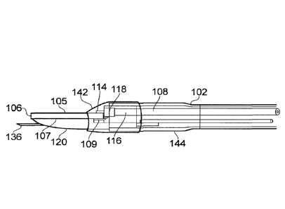
The instrument tip 104 comprises a dielectric block 106
that has layers of metallisation 105, 107 on its upper and
lower surfaces. The layers of metallisation correspond to the
first and second conductive elements of the invention. The
5 layers of metallisation are separated by the thickness of the
dielectric block 106 to form a bipolar radiating spatula
structure, similar to that disclosed in GB 2 472 972.
The layers of metallisation may be formed from high
melting point conductors, e.g. W or Ti. In such an
10 arrangement, lower melting point conductors may be deposited
around the regions where the coaxial cable connects to the
parallel plate planar transmission line to facilitate
soldering the coaxial arrangement to the planar transmission
line. The lower melting point conductors may be silver (Ag) or
15 gold (Au).
As seen most clearly in Fig. 2, the distal end of the
dielectric block is formed in a curved, e.g. parabolic, shape.
This shape is preferred so that the instrument does not
present sharp corners at its outer edges, and to enable use in
multiple directions of travel. Such sharp corners can be
undesirable when the instrument is used in environments with
delicate tissue structures, such as the gastrointestinal
tract, where the bowel wall is very thin.
The sleeve 102 defines a lumen which carries a flexible
coaxial feed cable 108 and a fluid delivery structure. In
this embodiment, the fluid delivery structure includes a
passageway formed by space in the lumen around the flexible
feed cable 108 and a retractable needle 110. The sleeve 102
carries a control wire 112 for both deploying and retracting
the needle 110. Operation of the needle is described below.
The inner conductor 114 of the coaxial feed cable 108
protrudes from the distal end of the coaxial feed cable 108
and is electrically bonded (e.g. using solder) to the upper
layer 105 of metallisation (first conductive element). The
outer conductor of the coaxial cable 116 is electrically
coupled to the lower layer of metallisation 107 (second
conductive element) by a braid termination 118. The braid
termination 118 comprises a tubular part that is electrically
bonded to the outer conductor and a distally extending plate
part 109 that fits under the dielectric block 106 and is
CA 02917130 2015-12-31
WO 2014/006369
PCT/GB2013/051672
16
electrically connected to the lower layer 107 of
metallisation.
In this embodiment, a shaped piece of dielectric material
120 is attached to the lower surface of the dielectric block
106. It may be secured to the lower layer 107 of
metallisation. The underside of the shaped piece of
dielectric material 120 has a configuration particularly
suited for use in procedures performed in the gastrointestinal
tract. In the longitudinal direction, the shaped piece of
dielectric material 120 comprises a distal part which
gradually tapers (e.g. in a curved manner) towards the
dielectric block 106. This part of the instrument is in
closest proximity to the tissue being treated in use, e.g. the
bowel wall, the wall of the oesophagus, the portal vein, or
the pancreatic duct. By presenting a curved surface in this
way, unwanted perforation of the bowel wall or the wall of the
oesophagus or damage to the portal vein or the pancreatic duct
can be avoided.
As can be seen most clearly in Fig. 2, the undersurface
of the shaped piece of dielectric material 120 has a
longitudinally extending recessed channel 122. The recessed
channel defines an access path for the retractable needle 110.
The recessed nature of the channel means that the access path
is flanked one both sides by longitudinally extending ridges
124 of the shaped piece of dielectric material.
The surface of the shaped piece of dielectric material
120 that engages with the underside of the radiating spatula
structure is shown in more detail in Figs. 12A and 12B. The
distal end of the shaped piece of dielectric material 120 has
a flat upper surface 126 for contacting the lower layer of
metallisation 107. A rectangular recess 129 is formed towards
the proximal end of the flat upper surface 126 for receiving
the plate part 109 of the braid termination 118.
The proximal end of the shaped piece of dielectric
material 120 is formed with a U-shaped channel 128 for
receiving and supporting the distal end of the coaxial feed
cable 108. Fig. 12B shows that a similar channel 130 is
formed on the underside of the proximal end of the shaped
piece of dielectric material 120 to receive a guide conduit
for the retractable needle (see Figs. 6 and 7). The outer
surface of the proximal end of the shaped piece of dielectric
CA 02917130 2015-12-31
WO 2014/006369
PCT/GB2013/051672
17
material 120 is cylindrical, with a diameter selected to fit
inside the sleeve.
At the sides of the shaped piece of dielectric material
120 between the proximal and distal ends, there are a pair of
upstanding wing portions 132, whose inner surfaces engage with
respective side edges of the radiating spatula structure and
whose outer surface engage in an interference fit with the
inner surface of the sleeve 102.
The shaped piece of dielectric material 120 is preferably
made from a ceramic or other material having low thermal
conductivity.
In another embodiment, the dielectric body 106 and the
shaped piece of dielectric 120 may be formed in one piece,
i.e. as a unitary body. The unitary body may have a planar
slot formed (e.g. cut) therein for receiving a conductive
material to form the lower layer of metallisation (second
conductive element). The thickness of the slot and therefore
the lower layer of metallisation may be 0.1 mm or more, but
preferably no more than 0.2 mm.
The overall size of the instrument may be such that it is
suitable for insertion through the instrument channel of an
endoscope. Thus, the outer diameter of the sleeve may be 2.8
mm or less, e.g. 2.7 mm.
Figs. 6, 7, and 7A illustrate operation of a control wire
138 for deploying and retracting a retractable needle 136.
The sleeve 102 and shaped piece of dielectric material 120 are
omitted in Figs. 6 and 7 for clarity. The retractable needle
136 is slidably mounted in a needle sleeve 134, which is fixed
in the channel 130 formed in the underside of the shaped piece
of dielectric material 120. The retractable needle 136 is
capable for sliding between a deployed position (shown in Fig.
6), where it protrudes from the distal end of the instrument,
and a retracted position (shown in Fig. 7) where the distal
end of the needle is set back from the distal end of the
instrument. The retractable needle 136 is attached at the end
of a needle base unit 140, which is itself slidable within the
sleeve by operating (i.e. pushing or pulling as appropriate) a
suitable control wire 138, as is conventional. The control
wire 138 is preferably welded in-line with the needle 138 as
shown in Figs. 6 and 7, as this allows a more compact
arrangement. Alternatively, the control wire may abut against
CA 02917130 2015-12-31
WO 2014/006369
PCT/GB2013/051672
18
a side surface of the needle or needle base unit, as shown in
Fig. 1.
When the control wire 138 pushes the needle 136 to its
forward-most (i.e. deployed) position the needle base unit 140
abuts the needle sleeve to create a seal. The needle base
unit 140 prevents the needle from being pushed too far out of
the instrument. As shown in Fig. 7A, the space 139 in the
lumen outside the coaxial cable 108 and retractable needle 136
forms a passageway for carrying fluid from the proximal end of
the sleeve, where for example it may be injected by a user.
An aperture 143 (seen in Fig. 7a) formed in a side wall of the
needle base unit 140 provides a fluid flow path between the
space 139 in the lumen and the proximal end of the needle 136.
This enables fluid that has travelled down the length of the
fluid conduit within the sleeve 102 to access the proximal end
of the needle and be injected out through the needle tip.
As shown in Fig. 7A, the control wire slides in a guide
conduit 141, which can prevent buckling of the control wire
when it is under compression, thereby improving accuracy of
control over the needles position. The guide conduit 141 may
be formed in a semi-rigid insert mounted in the sleeve, as
discussed below with reference to Figs. 10 and 11.
In the retracted position, the distal end of the needle
136 (i.e. the needle tip) may be enclosed by the needle sleeve
134 to prevent accidental snagging on either patient tissue or
the internal structure of an endoscope. The needle 136 may be
a hypodermic needle terminating with a sharp point for
penetrating biological tissue.
Injection of fluid (saline or the like) to plump up or
raise the biological tissue may be particularly useful where
the instrument is to treat the wall of the bowel or the wall
of the oesophagus. For example, the instrument may be
particular useful for removing sessile polyps, which sit flat
on the wall of the bowel. Plumping up the tissue in this
manner may help to reduce risk of bowel or oesophagus
perforation. It is advantageous to be able to use the same
instrument to deliver fluid as delivers RF and/or microwave
energy since deflation (e.g. due to fluid seepage) may occur
if a separate instrument is introduced into the region or
during treatment. The ability to introduce fluid using the
same treatment structure enables the level to be topped up as
CA 02917130 2015-12-31
WO 2014/006369
PCT/GB2013/051672
19
soon as deflation occurs. Moreover, the use of a single
instrument to perform desiccation or dissection as well as to
introduce fluid also reduces the time taken to perform the
polyp removal procedure, reduces the risk of causing harm to
the patient and also reduces the risk of infection. More
generally, injection of fluid may be used to flush the
treatment region, e.g. to remove waste products or removed
tissue to provide better visibility when treating. This may
be particularly useful in endoscopic procedures.
Fig. 8 shows a view of the instrument tip, in which the
distal end of the sleeve 102 is "potted" in a cap element 142,
which covers the electrical joint between the radiating
spatula structure and the coaxial cable. The cap element 142
may be formed from a suitable a heat shrink material or from
potting adhesive, e.g. UV curable adhesive such as Loctite0
4304 and/or Loctite 4305. Protecting the joint in this way
may prevent arcing from occurring at the electrical joint
during use. The adhesive used should not be lossy or absorb
energy at the microwave frequency of choice. Using a small
amount of adhesive will also minimise the amount of energy
coupled into it. If microwave power is absorbed by the
adhesive, it will cause local heating and loss of microwave
power available at the edges of the blade.
Fig. 9 shows a schematic cross-section view of the distal
end of the instrument. In this view the needle 136 is
deployed. Here the distal end of the sleeve 102 includes a
widened portion 144 having an increased diameter. The widened
portion 144 provides more space at the distal end, which gives
more room for the needle deployment mechanism and a more
robust connection between the coaxial cable 108, radiating
spatula structure 105, 106, 107 and shaped piece of dielectric
material 120.
Fig. 10 shows a cross-section view through the sleeve 102
facing towards the distal end of the instrument. Mounted
within the sleeve 102 is a semi-rigid insert 146 that is
arranged to maintain the position of the coaxial cable 108 and
push wire 112 along the length of the sleeve 102. The insert
146 may be a length of extruded plastic material or the like.
In Fig. 10 the insert 146 has a horse-shoe shaped cross-
section, with an outer surface for engaging the inner surface
of the sleeve, and a U-shaped channel for receiving the
CA 02917130 2015-12-31
WO 2014/006369
PCT/GB2013/051672
coaxial cable 108. Two longitudinally extending circular
passages are formed within the insert for carrying the push
wire and for providing space for a fluid path respectively.
Maintaining the position of the push wire is important,
5 because if movement of the push wire is unconstrained within
the lumen of the sleeve, control of the wire can be lost e.g.
due to the push wire moving laterally within the sleeve.
Although shown as a separate insert in this embodiment,
these passages may be incorporated into the sleeve itself,
10 e.g. as a single extrusion or through bonding or welding to
the inner surface of the sleeve 102. The insert may exhibit
lateral strength to provide crush resistance and durability to
the device.
Fig. 11 shows a similar view to Fig. 10 for another
15 extruded rigid insert 148. The effect of the semi-rigid
inserts 146, 148 is to provide multiple lumens within a common
sleeve 102.
When used to deliver microwave frequency energy, the
radiating spatula behaves as a resonant microwave structure,
20 fed from a coaxial transmission line. Its function is to pass
microwave energy into biological tissue that is close to or
touching the region near the tip of the spatula. As mentioned
above, the distal end of the radiating spatula blade is curved
to avoid presenting sharp edges or corners to tissue in use.
A discussion of the effect of changing the shape of the end of
the spatula on the delivery of microwave energy is presented
below with reference to Figs. 13 to 23.
The spatula is a low impedance planar transmission line,
that is to say that the ratio of the voltage between the top
and bottom metal plates to the (equal and opposite) currents
in the two plates is close to 30 fl (calculated using microwave
field modelling software). Typically, the transmission line
feeding the spatula has an impedance of 50 n. Thus, the
transmission line and the biological tissue touching the end
of the spatula appear as high impedances to the spatula.
The difference in impedance at each end would normally
present a partial obstacle to the passage of power into and
out of the spatula. However, when the spatula is close to a
whole number of half-wavelengths long, the voltages at the end
of the spatula increase, and the currents at the end decrease,
CA 02917130 2015-12-31
WO 2014/006369
PCT/GB2013/051672
21
both due to a resonant effect, so that power passes readily
from the coaxial line through the spatula into the tissue.
For this reason the length of the spatula, from the end of the
coaxial transmission line to the other end of the spatula (or
planar transmission line), plays a significant role in the
effectiveness of the spatula.
The length of the spatula is carefully adjusted so that,
taking into account the modification of the wavelength by the
shape of the spatula, the dielectric constant of the material
between the plates, and fringing fields at each end of the
spatula, the spatula is close to one half wavelength long at
the operating frequency. In practice this length can be found
empirically by numerical simulation and/or experiment.
The effect of changes in shape in the end of the spatula
can be understood in terms of a change in the capacitance of
the end of the spatula.
Under resonant conditions, the centre of the rectangular
spatula shown in Fig. 13 behaves in a similar way to an
electrical inductance (coil) and each end behaves similarly to
a capacitor, as shown schematically in Fig. 14. The product
of the capacitance and the inductance is proportional to the
inverse square of the frequency at which the spatula will
resonate. This is described by the standard electrical
relation f= ____________ for the resonant frequency f of a resonant
27rirCe
electrical circuit with a capacitance C and an inductance L.
If the shape of the end of the spatula is changed, this
results in a change in capacitance so that the resonant
frequency of the spatula changes, or to put it another way the
spatula is now not the correct length to resonate at the
operating frequency.
The overall length of the spatula can, however, be
adjusted to bring it back into resonance. A good
approximation to the length adjustment needed is that required
to return the area of the spatula to the value before the end
was rounded - this is equivalent to adjusting the capacitance
back to its previous value.
Capacitance is proportional to the area of the capacitor.
If the end of the spatula were rounded off to a semi-circle or
ellipse, then the length should be increased so that the extra
CA 02917130 2015-12-31
WO 2014/006369
PCT/GB2013/051672
22
rectangular part has the same area as the parts cut off to
make the semi-circular end, as indicated in Fig. 15.
The missing area in Fig. 15 is
n-rjr2
27-1r2 ___________________________ 2 2r1x0.2146r2
where r1 is half the width of the spatula and r2 is the
half-length of the ellipse that forms the curved end.
The area of the rectangle to be added shown in Fig. 16 is
2r1x = 2r1 X 0.2146r2
where x is the extra length required.
Thus the extra length required is approximately 0.215
times the length of the rounded part of the spatula. If the
rounded end is 3 mm long, the extra length required is about
0.64 mm. This increase in length was tested by simulation
with the actual shape of the spatula and found to be close to
the optimum. The length of the model was adjusted empirically
to find the optimum, which was actually 0.6 mm.
The change in resonant frequency may also be corrected by
changing the capacitance of the other end of the spatula, by
changing the geometry of the connection to the 50 fl coaxial
cable. A simple way to do this is to change the spacing
between the top plate of the spatula and the coaxial line.
The general shape of the spatula is shown in Fig. 17, and
a side view of the spatula with 0.4 mm gap is shown in Fig.
18. A side view of the spatula with 0.1 mm gap is shown in
Fig. 19, and a close up side view in Fig. 20.
In Fig. 20, the gap between the top plate of the spatula
and the coaxial line forms a capacitor that can be used to
adjust the resonant frequency of the spatula. If the gap is
reduced, the capacitance increases, and the resonant frequency
drops.
Fig. 21 shows the return loss for the 10.6 mm long
spatula with a 0.4 mm gap. The best return loss is close to
5.8 GHz.
Figs. 22 and 23 compare the return loss for a 10 mm long
spatula with 0.3 mm and 0.1 mm gaps respectively. It can be
seen in Fig. 22 that with the 0.3 mm gap the best return loss
is at 6 GHz, and in Fig. 23 with a 0.1 mm gap the best return
loss is close to 5.8 GHz.
It may be difficult to accurately manufacture the device
with a 0.1 mm gap, so the solution of increasing the spatula
CA 02917130 2015-12-31
W02014/006369
PCT/GB2013/051672
23
length to adjust for changing the overall shape may be
preferred. However, other ways of increasing the capacitance
at the cable end of the spatula might be used, such as
increasing the thickness of the top plate, which may happen
anyway when solder is applied.
Because it may be difficult to accurately describe the
geometry that is actually achieved around the connection
between the cable and the spatula, the best approach is to aim
for a geometry that is easily built and is repeatable.