Note: Descriptions are shown in the official language in which they were submitted.
CA 02917654 2016-01-14
SYSTEM AND METHOD FOR CONFIGURING POSITIONS IN A SURGICAL
POSITIONING SYSTEM
TECHNICAL FIELD
[0001] The present disclosure is generally related to image guided medical
procedures, and more specifically to a system and method for configuring
positions
in a surgical positioning system.
BACKGROUND
[0002] The present disclosure is generally related to image guided medical
procedures using a surgical instrument, such as an optical scope, an optical
coherence tomography (OCT) probe, a micro ultrasound transducer, an electronic
sensor or stimulator, or an access port based surgery.
[0003] In the example of a port-based surgery, a surgeon or robotic
surgical
system may perform a surgical procedure involving tumor resection in which the
residual tumor remaining after is minimized, while also minimizing the trauma
to
the intact white and grey matter of the brain. In such procedures, trauma may
occur, for example, due to contact with the access port, stress to the brain
matter,
unintentional impact with surgical devices, and/or accidental resection of
healthy
tissue. A key to minimizing trauma is ensuring that the surgeon is aware of
what is
transpiring in the operating room, has a proper view of the surgical site of
interest,
and has proper control of the surgical positioning system without undue
distraction.
[0004] FIG. 1 illustrates the insertion of an access port into a human
brain,
for providing access to internal brain tissue during a medical procedure. In
FIG. 1,
access port 12 is inserted into a human brain 10, providing access to internal
brain
tissue. Access port 12 may include such instruments as catheters, surgical
probes,
or cylindrical ports such as the NICO BrainPath. Surgical tools and
instruments
may then be inserted within the lumen of the access port in order to perform
1
CA 02917654 2016-01-14
surgical, diagnostic or therapeutic procedures, such as resecting tumors as
necessary. The present disclosure applies equally well to catheters, DBS
needles, a
biopsy procedure, and also to biopsies and/or catheters in other medical
procedures
performed on other parts of the body.
[0005] In the example of a port-based surgery, a straight or linear access
port
12 is typically guided down a sulci path of the brain. Surgical instruments
would
then be inserted down the access port 12. Optical tracking systems, used in
the
medical procedure, track the position of a part of the instrument that is
within line-
of-site of the optical tracking camera. The surgical positioning system will
often
have a camera mounted thereon and is responsible for maintaining a view of the
surgical site of interest by moving the camera to the proper positions, under
the
control of the surgeon.
[0006] During surgical procedures, a surgeon utilizing an external optical
system may want or need to view the surgical site from multiple angles. A
problem
can occur if it takes a significant amount of time to achieve this or if the
surgeon
needs to remove tools from the surgical field to move the optical system
between
these different angles. In many cases, these viewing angles are known before
the
procedure or can all be defined at the start of the procedure.
[0007] Conventional systems have not offered good solutions for ensuring
that a surgeon has a good view of the surgical site without constantly having
to
reconfigure the optical system positioning the camera. It would be desirable
to
have a system that helps a surgeon maintain the optical system in the
appropriate
positions without placing undue burden on the surgeon during the medical
procedure.
SUMMARY
[0008] One aspect of the present disclosure provides a medical navigation
system including a surgical positioning system for positioning a payload
during a
2
CA 02917654 2016-01-14
medical procedure. The medical navigation system has a robotic arm having a
plurality of joints, the robotic arm forming part of the surgical positioning
system
and having an end effector for holding the payload, an input device for
providing
input, and a controller electrically coupled to the robotic arm and the input
device.
The controller has a processor coupled to a memory and the controller is
configured
to perform the following during the medical procedure: position the robotic
arm in a
first position by providing a first positioning signal to the robotic arm;
save the first
position in the memory as a first saved position in response to a signal
received
from the input device; position the robotic arm in a second position by
providing a
second positioning signal to the robotic arm; and return the robotic arm to
the first
position by loading the first saved position from the memory and providing the
first
positioning signal to the robotic arm when an input is received from the input
device corresponding to a command to return to the first saved position.
[0009] Another aspect of the present disclosure provides a method of
positioning a payload during a medical procedure in a medical navigation
system
including a surgical positioning system. The medical navigation system has a
robotic arm, an input device, and a controller. The robotic arm has a
plurality of
joints and forms part of the surgical positioning system and has an end
effector for
holding the payload. The controller has a processor coupled to a memory, the
method comprising: positioning the robotic arm in a first position; saving the
first
position in the memory as a first saved position in response to a signal
received
from the input device; positioning the robotic arm in a second position; and
returning the robotic arm to the first position by loading the first saved
position
from the memory when an input is received from the input device corresponding
to
a command to return to the first saved position.
[0010] A further understanding of the functional and advantageous aspects
of
the disclosure can be realized by reference to the following detailed
description and
drawings.
3
CA 02917654 2016-01-14
BRIEF DESCRIPTION OF THE DRAWINGS
[0011] Embodiments will now be described, by way of example only, with
reference to the drawings, in which:
[0012] FIG. 1 illustrates the insertion of an access port into a human
brain,
for providing access to internal brain tissue during a medical procedure;
[0013] FIG. 2 shows an exemplary navigation system to support minimally
invasive surgery;
[0014] FIG. 3 is a block diagram illustrating a control and processing
system
that may be used in the navigation system shown in Fig. 2;
[0015] FIG. 4A is a flow chart illustrating a method involved in a surgical
procedure using the navigation system of FIG. 2;
[0016] FIG. 4B is a flow chart illustrating a method of registering a
patient for
a surgical procedure as outlined in FIG. 4A;
[0017] FIG. 5 is an exemplary navigation system similar to FIG. 2
illustrating
system components of an exemplary surgical system that.may be used for
configuring positions of the surgical system;
[0018] FIG. 6 is perspective drawing illustrating a conventional end
effector
holding a camera;
[0019] FIG. 7 is a flow chart is illustrating a method of configuring
positions in
a surgical positioning system according to one aspect of the present
description;
and
[0020] FIG. 8 is a block diagram illustrating an example of a virtual
reality
component according to one aspect of the present description.
4
CA 02917654 2016-01-14
DETAILED DESCRIPTION
[0021] Various embodiments and aspects of the disclosure will be described
with reference to details discussed below. The following description and
drawings
are illustrative of the disclosure and are not to be construed as limiting the
disclosure. Numerous specific details are described to provide a thorough
understanding of various embodiments of the present disclosure. !However, in
certain instances, well-known or conventional details are not described in
order to
provide a concise discussion of embodiments of the present disclosure.
[0022] As used herein, the terms, "comprises" and "comprising" are to be
construed as being inclusive and open ended, and not exclusive. Specifically,
when
used in the specification and claims, the terms, "comprises" and "comprising"
and
variations thereof mean the specified features, steps or components are
included.
These terms are not to be interpreted to exclude the presence of other
features,
steps or components.
[0023] As used herein, the term "exemplary" means "serving as an example,
instance, or illustration," and should not be construed as preferred or
advantageous
over other configurations disclosed herein.
[0024] As used herein, the terms "about", "approximately", and
"substantially" are meant to cover variations that may exist in the upper and
lower
limits of the ranges of values, such as variations in properties, parameters,
and
dimensions. In one non-limiting example, the terms "about", "approximately",
and
"substantially" mean plus or minus 10 percent or less.
[0025] Unless defined otherwise, all technical and scientific terms used
herein
are intended to have the same meaning as commonly understood by one of
ordinary skill in the art. Unless otherwise indicated, such as through
context, as
used herein, the following terms are intended to have the following meanings:
[0026] As used herein, the phrase "access port" refers to a cannula,
conduit,
sheath, port, tube, or other structure that is insertable into a subject, in
order to
CA 02917654 2016-01-14
provide access to internal tissue, organs, or other biological substances. In
some
embodiments, an access port may directly expose internal tissue, for example,
via
an opening or aperture at a distal end thereof, and/or via an opening or
aperture at
an intermediate location along a length thereof. In other embodiments, an
access
port may provide indirect access, via one or more surfaces that are
transparent, or
partially transparent, to one or more forms of energy or radiation, such as,
but not
limited to, electromagnetic waves and acoustic waves.
[0027] As used herein the phrase "intraoperative" refers to an action,
process,
method, event or step that occurs or is carried out during at least a portion
of a
medical procedure. Intraoperative, as defined herein, is not limited to
surgical
procedures, and may refer to other types of medical procedures, such as
diagnostic
and therapeutic procedures.
[0028] Embodiments of the present disclosure provide imaging devices that
are insertable into a subject or patient for imaging internal tissues, and
methods of
use thereof. Some embodiments of the present disclosure relate to minimally
invasive medical procedures that are performed via an access port, whereby
surgery, diagnostic imaging, therapy, or other medical procedures (e.g.,
minimally
invasive medical procedures) are performed based on access to internal tissue
through the access port.
[0029] Referring to FIG. 2, an exemplary navigation system environment 200
is shown, which may be used to support navigated image-guided surgery. As
shown in FIG. 2, surgeon 201 conducts a surgery on a patient 202 in an
operating
room (OR) environment. A medical navigation system 205 comprising an
equipment tower, tracking system, displays and tracked instruments assist the
surgeon 201 during his procedure. An operator 203 is also present to operate,
control and provide assistance for the medical navigation system 205.
[0030] Referring to FIG. 3, a block diagram is shown illustrating a control
and
processing system 300 that may be used in the medical navigation system 200
shown in FIG. 3 (e.g., as part of the equipment tower). As shown in FIG. 3, in
one
6
CA 02917654 2016-01-14
example, control and processing system 300 may include one or more processors
302, a memory 304, a system bus 306, one or more input/output interfaces 308,
a
communications interface 310, and storage device 312. Control and processing
system 300 may be interfaced with other external devices, such as tracking
system
321, data storage 342, and external user input and output devices 344, which
may
include, for example, one or more of a display, keyboard, mouse, sensors
attached
to medical equipment, foot pedal, and microphone and speaker. Data storage 342
may be any suitable data storage device, such as a local or remote computing
device (e.g. a computer, hard drive, digital media device, or server) having a
database stored thereon. In the example shown in FIG. 3, data storage device
342
includes identification data 350 for identifying one or more medical
instruments 360
and configuration data 352 that associates customized configuration parameters
with one or more medical instruments 360. Data storage device 342 may also
include preoperative image data 354 and/or medical procedure planning data
356.
Although data storage device 342 is shown as a single device in FIG. 3, it
will be
understood that in other embodiments, data storage device 342 may be provided
as multiple storage devices.
[0031] Medical
instruments 360 are identifiable by control and processing unit
300. Medical instruments 360 may be connected to and controlled by control and
processing unit 300, or medical instruments 360 may be operated or otherwise
employed independent of control and processing unit 300. Tracking system 321
may be employed to track one or more of medical instruments 360 and spatially
register the one or more tracked medical instruments to an intraoperative
reference
frame. For example, medical instruments 360 may include tracking markers such
as tracking spheres that may be recognizable by a tracking camera 307. In one
example, the tracking camera 307 may be an infrared (IR) tracking camera. In
another example, a sheath placed over a medical instrument 360 may be
connected
to and controlled by control and processing unit 300. In another example,
camera
307 may be a video camera.
7
CA 02917654 2016-01-14
[0032] Control and processing unit 300 may also interface with a number of
configurable devices, and may intraoperatively reconfigure one or more of such
devices based on configuration parameters obtained from configuration data
352.
Examples of devices 320, as shown in FIG. 3, include one or more external
imaging
devices 322, one or more illumination devices 324, a robotic arm 305, one or
more
projection devices 328, and one or more displays 311, and a scanner 309, which
in
one example may be a three dimensional (3D) scanner.
[0033] Exemplary aspects of the disclosure can be implemented via
processor(s) 302 and/or memory 304. For example, the functionalities described
herein can be partially implemented via hardware logic in processor 302 and
partially using the instructions stored in memory 304, as one or more
processing
modules or engines 370. Example processing modules include, but are not
limited
to, user interface engine 372, tracking module 374, motor controller 376,
image
processing engine 378, image registration engine 380, procedure planning
engine
382, navigation engine 384, and context analysis module 386. While the example
processing modules are shown separately in FIG. 3, in one example the
processing
modules 370 may be stored in the memory 304 and the processing modules may
be collectively referred to as processing modules 370. In some examples, the
set
of processing engines (370) may reside on a plurality of independent control
and
processing units (300), connected via a network, where the devices (320) may
be
distributed between the set of control and processing units (300), as well as
the
data device storage (342).
[0034] It is to be understood that the system is not intended to be limited
to
the components shown in FIG. 3. One or more components of the control and
processing system 300 may be provided as an external component or device. In
one example, navigation module 384 may be provided as an external navigation
system that is integrated with control and processing system 300.
[0035] Some embodiments may be implemented using processor 302 without
additional instructions stored in memory 304. Some embodiments may be
8
CA 02917654 2016-01-14
implemented using the instructions stored in memory 304 for execution by one
or
more general purpose microprocessors. Thus, the disclosure is not limited to a
specific configuration of hardware and/or software.
[0036] While some embodiments can be implemented in fully functioning
computers and computer systems, various embodiments are capable of being
distributed as a computing product in a variety of forms and are capable of
being
applied regardless of the particular type of machine or computer readable
media
used to actually effect the distribution.
[0037] At least some aspects disclosed can be embodied, at least in part,
in
software. That is, the techniques may be carried out in a computer system or
other
data processing system in response to its processor, such as a microprocessor,
executing sequences of instructions contained in a memory, such as ROM,
volatile
RAM, non-volatile memory, cache or a remote storage device.
[0038] A computer readable storage medium can be used to store software
and data which, when executed by a data processing system, causes the system
to
perform various methods. The executable software and data may be stored in
various places including for example ROM, volatile RAM, nonvolatile memory
and/or
cache. Portions of this software and/or data may be stored in any one of these
storage devices.
[0039] Examples of computer-readable storage media include, but are not
limited to, recordable and non-recordable type media such as volatile and non-
volatile memory devices, read only memory (ROM), random access memory (RAM),
flash memory devices, floppy and other removable disks, magnetic disk storage
media, optical storage media (e.g., compact discs (CDs), digital versatile
disks
(DVDs), etc.), among others. The instructions may be embodied in digital and
analog communication links for electrical, optical, acoustical or other forms
of
propagated signals, such as carrier waves, infrared signals, digital signals,
and the
like. The storage medium may be the internet cloud, or a computer readable
storage medium such as a disc.
9
CA 02917654 2016-01-14
[0040] At least some of the methods described herein are capable of being
distributed in a computer program product comprising a computer readable
medium that bears computer usable instructions for execution by one or more
processors, to perform aspects of the methods described. The medium may be
provided in various forms such as, but not limited to, one or more diskettes,
compact disks, tapes, chips, USB keys, external hard drives, wire-line
transmissions, satellite transmissions, internet transmissions or downloads,
magnetic and electronic storage media, digital and analog signals, and the
like. The
computer useable instructions may also be in various forms, including compiled
and
non-compiled code.
[0041] According to one aspect of the present application, one purpose of
the
navigation system 205, which may include control and processing unit 300, is
to
provide tools to the neurosurgeon that will lead to the most informed, least
damaging neurosurgical operations. In addition to removal of brain tumours and
intracranial hemorrhages (ICH), the navigation system 205 can also be applied
to a
brain biopsy, a functional/deep-brain stimulation, a catheter/shunt placement
procedure, open craniotomies, endonasal/skull-based/ENT, spine procedures, and
other parts of the body such as breast biopsies, liver biopsies, laparoscopic
surgery,
etc. While several examples have been provided, aspects of the present
disclosure
may be applied to any suitable medical procedure.
[0042] Referring to FIG. 4A, a flow chart is shown illustrating a method
400 of
performing a surgical procedure using a navigation system, such as the medical
navigation system 205 described in relation to FIG. 2. At a first block 402,
the
surgical plan is imported.
[0043] Once the plan has been imported into the navigation system at the
block 402, the patient is affixed into position using a body holding
mechanism. The
head position is also confirmed with the patient plan in the navigation system
(block 404), which in one example may be implemented by the computer or
controller forming part of the equipment tower of medical navigation system
205.
CA 02917654 2016-01-14
[0044] Next, registration of the patient is initiated (block 406). The
phrase
"registration" or "image registration" refers to the process of transforming
different
sets of data into one coordinate system. Data may include multiple
photographs,
data from different sensors, times, depths, or viewpoints. The process of
"registration" is used in the present application for medical imaging in which
images
from different imaging modalities are co-registered. Registration is used in
order to
be able to compare or integrate the data obtained from these different
modalities.
[0045] Those skilled in the relevant arts will appreciate that there are
numerous registration techniques available and one or more of the techniques
may
be applied to the present example. Non-limiting examples include intensity-
based
methods that compare intensity patterns in images via correlation metrics,
while
feature-based methods find correspondence between image features such as
points, lines, and contours. Image registration methods may also be classified
according to the transformation models they use to relate the target image
space to
the reference image space. Another classification can be made between single-
modality and multi-modality methods. Single-modality methods typically
register
images in the same modality acquired by the same scanner or sensor type, for
example, a series of magnetic resonance (MR) images may be co-registered,
while
multi-modality registration methods are used to register images acquired by
different scanner or sensor types, for example in magnetic resonance imaging
(MRI) and positron emission tomography (PET). In the present disclosure, multi-
modality registration methods may be used in medical imaging of the head
and/or
brain as images of a subject are frequently obtained from different scanners.
Examples include registration of brain computerized tomography (CT)/MRI images
or PET/CT images for tumor localization, registration of contrast-enhanced CT
images against non-contrast-enhanced CT images, and registration of ultrasound
and CT.
11
CA 02917654 2016-01-14
[0046] Referring now to FIG. 4B, a flow chart is shown illustrating a
method
involved in registration block 406 as outlined in FIG. 4A, in greater detail.
If the
use of fiducial touch points (440) is contemplated, the method involves first
identifying fiducials on images (block 442), then touching the touch points
with a
tracked instrument (block 444). Next, the navigation system computes the
registration to reference markers (block 446).
[0047] Alternately, registration can also be completed by conducting a
surface
scan procedure (block 450). The block 450 is presented to show an alternative
approach, but may not typically be used when using a fiducial pointer. First,
the
face is scanned using a 3D scanner (block 452). Next, the face surface is
extracted
from MR/CT data (block 454). Finally, surfaces are matched to determine
registration data points (block 456).
[0048] Upon completion of either the fiducial touch points (440) or surface
scan (450) procedures, the data extracted is computed and used to confirm
registration at block 408, shown in FIG. 4B.
[0049] Referring back to FIG. 4A, once registration is confirmed (block
408),
the patient is draped (block 410). Typically, draping involves covering the
patient
and surrounding areas with a sterile barrier to create and maintain a sterile
field
during the surgical procedure. The purpose of draping is to eliminate the
passage
of microorganisms (e.g., bacteria) between non-sterile and sterile areas. At
this
point, conventional navigation systems require that the non-sterile patient
reference is replaced with a sterile patient reference of identical geometry
location
and orientation.
[0050] Upon completion of draping (block 410), the patient engagement
points are confirmed (block 412) and then the craniotomy is prepared and
planned
(block 414).
12
CA 02917654 2016-01-14
[0051] Upon completion of the preparation and planning of the craniotomy
(block 414), the craniotomy is cut and a bone flap is temporarily removed from
the
skull to access the brain (block 416). Registration data is updated with the
navigation system at this point (block 422).
=
[0052] Next, the engagement within craniotomy and the motion range are
confirmed (block 418). Next, the procedure advances to cutting the dura at the
engagement points and identifying the sulcus (block 420).
[0053] Thereafter, the cannulation process is initiated (block 424).
Cannulation involves inserting a port into the brain, typically along a sulci
path as
identified at 420, along a trajectory plan. Cannulation is typically an
iterative
process that involves repeating the steps of aligning the port on engagement
and
setting the planned trajectory (block 432) and then cannulating to the target
depth
(block 434) until the complete trajectory plan is executed (block 424).
[0054] Once cannulation is complete, the surgeon then performs resection
(block 426) to remove part of the brain and/or tumor of interest. The surgeon
then
decannulates (block 428) by removing the port and any tracking instruments
from
the brain. Finally, the surgeon closes the dura and completes the craniotomy
(block 430). Some aspects of FIG. 4A are specific to port-based surgery, such
as
portions of blocks 428, 420, and 434, but the appropriate portions of these
blocks
may be skipped or suitably modified when performing non-port based surgery.
[0055] When performing a surgical procedure using a medical navigation
system 205, as outlined in connection with FIGS. 4A and 4B, the medical
navigation
system 205 must acquire and 'maintain a reference of the location of the tools
in
use as well as the patient in three dimensional (3D) space. In other words,
during
a navigated neurosurgery, there needs to be a tracked reference frame that is
fixed
relative to the patient's skull. During the registration phase of a navigated
neurosurgery (e.g., the step 406 shown in FIGS. 4A and 4B), a transformation
is
calculated that maps the frame of reference of preoperative MRI or CT imagery
to
13
CA 02917654 2016-01-14
the physical space of the surgery, specifically the patient's head. This may
be
accomplished by the navigation system 205 tracking locations of fiducial
markers
fixed to the patient's head, relative to the static patient reference frame.
The
patient reference frame is typically rigidly attached to the head fixation
device, such
as a Mayfield clamp. Registration is typically performed before the sterile
field has
been established (e.g., the step 410 shown in FIG. 4A).
[0056] FIG. 5 is a diagram illustrating components of an exemplary surgical
system that is similar to FIG. 2. FIG. 5 illustrates a navigation system 205
having
an equipment tower 502, tracking system 504, display 506, an intelligent
positioning system 508 and tracking markers 510 used to tracked instruments or
an
access port 12. Tracking system 504 may also be considered an optical tracking
device, tracking camera, video camera, 3D scanner, or any other suitable
camera
or scanner based system. In FIG. 5, a surgeon 201 is performing a tumor
resection
through a port 12, using an imaging device 512 (e.g., a scope and camera) to
view
down the port at a suffcient magnification to enable enhanced visibility of
the
instruments and tissue. The imaging device 512 may be an external scope,
videoscope, wide field camera, or an alternate image capturing device. The
imaging sensor view is depicted on the visual display 506 which surgeon 201
uses
for navigating the port's distal end through the anatomical region of
interest.
[0057] An intelligent positioning system 508 comprising an automated arm
514, a lifting column 516 and an end effector 518, is placed in proximity to
patient
202. Lifting column 516 is connected to a frame of intelligent positioning
system
508. As seen in FIG. 5, the proximal end of automated mechanical arm 514
(further known as automated arm 514 herein) is connected to lifting column
516.
In other embodiments, automated arm 514 may be connected to a horizontal
beam, which is then either connected to lifting column 516 or directly to
frame of
the intelligent positioning system 508. Automated arm 514 may have multiple
joints to enable 5, 6 or 7 degrees of freedom.
14
CA 02917654 2016-01-14
[0058] End effector 518 is attached to the distal end of automated arm 514.
End effector 518 may accommodate a plurality of instruments or tools that may
assist surgeon 201 in his procedure. End effector 518 is shown as holding an
external scope and camera, however it should be noted that this is merely an
example and alternate devices may be used with the end effector 518 such as a
wide field camera, microscope and OCT (Optical Coherence Tomography), video
camera, 3D scanner, or other imaging instruments. In another example, multiple
end effectors may be attached to the distal end of automated arm 518, and thus
assist the surgeon 201 in switching between multiple modalities. For example,
the
surgeon 201 may want the ability to move between microscope, and OCT with
stand-off optics. In a further example, the ability to attach a second, more
accurate, but smaller range end effector such as a laser based ablation system
with
micro-control may be contemplated.
[0059] In one example, the intelligent positioning system 508 receives as
input the spatial position and pose data of the automated arm 514 and target
(for
example the port 12) as determined by tracking system 504 by detection of the
tracking markers on the wide field camera on port 12. Further, it should be
noted
that the tracking markers may be used to track both the automated arm 514 as
well as the end effector 518 either collectively or independently. It should
be noted
that a wide field camera 520 is shown in FIG. 5 and that it is connected to
the
external scope (e.g., imaging device 512) and the two imaging devices together
are
held by the end effector 518. It should additionally be noted that although
these
are depicted together for illustration of the diagram that either could be
utilized
independently of the other, for example where an external video scope can be
used
independently of the wide field camera 520.
[0060] Intelligent positioning system 508 computes the desired joint
positions
for automated arm 514 so as to maneuver the end effector 518 mounted on the
automated arm's distal end to a predetermined spatial position and pose
relative to
the port 12. This redetermined relative spatial position and pose is termed
the
CA 02917654 2016-01-14
"Zero Position" where the sensor of imaging device 512 and port 12 are axially
alligned.
[0061] Further, the intelligent positioning system 508, optical tracking
device
504, automated arm 514, and tracking markers 510 may form a feedback loop.
This feedback loop works to keep the distal end of the port 12 (located inside
the
brain) in constant view and focus of the end effector 518 given that it is an
imaging
device as the port position may be dynamically manipulated by the surgeon
during
the procedure. Intelligent positioning system 508 may also include a foot
pedal for
use by the surgeon 201 to align the end effector 518 (i.e., holding a
videoscope) of
automated arm 514 with the port 12. Ensuring that the imaging device 512
and/or
wide field camera 520 remain focused on the surgical site of interest without
unduly
interfering with the surgeon during the medical procedure is one of the
objectives
of the present application and is discussed in more detail below, particularly
in
connection with FIGS. 7 and 8.
[0062] Referring to FIG. 6, a conventional end effector 518 is shown
attached
to automated arm 514. The end effector 518 includes a handle 602 and a scope
clamp 604. The scope clamp 604 holds imaging device 512. The end effector also
has wide field camera 520 attached thereto, which in one example could be a
still
camera, video camera, or 3D scanner used to monitor muscles of the patient for
movement, tremors, or twitching.
[0063] One aspect of the present disclosure provides a stored position
function that focuses on the reality that there are some arm positions that
will be
constant throughout most procedures and are unlikely to change often. These
constant positions can relate to draping, automated arm 514 orientation
positions
(e.g., left or right sided), storage positions, shipping positions, etc. These
constant
positions may be used before and/or after a surgical procedure and this
feature
may be controlled via a user interface, a foot pedal, voice control, etc. In
one
example, these constant positions may be stored as a list of joint angles
pertaining
16
CA 02917654 2016-01-14
to the arm configuration that is related to that particular position, since
the
automated arm 514 has a number of joints with an encoder associated with each
joint. In one example, the automated arm 514 may have six joints or even more.
However, the automated arm 514 may have any number of joints according to the
design criteria of a particular application. In one example, the automated arm
514
may go to a known position when the automated arm 514 is started up and may be
configured to toggle between different positions of interest.
[0064] In another example, the interface of the automated arm 514 may
provide the user with an undo function and a redo function. During a medical
procedure, the surgeon may wish to undo or redo automated arm 514 movements
or positions. This feature may both be used to correct an undesired movement
of
the automated arm 514 or to revisit a previous position that is clinically
relevant.
The undo and redo positions may be stored as a list of joint angles pertaining
to
sequential moves in a movement stack, for example saved in the memory 304 of
control and processing unit 300 (FIG. 3). The undo and redo feature may be
used
during a procedure but may also be useful during setup and wrap up of a
medical
procedure. The undo and redo feature may be controlled via a foot pedal, or by
other sources of control such as a user interface, voice control, etc. In one
example, when a successful move is made using the automated arm 514, the
surgeon may be provided with the option to save the move in the memory 304,
where the joint angles are saved. In one example, the joint angles may be
saved
relative in space to a patient reference based on a trajectory set in the
procedure
plan or guide.
[0065] Another aspect of the present application provides for a dynamic
memory position, During a procedure, the surgeon may wish to dynamically save
automated arm 514 movements so that the movements can be revisited at a later
time during the same procedure. In one example, the dynamic memory positions
may be stored as joint angles pertaining to the saved automated arm 514
position.
17
CA 02917654 2016-01-14
=
In another example, the dynamic memory positions may be stored relative to an
external frame of reference, such as the patient.
[0066] The dynamic memory position feature may be mainly used during a
procedure as dynamically stored positions but may not be as useful in the
setup
and wrap up phases of the procedure. In one example, the dynamic memory
position feature may be controlled via a foot pedal, user interface, voice
control,
etc. Further, the present application provides for dynamic allocation of pedal
mapping, where a foot pedal may be allocated to trigger the undo feature
instead of
requiring such input on a user interface of the control and processing unit
300. In
one example, stored positions may be recreated by aligning to tools that are
held in
the same position from when the desired position was first stored.
[0067] Another aspect of the present application provides for surgeons to
set
stored positions of the automated arm 514 during a medical procedure. These
stored positions often provide various perspectives on the surgical field.
Conventionally, a surgeon would have to verbally relay his or her desire to
save the
current automated arm 514 position to the clinical applications specialist
(e.g., the
operator 203 in FIG. 2) who is assisting with manipulating the controls of the
medical navigation system 205. When a surgeon wants the automated arm 514 to
move to one of the stored positions, the surgeon tries to recall the number
that the
specialist had given that stored position or tries to describe the view for
the
specialist to remember which one it was. This additional dialog introduces
another
barrier for changing the position of automated arm 514, leading to additional
time
being spent clarifying the view. Miscommunication between the surgeon and
operator would lead to the automated arm 514 going into a position that the
surgeon did not intend. In this case time is unnecessarily wasted and the
surgeon's
workflow and concentration would be broken. In fact, communication error in
the
operating room is one of the main sources of surgical failures, jeopardizing
patient
safety. In one example, this problem may be addressed by streaming stored
position view data to an augmented viewport, such that the surgeon is able to
see
18
CA 02917654 2016-01-14
virtual renderings of some or all of the available stored positions. The
virtual
renderings may also include sample snapshots that were taking using the optics
systems such as cameras at that particular stored position. The virtual
renderings
may be shown on a display, augmented reality goggles, etc.
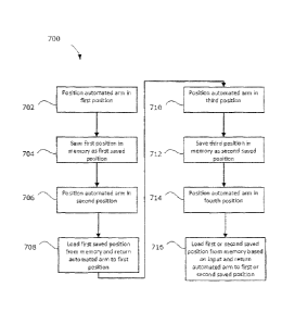
[0068] Referring now to FIG. 7, a flow chart is showing illustrating a
method
700 of configuring positions in a surgical positioning system according to one
aspect
of the present description. The method 700 may be executed on a medical
navigation system, such as the navigation system 205 (FIG. 2) that includes
the
control and processing unit 300 (FIG. 3), which may also include a surgical
positioning system, such as intelligent positioning system 508 (FIG. 5), for
positioning a payload during a medical procedure. The medical navigation
system
includes a robotic arm, such as automated arm 514, having a plurality of
joints.
The robotic arm forms part of the surgical positioning system and has an end
effector for holding the payload, such as end effector 518 (FIGS. 5 and 6).
The
medical navigation system further has an input device for providing input and
a
controller (e.g., control and processing unit 300) electrically coupled to the
robotic
arm and the input device. The controller may have a processor (e.g., processor
302) coupled to a memory (e.g., memory 304). The controller may be configured
to perform the method 700.
[0069] At a first block 702, the robotic arm (e.g., the automated arm 514)
is
positioned in a first position by providing a first positioning signal to the
robotic
arm. The robotic arm may be placed in the first position either under control
of the
navigation system 205 where a user is providing an associated input such as
through a graphical user interface, a foot pedal, a voice command, or any
other
suitable input, or the surgeon may simply grab the robotic arm and manually
position the robotic arm in the first position.
[0070] Next, at a block 704, the first position is saved in the memory as a
first saved position in response to a signal received from the input device.
The first
19
CA 02917654 2016-01-14
position may be saved in response to a control of the navigation system 205
where
a user is providing an associated input such as through a graphical user
interface, a
foot pedal, a voice command, or any other suitable input.
[0071] Next, at a block 706, the robotic arm (e.g., the automated arm 514)
is positioned in a second position by providing a second positioning signal to
the
robotic arm. The robotic arm may be placed in the second position either under
control of the navigation system 205 where a user is providing an associated
input
such as through a graphical user interface, a foot pedal, a voice command, or
any
other suitable input, or the surgeon may simply grab the robotic arm and
manually
position the robotic arm in the second position.
[0072] Next, at a block 708, the robotic arm may be returned to the first
position by loading the first saved position from the memory and providing the
first
positioning signal to the robotic arm when an associated input is received
from the
input device corresponding to a command to return to the first saved position.
The
input device may include a keyboard or mouse connected to the navigation
system
205. Input may be provided, such as through a graphical user interface, a foot
pedal, a voice command, or any other suitable input.
[0073] In one example, the first saved position may be defined by encoder
joint angles that are saved, where one angle is saved for each encoder
associated
with each of the plurality of joints. In one example, the automated arm may
have
six degrees of freedom, six joints, and six associated encoders. However, any
suitable number of joints and encoders may be used according to the design
criteria
of a particular application.
[0074] In one example, the input device may include any of a foot pedal, a
microphone providing voice input, a touch sensitive overlay on the display, a
mouse, and/or a keyboard. In one example, the payload includes a camera held
by
CA 02917654 2016-01-14
the end effector, such as the camera 307, 512, or 520, and electrically
coupled to
the controller, or a surgical tool held by the end effector.
[0075] In one example, the first position is manually positioned a first
time
based on input provided to the medical navigation system. The input may
include a
surgeon physically moving the robotic arm to the first position or the surgeon
moving the robotic arm to the first position by providing input using the
input
device.
[0076] Returning to FIG. 7, at a block 710, the robotic arm (e.g., the
automated arm 514) is positioned in a third position by providing a third
positioning
signal to the robotic arm. The robotic arm may be placed in the third position
either under control of the navigation system 205 where a user is providing an
associated input such as through a graphical user interface, a foot pedal, a
voice
command, or any other suitable input, or the surgeon may simply grab the
robotic
arm and manually position the robotic arm in the first position.
[0077] Next, at a block 712, the third position is saved in the memory as a
second saved position in response to a signal received from the input device.
The
third position may be saved in response to a control of the navigation system
205
where a user is providing an associated input such as through a graphical user
interface, a foot pedal, a voice command, or any other suitable input.
[0078] Next, at a block 714, the robotic arm (e.g., the automated arm 514)
is positioned in a fourth position by providing a fourth positioning signal to
the
robotic arm. The robotic arm may be placed in the fourth position either under
control of the navigation system 205 where a user is providing an associated
input
such as through a graphical user interface, a foot pedal, a voice command, or
any
other suitable input, or the surgeon may simply grab the robotic arm and
manually
position it in the fourth position.
21
CA 02917654 2016-01-14
[0079] Next, at a block 716, the robotic arm may be returned to either the
first saved position or the second saved position by loading the first saved
position
or the second saved from the memory and providing either the first or second
positioning signal to the robotic arm when an associated input is received
from the
input device corresponding to a command to return to the first or second saved
position. The input device may include a keyboard or mouse connected to the
navigation system 205, input such as through a graphical user interface, a
foot
pedal, a voice command, or any other suitable input.
[0080] In one example, each of the first and second saved positions
corresponds to a tool held by the end effector and the controller may be
further
configured to automatically position the robotic arm in one of the first and
second
saved positions when the corresponding tool is placed in the end effector. For
example, if a probe is placed in the end effector, the navigation system 205
may
automatically identify that the probe is in the end effector and automatically
go to a
corresponding position, such as the first saved position. Any saved position
may
correspond to any suitable tool, depending on the design criteria of a
particular
application and any user input to the navigation system 205.
[0081] While the example of a first and second saved position is provided
above, any suitable number of saved positions may be used according to the
requirements of the procedure being performed with the medical navigation
system
205.
[0082] In another example, the medical navigation system 205 may have a
tracking system (e.g., tracking system 321 interfacing with devices such as
camera
307 or 3D scanner 309) electrically coupled to the controller. The first saved
position or the second saved position may be defined relative to a position of
a
patient determined by the tracking system. The tracking system may be an
optical
tracking system having a camera for viewing optical tracking markers attached
to
the patient, a 3D scanning tracking system having a 3D scanner for tracking
the
22
CA 02917654 2016-01-14
patient, an electromagnetic tracking system having an electromagnetic sensor
for
tracking electromagnetic markers on the patient, or any other suitable type of
tracking system.
[0083] In yet another example, stored positions may automatically propagate
and be saved, at least temporarily, from a preplanned trajectory from the
navigation system 205 when using a tracking camera and patient reference. In
another example, when the robotic arm is automatically following a trajectory
based on the reference to the patient, at least some of the significant
positions
along this trajectory may automatically be saved as stored positions for use
later in
the procedure.
[0084] Referring now to FIG. 8, a block diagram is shown illustrating an
example of a virtual reality component view 800 according to one aspect of the
present description. In one example, the medical navigation system 205 may
have
an augmented reality component for providing a view of saved positions
including
the first saved position to a surgeon, thereby allowing the surgeon to easily
select
between saved positions. As shown in FIG. 8, a head of the patient 202 is
shown.
Relative to patient 202, the view 800 shows a current position 802 of the
automated arm 514, position 804 of the first saved position, and a position
806 of
the second saved position. The augmented reality component may include a
display, such as display 311, electrically coupled to the controller for
displaying
graphical illustrations of the saved positions to the surgeon, which makes it
much
easier for the surgeon to recall and identify which saved position he wishes
to
choose. In another example, the augmented reality component may include a
headset electrically coupled to the controller for displaying graphical
illustrations of
the saved positions to the surgeon. While a display and a headset are
described as
examples, any suitable device may be used to convey the information to the
surgeon.
23
CA 02917654 2016-01-14
[0085] In another example, the controller may be configured to execute a
redo command to return the automated arm 514 to the first position by loading
the
first saved position from the memory when an input is received from the input
device corresponding to the redo command. In another example, the controller
may be configured to execute the undo command to return the automated arm
to a previous position from a subsequent position by performing previous
movements in reverse even when the previous position has not been saved in
response to receipt of an input corresponding to the undo command. In other
words, the navigation system may automatically save some or all of previous
positions in memory, at least temporarily, in case the surgeon wishes to
revisit
these positions at a later time. The redo and/or undo commands may be
triggered
with any suitable input, including through a graphical user interface, a foot
pedal,
or a voice command.
[0086] The specific embodiments described above have been shown by way of
example, and it should be understood that these embodiments may be susceptible
to various modifications and alternative forms. It should be further
understood that
the claims are not intended to be limited to the particular forms disclosed,
but
rather to cover all modifications, equivalents, and alternatives falling
within the
spirit and scope of this disclosure.
24