Note: Descriptions are shown in the official language in which they were submitted.
CA 02917721 2016-01-07
Super-complex formed by cross-binding between
complexes of repeating chain and monomer and use
thereof
BACKGROUND OF THE INVENTION
1. Field of the Invention
The present invention relates to a super-complex
formed by a cross-binding between complexes of a
repeating chain and a monomer, and a method for
amplifying the biochemical effect of a monomer by the
same.
Particularly, a repeating chain/monomer complex
is formed by comprising a repeating chain of a binding
domain having a binding specificity to a monomer as an
active component, from which a super-complex is formed
by a cross-binding between the said complexes as the
form of an aggregate. This super-
complex comprises
multiple numbers of monomers, so that it can provide
the amplified monomer effect, specifically many times
greater biological and chemical effect on a monomer
target than a single monomer.
Herein the repeating chain can be prepared with
some domains or materials that display a binding
specificity to a natural monomer. The monomers
to
1
CA 029=1 2016-01-07
which the said binder could bind are in diversity.
Ligands, receptors, antibodies, and enzymes are the
examples. The effect
of a monomer can be increased
significantly by using multiple numbers of complexes
prepared with monomers and repeating chains and a
super-complex prepared with the complexes. In this
monomer, biological and chemical functional groups are
found as linked, conjugated, and fused, and at this
time the effect of such functional groups can be
amplified significantly.
2. Description of the Related Art
To detect a target, a monomer binds to the target
and generates a detection signal. At this time, the
intensity of the detection signal, the effect of the
monomer, is determined by the binding affinity between
the target concentration detection material and the
detection probe, the probe concentration, and the
strength of the probe signal. In
general, the probe
is a monomer, and the detection target material is a
target material of the monomer, and the signal
indicates the effect of the monomer.
Immunochromatographic assay, one of the detection
methods using antibody, is also called 'rapid antigen
test', 'lateral flow test', or simply 'strip test',
2
CA 02917721 2016-01-07
which is widely applied for the development of a
diagnostic kit. The target of diagnosis with the said
immunochromatographic assay, reported so far, includes
drug abuse, blood components analysis, group A
streptococcal antigen, Helicobacter pylori, human
Mycobacterium tuberculosis, hepatitis B surface
antigen/antibody, Dengue virus, influenza, parasite
(plasmodium falciparum for the diagnosis of malaria),
etc.
Immunochromatographic assay is simple and the
duration of the assay is only about 5 - 10 minutes,
which is very quick. The
preservation at room
temperature is excellent and the test cost is not
expensive. So, this
can be considered as the optimum
method for the diagnosis of a disease that can be
applied to various tests.
Colloidal gold is widely used as a chromogen for
the immunochromatographic assay, and the colloidal
gold-labeled immunoglobulin is widely used for the
diagnosis of disease. The immunoglobulin conjugated
colloidal gold particles have been applied for the
direct detection of antigen molecules. In 1981,
Leuvering et al developed gold particle agglutination
assay so called sol particle immunoassay (SPIA), and
further developed a pregnancy diagnostic kit using
thereof. Since then, membrane test methods using gold
3
CA 02917721 2016-01-07
have been reported for the diagnosis of diseases
caused by bacteria, viruses, parasites, and fungi,
etc. Rapid
antigen immunoassay has also been
developed as a quick field diagnosis method using
colloidal gold which is easy and simple and the
duration of the test is also as short as 5 - 10
minutes. Rapid
antigen test is important since it is
not only useful for the early diagnosis of human
infectious diseases but also useful for the early
diagnosis and prevention of animal infectious diseases
as well.
The problem of the rapid antigen test kit used
these days is that it is only effective within 2 - 3
days from the first acute disease symptom. For
example, in the case of diagnosis of a virus caused
disease, if the test is performed 3 days after the
first symptom, the rapid antigen test kit may show
negative result because the virus disappears rapidly 3
days after causing the symptoms. If a patient
is a
kid, the maintenance of the virus concentration is
longer, so that the test sample still can be picked up
after 5 days from the symptom development. However,
if a patient is an adult, the virus concentration
drops rapidly as time goes by, so the test sample has
4
CA 029=1 2016-01-07
to be picked up within 4 - 5 days from the symptom
development, suggesting that the antigen diagnosis is
limited according to ages. Another problem of the
conventional rapid antigen test kit is that the
sensitivity of the diagnostic kit reagent is lower
than expected, particularly to the low concentration
of the antigen concentration. Various
rapid antigen
test reagents for the diagnosis of various influenza
viruses have been developed world-widely and the
W sensitivity of these products increases continuously.
However, the sensitivity to seasonal diseases is only
60 - 83% and the sensitivity to the novel swine virus
is only 40 - 69%. The effect of three diagnostic
tests on the market in USA nowadays, 'BinaxNow', 'HZ
flu A+B (Becton, Dickinson and Company)', and
'Quickvue (Quidel)' was examined and as a result, the
H1N1 virus detection efficiency of BinaxNow was 40%,
which was the lowest, and Quickvue and HZ-flu A+B
displayed respectively the efficiency of 69% and 49%
(Centers for Disease Control and Prevention (CDC),
Evaluation of rapid influenza diagnostic tests for
detection of novel influenza A (H1N1) virus: United
States, 2009. Morb Mortal Wkly Rep 2009; 58:826-829).
The reason of such a low sensitivity of the
conventional rapid antigen test reagent is that the
5
CA 02917721 2016-01-07
virus concentration was not enough because either the
virus concentration in an infectee was very low or the
sample was not picked up from a patient under the
best/optimum condition to guarantee enough viruses for
the test. Therefore,
if a sample from a patient does
not contain enough amount of antigen even though the
patient has been diagnosed as infected with virus, it
is very difficult to diagnose a disease. So, the
study on the method and material useful for the
W detection of an antigen at a low concentration in a
sample is required.
Previously, the present inventors constructed a
complex comprising a monomer and a repeating chain by
using a repeating chain of the binder having a binding
specificity to the antigen Fab against such a monomer
as antigen-toxin as the matrix (scaffold); succeeded
in increasing the collision frequency of monomers by
increasing the local concentrations of monomers; and
accordingly developed a method to increase the yield
of a cross-linked multimer by promoting the formation
of a cross-binding (Korean Patent No. 10-1161323).
The present invention provides a method to mass-
produce a cross-linked multimer by promoting the
formation of a cross-binding between monomers by using
6
CA 02917721 2016-01-07
the repeating chain of a binder having a binding
specificity to the monomers. However,
the method to
amplify the effect of an antibody by applying such a
binding domain repeating chain having a binding
specificity to the monomer to immunoassay has not been
reported yet.
The study of the present inventors was focused on
the promotion of the detection sensitivity in the
course of diagnosis of a low concentration antigen.
As a result, the inventors confirmed that the
sensitivity to a low concentration antigen could be
increased when the repeating chain of a binding domain
having a binding specificity to an antibody monomer
was used as a signal amplifier for such antigen
detection methods as Western blotting, enzyme-linked
immunosorbent assay (ELISA), and FACS (fluorescence
activated cell sorter). The inventors accordingly
confirmed that an antibody could be efficiently used
as a detection monomer to amplify the effect of the
detection antibody monomer in an antigen detection
assay. There is
no limit in the pairs of a monomer
applicable to the present invention and a
corresponding binding domain.
7
CA 02917721 2016-01-07
SUMMARY OF THE INVENTION
It is an object of the present invention to
provide a super-complex formed by a cross-binding
between complexes of a repeating chain and a monomer,
precisely a super-complex generated by forming
complexes of a repeating chain and a monomer, which
contains the repeating chain of a binding domain
having a binding specificity to the monomer as an
active ingredient, and a cross-binding between the
complexes.
It is another object of the present invention to
provide a method for amplifying the effect of a
monomer by using the super-complex, wherein the super-
complex contains multiple numbers of monomers so that
it has many times higher biological/chemical effect on
the monomer targets than a single monomer can have,
resulting in the amplification of the monomer effect.
To achieve the above objects, the present
invention provides the following [1] - [13].
[1] The present invention provides a method for
preparing a repeating chain for the production of a
super-complex containing the step of producing a
8
CA 02917721 2016-01-07
repeating chain which comprises a single type of
binding domains or multiple types of binding domains
having at least two binding sites in a monomer and a
binding specificity to the monomer.
[2] The present invention provides a method for
preparing a complex of multiple numbers of
monomers/repeating chains for the production of a
super-complex, which comprises the following steps: 1)
preparing a repeating chain wherein a single type of
binding domains or multiple types of binding domains
having at least two binding sites in a monomer and a
binding specificity to the monomer are repeated; and
2) preparing a complex of multiple numbers of
monomers/repeating chains by mixing the repeating
chain of step 1) and the monomer having at least two
binding sites binding to the said repeating chain.
[3] The present invention provides a method for
preparing a super-complex, which comprises the
following steps: 1) preparing a repeating chain
wherein a single type of binding domains or multiple
types of binding domains having at least two binding
sites in a monomer and a binding specificity to the
monomer are repeated; 2) preparing a complex of
multiple numbers of monomers/repeating chains by
mixing the repeating chain of step 1) and the monomer
9
CA 02917721 2016-01-07
having at least two binding sites binding to the said
repeating chain; and 3) forming an aggregate of the
said complexes by forming a cross-binding between the
complexes of multiple numbers of monomers/repeating
chains of step 2).
[4] The present invention provides the repeating
chain prepared by the method of [1].
[5] The present invention provides the complex of
multiple numbers of monomers/repeating chains prepared
M by the method of [2].
[6] The present invention provides the super-
complex prepared by the method of [3].
[7] The present invention provides a method for
amplifying the effect of a monomer by forming the
super-complex binding to the monomer target by mixing
the repeating chain of [4], the complex of multiple
numbers of monomers/repeating chains of [5] or the
super-complex of [6], and the monomer target.
[8] The present invention provides a kit for the
analysis of biochemical functions, detection,
diagnosis, and treatment comprising the monomer which
is specific to a target of detection or to induce a
specific biochemical function and contains two or more
binding sites for the repeating chain in a monomer,
CA 02917721 2016-01-07
and a repeating chain of a binding domain having a
binding specificity to the monomer.
[9] The present invention provides a method for
preparing a repeating chain-biochemical functional
group which comprises the step of linking,
conjugating, or fusing a biological/chemical effector
group or a detection functional group to a repeating
chain of a binding domain having a binding specificity
to the monomer.
[10] The present invention provides a method for
preparing a complex of multiple monomers/repeating
chains-biological/chemical functional groups, which
comprises the following steps: 1) preparing a
repeating chain-biochemical functional group which
comprises the step of linking, conjugating, or fusing
a biological/chemical effector group or a detection
functional group to a repeating chain of a binding
domain having a binding specificity to the monomer;
and 2) preparing a complex of multiple
monomers /repeating chains-biological/chemical effector
groups by mixing the repeating chain-
biological/chemical effector group prepared in step 1)
and the monomer.
[11] The present invention provides the repeating
chain-biological/chemical effector group of a binding
11
CA 02917721 2016-01-07
domain that binds specifically to the monomer,
prepared by the method of [9].
[12] The present invention provides the complex
of multiple monomers/repeating chains-
biological/chemical effector groups prepared by the
method of [10].
[13] The present invention provides a method for
the analysis of biochemical functions, detection,
diagnosis, and treatment of a target, which comprises
M the step of forming a super-complex wherein complex of
multiple monomers /repeating chains-biological/chemical
effector groups of [12] is mixed with the monomer
target.
ADVANTAGEOUS EFFECT
In this invention, multiple numbers of monomers
bind to a repeating chain, leading to the formation of
a complex. Then, an insoluble super-complex is formed
by a cross-binding between the complexes, which is
precipitated when it is included at a high
concentration. The aggregation, precipitation, and
the size of the super-complex depend on the monomer
and the repeating chain structures. The
repeating
number of the binding domain in the repeating chain
CA 02917721 2016-01-07
affects the cross-binding between complexes. In the
meantime, the water-solubility and the molecular size
of the monomer affect the chances of cross-binding
between complexes. Since the
said super-complex
contains multiple numbers of monomers, it can provide
many times higher biological and chemical effect on
the monomer target than a single monomer, so that it
is useful for the amplification of a detection signal,
functional effect, and therapeutic effect by using an
antibody, the representative monomer.
BRIEF DESCRIPTION OF THE DRAWINGS
The application of the preferred embodiments of
the present invention is best understood with
reference to the accompanying drawings, wherein:
Figure 1 is a schematic diagram illustrating the
construction of an expression plasmid including GR1
10. Figure la
is a diagram illustrating the
construction of pGR2 vector pGR20 vector.
Precisely, pGR1 vector was first constructed and pGR2
pGR10 vectors were constructed by using the pGR1
vector and likewise pGR11 pGR20
vectors were
constructed. Each
plasmid has one G4S linker between
domain Ills. Figure lb illustrating that pGR2 series,
CA 02917721 2016-01-07
such as pGR2-2, pGR2-3, and pGR2-4, contain
respectively two, three, and four G4S linkers between
two domain Ills.
Figure 2 is a diagram illustrating the result of
SDS-PAGE with the repeating chain of the purified
protein G domain III. The
purified repeating chain
was analyzed by 16% SDS-PAGE. Lane 1 -
lane 13
indicate GR1, GR2, GR2-2, GR2-3, GR2-4, GR3, GR4, GR5,
GR6, GR7, GR8, GR9, and GR10 respectively.
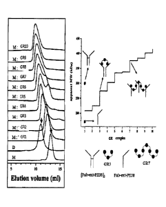
Figure 3 is a diagram illustrating the result of
size-exclusion chromatography of GR series complexes.
Two vertical arrows indicate the peaks of the
disulfide-dimer (left) [Fab-ext-PE38]2 and the monomer
(right) Fab-ext-PE38. The right table presents the
comparison of the molecular weights of GR complexes,
wherein the apparent molecular weights of a monomer
(U) and of disulfide-bridged dimer (0) are presented.
The schematic diagram illustrates the complex of GR3
or GR7 and Fab-ext-PE38.
Figure 4 is a diagram illustrating the result of
size-exclusion chromatography of GR2-2, GR2-3, and
GR2-4 complexes. A indicates size-exclusion
chromatography. One of the two vertical arrows, the
left arrow indicates the peak of [Fab-ext-9E38]2 and
the right arrow indicates the peak of Fab-ext-PE38. B
CA 02917721 2016-01-07
illustrates the result of size-
exclusion
chromatography with the complexes of Fab-ext-PE38 and
GR2-2, GR2-3, or GR2-4. The fractions were
electrophoresed on non-reducing 8% polyacrylamide gel.
The fractions corresponding to the elution volume of 9
ml - 15.5 ml were analyzed. The arrow indicates [Fab-
ext-PE38]2. The arrowhead indicates Fab-ext-PE38.
indicates the electrophoresis performed on reducing
12% polyacrylamide gel. The arrow, the arrowhead, and
W the white open arrow indicate respectively Fd-ext-
PE38, H6-L, and GR protein.
Figure 5 is a diagram presenting the result of
the analysis of complexes of [Fab-ext-PE38]2 and Fab-
ext-PE38 with GR2 or GR3. A indicates size-exclusion
chromatography. 395 gg of the
mixture of [Fab-ext-
PE38]2 and Fab-ext-PE38 was mixed with GR2 or GR3
protein. At this
time, 15 gg of GR protein was used.
The vertical arrow and the number indicate the
location of peaks. B presents
SDS-PAGE with the
eluted fraction. The mixture
of [Fab-ext-PE38]2 and
[Fab-ext-PE38] was used as the control, followed by
electrophoresis on non-reducing 8% acrylamide gel.
The fractions (#13 - #26) corresponding to the elution
volume of 7 ml - 12.5 ml were compared. The black
CA 02917721 2016-01-07
arrow and the white open arrow indicate respectively
[Fab-ext-PE38]2 and Fab-ext-PE38.
Figure 6 is a diagram illustrating the result of
size-exclusion chromatography with those proteins
prepared by mixing Fab-PE38 monomer and GR2-2, GR2-3,
or GR2-4. Those vertical arrows are the controls; the
right arrow indicates Fab-PE38 monomer and the left
arrow indicates disulfide-bridged dimer. The mixtures
of Fab-PE38 monomers and GR2-2 GR2-4 were
over-
lapped on chromatogram, suggesting that two Fab-PE38
monomers were included in the generated complex, which
became a complex in the form of monomer dimer.
Figure 7 is a diagram illustrating the result of
size-exclusion chromatography for the purification of
the complex of GR repeating chain and Fab-PE38
protein. The protein
was finally purified by size-
exclusion chromatography. All the chromatograms are
the records obtained at Opno- The chromatograms were
over-lapped according to the same elution volume. A:
Hiload superdex-75 pg (26/60) column was used for the
purification of GR1 - 6. B: Hiload
superdex-200 pg
(26/60) column was used for the purification of GR7
10. C: Hiload
superdex-200 pg (26/60) column was used
for the final purification of antibody-toxin.
CA 02917721 2016-01-07
Figure 8 is a diagram illustrating the result of
8% non-reducing SDS-PAGE proving the formation of
disulfide-bridged dimer from Fab-toxin monomer by
oxidation-reduction reaction in the complex of Fab-
toxin monomer and GR10 or GR2-2, GR2-3, or GR2-4.
First pathway: Fab-toxin monomer, the reaction
starting material. Second pathway: the sample reduced
at room temperature with 40 mM 2-mercaptoethanol for
30 minutes. Third pathway: the sample oxidized with 5
W mM glutathione oxidized form (GSSG) at 37 C for 2
hours. Arrows
indicate disulfide-bridged dimer, Fab-
toxin monomer, and Fd chain, respectively from the
top.
Figure 9 is a diagram illustrating the
amplification of chemiluminescence signal by Western
blotting with GR10 using the conventional Western
blotting reagents; Lane A: 20 gg of A431 whole cell
lysate (WCL); Lane 1: 2 jig of A431 WCL; Lane 2: 1 jig
of A431 WCL; Lane 3: 0.5 jig of A431 WCL; GR10 treated
Western blot: GR10 treated Western blotting provides
the 32-fold amplified signal, compared with the
conventional Western blotting.
Figure 10 is a diagram illustrating the
amplification of Western blot chemiluminescence signal
caused by GR10 repeating chain. a: 17 times
higher
CA 02917721 2016-01-07
signal provided by the super-complex prepared by the
complex of the mouse anti-3-actin monoclonal antibody
and GR10. b: the signal amplification by the super-
complex which was similar to that of nitrocellulose
membrane, wherein the super-complex was obtained after
the separation of A431 clear cell lysate by 10%
denaturing SDS-PAGE, followed by the transfer of the
product onto PVDF membrane.
Figure 11 is a diagram illustrating the increase
W of ELISA sensitivity by GR10. a: Each well was coated
with 1 g of AGS cell lysate. The primary antibody was
serially diluted. b: A graph
illustrating the
increase of OD of A450, compared with when the primary
antibody alone was treated, according to the different
molar ratios of the primary antibody to GR10 which was
added in order to form a super-complex, considering
the dilution rate of the primary antibody. c: The
primary antibody was fixed at the dilution rate of
1:120 and the AGS cell lysate was serially diluted.
Then, each well of a 96-well plate was coated with the
cell lysate. d: A graph
illustrating the increase of
OD of A450, compared with when the primary antibody
alone was treated, according to the molar ratio of the
primary antibody to GR10, which was added in order to
CA 02917721 2016-01-07
form a super-complex with considering the
concentration of the cell lysate coating each well.
Figure 12 is a diagram illustrating the effect of
signal amplification according to the serial dilution
of the secondary antibody.
Figure 13 is a diagram illustrating the increase
of detection sensitivity of a rapid antigen test kit
by GR10.
Figure 14 is a diagram illustrating the result of
W immunofluorescence with the human squamouse carcinoma
cell line A431 by using the GR1-FITC conjugate.
Figure 15 is a diagram illustrating the cross-
binding between the GR repeating chain and the test
line antibody in the course of rapid antigen test.
Figure 16 is a diagram illustrating the cross-
binding between the AR, LR, or LAR repeating chain and
the test line antibody in the course of rapid antigen
test.
Figure 17 is a diagram illustrating the increase
of detection sensitivity of a rapid antigen test kit
by GR5.
Figure 18 is a diagram illustrating the increase
of detection sensitivity of a rapid antigen test kit
by GR10.
CA 02917721 2016-01-07
Figure 19 is a diagram illustrating the increase
of detection sensitivity of a rapid antigen test kit
by GR15.
Figure 20 is a diagram illustrating the increase
of detection sensitivity of a rapid antigen test kit
by GR20.
Figure 21 is a diagram illustrating the increase
of detection sensitivity of a rapid antigen test kit
by AR5.
Figure 22 is a diagram illustrating the increase
of detection sensitivity of a rapid antigen test kit
by LR5.
Figure 23 is a diagram illustrating the increase
of detection sensitivity of a rapid antigen test kit
by LAR3.
Figure 24 is a diagram illustrating that GR10
formed a super-complex with IgG, and the super-complex
was precipitated without being dissolved in an aqueous
solution. IgG and
GR10 were mixed at the molar ratio
of 1:1, 5:1, and 10: 1, starting from the left,
followed by reaction.
Figure 25 is a diagram illustrating the result of
SDS-PAGE confirming that the GR/IgG super-complex
precipitate was greater when IgG was mixed with GR10
than when IgG was mixed with GR1 or GR2. 1/36 of each
CA 02917721 2016-01-07
sample was examined by non-reducing 15% SDS-
polyacrylamide gel. Lane 1 and lane 2 are
respectively the precipitates generated from the
reaction of IgG and GR10 and the supernatant. Lane 3
- lane 6 are the precipitates generated from the
reaction of IgG with GR1 and GR2 and the supernatants
in that order. Lane 7 and
lane 8 are the control
samples prepared by the reaction of BSA and IgG, and
lane 9 and lane 10 are the control samples containing
N IgG only.
Figure 26 is a diagram showing the precipitate
generated by the formation of GR/IgG super-complex.
GR1 GR10 were reacted with IgG. As the
controls,
BSA was reacted with IgG and IgG alone was selected,
followed by centrifugation at 13,000 rpm at 20 C for
30 minutes. The micro-
centrifuge tube was turned
upside down and the precipitate was confirmed by the
naked eye. The circle
indicates the formed
precipitate.
Figure 27 is a diagram illustrating the result of
SDS-PAGE that confirmed the precipitate generated by
the formation of GR/IgG super-complex. Lane 1 -
lane
20 indicate the precipitates generated by the reaction
of GR1 - 10 with IgG and the supernatants in that
order. Lane 21 and lane 22 are the controls which are
CA 02917721 2016-01-07
the result of reaction between BSA and IgG. Lane 21
and lane 22 are the control samples prepared by the
reaction of BSA and IgG, and lane 23 and lane 24 are
the control samples containing IgG only. 1/30 of each
sample was examined by reducing 15% SDS-polyacrylamide
gel.
Figure 28 is a diagram illustating the result of
SDS-PAGE to investigate whether or not GR greater than
GR10 also could form a super-complex with IgG. Lane 1
N - lane 12 are the precipitates generated from the
reaction of GR1, 3, 5, 10, 15, and 20 with IgG and the
suerpnatants in that order. Lane 13
and lane 14 are
the control sample precipitate made of only IgG and
the supernatant. 1/10 of
each sample was examined by
reducing 15% SDS-polyacrylamide gel.
Figure 29A is a diagram illustrating the
comparison of the two Western blotting results,
wherein the detection limit of Western blotting was
extended by GR which was compared with the result of
Western blotting without using GR. In Figure 298, the
result of Figure 29A was schemetized.
Figure 30A is a diagram illustrating the result
of Western blotting confirming the amplification of
chemiluminescence signal by GR in the presence of
22
CA 02917721 2016-01-07
equal amount of antigen. Figure 30B
is a graph
illustrating the result of Figure 30A.
Figure 31A is a diagram illustrating that the
amplification of Western blot signal by GR super-
complex was confirmed using other antigens. Figure
31B is a diagram illustrating that the amplification
of Western blot signal could be achieved by repeating
chain in the presence of different primary antibody.
Figure 32A is a diagram illustrating the
N comparison of the amplification levels of Western blot
signal by repeating chain. Precisely,
this is a
diagram illustrating the comparison of the
amplification levels Western blot signal by GR10,
AR10, and MAR5 (LAR5 repeating chain wherein L is
different.) in the same condition. Figure 32B is
a
diagram showing the result of Western blotting using
LR10.
Figure 33 is a diagram illustrating the cross-
binding between the repeating chains of GR series
proteins and the gold antibody complx or the super-
complex detection line antibody.
Figure 34 is a diagram illustrating the cross-
binding between the repeating chains of AR series
proteins and the gold antibody complx or the super-
complex detection line antibody.
CA 02917721 2016-01-07
Figure 35 is a diagram illustrating the cross-
binding between the repeating chains of LR series
proteins and the gold antibody complx or the super-
complex detection line antibody.
Figure 36 is a diagram illustrating the cross-
binding between the repeating chains of MAR series
proteins and the gold antibody complx or the super-
complex detection line antibody.
Figure 37 is a diagram illustrating the result of
M non-reducing SDS-PAGE by using the purified TR1, 3, 5,
10, 15, and 20 (=GR1, 3, 5, 10, 15, 20) proteins and
B3(Fab)-ext-PE38 attached thereon that were reduced by
2-mercaptoethanol and then oxidized into glutathione
oxidized form (GSSG) in order to produce [B3(Fab)-ext-
Figure 38 is a diagram illustrating the result of
SDS-PAGE by using the purified GR5, GR10, GR15, and
GR20 (=TR5, TRIO, TR15, TR20) proteins and
Herceptin(Fab)-ext-PE38 attached thereon that were
reduced by 2-mercaptoethanol and then oxidized into
glutathione oxidized form (GSSG) in order to produce
[Herceptin(Fab)-ext-PE38]2.
Figure 39 is a diagram illustrating the cytotoxic
effect of the complex of GR repeating chain protein
24
CA 02917721 2016-01-07
and [Herceptin(Fab)-ext-PE38]2 monomer on SKBR3 cells
and BT 474 cells.
Figure 40 is a diagram illustrating the cytotoxic
effect of the complex of GR repeating chain protein
and [e23(Fab)-ext-PE38] monomer on SKBR3 cells and BT
474 cells.
DESCRIPTION OF THE PREFERRED EMBODIMENTS
Hereinafter, the terms used in this invention are
defined.
The term "binder repeating chain" herein
indicates the recombinant protein generated by the
material wherein the site that has a binding
specificity to a monomer such as an antibody is
repeated. The site
that has a binding specificity
herein indicates a binding domain showing a binding
specificity to a monomer. In a
natural protein, the
domain that binds specifically to a monomer is the
site indicated.
The term "antibody monomer" used in this
invention indicates the molecule originated from an
antibody, which includes an antibody fragment or an
antibody conjugated with other proteins or functional
CA 02917721 2016-01-07
biological-chemical molecules, which is supposed to
bind to the repeating chain. A natural
antibody has
two heavy chains and two light chains. When one heavy
chain and one light chain make a pair in one unit, it
is called a dimer, but in this invention, when a
natural general antibody is conjugated with a
repeating chain, it is called an "antibody monomer".
The term "multiple antibody monomers/repeating
chain complex" in this invention indicates the complex
W prepared by contacting the said antibody monomer to
the repeating chain of a binder that specifically
binds to the said monomer.
The term "super-complex of multiple antibody
monomers/repeating chain complex" indicates the super-
complex that is the aggregate of complexes formed by a
cross-binding between the complexes prepared by
contacting the said antibody monomer to the repeating
chain of a binder that binds specifically to the said
monomer.
In addition, the term "antigen/multiple antibody
monomers/repeating chain complex" used in this
invention indicates the complex prepared by contacting
the said multiple antibody monomers/repeating chain
complex to an antigen.
CA 02917721 2016-01-07
The term "super-complex of antigen/multiple
antibody monomers/repeating chain complex" in this
invention indicates the conjugate of the super-complex
and the antigen generated by binding between an
antigen and the super-complex prepared by contacting
the super-complex that is the aggregate of the
complexes formed by a cross-binding between the
multiple antibody monomers/repeating chain complexes
to an antigen or by preparing the antigen/repeating
W chain mixture first and then adding this mixture to
the antibody monomer in the presence of an antigen to
form a super-complex.
Hereinafter, the present invention is described
in detail.
The present invention provides a method for
preparing a repeating chain containing the step of
producing a repeating chain wherein a single type of
binding domains or multiple types of binding domains
which bind specifically to the monomer and contain at
least two binding sites in one monomer are repeated.
The present invention also provides a method for
preparing a complex of multiple monomers/repeating
chain containing the step of preparing the multiple
CA 02917721 2016-01-07
monomers/repeating chain complex by mixing the
repeating chain prepared by the above method with
multiple numbers of monomers.
The present invention also provides a method for
preparing a super-complex containing the step of
forming an aggregate of the said complexes by a cross-
binding between the multiple monomers/repeating chain
complexes prepared by the method above.
In the above method, the monomer is preferably a
W protein, which is more preferably selected from the
group consisting of antibodies, ligands and receptors,
fragments thereof, recombinant conjugates thereof,
derivatives thereof, and biological/chemical effector
group conjugates.
In the above method, the antibody is preferably
selected from the group consisting of antibody
fragments, Fab fragments, Fab fragment containing
fragments, Fv fragments, Fv fragment containing
fragments, Fc fragments, and Fc fragment containing
fragments.
In the above method, the binding domain is
preferably a protein, and more preferably a
microorganism originated protein, which is more
preferably selected from the group consisting of
streptococcal protein G, Staphylococcus aureus protein
28
CA 02917721 2016-01-07
A, Peptostreptococcus magnus protein L, and
derivatives thereof.
In the repeating chain of the present invention,
a flexible linker chain can be included in order for
each domain to rotate freely in between the repeated
binding domains therein and to maintain a distance
between the domains. When monomers bind together, the
collision between the monomers cause stereoscopic
hindrance, resulting in the decrease of binding
reaction rate constant, which suggests that the
decrease of binding reaction equilibrium constant is
prevented so that high binding reaction equilibrium
can be obtained.
In the binding domain repeating chain, the
binding domain can rotate freely and chain bending is
also possible owing to the flexible linker in between
the binding domains. Therefore, in the complex formed
with a monomer and the repeating chain, each binding
monomer can have rotational freedom and vibrational
freedom along with bending freedom. So, each
binding
monomer can avoid collision between each other because
it is allowed to move freely not just in a limited
rotation direction or in a narrow bending angle but in
a wide direction range and a wide angle range, so that
29
CA 02917721 2016-01-07
multiple numbers of monomers can be bound to the
repeating chain at the same time. The flexible linker
sequence makes a big room between the binding domains,
so that many monomers that are approaching to the
repeating chain for the conjugation can bind to the
repeating chain without any stereoscopic hindrance
between each other. A natural
protein having the
binding domain that can be used for the repeating
chain of the present invention does not have such
flexibility between the binding domains. Natural
proteins do not allow such degree of freedom to their
binding domains inside, suggesting that a monomer that
is approaching thereto in order to form a complex with
multiple monomers or a monomer that has been bound
already thereto cannot have such a high level of
rotation freedom and vibration freedom.
The binding domain used for the repeating chain
of the present invention is preferably a natural
protein fragment, and this kind of natural protein
fragment has a lower molecular weight than a whole
natural protein. The
repeating chain having more
binding domains than a natural protein having a
comparatively high molecular weight, from which the
above fragment has been derived, and having a low
molecular weight can be constructed with the
CA 02917721 2016-01-07
artificially designed repeating chain prepared by
using such low molecular weight binding domains.
Accordingly, many monomers can bind to a low molecular
weight repeating chain. The
repeating chain that has
many binding domains but has a low molecular weight is
easy to produce and purify, which is a huge advantage
that the natural protein cannot have. In this
invention, a repeating chain wherein binding domain
repeats 20 times has been constructed, but the number
of repeating is not limited thereto.
When the repeating chain characterized by many
binding domains but a low molecular weight is used,
the effect of monomer can be amplified because the
rate of the monomer to binding molecule (natural
microorganism protein molecule, repeating chain in
this invention) is high at this time, which cannot be
obtained from a natural molecule. Since such
a
repeating chain has a low molecular weight, it is
useful for the production of a synthetic protein.
In this invention, the monomer has to have the
different sites a and b' and the binder has to have
the corresponding binding sites a and b, so that the
repeating chain has to have at least one of the
repeating a and b and the monomer has to have at least
CA 02917721 2016-01-07
one of each a' and b' in order to form a super-complex
comprising multiple monomers/repeating chain
complexes.
In this invention, the monomer has to have a' and
b' together in the form of (a'br), and the binding
domain in a repeating chain has to have the binding
sites a and b together to form the repeats of (ab)-
(ab)-(ab)----(ab) or the binding sites a and b stay
independently in different binding domains that make
the repeating in the form of a-b-a-b-a-b----a-b that
means the independent binding sites a and b are
repeated separately. The numbers and the order of the
independent domains a and b in the repeating chain are
not limited. When a=b,
the monomer is (a'a') and the
repeating chain is in the form of (aa)-(aa)-(aa)--- or
a-a a --- a a
In this invention, the repeating chain for the
formation of a complex by cross-binding can have the
' binding domains, c and d, that bind to each other, and
at this time these domains are independent and not
involved in the binding between the multiple monomers
and the repeating chain. If the
repeating chain is
composed of c-a a a ----------------------------------------------- d, the
said monomer binds to
the domain a and the cross-binding between complexes
is achieved by the binding between c and d. For
32
CA 02917721 2016-01-07
example, a super-complex can be composed of c-a-a-a---
--a-d...c a a ----- a a-d....c-a a a ------ a-d, wherein
indicates the cross-binding between the
repeating chains. The super-complex formed thereafter
can amplify the effect of the monomer bound to the
repeating chain. At this time in the structure of the
repeating chain, the domains c and d in one repeating
chain need to be firm not to be bended so as not to
bind to each other. If the
domains c and d bind to
each other in one repeating chain, the chances of the
cross-binding between the multiple monomers/repeating
chain complexes are very low, and accordingly the
chances of the formation of a super-complex becomes
very low. It is not
desirable either that the domain
c of one repeating chain binds to domain d of another
repeating chain before the cross-binding with a
monomer. If such a
linking between two different
repeating chains is made, the repeating chain is
difficult to control and therefore it is hard to form
a complex with monomers.
The present invention also provides a repeating
chain wherein a kind of binding domain or different
kinds of binding domains which bind specifically to
CA 02917721 2016-01-07
the monomer and have at least two binding sites in one
monomer are repeated.
The present invention also provides a complex of
multiple monomers/repeating chain wherein multiple
numbers of monomers are linked to the said repeating
chain.
The present invention also provides a super-
complex that is an aggregate of the complexes resulted
from the cross-binding between the multiple
monomers/repeating chain complexes.
The monomer herein is preferably a protein, which
is more preferably selected from the group consisting
of antibodies, ligands and receptors, fragments
thereof, recombinant conjugates thereof, derivatives
thereof, and biological/chemical effector group
conjugates.
The antibody herein is preferably selected from
the group consisting of antibody fragments, Fab
fragments, Fab fragment containing fragments, Fv
fragments, Fv fragment containing fragments, Fc
fragments, and Fc fragment containing fragments.
The binding domain herein is preferably a
protein, and more preferably a microorganism
originated protein, which is more preferably selected
from the group consisting of streptococcal protein G,
34
CA 02917721 2016-01-07
Staphylococcus aureus protein A, Peptostreptococcus
magnus protein L, and derivatives thereof.
The present invention also provides a method for
amplifying the effect of a monomer containing the step
of forming a super-complex on the target of the
monomer by mixing the repeating chain of the present
invention, the multiple monomers/repeating chain
complexes of the invention, or the super-complexes
W thereof.
In the above method, the step of measuring the
effect of the monomer on the target can be
additionally included.
In the above method, the target of the monomer is
preferably selected from the group consisting of
antigens, antibodies, peptides, proteins, bacteria,
viruses, and fungi, or their fragments, but not always
limited thereto.
The said bacteria are preferably selected from
the group consisting of Helicobacter pylori,
Mycobacterium tuberculosis, and Chlamydia trachomatis,
but not always limited thereto.
The said viruses are preferably selected from the
group consisting of influenza, foot-and-mouth disease
virus, human papilloma virus (HPV), dengue fever
CA 02917721 2016-01-07
virus, hepatitis C virus, hepatitis B surface antigen,
and hepatitis B surface antibody, but not always
limited thereto.
In the above method, the measurement of the
effect of the monomer is preferably performed by using
a monomer-marker conjugate or a secondary
detector(antibody)-marker conjugate, and a substrate
of a marker, but not always limited thereto.
The formation of a super-complex in this
invention is advantageous for the amplification of the
effect of a monomer including signal amplification
because the super-complex of the invention contains
multiple numbers of monomers to increase the
biological and chemical effect on the target of the
monomer which is many times higher than a single
monomer can provide.
The present invention provides an analysis kit
comprising the repeating chain of a binding domain
which is specific to a detection target and contains
multiple numbers of monomers having at least two
binding sites in one monomer and has a binding
specificity to the monomer.
The kit is preferably composed of the followings:
36
CA 02917721 2016-01-07
1) repeating chain of binding domain having a
binding specificity to monomer;
2) monomer specifically binding to the detection
target;
3) secondary probe conjugate labeled with a
marker activated by the reaction with a substrate;
4) substrate solution to react with the marker;
5) washing buffer for each reaction stage; and
6) stop solution to terminate the enzyme
W reaction, but not always limited thereto.
The kit herein can be used for the analysis
method selected from the group consisting of
immunohistochemical techniques,
immunoblot,
immunoprecipitation, enzyme linked immunosorbent assay
(ELISA), agglutination, immunochromatographic assay,
and radio-immuno assay.
The marker herein is preferably selected from the
group consisting of horseradish peroxidase (HRP),
alkaline phosphatase, colloid gold, fluorescein,
Quantum dot, glucose oxidase, luciferase, beta-D-
galactosidase, malate dehydrogenase (MDH),
acetylcholinesterase, isotope, and dye, but not always
limited thereto.
The substrate herein is preferably selected from
the group consisting of 3,3', 5,5'-tetramethyl
CA 029=1 2016-01-07
bezidine (TMB), 2,2'-azino-bis(3-ethylbenzothiazoline-
6-sulfonic acid) (ABTS), o-phenylenediamine (OPD),
diaminobenzidine (DAB), 3-amino-9-ethylcarbasole, 5-
bromo-4-chloro-3-indoly1
phosphate/iodonitrotetrazolium (BCIP/INT), new fuchin
(NF), and fast red TR salts, but not always limited
thereto.
The present invention provides a method for
N preparing a repeating chain-detection functional group
containing the step of linking, conjugating, or fusing
a detection functional group to a repeating chain of a
binding domain that binds to a monomer.
The present invention also provides a method for
preparing a complex of multiple monomers/repeating
chain-detection functional group comprising the
following steps:
1) preparing a
repeating chain-detection
functional group by linking, conjugating, or fusing a
detection functional group to a repeating chain of a
binding domain that binds to a monomer; and
2) preparing a
complex of multiple
monomers/repeating chain-detection functional group by
mixing multiple monomers with the repeating chain-
detection functional group prepared in step 1).
CA 02917721 2016-01-07
The present invention also provides a monomer
binding domain repeating chain-detection functional
group prepared by linking, conjugating, or fusing a
detection functional group to a repeating chain of a
binding domain that binds to a monomer.
The present invention also provides a complex of
multiple monomers/repeating chain-detection functional
group prepared by conjugating multiple numbers of
monomers to the monomer binding domain repeating
chain-detection functional group.
The present invention also provides a method for
detecting a target of a monomer containing the step of
forming a super-complex by mixing the multiple
monomers/repeating chain-detection functional group
complex of the invention with the monomer target.
In the above method, an additional step of
measuring the target detection level of a monomer can
be included.
In the above method, the detection functional
group is preferably selected from the group consisting
of Cy-3, Cy-5, FITC, GFP (green fluorescent protein),
REP (red fluorescent protein), and Texas Red, but not
always limited thereto.
In the above method, in case the monomer is an
antibody, the antibody is preferably selected from the
39
CA 02917721 2016-01-07
group consisting of antibody fragments, Fab fragments,
Fab fragment containing fragments, Fv fragments, Fv
fragment containing fragments, Fc fragments, and Fc
fragment containing fragments.
The present invention can be used for the
production of a cancer cell specific antibody-toxin
super-complex by using antibody-toxin (immunotoxin),
the anticancer agent. This
produced antibody-toxin
super-complex has an excellent drug efficacy so that
M the delivery of a large volume of cell killing
functional groups to a target cancer cell will be
possible, by which positive cancer treatment effect is
expected.
Practical and presently preferred embodiments of
the present invention are illustrative as shown in
the following Examples.
However, it will be appreciated that those
skilled in the art, on consideration of this
disclosure, may make modifications and improvements
within the spirit and scope of the present invention.
Example I: Preparation of repeating chains GR8 -
GR20, GR2-2, 2-3, 2-4 of protein G antibody binding
domain III
CA 02917721 2016-01-07
Protein G domain III gene was obtained from
chromosomal DNA of KCTC 3098 distributed from Korean
Collection for Type Cultures (KCTC). The plasmids
pGR2-2 pGR2-4
wherein G4S linker was repeated 2 - 4
times between two domain Fas were constructed. The
plasmid pGR1 (Y. Lee et al, Enhanced Formation of
Disulfide-bridged Dimer(Fab-9E38)2 Utilizing Repeats
of the Fab Binding Domain of Protein G (2010) J. Biol.
Chem. 285, 5127-5131) containing protein G domain III
W was inserted in the additional Agel restriction enzyme
site in front of the G4S linker at the end of the
domain III by using site-directed mutagenesis,
resulting in the construction of pGR1-A.
Quick-change site-directed mutagenesis was
performed with two primers P3 [5'-AGACCTTTAC
GGTAACTCAA ACCGGTGGAG GCGGGTCCGG ATA-3' (SEQ. ID. NO:
1)] and P4 [5'-TATCCGGACC CGCCTCCACC GGTTTCAGTT
ACCGTAAAGG TCT-3' (SEQ. ID. NO: 2)]. After the
mutagenesis, the nucleic acid sequence of pGR1-A was
confirmed by dedeoxy DNA sequencing. The plasmid
pGR1-A was digested with NdeI and BspEI and the
fragments obtained thereby were purified. Then, pGR1
was digested with AgeI located behind Ndel site and 6
His tag site. PGR2-A was
constructed by the ligation
of two fragments; the big fragment obtained by
CA 02917721 2016-01-07
digesting pGR1 with NdeI and AgeI and the small
fragment obtained by digesting pGR1-A with NdeI and
BspEI. And the
resultant pGR2-A was digested with
NdeI and AgeI to obtain a big fragment still harboring
the second domain III of protein G and G4S linker in
front of that. This big fragment was ligated with the
small fragment obtained by digesting pGR1-A with NdeI
and BspEI, resulting in the construction of pGR2-2
containing two G4S linkers in between the first domain
M III and the second domain III. Further, pGR2-3 having
three G4S linkers and pGR2-4 having 4 G4S linkers were
constructed by the same manner as used for the
construction of the above pGR2-2.
GR8- GR20 were prepared by the method described
in the paper of pGR1 (Y. Lee et al, Enhanced Formation
of Disulfide-bridged Dimer(Fab-PE38)2 Utilizing
Repeats of the Fab Binding Domain of Protein G (2010)
J. Biol. Chem. 285, 5127-5131). The construction of
an expression plasmid was performed by the method
shown in Figure 1 and the purified repeating chain was
analyzed by 16% SDS-PAGE (Figure 2).
Example 2: Size-exclusion chromatography with the
complex of Fab-toxin monomer and protein G domain III
repeating chains GR1 GR10, GR2-2,
2-3, and 2-4 and
42
CA 02917721 2016-01-07
the formation of disulfide-bridged dimer in the
complex
Protein G domain III that binds to immunoglobulin
can bind to IgG Fc and Fab fragments. Protein G
immunoglobulin binding site (domain III) is known to
bind to Fab fragment CH1, according to the previous
reports. The second
13-strand of protein G domain III
binds to the seventh 13-strand of Fab CH1 domain in
anti-parallel position. The p/p
interaction between
N these two proteins allows five hydrogen bonds between
CH1 domain and domain III. Besides,
three other
hydrogen bonds can also be found among major atoms of
CH1 domain. Despite
these two proteins form a
complex, no changes in the structure of domain III or
CH1 domain are found.
In this invention, two different types of
repeating chains having the repeats of domain III were
prepared. First, the
repeating chain having one G4S
amino acid between domain Ills was prepared. The
repeating chain wherein domain Ills were repeated 10
times therein was used for the association with Fab-
PE38 monomer to form a complex. The
repeating chains
used herein were GR1 GR10. When this
type of
repeating chain was associated with a monomer, the
CA 02917721 2016-01-07
complex comprising multiple numbers of monomers (2, 3,
or 4 monomers) could be prepared.
The present inventors also prepared the repeating
chain having G4S linkers in different lengths between
two domain Ills. There is a difference according to
the linker in the association of Fab-PE38 domain and
domain III. The repeating chains GR2-2, GR2-3, and
GR2-4 were accordingly prepared and they had 2, 3, and
4 G4S linkers between two domain Ills. So, two
Fab-
PE38 monomers were linked to a repeating chain to form
a complex and as a result the length of a dimer of the
monomers could be regulated.
The complexes formed by a noncovalent bond with
GR2-2, GR2-3, and GR2-4 were analyzed by size
exclusion column chromatography.
The results of size-exclusion chromatography with
GR series complexes are shown in Figure 3. The size-
exclusion chromatography was performed with Superdex-
200 TM HR column. The
association of Fab-ext-P538
with GR series was performed at different ratios. 715
jig of Fab-ext-PE38 was associated with each GR
protein. The association of Fab-ext-9E38 with GR1 -
GR3 was performed at the molar ratio of 2:1. 715 jig
of Fab-ext-PE38 was associated with 28 jig of GR4
GR10. All the
chromatograms were presented as over-
44
CA 02917721 2016-01-07
lapped according to the elution volume. Chromatograms
of [Fab-ext-PE38)2(D) and Fab-ext-PE38(M), used as the
standard materials, were obtained by the elution under
the same condition.
The results of size-exclusion chromatography with
GR2-2, 2-3, and 2-4 are shown in Figure 4.
The results of the investigation of the complex
of [Fab-ext-PE38]2 and Fab-ext-PE38 mixture with GR2
or GR3 are shown in Figure 5.
The results of size-exclusion chromatography with
the complex of Fab-PE38 monomer and GR2-2, 2-3, and 2-
4 are shown in Figure 6.
The results of size-exclusion chromatography for
the purification of the complex of GR repeating chain
and Fab-PE38 protein are shown in Figure 7.
The results of 8% non-reducing SDS-PAGE
confirming the formation of disulfide-bridged dimer
from Fab-toxin monomer by reduction/oxidation mixed
reaction in the complex of Fab-toxin monomer and GR10
or GR2-2, 0R2-3, and GR2-4 are shown in Figure 8
(Figures 3 - 8).
As a result, it was confirmed that two Fab-PE38
monomers could form complexes with a repeating chain
respectively with forming a dimer as well.
45
CA 02917721 2016-01-07
[Table 1]
Ratio of the disulfide-bridged dimer formed by
reduction/oxidation mixed reaction from Fab-toxin
monomer
Antibody-toxin bound to metal
chelating bead action of
disulfide dimer
B3(Fab)-ext-PE38] n.d.*
B3(Fab)-ext-PE38]:GR1 n.d.*
B3(Fab)-ext-PE38]:GR2 n.d.*
33(Fab)-ext-PE38]:GR3 0.7
B3(Fab)-ext-PE38]:GR4 0.5
B3(Fab)-ext-PE38]:GR5 0.6
B3(Fab)-ext-2E38]:GR6 0.3
83(Fab)-ext-9E381:GR7 0.6
B3(Fab)-ext-PE38]:GR8 0.1
B3(Fab)-ext-9E38]:GR9 0.1
B3(Fab)-ext-PE38]:GR10 0.3
B3(Fab)-ext-PE38]:GR2-2 0.2
B3(Fab)-ext-PE38]:GR2-3 0.4
B3(Fab)-ext-PE38]:GR2-4 0.3
*n.d: not determined.
Example 3: Amplification of antibody signal by protein
G domain III repeating chain GR10 confirmed by Western
W blotting
46
CA 02917721 2016-01-07
Western blotting protocol provided by cell
signaling technology was used with slight
modification and the revised version of direct ELISA
protocol provided by abcom was used.
Solutions and reagents used for this experiment
were prepared as follows: Solutions were prepared by
using water filtered with Milli-Q or water with equal
purity. 1) 1X SDS
sample buffer: 62.5 mM Tris-HC1
(25 C, pH 6.8), 2% W/V SDS, 10 % glycerol, 50 mM DTT,
0.01 % W/V bromophenol blue or phenol red. 2) mobile
buffer : 25 mM tris base, 0.2 M glycine. 3) 10X
tris
buffered saline (TBS) : 10X TBS 1 L: 24.2 g tris base,
80 g sodium chloride, acidity was regulated with HC1
(pH 7.6) (1X). 4) ovalbumin: (weight to volume [W /
V]). 5) blocking
buffer: 1X TBS, 0.1% tween-20, and
2% W/V chicken serum albumin. 6) washing
buffer: 1X
TBS, 0.1% tween-20 (TBS/T). 7) primary
antibody:
anti-beta-actin mouse antibody (Santa Cruz Biotech).
8) primary antibody dilution buffer: 1X TBS, 0.1%
tween-20, and 2% W/V chicken serum albumin. 9)
secondary antibody: goat anti-mouse beta actin HRP.
10) blotting membrane: nitrocellulose membrane
(Wattman), PVDF membrane (PALL). 11) GR
recombinant
protein: GR10. 12)
luminal solution: 100 mM Tris/HC1
pH8.8, 1.25 mM luminol, 2 mM 4IPBA, 5.3 mM hydrogen
47
CA 02917721 2016-01-07
peroxide. 13) super
signal femto maximum sensitivity
reagent (Thermo Scientific).
Western blotting was performed as follows. A
cell lysate was first prepared by using A431 or AGS
cancer cell line and then the culture medium was
eliminated by suction.
Particularly, the cells were
washed with 1X PBS and then PBS was also eliminated by
suction. Then, 1X SDS sample buffer was added thereto
in order to lyse the cells, followed by heating at 95
M 100r for 5 minutes.
Centrifugation was performed
for 5 minutes to precipitate the sample. Then, the
precipitate was placed on SDS-PAGE gel (10 cm x 10
cm), followed by electrophoresis. Then, the
sample
was transferred onto a nitrocellulose membrane or a
PVDF membrane.
Blocking the membrane surface and antibody
binding reaction were performed as follows. The
volume of solution was adjusted to fit the size of the
membrane which was 10 cm x 10 cm (100 cm2). The
volume was regulated according to the size of
membranes.
For the membrane surface blocking, the
nitrocellulose or PVDF membrane was washed with a
proper volume of TBS at room temperature for 5 minutes
after the transfer. The membrane
was loaded in a
48
CA 02917721 2016-01-07
proper volume of membrane surface blocking buffer,
which was warmed up at room temperature for 1 hour.
The membrane was washed with TBS/T for 5 minutes three
times. Then, the
super-complex was prepared.
Particularly, GR10 was mixed with the primary antibody
according to the molar ratio listed in the shown
result, followed by warming at 37 C for 1 hour. The
produced super-complex was placed in ice and stored as
in ice until it would be used. Then, 10 in of primary
antibody dilution buffer was mixed with the
membrane/primary antibody complex or the primary
antibody/repeating chain complex at the dilution ratio
listed in the shown result in order to induce primary
antibody reaction, followed by warming at room
temperature for 1 hour. The mixture
was washed with
15 la of TBS/T three times for 5 minutes each. HRP-
conjugated secondary antibody (1:2000) which had been
properly selected to match the primary antibody was
mixed with the membrane surface blocking buffer at a
proper volume, followed by stirring softly for 1 hour
at room temperature to keep it warm. The mixture was
washed with 15 of TBS/T
three times for 5 minutes
each. Detection in section D was performed. Lastly,
for the detection of an antigen protein, luminol
solution or super signal femto maximum sensitivity
49
CA 02917721 2016-01-07
reagent (Thermo Scientific) was mixed with the
membrane, which was softly stirred at room temperature
to keep it warm. The
excessive developing solution
was taken away with leaving it not to dry and then the
membrane was rapped with plastic lab, followed by
exposure on X-ray film.
The results of the chemiluminescence signal
amplification by GR10, confirmed by Western blotting,
are shown in Figure 9 (Figure 9).
The results of the chemiluminescence signal
amplification by GR10 repeating chain, confirmed by
Western blotting, are shown in Figure 10 (Figure 10).
Particularly, the super-complex of GR10 and the
primary antibody was prepared according to the molar
ratio shown in the bracket in order to form a super-
complex. The
primary antibody was diluted at the
ratio of 1:1000 and the super-complex was prepared at
the same concentration as that of the primary
antibody. The goat anti-mouse-HRP conjugated antibody
was used as the secondary antibody for both
experiments. The primary and the secondary antibodies
were all kept warm at room temperature for 1 hour.
All the cell lysate samples were separated by 10%
denaturing SDS-PAGE, which were then transferred onto
nitrocellulose membrane. This membrane
was then cut
CA 02917721 2016-01-07
into three pieces, which were detected with the
primary antibody. Theme
Supersignal Femto substrate
displaying a high sensitivity was used as the
fluorescence color developing reagent. A431 clear
cell lysate was separated by 10% denaturing SDS-PAGE
and then transferred onto PVDF membrane in order to
investigate whether or not the resultant super-complex
could give as much signal amplification effect as
nitrocellulose membrane. The
membrane was first
detected only with the primary antibody and then color
development was induced by using the secondary
antibody and the conventional ECL reagent. The
membrane was washed with TEST solution, followed by
inducing color development by using Supersignal Femto
substrate. As a result, a very strong background
noise signal was detected along with the increase of
the regular signal. This
background noise signal was
attributed to the non-specific conjugation of the
secondary antibody to PVDF membrane and the high
sensitivity of Supersignal femto substrate.
Thereafter, the antibodies were peeled off from the
membrane by using SDS and 2-mercaptoethanol. The
super-complex prepared by mixing the primary antibody
and the repeating chain was linked thereto, followed
by detection by using the secondary antibody and the
CA 02917721 2016-01-07
conventional ECL color developing reagent. At this
time, the super-complex demonstrated 15 times higher
sensitivity than the conventional one, which was
consistent result with the one observed on
nitrocellulose membrane. In this
experiment, the
conventional chemiluminescence reagent displaying a
medium level of sensitivity was used and the ECL
reagent prepared by the method of Haan and Behmann
(2007) was used. The signal
amplification effect was
W similar to that observed on the nitrocellulose
membrane and the amplified signal was as high as when
Supersignal Femto substrate was used. However, this
time the background noise signal was not as increased
as before, suggesting that the signal amplification
this time yielded clearer signals. A cell lysate
tends to attach on PVDF easily. So, the
color
developing reagent displaying a medium level of
sensitivity was good enough to detect a small amount
of sample.
Example 4: Amplification of antibody signal by
repeating chain GR10 of protein G domain III confirmed
by enzyme-linked immunosorbent assay (ELISA)
Indirect ELISA was performed as follows.
52
CA 02917721 2016-01-07
Solutions and reagents used for this experiment
were prepared as follows: Solutions were prepared by
using water filtered with Milli-Q or water with equal
purity. 1)
bicarbonate/carbonate coating buffer (100
mM), Antigen or antibody was diluted in a coating
buffer for fixation in wells: 3.03 g of Na2CO3, 6.0 g
of NaHCO3, 1000 HO, of distilled water (pH 9.6). 2)
PBS. 3)
blocking buffer: PBS containing 1% BSA serum.
4) washing buffer: PBS containing 0.05% (v/v) Tween
W 20. 5)
antibody dilution buffer: Antibodies were
diluted in 1X blocking buffer in order to reduce non-
specific binding of the primary and secondary
antibodies.
For the antigen coating on a microplate, A431 or
AGS cell line was used for the preparation of a cell
lysate. The cell
lysate was diluted with carbonate
coating buffer to make the final concentration 20
jig/iii. Then, 50
ig of the diluted cell lysate was
distributed on the top of the micro-plate well,
followed by coating. The plate was covered and stayed
at 4 C for overnight. The
coating buffer was
eliminated and each well was washed with 200 ge of
PBS. The plate
was turned upside down and shaken in
the sink in order to eliminate the used coating buffer
CA 02917721 2016-01-07
or washing buffer. The
remaining solution was
eliminated with paper towel by tapping softly.
For the membrane surface blocking, 200 gi of 1%
BSA/PBS blocking buffer was added to each well of the
plate to block the protein binding site that remained
uncoated in the coated well. Then, the
plate was
covered with a lid and kept warm at room temperature
for at least 2 hours, followed by washing with PBS
twice. To induce
antibody reaction, 100 a of the
primary antibody diluted at the designated
concentration right before being used or the super-
complex was added to the blocking buffer, and then the
plate was covered with a lid and kept warm at room
temperature for 1 hour. Then, the
plate was washed
with PBS twice and 100 0 of the secondary antibody-
HRP diluted at the optimum concentration was added to
each well of the plate. The plate was covered with a
lid and kept warm at room temperature for 1 hour. The
plate was washed with PBS twice.
For the detection, 100 Ite of TMB substrate
solution was added to each well of the plate by using
a multichannel pipette. When the color development
was fully induced (30 minutes), 100 0 of stop
solution was added to each well of the plate.
54
CA 02917721 2016-01-07
Absorbance (optical density) of each well was measured
by using a plate reader.
As a result, the sensitivity of ELISA was
significantly increased by GR10, which is shown in
Figure 11 (Figure 11). Precisely,
two different
ELISAs were performed. The
significant amplification
of ELISA signal was observed when the super-complex of
monoclonal anti-p-actin antibody and GR10 was used as
the primary antibody. AGS cell lysate was placed in a
W general 96-well cell culture plate, which stayed at
4 C for overnight. ELISA was
performed according to
the standard protocol. The super-
complex of GRID and
the primary antibody was prepared according to the
molar ratio shown in the bracket. The
primary
antibody used herein was the mouse monoclonal anti-3-
actin antibody. The goat
anti-mouse-HRP conjugated
antibody was used as the secondary antibody for both
experiments. Both the
primary and the secondary
antibodies were reacted at room temperature for 1
hour. The substrate
TMB was reacted at room
temperature for 30 minutes.
The signal amplification effect according to the
serial dilution of the secondary antibody is shown in
Figure 12. ELISA signal was significantly increased
by the super-complex of the primary antibody and GR10.
CA 02917721 2016-01-07
The signal was quite regular in the tested dilution
range of the secondary antibody. When the super-
complex of the mouse monoclonal anti-3-actin antibody
and GR10 was used as the primary antibody for the
above ELISA, a significant signal amplification was
observed. At this time, A431 cell lysate was used.
The complex of GR10 and the primary antibody was
prepared according to the molar ratio of 1:10. The
goat anti-mouse HRP conjugate was used as the
W secondary antibody. Each well was coated with 1 g of
A431 cell lysate. The primary
antibody was serially
diluted 10-fold. The primary
antibody was diluted
according to the designated dilution ratio and the
secondary antibody was serially diluted 2-fold.
Example 5: Increase of sensitivity of influenza rapid
antigen test kit by repeating chain GR10 of protein G
domain III
The increase of sensitivity of influenza rapid
antigen test kit, the most representative diagnostic
kit using an antibody, by GR10 was observed. SD
Bioline influenza antigen rapid test kit or the kit
provided by Korea Green Cross Co was used herein. The
kit included antigen buffer, dropper, tube, cotton
swab for the collection of sample, and strip. The
56
CA 02917721 2016-01-07
antigen buffer was sucked up to the dropper line and
then the antigen buffer was placed in the tube. An
antigen sample was loaded in the tube containing the
antigen buffer, followed by well-mixing at least 5
times. At this
time, GR10 protein was simply diluted
with an antigen in the antigen buffer. The strip
was
added into the tube, which was read 10 - 15 minutes
later.
As a result, when GR10 protein was simply diluted
W together with an antigen in the conventional rapid
antigen test kit antigen dilution buffer, the antigen
could be detected until it was 1000-times diluted,
unlike in the absence of GR10 (Figure 13).
Example 6: Immunofluorescence with a cancer cell line
using GR protein as the antibody labeling reagent
GR1 was conjugated with
fluorescein
isothiocyanate (FITC), the fluorochrome, and the
conjugated GR1-FITC was used for immunofluorescent
staining of A431 (human squamous carcinoma), the
cultured cancer cell line. Mouse anti-LC3 antibody
was used as the primary antibody. The
prepared GR1-
FITC was used instead of the secondary antibody for
the detection. The cells
detected by fluorescence
were stained with F-actin specific rhodamine-
57
CA 02917721 2016-01-07
phalloidin (Sigma Aldrich). The color development was
observed under fluorescent microscope.
As a result, compared with when the secondary
antibody-fluorochrome conjugate was used, much smaller
amount of protein could be detected by using the GR1-
FITC conjugate. Clear immunofluorescent staining
images could be obtained (Figure 14).
Therefore, like the GR1-FITC, GR protein can be
linked, conjugated, or fused with a detection
W functional group, and the resultant conjugate can be
usable for any kinds of antibodies and thus there is
no need to conjugate a detection functional group to
each antibody. GR protein
is a very small molecule,
so that the production of GR protein is easy and the
handling thereof is also simple and easy.
Example 7: Construction of the plasmid expressing
repeating chain of Staphylococcus aureus protein A
antibody binding domain B
The plasmid prepared by introducing the
synthesized protein A domain B (Table 1) DNA sequence
into a vector was distributed from Bioneer
Corporation. E. coil
DH5a was transfected with the
plasmid and cloned.
58
CA 02917721 2016-01-07
The said plasmid was digested with NdeI and
BspEI. The resultant small DNA fragment (245 bp) was
purified. The DNA
fragment was cloned into pGR1
(pTR1) vector which was digested with the same
enzymes, resulting in the construction of pAR1
containing one protein A domain B. Then, DNA sequence
of the above domain B was confirmed by dideoxy DNA
sequencing.
There is (G4S)2 sequence composed of 10 amino
W acids as a spacer between each domain B. To construct
pAR2 wherein protein A domain B repeats two times, the
pAR1 (Table 2) constructed above was digested with
NdeI and BspEI. Then, the small DNA fragment (245 bp)
encoding two G4Ss and domain B was conjugated to the
big fragment of the same plasmid digested with NdeI
and AgeI. The
resultant pAR2 was digested with NdeI
and AgeI. The obtained big fragment was conjugated to
the small fragment (245 bp), resulting in the
construction of pAR3 wherein protein A domain B
repeats three times. By the same
cloning method as
described above, pAR5 wherein protein A domain B
repeats 5 times has been constructed.
[Table 21
Nucleotide sequence of protein A domain B
59
CA 02917721 2016-01-07
Nucleotide sequence of protein A domain B
5'-
CATATGCATCATCATCATCACCACACCGGTTCTCAAGCCCCAAAAGCCGACAA
TAAATTTAATAAAGAGCAGCAGAACGCGTTTTATGAAATCTTGCATCTGCCGA
ATCTGAATGAAGAACAACGTAACGGATTCATTCAGAGCCTTAAAGATGATCCT
AGTCAGTCCGCTAACTTACTCGCAGAAGCTAAGAAACTGAATGATGCACAGGC
GCCGAAGGGAGGGGGTGGATCCGGTGGTGGCGGCTCCGGATAAGAATTC-
3'(SEQ. ID. NO: 3)
* underlined part: coding sequence
[Table 3]
Plasmid and protein used in this invention
Plasmid Protein Reference
pLR1-5 LR1-5 : (His ) (B1¨G4S¨GLIS)rif This
n=1-5. a, b, c.
invention
aG4S: amino acid sequence of GGGGS (SEQ. ID. NO:
4);
b(His)6: 6x histidine tag; and
CB: Staphylococcus aureus protein A domain B.
The protein was over-expressed from the repeating
chain construct via the conventional method reported
previously (J.H. Park, et al., Mol Cells 12 (2001)
398-402).
The pure lysate was separated by Ni-chelating
sepharose fast flow chromatography (Amersham
CA 02917721 2016-01-07
Bioscience, Sweden), followed by size-exclusion
chromatography using Hiload Superdex-75 pg or Hiload
Superdex-200 pg(26/60) (Amersham Bioscience, Sweden).
Example 8: Construction of the plasmid expressing
repeating chain of Peptostreptococcus magnus protein L
antibody binding domain B1
The plasmid prepared by introducing the
synthesized protein L domain B1 DNA sequence into a
W vector was distributed from Bioneer Corporation. E.
coil DH5a was transfected with the plasmid and cloned.
The said plasmid was digested with NdeI and
BspEI. The resultant small DNA fragment (299 bp) was
purified. The DNA
fragment was cloned into pGR1
(pTR1) vector which was digested with the same
enzymes, resulting in the construction of pLR1
containing one protein L domain B1. Then, DNA
sequence of the above domain B1 was confirmed by
dideoxy DNA sequencing.
There is (G4S)2 sequence composed of 10 amino
acids as a spacer between each domain Bl. To
construct pLR2 wherein protein L domain B1 repeats two
times, the pLR1 (Table 5) constructed above was
digested with NdeI and BspEI. Then, the
small DNA
fragment (299 bp) encoding two G4Ss and domain B1 was
CA 02917721 2016-01-07
conjugated to the big fragment of the same plasmid
digested with NdeI and AgeI. The
resultant pLR2 was
digested with NdeI and AgeI. The
obtained big
fragment was conjugated to the small fragment (299
bp), resulting in the construction of pLR3 wherein
protein L domain B1 repeats three times. By the same
cloning method as described above, pLR5 wherein
protein L domain B1 repeats 5 times has been
constructed.
[Table 4]
Nucleotide sequence of protein L domain B1
Nucleotide sequence of protein L domain B1
5'-
CATATGCATCACCATCACCATCATACCGGTATCAAGTTCGCCGGTAAAGAAGAAACGC
CGGAAACCCCTGAGACAGACAGTGAAGAGGAAGTGACAATAAAAGCAAATCTGATTTT
CGCCAACGGGTCAACCCAGACGGCCGAATTCAAAGGGACATTTGAAAAAGCAACTTCT
GAGGCTTATGCATACGCGGACACTCTGAAGAAGGATAATGGTGAATATACCGTAGATG
TTGCTGATAAAGGTTATACCCTGAATATTAAATTTGCGGGTGGCGGCGGCGGAAGCGG
TGGCGGAGGTTCCGGATAAGAATTC-3'(SEQ. ID. NO: 5)
* underlined part: coding sequence
[Table 5]
Plasmid and protein used in this invention
Plasmid Protein Reference
pLR1-5 LR1-5: (His)6-(B1-G4S-G4S)n, This
invention
62
CA 02917721 2016-01-07
n=1-5. a'
aG4S: amino acid sequence of GGGGS (SEQ. ID. NO:
4);
(His)6: 6x histidine tag; and
CB1: Peptostreptococcus protein L domain Bl.
The protein was over-expressed from the repeating
chain construct via the conventional method reported
previously (J.H. Park, et al., Mol Cells 12 (2001)
398-402).
The pure lysate was separated by Ni2+-chelating
sepharose fast flow chromatography (Amersham
Bioscience, Sweden), followed by size-exclusion
chromatography using Hiload Superdex-75 pg or Hiload
Superdex-200 pg(26/60) (Amersham Bioscience, Sweden).
Example 9: Construction of the plasmid expressing
repeating chain of Peptostreptococcus magnus protein L
antibody binding domain B1 and Staphylococcus aureus
protein A antibody binding domain B
The plasmid prepared by introducing the
synthesized protein L domain Bl DNA sequence into a
vector was distributed from Bioneer Corporation for
the construction of pLR1.
CA 02917721 2016-01-07
The said plasmid pLR1 was digested with NdeI and
BspEI. The resultant small DNA fragment (299 bp) was
purified. The DNA
fragment was cloned into pAR1
(including protein A domain B) vector which was
digested with the same enzymes, resulting in the
construction of pLAR1 containing one protein L domain
B1 linked to one protein A domain B (131-B). Then, DNA
sequences of the above domain El (protein L domain Bl)
and domain B (protein A domain B) were confirmed by
W dideoxy DNA sequencing.
There is (G4S)2 sequence composed of 10 amino
acids as a spacer between each domain B1 (protein L
domain B1) and domain B (protein A domain B). To
construct pLAR2 wherein LA sequence (B1-B, protein L
domain Bl - protein A domain B) repeats two times, the
plasmid pLAR1 (Table 6) obtained above was digested
with NdeI and BspEI. Then, the
small DNA fragment
(521 bp) encoding B1-(G4S)2-B-(G4S)2 (131-B) Was
conjugated to the big fragment of the same plasmid
digested with NdeI and AgeI. The resultant
pLAR2 was
digested with NdeI and AgeI. The
obtained big
fragment was conjugated to the small fragment (521
bp), resulting in the construction of pLAR3 wherein LA
sequence repeats three times. By the same cloning
method as described above, pLAR3 wherein protein L
64
GA 020=1 2016-01-07
domain B1 - protein A domain B sequence repeats 3
times has been constructed.
[Table 6]
Nucleotide sequence of protein LA domain Bl-B
Nucleotide sequence of protein LA domain El-B
5'-
CATATGCATCACCATCACCATCATACCGGTATCAAGTTCGCCGGTAAAGAAGAAACGC
CGGAAACCCCTGAGACAGACAGTGAAGAGGAAGTGACAATAAAAGCAAATCTGATTTT
CGCCAACGGGTCAACCCAGACGGCCGAATTCAAAGGGACATTTGAAAAAGCAACTTCT
GAGGCTTATGCATACGCGGACACTCTGAAGAAGGATAATGGTGAATATACCGTAGATG
TTGCTGATAAAGGTTATACCCTGAATATTAAATTTGCGGGTGGCGGCGGCGGAAGCGG
TGGCGGAGGTTCCGGTTCTCAAGCCCCAAAAGCCGACAATAAATTTAATAAAGAGCAG
CAGAACGCGTTTTATGAAATCTTGCATCTGCCGAATCTGAATGAAGAACAACGTAACG
GATTCATTCAGAGCCTTAAAGATGATCCTAGTCAGTCCGCTAACTTACTCGCAGAAGC
TAAGAAACTGAATGATGCACAGGCGCCGAAGGGAGGGGGTGGATCCGGTGGTGGCGGC
TCCGGATAAGAATTC-3'(SEQ. ID. NO: 6)
* underlined part: coding sequence
[Table 7]
Plasmid and protein used in this invention
Plasmid Protein Reference
pLAR1-3 LAR1-3: (His)6-(B1-G4S-G4S-B-G4S-G4S), This
n=1-3. a,b,c,d invention
aG4: amino acid sequence of GGGG (SEQ. ID. NO:
7);
CA 02917721 2016-01-07
(His)6: 6x histidine tag;
cB1: Peptostreptococcus protein L domain Bl; and
B: Staphylococcal protein A domain B.
The repeating chain construct was over-expressed
by the conventional method reported previously (J.H.
Park, et al., Mol Cells 12 (2001) 398-402).
The pure lysate was separated by Ni2+-chelating
sepharose fast flow chromatography (Amersham
Bioscience, Sweden), followed by size-exclusion
chromatography using Hiload Superdex-75 pg or Hiload
Superdex-200 pg(26/60) (Amersham Bioscience, Sweden).
Example 10: Investigation of cross-binding between
repeating chain and gold antibody complex or super-
complex and detection line antibody by using rapid
antigen test
The most representative detection method using an
antibody, influenza rapid antigen test kit was used to
detect gold antibody by using a cross-binding between
the repeating chain linked gold antibody complex or
the super-complex and the detection line antibody in
the absence of an antigen. The
detection line
generated therein was due to the cross-binding between
the detection line antibody and the empty space of the
66
CA 02917721 2016-01-07
antibody binding domain of the repeating chain of the
repeating chain/gold antibody complex or the super
complex. Herein, the
influenza antigen test kit
provided by SD Bioline and the influenza antigen test
kit provided by Korea Green Cross Co were used. The
kit included antigen buffer, cotton swab for the
collection of sample, and strip. The antigen
buffer
was opened and the buffer was poured in a tube. A
series of GR, AR, LA, and LAR repeating chain was
added to the tube containing the antigen buffer,
followed by mixing at least 5 times. The strip
was
added into the tube, which was read 10 - 15 minutes
later.
On the SD strip, when GR4, GR5, GR7, GR10, GR15,
and GR20 proteins simply diluted in antigen buffer (10
gg and 1 jig) were used, a band was observed,
suggesting that there was a cross-binding between
them. On the
Korea Green Cross strip, when 1 jig of
the protein was used, a stronger band was observed.
However, at the concentration of 0.1 fig, both of the
strips did not produce any band, suggesting that the
concentration of 0.1 jig was the amount of repeating
chain that did not make a detection line in the
absence of an antigen. This concentration of
repeating chain is adequate for the examination of the
67
CA 02917721 2016-01-07
effect of repeating chain on the antigen detection in
the presence of an antigen (Figure 15).
On the SD strip, when AR5, LR3, LR5, LAR1, LAR2,
and LAR3 were used, the detection band generated by
the gold antibody was observed. However, on the Korea
Green Cross strip, no detection band was observed when
LR series was used. This result indicates that the
antibody included in the Korea Green Cross kit was not
the one composed of kappa light chain since LR
M antibody binding domain only binds to kappa light
chain among the light chains of antibody. At the
repeating chain concentration of 0.1 jig, neither SD
nor Korea Green Cross strip produced a band, so this
amount is adequate only for the experiment measuring
the effect of a repeating chain on the antigen
detection (Figure 16).
Example 11: Effect of repeating chain on signal
amplification in influenza rapid antigen test kit
The amplification of sensitivity of influenza
rapid antigen test kit, the most representative
antigen detection kit using an antibody, by GR, AR,
LR, and LAR proteins was investigated. Herein, the
influenza antigen test kit provided by SD Bioline was
used. The kit included extraction solution, cotton
68
CA 02917721 2016-01-07
swab for the collection of sample, and strip. The
extraction solution was opened and the solution was
poured in a tube. The
antigen H1N1 was added in the
tube containing the extraction solution, followed by
mixing at least 5 times. At this time, GR, AR, LR,
and LAR proteins were simply diluted along with an
antigen in the antigen dilution buffer. The strip was
added into the tube, which was read 10 - 15 minutes
later.
As a result, compared with when GR5 was not
included, when GR5 was diluted together with the
antigen, the antigen was detected until it was 10-7
times diluted (Figure 17).
When GR10 was diluted together with the antigen,
the antigen was detected until it was 10-8 times
diluted, compared with when GR10 was not included
(Figure 18).
When GR15 was diluted together with the antigen,
the antigen was detected until it was 10-8 times
diluted, compared with when GR15 was not included
(Figure 19).
When GR20 was diluted together with the antigen,
the antigen was detected until it was 10-10 times
diluted, compared with when GR20 was not included
(Figure 20).
CA 02917721 2016-01-07
When AR5 protein was diluted together with the
antigen, there was no specific effect by that,
compared with when AR5 was not included (Figure 21).
When LR5 was diluted together with the antigen,
the antigen was detected until it was 10-6 times
diluted, compared with when LR5 was not included
(Figure 22).
When LAR3 was diluted together with the antigen,
the antigen was detected until it was 10-6 times
N diluted, compared with when LAR3 was not included
(Figure 23).
Example 12: Precipitation of the super-complexes of
the repeating chains GR, AR, LR, and LAR and IgG
IgG solution was prepared by dissolving mouse IgG
powder provided from Equitech Bio Co in PBS. The IgG
solution dissolved in PBS was rocked on a rocker for 2
hours, followed by centrifugation at 21,000 rpm at 4r
for 2 hours to eliminate completely the IgG particles
that were not dissolved in the solution. GR1 GR20
and IgG were all quantified by BCA protein
quantification method (Bicinchoninic Acid protein
assay), which would be diluted at the proper
concentrations for the experiment. A proper amount of
GR1 GR20 solution
and IgG solution were mixed in a
CA 02917721 2016-01-07
microcentrifuge tube, which stayed at room temperature
for overnight for binding. Then,
centrifugation was
performed at 13,000 rpm at 20r for 30 minutes. At
this time, if a super-complex was formed and
precipitated, it would be observed by the naked eye.
The supernatant was eliminated by using a micro-
pipette. 500 ta of
75% ethanol was added thereto and
then eliminated again, by which all the remaining
solution on the wall of the microcentrifuge tube was
W washed off. The
remaining 75% ethanol therein was
eliminated by evaporation, followed by SDS-PAGE.
As a result, when IgG was mixed with GR10 at the
molar ratio of 5:1, more precipitates were formed than
when IgG was mixed with GR10 at the molar ratio of 1:1
or 10:1 (Figure 24).
After mixing IgG and GR10 at the molar ratio of
5:1, GR1 and GR2 at the same concentration as that of
GR10 were reacted thereto. When GR10 was added
thereto, more precipitates were formed compared with
when GR1 and GR2 were used. This result was observed
not only by the naked eye but also on SDS-PAGE (Figure
25). When the equal volume was added, the total mole
number of the antibody binding domain DIII in the
reaction solution was the same. In GR10, 10
DIIIs
were repeated and linked together in one chain. In
CA 02917721 2016-01-07
GR2, 2 DIIIs were repeated and linked together in one
chain. If the
total amount of GR protein added
thereto was the same, the total amount of domain DIII
was the same. At this time, GR10 favored the
formation of a super-complex since GR10 is the longer
chain than GR1 or GR2.
IgG was mixed with GR10 at the molar ratio of
5:1, and GR1 GR9 were
also mixed with IgG at the
same concentration as GR10. The final concentration
N of IgG, after mixed with GR protein, was 400 J1g/rn6.
GR3- GR10 produced precipitates, observed by the naked
eye (Figure 26). The consistent result was obtained
by SDS-PAGE (Figure 27). From the
above results, it
was confirmed that a super-complex, which can be
precipitated, was formed by the cross-binding between
IgG and GR protein complexes.
A precipitate was not generated in the control
sample containing IgG alone. A
precipitate was not
formed either in another control treated not with GR
protein but with BSA. Therefore, it
was suggested
that a precipitate was not due to the decrease of
protein solubility caused by the introduction of a
heterologous protein but due to the formation of a
super-complex with a big molecular weight resulted
from the repeating chain.
72
CA 02917721 2016-01-07
To investigate whether or not a super-complex was
formed when domain DIII repeats outnumbered the number
of GR10, IgG was mixed with GR10 at the molar ratio of
5:1 and then GR1, 3, 5, 15, and 20 were also mixed
with IgG at the same concentration as GR10. Except
the case using GR1 and IgG, precipitate was formed in
all the other cases. The result of SDS-PAGE also
confirmed that the complexes (GR3 GR20)
produced
precipitates (Figure 28). So, GR15 or GR20 which is
N bigger than GR10 could also form a super-complex with
IgG, which can be precipitated.
The same experiment was performed again with
reducing the final concentration of IgG. At this
time, when the final concentration of IgG was reduced
lower than 25 1100, a precipitate by GR was not
observed by the naked eye. So, the
formation of a
precipitate resulted from the formation of a super-
complex was confirmed to be dependent on the
concentration of IgG. In addition to GR, AR1, 3, 5,
LR1, 3, 5, LAR1, 2, and 3 were also reacted with IgG
at the molar ratio of 5:1. At that
time, the final
concentration of IgG was 500 fig/id. Unlike GR,
a
precipitate was not generated by AR, LR, or LAR, which
suggested that these repeating chains did not bind to
CA 02917721 2016-01-07
IgG to form an insoluble precipitate that could be
observed by the naked eye.
Example 13: Construction and expression of the plasmid
prepared by the fusion between protein G antibody
binding domain III repeating chain and green
fluorescent protein (GFP)
GR1, GR5, and GR10 including Streptococcus
protein G domain III repeating chain were fused with
GFP DNA sequence (Table 8), resulting in the
construction of the plasmids GR1-GFP, GR5-GFP, and
GR10-GFP.
PCR primers that could over-lap the sequences of
GR1 and GFP were constructed (Table 9 and Table 10).
First, PCR was performed with GR1 forward and reverse
primers by using GR1 as a template, and as a result
PCR product 1 was obtained.
PCR was performed with GFP forward and reverse
primers by using GFP as a template, and as a result
PCR product 2 was obtained. PCR was
performed with
GR1 forward primer and GFP reverse primer by using the
above PCR product 1 and PCR product 2 as templates.
GR1 and GFP were linked together by G45 linker.
GR1 and GFP were fused together and the resultant
amplified fragments were digested with the restriction
74
CA 02917721 2016-01-07
enzymes Ndel and EcoR1Nde1 an EcoR1 (950 bp). And the
fragment was inserted in the vector digested with the
same enzymes as the above. DNA
sequence of the
plasmid was confirmed by dideoxy DNA sequencing.
The completed GR1-GFP plasmid was digested with
the restriction enzymes Ndel and Agel, resulting in
the preparation of a vector for this experiment. GR4-
GFP plasmid was digested with Ndel and BspEl. The
obtained small fragment was inserted in the vector,
resulting in the preparation of GR5-GFP plasmid.
GR1-GFP plasmid was digested with the restriction
enzymes Ndel and Agel by the same method above,
resulting in the preparation of a vector for this
experiment. GR9-GFP
plasmid was digested with Ndel
and BspEl. The obtained
fragment was inserted in the
vector, resulting in the preparation of GR10-GFP
plasmid. DNA sequence of the plasmid was confirmed by
dideoxy DNA sequencing.
The protein was over-expressed by the
conventional method reported previously (J.H. Park, at
al., Mol Cells 12 (2001) 398-402). The lysate was
separated by Ni24.-chelating sepharose fast flow
chromatography (Amersham Bioscience, Sweden), followed
by size-exclusion chromatography using Hiload
CA 02917721 2016-01-07
Superdex-75 pg or Hiload Superdex-200 pg(26/60)
(Amersham Bioscience, Sweden).
[Table 8]
Nucleotide sequence of GFP
5'-
ATGGTGAGCAAGGGCGAGGAGCTGTTCACCGGGGTGGTGCCCATCCTGGTCGAGCTGG
ACGGCGACGTAAACGGCCACAAGTTCAGCGTGTCCGGCGAGGGCGAGGGCGATGCCAC
CTACGGCAAGCTGACCCTGAAGTTCATCTGCACCACCGGCAAGCTGCCCGTGCCCTGG
CCCACCCTCGTGACCACCCTGACCTACGGCGTGCAGTGCTTCAGCCGCTACCCCGACC
ACATGAAGCAGCACGACTTCTTCAAGTCCGCCATGCCCGAAGGCTACGTCCAGGAGCG
CACCATCTTCTTCAAGGACGACGGCAACTACAAGACCCGCGCCGAGGTGAAGTTCGAG
GGCGACACCCTGGTGAACCGCATCGAGCTGAAGGGCATCGACTTCAAGGAGGACGGCA
ACATCCTGGGGCACAAGCTGGAGTACAACTACAACAGCCACAACGTCTATATCATGGC
CGACAAGCAGAAGAACGGCATCAAGGTGAACTTCAAGATCCGCCACAACATCGAGGAC
GGCAGCGTGCAGCTCGCCGACCACTACCAGCAGAACACCCCCATCGGCGACGGCCCCG
TGCTGCTGCCCGACAACCACTACCTGAGCACCCAGTCCGCCCTGAGCAAAGACCCCAA
CGAGAAGCGCGATCACATGGTCCTGCTGGAGTTCGTGACCGCCGCCGGGATCACTCTC
GGCATGGACGAGCTGTACAAGTAA-3' (SEQ. ID. NO:: 8)
[Table 91
GR1 Forward primer sequence
5'-GCC CAT ATG CAT CAC CAT CAC-3' (SEQ. ID. NO: 9)
GR1 Reverse primer sequence
5'-GCC CTT GCT CAC CAT TCC GGA GGA CCC GCC TCC ACC-3'
(SEQ. ID. NO: 10)
76
CA 02917721 2016-01-07
[Table 10]
GFP Forward primer sequence
5'-GCC TOO GGA ATG GTG AGO AAG GGC GAG-3' (SEQ. ID. NO:
11)
GFP Reverse primer sequence
5'-GOO GAA TTC TTA OTT GTA CAG CTC GTC-3' (SEQ. ID. NO:
12)
Example 14: Amplification of antibody signal by the
repeating chains GR, AR, LA, and LAR confirmed by
Western blotting
Western blotting was performed by the same manner
as described in Example 3.
As shown in Figure 29a, the detection limit of
Western blotting was extended by GR, confirmed by the
comparison with the detection limit of Western
blotting without using GR. Herein,
GR5, 10, 15, and
were used. The molar
ratios of them are as shown
in the Figure. It was also confirmed that the optimum
ratio was changed by the change of the length of
15 repeating chain. As the
repeating chain became
longer, the amplification effect was increased and
thereby the sensitivity was increased. When GR20
was
used, the sensitivity was increased 8 times more. In
Figure 29b, the content of Figure 29a is schematized
77
CA029=12016-01-07
displaying the comparison of the detectable minimum
antigen concentration (Figure 29b).
As shown in Figure 30A, the chemiluminescence
signal was amplified by GR even in the presence of the
same amount of antigen, confirmed by Western blotting.
When GR20 was used, it formed a super-complex with the
primary Ab, which caused the amplification of the
signal as high as at maximum 42 times (Figure 30).
Figure 30B is the graph illustrating the content of
N Figure 30A (Figure 30).
Figure 31 presents the signal amplification by
the formation of GR super-complex in Western blotting
with different types of antigens. At this time, A431
whole cell lysate was used instead of the conventional
bovine tissue lysate and as a result the amplification
of signal was also confirmed (Figure 31).
Figure 31B presents that the amplification of
Western blot signal could be possible by the repeating
chain in the presence of a different primary antibody.
At this time, anti-E-cadherin mouse monoclonal
antibody was used instead of anti-)3-actin mouse
monoclonal antibody (Figure 31).
Figure 32A presents the comparison of the
amplification levels in Western blotting by different
repeating chains. The
amplification caused by each
78
CA 02917721 2016-01-07
GR10, AR10, and MAR5 (repeating chain having a
different type of L from LARS) was compared under the
same condition. When AR10 was compared with MARS, the
amplification effect was greater when MARS wherein two
different domains (A and M) were repeated 5 times was
used than when AR10 wherein only one domain (A) was
repeated 10 times was used (Figure 32).
Figure 32B presents the result of Western
blotting using LR10. Unlike the other repeating
chains, LR did not cause any big changes in the
amplification (Figure 32).
Example 15: Investigation of cross-binding between
repeating chain and gold antibody complex or super-
complex and detection line antibody by using rapid
antigen test
The most representative detection method using an
antibody, influenza rapid antigen test kit was used to
detect gold antibody by using a cross-binding between
the repeating chain linked gold antibody complex or
the super-complex and the detection line antibody in
the absence of an antigen. The
detection line
generated therein was due to the cross-binding between
the detection line antibody and the empty space of the
antibody binding domain of the repeating chain of the
79
CA 02917721 2016-01-07
repeating chain/gold antibody complex or the super-
complex. Herein, the influenza antigen test kit
provided by SD Bioline and the influenza antigen test
kit provided by Korea Green Cross Co were used. The
kit included antigen buffer, cotton swab for the
collection of sample, and strip. The antigen
buffer
was opened and the buffer was poured in a tube. A
series of GR, AR, LR, and MAR repeating chain was
added to the tube containing the antigen buffer,
W followed by mixing at least 5 times. The strip
was
added into the tube, which was read 10 - 15 minutes
later.
In the case of GR, GR5, GR10, GR15, and GT20
produced a gold line on each strip (Figure 33). In
the case of AR, AR10 produced gold lines on all the
strips. AR5 did not
produce a gold line at any
concentration (Figure 34). In the case
of LR, LRS and
LR10 produced a gold line on SD strip but no LR
protein displayed a cross-binding on Korea Green Cross
strip. This was
because the antibody VI provided by
Korea Green Cross Co was not the kappa chain to which
LR protein could bind (Figure 35). In the case of MAR
(repeating chain having a different type of L from
LAR5), MARI, MARS, and MARIO produced a gold line on
SD strip, and MARS and MARIO produced a gold line on
CA 02917721 2016-01-07
Korea Green Cross strip. There was
a difference in
the gold line formation pattern between the above two
kinds of strips. A much
stronger gold line was
observed on SD strip than on Korea Green Cross strip
(Figure 36). The pattern
was changed according to
whether or not the detection line antibody was VL
kappa chain to which L binding domain could bind. On
Korea Green Cross strip wherein the detection line
antibody did not contain VL kappa, MARIO gold line was
N weaker than that shown on SD strip because the MARIO
gold line was resulted only from A binding domain
prepared from protein A domain B.
GR1, AR1, and LA1, all of which included one
domain each, did not form a gold line on any of those
strips because the cross-binding between gold antibody
and detection line antibody was not possible with only
one domain.
Example 16: Amplification of antibody signal by GRN
(1, 5, 10, 15, 20), ARN (1, 5, 10), LRN (1, 5, 10),
and MARN (1, 5, 10) confirmed by enzyme-linked
immunosorbent assay (ELISA)
A bovine tissue lysate was diluted in coating
buffer at the final concentration of 100 nging. The
diluted lysate was loaded in a microtiter plate (50
CA 02917721 2016-01-07
Re/well) by pipetting, leading to the coating of the
plate. The plate was covered with a lid and kept warm
at 4 C for overnight. Then, the
coating buffer was
eliminated and the plate was washed twice with 200 gi
of washing buffer (phosphate buffered saline). The
washing buffer (PBS containing 0.1% (v/v) Tween 20)
was eliminated by shaking the plate in the sink. The
remaining water drop was eliminated by paper towel
with tapping softly. For
blocking the plate, 200 0
of blocking solution (3% bovine serum albumin (BSA),
phosphate buffered saline) was added to each well of
the plate to block the protein binding site remaining
on the coated well. After
covering the plate with a
lid, the plate stayed warm at room temperature for 2
hours, followed by washing with PBS twice. 100 0 of
the mixture of dilution buffer/primary antibody (mouse
anti-actin antibody (Santacruz Biotech)) or the
mixture of primary antibody/repeating chain was added
thereto right before use. After covering the plate,
the plate stayed warm again at room temperature for 1
hour, followed by washing with PBS twice. 100 jL of
the mixture of dilution buffer/Goat anti-mouse beta
actin-HRP antibody (1:2000) was added right before
use. The plate
was covered again and kept warm at
room temperature for 1 hour, followed by washing with
82
CA 02917721 2016-01-07
PBS twice. 100 ge of
TMB (HRP substrate) was added
thereto, and the plate stayed warm at room temperature
for 20 minutes. 100 id of
2 M H2SO4 was added thereto,
and then plate reading was performed at 450 nm with a
plate reader.
It was confirmed that the signal amplification
was induced by GR5, GR10, GR15, and GR20. In the case
of using GR5, when it was mixed with the primary
antibody at the molar ratio of 1:2, the signal
amplification effect was the greatest, suggesting that
the most effective complex was formed. The
amplification was 6 times higher than the regular
antibody signal. In the
case of using GR10, when it
was mixed with the primary antibody at the molar ratio
of 1:2, like GR5, the amplification was 9.8 times
higher and the effective complexes were also formed by
GR15 and GR20 added at the molar ratio of 1:1/2
respectively. At this
time, the amplification was
respectively 10.5 and 12.9 times higher (Table 11).
In the case of using AR and MAR, when AR10 and MARIO
were added at the molar ratio of 1:2 and 1:1, the
amplification level was increased 1.8 times and 1.6
times respectively. On the
other hand, the
amplification effect was not observed in the case of
using LR. This was
because the primary antibody VL
CA 02917721 2016-01-07
used in this invention was not kappa chain, so that
the amplification was not induced.
GR1, AR1, and LR1 which included only one domain,
did not cause signal amplification at any
concentration. This was because the super-complex
could not be formed by a cross-binding between the
complexes having only one domain.
[Table 11]
Measurement of antibody signal amplification by
GRN (1, 5, 10, 15, 20), ARN (1, 5, 10), LRN (1, 5,
10), and MARN (1, 5, 10) with indirect ELISA
Sample A450 Value Fold increase
IgG only 0.188 1.0
IgG+GR1 0.207 1.1
IgG+GR5 1.128 6.0
IgG+GR10 1.842 9.8
IgG+GR15 1.974 10.5
IgG+GR20 2.425 12.9
IgG+AR1 0.195 1.0
IgG+AR5 0.183 1.0
IgG+AR10 0.338 1.8
IgG+LR1 0.190 1.0
IgG+LR5 0.189 1.0
IgG+LR10 0.188 1.0
IgG+MAR1 0.191 1.0
84
CA 02917721 2016-01-07
IgG+MAR5 0.190 1.0
IgG+MAR10 0.300 1.6
Example 17: Preparation of [B3(Fab)-ext-PE38]2 from
the complex of protein G domain III repeating chain
recombinant protein and B3(Fab)-ext-PE38
To construct [B3(Fab)-ext-9E38]2 by using the
complex of the purified TR1, 3, 5, 10, 15, and 20 (-
GR1, 3, 5, 10, 15, 20) proteins and B3(Fab)-ext-PE38,
the complex was reduced by 2-mercaptoethanol and then
oxidized into glutathione oxidized form (GSSG),
M followed by analysis with non-reducing SDS-PAGE
(Figure 37).
Particularly, the protein complex was fixed on
metal chelating sepharose beads at 10t for 1 hour.
40 of the
metal chelating sepharose beads
[suspended in 100 mM Tris-HC1 (pH 8.2) at the
concentration of 50%] was added to each reaction
mixture. The fixed protein complex was added with 100
mM Tris-HC1 (pH 8.2) supplemented with 40 mM 2-
merchaptoethanol at room temperature to induce
reduction. To determine
the concentration of 2-
mercaptoethanol necessary for the reduction of
cysteine residue of B3(Fab)-ext-PE38, a preliminary
experiment was first performed, wherein the complete
CA 02917721 2016-01-07
reduction of B3(Fab)-ext-P538 cysteine residue was
confirmed in the presence of 2-mercaptoethanol at the
concentration of 20 - 40 mM. The
reduced protein
complex was washed with washing buffer containing 100
mM MOPS (pH 6.5) once, and then washed three times
more with 100 mM Tris-HC1 (pH 8.2). After washing,
the protein complex was oxidized with the oxidizing
buffer comprising 5 mM GSSG and 100 mM Tris-HC1 (pH
8.2). Upon completion of the oxidization, the complex
M stayed warm at 37r for 2 hours.
Thereafter, 2X SDS
sample buffer was added thereto, followed by SDS-PAGE
and Coomassie staining. The
density was analyzed by
using a densitometer to investigate the yield of
[B3(Fab)-ext-PE38]2= The increase rate of [B3(Fab)-
was calculated by dividing the value of
strength of the band of [B3(Fab)-ext-PE38]2 finished
with the oxidization/reduction on SDS-PAGE in the
presence of TR (Figure 37, Lane 4) by the value of
strength of the band of [B3(Fab)-ext-PE38]2 finished
with the oxidization/reduction in the absence of TR
(Figure 37, Lane 5) (Table 12).
As a result, the band strength of [B3(Fab)-ext-
PE38]2 was not increased when it was alone or in the
form of the complex with TR1. In the
complexes with
TR3, 5, 10, 15, and 20, the increased collision
86
CA 02917721 2016-01-07
frequency between monomers induced the production of
disulfide-bridged dimer. The
production of [B3(Fab)-
ext-PE38]2 in the complexes with TR3, TR5, TRIO, TR15,
and TR20 was 4.6 times, 3.4 times, 16.7 times, 3.5
times, and 2.7 times increased. The
production of
[B3(Fab)-ext-PE38]2 was achieved within 2 hours from
the addition of an oxidizing agent. However,
the
additional time for warming up did not increase a
significant increase of the production. Therefore, it
was confirmed that the cysteine residue in the
molecule [B3(Fab)-ext-PE38] binding to TR protein
could approach and bind easily to the protein through
the repeating chain.
[Table 12]
Increase rate of [B3(Fab)-ext-PE38]2 production
Antibody-toxin Increase rate of production
(times)
B3(Fab)-ext-PE38 Not detected
B3(Fab)-ext-PE38: TR1 Not detected
B3(Fab)-ext-PE38: TR3 4.6
B3(Fab)-ext-PE38: TR5 3.6
B3(Fab)-ext-PE38: TRIO 16.7
B3(Fab)-ext-PE38: TR15 3.5
B3(Fab)-ext-PE38: TR20 2.7
87
CA 02917721 2016-01-07
Example 18: Preparation of [Herceptin(Fab)-ext-PE38]2
from the complex of protein G domain III repeating
chain recombinant protein and Herceptin(Fab)-ext-PE38
To construct [Herceptin(Fab)-ext-PE38]2 by using
the complex of the purified GR5, GR10, GR15, and GR20
(= TR5, TRIO, TR15, and TR20) proteins and
Herceptin(Fab)-ext-PE38, the complex was reduced by 2-
mercaptoethanol and then oxidized into glutathione
oxidized form (GSSG), followed by analysis with SDS-
PAGE.
Particularly, the protein complex was fixed on
metal chelating sepharose beads at 4 C for 1 hour
(Figure 38, lane 1). 40 a of the
metal chelating
sepharose beads [suspended in 100 mM Tris-HC1 (pH 8.2)
at the concentration of 5096] was added to each
reaction mixture. The fixed protein complex was added
with 100 mM Tris-HC1 (pH 8.2) supplemented with 40 mM
2-merchaptoethanol at room temperature to induce
reduction (Figure 38, lane 2). The reduced protein
complex was washed with washing buffer containing 100
mM MOPS (pH 6.5) once, and then washed three times
more with 100 mM Tris-HC1 (pH 8.2). After
washing,
the protein complex was oxidized with the oxidizing
buffer comprising 5 mM GSSG and 100 mM Tris-HC1 (pH
8.2). Upon completion of the oxidization, the complex
88
CA 02917721 2016-01-07
stayed warm at 37 C for 2 hours (Figure 38, lane 3).
Thereafter, 2X SDS sample buffer was added thereto,
followed by SDS-PAGE and Coomassie staining. The
density was analyzed by using a densitometer to
investigate the yield of [Herceptin(Fab)-ext-PE38]2
(Table 13). The increase rate of [Herceptin(Fab)-ext-
PE38]2 was calculated by dividing the value of
strength of the band of [Herceptin(Fab)-ext-PE38]2
finished with the oxidization/reduction on SDS-PAGE in
W the presence of TR (Lane 4) by the value of strength
of the band of [Herceptin(Fab)-ext-PE38]2 finished
with the oxidization/reduction in the absence of TR
(Lane 5) (Table 13). At this time, Herceptin(Fab)-
ext-PE38 that had been reduced only (Figure 38, Lane
2), Herceptin(Fab)-ext-PE38 that had been oxidized
only (Figure 38, Lane 3) and Herceptin(Fab)-ext-PE38
alone (-TR) were used as the negative controls.
As a result, the production of [Herceptin(Fab)-
ext-PE38]2 was 2.3 times, 2.3 times, 1.7 times, and
3.3 times higher in the complexes with TR5, TRIO,
TR15, and TR20 than in Herceptin(Fab)-ext-PE38 alone
(-TR). In the
complexes with TR5, TRIO, TR15, and
TR20, the increased collision frequency between
monomers induced the production of disulfide-bridged
dimer. The
production of [Herceptin(Fab)-ext-PE38]2
89
CA 02917721 2016-01-07
was achieved within 2 hours from the addition of an
oxidizing agent. However, the additional time for
warming up did not increase a significant increase of
the production. Therefore, it was confirmed that the
cysteine residue in the molecule [B3(Fab)-ext-PE38]
binding to TR protein could approach and bind easily
to the protein through the repeating chain.
[Table 13]
Increase rate of [Herceptin(Fab)-ext-PE38l2
Antibody-toxin Increase rate
Herceptin(Fab)-ext-PE38 + no 1.0
TR
Herceptin (Fab)-ext-PE38 + 2.3
TR5
Herceptin (Fab)-ext-PE38 + 2.3
TRIO
Herceptin (Fab)-ext-PE38 + 1.7
TR15
Herceptin (Fab)-ext-9E38 + 3.3
TR20
Example 19: Cytotoxic effect of the complex of GR
repeating chain protein and [Herceptin(Fab)-ext-PE38]
monomer
CA 02917721 2016-01-07
The over-expression, preparation, and refolding
of Herceptin(Fd)-ext-PE38, H6-Herceptin(L), e23(Fd)-
ext-PE38, and H6-e23(L) inclusion body were performed
by the conventional methods reported earlier (J.H.
Park, et al., Mol Cells 12 (2001) 398-402). To
confirm the cytotoxic effect after the association
with GR protein, [Herceptin(Fab)-ext-PE38] was
associated with GR5 GR20.
Particularly, [Herceptin(Fab)-ext-PE38] and TR5
TR20, and [e23(Fab)-ext-PE38] and TR5 TR20 were
cultured at 37 C for 1 hour. They were
treated to
breast cancer cells at different concentrations,
followed by reaction at 37 C for 24 hours. Then, CCK-
8 solution was treated to the medium (1/10 of the
medium), followed by reaction at 37 C for 4 hours.
Then, the effect was observed (Figures 39 and 40,
Tables 14 and 15).
The cytotoxic effect of the complexes with TR5,
TRIO, TR15, and TR20 on SKBR3 cells was respectively
2.8 times, 2.2 times, 1.7 times, and 2.3 times higher
than that of Herceptin(Fab)-ext-PE38 alone. The
cytotoxic effect of the complexes with TR5, TRIO,
TR15, and TR20 on SKBR3 cells was respectively 4.5
times, 1.4 times, 2.4 times, and 1.7 times higher than
that of e23(Fab)-ext-PE38 alone. The cytotoxic effect
CA 02917721 2016-01-07
of the complexes with TR5, TRIO, TR15, and TR20 on
BT474 cells was respectively 1.9 times, 2.8 times, 3.4
times, and 2.9 times higher than that of
Herceptin(Fab)-ext-PE38 alone. And the cytotoxic
effect of the complexes with TR5, TRIO, TR15, and TR20
on SKBR3 cells was respectively 3.8 times, 5 times, 23
times, and 11.3 times higher than that of e23(Fab)-
ext-PE38 alone.
[Table 14]
SKBR3 cell line BT 474 cell line
Herceptin(Fab)-PE38 + 1.0 Herceptin(Fab)-PE38 + 1.0
no GR no GR
Herceptin(Fab)-PE38 + 2.8 Herceptin(Fab)-PE38 + 1.9
GR5 GR5
Herceptin(Fab)-PE38 + 2.2 Herceptin(Fab)-PE38 + 2.8
GR10 GR10
Herceptin(Fab)-PE38 + 1.7 Herceptin(Fab)-PE38 + 3.4
GR15 GR15
Herceptin(Fab)-9E38 + 2.3 Herceptin(Fab)-PE38 + 2.9
GR20 GR20
[Table 15]
SKBR3 cell line BT 474 cell line
e23(Fab)-PE38 + no GR 1.0 e23(Fab)-PE38 + no GR 1.0
e23(Fab)-PE38 + GR5 4.5 e23(Fab)-PE38 + GR5 3.8
92
CA 02917721 2016-01-07
e23(Fab)-PE38 + GR10 1.4 e23(Fab)-PE38 + GR10 5.0
e23(Fab)-PE38 + GR15 2.4 e23(Fab)-9E38 + GR15 23.0
e23(Fab)-PE38 + GR20 1.7 e23(Fab)-PE38 + GR20 11.3
Those skilled in the art will appreciate that
the conceptions and specific embodiments disclosed in
the foregoing description may be readily utilized as
a basis for modifying or designing other embodiments
for carrying out the same purposes of the present
invention. Those skilled in the art will also
appreciate that such equivalent embodiments do not
depart from the spirit and scope of the invention as
M set forth in the appended Claims.